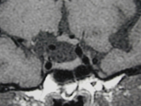
Figure 21B:Sagittal (A) and coronal (B) unenhanced T1 weighted images show an arachnoid cyst in the suprasellar region. This is markedly elevating the hypothalamus and stretching the pituitary stalk. Image B shows elevation of the right side of the chiasm by the cyst. No cyst wall is evident and the pituitary tissue itself is normal.