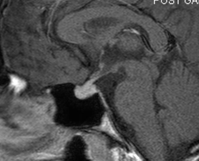
Figure 25A.Sagittal (A) and coronal (B) T1 weighted enhanced images show thickening of the pituitary stalk and nodular enhancement of the Rt side of the chiasm (arrow) in a patient with neurosarcoid.
Figure 25A.Sagittal (A) and coronal (B) T1 weighted enhanced images show thickening of the pituitary stalk and nodular enhancement of the Rt side of the chiasm (arrow) in a patient with neurosarcoid.