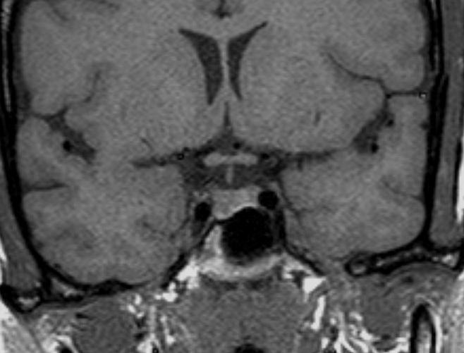
Figure 3A, B and C: Coronal T1 weighted images of the pituitary gland before (A) and immediately after contrast (B) There is a microadenoma in the right side of the gland. On the unenhanced image (A) there is evidence of depression of the floor of the sella on the right side but the microadenoma cannot be visualised within the gland. After contrast a dynamic acquisition (B) shows an area of lesser enhancement indicative of a microadenoma. Figure C demonstrates a left sided microadenoma (arrow) which was best seen on this nondynamic post-contrast sequence.