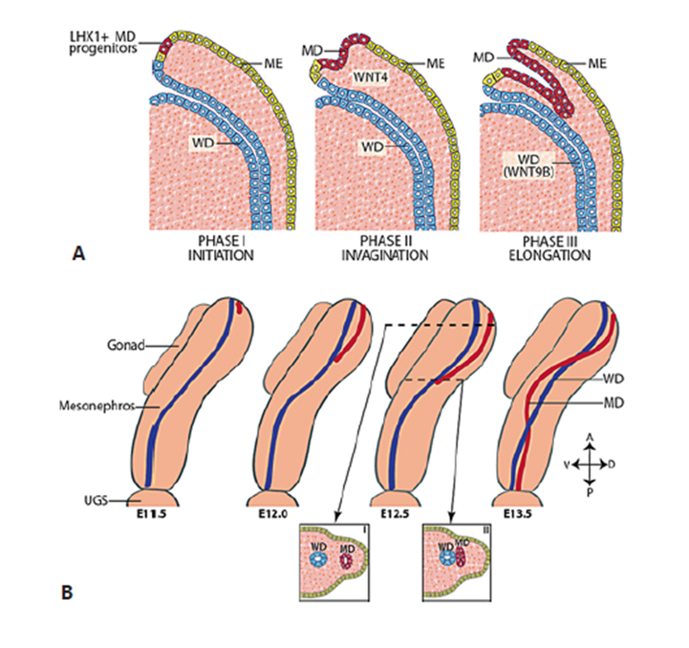
FIGURE 12. Müllerian duct (MD) development can be subdivided into three phases. A. Phase I (initiation): MD progenitor cells in the mesonephric epithelium (ME) (yellow) are specified and begin to express LHX1. Phase II (invagination): in response to WNT4 signaling from the mesenchyme, LHX1+ MD progenitor cells invaginate caudally into the mesonephros towards the WD (blue). Phase III (elongation): the tip of the MD contacts the WD and elongates caudally in close proximity to the WD requiring structure and WNT9B signaling from the WD. B. Beginning at ∼ E11.5 in mice, the MD invaginates and extends posteriorly guided by the WD. During elongation, mesenchymal cells separate the WD and MD anterior to the growing tip (inset I). However, at the MD tip, the MD and WD are in contact (inset II). At ∼ E12.5, the MD crosses over the WD to be located medially. Elongation is complete by ∼ E13.5 with the MD reaching the urogenital sinus (UGS). A = anterior (rostral); D = dorsal; P = posterior (caudal); V = ventral. Reprinted with permission from ref. (303): Mullen RD, Behringer RR. Molecular Genetics of Müllerian Duct Formation, Regression and Differentiation. Sexual Development 8:281-296 (2014), Copyright 2014, Karger.