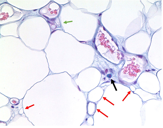
Figure 6. Angiolipoma with mast cell with enlarged multiple vessel lumens and degraded tissue. The black arrow points to a classic fried egg appearance of a mast cell stained with Alcian Blue in angiolipoma tissue. Red arrows point to small fat cell remnants likely non-functional as evidenced by the absence of nuclei. Blood vessels are numerous and large for location. The green arrow demonstrates the remnant of a capillary. Connective tissue is evident especially in the area surrounding the mast cell as bluish fibers. Magnification 100X.