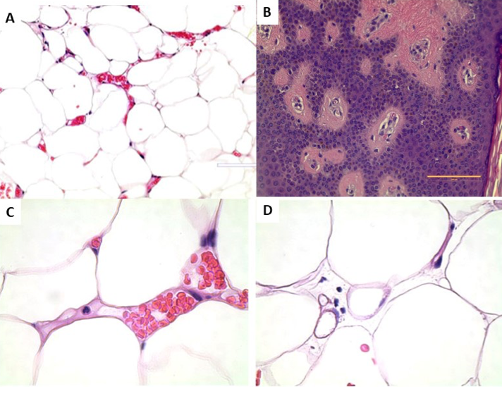
Figure 8. Histological features of angiolipomas. A. Small area of hypervascularity in an angiolipoma (40X). B. Blood vessels in an angiolipoma grow up and through the epidermis and are palpable on the skin (40X). C. Empty and presumed dead and non-functional vessel on the left containing an eosinophil next to two functional blood vessel lumens containing red blood cells (100X). Microthrombi can be seen as pale areas especially between the right side of the dead vessel and the lumen of the active vessel. Dead vessels may result in hypoxia and ischemia causing pain. D. Non-functioning blood vessel to the right and smaller fat cells surrounded by an enlarged interstitial organ (40X).