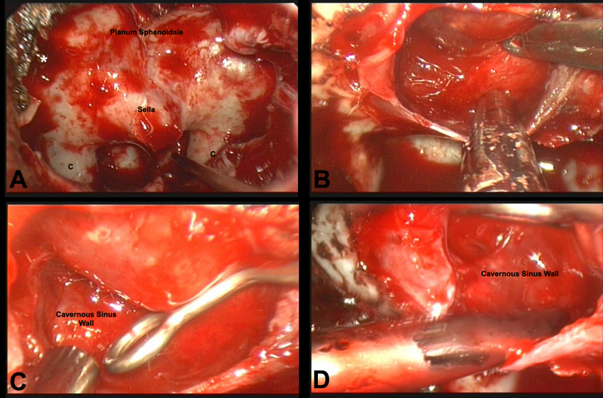
Figure 8. Endoscopic views. A. After the anterior wall of the sphenoid sinus is opened, the endoscope provides a panoramic view of the sella and surrounding anatomy. B. Endoscopic view of the tumor bed after resection. C. Endoscopic view of the right cavernous sinus wall using the 0 degree endoscope. D. Note the dramatically improved view of the right cavernous sinus wall in the same patient using the 45 degree endoscope. (arrowhead= carotid artery)