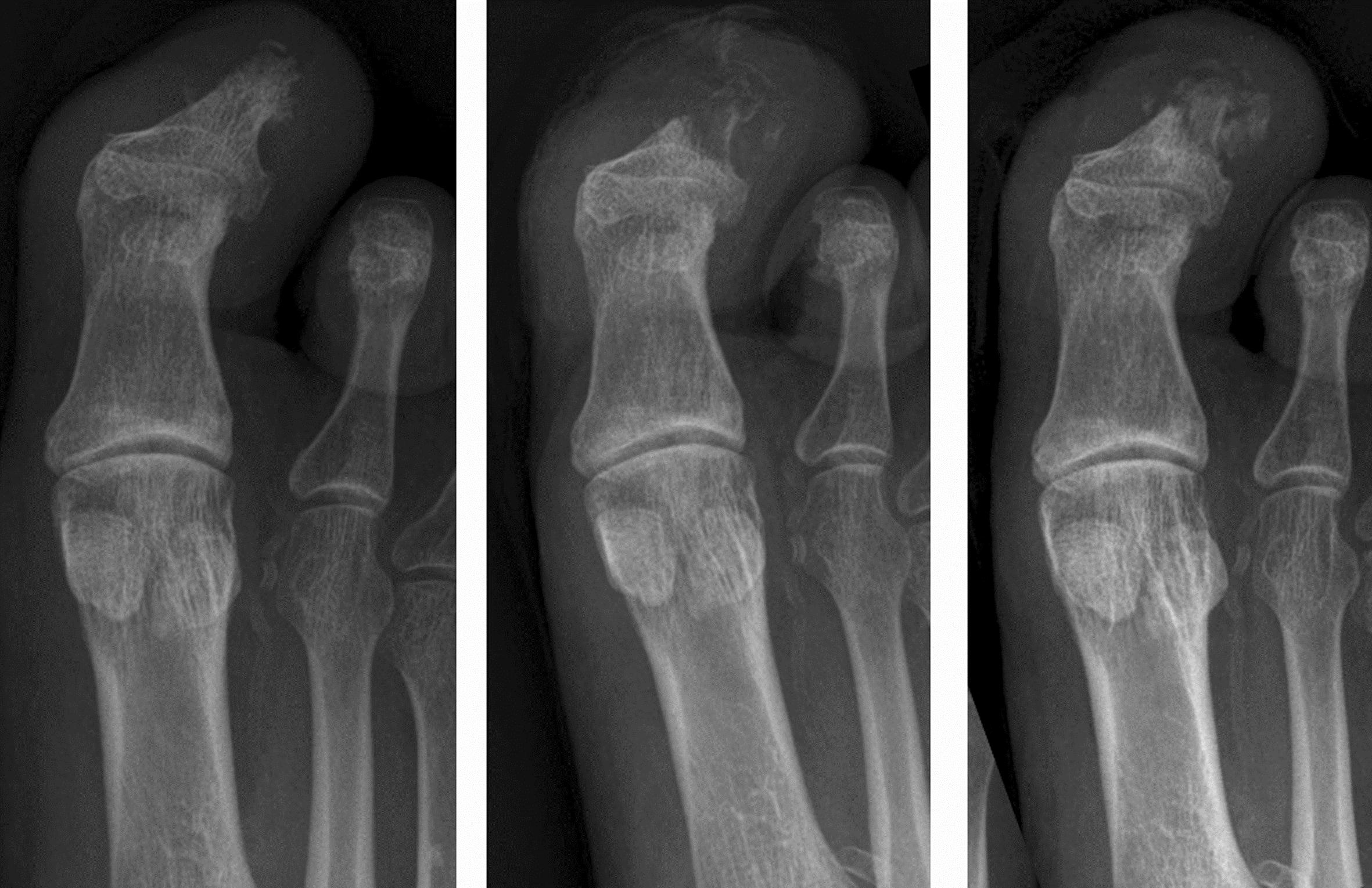
Figure 3. Acute presentation with an ulcer at the tip of the great toe, probing to bone. The terminal phalangeal tuft does show some irregularity (left panel). b) two weeks later there is marked bone demineralisation consistent with osteomyelitis (middle panel). C) After 2 months of treatment there has been partial remineralisation of the bone but with an underlying pathological fracture (right panel).