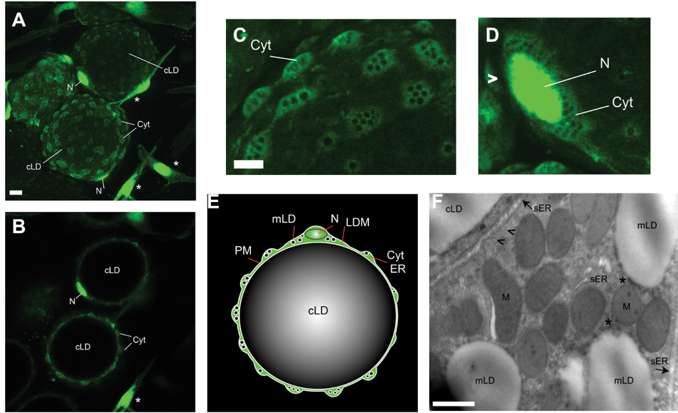
Figure 3. Architecture of primary unilocular adipocytes. Figure adapted from (32). The cytoplasm and nuclei of adipocytes and stromovascular cells were labeled by infecting visceral WAT explants from nonhuman primates with an adenoviral vector encoding enhanced green fluorescent protein (eGFP). Two days after infection, live explants were examined by for GFP expression using confocal microscopy. Cellular and subcellular features are labeled: cLD, central lipid droplet; Cyt, cytoplasm; LDM, lipid droplet membrane; mLD, micro-LD; N, nucleus; PM, plasma membrane; sER, smooth ER. (A) GFP-positive unilocular adipocytes (spheres) and stromovascular cells (asterisks) residing in WAT. The image represents the sum of all confocal slices. Bar, 10 um. (B) Single confocal section of the image in A. Enhanced magnification of adipocytes containing cytoplasmic nodules (C) and perinuclear cytoplasm (D). (E) Schematic representation a unilocular adipocyte demonstrates that the cLD is a sphere tightly fitted within the cell, whereas the cytoplasm collects in multiple organelle- and mLD-containing nodules. (F) Electron micrograph of a unilocular adipocyte from a visceral WAT explant that was fixed and processed for electron microscopy. Asterisks mark contact sites between mitochondria and mLDs, whereas arrowheads point towards vesicles budding off the ER tubules. Bar, 500 nm.