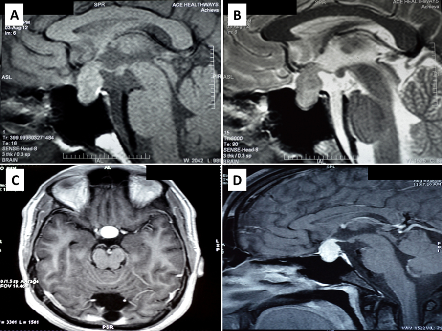
Figure 8. Magnetic resonance imaging of a patient with pituitary tuberculosis shows a sellar mass lesion measuring 2.1 cm x 1.9 cm x 1.4 cm with suprasellar extension A) heterogenous predominantly increased signal intensity on T2 weighted imaging and B) hypointense on T1 weighted imaging. C and D) Significant homogenous post contrast enhancement of the mass lesion on axial (C) and sagittal (D) views, respectively. Involvement of the pituitary stalk and superior displacement of the optic chiasma is also seen. Bright signal of posterior pituitary is maintained.