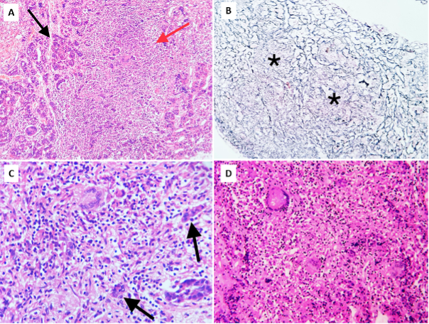
Figure 9. Pituitary tuberculosis. Biopsy of the pituitary showing nests of pituicytes (black arrows) destroyed and separated by confluent granulomas (red arrow). A) low power view, Hematoxylin and Eosin (H&E), 100x; B) corresponding area showing reticulin-free zones (asterisks) occupied by granulomas, Gordon and Sweet’s silver reticulin stain, 100x; C & D) higher power views showing non-caseating granulomas comprised of epithelioid cells with occasional Langhan’s giant cells (top left) and lymphocytes, H&E, 400x.