Archives

Adolescent Bariatric Surgery

ABSTRACT


The prevalence of adolescent obesity has rapidly increased over the past several decades. With this increase, there has also been a rise in the prevalence of complications of obesity leading to premature mortality. While lifestyle and medical management remain a part of the initial management of obesity, these therapies have been shown to be inferior when compared to metabolic and bariatric surgery (MBS) for adolescents with severe obesity. A multidisciplinary approach is recommended to evaluate medically eligible candidates for MBS, prepare patients for surgery, and guide postoperative management. Laparoscopic sleeve gastrectomy (SG) and Roux-en-Y gastric bypass (RYGB) are the most common MBS procedures performed in both adolescent and adult patients. Postoperative hospital stays are generally short and long-term routine follow-up with the MBS team is recommended to monitor weight loss, resolution of complications of obesity, and to monitor for postoperative complications. Most adolescent MBS studies demonstrate average percent body mass index loss between 25-29% after surgery. This is also associated with resolution or improvement of most complications of obesity at rates that are similar or superior to adult studies. Resolution and prevention of type 2 diabetes mellitus (T2DM) after MBS is a particularly compelling reason to pursue surgical treatment due to the complications from T2DM that occur over a patient’s lifetime as well as the overall burden of health-related costs. These adverse consequences of T2DM can be mitigated by early use of MBS. MBS is generally well tolerated. Complication rates are similar to adult patients therefore it is recommended to refer patients for MBS whenever they are medically qualified. Most common short-term (<30 days) complications include leak, bleeding, and surgical site infections. Most common long-term (>30 days) complications are nutritional deficiencies.


INTRODUCTION


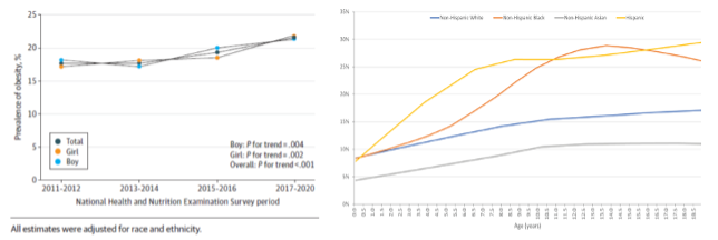
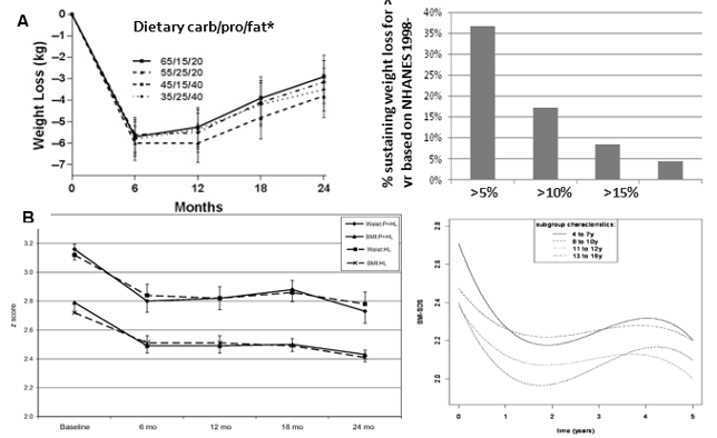
The prevalence of worldwide overweight and obesity in adolescents has more than quadrupled since 1975. Currently, it is estimated that over 14 million children age 2-19 years suffer from obesity in the United States alone (1, 2). Adolescents with obesity are at risk for developing significant comorbidities including insulin resistance, type 2 diabetes mellitus (T2DM), hypertension, dyslipidemia, obstructive sleep apnea, nonalcoholic fatty liver disease, depression, polycystic ovarian syndrome, impaired quality of life, cardiovascular disease, and longer term, certain malignancies (3-9). Similar to obesity, the prevalence of T2DM has been increasing dramatically (3). Obesity is a major risk factor the development of T2DM with overweight adolescents having close to a three times greater risk of developing T2DM when compared to adolescents with normal weight (10-12). Additionally, obesity in adolescence is associated with persistent obesity into adulthood, increased risk for obesity related comorbidities, and premature mortality in adulthood (13-15). Lifestyle and medical management remain the first-line treatment for adolescent obesity; however, current evidence suggests that pharmacotherapy, dietary, and behavioral modifications rarely lead to long-term weight loss in adolescents with severe obesity (16-18). The use of metabolic and bariatric surgery (MBS) in adolescents with severe obesity and complications of obesity has been shown to have superior results in both efficacy and durability (19).


PREOPERATIVE EVALUATION


Multidisciplinary Program


A multidisciplinary approach is recommended when considering MBS for an adolescent (20, 21). At a minimum, this includes a bariatric surgeon with adolescent experience, pediatrician, dietitian, nurse, and pediatric psychologist. It is also important the core providers have access to additional pediatric specialists including anesthesiologists, radiologists, and appropriate specialists to aid the management of complications of obesity (e.g., pulmonology, endocrinology, gastroenterology/hepatology). Adolescents undergoing preoperative work-up should be evaluated for the presence and severity of complications of obesity. Additionally, it is important for the multidisciplinary team to determine a potential patient and caregivers’ ability to assess the risks and benefits of surgery as well as to adhere to postoperative requirements including daily vitamin regimens and attending postoperative visits.


Patient Selection


BODY MASS INDEX (BMI)


The following criteria have been recommended by multiple panels of experts for consideration of weight loss surgery in adolescents under 18 years old: (4, 19)

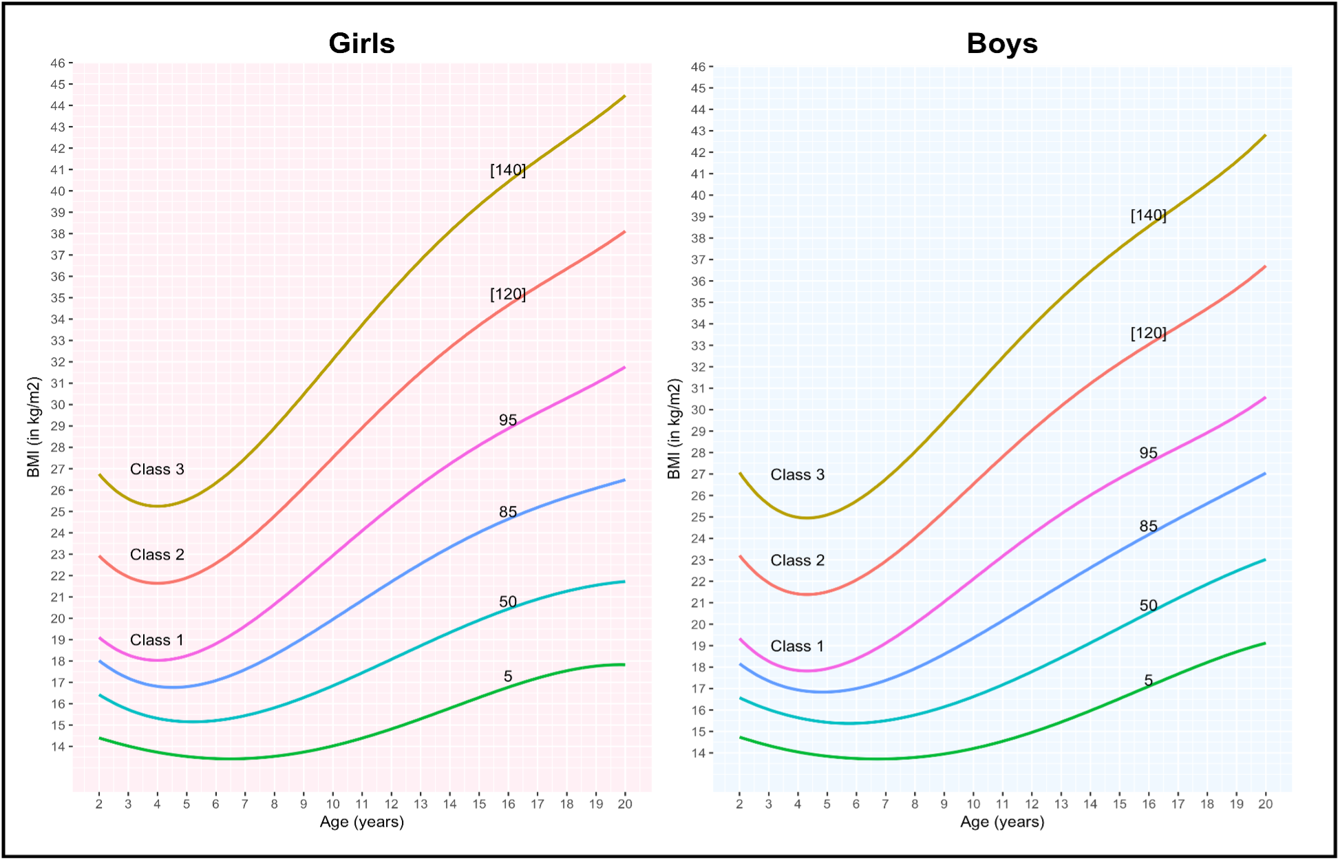
  • BMI ≥ 120 percent of the 95th percentile for BMI for age or BMI ≥ 35kg/m2, whichever is lower, with complications of obesity that has a significant effect on health (Table 1).
  • OR -
  • BMI ≥ 140 percent of the 95th percentile of BMI for age or BMI ≥ 40 kg/m2, whichever is lower


Table 1. Qualifying Comorbidities for Consideration of MBS in Adolescents (4).

Obstructive sleep apnea (apnea-hypoxia index > 5)

Type 2 diabetes mellitus

Idiopathic intracranial hypertension

Nonalcoholic steatohepatitis

Blount’s disease

Slipped capital femoral epiphysis

Gastroesophageal Reflux Disease

Hypertension


CONTRAINDICATIONS


Contraindications to adolescent MBS are listed in Table 2.


Table 2. Contraindications to Adolescent MBS

Medically correctable cause of obesity

Ongoing substance abuse problem (within the preceding year)

Medical, psychiatric, psychosocial, or cognitive condition that prevents adherence to postoperative dietary and medication regimens or impairs decisional capacity

Current or planned pregnancy within 18 months of the procedure

Inability for patient or caregivers to comprehend risks and benefits of surgical weight loss procedure


AGE


A recent retrospective review of the Metabolic and Bariatric Surgery Accreditation and Quality Improvement Program (MBSAQIP) data registry from 2015 to 2018 demonstrated that adolescents and young adults only represented 3.7% of total MBS cases performed suggesting significant underutilization within this population (22). Multiple studies have evaluated the safety and efficacy of MBS in younger adolescents. Current evidence suggests there are no significant clinical differences in outcomes between MBS in younger (e.g., <16 years) versus older adolescents (e.g., ≥16 years)(23-27). It is therefore not recommended to limit access to MBS based on patient’s age, physical maturity (e.g., bone age), or pubertal status. These findings have prompted increase advocacy for the use of MBS in the adolescent population by the American Academy of Pediatrics (19).


TYPES OF SURGERY


Sleeve Gastrectomy


A laparoscopic sleeve gastrectomy (SG) results in the removal of the greater curvature of the stomach resulting in a smaller, tubular stomach that has a reduced capacity (Figure 1). Given the procedure is less complex than the Roux-en-Y gastric bypass (RYGB) and has less risk for micronutrient deficiencies, it is an appealing option for adolescents. SG currently accounts for approximately 80% of bariatric procedures in adolescents (22, 28-30). A sleeve gastrectomy may also be converted to RYGB in the event additional MBS is indicated or in the setting of postoperative medically refractory gastroesophageal reflux disease (GERD).

Figure 1. Sleeve Gastrectomy

Roux-en-Y Gastric Bypass


Laparoscopic Roux-en-Y gastric bypass involves creating a small, proximal gastric pouch which is separated from the remnant stomach and anastomosed to a Roux-limb of small bowel 70-150cm distally (Figure 2). The RYGB results in similar weight loss when compared to SG and dramatically improves glycemic control (29, 31). The incidence of postoperative GERD is significantly less following RYGB compared to SG, making the procedure an attractive option for adolescents with GERD at baseline (32).

Figure 2. Roux-en-Y Gastric Bypass


Others


Additional procedures including intragastric balloons are not currently approved by the United States Food and Drug Administration (FDA) for use in adolescents. Adjustable gastric bands have been previously used in the adolescent population; however, they have fallen out of favor due inferior efficacy compared to SG and RYGB (33).


POSTOPERATIVE MANAGEMENT


Inpatient  


Average inpatient stay is typically 1-3 days following both a SG and RYGB (34, 35). Patients are monitored for immediate postoperative complications including a leak, bleeding, and venous thromboembolism (VTE). Following discharge, patients are seen at regular postoperative visits to monitor body weight, nutritional status, and to manage complications of obesity.


Diet


Following a SG or RYGB, patients are gradually progressed from a high protein liquid diet to incorporating small volumes of regular food. Patients are encouraged to eat three to four protein-rich meals a day while avoiding carbohydrate rich foods. Supplemental sugar-free fluids between meals are also essential following surgery in order to avoid dehydration. Patients are typically encouraged to avoid excessive fluids with meals in order to minimize nausea and maximize nutritional intake with meals due to the restrictive component of both procedures.


Postoperative nausea is not uncommon following surgery but typically self resolves. Meals high in carbohydrates or sugar can result in dumping syndrome or weight regain following surgery. Some providers recommend limiting carbonated or caffeinated beverages following MBS based on theoretical concerns, however there is minimal evidence to support this apprehension. Similar to non-operative weight loss recommendations, general recommendations including exercising for 30 to 60 minutes daily, drinking sugar-free fluids, and portion-controlled protein rich meals are the same. Overall, it is recommended that patient and caregiver meet with a dietitian prior to discharge to develop a plan tailored to patient’s specific nutritional needs. Regular follow-up visits with a dietitian are also recommended to assist with postoperative weight management and to monitor for nutritional deficiencies.


Nutritional Supplements and Monitoring


Although SG may be associated with a decreased risk of nutritional deficiencies when compared to RYGB, lifelong supplementation with vitamins and minerals is recommended following both operations (Table 3). Patients are particularly at risk for deficiencies in iron, vitamin B12, and vitamin D. Additionally, lifelong annual monitoring of nutritional and micronutrient status is recommended with annual laboratory testing (Table 3). Adjustments in supplements may need to be made over time as specific deficiencies emerge. 


Table 3. Nutritional Supplementation and Monitoring Recommendations (36)

Nutritional Supplements

Standard multivitamin with folate or iron, or prenatal vitamin if female (once or twice daily)

Vitamin B12, 500mcg sublingually daily, or 1000mcg intramuscularly month

Calcium, 1200 to 1500mg daily (measured as elemental calcium) with 800 to 1000 international units of vitamin D.

Annual Nutritional Monitoring

Complete blood cell count with differential

Serum iron and ferritin

Red blood cell folate, serum vitamin B12, and serum homocysteine

Serum thiamin (vitamin B1)

Hepatic panel (including albumin, total protein, serum aminotransferase levels, gamma-glutamyl transpeptidase, and alkaline phosphatase

Calcium, 25-hydroxyvitamin D, and parathyroid hormone

Dual-energy x-ray absorptiometry (DXA) scan to monitor bone density (optimal frequency not yet established)


Pregnancy Prevention


Pregnancy should be avoided for 12 to 18 months following MBS to allow patients to achieve weight maintenance and to avoid potential micronutrient deficiencies which may affect both patient and fetus (37). Obesity can result in decreased fertility secondary to irregular menstruation and ovulatory dysfunction (38, 39). Weight loss after MBS has been shown to result in more regular ovulation and improved fertility (40, 41). In a retrospective review of 47 adolescents who underwent MBS surgery, seven pregnancies occurred, six of them within 10 to 22 months following surgery (42). While all six deliveries were healthy and at term, the twofold higher than anticipated pregnancy rate highlights the need for contraception counseling following MBS.


Multiple studies have evaluated the efficacy of hormonal contraceptive methods in patients with elevated BMIs and no definitive association was found between higher BMI and effectiveness of hormonal contraceptives (43). Due to concern for malabsorption after intestinal bypass procedures and the subsequent potential for decreased oral contraceptive efficacy, the American College of Obstetricians and Gynecologist recommend using non-oral forms of hormonal contraception in patients who have undergone malabsorptive MBS (44). Additionally, oral contraceptives are associated with increased risk of venous thromboembolism (VTE) which may be worrisome for adolescents with elevated BMIs who already have a higher predisposition for VTE (45, 46).


Intrauterine devices (IUDs) are an appealing option following MBS in adolescent patients as they are one of the most effective contraception methods, do not increase risk of VTE, and can be placed at the time of surgery (47). Levonorgestrel-releasing IUDs have the added benefit of promoting amenorrhea which could help reduce the risk of iron deficiency anemia following surgery (48). Regardless of the form of contraception selection, adolescents should be counseled on safe sex practices including the use of barrier protection against sexually transmitted infections.


Adolescent patients who become pregnant following MBS should be counseled on adequate nutritional intake with close monitoring of iron, folate, and vitamin B12 levels. Additionally, one must be cautions when screening for gestational diabetes in pregnant patients who have undergone MBS. In a study of a 119 post-bariatric surgery pregnant patients, oral glucose tolerance test resulted in hypoglycemia in 83% of patients with history of RYGB and 55% of patients with history of SG (49). Alternative methods for screening such as capillary blood glucose measurements are therefore recommended (50, 51).


Comorbidity Reassessment


Regular reassessment of complications of obesity should occur at routine intervals in the postoperative phase to monitor for resolution or need for continued management. Patients with T2DM should be evaluated by their endocrinologist every three months. Repeat polysomnography are generally obtained between three to six months after surgery for patients previously on continuous positive airway pressure therapy (52, 53). Twenty-four-hour blood pressure monitoring can also be repeated three months after surgery to demonstrate resolution or persistence of hypertension. Medication may be restarted if blood pressure is consistently ≥120 mmHg systolic or ≥80 mmHg diastolic. Patients with biopsy proven nonalcoholic fatty liver disease may be re-biopsied 12 months after surgery to document regression. Finally, patients’ mental health needs should be re-evaluated by a pediatric psychologist at 6 and 12 months after surgery.


In the setting of weight regain, patients should be monitored for complications of obesity. There is emerging evidence however, that some complications of obesity may be weight dependent and others non-weight dependent (54). Some surgeons will routinely obtain an upper gastrointestinal contrast study at 12 months after surgery or as needed to assess anatomy which may lead to weight regain. Anatomical abnormalities that may contribute to weight regain include a dilated gastric sleeve or gastrogastric fistula.


Follow Up


Close follow up with the multidisciplinary team including the bariatric surgeon, pediatrician, dietitian, and pediatric psychologist is strongly recommended. Patients are typically followed by a pediatrician to ensure ongoing continuity of care. It is important for the core providers to have access to pediatric specialists including endocrinology, gastroenterology/hepatology, and pulmonology as needed in those with complications of obesity that require ongoing monitoring or management. Additionally, a gynecologist for contraception counseling may be required for female patients. The transition from pediatric to adult medicine can be challenging in patients with chronic medical conditions and frequently requires assistance from multiple members of the team for transition care coordination and preparation as well as to ensure adequate communication, support, and education (55-57). 


OUTCOMES


Percent BMI Loss


Both SG and RYGB have resulted in clinically significant weight loss in adolescents. The efficacy of both procedures appears to be similar in the adolescent population with potentially slightly greater weight loss following RYBG. In a large, multicenter analysis of 177 adolescents who underwent RYGB and 306 adolescents who underwent SG, there was a three-year postoperative average percent BMI loss of -29% (95% CI, -26 to -33) and -25% (95% CI, -22 to -28) for RYGB and SG, respectively (29). Similar results were seen in the largest prospective study to date of 228 adolescents undergoing either RYGB or SG with an average 28% reduction in BMI at three years following RYGB compared to 26% reduction following SG (53). Smaller, long-term studies demonstrate the durability of weight loss with MBS with an average percent BMI loss of 29% in adolescents undergoing RYGB up to 12 years after surgery(58, 59). 


Complications of Obesity


TYPE 2 DIABETES MELLITUS  


Multiple studies have demonstrated improved glycemic control, even remission as well as prevention of T2DM following MBS, making a compelling case of MBS as a treatment for T2DM (31, 59-63). The Teen-Longitudinal Assessment of Bariatric Surgery (Teen-LABS) is a prospective, observational study of pediatric patients undergoing MBS at 5 children’s hospitals in the United States with 3 and 5 year of follow-up data published to date. Of the 242 adolescents with obesity who underwent MBS, 29 had T2DM. By 3 years after the procedure, remission of T2DM occurred in 95% (95% CI, 85-100) of participates who had diabetes at baseline with no new cases of T2DM in those without the condition at baseline (53). Additionally, 19 participants had prediabetes at baseline with a 76% (95% CI, 56-97) rate of remission at 3 years (53). These remission rates in Teen-LABS were compared to adults who underwent MBS. Among those who underwent RYGB, adolescents were more likely to have remission of T2DM at 5 years with a remission rate of 86% compared to 53% in adults (64).


Similar findings were demonstrated in another study of 226 adolescents undergoing SG, of which 23% of patients were found to have T2DM. Eighty-five percent of patients with T2DM were on medication for diabetes prior to surgery and 89% achieved normal fasting plasma glucose and hemoglobin A1c levels without the use of medication postoperatively (52).


In an effort to compare surgical versus medical therapy for T2DM in adolescents with severe obesity, data from participants with T2DM enrolled in the Teen-LABS study were compared to participants of similar age and racial distribution from the Treatment Options of Type 2 Diabetes in Adolescents and Youth (TODAY) studies. Teen-LABS participants underwent MBS. TODAY participants were randomized to metformin alone or in combination with rosiglitazone or intensive lifestyle intervention, with insulin therapy given for glycemic progression. At two years, mean hemoglobin A1c concentration decreased from 6.8% to 5.5% in patients who underwent MBS compared to an increase from 6.4% to 7.8% in those enrolled in the TODAY study. Compared to baseline, average BMI decreased by 29% in Teen-LABS participants while the average BMI increased by 3.7% in TODAY participants (65). Cardiovascular disease (CVD) risk reduction was also explored in a secondary analysis of this study and despite higher pretreatment risk for CVD, treatment with MBS resulted in reduction of estimated CVD that were sustained at 5-year follow-up where medical therapy was associated with an increase in risk of CVD in adolescents with T2DM and severe obesity (66).


While these initial results are promising of the beneficial effects of MBS for the treatment of T2DM, no studies have prospectively compared the efficacy of MBS with that of medical therapy for the treatment of T2DM in adolescents with obesity. Additionally, the majority of initial MBS data in adolescents were from those who underwent RYGB which is no longer the primary MBS procedure performed in adolescents due to its inferior safety profile. In 2019, the National Institute of Health funded the Surgical or Medical Treatment for Pediatric T2DM (ST2OMP) trial which will compare SG to advanced medical therapy (67, 68).


OTHER COMORBIDITIES


In the Teen-LABS study described above, a mean 74% (95% CI, 64 to 84) remission of hypertension (HTN), 66% (95% CI 57 to 74) remission of dyslipidemia, and 86% (95% CI 72 to 100) resolution of abnormal kidney function was found at 3 years (53). In a secondary analysis of Teen-LABS and TODAY data, medical management of adolescents with obesity was associated with higher odds of diabetic kidney disease when compared to MBS (69). Greater weight loss after MBS in adolescents has also been associated with greater remission of T2DM, HTN, and dyslipidemia (54, 70). In a comparison of adolescents and adults who underwent RYGB, adolescents were more likely to have remission of HTN at 5 years compared to adults (68% vs 41%) (64).


Additional studies have demonstrated a 66% to 84% remission of obstructive sleep apnea as well as improvements in liver disease and polycystic ovarian syndrome (8, 52, 71). Improvements in functional mobility as well as reduction in musculoskeletal pain have also been well described (72, 73).


Mental Health


Multiple studies have reported higher rates of depression, emotional and behavioral disorders, and suicidal ideation in adolescents with obesity (74-77). Additionally, binge and loss of control eating is prevalent among more than one quarter of adolescents with overweight and obesity (78, 79). A recent prospective study demonstrated that undergoing MBS in adolescence did not heighten or lower the risk of suicidal thoughts or behaviors following the initial 4 years after surgery (80). While still unclear whether obesity leads to psychopathology, or vice versa, the association highlights the need for appropriate psychological services in the pre- and postoperative period (74).


MBS can lead to improvements in psychosocial outcomes, although the improvements appear too often be transient. In the TEEN-Labs study, quality of life measured by the Impact of Weight on Quality of Life and Short Form 36 Health Survey improved after MBS (53, 73). Several studies have demonstrated improved depressive and anxiety symptoms in the months following MBS, although the results were not maintained after the first postoperative year (81, 82). In a multisite study assessing two year follow up of psychopathology prevalence in adolescents undergoing MBS, most patients retained their symptomatic or non-symptomatic psychopathology status at two years, although remission of symptoms was more prevalent than the development of new symptoms (83). These results emphasize the need for long-term psychosocial monitoring following MBS as well as early treatment in those with psychopathology


Short-Term Complications


Short-term complications (<30 days after surgery) in adolescents undergoing MBS are similar to those seen in adults. Early postoperative complications, though rare, include surgical site infections, bleeding, leak, strictures, and pulmonary embolism. In a retrospective review of 21,592 adolescents and young adults who underwent SG or RYGB between 2015 and 2018, 3.7% of patients required readmission, 1.1% of patients required reoperation, and 3.3% required percutaneous, endoscopic, or other intervention (22). Major complications were rare; the most common complication was bleeding (0.4%), followed by leak (0.4%), and deep surgical site infections (0.2%). RYGB was associated with higher rates of reoperation (2.1% vs. 0.8%), readmission (6.3% vs. 3.0%), and serious complications (5.5% vs. 1.8%) compared to SG. Mortality occurred in 0.05% of patients and there were no differences in mortality noted between groups (22). In an additional retrospective review of 483 adolescents (SG n=306, RYGB n=177) no perioperative deaths occurred and the rate of major adverse events were too rare for statistical comparison. VTEs occurred in only 0.4% of patients and failure to discharge in 30 days was observed in 0.7% of patients (29).


Multiple studies have also suggested that MBS may be safer in adolescents when compared with adults. In a large study evaluating perioperative outcomes of MBS between 309 adolescents and 55,192 adults, the overall 30-day complication rate was significantly lower in adolescents (5.5%) as compared with adults (9.8%). No in-hospital mortalities occurred in the adolescent group compared to 0.2% the adult group. The 30-day morbidity for adolescents following SG was zero compared to 4.3% following RYGB (84). In an additional study evaluating 1047 adolescents,10,429 college-aged individuals, and 24,841 young adults who underwent SG or RYGB, there were no differences in 30-day complication rates between age groups (85).


Long-Term Complications


NUTRITIONAL DEFICIENCIES  


Long-term complications after MBS in adolescents are primarily nutritional. Patients are particularly at risk for deficiencies in iron, vitamin B12, and vitamin D. Iron deficiency is common in premenopausal females due to menstruation. Some patients may require iron infusion if oral supplementation is not adequate. Symptomatic thiamine deficiency following MBS is rare, however can have serious consequences (86-88).These risks are higher for patients who undergo RYGB compared to SG due to potential malabsorption. In a Teen-LABS study evaluating nutritional deficiencies at 5 years postoperatively, low serum ferritin levels were seen in 71% of patients who underwent RYGB compared to 45% following a SG indicating iron deficiency (86). Iron deficiency anemia can occasionally be severe in adolescent women following MBS which can be compounded by menstruation and challenges in recognizing symptoms therefore daily supplementation and routine nutritional monitoring is essential following MBS.


Vitamin B12 deficiency was seen in approximately 12% of patients after either procedure. Approximately 40% of patients had low vitamin D levels at baseline with no significant change at follow up. Parathyroid hormone concentrations increased in patients undergoing RYGB from an average baseline concentration of 44 pg/ml to 59 pg/ml at 5 years with the risk of abnormal parathyroid hormone nearly sixfold higher after RYGB compared with SG (86). Elevated parathyroid hormone is utilized as a surrogate for calcium deficiency and raises concerns about long-term bone health. In adolescents, reduced bone mass has been noted two years after MBS although the bone mass remains appropriate for the patients’ age and new body weight (89). Concerns of growth retardation after MBS have been refuted and the most recent adolescent ASMBS guidelines have removed the recommendation of patients reaching physical maturity prior to MBS (4, 24).


The risk of nutritional deficiencies decreases with adherence to prescribed micronutrient supplements and increases with pregnancy (86). Given the high prevalence of nutritional deficiencies, lifelong micronutrient supplementation is required following surgery. One concern emphasized in the adolescent population is adherence to regular multivitamin use. In a prospective study of 41 adolescents who underwent MBS, multivitamin adherence was only 29.8%  23.9 (90).


WEIGHT REGAIN


Current data demonstrates adequate maintenance of weight loss at 5-9 years (29, 91). Several studies have suggested a long-term weight loss advantage in adolescents undergoing RYGB compared to SG, although there is still insufficient evidence to directly compare long-term outcomes of both procedures (29, 53, 92, 93). More research is needed to fully understand the mechanisms behind long-term weight maintenance after MBS.


OTHER COMPLICATIONS  


Cholelithiasis is a common complication due to rapid weight loss following MBS in both adolescents and adults. In the Teen-LABS study, cholecystectomy was required within three years in 9.9% of adolescents who underwent RYGB and 5.1% who underwent SG (53). Five percent of Teen-LABS participants required other abdominal operations including lysis of adhesions, gastrostomy, ventral hernia repair, or internal hernia repair (53). Symptoms of GERD, nausea, bloating, and diarrhea can also increase following MBS. During five years of follow up, the incidence of GERD increased from 2% to 8% in adolescents who underwent RYGB and from 11% to 24% in those who underwent SG. At five years postoperatively, the SG group had more than fourfold greater odds of having gastrointestinal distress symptoms when compared to RYGB (32). Dumping syndrome can been seen after both procedures, however it is much more common after RYGB compared to SG (94, 95). The incidence of dumping syndrome (~12%) in adolescents after RYGB was similar to adult patients two years after surgery(96).


There are no current established guidelines for surveillance of Barrett’s esophagus after SG for adolescent patients, however routine screening is recommended for adult patients after SG, therefore it would be prudent for adolescent patients to undergo intermittent surveillance also as the length of possible GERD exposure is theoretically longer (97). Similarly, there are no established guidelines for monitoring of bone density following use of MBS in adolescence, but due to inadequate vitamin D levels and rising PTH at least in those who underwent RYGB, periodic monitoring with DEXA may be prudent.


Emerging Evidence


Current evidence evaluating the outcomes and efficacy of adolescent MBS is generally limited to ≤10 years of follow up. Smaller, long-term studies with data available for 7 to 12 post-operatively in patients who primary underwent RYGB demonstrate the durability of weight loss and similar rates of complications, although inference is limited due to small sample sizes with reduced attrition rates (59, 60, 98, 99). Characteristics including study size, length of follow up, and attrition rate of available studies on MBS published from 2012 to present are available in Table 4. As SG is now the most predominate MBS procedure performed in the Unites States long-term data with this procedure is required. While some longitudinal studies are ongoing (Table 4), there remains a paucity of long-term data in the adolescent population.


Table 4. Characteristics of Studies on MBS, 2012 – Present

Author; Year

Study Design

Sample Size (N)

Type of MBS


RYBG     SG

Longest follow up

N

(1 yr)

N

(3 yr)

N

(5 yr)

Comments

Inge;

2018 (29)

RO 

483

177

306

5 yr

466 (96%)

153 (32%)

41 (8%)

The PCORnet bariatric study (2005 – 2015)

Olbers; 2017 (58)

CC

81

81

0

5 yr

81

(100%)

n/a

81 (100%)

Adolescent Morbid Obesity Surgery (AMOS) study

Inge; 2017(59)

PO

74

74

0

12.5 yr

n/a

n/a

58 (81%)

Adolescent Bariatric Surgery at 5 Plus Years (FABS-5+) study (2001-2007); mean follow up 8.0 yr

Inge; 2016 (53)

PO 

228

161

67

3 yr

205 (90%)

194 (85%)

n/a

Teen-Longitudinal Assessment of Bariatric Surgery (Teen-LABS) study; (2007-2012)

Vilallonga; 2016 (60)

RO

19

19

0

10.2 yr

n/a

n/a

n/a

Mean follow up 7.2 years; (2003-2008)

Al-Sabah; 2015 (61)

RO

125

0

135

4 yr

54 (40%)

n/a

n/a

2 yr follow up: 46 (34%); (2008-2012)

Cozacov; 2014 (98)

RO

18

8

10

7 yr

15 (83%)

10 (56%)

n/a

7 yr follow up: 3 (17%); (2002 – 2011)

Messiah; 2013 (71)

PO

454

454

0

1 yr

108 (24%)

n/a

n/a

Bariatric Outcomes Longitudinal Database (BOLD) (2004-2010)

Alqahtani; 2012 (25)

RO

108

0

108

2 yr

41 (38%)

n/a

n/a

2 yr follow-up: 8 (7%); (2008 – 2011)

Nijhawan; 2012 (99)

RO

25

25

0

9 yr

n/a

n/a

20 (80%)

Study dates not provided

RO- Retrospective observational; CC- Case-control; PO- Prospective observational


DISCUSSION


Surgical weight loss is an appropriate consideration for adolescents with severe obesity and/or complications of obesity who have failed to lose weight through conservative management. It is essential that adolescents undergoing evaluation for MBS due so in the context of a multidisciplinary program with specific expertise in adolescent medicine and MBS. SG and RYGB are safe and effective treatment options in adolescents. Weight loss outcomes are comparable between SG and RYGB. Both procedures also result in substantial improvement in complications of obesity, including T2DM. SG appears to have an improved safety profile when compared to RYGB and is now the most common adolescent bariatric procedure performed in the United States. Emerging evidence demonstrates advantages of earlier surgical intervention in those with obesity including improved weight loss, increased resolution of comorbidities, and decreased adverse events when compared to adults (64, 100). Perioperative complications in adolescents undergoing MBS are similar to those in adults but occur less frequently (84, 85). Long-term complications are primarily nutritional and life-long vitamin and mineral supplementation is recommended. Regular follow up is required following MBS and it is important for patients to have access to appropriate medical, dietary, and psychological care.


REFERENCES


  1. Obesity and overweight World Health Organization2020 [Available from: https://www.who.int/news-room/fact-sheets/detail/obesity-and-overweight.
  2. Childhood Obesity Facts Centers for Disease Control and Prevention: Centers for Disease Control and Prevention; 2021 [Available from: https://www.cdc.gov/obesity/data/childhood.html.
  3. Rosenbaum M. Special Considerations Relevant to Pediatric Obesity. In: Feingold KR, Anawalt B, Boyce A, Chrousos G, de Herder WW, Dhatariya K, et al., editors. Endotext. South Dartmouth (MA)2000.
  4. Pratt JSA, Browne A, Browne NT, Bruzoni M, Cohen M, Desai A, et al. ASMBS pediatric metabolic and bariatric surgery guidelines, 2018. Surg Obes Relat Dis. 2018;14(7):882-901.
  5. Xanthakos SA, Jenkins TM, Kleiner DE, Boyce TW, Mourya R, Karns R, et al. High Prevalence of Nonalcoholic Fatty Liver Disease in Adolescents Undergoing Bariatric Surgery. Gastroenterology. 2015;149(3):623-34.e8.
  6. Zeller MH, Roehrig HR, Modi AC, Daniels SR, Inge TH. Health-related quality of life and depressive symptoms in adolescents with extreme obesity presenting for bariatric surgery. Pediatrics. 2006;117(4):1155-61.
  7. Legro RS, Arslanian SA, Ehrmann DA, Hoeger KM, Murad MH, Pasquali R, et al. Diagnosis and treatment of polycystic ovary syndrome: an Endocrine Society clinical practice guideline. J Clin Endocrinol Metab. 2013;98(12):4565-92.
  8. Kaar JL, Morelli N, Russell SP, Talker I, Moore JM, Inge TH, et al. Obstructive sleep apnea and early weight loss among adolescents undergoing bariatric surgery. Surg Obes Relat Dis. 2021;17(4):711-7.
  9. Avgerinos KI, Spyrou N, Mantzoros CS, Dalamaga M. Obesity and cancer risk: Emerging biological mechanisms and perspectives. Metabolism. 2019;92:121-35.
  10. Tirosh A, Shai I, Afek A, Dubnov-Raz G, Ayalon N, Gordon B, et al. Adolescent BMI trajectory and risk of diabetes versus coronary disease. N Engl J Med. 2011;364(14):1315-25.
  11. Abdullah A, Stoelwinder J, Shortreed S, Wolfe R, Stevenson C, Walls H, et al. The duration of obesity and the risk of type 2 diabetes. Public Health Nutr. 2011;14(1):119-26.
  12. Lee JM, Gebremariam A, Vijan S, Gurney JG. Excess body mass index-years, a measure of degree and duration of excess weight, and risk for incident diabetes. Arch Pediatr Adolesc Med. 2012;166(1):42-8.
  13. Fontaine KR, Redden DT, Wang C, Westfall AO, Allison DB. Years of life lost due to obesity. Jama. 2003;289(2):187-93.
  14. Greenberg JA. Obesity and early mortality in the United States. Obesity (Silver Spring). 2013;21(2):405-12.
  15. Inge TH, King WC, Jenkins TM, Courcoulas AP, Mitsnefes M, Flum DR, et al. The effect of obesity in adolescence on adult health status. Pediatrics. 2013;132(6):1098-104.
  16. Berkowitz RI, Wadden TA, Gehrman CA, Bishop-Gilyard CT, Moore RH, Womble LG, et al. Meal replacements in the treatment of adolescent obesity: a randomized controlled trial. Obesity (Silver Spring). 2011;19(6):1193-9.
  17. Andela S, Burrows TL, Baur LA, Coyle DH, Collins CE, Gow ML. Efficacy of very low-energy diet programs for weight loss: A systematic review with meta-analysis of intervention studies in children and adolescents with obesity. Obes Rev. 2019;20(6):871-82.
  18. Srivastava G, Fox CK, Kelly AS, Jastreboff AM, Browne AF, Browne NT, et al. Clinical Considerations Regarding the Use of Obesity Pharmacotherapy in Adolescents with Obesity. Obesity (Silver Spring). 2019;27(2):190-204.
  19. Armstrong SC, Bolling CF, Michalsky MP, Reichard KW. Pediatric Metabolic and Bariatric Surgery: Evidence, Barriers, and Best Practices. Pediatrics. 2019;144(6).
  20. Inge TH, Krebs NF, Garcia VF, Skelton JA, Guice KS, Strauss RS, et al. Bariatric surgery for severely overweight adolescents: concerns and recommendations. Pediatrics. 2004;114(1):217-23.
  21. Styne DM, Arslanian SA, Connor EL, Farooqi IS, Murad MH, Silverstein JH, et al. Pediatric Obesity-Assessment, Treatment, and Prevention: An Endocrine Society Clinical Practice Guideline. J Clin Endocrinol Metab. 2017;102(3):709-57.
  22. Mocanu V, Lai K, Dang JT, Switzer NJ, Birch DW, Ball GDC, et al. Evaluation of the Trends, Characteristics, and Outcomes in North American Youth Undergoing Elective Bariatric Surgery. Obes Surg. 2021;31(5):2180-7.
  23. Ogle SB, Dewberry LC, Jenkins TM, Inge TH, Kelsey M, Bruzoni M, et al. Outcomes of Bariatric Surgery in Older Versus Younger Adolescents. Pediatrics. 2021;147(3).
  24. Alqahtani A, Elahmedi M, Qahtani AR. Laparoscopic Sleeve Gastrectomy in Children Younger Than 14 Years: Refuting the Concerns. Ann Surg. 2016;263(2):312-9.
  25. Alqahtani AR, Antonisamy B, Alamri H, Elahmedi M, Zimmerman VA. Laparoscopic sleeve gastrectomy in 108 obese children and adolescents aged 5 to 21 years. Ann Surg. 2012;256(2):266-73.
  26. El Chaar M, King K, Al-Mardini A, Galvez A, Claros L, Stoltzfus J. Thirty-Day Outcomes of Bariatric Surgery in Adolescents: a First Look at the MBSAQIP Database. Obes Surg. 2021;31(1):194-9.
  27. Poliakin L, Roberts A, Thompson KJ, Raheem E, McKillop IH, Nimeri A. Outcomes of adolescents compared with young adults after bariatric surgery: an analysis of 227,671 patients using the MBSAQIP data registry. Surg Obes Relat Dis. 2020;16(10):1463-73.
  28. Alqahtani A, Alamri H, Elahmedi M, Mohammed R. Laparoscopic sleeve gastrectomy in adult and pediatric obese patients: a comparative study. Surg Endosc. 2012;26(11):3094-100.
  29. Inge TH, Coley RY, Bazzano LA, Xanthakos SA, McTigue K, Arterburn D, et al. Comparative effectiveness of bariatric procedures among adolescents: the PCORnet bariatric study. Surg Obes Relat Dis. 2018;14(9):1374-86.
  30. Jackson WL, Lewis SR, Bagby JP, Hilton LR, Milad M, Bledsoe SE. Laparoscopic sleeve gastrectomy versus laparoscopic Roux-en-Y gastric bypass in the pediatric population: a MBSAQIP analysis. Surg Obes Relat Dis. 2020;16(2):254-60.
  31. Inge TH, Miyano G, Bean J, Helmrath M, Courcoulas A, Harmon CM, et al. Reversal of type 2 diabetes mellitus and improvements in cardiovascular risk factors after surgical weight loss in adolescents. Pediatrics. 2009;123(1):214-22.
  32. Dewberry LC, Khoury JC, Ehrlich S, Jenkins TM, Beamish AJ, Kalkwarf HJ, et al. Change in gastrointestinal symptoms over the first 5 years after bariatric surgery in a multicenter cohort of adolescents. J Pediatr Surg. 2019;54(6):1220-5.
  33. Dewberry LC, Jalivand A, Gupta R, Jenkins TM, Beamish A, Inge TH, et al. Weight Loss and Health Status 5 Years After Adjustable Gastric Banding in Adolescents. Obes Surg. 2020;30(6):2388-94.
  34. Cummins CB, Nunez Lopez O, Hughes BD, Adhikari D, Guidry CA, Stubbs S, et al. Adolescent Bariatric Surgery: Effects of Socioeconomic, Demographic, and Hospital Characteristics on Cost, Length of Stay, and Type of Procedure Performed. Obes Surg. 2019;29(3):757-64.
  35. Kyler KE, Bettenhausen JL, Hall M, Fraser JD, Sweeney B. Trends in Volume and Utilization Outcomes in Adolescent Metabolic and Bariatric Surgery at Children's Hospitals. J Adolesc Health. 2019;65(3):331-6.
  36. Mechanick JI, Apovian C, Brethauer S, Timothy Garvey W, Joffe AM, Kim J, et al. Clinical Practice Guidelines for the Perioperative Nutrition, Metabolic, and Nonsurgical Support of Patients Undergoing Bariatric Procedures - 2019 Update: Cosponsored by American Association of Clinical Endocrinologists/American College of Endocrinology, The Obesity Society, American Society for Metabolic and Bariatric Surgery, Obesity Medicine Association, and American Society of Anesthesiologists. Obesity (Silver Spring). 2020;28(4):O1-o58.
  37. Mechanick JI, Youdim A, Jones DB, Garvey WT, Hurley DL, McMahon MM, et al. Clinical practice guidelines for the perioperative nutritional, metabolic, and nonsurgical support of the bariatric surgery patient--2013 update: cosponsored by American Association of Clinical Endocrinologists, The Obesity Society, and American Society for Metabolic & Bariatric Surgery. Obesity (Silver Spring). 2013;21 Suppl 1(0 1):S1-27.
  38. Broughton DE, Moley KH. Obesity and female infertility: potential mediators of obesity's impact. Fertil Steril. 2017;107(4):840-7.
  39. Silvestris E, de Pergola G, Rosania R, Loverro G. Obesity as disruptor of the female fertility. Reprod Biol Endocrinol. 2018;16(1):22.
  40. Teitelman M, Grotegut CA, Williams NN, Lewis JD. The impact of bariatric surgery on menstrual patterns. Obes Surg. 2006;16(11):1457-63.
  41. Escobar-Morreale HF, Santacruz E, Luque-Ramírez M, Botella Carretero JI. Prevalence of 'obesity-associated gonadal dysfunction' in severely obese men and women and its resolution after bariatric surgery: a systematic review and meta-analysis. Hum Reprod Update. 2017;23(4):390-408.
  42. Roehrig HR, Xanthakos SA, Sweeney J, Zeller MH, Inge TH. Pregnancy after gastric bypass surgery in adolescents. Obes Surg. 2007;17(7):873-7.
  43. Lopez LM, Bernholc A, Chen M, Grey TW, Otterness C, Westhoff C, et al. Hormonal contraceptives for contraception in overweight or obese women. Cochrane Database Syst Rev. 2016(8):Cd008452.
  44. ACOG practice bulletin no. 105: bariatric surgery and pregnancy. Obstet Gynecol. 2009;113(6):1405-13.
  45. de Bastos M, Stegeman BH, Rosendaal FR, Van Hylckama Vlieg A, Helmerhorst FM, Stijnen T, et al. Combined oral contraceptives: venous thrombosis. Cochrane Database Syst Rev. 2014(3):Cd010813.
  46. Lassandro G, Palmieri VV, Palladino V, Amoruso A, Faienza MF, Giordano P. Venous Thromboembolism in Children: From Diagnosis to Management. Int J Environ Res Public Health. 2020;17(14).
  47. ACOG Committee Opinion No. 735: Adolescents and Long-Acting Reversible Contraception: Implants and Intrauterine Devices. Obstet Gynecol. 2018;131(5):e130-e9.
  48. Apter D. International Perspectives: IUDs and Adolescents. J Pediatr Adolesc Gynecol. 2019;32(5s):S36-s42.
  49. Rottenstreich A, Elazary R, Ezra Y, Kleinstern G, Beglaibter N, Elchalal U. Hypoglycemia during oral glucose tolerance test among post-bariatric surgery pregnant patients: incidence and perinatal significance. Surg Obes Relat Dis. 2018;14(3):347-53.
  50. Benhalima K, Minschart C, Ceulemans D, Bogaerts A, Van Der Schueren B, Mathieu C, et al. Screening and Management of Gestational Diabetes Mellitus after Bariatric Surgery. Nutrients. 2018;10(10).
  51. Minschart C, Beunen K, Benhalima K. An Update on Screening Strategies for Gestational Diabetes Mellitus: A Narrative Review. Diabetes Metab Syndr Obes. 2021;14:3047-76.
  52. Alqahtani AR, Elahmedi MO, Al Qahtani A. Co-morbidity resolution in morbidly obese children and adolescents undergoing sleeve gastrectomy. Surg Obes Relat Dis. 2014;10(5):842-50.
  53. Inge TH, Courcoulas AP, Jenkins TM, Michalsky MP, Helmrath MA, Brandt ML, et al. Weight Loss and Health Status 3 Years after Bariatric Surgery in Adolescents. N Engl J Med. 2016;374(2):113-23.
  54. Derderian SC, Patten L, Kaizer AM, Moore JM, Ogle S, Jenkins TM, et al. Influence of Weight Loss on Obesity-Associated Complications After Metabolic and Bariatric Surgery in Adolescents. Obesity (Silver Spring). 2020;28(12):2397-404.
  55. Wafa S, Nakhla M. Improving the Transition from Pediatric to Adult Diabetes Healthcare: A Literature Review. Can J Diabetes. 2015;39(6):520-8.
  56. Coyne I, Sheehan A, Heery E, While AE. Healthcare transition for adolescents and young adults with long-term conditions: Qualitative study of patients, parents and healthcare professionals' experiences. J Clin Nurs. 2019;28(21-22):4062-76.
  57. Butalia S, McGuire KA, Dyjur D, Mercer J, Pacaud D. Youth with diabetes and their parents' perspectives on transition care from pediatric to adult diabetes care services: A qualitative study. Health Sci Rep. 2020;3(3):e181.
  58. Olbers T, Beamish AJ, Gronowitz E, Flodmark CE, Dahlgren J, Bruze G, et al. Laparoscopic Roux-en-Y gastric bypass in adolescents with severe obesity (AMOS): a prospective, 5-year, Swedish nationwide study. Lancet Diabetes Endocrinol. 2017;5(3):174-83.
  59. Inge TH, Jenkins TM, Xanthakos SA, Dixon JB, Daniels SR, Zeller MH, et al. Long-term outcomes of bariatric surgery in adolescents with severe obesity (FABS-5+): a prospective follow-up analysis. Lancet Diabetes Endocrinol. 2017;5(3):165-73.
  60. Vilallonga R, Himpens J, van de Vrande S. Long-Term (7 Years) Follow-Up of Roux-en-Y Gastric Bypass on Obese Adolescent Patients (<18 Years). Obes Facts. 2016;9(2):91-100.
  61. Al-Sabah SK, Almazeedi SM, Dashti SA, Al-Mulla AY, Ali DA, Jumaa TH. The efficacy of laparoscopic sleeve gastrectomy in treating adolescent obesity. Obes Surg. 2015;25(1):50-4.
  62. Syn NL, Cummings DE, Wang LZ, Lin DJ, Zhao JJ, Loh M, et al. Association of metabolic-bariatric surgery with long-term survival in adults with and without diabetes: a one-stage meta-analysis of matched cohort and prospective controlled studies with 174 772 participants. Lancet. 2021;397(10287):1830-41.
  63. Stefater MA, Inge TH. Bariatric Surgery for Adolescents with Type 2 Diabetes: an Emerging Therapeutic Strategy. Curr Diab Rep. 2017;17(8):62.
  64. Inge TH, Courcoulas AP, Jenkins TM, Michalsky MP, Brandt ML, Xanthakos SA, et al. Five-Year Outcomes of Gastric Bypass in Adolescents as Compared with Adults. N Engl J Med. 2019;380(22):2136-45.
  65. Inge TH, Laffel LM, Jenkins TM, Marcus MD, Leibel NI, Brandt ML, et al. Comparison of Surgical and Medical Therapy for Type 2 Diabetes in Severely Obese Adolescents. JAMA Pediatr. 2018;172(5):452-60.
  66. Ryder JR, Xu P, Nadeau KJ, Kelsey MM, Xie C, Jenkins T, et al. Effect of surgical versus medical therapy on estimated cardiovascular event risk among adolescents with type 2 diabetes and severe obesity. Surg Obes Relat Dis. 2021;17(1):23-33.
  67. Shah AS, Nadeau KJ, Helmrath MA, Inge TH, Xanthakos SA, Kelsey MM. Metabolic outcomes of surgery in youth with type 2 diabetes. Semin Pediatr Surg. 2020;29(1):150893.
  68. Shah AS, Helmrath MA, Inge TH, Xanthakos SA, Kelsey MM, Jenkins T, et al. Study protocol: a prospective controlled clinical trial to assess surgical or medical treatment for paediatric type 2 diabetes (ST(2)OMP). BMJ Open. 2021;11(8):e047766.
  69. Bjornstad P, Hughan K, Kelsey MM, Shah AS, Lynch J, Nehus E, et al. Effect of Surgical Versus Medical Therapy on Diabetic Kidney Disease Over 5 Years in Severely Obese Adolescents With Type 2 Diabetes. Diabetes Care. 2020;43(1):187-95.
  70. Michalsky MP, Inge TH, Jenkins TM, Xie C, Courcoulas A, Helmrath M, et al. Cardiovascular Risk Factors After Adolescent Bariatric Surgery. Pediatrics. 2018;141(2).
  71. Messiah SE, Lopez-Mitnik G, Winegar D, Sherif B, Arheart KL, Reichard KW, et al. Changes in weight and co-morbidities among adolescents undergoing bariatric surgery: 1-year results from the Bariatric Outcomes Longitudinal Database. Surg Obes Relat Dis. 2013;9(4):503-13.
  72. Ryder JR, Edwards NM, Gupta R, Khoury J, Jenkins TM, Bout-Tabaku S, et al. Changes in Functional Mobility and Musculoskeletal Pain After Bariatric Surgery in Teens With Severe Obesity: Teen-Longitudinal Assessment of Bariatric Surgery (LABS) Study. JAMA Pediatr. 2016;170(9):871-7.
  73. Bout-Tabaku S, Gupta R, Jenkins TM, Ryder JR, Baughcum AE, Jackson RD, et al. Musculoskeletal Pain, Physical Function, and Quality of Life After Bariatric Surgery. Pediatrics. 2019;144(6).
  74. Burton ET, Mackey ER, Reynolds K, Cadieux A, Gaffka BJ, Shaffer LA. Psychopathology and Adolescent Bariatric Surgery: A Topical Review to Support Psychologists in Assessment and Treatment Considerations. J Clin Psychol Med Settings. 2020;27(2):235-46.
  75. Kalarchian MA, Marcus MD. Psychiatric comorbidity of childhood obesity. Int Rev Psychiatry. 2012;24(3):241-6.
  76. Rankin J, Matthews L, Cobley S, Han A, Sanders R, Wiltshire HD, et al. Psychological consequences of childhood obesity: psychiatric comorbidity and prevention. Adolesc Health Med Ther. 2016;7:125-46.
  77. Zeller MH, Reiter-Purtill J, Jenkins TM, Ratcliff MB. Adolescent suicidal behavior across the excess weight status spectrum. Obesity (Silver Spring). 2013;21(5):1039-45.
  78. He J, Cai Z, Fan X. Prevalence of binge and loss of control eating among children and adolescents with overweight and obesity: An exploratory meta-analysis. Int J Eat Disord. 2017;50(2):91-103.
  79. Goldschmidt AB, Khoury JC, Mitchell JE, Jenkins TM, Bond DS, Zeller MH, et al. Loss of Control Eating and Health Indicators Over 6 Years in Adolescents Undergoing Metabolic and Bariatric Surgery. Obesity (Silver Spring). 2021;29(4):740-7.
  80. Zeller MH, Reiter-Purtill J, Jenkins TM, Kidwell KM, Bensman HE, Mitchell JE, et al. Suicidal thoughts and behaviors in adolescents who underwent bariatric surgery. Surg Obes Relat Dis. 2020;16(4):568-80.
  81. Järvholm K, Olbers T, Marcus C, Mårild S, Gronowitz E, Friberg P, et al. Short-term psychological outcomes in severely obese adolescents after bariatric surgery. Obesity (Silver Spring). 2012;20(2):318-23.
  82. Zeller MH, Reiter-Purtill J, Ratcliff MB, Inge TH, Noll JG. Two-year trends in psychosocial functioning after adolescent Roux-en-Y gastric bypass. Surg Obes Relat Dis. 2011;7(6):727-32.
  83. Hunsaker SL, Garland BH, Rofey D, Reiter-Purtill J, Mitchell J, Courcoulas A, et al. A Multisite 2-Year Follow Up of Psychopathology Prevalence, Predictors, and Correlates Among Adolescents Who Did or Did Not Undergo Weight Loss Surgery. J Adolesc Health. 2018;63(2):142-50.
  84. Varela JE, Hinojosa MW, Nguyen NT. Perioperative outcomes of bariatric surgery in adolescents compared with adults at academic medical centers. Surg Obes Relat Dis. 2007;3(5):537-40; discussion 41-2.
  85. Grant HM, Perez-Caraballo A, Romanelli JR, Tirabassi MV. Metabolic and bariatric surgery is likely safe, but underutilized in adolescents aged 13-17 years. Surg Obes Relat Dis. 2021;17(6):1146-51.
  86. Xanthakos SA, Khoury JC, Inge TH, Jenkins TM, Modi AC, Michalsky MP, et al. Nutritional Risks in Adolescents After Bariatric Surgery. Clin Gastroenterol Hepatol. 2020;18(5):1070-81.e5.
  87. Armstrong-Javors A, Pratt J, Kharasch S. Wernicke Encephalopathy in Adolescents After Bariatric Surgery: Case Report and Review. Pediatrics. 2016;138(6).
  88. Towbin A, Inge TH, Garcia VF, Roehrig HR, Clements RH, Harmon CM, et al. Beriberi after gastric bypass surgery in adolescence. J Pediatr. 2004;145(2):263-7.
  89. Kaulfers AM, Bean JA, Inge TH, Dolan LM, Kalkwarf HJ. Bone loss in adolescents after bariatric surgery. Pediatrics. 2011;127(4):e956-61.
  90. Modi AC, Zeller MH, Xanthakos SA, Jenkins TM, Inge TH. Adherence to vitamin supplementation following adolescent bariatric surgery. Obesity (Silver Spring). 2013;21(3):E190-5.
  91. Elhag W, El Ansari W. Durability of Cardiometabolic Outcomes Among Adolescents After Sleeve Gastrectomy: First Study with 9-Year Follow-up. Obes Surg. 2021;31(7):2869-77.
  92. Pedroso FE, Angriman F, Endo A, Dasenbrock H, Storino A, Castillo R, et al. Weight loss after bariatric surgery in obese adolescents: a systematic review and meta-analysis. Surg Obes Relat Dis. 2018;14(3):413-22.
  93. Shoar S, Mahmoudzadeh H, Naderan M, Bagheri-Hariri S, Wong C, Parizi AS, et al. Long-Term Outcome of Bariatric Surgery in Morbidly Obese Adolescents: a Systematic Review and Meta-Analysis of 950 Patients with a Minimum of 3 years Follow-Up. Obes Surg. 2017;27(12):3110-7.
  94. Ramadan M, Loureiro M, Laughlan K, Caiazzo R, Iannelli A, Brunaud L, et al. Risk of Dumping Syndrome after Sleeve Gastrectomy and Roux-en-Y Gastric Bypass: Early Results of a Multicentre Prospective Study. Gastroenterol Res Pract. 2016;2016:2570237.
  95. Ahmad A, Kornrich DB, Krasner H, Eckardt S, Ahmad Z, Braslow A, et al. Prevalence of Dumping Syndrome After Laparoscopic Sleeve Gastrectomy and Comparison with Laparoscopic Roux-en-Y Gastric Bypass. Obes Surg. 2019;29(5):1506-13.
  96. Laurenius A, Olbers T, Naslund I, Karlsson J. Dumping syndrome following gastric bypass: validation of the dumping symptom rating scale. Obes Surg. 2013;23(6):740-55.
  97. Campos GM, Mazzini GS, Altieri MS, Docimo S, Jr., DeMaria EJ, Rogers AM. ASMBS position statement on the rationale for performance of upper gastrointestinal endoscopy before and after metabolic and bariatric surgery. Surg Obes Relat Dis. 2021;17(5):837-47.
  98. Cozacov Y, Roy M, Moon S, Marin P, Lo Menzo E, Szomstein S, et al. Mid-term results of laparoscopic sleeve gastrectomy and Roux-en-Y gastric bypass in adolescent patients. Obes Surg. 2014;24(5):747-52.
  99. Nijhawan S, Martinez T, Wittgrove AC. Laparoscopic gastric bypass for the adolescent patient: long-term results. Obes Surg. 2012;22(9):1445-9.
  100. Benedix F, Krause T, Adolf D, Wolff S, Lippert H, Manger T, et al. Perioperative Course, Weight Loss and Resolution of Comorbidities After Primary Sleeve Gastrectomy for Morbid Obesity: Are There Differences Between Adolescents and Adults? Obes Surg. 2017;27(9):2388-97.

Familial Hypercholesterolemia: Genes and Beyond

ABSTRACT


Genetic disorders resulting in familial hypercholesterolemia (FH) include autosomal dominant hypercholesterolemia (ADH), polygenic hypercholesterolemia, as well as other rare conditions such as autosomal recessive hypercholesterolemia (ARH). All of these disorders cause elevations in low-density lipoprotein (LDL)-cholesterol (LDL-C) and, as a result, greatly increase the risk of cardiovascular disease (CVD). Genetic loci involved in ADH include the LDLR, which codes for the LDL receptor (LDLR), APOB, which codes for apolipoprotein B-100 (apoB-100), the major protein component of LDL, PCSK9, which codes for Proprotein Convertase Subtilisin/Kexin Type 9 (PCSK9), the low abundance circulatory protein that terminates the lifecycle of the LDLR, and apolipoprotein E (APOE), which synthesizes the main protein of triglyceride rich lipoproteins. Importantly, a large percentage of people with the severe hypercholesterolemic phenotype do not possess a readily identifiable gene defect and many likely have polygenic hypercholesterolemia. Thus, identification of a specific genetic pathologic variant is not a necessary condition for the diagnosis of a genetic hypercholesterolemia. Several formal diagnostic criteria exist for FH and include lipid levels, family history, personal history, physical exam findings, and genetic testing. As all individuals with severe hypercholesterolemia are at high risk for CVD, treatment is centered on dietary and lifestyle modifications and early institution of lipid-lowering pharmacotherapy. Treatment should initially be statin-based, but most patients require adjunctive medications such as ezetimibe and PCSK9 blocking monoclonal antibodies. Three large cardiovascular outcome trials have shown a reduction in atherosclerotic CVD when ezetimibe or PCSK9 blocking monoclonal antibodies were added to a background of statin therapy and consequently have assisted in shaping international guidelines and consensus recommendations. Novel therapeutics recently developed, include: inclisiran – a small interfering ribonucleic acid (siRNA)-based gene-silencing technology that inhibits PCSK9 production (approved in the European Union only thus far), bempedoic acid –  an inhibitor of adenosine triphosphate (ATP)-citrate lyase (FDA approved for heterozygous FH and patients with prior CVD and elevated LDL-C), and evinacumab – a fully human monoclonal antibody inhibiting angiopoietin-like 3 (ANGPTL3) (FDA approved for homozygous FH only). Patients with extreme and unresponsive elevations in LDL-C will require more aggressive therapies such as lipoprotein apheresis and agents for the treatment of severe hypercholesterolemia such as microsomal triglyceride transfer protein (MTP) inhibitors and evinacumab.


INTRODUCTION


Genetic disorders resulting in familial hypercholesterolemia (FH) consist of autosomal dominant hypercholesterolemia (ADH), autosomal recessive hypercholesterolemia (ARH), and polygenic hypercholesterolemia. The genetic architecture of FH is more complex than previously recognized and in fact is now believed to be associated with at least nine different genes with thousands of variants, the details of which are beyond the scope of this chapter. But briefly, the term “autosomal dominant hypercholesterolemia” refers to those patients with dominantly inherited severe hypercholesterolemia – low-density lipoprotein (LDL)-cholesterol (LDL-C) greater than 190 mg/dL, who likely harbor mutations in genes regulating serum LDL levels. Historically, the more common causes of ADH include “classic” FH, which is a codominant disorder involving aberrations in the LDL receptor (LDLR), as well as other codominant forms of “nonclassical” FH, which involve defects in two other genes that regulate plasma clearance of LDL, apolipoprotein B (APOB), which synthesizes the main protein of LDL (a ligand for the LDLR), and Proprotein Convertase Subtilisin/Kexin Type 9 (PCSK9), which synthesizes a low abundance circulatory protein that limits the LDLR lifespan (1,2). Autosomal dominant forms of ADH previously include mutations in apolipoprotein E (APOE), which synthesizes the main protein of triglyceride rich lipoproteins and signal transducing adaptor family member 1 (STAP1), whose function in cholesterol homeostasis remains largely unknown (2,3). However, recent data have determined that mutations in STAP1 are not a causative factor in FH (4-7). An autosomal recessive form of FH, ARH, is very rare and results from pathogenic variants in LDLR accessory protein 1 (LDL-RAP1). Other forms may be due to defects in lysosomal acid lipase (LIPA), ATP- binding cassette sub-family G member 5 and 8 (ABCG5/8), and cholesterol 7 alpha-hydroxylase (CYP7A1) (2,8). Finally, a common form of FH is attributable to multiple variations in several genes each with minor effects on cholesterol regulation (more than 50 loci identified, as opposed to a single large effect as seen in the textbook version of FH). Polygenic causes are relatively common and likely explain many of the patients who are genotype negative. In fact, up to 50% of patients referred to lipid clinics for possible or probable HeFH have a polygenic basis (2). Additionally, only approximately 2% of patients with an LDL >190 mg/dL and no additional clinical or family history compatible with FH have a pathogenic variant in one of the FH genes (9,10). Thus, having an elevated LDL-C does not necessarily indicate that the patient has ADH due to a pathogenic variation in the LDLR, APOB, PCSK9, or APOE genes. All forms of FH result in very high levels of LDL-C and increase the risk of early and accelerated coronary artery disease (CAD) (8). Yet, FH remains vastly underdiagnosed and thus, undertreated, representing an extraordinary missed opportunity for maintenance of cardiovascular healthy and prevention of cardiovascular events. It has been estimated that less than 10% of the patients in the US with FH have been diagnosed (11,12). This chapter will largely focus on the canonical forms of FH involving the five traditional genetic loci described above.


GENETICS


The LDLR is an 893-amino acid cell surface glycoprotein that binds and internalizes LDL particles, primarily in the liver. Mutations in LDLR (i.e., “classic” FH) give rise to nearly 90% of cases of clinical ADH (13). Over 2000 such mutations in LDLR have been identified, including deletions, insertions, missense, copy number variants, and nonsense mutations (2). FH patients can be homozygous (traditionally with a prevalence thought to be 1 in 1,000,000 but based on contemporary genetic studies, the prevalence is thought to be closer to 1 in 300,000), carrying mutations in both alleles encoding for LDLR, or heterozygous (traditionally with a prevalence thought to be 1 in 500, with newer data suggesting as frequent as 1 in 200), possessing mutations in only one allele (2,14). Homozygous FH (HoFH) should be suspected when LDL-C exceeds 400 mg/dL, whereas heterozygous FH (HeFH) should be suspected when LDL-C is greater than 190 mg/dL in adults and 160 mg/dL in children (8). Patients with HoFH can be true FH homozygotes, with two identical mutations in each allele, versus compound heterozygotes, with a different mutation in each allele. In addition, FH can result in elevated levels of lipoprotein(a) (Lp[a]) through an unclear mechanism, not necessarily linked to the dysfunctional LDLR pathway (15-19). Elevated Lp(a), which is composed of 30-45% cholesterol by mass and reported as part of the LDL-C laboratory measurement, amplifies the already increased risk of incident CAD seen in those with FH (19). It is also important to be aware of “founder effects” in some populations. Founder effects influencing the type and frequency of mutations causing FH are seen among Afrikaners, French Canadians, Ashkenazi Jews, Christian Lebanese, and some Tunisian groups. Slimane et al. estimated the prevalence of individuals with HoFH and HeFH in Tunisia to be 1:125,000 and 1:165, respectively (20).


“Nonclassical” FH, which phenotypically resembles classic FH in presentation and severity, involves dominantly inherited gene defects in APOB, PCSK9, and APOE, which code for proteins that modulate ligand-LDL/LDL-like receptor interaction (2,21,22). ApoB is the major protein constituent of LDL and acts as a ligand for LDLR. Mutations in ApoB (most commonly a single base change at a position near amino-acid 3,500) block the binding of LDL containing the apoB-100 to LDLR, resulting in severely elevated levels of LDL-C. This condition was originally defined as “familial defective APOB-100” or FDAB (23). PCSK9, on the other hand, is a circulating protein that terminates the lifecycle of LDLR by binding to it and targeting it to lysosomal degradation. Gain-of-function (GOF) mutations in PCSK9 lead to a FH phenotype, whereas loss-of-function (LOF) mutations lead to lower LDL-C and protection from coronary atherosclerotic events (24,25). Absence of circulating PCSK9 has been reported in a few subjects, who were reportedly healthy and had LDL levels around 20 mg/dL (26,27). Collectively, these observations spurred a frenzy of targeted research that led to the development and FDA approval of therapeutic antibodies against PCSK9 to reduce LDL levels in individuals with atherosclerotic cardiovascular disease (ASCVD) and/or in FH. Mutations in ApoE (an in-frame three base-pair deletion at position 167 in exon 4) block the binding of triglyceride rich lipoproteins (i.e., chylomicrons, chylomicron remnants, very low-density lipoprotein [VLDL-C], intermediate-density lipoprotein [IDL-C]) to the E receptor (belongs to the LDLR superfamily), and limits clearance of these particles from plasma (28). The prevalence of ADH resulting from mutations in APOB, PCSK9, and APOE has been difficult to estimate, but it is agreed that these are 5-10%, <1%, and <<1% respectively (2).


Finally, an additional ultra-rare recessive genetic disorder causing hypercholesterolemia bears mentioning. It involves a homozygous deletion mutation in the gene CYP7A1. This gene codes for the enzyme cholesterol 7α-hydroxylase, which catalyzes the initial step in cholesterol catabolism and bile acid synthesis. The mutation results in loss of enzymatic function and high levels of LDL-C, and was first identified in three homozygotes within a single kindred of English and Celtic descent (29). There are a number of other rare recessive genetic disorders that are associated with hypercholesterolemia, see table 1 below and other Endotext chapters on these rare genetic disorders.


TABLE 1. RARE GENETIC DISORDERS THAT CAN BE CONFUSED WITH ADH

Disorder

Description

Sitosterolemia

Autosomal recessive disorder due to mutations in ABCG5/8. Manifestations include hypercholesterolemia, marked elevations of plasma plant sterols, tendon and tuberous xanthomas, and premature ASCVD

Lysosomal acid lipase deficiency

Autosomal recessive disorder due to mutations in LIPA, which codes for lysosomal acid lipase. Manifestations include moderate hypercholesterolemia with depressed high-density lipoprotein cholesterol (HDL-C), accelerated atherosclerosis, and progressive liver disease.

Deficiency of LDL receptor adapter protein 1

Autosomal recessive disorder due to mutations in LDL-RAP1. Typically presents with very high LDL cholesterol levels.

Deficiency of cholesterol 7-alpha hydroxylase

Autosomal recessive disorder due to mutations in CYP7A1, which codes for the enzyme that catalyzes the first step in bile acid synthesis, resulting in high intrahepatic cholesterol and reduced surface expression of LDLR.


An important practical point is that 30-50% of people with the FH phenotype have no readily identifiable defects in any of the genes that have been mentioned here; thus, diagnosing an individual with the FH phenotype does not necessarily mean the presence of a monogenic defect in the LDLR pathway (30). The genetic confirmation for FH-causing mutations in ADH varies considerably (2,10). The rate of positive genetics is related to clinical criteria - in patients with definite FH based on clinical criteria 60-80% are positive whereas in patients with possible FH based on clinical criteria only 21-44% are positive (31). Additionally, the LDL-C level is crucial. When LDL-C is very high (i.e., >310 mg/dL), the frequency of monogenic pathogenic variants is as high as 92% (32). In patients without an identifiable pathologic genetic variant the etiology may be due to an as-yet-unidentified genetic error or to polygenic, epigenetic, or non-genetic factors, including co-morbid and environmental modifiers. For those without a mutation, an elevated LDL-C still confers elevated cardiovascular disease (CVD) risk. However, for any given LDL-C strata, those with a causal mutation compared to those without have higher risk for CVD, likely due to lifelong exposure to high LDL-C (10,33). In addition, unintended consequences of genetic testing (i.e., genetic discrimination for life or long-term care insurance and increased expense) must be taken into account. For these reasons, genetic testing should not be mandated, but should involve a shared-decision making model between patient and provider, encompassing benefits and risks of genetic testing, as well as patient values and preferences (34). On the other hand, others advocate for routine genetic testing citing as rationale 1) facilitate a definitive diagnosis; 2) identify pathogenic variants with higher cardiovascular risk and therefore needing more aggressive treatment; 3) increase initiation of and adherence to therapy; 4) facilitate insurance approval for novel lipid-lowering therapies; and 5) cascade testing of first-degree relatives (25). Cascade screening on the basis of LDL-C alone (i.e., ≥190 mg/dL) has low sensitivity and specificity, however, identification of a pathogenic variant with genetic testing followed by cascade screening results in very high sensitivity and specificity. This is likely due to missed diagnoses of FH with reduced penetrance where LDL-C is <190 mg/dL (31). Nonetheless, the reduced costs and more widespread availability of genetic testing warrant performance of this test to obtain information that can help the physician familiarize with genotype-phenotype correlations and identify subjects that can be studied for the discovery of novel pathways leading to severe hypercholesterolemia. It must be noted that the FDA does not mention genetic testing as a measure to define FH (either heterozygous or homozygous).


PATHOPHYSIOLOGY


Most circulating LDL particles end up in the liver. The LDLR pathway is the predominant method for LDL uptake (35,36).  ApoB binds to a specific binding site on LDLR and the receptor-ligand complex is subsequently internalized from clathrin-coated pits on the cell membrane. The receptor-ligand complex undergoes endocytosis and is targeted to the lysosome, where LDL is released for degradation while the LDLR is recycled back to the cell surface approximately 100-150 times in its 24 hour life cycle (2). PCSK9 terminates LDLR lifespan by disallowing its recycling, thus providing a physiologic mechanism of protein removal much different from, and stronger than, that caused by inducible degrader of LDL, an E3 ubiquitin ligase (24,37). There are other nonspecific and constitutively active pathways of LDL-C clearance as well (35,38). In HeFH, though transport through the LDLR pathway is reduced by 50%, LDL-C clearance is doubled through these other, non-LDLR pathways. The same holds true for HoFH, where despite a near-absolute reduction in LDLR transport, total LDL-C clearance via non-specific pathways is increased by 4-fold (39). Excess LDL-C, which accumulates in liver cells, is then re-exported via the apoB system back into the plasma, secreted into bile unchanged, or transformed into bile acids. This increased production of LDL adds to inefficient clearance via LDLR to cause elevated serum LDL levels typical of the FH phenotype (40).


ADH can thus be further classified into subtypes 1, 2, and 3, based on which protein of the LDLR pathway is causative (Figure 1). ADH-1 comprises mutations within LDLR, the canonical form of FH. There are six major classes of ADH-1 (see table 2 below), based on the type of mutation. These include those that: inhibit synthesis of LDLR; impede exit of mature LDLR from the endoplasmic reticulum; affect the binding site of LDLR to apoB-100; prevent the ligand-receptor interaction; prevent endocytosis of the LDLR-apoB-100 complex; or inhibit recycling of LDLR to the cell surface for further rounds of lipid uptake (not shown). ADH-2 comprises mutations of APOB that block the association of apoB-100 to LDLR. ADH-3 is due to GOF mutations of PCSK9, which reduce LDLR recycling and accelerate its lysosomal degradation (12). Some authors have suggested that mutations that affect binding of apoB-100 to LDLR carry a less severe phenotype than those that affect LDLR directly (41-45).

FIGURE 1 (22): ADH-1 comprises mutations within LDLR. There are six major classes of ADH-1, affecting: a) synthesis of LDLR; b) exit of mature LDLR from the endoplasmic reticulum; c) binding site of LDLR to apoB-100; d) endocytosis of LDLR-apoB-100 complex; and recycling of LDLR to the cell surface (not shown). ADH-2 comprises mutations in apoB that block the association of apoB-100 to LDLR. ADH-3 is due to GOF mutations of PCSK9, which reduce LDLR recycling and accelerate its lysosomal degradation. Adapted from Calandra et al. J. Lipid Research 2011; 52: 1885-926.


TABLE 2. THE SIX CLASSES OF LDLR MUTATIONS (46)

Class 1: synthesis of receptor or precursor protein is absent

The so-called null allele is a prevalent class of mutations and is generally associated with very high LDL-C levels. The molecular basis of this type of mutation shows a wide variety: point mutations introducing a stop codon, mutations in the promoter region completely blocking transcription, mutations giving rise to incorrect excision of mRNA, and finally, large deletions preventing the assembly of a normal receptor.


Class 2: absent or impaired formation of receptor protein

This class comprises mutations in which the normal routing through the cell is not complete or is only very slowly completed. Usually, there is a complete blockade of transport, and LDL receptors are unable to leave the ER. The Golgi apparatus is not reached, and the increase of 40,000 Da in molecular weight does not take place. Truncated proteins, as a result of a premature stop codon, and misfolded proteins, as a result of mutations in cysteine-rich regions leading to free or unpaired cysteine residues, are retained in the ER. However, quality control by the ER is not perfect, given the observation that sometimes misfolded proteins leave the ER but are processed more slowly. Such mutations give rise to class 2B mutations, in contrast to class 2A mutations that cause complete retaining in the ER.


Class 3: normal synthesis of receptor protein, abnormal LDL binding

Receptors characterized by this class of alleles show the normal rate of synthesis, exhibit normal conversion into receptor protein, and are transported to the cell surface, but binding to LDL is impaired. It is obvious that mutations in the binding domain underlie this class of receptors.


Class 4: clustering in coated pits, internalization of the receptor complex does not take place

The receptors in this class lack the property to cluster in coated pits (class 4A). This phenomenon, which makes interaction of receptors with the fuzzy coat impossible, is caused by mutations in the carboxyterminal part of the receptor protein. These mutated receptors are synthesized normally, folding and transport are normal, but clustering in coated pits is impossible, and sometimes the receptors are secreted even after they have reached the cell surface (class 4B).


Class 5: receptors are not recycled and are rapidly degraded

All mutations in this class are localized in the EGF-precursor homologous domain of the LDL receptor protein. This domain seems to be involved in the acid-dependent dissociation of the receptor-ligand complex in endosomes, after which the receptor can be recycled. When the entire EGF-precursor homologous domain is deleted by site-directed mutagenesis or when such a deletion occurs naturally in a homozygous FH patient, the receptor is trapped in the endosomes, and rapid degradation subsequently is observed.


Class 6: receptors fail to be targeted to the basolateral membrane


The class of mutations was recently discovered and is caused by alterations in the cytoplasmic tail of the protein. Such receptors do not reach the liver cell membrane and are probably rapidly degraded.


*Adapted from Gidding SS, et al. Circulation 2015;132:2167-92.


CLINICAL MANIFESTATIONS


Lipid Abnormalities in Heterozygous Familial Hypercholesterolemia


LDL-C levels are frequently greater than the 90th percentile for age and gender. The magnitude of the LDL-C elevation is affected by the specific mutations causing FH with mutations in the LDLR leading to greater elevations in LDL-C levels than mutations in ApoB or PCSK9 (32,41-44,47). Null mutations in the LDLR are more severe than non-null mutations (48). Additionally, other genes that regulate LDL-C and environmental factors, such as diet, also influence the magnitude of the elevation in LDL-C (49,50). It should be recognized that a significant number of patients with genetically diagnosed FH have LDL-C <190 mg/dL. In some studies, approximately 50% of patients with genetically diagnosed FH have LDL-C levels <190 mg/dL (9,10). HDL-C and triglyceride levels are usually normal or only modestly altered (51-58). Elevated triglycerides and/or low HDL-C do not rule out the diagnosis of FH. Lp(a) levels are frequently elevated in patients with FH and may contribute to the increased risk of ASCVD (15-19). One should exclude secondary causes of marked elevations in LDL-C particularly hypothyroidism, renal disease, autoimmunity, and iatrogenic conditions (see chapter on Approach to the Patient with Dyslipidemia) (59).


Atherosclerotic Cardiovascular Disease in Heterozygous Familial Hypercholesterolemia


Patients with FH have a 3-13 fold higher risk of ASCVD (60). Untreated males with FH have a 50% risk for a fatal or non-fatal myocardial infarction by 50 years of age whereas untreated females have a 30% chance by age 60 (61,62). However, it should be recognized that there is heterogeneity of ASCVD risk. Other cardiovascular risk factors, such as male sex, BMI, diabetes, hypertension, smoking, low HDL-C levels, and Lp(a) levels modulate the risk of ASCVD (60). Patients with FH who have corneal arcus or xanthomas are more likely to have ASCVD (63-65). Of particular note, patients with mutations that result in FH have a greater ASCVD risk than patients with equivalent LDL-C levels (10,33). This is likely due to the LDL-C elevations being present from birth (lifelong exposure to elevated LDL-C).


Other Manifestations


Early onset corneal arcus (age < 45) and tendinous xanthomas, particularly the Achilles tendon and dorsum of hands, are classical abnormalities that occur in patients with FH. Xanthelasma (xanthomas in eyelids) and tuberous xanthoma may also be seen. However, it should be recognized that in the modern era with the increased treatment of elevated LDL-C levels these abnormalities are no longer frequently seen (only 5-35% of patients have xanthoma or corneal arcus currently) (66,67).


Homozygous Familiar Hypercholesterolemia


This is a rare disorder with untreated LDL-C levels that vary but are markedly elevated (usually > 300 mg/dL but often > 500 mg/dL) (68). Patients who are LDLR negative have higher LDL-C and a poorer clinical prognosis than LDLR defective patients (68). Lp(a) levels tend to be higher than observed in patients with HeFH (16). Additionally, HDL-C levels tend to be decreased in HoFH (68). Tendinous xanthoma, tuberous xanthomas, and arcus cornea may appear in childhood (68). If untreated >50% of patient with HoFH develop clinically significant ACVD by age of 30 and cardiovascular events can occur before age 10 in some patients (69). Almost 90% of patients with HoFH suffered a cardiovascular event by age 40 (69). Cholesterol and calcium deposits can lead to aortic stenosis and occasionally to mitral regurgitation (68,70-72).


DIAGNOSIS


FH, despite its different underlying gene abnormalities, leads to severe hypercholesterolemia and a distinct FH phenotype with markedly increased risk of developing CAD. In general, there are five major clinical criteria for diagnosing FH (see table 3): a family history of early CAD (less than age 55 in a first-degree relative in men, and less than age 65 in women), early CAD in the index case, elevated LDL-C (greater than 190 mg/dL), tendon xanthomas (especially in the Achilles and finger extensor tendons), and corneal arcus (which is highly specific in younger patients, but overall an insensitive finding). Mutations in any of the aforementioned genes of the LDLR pathway, when they are identified, are diagnostic.


TABLE 3. MAJOR CLINICAL CRITERIA FOR DIAGNOSING FH

When to Suspect FH

1)    If LDL-C levels are > 190 mg/dL (4.92 mMol/L) or non-HDL-C levels are >220 mg/dL (5.70mMol/L)

2)    Patients with premature ASCVD (<55 years of age in male and <65 years of age in females)

3)    Family history of hypercholesterolemia

4)    When there is a positive family history of premature ASCVD (<55 years of age in male and <65 years of age in females)

5)    When tendon xanthomas or corneal arcus (< age 45) are present on physical exam


As has been mentioned in other chapters of this text, when evaluating a patient suspected of having FH, it is critical to rule out secondary causes of hypercholesterolemia, such as hypothyroidism, nephrotic syndrome, and liver disease. Another extremely rare cause of non-FH has been described, involving autoantibodies to LDLR that inhibit receptor-mediated binding and catabolism of LDL-C (73).


There are three sets of statistically validated criteria that are most commonly used in the diagnosis of FH: the Dutch Lipid Network criteria, Simon Broome Register criteria, and Make Early Diagnosis to Prevent Early Deaths (MEDPED) criteria (74,75). These are summarized in Table 4, below (76).


TABLE 4. SCORING SYSTEMS FOR DIAGNOSING FH

MEDPED Criteria (USA)


FH diagnostic if total cholesterol (LDL-C) levels exceed these cut points in mg/dL

Age

1st degree relative

2nd degree relative

3rd degree relative

General population

< 18

220 (155)

230 (165)

240 (170)

270 (200)

20

240 (170)

250 (180)

260 (185)

290 (220)

30

270 (190)

280 (200)

290 (210)

340 (240)

> 40

290 (205)

300 (215)

310 (225)

360 (260)

Simon Broome Criteria (UK)

Total cholesterol (LDL-C) 290 (190) mg/dL in adults, or 260 (155) mg/dL in children

AND

DNA mutation                                                  Definite FH   

Tendon xanthomas in patient or 1st or 2nd       Probable FH degree relative


Family history of MI at age < 50 in 2nd              Possible FH degree relative or < 60 in 1st degree relative

OR

Family history of total cholesterol > 290

mg/dL in 1st or 2nd degree relative

                                                Dutch Criteria (Netherlands)

1 point

1st degree relative with premature CVD or LDL-C > 95thpercentile, OR

Personal history of premature peripheral or cerebrovascular disease, OR

LDL-C 155-189 mg/dL

Definite FH

(8 points or more)


Probable FH (6-7 points)


Possible FH

(3-5 points)


2 points

1st degree relative with tendon xanthoma or corneal arcus, OR

1st degree relative child (< 18 years) with LDL-C > 95thpercentile, OR

Personal history of CAD

3 points

LDL-C 190-249 mg/dL

4 points

Presence of corneal arcus in patient < 45 years old

5 points

LDL-C 250-329 mg/dL

6 points

Presence of a tendon xanthoma

8 points

LDL-C > 330 mg/dL, OR

Functional mutation of the LDLR gene


Adapted from Fahed et al., Nutrition & Metabolism 2011;8:23.


Unlike MEDPED criteria, which use only lipid levels, the Simon Broome and Dutch criteria also use family history, personal history, physical exam findings, and genetic testing to establish an FH diagnosis. Again, however, it should be emphasized that FH should be diagnosed phenotypically, as opposed to genetically—most FH patients are genotype-negative and do not possess a clear genetic substrate for their hyperlipidemic phenotype, but they clearly warrant aggressive intervention.


It is important to note that in the modern era of earlier recognition, wide-spread statin use and possibly improved dietary messages, it is often difficult to make a definitive or probable diagnosis of FH using clinical criteria (i.e., treatment reduces the development of xanthomas and corneal arcus and reduces or delays the occurrence of ASCVD events in patients and relatives). Similarly, it is often very difficult to know the before treatment LDL-C levels (31).


While genetic screening is not required for clinical management, lipid screening in family members should be undertaken in all individuals by age 20, starting as early as age 2 (42). Cascade screening—i.e., lipid screening of first-degree relatives of the proband—is infrequently employed, but is recommended as the most economical method of identifying new cases of FH (43). It is the responsibility of the examining clinician to attempt identification of other cases when making the diagnosis of FH in any given patient.


A potential novel solution to the underdiagnosis and undertreatment issues that plague the FH community, lies in leveraging health information technology. The FIND FH study demonstrated the use of a machine learning algorithm can successfully utilize medical profiles within the electronic health record to consistently identify individuals with probable FH (77). This new approach possesses the promise of identifying FH patients on a national scale and will hopefully lead to increased initiation of effective preventive therapies and at an earlier time-point as well.


TREATMENT


Goals of Therapy


In genetic disorders causing hypercholesterolemia, aggressive lipid-lowering through lifestyle modification, pharmacologic treatment, and invasive treatments such as apheresis has been shown to decrease angiographically-apparent CAD and reduce cardiovascular events (69,78,79). However, traditional risk assessment tools like the Framingham risk score do not apply to FH patients. Recent guidelines suggest that drug therapy should be initiated when LDL-C is greater than 190 mg/dL in all patients, including children over the age of eight (80,81). Most recommend at least a 50% reduction in LDL-C with statin therapy as a starting goal, with some advocating for targeting LDL-C less than 100 mg/dL and consideration of non-statin options (ezetimibe, bile-acid sequestrants, and PCSK9 inhibiting (PCSK9i) monoclonal antibodies) should these thresholds not be met (81,82). The European Atherosclerosis Society suggests LDL-C goals of less than 135 mg/dL in pediatric patients, less than 100 mg/dL in adults, and less than 70 mg/dL in adults with known CAD or diabetes mellitus (a goal that is seldom attained in most FH patients) (11).


TABLE 5. GUIDELINE RECOMMENDATIONS FOR TREATING FH


NLA Expert panel on pediatric FH(80)

AHA/ACC 2018 cholesterol guideline(81)

NLA Expert panel on adult FH(82)

EAS guideline on FH(11)

Age to initiate treatment

≥ 8 years (earlier in special cases i.e., HoFH)

≥ 20 years

Not specified – “After a confirmatory diagnosis of FH …adult FH patients

should receive initial treatment”

≥ 8 years

Agent recommended

Statins are preferred


Non-statin options (ezetimibe, BAS, fibrates, niacin) are discussed but not routinely recommended due to lack of FDA approval or adverse drug events

Statins are preferred


Non-statin options (ezetimibe, BAS, PCSK9i monoclonal antibodies) are also recommended as add on therapy

Statins are preferred


Non-statin options (ezetimibe, BAS, niacin) or LDL apheresis are also recommended as add on therapy or in statin intolerant patients

Statins are preferred


Non-statin options (ezetimibe, BAS) or LDL apheresis are also recommended as add on therapy or in statin intolerant patients

Goal of therapy

≥ 50% reduction in LDL-C or LDL-C < 130 mg/dL

≥ 50% reduction in LDL-C or LDL-C < 100 mg/dL

≥ 50% reduction in LDL-C

LDL-C < 135 mg/dL in pediatrics, < 100 mg/dL in adults, < 70 mg/dL in adults with CAD or diabetes

AHA / ACC: American Heart Association / American College of Cardiology; EAS: European Atherosclerosis Society; NLA: National Lipid Association; BAS: bile-acid sequestrants


In our view in FH patients without clinical ASCVD one should aim for an LDL-C level <100 mg/dL (2.59 mMol/L). In FH patients with clinical ASCVD the goal, at a minimum should be an LDL-C level <70 mg/dL (1.81 mMol/L) with many experts recommending LDL-C levels <55 mg/dL (1.4 2mMol/L), particularly when patients have additional risk factors (acute coronary syndrome, diabetes, polyvascular disease, etc.) (83,84). In patients without ASCVD but who are at high risk due to other risk factors such as diabetes, Lp(a) >50 mg/dL, smokers, a strong family history of premature ASCVD, etc. many experts would recommend an LDL-C goal of <70 mg/dL (1.81 mMol/L). In patients with FH it may be difficult to achieve these goals.


Treatment of Patients with Heterozygous Familial Hypercholesterolemia


As with almost all metabolic disorder we should encourage the patient to follow a lifestyle that will reduce disease manifestations. However, lifestyle changes are very rarely sufficient to lower LDL-C levels to the desired range in patients with FH and therefore cholesterol lowering drugs will be required (see figure 2 and tables 6 and 7) (for detailed information on cholesterol lowering drugs see the chapter on cholesterol lowering drugs (85). In patients with HeFH, statins are a mainstay of treatment, despite the dearth of randomized clinical trials of statin efficacy in this special population. Statins are FDA approved for use in pediatric FH patients beginning at age 8-10 years for HeFH and in the first year of life or at initial diagnosis for HoFH (46,68,83,86-90). Data from longitudinal observational studies suggest statin initiation in childhood is both safe and effective, reducing LDL-C burden and corresponding atherosclerosis rates over follow-up of up to 20 years (46,91). The major early statin trials (4S and WOSCOPS) likely had study populations that were enriched with FH patients, given that mean baseline LDL-C ranged from 189 to 216 mg/dL (92,93). Patients should be treated with atorvastatin 40-80 mg per day or rosuvastatin 20-40 mg per day (i.e., high-intensity statin therapy). As monotherapy, statins can reduce LDL-C by up to 60% in HeFH patients but typically display a blunted response (10-25%) in HoFH patients depending on LDLR functionality (68,94). The vast majority of patients, however, require additional pharmacotherapy. When intensive statin therapy does not result in an LDL-C level in the desired range additional therapy should be added. Given that ezetimibe is now generic, relatively inexpensive, well tolerated, and has evidence for ASCVD risk reduction in a large cardiovascular outcome trial, this is frequently the next drug used (95). One can anticipate that ezetimibe 10 mg per day added to intensive statin therapy will result in an approximate 20% further reduction in LDL-C. If this is not sufficient one can then use a PCSK9i monoclonal antibody to achieve the desired cholesterol goal. Adding a PCSK9i monoclonal antibody will result in a further 50-60% decrease in LDL-C levels and in most patients will result in LDL-C levels in the desired range. In some instances, if the LDL-C level is far from goal (>25%) on intensive statin therapy one can skip treating with ezetimibe and proceed directly to adding a PCSK9i monoclonal antibody. Bempedoic acid (discussed in more detail below) is an acceptable third- or fourth-line add-on agent if additional LDL-C lowering is needed. In certain instances, bile resin binders may be useful in the treatment of FH (for example pregnant and lactating patients).

FIGURE 2. Approach to the Pharmacologic Treatment of Patients with Heterozygous Familial Hypercholesterolemia

PCSK9 inhibitors consist of two therapeutic modalities, 1) monoclonal antibodies (alirocumab and evolocumab) that work extracellularly to sequester the PCSK9 protein, and 2) inclisiran, a novel, small interfering ribonucleic acid (siRNA)-based gene-silencing technology that inhibits mRNA translation and intracellular production of PCSK9 by the liver.(96)


The effect of PCSK9i monoclonal antibodies in patients with HeFH has been extensively studied and consistently demonstrate potent LDL-C lowering on the order of 50-60% (58,97-111). An analysis of the ODYSSEY trials (FH I, FH II, LONG TERM, and HIGH FH) evaluated alirocumab use (75 or 150 mg subcutaneously every 14 days) in 1,257 HeFH patients. The primary endpoint was LDL-C at 24 weeks; alirocumab resulted in a more than 55% reduction in LDL-C compared with placebo (107). In another trial, alirocumab 150 mg every 14 days in 62 apheresis patients reduced the primary endpoint, rate of apheresis treatments over 12 weeks, by 75%, with 63.4% of patients completely discontinuing apheresis treatments due to well controlled LDL-C values (106). The RUTHERFORD-2 trial, evaluated more than 300 patients with HeFH randomized to evolocumab (140 mg subcutaneously every 2 weeks or 420 mg subcutaneously monthly) versus placebo. At both dosing regimens, evolocumab resulted in significantly reduced LDL-C at 12 weeks compared with placebo (>60%) (58). In all trials, PCSK9i monoclonal antibodies were well tolerated, with most demonstrating treatment-emergent adverse events (TEAEs) similar to placebo. Clinically, the most common adverse events include: injection site reactions, mild cold or flu-like symptoms, nasopharyngitis, and myalgias (112).Thus, PCSK9i monoclonal antibodies are very effective at lowering LDL cholesterol levels and safe in HeFH patients.


Additionally, there are two cardiovascular outcomes trials that evaluated the FDA approved monoclonal antibodies targeting PCSK9. The FOURIER trial evaluated almost 28,000 subjects with stable vascular disease (CAD, stroke, peripheral arterial disease) on optimized statin therapy and randomized them to either placebo or evolocumab. Evolocumab therapy was associated with a 60% reduction in LDL-C and a 15% reduction in the primary 5-point major adverse cardiovascular outcome endpoint (113). ODYSSEY OUTCOMES enrolled approximately 18,000 subjects with recent acute coronary syndrome (1-12 months prior to enrollment in the trial) who were on high-intensity statin therapy at baseline and randomized them to placebo or alirocumab. Alirocumab was also associated with a 15% reduction in the primary outcome, in this case a 4-point composite of major adverse cardiovascular events (114). Major guidelines, consensus documents, and expert recommendations suggest consideration of a PCSK9 inhibitor (in addition to background statin +/- ezetimibe therapy) in high-risk patients with established ASCVD and/or in patients with severe hypercholesterolemia when LDL-C values exceed 70 or 100 mg/dL respectively (81,83,84,115-118). Both monoclonal antibodies have an FDA labeled indication for ASCVD risk reduction in patients with established ASCVD.


Inclisiran has been thoroughly studied in the ORION clinical development program, a series of phase 1 to 3 trials designed to investigate the pharmacokinetics, pharmacodynamics, optimal dose, efficacy, and safety of inclisiran in specific populations (96). ORION-9 was a phase 3, randomized, double-blinded, placebo-controlled trial evaluating use of inclisiran 300 mg given subcutaneously on days 1, 90, 270, and 540, in 482 HeFH patients with baseline LDL-C of ≥100 mg/dL(119). Treatment with inclisiran produced a placebo-corrected LDL-C reduction of 47.9% (P<0.001) at day 510. Response based on genotype was as expected, with LDL-C reductions of 41.2% to 59.2% for all except PCSK9 GOF variants which were associated with dramatic LDL-C reduction of 89.7%. Pre-specified exploratory cardiovascular event (CV death, cardiac arrest, non-fatal MI, or non-fatal stroke) rates were comparable in both treatment groups (4.1% inclisiran vs. 4.2% placebo). Inclisiran was tolerated well during the trial with adverse events similar between treatment groups, with most adverse reactions mild to moderate in nature. The most common adverse event was injection site reactions, which was 10-fold higher in the inclisiran group (17%) compared to placebo (1.7%), however, 90% of these were graded as mild severity. Future studies will evaluate inclisiran in an adolescent HeFH population in the ORION-16 trial (120). Inclisiran remains experimental in the US but was approved for clinical use in the European Union (EU) in December 2020 (121). Though PCSK9 inhibition by monoclonal antibodies and inclisiran are comparable in LDL-C reducing capacity, there are important differences to acknowledge 1) inclisiran has a long biological half-life allowing for twice yearly dosing and possible medication adherence advantages, 2) PCSK9i monoclonal antibodies are administered by patients in their home and inclisiran is to be administered by a healthcare professional in a healthcare setting, and 3) while PCSK9i monoclonal antibodies have robust data proving reduction in ASCVD events, clinical outcome trials with inclisiran are still in progress.


Bempedoic acid, a novel inhibitor of adenosine triphosphate (ATP)-citrate lyase (ACL), an enzyme upstream from 3–hydroxy–3–methylglutaryl coenzyme A (HMG–CoA) in the cholesterol synthesis pathway, is the newest orally administered lipid-lowering therapy (122). Bempedoic acid is a prodrug, that is converted into its active form (bempedoyl-CoA) by very long-chain acyl-CoA synthetase 1 (ACSVL1), which is expressed in hepatocytes but is undetectable in muscle. Development of this agent was designed to circumvent the myotoxicity commonly associated with historical lipid-lowering therapies, primarily statins. Bempedoic acid was FDA approved on February 21, 2020 for use in patients with established ASCVD and/or HeFH who require additional LDL-C lowering as an adjunct to dietary intervention and maximally tolerated statin therapy (123). Bempedoic acid was evaluated in over 3600 patients in the phase 3 CLEAR program trials, of which, only 3.7% were HeFH patients (122). A pooled analysis from two phase 3 trials, CLEAR Harmony and CLEAR Wisdom, demonstrated a similarly modest placebo-corrected LDL-C reduction at 12 weeks of bempedoic acid treatment in the HeFH cohort (22.3%) as compared to the overall population (18.3%) (124). Overall bempedoic acid was well tolerated in the phase 3 trials but occurrence of TEAEs was higher in the HeFH cohort compared to those without HeFH but was not increased with bempedoic acid versus placebo. In a real-world analysis of bempedoic acid, which was enriched in patients with HeFH (64%) and statin intolerance (74%), the drug was associated with clinically meaningful LDL-C lowering (mean reductions 20.3% to 36.7%), but high rates of TEAEs (50%) and drug discontinuations (35.9%) (unpublished observations – manuscript under submission).


Treatment of Patients with Homozygous Familial Hypercholesterolemia


The initial therapy in patients with HoFH is identical to the treatment of patients with HeFH. However, statins and ezetimibe may prove relatively ineffective in the treatment of HoFH because the mode of action of these drugs largely depends on the upregulation of functional LDLR in the liver. In HoFH, measurable activity of both copies of the LDLR is absent or greatly reduced (69). Drug-induced LDL-C lowering is diminished by more than 50% comparing HeFH to HoFH – high intensity statins lower LDL-C 50-60% vs 10-25% and ezetimibe lowers LDL-C by 15-25% vs <10%, respectively (see table 6) (125).


In patients with HoFH, response to PCSK9i monoclonal antibodies varies depending on the specific gene defect. In the TESLA-B trial, 49 patients with HoFH were treated with evolocumab or placebo every four weeks for 12 weeks. LDL-C in the evolocumab treatment arm was significantly reduced by almost 31% compared with placebo. In addition to overall reduction in LDL-C, the trial investigators examined the treatment effect by LDLR mutation status. They found that the response to evolocumab aligns with the genetic cause of HoFH, with a greater reduction in LDL-C observed in subjects with two LDL receptor-defective mutations (i.e., abnormal receptor functionality in both alleles) when compared with those patients with even just one LDL receptor-negative mutation (i.e., nonexistent receptor functionality in one allele). Evolocumab was well tolerated among the HoFH patients (126). Similar results were seen in other HoFH trials examining the PCSK9i monoclonal antibody (alirocumab) and over longer follow up durations, providing confirmation of durable LDL-C efficacy and safety with this therapeutic class (127-129).


Inclisiran, dosed as 300 mg subcutaneously on days 1 and 90 (or 104 if PCSK9 level was suppressed by >70%), was evaluated in 4 HoFH patients enrolled in the ORION-2 trial (130). Patients were ages 50, 46, 23, and 29 years, 50% female, and had baseline LDL-C 540 mg/dL, 547 mg/dL, 614 mg/dL, and 189 mg/dL, respectively. All had biallelic causative genetic variants in LDLR, and all were on high-intensity statins and ezetimibe. Inclisiran-induced LDL-C reductions ranged 11.7% to 33.1% at day 90, and 17.5% to 37.0% at day 180 in three of the four patients, similar results to prior trials of PCSK9i monoclonal antibodies in HoFH patients with residual LDLR function. One patient (LDLR c.681C/G [defective]) who had a history of hypo-responsiveness to both PCSK9i monoclonal antibodies (<20% lowering), also exhibited no change in LDL-C with inclisiran treatment. No adverse events were recorded over a 10 month follow up. Future studies will evaluate inclisiran in a larger set of adult (ORION-5), and adolescent (ORION-13) HoFH patients (131,132).


Standard triple drug therapy (statin, ezetimibe, PCSK9 inhibitor) often does not result in a sufficient lowering of LDL-C due to the combination of the very high baseline LDL-C and the relative resistance of patients with HoFH to drug therapy. Novel agents approved specifically for the treatment of severe hypercholesterolemia include microsomal triglyceride transfer protein (MTP) inhibitors and apoB-100 antisense oligonucleotides (ASO). MTP is involved in the transfer of lipid droplets to apoB as well as assembly and secretion of apoB-containing lipoproteins in the liver and gut. MTP inhibition thus reduces production and secretion of chylomicrons and VLDL-C. In one study, 29 patients with HoFH were treated with the MTP inhibitor lomitapide for 26 weeks and were followed until week 78. Average LDL-C reductions were 50% (to 166 mg/dl) at week 26, 44% (to 197 mg/dL) at week 52, and 38% (to 208 mg/dL) at week 78 (133). Though lomitapide displays potent LDL-C lowering capacity, use is limited as a result of a significant side effect profile consisting of severe gastrointestinal complications and hepatotoxicity risk, as well as high medication cost.


Anti-sense oligonucleotide (ASO) molecules bind to specific mRNAs and target them for degradation, reducing protein synthesis in the process. Mipomersen, which was removed from the market in June 2018, is an ASO that binds to apoB-100 mRNA and thus prevents the formation of apoB-100. Mipomersen results in decreased synthesis of apoB-containing lipoproteins, mostly VLDL-C, eventually leading to a drastic reduction of LDL-C levels in plasma. In one trial, 51 patients with either genetically-defined HoFH, untreated LDL-C levels of >500 mg/dL plus xanthomas, or evidence of HeFH in both parents were randomized to placebo versus mipomersen for a treatment duration of 26 weeks. In the placebo group, baseline LDL-C was 402 mg/dL and declined to 390 mg/dL; in the treatment group, baseline LDL-C dropped from 440 mg/dl to 324 mg/dL (134).


Evinacumab is the newest addition to the lipid-lowering treatment armamentarium, receiving FDA approval in February 2021 as an adjunct to other LDL-lowering therapies for treatment of adults and pediatric patients ≥12 years of age with HoFH (125). Evinacumab is a fully human IgG4 isotype monoclonal antibody targeting a novel, non-LDLR pathway for LDL-C lowering by inhibiting angiopoietin-like 3 (ANGPTL3) (135). ANGPTL3 inhibition overrides the inhibitory effect on lipoprotein lipase (LPL) and endothelial lipase (EL) increasing the activity of these enzymes, producing a panlipid-lowering effect of apo-B containing lipoproteins (with the exception Lp[a]), reducing lipoproteins by approximately 50% from baseline (125). Specifically, evinacumab promotes LDL-C lowering through EL-dependent VLDL-C processing and clearance by LDLR independent pathways, thereby decreasing formation of LDL-C from VLDL-C.(136) The magnitude of LDL-C lowering was seen in both HoFH and HeFH populations and was similar regardless of genotype. Serial coronary computed tomography angiography (CCTA) evaluations of two HoFH patients enrolled in the ELIPSE HoFH trial demonstrated reductions in coronary total plaque volume (TPV) of 76% to 85% over 10 months of treatment with evinacumab (137). Evinacumab is dosed at 15 mg/kg of body weight over a 60 minute intravenous infusion (125). The drug is well tolerated with adverse effects being infrequent, mild, and transient, consisting primarily of injection site reactions, flu/cold-like symptoms, pain, and fatigue. Long-term safety of evinacumab remains unknown. Evinacumab is poised to play a significant role in HoFH management as it offers one of the most potent LDL-C lowering capabilities among existing treatments. Use in HoFH will likely be as an add-on therapy after high-intensity statin, ezetimibe, and PCSK9 inhibition. However, potential barriers to use may include high cost, intravenous administration, and requirement for administration in a healthcare or home infusion setting. Future use of evinacumab may extend beyond HoFH and could entail use in other therapeutic areas such as HeFH and severe hypertriglyceridemia.


In patients in which drug therapy is either not successful at lowering LDL-C or not well tolerated one can consider lipoprotein apheresis. The FDA has approved lipoprotein apheresis for subjects with CVD and LDL-C >200 mg/dL or without CVD and LDL-C >300 mg/dL (138). Recently, this threshold has been moved to 160 mg/dL, thus increasing the target population for cholesterol dialysis at a time when arrival of stronger medications is curtailing patient entry into this therapeutic program. The process, which involves removing apoB-containing lipoproteins from plasma, is usually performed every two weeks and results in a 60-70% reduction of LDL-C and Lp(a) in the immediate post-procedure period, with time-averaged reductions of 20-50%. Levels tend to revert to baseline within two weeks. For more detailed information on lipoprotein apheresis see the Endotext chapter on this topic (139).


TABLE 6. COMMON LIPID-LOWERING TREATMENTS FOR FH

Agent

Niacin

BAS

Fibrate

Statin

Ezetimibe

FDA approval date

1997*

1973

1981†

1993

1987

2002

Administration

PO

PO

PO

PO

PO

Dosing

Daily

Daily

Daily

Daily

Daily

LDL-C lowering

(HeFH)

10%-25%

15%-30%

10%-20%

20%-60%

15%-25%

LDL-C lowering

(HoFH)

<10%

<10%

<10%

10%-25%

<10%

Lp(a) lowering

20-30%

N/A

N/A

N/A

N/A

Relative cost

+/++

++

+

+

+

Safety concerns

Flushing, moderate GI intolerance (abd pain, nausea, vomiting, peptic ulcer), hyperglycemia, gout, hepatotoxicity

Moderate GI symptoms (abd pain, constipation, bloating, nausea), hypertriglyceridemia, fat-soluble vitamin deficiencies

Myalgia, mild GI symptoms (abd pain, diarrhea), cholelithiasis, increased LFT

LFT elevation, myalgia, DM risk

Mild GI symptoms (loose stool, diarrhea, cramping), myalgia, increased LFT

Other consider-ations


High pill burden, separate from other meds (binding)

Primarily used for triglyceride lowering, renal dose adjustments

Usually well tolerated


Adapted from Warden BA, et al. Expert Rev Cardiovasc Ther. 2021;1-13.

*Approval date is for niacin extended-release. Niacin has been used clinically for hypercholesterolemia since the 1950’s

†Gemfibrozil FDA approved in 1981, fenofibrate in 1993


TABLE 7. ADVANCED LIPID-LOWERING TREATMENTS FOR FH

Agent

Lipoprotein

apheresis

Lomitapide

PCSK9i*

Bempedoic acid

Evinacumab

FDA approval date

1996

2012

2015

2020

2021

Administration

IV

PO

SQ

PO

IV

Dosing

2-4x monthly

Daily

1-2x monthly

Daily

Monthly

LDL-C lowering

(HeFH)

50-85% (acute)

23-50%

(time-average)

N/A

50%-60%

15%-25%

49%


LDL-C lowering

(HoFH)

50-85% (acute)

23-50%

(time-average)

20%-50%

zero-30%†

Unknown

49%

Lp(a) lowering

50-75% (acute)

20-40%

(time-average)

zero-30%

20-30%

N/A

N/A

Relative cost

++++

++++

+++

++/+++

+++++

Safety concerns

IV access issues, hypotension, vasovagal episodes, fatigue, bleeding, hypocalcemia, anemia

Severe GI intolerance (diarrhea, nausea, vomiting, abd pain, cramping), fat malabsorption, hepatic steatosis,

hepatotoxicity (REMS)

ISR, flu/cold-like symptoms

Mild GI symptoms (diarrhea, abd discomfort), gout, tendon injury, transient lab changes (SCr, BUN, LFT, Hgb, HCT, uric acid)

Nasopharyngitis, ISR, flu-like symptoms, fatigue, pain, headache, rare hypersensitivity reaction

Other consider-ations

Lengthy and frequent treatments, need for patient travel, DDI (heparin, ACEi)

Significant DDI, renal dose adjustments

Access and cost issues

Improves glycaemia, option for patients with SAMS

Reduces all non-Lp(a) apoB-containing lipoproteins

Adapted from Warden BA, et al. Expert Rev Cardiovasc Ther. 2021;1-13.

*PCSK9i: represent PCSK9 blocking monoclonal antibodies approved in the United States. Inclisiran, a novel small interfering ribonucleic acid (siRNA)-based medication that reduces PCSK9 production, was approved in Europe as of December 2020.

†Response aligns with genetic cause of HoFH with no reduction was seen in those with null-null LDLR mutations


REFERENCES


  1. Brown MS, Goldstein JL. A receptor-mediated pathway for cholesterol homeostasis. Science 1986; 232:34-47
  2. Berberich AJ, Hegele RA. The complex molecular genetics of familial hypercholesterolaemia. Nat Rev Cardiol2019; 16:9-20
  3. Fouchier SW, Dallinga-Thie GM, Meijers JC, Zelcer N, Kastelein JJ, Defesche JC, Hovingh GK. Mutations in STAP1 are associated with autosomal dominant hypercholesterolemia. Circ Res 2014; 115:552-555
  4. Kanuri B, Fong V, Haller A, Hui DY, Patel SB. Mice lacking global Stap1 expression do not manifest hypercholesterolemia. BMC Med Genet 2020; 21:234
  5. Mohebi R, Chen Q, Hegele RA, Rosenson RS. Failure of cosegregation between a rare STAP1 missense variant and hypercholesterolemia. J Clin Lipidol 2020; 14:636-638
  6. Loaiza N, Hartgers ML, Reeskamp LF, Balder JW, Rimbert A, Bazioti V, Wolters JC, Winkelmeijer M, Jansen HPG, Dallinga-Thie GM, Volta A, Huijkman N, Smit M, Kloosterhuis N, Koster M, Svendsen AF, van de Sluis B, Hovingh GK, Grefhorst A, Kuivenhoven JA. Taking One Step Back in Familial Hypercholesterolemia: STAP1 Does Not Alter Plasma LDL (Low-Density Lipoprotein) Cholesterol in Mice and Humans. Arterioscler Thromb Vasc Biol 2020; 40:973-985
  7. Lamiquiz-Moneo I, Restrepo-Córdoba MA, Mateo-Gallego R, Bea AM, Del Pino Alberiche-Ruano M, García-Pavía P, Cenarro A, Martín C, Civeira F, Sánchez-Hernández RM. Predicted pathogenic mutations in STAP1 are not associated with clinically defined familial hypercholesterolemia. Atherosclerosis 2020; 292:143-151
  8. Hopkins PN, Toth PP, Ballantyne CM, Rader DJ. Familial hypercholesterolemias: prevalence, genetics, diagnosis and screening recommendations from the National Lipid Association Expert Panel on Familial Hypercholesterolemia. J Clin Lipidol 2011; 5:S9-17
  9. Abul-Husn NS, Manickam K, Jones LK, Wright EA, Hartzel DN, Gonzaga-Jauregui C, O'Dushlaine C, Leader JB, Lester Kirchner H, Lindbuchler DM, Barr ML, Giovanni MA, Ritchie MD, Overton JD, Reid JG, Metpally RP, Wardeh AH, Borecki IB, Yancopoulos GD, Baras A, Shuldiner AR, Gottesman O, Ledbetter DH, Carey DJ, Dewey FE, Murray MF. Genetic identification of familial hypercholesterolemia within a single U.S. health care system. Science 2016; 354
  10. Khera AV, Won HH, Peloso GM, Lawson KS, Bartz TM, Deng X, van Leeuwen EM, Natarajan P, Emdin CA, Bick AG, Morrison AC, Brody JA, Gupta N, Nomura A, Kessler T, Duga S, Bis JC, van Duijn CM, Cupples LA, Psaty B, Rader DJ, Danesh J, Schunkert H, McPherson R, Farrall M, Watkins H, Lander E, Wilson JG, Correa A, Boerwinkle E, Merlini PA, Ardissino D, Saleheen D, Gabriel S, Kathiresan S. Diagnostic Yield and Clinical Utility of Sequencing Familial Hypercholesterolemia Genes in Patients With Severe Hypercholesterolemia. J Am Coll Cardiol 2016; 67:2578-2589
  11. Nordestgaard BG, Chapman MJ, Humphries SE, Ginsberg HN, Masana L, Descamps OS, Wiklund O, Hegele RA, Raal FJ, Defesche JC, Wiegman A, Santos RD, Watts GF, Parhofer KG, Hovingh GK, Kovanen PT, Boileau C, Averna M, Boren J, Bruckert E, Catapano AL, Kuivenhoven JA, Pajukanta P, Ray K, Stalenhoef AF, Stroes E, Taskinen MR, Tybjaerg-Hansen A. Familial hypercholesterolaemia is underdiagnosed and undertreated in the general population: guidance for clinicians to prevent coronary heart disease: consensus statement of the European Atherosclerosis Society. Eur Heart J 2013; 34:3478-3490a
  12. Knowles JW, Rader DJ, Khoury MJ. Cascade Screening for Familial Hypercholesterolemia and the Use of Genetic Testing. JAMA 2017; 318:381-382
  13. Haase A, Goldberg AC. Identification of people with heterozygous familial hypercholesterolemia. Curr Opin Lipidol 2012; 23:282-289
  14. Sniderman AD, Tsimikas S, Fazio S. The severe hypercholesterolemia phenotype: clinical diagnosis, management, and emerging therapies. J Am Coll Cardiol 2014; 63:1935-1947
  15. Tsimikas S, Hall JL. Lipoprotein(a) as a potential causal genetic risk factor of cardiovascular disease: a rationale for increased efforts to understand its pathophysiology and develop targeted therapies. J Am Coll Cardiol 2012; 60:716-721
  16. Kraft HG, Lingenhel A, Raal FJ, Hohenegger M, Utermann G. Lipoprotein(a) in homozygous familial hypercholesterolemia. Arterioscler Thromb Vasc Biol 2000; 20:522-528
  17. Rader DJ, Mann WA, Cain W, Kraft HG, Usher D, Zech LA, Hoeg JM, Davignon J, Lupien P, Grossman M, et al. The low density lipoprotein receptor is not required for normal catabolism of Lp(a) in humans. J Clin Invest 1995; 95:1403-1408
  18. Cain WJ, Millar JS, Himebauch AS, Tietge UJ, Maugeais C, Usher D, Rader DJ. Lipoprotein [a] is cleared from the plasma primarily by the liver in a process mediated by apolipoprotein [a]. J Lipid Res 2005; 46:2681-2691
  19. Tsimikas S, Fazio S, Ferdinand KC, Ginsberg HN, Koschinsky ML, Marcovina SM, Moriarty PM, Rader DJ, Remaley AT, Reyes-Soffer G, Santos RD, Thanassoulis G, Witztum JL, Danthi S, Olive M, Liu L. NHLBI Working Group Recommendations to Reduce Lipoprotein(a)-Mediated Risk of Cardiovascular Disease and Aortic Stenosis. J Am Coll Cardiol 2018; 71:177-192
  20. Slimane MN, Pousse H, Maatoug F, Hammami M, Ben Farhat MH. Phenotypic expression of familial hypercholesterolaemia in central and southern Tunisia. Atherosclerosis 1993; 104:153-158
  21. Goldberg AC, Hopkins PN, Toth PP, Ballantyne CM, Rader DJ, Robinson JG, Daniels SR, Gidding SS, de Ferranti SD, Ito MK, McGowan MP, Moriarty PM, Cromwell WC, Ross JL, Ziajka PE. Familial hypercholesterolemia: screening, diagnosis and management of pediatric and adult patients: clinical guidance from the National Lipid Association Expert Panel on Familial Hypercholesterolemia. J Clin Lipidol 2011; 5:S1-8
  22. Calandra S, Tarugi P, Speedy HE, Dean AF, Bertolini S, Shoulders CC. Mechanisms and genetic determinants regulating sterol absorption, circulating LDL levels, and sterol elimination: implications for classification and disease risk. J Lipid Res 2011; 52:1885-1926
  23. Innerarity TL, Mahley RW, Weisgraber KH, Bersot TP, Krauss RM, Vega GL, Grundy SM, Friedl W, Davignon J, McCarthy BJ. Familial defective apolipoprotein B-100: a mutation of apolipoprotein B that causes hypercholesterolemia. J Lipid Res 1990; 31:1337-1349
  24. Lambert G, Sjouke B, Choque B, Kastelein JJ, Hovingh GK. The PCSK9 decade. J Lipid Res 2012; 53:2515-2524
  25. Cohen JC, Boerwinkle E, Mosley TH, Jr., Hobbs HH. Sequence variations in PCSK9, low LDL, and protection against coronary heart disease. N Engl J Med 2006; 354:1264-1272
  26. Zhao Z, Tuakli-Wosornu Y, Lagace TA, Kinch L, Grishin NV, Horton JD, Cohen JC, Hobbs HH. Molecular characterization of loss-of-function mutations in PCSK9 and identification of a compound heterozygote. Am J Hum Genet 2006; 79:514-523
  27. Hooper AJ, Marais AD, Tanyanyiwa DM, Burnett JR. The C679X mutation in PCSK9 is present and lowers blood cholesterol in a Southern African population. Atherosclerosis 2007; 193:445-448
  28. Awan Z, Choi HY, Stitziel N, Ruel I, Bamimore MA, Husa R, Gagnon MH, Wang RH, Peloso GM, Hegele RA, Seidah NG, Kathiresan S, Genest J. APOE p.Leu167del mutation in familial hypercholesterolemia. Atherosclerosis 2013; 231:218-222
  29. Pullinger CR, Eng C, Salen G, Shefer S, Batta AK, Erickson SK, Verhagen A, Rivera CR, Mulvihill SJ, Malloy MJ, Kane JP. Human cholesterol 7alpha-hydroxylase (CYP7A1) deficiency has a hypercholesterolemic phenotype. J Clin Invest 2002; 110:109-117
  30. Ahmad Z, Adams-Huet B, Chen C, Garg A. Low prevalence of mutations in known loci for autosomal dominant hypercholesterolemia in a multiethnic patient cohort. Circ Cardiovasc Genet 2012; 5:666-675
  31. Sturm AC, Knowles JW, Gidding SS, Ahmad ZS, Ahmed CD, Ballantyne CM, Baum SJ, Bourbon M, Carrie A, Cuchel M, de Ferranti SD, Defesche JC, Freiberger T, Hershberger RE, Hovingh GK, Karayan L, Kastelein JJP, Kindt I, Lane SR, Leigh SE, Linton MF, Mata P, Neal WA, Nordestgaard BG, Santos RD, Harada-Shiba M, Sijbrands EJ, Stitziel NO, Yamashita S, Wilemon KA, Ledbetter DH, Rader DJ. Clinical Genetic Testing for Familial Hypercholesterolemia: JACC Scientific Expert Panel. J Am Coll Cardiol 2018; 72:662-680
  32. Wang J, Dron JS, Ban MR, Robinson JF, McIntyre AD, Alazzam M, Zhao PJ, Dilliott AA, Cao H, Huff MW, Rhainds D, Low-Kam C, Dube MP, Lettre G, Tardif JC, Hegele RA. Polygenic Versus Monogenic Causes of Hypercholesterolemia Ascertained Clinically. Arterioscler Thromb Vasc Biol 2016; 36:2439-2445
  33. Stein EA, Raal FJ. Polygenic familial hypercholesterolaemia: does it matter? Lancet 2013; 381:1255-1257
  34. Brown EE, Sturm AC, Cuchel M, Braun LT, Duell PB, Underberg JA, Jacobson TA, Hegele RA. Genetic testing in dyslipidemia: A scientific statement from the National Lipid Association. J Clin Lipidol 2020; 14:398-413
  35. Harders-Spengel K, Wood CB, Thompson GR, Myant NB, Soutar AK. Difference in saturable binding of low density lipoprotein to liver membranes from normocholesterolemic subjects and patients with heterozygous familial hypercholesterolemia. Proc Natl Acad Sci U S A 1982; 79:6355-6359
  36. Havekes LM, Verboom H, de Wit E, Yap SH, Princen HM. Regulation of low density lipoprotein receptor activity in primary cultures of human hepatocytes by serum lipoproteins. Hepatology 1986; 6:1356-1360
  37. Zelcer N, Hong C, Boyadjian R, Tontonoz P. LXR regulates cholesterol uptake through Idol-dependent ubiquitination of the LDL receptor. Science 2009; 325:100-104
  38. Goldstein JL, Schrott HG, Hazzard WR, Bierman EL, Motulsky AG. Hyperlipidemia in coronary heart disease. II. Genetic analysis of lipid levels in 176 families and delineation of a new inherited disorder, combined hyperlipidemia. J Clin Invest 1973; 52:1544-1568
  39. Bilheimer DW, Stone NJ, Grundy SM. Metabolic studies in familial hypercholesterolemia. Evidence for a gene-dosage effect in vivo. J Clin Invest 1979; 64:524-533
  40. Kesaniemi YA, Grundy SM. Significance of low density lipoprotein production in the regulations of plasma cholesterol level in man. J Clin Invest 1982; 70:13-22
  41. Real JT, Chaves FJ, Ejarque I, Garcia-Garcia AB, Valldecabres C, Ascaso JF, Armengod ME, Carmena R. Influence of LDL receptor gene mutations and the R3500Q mutation of the apoB gene on lipoprotein phenotype of familial hypercholesterolemic patients from a South European population. Eur J Hum Genet 2003; 11:959-965
  42. Ejarque I, Real JT, Martinez-Hervas S, Chaves FJ, Blesa S, Garcia-Garcia AB, Millan E, Ascaso JF, Carmena R. Evaluation of clinical diagnosis criteria of familial ligand defective apoB 100 and lipoprotein phenotype comparison between LDL receptor gene mutations affecting ligand-binding domain and the R3500Q mutation of the apoB gene in patients from a South European population. Transl Res 2008; 151:162-167
  43. Pimstone SN, Defesche JC, Clee SM, Bakker HD, Hayden MR, Kastelein JJ. Differences in the phenotype between children with familial defective apolipoprotein B-100 and familial hypercholesterolemia. Arterioscler Thromb Vasc Biol 1997; 17:826-833
  44. Miserez AR, Keller U. Differences in the phenotypic characteristics of subjects with familial defective apolipoprotein B-100 and familial hypercholesterolemia. Arterioscler Thromb Vasc Biol 1995; 15:1719-1729
  45. Defesche JC, Pricker KL, Hayden MR, van der Ende BE, Kastelein JJ. Familial defective apolipoprotein B-100 is clinically indistinguishable from familial hypercholesterolemia. Arch Intern Med 1993; 153:2349-2356
  46. Gidding SS, Champagne MA, de Ferranti SD, Defesche J, Ito MK, Knowles JW, McCrindle B, Raal F, Rader D, Santos RD, Lopes-Virella M, Watts GF, Wierzbicki AS. The Agenda for Familial Hypercholesterolemia: A Scientific Statement From the American Heart Association. Circulation 2015; 132:2167-2192
  47. Garcia-Alvarez I, Castillo S, Mozas P, Tejedor D, Reyes G, Artieda M, Cenarro A, Alonso R, Mata P, Pocovi M, Civeira F. [Differences in clinical presentation between subjects with a phenotype of familial hypercholesterolemia determined by defects in the LDL-receptor and defects in Apo B-100]. Rev Esp Cardiol2003; 56:769-774
  48. Bourbon M, Alves AC, Alonso R, Mata N, Aguiar P, Padro T, Mata P. Mutational analysis and genotype-phenotype relation in familial hypercholesterolemia: The SAFEHEART registry. Atherosclerosis 2017; 262:8-13
  49. Talmud PJ, Shah S, Whittall R, Futema M, Howard P, Cooper JA, Harrison SC, Li K, Drenos F, Karpe F, Neil HA, Descamps OS, Langenberg C, Lench N, Kivimaki M, Whittaker J, Hingorani AD, Kumari M, Humphries SE. Use of low-density lipoprotein cholesterol gene score to distinguish patients with polygenic and monogenic familial hypercholesterolaemia: a case-control study. Lancet 2013; 381:1293-1301
  50. Ghaleb Y, Elbitar S, El Khoury P, Bruckert E, Carreau V, Carrie A, Moulin P, Di-Filippo M, Charriere S, Iliozer H, Farnier M, Luc G, Rabes JP, Boileau C, Abifadel M, Varret M. Usefulness of the genetic risk score to identify phenocopies in families with familial hypercholesterolemia? Eur J Hum Genet 2018; 26:570-578
  51. Kastelein JJ, van Leuven SI, Burgess L, Evans GW, Kuivenhoven JA, Barter PJ, Revkin JH, Grobbee DE, Riley WA, Shear CL, Duggan WT, Bots ML. Effect of torcetrapib on carotid atherosclerosis in familial hypercholesterolemia. N Engl J Med 2007; 356:1620-1630
  52. Kane JP, Malloy MJ, Tun P, Phillips NR, Freedman DD, Williams ML, Rowe JS, Havel RJ. Normalization of low-density-lipoprotein levels in heterozygous familial hypercholesterolemia with a combined drug regimen. N Engl J Med 1981; 304:251-258
  53. Kastelein JJ, Akdim F, Stroes ES, Zwinderman AH, Bots ML, Stalenhoef AF, Visseren FL, Sijbrands EJ, Trip MD, Stein EA, Gaudet D, Duivenvoorden R, Veltri EP, Marais AD, de Groot E. Simvastatin with or without ezetimibe in familial hypercholesterolemia. N Engl J Med 2008; 358:1431-1443
  54. Stein EA, Illingworth DR, Kwiterovich PO, Jr., Liacouras CA, Siimes MA, Jacobson MS, Brewster TG, Hopkins P, Davidson M, Graham K, Arensman F, Knopp RH, DuJovne C, Williams CL, Isaacsohn JL, Jacobsen CA, Laskarzewski PM, Ames S, Gormley GJ. Efficacy and safety of lovastatin in adolescent males with heterozygous familial hypercholesterolemia: a randomized controlled trial. JAMA 1999; 281:137-144
  55. Pang J, David Marais A, Blom DJ, Brice BC, Silva PR, Jannes CE, Pereira AC, Hooper AJ, Ray KK, Santos RD, Watts GF. Heterozygous familial hypercholesterolaemia in specialist centres in South Africa, Australia and Brazil: Importance of early detection and lifestyle advice. Atherosclerosis 2018; 277:470-476
  56. Ridker PM, Rose LM, Kastelein JJP, Santos RD, Wei C, Revkin J, Yunis C, Tardif JC, Shear CL. Cardiovascular event reduction with PCSK9 inhibition among 1578 patients with familial hypercholesterolemia: Results from the SPIRE randomized trials of bococizumab. J Clin Lipidol 2018; 12:958-965
  57. Kastelein JJ, Ginsberg HN, Langslet G, Hovingh GK, Ceska R, Dufour R, Blom D, Civeira F, Krempf M, Lorenzato C, Zhao J, Pordy R, Baccara-Dinet MT, Gipe DA, Geiger MJ, Farnier M. ODYSSEY FH I and FH II: 78 week results with alirocumab treatment in 735 patients with heterozygous familial hypercholesterolaemia. Eur Heart J 2015; 36:2996-3003
  58. Raal FJ, Stein EA, Dufour R, Turner T, Civeira F, Burgess L, Langslet G, Scott R, Olsson AG, Sullivan D, Hovingh GK, Cariou B, Gouni-Berthold I, Somaratne R, Bridges I, Scott R, Wasserman SM, Gaudet D. PCSK9 inhibition with evolocumab (AMG 145) in heterozygous familial hypercholesterolaemia (RUTHERFORD-2): a randomised, double-blind, placebo-controlled trial. Lancet 2015; 385:331-340
  59. Feingold KR, Grunfeld C. Approach to the Patient with Dyslipidemia. In: De Groot LJ, Chrousos G, Dungan K, Feingold KR, Grossman A, Hershman JM, Koch C, Korbonits M, McLachlan R, New M, Purnell J, Rebar R, Singer F, Vinik A, eds. Endotext. South Dartmouth (MA): MDText.com, Inc.; 2000.
  60. Santos RD. Phenotype vs. genotype in severe familial hypercholesterolemia: what matters most for the clinician? Curr Opin Lipidol 2017; 28:130-135
  61. Slack J. Risks of ischaemic heart-disease in familial hyperlipoproteinaemic states. Lancet 1969; 2:1380-1382
  62. Stone NJ, Levy RI, Fredrickson DS, Verter J. Coronary artery disease in 116 kindred with familial type II hyperlipoproteinemia. Circulation 1974; 49:476-488
  63. Civeira F, Castillo S, Alonso R, Merino-Ibarra E, Cenarro A, Artied M, Martin-Fuentes P, Ros E, Pocovi M, Mata P. Tendon xanthomas in familial hypercholesterolemia are associated with cardiovascular risk independently of the low-density lipoprotein receptor gene mutation. Arterioscler Thromb Vasc Biol 2005; 25:1960-1965
  64. Oosterveer DM, Versmissen J, Yazdanpanah M, Hamza TH, Sijbrands EJ. Differences in characteristics and risk of cardiovascular disease in familial hypercholesterolemia patients with and without tendon xanthomas: a systematic review and meta-analysis. Atherosclerosis 2009; 207:311-317
  65. Silva PR, Jannes CE, Marsiglia JD, Krieger JE, Santos RD, Pereira AC. Predictors of cardiovascular events after one year of molecular screening for Familial hypercholesterolemia. Atherosclerosis 2016; 250:144-150
  66. Perez de Isla L, Alonso R, Mata N, Fernandez-Perez C, Muniz O, Diaz-Diaz JL, Saltijeral A, Fuentes-Jimenez F, de Andres R, Zambon D, Piedecausa M, Cepeda JM, Mauri M, Galiana J, Brea A, Sanchez Munoz-Torrero JF, Padro T, Argueso R, Miramontes-Gonzalez JP, Badimon L, Santos RD, Watts GF, Mata P. Predicting Cardiovascular Events in Familial Hypercholesterolemia: The SAFEHEART Registry (Spanish Familial Hypercholesterolemia Cohort Study). Circulation 2017; 135:2133-2144
  67. Ahmad ZS, Andersen RL, Andersen LH, O'Brien EC, Kindt I, Shrader P, Vasandani C, Newman CB, deGoma EM, Baum SJ, Hemphill LC, Hudgins LC, Ahmed CD, Kullo IJ, Gidding SS, Duffy D, Neal W, Wilemon K, Roe MT, Rader DJ, Ballantyne CM, Linton MF, Duell PB, Shapiro MD, Moriarty PM, Knowles JW. US physician practices for diagnosing familial hypercholesterolemia: data from the CASCADE-FH registry. J Clin Lipidol 2016; 10:1223-1229
  68. Cuchel M, Bruckert E, Ginsberg HN, Raal FJ, Santos RD, Hegele RA, Kuivenhoven JA, Nordestgaard BG, Descamps OS, Steinhagen-Thiessen E, Tybjaerg-Hansen A, Watts GF, Averna M, Boileau C, Boren J, Catapano AL, Defesche JC, Hovingh GK, Humphries SE, Kovanen PT, Masana L, Pajukanta P, Parhofer KG, Ray KK, Stalenhoef AF, Stroes E, Taskinen MR, Wiegman A, Wiklund O, Chapman MJ. Homozygous familial hypercholesterolaemia: new insights and guidance for clinicians to improve detection and clinical management. A position paper from the Consensus Panel on Familial Hypercholesterolaemia of the European Atherosclerosis Society. Eur Heart J 2014; 35:2146-2157
  69. Raal FJ, Pilcher GJ, Panz VR, van Deventer HE, Brice BC, Blom DJ, Marais AD. Reduction in mortality in subjects with homozygous familial hypercholesterolemia associated with advances in lipid-lowering therapy. Circulation 2011; 124:2202-2207
  70. Raal FJ, Santos RD. Homozygous familial hypercholesterolemia: current perspectives on diagnosis and treatment. Atherosclerosis 2012; 223:262-268
  71. Sprecher DL, Schaefer EJ, Kent KM, Gregg RE, Zech LA, Hoeg JM, McManus B, Roberts WC, Brewer HB, Jr. Cardiovascular features of homozygous familial hypercholesterolemia: analysis of 16 patients. Am J Cardiol1984; 54:20-30
  72. Allen JM, Thompson GR, Myant NB, Steiner R, Oakley CM. Cadiovascular complications of homozygous familial hypercholesterolaemia. Br Heart J 1980; 44:361-368
  73. Corsini A, Roma P, Sommariva D, Fumagalli R, Catapano AL. Autoantibodies to the low density lipoprotein receptor in a subject affected by severe hypercholesterolemia. J Clin Invest 1986; 78:940-946
  74. van Aalst-Cohen ES, Jansen AC, Tanck MW, Defesche JC, Trip MD, Lansberg PJ, Stalenhoef AF, Kastelein JJ. Diagnosing familial hypercholesterolaemia: the relevance of genetic testing. Eur Heart J 2006; 27:2240-2246
  75. Risk of fatal coronary heart disease in familial hypercholesterolaemia. Scientific Steering Committee on behalf of the Simon Broome Register Group. BMJ (Clinical research ed) 1991; 303:893-896
  76. Fahed AC, Nemer GM. Familial hypercholesterolemia: the lipids or the genes? Nutr Metab (Lond) 2011; 8:23
  77. Myers KD, Knowles JW, Staszak D, Shapiro MD, Howard W, Yadava M, Zuzick D, Williamson L, Shah NH, Banda JM, Leader J, Cromwell WC, Trautman E, Murray MF, Baum SJ, Myers S, Gidding SS, Wilemon K, Rader DJ. Precision screening for familial hypercholesterolaemia: a machine learning study applied to electronic health encounter data. Lancet Digit Health 2019; 1:e393-e402
  78. Kane JP, Malloy MJ, Ports TA, Phillips NR, Diehl JC, Havel RJ. Regression of coronary atherosclerosis during treatment of familial hypercholesterolemia with combined drug regimens. JAMA 1990; 264:3007-3012
  79. Neil A, Cooper J, Betteridge J, Capps N, McDowell I, Durrington P, Seed M, Humphries SE. Reductions in all-cause, cancer, and coronary mortality in statin-treated patients with heterozygous familial hypercholesterolaemia: a prospective registry study. Eur Heart J 2008; 29:2625-2633
  80. Daniels SR, Gidding SS, de Ferranti SD. Pediatric aspects of familial hypercholesterolemias: recommendations from the National Lipid Association Expert Panel on Familial Hypercholesterolemia. J Clin Lipidol 2011; 5:S30-37
  81. Grundy SM, Stone NJ, Bailey AL, Beam C, Birtcher KK, Blumenthal RS, Braun LT, de Ferranti S, Faiella-Tommasino J, Forman DE, Goldberg R, Heidenreich PA, Hlatky MA, Jones DW, Lloyd-Jones D, Lopez-Pajares N, Ndumele CE, Orringer CE, Peralta CA, Saseen JJ, Smith SC, Jr., Sperling L, Virani SS, Yeboah J. 2018 AHA/ACC/AACVPR/AAPA/ABC/ACPM/ADA/AGS/APhA/ASPC/NLA/PCNA Guideline on the Management of Blood Cholesterol: A Report of the American College of Cardiology/American Heart Association Task Force on Clinical Practice Guidelines. J Am Coll Cardiol 2018;
  82. Ito MK, McGowan MP, Moriarty PM. Management of familial hypercholesterolemias in adult patients: recommendations from the National Lipid Association Expert Panel on Familial Hypercholesterolemia. J Clin Lipidol 2011; 5:S38-45
  83. Mach F, Baigent C, Catapano AL, Koskinas KC, Casula M, Badimon L, Chapman MJ, De Backer GG, Delgado V, Ference BA, Graham IM, Halliday A, Landmesser U, Mihaylova B, Pedersen TR, Riccardi G, Richter DJ, Sabatine MS, Taskinen MR, Tokgozoglu L, Wiklund O. 2019 ESC/EAS Guidelines for the management of dyslipidaemias: lipid modification to reduce cardiovascular risk. Eur Heart J 2020; 41:111-188
  84. Handelsman Y, Jellinger PS, Guerin CK, Bloomgarden ZT, Brinton EA, Budoff MJ, Davidson MH, Einhorn D, Fazio S, Fonseca VA, Garber AJ, Grunberger G, Krauss RM, Mechanick JI, Rosenblit PD, Smith DA, Wyne KL. Consensus Statement by the American Association of Clinical Endocrinologists and American College of Endocrinology on the Management of Dyslipidemia and Prevention of Cardiovascular Disease Algorithm - 2020 Executive Summary. Endocr Pract 2020; 26:1196-1224
  85. Feingold KR. Cholesterol Lowering Drugs. In: De Groot LJ, Chrousos G, Dungan K, Feingold KR, Grossman A, Hershman JM, Koch C, Korbonits M, McLachlan R, New M, Purnell J, Rebar R, Singer F, Vinik A, eds. Endotext. South Dartmouth (MA): MDText.com, Inc.; 2021.
  86. Kusters DM, Wiegman A, Kastelein JJ, Hutten BA. Carotid intima-media thickness in children with familial hypercholesterolemia. Circ Res 2014; 114:307-310
  87. Kusters DM, Avis HJ, de Groot E, Wijburg FA, Kastelein JJ, Wiegman A, Hutten BA. Ten-year follow-up after initiation of statin therapy in children with familial hypercholesterolemia. JAMA 2014; 312:1055-1057
  88. Vuorio A, Kuoppala J, Kovanen PT, Humphries SE, Tonstad S, Wiegman A, Drogari E, Ramaswami U. Statins for children with familial hypercholesterolemia. Cochrane Database Syst Rev 2017; 7:Cd006401
  89. Braamskamp M, Langslet G, McCrindle BW, Cassiman D, Francis GA, Gagne C, Gaudet D, Morrison KM, Wiegman A, Turner T, Miller E, Kusters DM, Raichlen JS, Martin PD, Stein EA, Kastelein JJP, Hutten BA. Effect of Rosuvastatin on Carotid Intima-Media Thickness in Children With Heterozygous Familial Hypercholesterolemia: The CHARON Study (Hypercholesterolemia in Children and Adolescents Taking Rosuvastatin Open Label). Circulation 2017; 136:359-366
  90. Luirink IK, Wiegman A, Kusters DM, Hof MH, Groothoff JW, de Groot E, Kastelein JJP, Hutten BA. 20-Year Follow-up of Statins in Children with Familial Hypercholesterolemia. N Engl J Med 2019; 381:1547-1556
  91. Wiegman A, Hutten BA, de Groot E, Rodenburg J, Bakker HD, Büller HR, Sijbrands EJ, Kastelein JJ. Efficacy and safety of statin therapy in children with familial hypercholesterolemia: a randomized controlled trial. JAMA2004; 292:331-337
  92. Randomised trial of cholesterol lowering in 4444 patients with coronary heart disease: the Scandinavian Simvastatin Survival Study (4S). Lancet 1994; 344:1383-1389
  93. Shepherd J, Cobbe SM, Ford I, Isles CG, Lorimer AR, MacFarlane PW, McKillop JH, Packard CJ. Prevention of coronary heart disease with pravastatin in men with hypercholesterolemia. West of Scotland Coronary Prevention Study Group. N Engl J Med 1995; 333:1301-1307
  94. Stein EA, Strutt K, Southworth H, Diggle PJ, Miller E. Comparison of rosuvastatin versus atorvastatin in patients with heterozygous familial hypercholesterolemia. Am J Cardiol 2003; 92:1287-1293
  95. Cannon CP, Blazing MA, Giugliano RP, McCagg A, White JA, Theroux P, Darius H, Lewis BS, Ophuis TO, Jukema JW, De Ferrari GM, Ruzyllo W, De Lucca P, Im K, Bohula EA, Reist C, Wiviott SD, Tershakovec AM, Musliner TA, Braunwald E, Califf RM. Ezetimibe Added to Statin Therapy after Acute Coronary Syndromes. N Engl J Med 2015; 372:2387-2397
  96. Warden BA, Duell PB. Inclisiran: A Novel Agent for Lowering Apolipoprotein B-Containing Lipoproteins. J Cardiovasc Pharmacol 2021;
  97. Stein EA, Mellis S, Yancopoulos GD, Stahl N, Logan D, Smith WB, Lisbon E, Gutierrez M, Webb C, Wu R, Du Y, Kranz T, Gasparino E, Swergold GD. Effect of a monoclonal antibody to PCSK9 on LDL cholesterol. N Engl J Med 2012; 366:1108-1118
  98. Roth EM, McKenney JM, Hanotin C, Asset G, Stein EA. Atorvastatin with or without an antibody to PCSK9 in primary hypercholesterolemia. N Engl J Med 2012; 367:1891-1900
  99. McKenney JM, Koren MJ, Kereiakes DJ, Hanotin C, Ferrand AC, Stein EA. Safety and efficacy of a monoclonal antibody to proprotein convertase subtilisin/kexin type 9 serine protease, SAR236553/REGN727, in patients with primary hypercholesterolemia receiving ongoing stable atorvastatin therapy. J Am Coll Cardiol 2012; 59:2344-2353
  100. Stein EA, Gipe D, Bergeron J, Gaudet D, Weiss R, Dufour R, Wu R, Pordy R. Effect of a monoclonal antibody to PCSK9, REGN727/SAR236553, to reduce low-density lipoprotein cholesterol in patients with heterozygous familial hypercholesterolaemia on stable statin dose with or without ezetimibe therapy: a phase 2 randomised controlled trial. Lancet 2012; 380:29-36
  101. Koren MJ, Scott R, Kim JB, Knusel B, Liu T, Lei L, Bolognese M, Wasserman SM. Efficacy, safety, and tolerability of a monoclonal antibody to proprotein convertase subtilisin/kexin type 9 as monotherapy in patients with hypercholesterolaemia (MENDEL): a randomised, double-blind, placebo-controlled, phase 2 study. Lancet2012; 380:1995-2006
  102. Dias CS, Shaywitz AJ, Wasserman SM, Smith BP, Gao B, Stolman DS, Crispino CP, Smirnakis KV, Emery MG, Colbert A, Gibbs JP, Retter MW, Cooke BP, Uy ST, Matson M, Stein EA. Effects of AMG 145 on low-density lipoprotein cholesterol levels: results from 2 randomized, double-blind, placebo-controlled, ascending-dose phase 1 studies in healthy volunteers and hypercholesterolemic subjects on statins. J Am Coll Cardiol 2012; 60:1888-1898
  103. Raal F, Scott R, Somaratne R, Bridges I, Li G, Wasserman SM, Stein EA. Low-density lipoprotein cholesterol-lowering effects of AMG 145, a monoclonal antibody to proprotein convertase subtilisin/kexin type 9 serine protease in patients with heterozygous familial hypercholesterolemia: the Reduction of LDL-C with PCSK9 Inhibition in Heterozygous Familial Hypercholesterolemia Disorder (RUTHERFORD) randomized trial. Circulation 2012; 126:2408-2417
  104. Giugliano RP, Desai NR, Kohli P, Rogers WJ, Somaratne R, Huang F, Liu T, Mohanavelu S, Hoffman EB, McDonald ST, Abrahamsen TE, Wasserman SM, Scott R, Sabatine MS. Efficacy, safety, and tolerability of a monoclonal antibody to proprotein convertase subtilisin/kexin type 9 in combination with a statin in patients with hypercholesterolaemia (LAPLACE-TIMI 57): a randomised, placebo-controlled, dose-ranging, phase 2 study. Lancet 2012; 380:2007-2017
  105. Sullivan D, Olsson AG, Scott R, Kim JB, Xue A, Gebski V, Wasserman SM, Stein EA. Effect of a monoclonal antibody to PCSK9 on low-density lipoprotein cholesterol levels in statin-intolerant patients: the GAUSS randomized trial. JAMA 2012; 308:2497-2506
  106. Moriarty PM, Parhofer KG, Babirak SP, Cornier MA, Duell PB, Hohenstein B, Leebmann J, Ramlow W, Schettler V, Simha V, Steinhagen-Thiessen E, Thompson PD, Vogt A, von Stritzky B, Du Y, Manvelian G. Alirocumab in patients with heterozygous familial hypercholesterolaemia undergoing lipoprotein apheresis: the ODYSSEY ESCAPE trial. Eur Heart J 2016; 37:3588-3595
  107. Kastelein JJ, Hovingh GK, Langslet G, Baccara-Dinet MT, Gipe DA, Chaudhari U, Zhao J, Minini P, Farnier M. Efficacy and safety of the proprotein convertase subtilisin/kexin type 9 monoclonal antibody alirocumab vs placebo in patients with heterozygous familial hypercholesterolemia. J Clin Lipidol 2017; 11:195-203.e194
  108. Robinson JG, Farnier M, Krempf M, Bergeron J, Luc G, Averna M, Stroes ES, Langslet G, Raal FJ, El Shahawy M, Koren MJ, Lepor NE, Lorenzato C, Pordy R, Chaudhari U, Kastelein JJ. Efficacy and safety of alirocumab in reducing lipids and cardiovascular events. N Engl J Med 2015; 372:1489-1499
  109. Daniels S, Caprio S, Chaudhari U, Manvelian G, Baccara-Dinet MT, Brunet A, Scemama M, Loizeau V, Bruckert E. PCSK9 inhibition with alirocumab in pediatric patients with heterozygous familial hypercholesterolemia: The ODYSSEY KIDS study. J Clin Lipidol 2020; 14:322-330.e325
  110. Santos RD, Ruzza A, Hovingh GK, Wiegman A, Mach F, Kurtz CE, Hamer A, Bridges I, Bartuli A, Bergeron J, Szamosi T, Santra S, Stefanutti C, Descamps OS, Greber-Platzer S, Luirink I, Kastelein JJP, Gaudet D. Evolocumab in Pediatric Heterozygous Familial Hypercholesterolemia. N Engl J Med 2020; 383:1317-1327
  111. Alonso R, Muñiz-Grijalvo O, Díaz-Díaz JL, Zambón D, de Andrés R, Arroyo-Olivares R, Fuentes-Jimenez F, Muñoz-Torrero JS, Cepeda J, Aguado R, Alvarez-Baños P, Casañas M, Dieguez M, Mañas MD, Rubio P, Argueso R, Arrieta F, Gonzalez-Bustos P, Perez-Isla L, Mata P. Efficacy of PCSK9 inhibitors in the treatment of heterozygous familial hypercholesterolemia: A clinical practice experience. J Clin Lipidol 2021;
  112. Kaufman TM, Warden BA, Minnier J, Miles JR, Duell PB, Purnell JQ, Wojcik C, Fazio S, Shapiro MD. Application of PCSK9 Inhibitors in Practice. Circ Res 2019; 124:32-37
  113. Sabatine MS, Giugliano RP, Keech AC, Honarpour N, Wiviott SD, Murphy SA, Kuder JF, Wang H, Liu T, Wasserman SM, Sever PS, Pedersen TR. Evolocumab and Clinical Outcomes in Patients with Cardiovascular Disease. N Engl J Med 2017; 376:1713-1722
  114. Schwartz GG, Steg PG, Szarek M, Bhatt DL, Bittner VA, Diaz R, Edelberg JM, Goodman SG, Hanotin C, Harrington RA, Jukema JW, Lecorps G, Mahaffey KW, Moryusef A, Pordy R, Quintero K, Roe MT, Sasiela WJ, Tamby JF, Tricoci P, White HD, Zeiher AM. Alirocumab and Cardiovascular Outcomes after Acute Coronary Syndrome. N Engl J Med 2018; 379:2097-2107
  115. Orringer CE, Jacobson TA, Saseen JJ, Brown AS, Gotto AM, Ross JL, Underberg JA. Update on the use of PCSK9 inhibitors in adults: Recommendations from an Expert Panel of the National Lipid Association. J Clin Lipidol 2017; 11:880-890
  116. Lloyd-Jones DM, Morris PB, Ballantyne CM, Birtcher KK, Daly DD, Jr., DePalma SM, Minissian MB, Orringer CE, Smith SC, Jr. 2017 Focused Update of the 2016 ACC Expert Consensus Decision Pathway on the Role of Non-Statin Therapies for LDL-Cholesterol Lowering in the Management of Atherosclerotic Cardiovascular Disease Risk: A Report of the American College of Cardiology Task Force on Expert Consensus Decision Pathways. J Am Coll Cardiol 2017; 70:1785-1822
  117. Catapano AL, Graham I, De Backer G, Wiklund O, Chapman MJ, Drexel H, Hoes AW, Jennings CS, Landmesser U, Pedersen TR, Reiner Z, Riccardi G, Taskinen MR, Tokgozoglu L, Verschuren WMM, Vlachopoulos C, Wood DA, Zamorano JL, Cooney MT. 2016 ESC/EAS Guidelines for the Management of Dyslipidaemias. Eur Heart J 2016; 37:2999-3058
  118. Rosenson RS, Hegele RA, Fazio S, Cannon CP. The Evolving Future of PCSK9 Inhibitors. J Am Coll Cardiol2018; 72:314-329
  119. Raal FJ, Kallend D, Ray KK, Turner T, Koenig W, Wright RS, Wijngaard PLJ, Curcio D, Jaros MJ, Leiter LA, Kastelein JJP. Inclisiran for the Treatment of Heterozygous Familial Hypercholesterolemia. N Engl J Med 2020; 382:1520-1530
  120. Novartis Pharmaceuticals. Study to Evaluate Efficacy and Safety of Inclisiran in Adolescents With Heterozygous Familial Hypercholesterolemia (ORION-16) [ClinicalTrials.gov identifier NCT04652726]. National Institutes of Health. https://www.clinicaltrials.gov/ct2/show/NCT04652726?cond=inclisiran&draw=2&rank=10. Accessed 16 Jan 2021.
  121. Novartis Pharmaceuticals. Novartis receives EU approval for Leqvio®* (inclisiran), a first-in-class siRNA to lower cholesterol with two doses a year**. https://www.novartis.com/news/media-releases/novartis-receives-eu-approval-leqvio-inclisiran-first-class-sirna-lower-cholesterol-two-doses-year. Accessed 18 Jan 2021.
  122. Ballantyne CM, Bays H, Catapano AL, Goldberg A, Ray KK, Saseen JJ. Role of Bempedoic Acid in Clinical Practice. Cardiovasc Drugs Ther 2021; 35:853-864
  123. Prescribing information. Nexletol (Bempedoic acid). Ann Arbor, MI: Esperion Therapeutics, Inc. 2020.
  124. Duell PB, Banach M, Catapano AL, Laufs U, Mancini GBJ, Ray KK, Bloedon LT, Ye Z and Goldberg AC. Efficacy and safety of bempedoic acid in patients with heterozygous familial hypercholesterolemia: Analysis of pooled patient-level data from phase 3 clinical trials. Atherosclerosis. 2020;315:e12-e13 [abstract].
  125. Warden BA, Duell PB. Evinacumab for treatment of familial hypercholesterolemia. Expert Rev Cardiovasc Ther2021:1-13
  126. Raal FJ, Honarpour N, Blom DJ, Hovingh GK, Xu F, Scott R, Wasserman SM, Stein EA. Inhibition of PCSK9 with evolocumab in homozygous familial hypercholesterolaemia (TESLA Part B): a randomised, double-blind, placebo-controlled trial. Lancet 2015; 385:341-350
  127. Stein EA, Honarpour N, Wasserman SM, Xu F, Scott R, Raal FJ. Effect of the proprotein convertase subtilisin/kexin 9 monoclonal antibody, AMG 145, in homozygous familial hypercholesterolemia. Circulation2013; 128:2113-2120
  128. Blom DJ, Harada-Shiba M, Rubba P, Gaudet D, Kastelein JJP, Charng MJ, Pordy R, Donahue S, Ali S, Dong Y, Khilla N, Banerjee P, Baccara-Dinet M, Rosenson RS. Efficacy and Safety of Alirocumab in Adults With Homozygous Familial Hypercholesterolemia: The ODYSSEY HoFH Trial. J Am Coll Cardiol 2020; 76:131-142
  129. Santos RD, Stein EA, Hovingh GK, Blom DJ, Soran H, Watts GF, López JAG, Bray S, Kurtz CE, Hamer AW, Raal FJ. Long-Term Evolocumab in Patients With Familial Hypercholesterolemia. J Am Coll Cardiol 2020; 75:565-574
  130. Hovingh GK, Lepor NE, Kallend D, Stoekenbroek RM, Wijngaard PLJ, Raal FJ. Inclisiran Durably Lowers Low-Density Lipoprotein Cholesterol and Proprotein Convertase Subtilisin/Kexin Type 9 Expression in Homozygous Familial Hypercholesterolemia: The ORION-2 Pilot Study. Circulation 2020; 141:1829-1831
  131. Novartis Pharmaceuticals. A Study of Inclisiran in Participants With Homozygous Familial Hypercholesterolemia (HoFH) (ORION-5) [ClinicalTrials.gov identifier NCT03851705]. National Institutes of Health. https://www.clinicaltrials.gov/ct2/show/NCT03851705?cond=inclisiran&draw=2&rank=7. Accessed 16 Jan 2021.
  132. Novartis Pharmaceuticals. Study to Evaluate Efficacy and Safety of Inclisiran in Adolescents With Homozygous Familial Hypercholesterolemia (ORION-13) [ClinicalTrials.gov identifier NCT04659863]. National Institutes of Health. https://www.clinicaltrials.gov/ct2/show/NCT04659863?cond=inclisiran&draw=2&rank=9. Accessed 16 Jan 2021.
  133. Cuchel M, Meagher EA, du Toit Theron H, Blom DJ, Marais AD, Hegele RA, Averna MR, Sirtori CR, Shah PK, Gaudet D, Stefanutti C, Vigna GB, Du Plessis AM, Propert KJ, Sasiela WJ, Bloedon LT, Rader DJ. Efficacy and safety of a microsomal triglyceride transfer protein inhibitor in patients with homozygous familial hypercholesterolaemia: a single-arm, open-label, phase 3 study. Lancet 2013; 381:40-46
  134. Raal FJ, Santos RD, Blom DJ, Marais AD, Charng MJ, Cromwell WC, Lachmann RH, Gaudet D, Tan JL, Chasan-Taber S, Tribble DL, Flaim JD, Crooke ST. Mipomersen, an apolipoprotein B synthesis inhibitor, for lowering of LDL cholesterol concentrations in patients with homozygous familial hypercholesterolaemia: a randomised, double-blind, placebo-controlled trial. Lancet 2010; 375:998-1006
  135. Warden BA, Duell, P.B. Evinacumab: Anti-ANGPTL3 (angiopoietin-like protein 3) monoclonal antibody Treatment of homozygous familial hypercholesterolemia Treatment of dyslipidemia and cardiovascular disease. Drugs of the Future 2020; 45:619-631
  136. Adam RC, Mintah IJ, Alexa-Braun CA, Shihanian LM, Lee JS, Banerjee P, Hamon SC, Kim HI, Cohen JC, Hobbs HH, Van Hout C, Gromada J, Murphy AJ, Yancopoulos GD, Sleeman MW, Gusarova V. Angiopoietin-like protein 3 governs LDL-cholesterol levels through endothelial lipase-dependent VLDL clearance. J Lipid Res2020; 61:1271-1286
  137. Reeskamp LF, Nurmohamed NS, Bom MJ, Planken RN, Driessen RS, van Diemen PA, Luirink IK, Groothoff JW, Kuipers IM, Knaapen P, Stroes ESG, Wiegman A, Hovingh GK. Marked plaque regression in homozygous familial hypercholesterolemia. Atherosclerosis 2021; 327:13-17
  138. McGowan MP. Emerging low-density lipoprotein (LDL) therapies: Management of severely elevated LDL cholesterol--the role of LDL-apheresis. J Clin Lipidol 2013; 7:S21-26
  139. Feingold K, Grunfeld C. Lipoprotein Apheresis. In: De Groot LJ, Chrousos G, Dungan K, Feingold KR, Grossman A, Hershman JM, Koch C, Korbonits M, McLachlan R, New M, Purnell J, Rebar R, Singer F, Vinik A, eds. Endotext. South Dartmouth (MA): MDText.com, Inc.; 2020.

Non-Pharmaceutical Intervention Options for Type 2 Diabetes: Complementary Health Approaches and Integrative Health (Including Natural Products and Mind/Body Practices)

ABSTRACT


Complementary health approaches, otherwise known as non-mainstream practices, are commonly used by patients with diabetes. Natural products, including dietary supplements, are the most frequently used complementary approach by patients with diabetes. While popular, there are regulatory, safety, and efficacy concerns regarding natural products. Commonly used dietary supplements for diabetes can be categorized as hypoglycemic agents, carbohydrate absorption inhibitors, and insulin sensitizers. Hypoglycemic agents of interest include banaba, bitter melon, fenugreek, and gymnema. American ginseng, banaba, berberine, chromium, cinnamon, gymnema, milk thistle, prickly pear cactus, soy, and vanadium are insulin sensitizers that have been studied in patients with diabetes. The carbohydrate absorption inhibitors aloe vera gel, fenugreek, flaxseed, prickly pear cactus, soy, and turmeric may be used in patients with diabetes. Mind body therapies including yoga, massage, and tai chi have preliminary evidence to support use in patients with diabetes. Deceptive marketing tactics may be employed by sellers of natural products. Consumers and clinicians must be aware of potential risks and make informed choices. Resources such as the Food and Drug Administration’s (FDA’s) MedWatch may be helpful. The FDA’s online health fraud website informs consumers on various types of fraud and how to avoid them.


BACKGROUND OF COMPLEMENTARY AND ALTERNATIVE MEDICINE


This chapter reviews information regarding complementary health approaches used to treat diabetes. First, background information on complementary health approaches (sometimes referred to as complementary and alternative medicine or CAM) will be presented. This will be followed by a description of non-mainstream practices used by patients with diabetes. An evidence-based description of specific natural products used to treat diabetes will be next. Mind body practices will be addressed. The chapter will conclude with specific ways clinicians can assist patients in choosing safe natural products. Please note information regarding therapies to treat comorbidities of diabetes are covered elsewhere in this text.


To help clarify the contents of this chapter, nomenclature definitions relevant to complementary and alternative medicine will be provided as defined by the National Institute of Health (NIH). Complementary medicine is defined as non-mainstream practices that are used together with conventional medicine. In contrast, alternative medicine describes non-mainstream practices used instead of conventional medicine. Complementary health approaches are non-mainstream practices. The term integrative medicine refers to medicine that brings complementary and conventional health approaches together in a coordinated fashion. Lastly, integrative health describes complementary approaches that are incorporated into mainstream healthcare (1).


Table 1. Definitions of Terms Relevant to Complementary and Alternative Medicine

Term

Definition

Alternative Medicine

Non-mainstream practice used instead of conventional medicine

Complementary Health Approaches

Non-mainstream practices

Complementary Medicine

Non-mainstream practice used together with conventional medicine

Integrative Health

Complementary approaches being incorporated into mainstream healthcare

Integrative Medicine

Medicine that brings complementary and conventional health approaches together in a coordinated fashion

Table Data Source- National Institutes of Health, Last updated April 1, 2021 (1)


“Complementary health approaches” is an umbrella term for non-mainstream practices. Complementary health approaches include both natural products and mind and body practices. “Natural products” refers to herbs, vitamins, minerals, and probiotics. Mind and body practices include yoga, chiropractic and osteopathic manipulation, meditation, massage, acupuncture, tai chi, healing touch, hypnotherapy, and movement therapy. Definitions of terms relevant to complementary and alternative medicine are provided in Table 1. The concepts behind complementary health are provided in Figure 1.

Figure 1. Complementary Health Approaches

REGULATIONS OF DIETARY SUPPLEMENTS AND NATURAL PRODUCTS


Background Information


The Dietary Supplement Health and Education Act (DSHEA) was passed in 1994. This legislation created the category of dietary supplements. Prior to DSHEA, natural products (herbs, vitamins, minerals, probiotics) were classified as either food or drug. Even though natural products are biologically active, they are considered food products and are exempt from the same approval process as drugs (2).


Under DSHEA, natural product manufacturers are not allowed to sell any adulterated or misbranded product. Manufacturers are expected to ensure natural products are effective and safe. However, manufacturers are not required to provide proof of efficacy or safety before marketing and selling a particular product (2).


When DSHEA was passed, it required that good manufacturing practices (GMPs) be established. Several years later the Food and Drug Administration (FDA) provided these standards. The standards say products must be labeled correctly and be free of impurities or adulterants (3).


Labeling requirements for dietary supplements exist. Products sold as dietary supplements must contain certain informational pieces on their labels. Items such as the product name, the word “supplement”, the net content quantity, the name and place of business of the manufacturer/packer/distributor, directions for use, a “Supplement Facts” panel, and a listing of all nondietary ingredients must be included. Table 2 reviews labeling requirements and Figure 2 is an example “Supplement Facts” panel (4).


Table 2. Information Required to Appear on Dietary Supplement Labels

Dietary Supplement Labeling Requirements

Product Name

The word “supplement” or a statement the product is a supplement

Net content quantity

Manufacturer’s, packer’s, or distributor’s name

Manufacturer’s, packer’s, or distributor’s place of business

Directions for use

“Supplement Facts” panel listing serving size, dietary ingredients, amount per serving size, and percent daily value (if established)

Nondietary ingredients such as fillers, artificial colors, sweeteners, binders,

Table Source (4)


Figure 2. Example Supplement Facts Label. https://www.fda.gov/food/guidanceregulation/guidancedocumentsregulatoryinformation/dietarysupplements/ucm070597.htm#4-59

Dietary supplement products are allowed to make claims about maintaining structure or function of the body. However, products are not allowed to make claims about diagnosis, treatment, cure, or prevention of a disease. For example, a product may claim to “maintain a healthy pancreas.” Conversely, a product may not claim to “treat diabetes.” If a product does make a health maintenance claim, the label must include the following statement: “This statement has not been evaluated by the Food and Drug Administration (FDA). This product is not intended to diagnose, treat, cure, or prevent any disease” (4).


Reporting of Adverse Events


The Dietary Supplement and Nonprescription Drug Consumer Protection Act was signed into law in 2006. This act required manufacturers to report adverse events for dietary supplements and nonprescription drugs. In addition, individuals are encouraged to report supplement and nonprescription drug adverse events to the FDA (5). Despite the Dietary Supplement and Nonprescription Drug Consumer Protection Act, there is concern of underreporting of adverse events.


Safety Concerns


Safety surrounding dietary supplements and natural products is a concern for clinicians and consumers alike. Despite DSHEA and the Dietary Supplement and Nonprescription Drug Consumer Protection Act, adverse events and safety issues regarding natural products abound. In fact, an article published in 2015 in The New England Journal of Medicine estimated 23,005 emergency room visits annually were a result of adverse effects related to dietary supplements (6).


The FDA publishes recalls for prescription drugs, nonprescription drugs, and dietary supplements. The most serious recalls are classified as Class I. A Class I drug recall is one where the product in question has “reasonable probability that the use of or exposure to . . . will cause serious adverse health consequences or death”(7). From 2008 to 2012, half of all Class I drug recalls were from dietary supplements (8).


INTEGRATIVE HEALTH USE IN THE UNITED STATES


According to the Council for Responsible Nutrition (CRN), Americans spend an estimated $35 billion on dietary supplements each year. The market appears to be growing each year. CRN estimates 77% of adults in the United States use supplements (9).


Most users of natural products take a multivitamin. Other commonly used supplements include specific vitamins (D, C, and B), calcium, omega-3 fatty acids/fish oil, probiotics, green tea, protein bars, whey protein powders, and energy drinks. Mass merchandizers and pharmacies are the most common places where dietary supplements are purchased (9).


Reasons for dietary supplement use vary. The most popular was to support overall health and wellness. Other popular reasons include filling nutrient gaps, heart health, healthy aging, immune health, energy, bone health, preventing illness, and joint health (9).


Integrative Health Use Amongst Patients with Diabetes


According to a meta-analysis published in 2021, 51% of patients with diabetes globally use some form of CAM. Use prevalence was highest in Europe, where 76% of patients with diabetes in France using CAM; prevalence was lowest in North America, where 45% of patients used CAM (10). Reasons for CAM use specific to the United States (US) varies from overall wellness (28% of users), treatment of diabetes (15%), or a combination of the two (57%) (11). Figure 3 illustrates the reasons for CAM use in patients with diabetes.

Figure 3. Reasons for CAM Use in Patients with Diabetes in the United States (11)

Overall, the most common forms of CAM used in US patients with diabetes were herbal therapies (56.7% of users), chiropractic (25.3%), and massage (20.2%). See Figure 4 for an illustration. For those citing treatment alone as their reason for complementary health approach use, the most common types were chiropractic, herbal therapies, and massage. Those that cited wellness alone as their reason, the most common types of CAM utilized were herbal therapies, massage, and chiropractic (11).


Figure 4. Most Common Types of CAM Used in Patients with Diabetes (11)


In terms of sociodemographics, there are several differences in US CAM users with diabetes. The racial/ethnic group most likely to utilize complementary health approaches were non-Hispanic Whites. Those employed and with higher education attainment were also more likely to use CAM (11).


Due to the high usage, clinicians should gather comprehensive complementary health approach use histories from patients. This may prevent dangerous CAM-herb and CAM-disease interactions.


Older Adults


In particular, managing older adults with diabetes that utilize CAM can present unique challenges. Older adults tend to have more chronic medical conditions and diabetes complications. Additionally, older adults utilize more medications compared to the general population.


One-quarter of older adults with diabetes utilize complementary, alternative, or integrative medicine (12). Of these older adults, 62.8% utilize herbal therapies specifically (12). Chiropractic (23.9%), massage (14.7%), acupuncture (10.2%), and yoga (5.2%) were the other most popular therapies used.


Clinicians should query older adult patients with diabetes on active CAM use. This may prevent dangerous CAM-herb and CAM-disease interactions.


NATURAL PRODUCTS


Natural products have been used for thousands of years. Natural products were depicted on clay tablets in ancient Mesopotamia (2600 BC). An ancient Egyptian pharmaceutical record, the Ebers Papyrus, dates back to 2900 BC and documents hundreds of natural therapies. Documented records of natural product use have also been found in China (the Chinese Materia Medica) and Greece (from the physician Dioscorides) (13).


This section will discuss natural products that are commonly used in patients with type 2 diabetes for glycemic control. A brief description of each product will be followed by an overview of proposed mechanisms of action. Next, currently available evidence for the product will be reviewed. A brief discussion of adverse effects and drug interactions will also be included.


While botanical products are often hypothesized to work by multiple mechanisms, each product is categorized below by the major mechanism thought to exert its effect. For your convenience, an alphabetized table of botanical products is also presented below.


Hypoglycemic Agents


The natural products covered in this section are all agents that theoretically lower blood glucose. Each individual product may have additional mechanisms of action, which are also covered.


BANABA (LAGERSTROEMIA SPECIOSA)  


Banaba is a crepe myrtle species indigenous to India. The first published report of banaba use is from 1940. Banaba is used for diabetes and weight loss. See Figure 5 for an illustration of the banaba plant. The banaba leaf is the portion thought to exert beneficial effects. It is thought the active constituents of the banaba leaf are corosolic acid and ellagitannins (lagerstroemin, flosin B, and reginin A) ((14-16).

Figure 5. Banaba Plant
https://commons.wikimedia.org/wiki/File:Inflorescence_of_Lagerstroemia_speciosa.JPG

Mechanism of Action


It is hypothesized that banaba lowers blood glucose by having an insulin-like effect and by activation of insulin receptors (15-17).


Evidence


A randomized controlled trial studied the activity of 1% corosolic acid (an active constituent of banaba) on glucose control in patients with type 2 diabetes. This was a dose-response study of 10 subjects aged 55-70 years. Subjects did not use any oral hypoglycemic medications for 45 days prior to the clinical trial. Five subjects in each group received either a hard or soft gelatin capsule containing 1% corosolic acid of 16, 32, and 48 mg doses (equivalent to 0.16 mg, 0.32 mg, and 0.48 mg of corosolic acid). Doses were given sequentially with a 10-day wash-out period between dose escalation.  Blood glucose levels were measured via finger-prick sample. Compared to control blood glucose levels, 1% corosolic acid from a soft gelatin capsule resulted in a statistically significant percent reduction in blood glucose levels at the 32 mg (10.7% ± 1.4) and 48 mg (30.0% ± 3.4) doses (p <0.05). The hard gel formulation resulted in a significant (p <0.05) percentage reduction in blood glucose for only the 48 mg dose (20.2% ± 1.29). Results are summarized in Table 3 (16).

Table 3. Percent Reduction in Basal Blood Glucose Levels in Patients with Type 2 Diabetes After 15 Days of Treatment with Different Doses of 1% Corosolic Acid

Dosage Form

Dose of 1% Corosolic Acid (Equivalent Corosolic Acid Dose)

Percent Reduction in Blood Glucose Levels (± SD)

p-value


32 mg (0.32 mg)

10.7 ± 1.4

≤0.01

48 mg (0.48 mg)

30.0 ± 3.4

≤0.002

32 mg (0.32 mg)

6.5 ± 1.13

≤0.09

48 mg (0.48 mg)

20.2 ± 1.29

≤0.001

Data Source (16)


Fukushima and colleagues performed a double-blind cross-over design trial in 31 subjects with type 2 diabetes. In this study, subjects were not randomized. Subjects received oral supplementation with a 10 mg corosolic acid (an active constituent of banaba) capsule or placebo five minutes prior to a 75-g oral glucose tolerance test. The majority of the subjects had type 2 diabetes (n=19) while seven had impaired glucose tolerance, one had impaired fasting glucose, and four had normal glucose according to 1998 WHO criteria. Subjects with diagnosed hypertension, hepatic, or renal disease; engaged in heavy exercise; or took any medication were excluded. Thirty minutes after the oral glucose tolerance test, there were no differences in plasma glucose levels. The corosolic acid treatment group showed lower glucose levels from 60 minutes until 120 minutes after glucose administration. Statistical significance was reached at 90 minutes (p<0.05). (18)


Adverse Effects and Warnings


Banaba extract appears to be well tolerated when used orally. Dizziness, headache, tremor, weakness, diaphoresis, and nausea have been reported.


Interactions


As banaba lowers blood glucose, it should be used with caution in those using other hypoglycemic agents. Banaba may also lower blood pressure and may have an additive effect with other antihypertensives (16).


Summary


Banaba is possibly effective for the treatment of type 2 diabetes. Data from human studies investigating banaba demonstrate corosolic acid’s potential to lower blood glucose. Small sample size and short study duration limit applicability of results.


BITTER MELON (MOMORDICA CHARANTIA)


Bitter melon is a plant cultivated in India, Asia, South America, and the Caribbean. Local nomenclature for Bitter melon varies – in India it is known as karela, bitter melon, and bitter gourd. It can also be known as wild cucumber, ampalaya, and cundeamor. (19 20). A member of the melon family, bitter melon is consumed in Asian cuisine and used both orally and topically. Uses for bitter melon include diabetes, cancer, and HIV. See Figure 6 for an image of the bitter melon plant (21).

Figure 6. Bitter Melon Plant
https://commons.wikimedia.org/wiki/File:Momordica_charantia_(Bitter_melon)_leaves_and_a_flower.jpg

Mechanism of Action


It is thought bitter melon has insulin-like properties. Additionally, it is hypothesized bitter melon decreases hepatic gluconeogenesis, increases hepatic glycogen synthesis, and increases peripheral glucose oxidation in erythrocytes and adipocytes (22).


Evidence


A randomized, double-blind, placebo-controlled trial studied the effect of bitter melon on hemoglobin A1c (HbA1c) in patients with diabetes. Forty patients with newly diagnosed or inadequately controlled type 2 diabetes with HbA1c between 7 and 9% were included. In addition to standard treatment, patients were randomized to receive bitter melon capsules or placebo capsules. After 12 weeks, the mean difference in HbA1c was 0.22% lower in the treatment group (95% CI: -0.40 to 0.84). This result was not statistically significant (p=0.4825) (23).


A meta-analysis was conducted to evaluate the efficacy of bitter melon in lowering elevated plasma glucose levels in patients with prediabetes and type 2 diabetes mellitus. Ten studies were included in the meta-analysis and ranged from 4 to 16 weeks in follow up. Overall, bitter melon lowered fasting plasma glucose by 13 mg/dL (95% CI: -23.9 to -2.2). Postprandial glucose decreased by 25.7 mg/dL (95% CI: -39.2 to -12.1) and HbA1c decreased by 0.26% (95% CI, -0.49 to 0.03). Of note, the studies had overall bias risk of moderate to high. The study authors rated the evidence quality low due to risk of bias and inadequate sample size (24).


A Cochrane Review of four randomized controlled trials found no statistically significant difference in glycemic control with bitter melon compared to placebo. Additionally, there was no significant change in glycemic control compared to metformin and sulfonylureas. Of note, no significant interactions were noted (25).


Adverse Effects and Warnings


Adverse effects reported include abdominal discomfort, pain, and diarrhea (23).


Bitter melon has been used as an abortifacient agent. Animal research confirms two proteins isolated from the plant possess abortifacient properties and may decrease fertility (26).


Interactions


As bitter melon may lower blood glucose, it should be used with caution in those using other hypoglycemic agents.


Bitter melon may increase levels of drugs that are P-glycoprotein substrates. For example, levels of apixaban, cimetidine, corticosteroids, diltiazem, erythromycin, fexofenadine, linagliptin, rivaroxaban, and verapamil may be increased with concurrent bitter melon use.


Those with G6PD deficiency should avoid use due to risk of developing favism (22).


Summary


Bitter melon is commonly used medicinally and in cuisine. There is a paucity of evidence to support its blood glucose lowering efficacy in patients with type 2 diabetes. Bitter melon has abortifacient properties and should be avoided in pregnancy.


FENUGREEK (TRIGONELLA FOENUM-GRAECUM)


Fenugreek is an aromatic herb native to the Mediterranean region, southern Europe, and western Asia. The plant appears clover-like and leaves are used in Indian cuisine (see Figure 7 for an illustration). The seed is considered to be the pharmaceutically active portion of Trigonella foenum-graecum. If consuming fenugreek seeds, one must crush them to release the viscous gel fiber to increase efficacy. The flavor and fragrance of the seed is similar to maple syrup. Fenugreek seeds are approximately 50% fiber (30% soluble and 20% insoluble fiber) (27).


Figure 7. Fenugreek Plant https://en.wikipedia.org/wiki/Fenugreek

Mechanism of Action


Fenugreek is thought to lower blood glucose via multiple mechanisms. As fenugreek seeds are fiber rich, it is thought this may slow postprandial glucose absorption (27). Bioactive compounds in fenugreek include 4-hydroxyisoleucine (which accounts for the majority), saponins, alkaloids, and coumarins (28). 4-hydroxyisoleucine increases glucose-dependent insulin secretion in human beta-islet cells (29). Fenugreek, along with other herbal products used for the treatment of type 2 diabetes, contains biguanide-related compounds. In fact, the history of metformin can be traced back to the use of French lilac as herbal medicine in medieval Europe (30).

Evidence


A meta-analysis evaluating ten trials of fenugreek found both fasting blood glucose and HbA1c significantly decreased with fenugreek compared to placebo. The weighted mean difference in HbA1c was -0.58% (95% CI: -0.99 to -0.17, p<0.05). The same analysis found a difference in fasting blood glucose of – 12.94 mg/dL (95% CI: -21.39 to -4.49, p<0.05). See Table 4 for a summary of the results of the meta-analysis (31).


Table 4. Fenugreek Meta-Analysis Results

Parameter

Pooled Mean Difference

(95% CI)

Heterogeneity

(I2)

p-value Heterogeneity

HbA1c

-0.58% (-0.99 to -0.17)

0%

0.61

Fasting blood glucose

-12.94 mg /dL

85%

0.0001

Data Source (31)


A study evaluating the effect of fenugreek on glycemic control enrolled 25 patients with type 2 diabetes mellitus. Baseline fasting glucose was less than 200 mg/dL in all study participants. Half of patients received 1 g of hydroalcoholic extract of fenugreek seeds while the other half received placebo. At the end of two months, the fenugreek group’s fasting and two hour post prandial glucose levels were not different from placebo. There was, however a difference favoring fenugreek in area under the curve of blood glucose (2375 +/- 574 vs 27597 +/- 274) as well as insulin (2492 +/- 2536 vs. 5631 +/- 2428) (p < 0.001). Homeostatic model assessment for insulin resistance (HOMA-IR) was lower in the fenugreek group (86.3 +/- 32 vs. 70.1 +/- 52) and insulin sensitivity increased (112.9 +/- 67 vs 92.2 +/- 57) (p < 0.05) (32).


Adverse Effects and Warnings


Adverse effects of fenugreek include diarrhea, heartburn, and flatulence (27).


Fenugreek is aromatic and smells similar to maple syrup. Consumption prior to delivery may cause the neonate to have this odor, which may lead to confusion with maple syrup urine disease (33).


Patients with chickpea allergies should use fenugreek with caution as there is potential for cross-reactivity. Furthermore, fenugreek may cause uterine contractions and should be avoided in pregnancy (27).


Interactions


Fenugreek preparations may contain coumarin and increase the risk of bleeding with anticoagulants. Theophylline levels may be decreased with concomitant use. As fenugreek may lower blood glucose levels, use cautiously with other agents that decrease glucose (27).


Summary


Fenugreek is a commonly used herb that is possibly effective for the treatment of hyperglycemia in patients with type 2 diabetes. Due to the risk of uterine contractions, fenugreek should not be used in pregnancy. Fenugreek may contain coumarin and should be used cautiously with anticoagulants.


GYMNEMA (GYMNEMA SYLVESTRE)


Gymnema is a plant native to tropical and subtropical regions of Asia, Africa, and Australia. Ayurvedic medicine has long utilized gymnema and its Hindi name is gurmar, which means “destroyer of sugar” (34). The pharmaceutically active parts of gymnema are the leaves and roots. See Figure 8 for an image of the gymnema plant (35).

Figure 8. Gymnema Plant HTTPS://COMMONS.WIKIMEDIA.ORG/WIKI/FILE:GYMNEMA_SYLVESTRE_R.BR_-_FLICKR_-_LALITHAMBA.JPG

Mechanism of Action


The active constituents of gymnema appear to be gymnemosides, saponins, stigmasterol, and various amino acid derivatives. In terms of glucose lowering, it appears gymnema reduces intestinal absorption of glucose. It may also increase enzymes that promote cellular glucose update. It is hypothesized gymnema impacts beta cells – particularly by increasing cell quantity and by stimulation of insulin release (36-38).


Evidence


Despite gymnema’s historical use, there are few human studies evaluating its efficacy in patients with diabetes.


Gymnema was studied in 64 patients with type 1 diabetes in an open-label, non-randomized, controlled trial. The study group received 200 mg twice daily of a water-soluble extract of gymnema by mouth for 6 to 60 months. All participants remained on insulin throughout the duration of the study. Insulin requirements were reduced by 50% in those in the gymnema group. Additionally, blood glucose and HbA1c levels were reduced (39).


An open-label and non-randomized trial studied gymnema in 47 patients with type 2 diabetes mellitus. In addition to conventional oral hypoglycemic agents, 400 mg of a water-soluble extract of gymnema was administered for 18 to 20 months to 22 of the participants.  At the conclusion of the study, the gymnema group had significantly lower fasting glucose (29% reduction, p<0.001). HbA1c levels were also significantly (p<0.001) lower in the gymnema group (baseline 11.9% to 8.48%). Results are summarized in Table 5 (40).


Table 5. Effect of Gymnema on Patients with Type 2 Diabetes Mellitus


Gymnema Group

Control Group


Baseline

18-20 months

p-value

Baseline

18-20 months

p-value

Fasting blood glucose (mg/dL)

 174 ± 7

 124 ± 5

<0.001

 150 ± 4

157 ± 4

>0.05

HbA1c (%)

11.91 ± 0.30

 8.48 ± 0.13

<0.001

 10.24 ± 0.15


 10.47 ± 0.14

>0.05

Data Source (40)


Adverse Effects and Warnings


A case of acute hepatitis secondary to gymnema use has been reported (34).


Interactions


Gymnema may potentiate the effects of other agents that lower glucose. There is evidence to suggest gymnema can inhibit and induce certain liver enzymes. For example, gymnema may inhibit cytochrome P450 (CYP) 1A2 and increase circulating levels of medications such as clozapine, cyclobenzaprine, mexiletine, olanzapine, propranolol, theophylline, and zolmitriptan. Gymnema may induce CYP 2C9 and decrease levels of concurrently used medications such as nonsteroidal anti-inflammatory drugs, glipizide, losartan, and warfarin (41).


Summary


Gymnema has been long used in Ayurvedic medicine. It is thought to lower blood glucose via multiple mechanisms. There are minimal human studies evaluating gymnema’s efficacy, but preliminary evidence shows promise.


Insulin Sensitizers


The natural products contained in this section are known to be insulin sensitizers. This means they improve the sensitivity of cells to the effects of insulin. Products may also employ other mechanisms of action, which are addressed.


AMERICAN GINSENG (PANAX QUINQUEFOLIUS)


American ginseng is from the Panax genus. While named similarly, American ginseng (Panax quinquefolius) is different from Asian ginseng (Panax ginseng). As the name suggests, American ginseng is found mostly in North America and is considered endangered in some states (42). Currently in the United States, only eighteen states allow for the harvesting of American ginseng. See Figure 9 for an image of the American ginseng plant (43).


Figure 9. American Ginseng Plant HTTPS://COMMONS.WIKIMEDIA.ORG/WIKI/FILE:AMERICAN-GINSENG-WITH-FRUIT.JPG

Mechanism of Action


The root of the American ginseng plant is the portion that exerts a pharmaceutical effect. Saponins, specifically ginsenosides, are hypothesized to reduce glucose levels (44 45). The primary mechanism is thought to be insulin sensitization, but increased insulin secretion may also play a part (45).


Evidence


Vuksan and colleagues performed a randomized, double-blind, cross-over trial to evaluate the efficacy and safety of American ginseng as complementary therapy in patients with type 2 diabetes mellitus using conventional therapy. Participants (n=24) received either 1 g of American ginseng extract or placebo for 8-weeks while continuing their original conventional therapies. After a 4-week washout period, participants were crossed over to the opposite 8-week treatment arm. American ginseng reduced HbA1c by 0.29% (p = 0.041) and plasma blood glucose by 12.8 mg/mL (p=0.008). The safety parameters studied, liver and kidney function, were not affected (46). 


Vuksan and colleagues performed a randomized, single-blind, controlled study in ten patients to evaluate the efficacy of various doses of American ginseng. Participants received either placebo or 3, 6, or 9 g of American ginseng prior to a 25-g oral glucose challenge. Capillary glucose was measured during the study duration. Compared to placebo, American ginseng significantly decreased glucose (p<0.05) for the 3, 6, and 9 g doses. Glucose area under the curve was also reduced (19.7% for the 3 g dose, 15.3% for the 6 g dose, and 15.9% for the 9 g dose). There was no difference between the various American ginseng doses (45).


Due to concern over varying ginsenoside concentrations in different products, a follow up study was completed. The objective of this study was to determine the efficacy of American ginseng with a different ginsenoside profile. Twelve participants received 6 g of American ginseng or placebo after a 75-g oral glucose load. There was no significant difference in venous blood glucose levels at -40, 0, 15, 30, 45, 60, 90, and 120 minutes between the groups. There was also no difference in plasma insulin levels. The American ginseng used in this study contained 1.66% total ginsenosides with the breakdown being 0.90% protopanaxodiol ginsenosides (PPD) and 0.75% protopanaxatriol ginsenosides (PPT) (47).


Adverse Effects and Warnings


Headache is the most common side effect reported with American ginseng use (48).


American ginseng should not be confused for Asian ginseng (Panax ginseng) or Siberian ginseng (Eleutherococcus senticosus) (42).


Interactions


American ginseng may stimulate immune function and may theoretically decrease the effect of immunosuppressants(49).


American ginseng can decrease the efficacy of warfarin. Concomitant use is not advised (50).


As American ginseng can lower glucose, it should be used cautiously with other glucose lowering agents (45). 


Summary


Limited trials suggest American ginseng (Panax quinquefolius) may lower blood glucose. However, variability of product ginsenoside profile can impact efficacy. Concomitant warfarin use is not recommended due to decreased warfarin effectiveness. American ginseng is often confused with Asian (Panax ginseng) or Siberian ginseng (Eleutherococcus senticosus).


BANABA (LAGERSTROEMIA SPECIOSA)  


Banaba is a natural product with multiple mechanisms. It was previously covered in the “Hypoglycemia Agents” section.


BERBERINE


Berberine is a bitter tasting plant alkaloid extracted from various plants. Goldenseal, goldthread, Oregon grape, European barberry, phellodendron, and tree turmeric are all sources. See Figure 10 for an image of European barberry, a source of berberine. Berberine is used for glucose lowering, dyslipidemia, hypertension, and infections (51).


Figure 10. European Barberry (Berberis vulgaris) – A Source of Berberine HTTPS://COMMONS.WIKIMEDIA.ORG/WIKI/FILE:BERBERIS_VULGARIS_%27ATROPURPUREA%27_003.JPG

Mechanism of Action


Berberine’s glucose lowering properties are thought to be from increased insulin secretion, increased glycolysis, increased levels of GLUT-4 and GLP-1, activation of PPAR gamma receptors, and alpha-glucosidase inhibition (52-54).


Evidence

A systematic review and meta-analysis evaluated the effect of berberine on glucose control in patients with type 2 diabetes mellitus. Twenty-eight studies were included and weighted mean differences (WMD) were calculated for fasting blood glucose, post-prandial glucose, and HbA1c. Berberine appeared to decrease all parameters. However, there was significant heterogeneity between the studies in all parameters. Results of the meta-analysis are shown in Table 6 (55).


Table 6. Effect of Berberine on Glucose in Patients with Type 2 Diabetes Mellitus

Parameter

Weighted Mean Distribution

(95% CI)

Heterogeneity

(I2)

p-value Heterogeneity

p-value Egger’s test

p-value Begg’s Test

HbA1c (%)

-2.0 (-2.2 to -1.7)

97.6%

0.000

0.048


0.045


Fasting blood glucose (mg/dL)

-9.7

(-13.9 to -5.4)

88.1%

0.000

.797


0.902


Post prandial glucose (mg/dL)

-16.9

(-22.9 to -11.0)

71.3%

0.000

0.846


0.573


Data Source (55)


Berberine has also been studied in a randomized controlled trial compared to metformin, a medication in the biguanide class. In this study, 36 patients that were recently diagnosed with type 2 diabetes were randomized to either berberine 500 mg three times daily or metformin 500 mg three times daily for three months. In the berberine group, HbA1c decreased from 9.5 ± 0.5% to 7.5 ± 0.4% (p<.01). Fasting blood glucose changed from 191 ± 16 mg/dL to 124 ± 9 mg/dL (p<0.01). Postprandial glucose also decreased from 357 ± 31 mg/dL to 214 ± 16 mg/dL (p<0.01). These differences were similar to metformin. At the end of the trial, the HbA1c lowering effect of berberine was similar to metformin (56).


Adverse Effects and Warnings


Gastrointestinal side effects are most common with berberine (diarrhea, constipation, flatulence, abdominal pain, and vomiting).


Uterine contractions are a side effect of berberine. Berberine is also thought to cross the placenta and neonatal kernicterus may result when ingested during pregnancy. Use in pregnancy is not recommended. Berberine can be transferred through breastmilk (57 58).


Interactions


Berberine may inhibit cytochrome P450 3A4, 2D6, and 2C9 and should be used cautiously with other agents that are substrates, inhibitors, or inducers of these hepatic enzymes. Of note, cyclosporine levels can be increased and concomitant use is not advised (59 60).


As berberine lowers glucose, caution should be exercised when used with other agents that lower glucose. Berberine may increase the risk of bleeding when used with anticoagulants (61).


Summary


Berberine is an alkaloid extract derived from various plants. There is evidence to suggest berberine lowers fasting glucose, postprandial glucose, and HbA1c. Berberine may cause uterine contractions and kernicterus so should be avoided during pregnancy. There is concern that berberine inhibits several CYP enzymes and may contribute to multiple drug interactions.


CHROMIUM


Chromium is a mineral essential to humans. It is found naturally in brewer’s yeast (where it was first discovered), oysters, mushrooms, liver, potatoes, beef, cheese, and fresh vegetables. Chromium exists in two valences – trivalent and hexavalent. Trivalent chromium (Cr+3 or Cr III) is the biologically active form found in food and supplements. Hexavalent chromium (Cr+6 of Cr VI) is a toxic manufacturing byproduct and may cause lung cancer, dermatologic issues, and perforated nasal septum with chronic exposure (62 63). Chromium in this section will refer to the commercially available trivalent chromium.


Chromium may be referred to as glucose tolerance factor (63 64). However, glucose tolerance factor is a complex that contains, amongst other molecules, chromium. There is an apparent association between low chromium levels and impaired glycemic control (65).


The Food and Nutrition Board of the Institute of Medicine determined there was not sufficient evidence to set an Estimated Average Requirement for chromium consumption. However, they did suggest Adequate Intake (AI) levels. For adults, the AI is 35 mcg per day for men and 25 mcg per day for women. Due to the fact few serious adverse effects are associated with excess chromium from food, there is no designated Tolerable Upper Intake Level (63).


Chromium is typically found in the chloride, nicotinate, and picolinate salt forms. It is thought the picolinate salt is absorbed by humans best (62 66).


Mechanism of Action


The exact mechanism of chromium has not been elucidated. Chromium has an insulin sensitizing effect by reducing the content and activity of the tyrosine phosphatase PTP-1B (67). Alternatively, chromium might act directly on the insulin receptor (68 69).


Evidence


Despite plausible mechanisms of action, there is mixed evidence surrounding chromium for the treatment of diabetes (64 70).


A meta-analysis of 28 randomized controlled studies was published in 2020. It was conducted to investigate the effect of chromium on glycemic control in patients with type 2 diabetes mellitus. The weighted mean differences revealed significant reductions in HbA1c (-0.71%, p = 0.004) and fasting blood glucose (-19.0 mg/dL, p = 0.030) with chromium use. Insulin levels (-12.35 pmol/L, p <0.001) and homeostatic model assessment for insulin resistance (HOMA-IR) were also significantly lower with chromium. HOMA-IR levels decreased by 1.53 (p <0.001). There was significant heterogeneity between studies for HbA1c, fasting blood glucose, insulin, and HOMA-IR. Results are shown in Table 7 (71).


Table 7. Meta-Analysis Results of the Effect of Chromium on Glycemic Control in Patients with Diabetes

Parameter

Weighted Mean Distribution

(95% CI)

p-value Weighted Mean Distribution

Heterogeneity

(I2)

p-value Heterogeneity

p-value Begg’s test

HbA1c (%)

-0.71

(-1.19 to -0.23)

0.004

99.2%

<0.001

0.143


Fasting blood glucose (mg/dL)

-19.0

(-36.15 to -1.85)

0.030

99.8%

<0.001

0.086


Insulin level (pmol/L)

-12.35

(-17.86 to -6.83)

<0.001

98.1%

<0.001

0.363


HOMA-IR

-1.53

(-2.35 to -0.72)

<0.001

89.9%

<0.001

0.466


Data Source: (71)


Another meta-analysis of 25 randomized controlled trials evaluating the efficacy of chromium supplementation was published in 2014. Of these trials, the majority studied chromium monosupplementation (22) while two trials studied chromium in combination with biotin and one trial studied chromium with vitamins C and E. Trial duration varied from 4 to 24 weeks. In the fourteen included trials that assessed HbA1c, there was a statistically significant change of -0.55% (95% CI, -0.88 to -0.22). Twenty-four studies evaluated fasting glucose and the pooled mean change was -20.7 mg/dL (95% CI, -33.1 to -8.5). Monosupplementation with chromium significantly decreased triglycerides (-26.6 mg/dL, p=0.002) and increased high density lipoprotein concentration (4.6 mg/dL, p=0.01). There was no change in total cholesterol or low-density lipoprotein concentrations. The meta-analysis authors concluded chromium supplementation had favorable effects on HbA1c and fasting glucose in patients with diabetes (64). Results from this meta-analysis are presented in Table 8.


Table 8. Meta-Analysis Results of the Effect of Chromium on Glycemic Control in Patients with Diabetes

Parameter

Pooled Mean Difference (95% CI)

Heterogeneity P-Value

HbA1c (%)

-0.55% (-0.88 to -0.22)

<0.00001

Fasting glucose (mg/dL)

-20.7 mg/dL (-33.1 to -8.5)

<0.00001

Data Source: (64)


A meta-analysis from 2002 of 15 randomized controlled trials was conducted to determine the efficacy of chromium on glycemic control. Doses in the included trials ranged from 10 to 1,000 micrograms of chromium daily and varied in terms of source (brewer’s yeast, chromium chloride, chromium nicotinate, chromium picolinate, or chromium-niacin). Study duration ranged from one to 16 months. In terms of fasting glucose, 14 studies and 463 patients were included (n=38 with diabetes and n=425 without diabetes). For all included patients, the fasting glucose pooled mean difference was 0.5 mg/dL (95% CI, -1.6 to 2.7) with no evidence of heterogeneity (p=0.97).  The effect of chromium supplementation on two-hour OGTT results were included in five of the 14 trials. The majority of patients did not have a diabetes diagnosis (n=133 versus 8 with a diabetes diagnosis). The pooled mean difference was 4.7 mg/dL (95% CI, -4.3 to 13.7) with no evidence of heterogeneity (p=0.98). Fasting insulin levels were recorded in 10 of the studies (8 patients with diabetes and 326 without diabetes). The pooled mean difference in fasting insulin with chromium use was 0.28 pmol/L (95% CI, -7.0 to 7.5, heterogeneity p=0.097). Three of the trials assessed HbA1c (33 healthy subjects, 24 with glucose intolerance, and 155 with diabetes). There was no association between chromium supplementation and HbA1c in the study of healthy subjects. The single study that included subjects with glucose intolerance showed chromium supplementation was associated with a nonsignificant reduction in HbA1c (mean difference -0.30%; 95% CI, -0.86 to 0.25). The study that included subjects with diabetes showed a reduction in HbA1c for different chromium doses (mean difference for 1000 micrograms: -1.90%; 95% CI, -2.34 to -1.46; mean difference for 200 micrograms: -1.00%, 95% CI -1.55 to -0.45). Data from the meta-analysis is presented in the following table. The authors of the meta-analysis concluded there was no effect of chromium on glucose or insulin concentrations in subjects without diabetes. The data for those with diabetes was inconclusive. Table 9 summarizes these results (70).


Table 9. Meta-Analysis Results of the Impact of Chromium on Glycemic Control

Parameter

Number of Studies

N Patients with Diabetes

N Patients without Diabetes

Chromium Supplementation Pooled Mean Difference (95% CI)

Heterogeneity P Value

Fasting glucose (mg/dL)

14

38

425

0.5 (-1.6 to 2.7)

0.97

2-hour glucose tolerance test (mg/dL)

5

8

133

4.7 (-4.3 to 13.7)

0.98

Fasting insulin (pmol/L)

10

8

326

0.28 (-7.0 to 7.5)

0.097

Data Source: (70)


Adverse Effects and Warnings


Trivalent chromium has demonstrated safety in large doses (63 64). The picolinate form may cause cognitive, perceptual, and moto dysfunction (72).


Hexavalent chromium is toxic and is listed as a known carcinogen (63).


Interactions


There is a theoretical interaction between chromium and iron (63).


Summary


Chromium is a mineral essential to humans and may be referred to as glucose tolerance factor. There is conflicting evidence in terms of efficacy. Meta-analyses published in 2020 and 2014 suggested chromium decreased HbA1c and fasting glucose in patients with diabetes. Another meta-analysis published in 2002 found chromium to have no impact on glycemic control in those without diabetes. 


CINNAMON (CINNAMOMUM AROMATICUM, CINNAMOMUM CASSIA)


Cinnamon is a natural product derived from the dried inner bark of the evergreen tree. It is commonly used in many cuisines. Cinnamon commonly found in grocery stores for culinary purposes is usually Cinnamomum cassia, but may be Ceylon cinnamon (73 74).


Mechanism of Action


Procyanidin polymers appear to be responsible for cinnamon’s insulin sensitizing actions. It may also stimulate insulin release and increase GLP-1 and GLUT-4 levels. Evidence also suggests cinnamon increases cellular glucose uptake (75-77).


Evidence


Cinnamon has shown mixed results in various trials in patients with diabetes (78-80).


In 2012, a Cochrane Review was published evaluating the effectiveness of cinnamon in patients with type 2 diabetes mellitus. The primary outcomes included fasting glucose, postprandial glucose, and adverse effects. Change in HbA1c was a secondary outcome. There was not a statistically significant change in fasting glucose (-1.4 mg/dL (95% CI, -6.1, 3.2)), post-prandial glucose (-7.0 (95% CI, -14.9, 0.9)), or HbA1c -0.06% (95% CI, -0.29, 0.18). There was no difference in adverse effects between users and non-users of cinnamon (OR 0.83 (95% CI, 0.22, 3.07), P = 0.77; n = 264; 4 trials). Table 10 presents results from this review (81).


Table 10. Cochrane Review Results of the Effect of Cinnamon on Glycemic Control in Patients with Diabetes

Parameter

Weighted Mean Distribution

(95% CI)

p-value Weighted Mean Distribution

Heterogeneity

(I2)

Number of Trials

HbA1c (%)

-0.06%

(-0.29 to 0.18)

0.63

0%

6

Fasting blood glucose (mg/dL)

-1.4

(-6.1 to 3.2)

0.06

0%

8


Postprandial glucose (mg/dL)

-7.0

(-14.9 to 0.9)

0.08

n/a

1


Data Source: (81)


More recently, a systematic review and meta-analysis by Deyno and colleagues was published to evaluate the efficacy of cinnamon in patients with type 2 diabetes mellitus and pre-diabetes. Sixteen randomized controlled studies were included in the meta-analysis. There was no significant change in weighted mean difference of HbA1c and lipid profiles. There was, however, a statistically significant difference in fasting blood glucose and Homeostatic Model Assessment for Insulin Resistance (HOMA-IR). High heterogeneity was observed in the included studies and cinnamon doses ranged from 1 g to 14.4 g a day. Results can be found in Table 11 (82).


Table 11. Meta-Analysis Results for the Effect of Cinnamon on Glycemia and Lipoprotein Levels

Parameter

Weighted Mean Difference (95% CI)

Heterogeneity

(I2)

Glycemic


Fasting plasma glucose (mg/dL)

-9.8 (-16.4 to -3.2)

83.6%

HbA1c (%)

-0.104 (-0.138, 0.110)

69.6%

HOMA-IR

-0.714 (-1.388, -0.04)

 84.4%

Lipoprotein


Total Cholesterol (mg/dL)

-3.6 (-7.3, 0.2)

86.4%,

Low density lipoprotein concentration (mg/dL)

-2.1 (-4.9 to 0.7)

86.0%

High density lipoprotein concentration (mg/dL)

-0.1 (-1.1 to 0.9)

81.0%

Triglycerides (mg/dL)

-1.8 (-4.0 to 0.4)

69.0%

Data Source: (82)


A meta-analysis by Allen and colleagues published 5 years prior aimed to evaluate the efficacy of cinnamon on glycemia and lipoprotein levels. This meta-analysis reviewed ten randomized controlled cinnamon studies (n=543). The doses of cinnamon ranged from 120 mg daily to 6 g daily for one to four months. Fasting plasma glucose was significantly lower in cinnamon users (24.59 mg/dL; 95% CI, -40.52 to -8.67). HbA1c was not significantly different (-0.16%; 95% CI, -0.39 to 0.02). LDL-C, or low- density lipoprotein concentration, and triglycerides were significantly lower. High density lipoprotein concentration (or HDL-C) significantly increased. The authors suggested a major limitation to the analysis was the high degree of heterogeneity of the various studies analyzed (72).  


A randomized, double-blind, placebo-controlled trial evaluated the effect of cinnamon on 66 Chinese patients with type 2 diabetes (HbA1c greater than 7% and fasting glucose greater than 144 mg/dL). Patients were not receiving insulin or other glucose-lowering agents aside from glicazide, a sulfonylurea, of which all participants were taking 30 mg daily. Patients were randomized to 120 mg daily of cinnamon, 360 mg daily of cinnamon, or placebo for 12 weeks. Both the 120 mg and 360 mg cinnamon groups significantly lowered HbA1c and fasting plasma glucose. There was no significant change in either parameter for the placebo group Results are provided in Table 12 (83).


Table 12. Effect of Various Cinnamon Doses on Glycemic Parameters


Cinnamon 120 mg Daily Group

Cinnamon 360 mg Daily Group

Placebo Group

Change in HbA1c (%) (95% CI)

-0.67 (-1.1 to -0.25)

-0.93 (-1.38 to -0.47)

0.00 (-0.61 to 0.61)

Change in fasting plasma glucose (mg/dL) (95% CI)

-18.4 (-29.0 to -7.57)

-29.2 (-41.8 to -16.8)

-3.96 (-24 to 16)

Data Source: (83)


Adverse Effects and Warnings


Cinnamon is typically tolerated well (84-86).


Cinnamon is a natural source of coumarin and use therefore presents a theoretical risk of hepatic injury (87).


Interactions


Due to concerns of hepatic injury when large doses of cinnamon are used, use cautiously with other hepatotoxic agents.


Cinnamon may decrease glucose levels and should be used cautiously with other agents that lower glucose.


Summary


Cinnamon is derived from the dried inner bark of evergreen trees and is commonly used as a spice in cuisine. In terms of glycemic lowering, cinnamon studies have shown varying results. However, the most recent meta-analysis published suggests a significant decrease in fasting plasma glucose with a non-significant decrease in HbA1c. Cinnamon is typically well tolerated.


GYMNEMA (GYMNEMA SYLVESTRE)


Gymnema has multiple mechanisms of action is addressed under the “Hypoglycemic Agents” section.


MILK THISTLE (SILYBUM MARIANUM)


Milk thistle (Silybum marianum) is a member of the aster family which also includes daisies and thistles (88). The plant itself is edible and was native to Europe before introduction to North America. Currently, milk thistle is found in Europe, North America, India, China, South America, Africa, and Australia (89).


Milk thistle has a long history of medicinal use. Use dates back to the time of ancient Greece. Milk thistle is used for diabetes, liver support, and menstrual support. An image of the milk thistle plant can be found in Figure 11 (89, 90).

Figure 11. Milk Thistle Plant https://commons.wikimedia.org/wiki/File:(1)_Milk_thistle.jpg

Mechanism of Action


The mechanism of action of milk thistle for glycemic control is not fully understood. The pharmaceutically active portions of the plant are the seeds and the above ground portions. Milk thistle seed extract is comprised primarily (up to 80%) of silymarin. Silymarin contains various flavonolignans, the most active being silybin or silibinin (88 89 91).


Silymarin decreases insulin resistance and may have a protective pancreatic effect through a mechanism thought to involve antioxidant properties (92 93). Carbohydrate-induced glycolysis is decreased by silibinin through pyruvate kinase inhibition (89 94).


Evidence


Milk thistle has been studied in randomized controlled trials. In 2020, a meta-analysis was published to evaluate the efficacy and safety of milk thistle in patients with glucose or lipid metabolic dysfunction. Sixteen studies (n=1358) were included in the analysis. Fasting blood glucose levels and HbA1c were reduced significantly in milk thistle users compared to placebo. There was no difference between the groups in liver enzymes, creatinine phosphokinase, or creatinine. Results are shown in Table 13 (95).


Table 13. Effect of Silymarin on Glucose and Lipid Parameters

Parameter

Weighted Mean Difference (95% CI)

p-value

Fasting plasma glucose (mg/dL)

-22.9 mg/dL (-32 to -13.7)

<0.001

HbA1c (%)

-1.88 (-2.57 to -1.20)

<0.001

Data Source: (95)


A meta-analysis of trials was published in 2011 that evaluated the impact of milk thistle on glycemic control in patients with type 2 diabetes. Two studies (n=89) were identified that met analysis criteria. The mean pooled difference in fasting glucose was -38.1 mg/dL (95% CI, -66.6 to -9.5). The mean pooled difference in HbA1c was -1.92% (95% CI, -3.32 to -0.51). Heterogeneity for both had p-values of less than 0.05. The authors concluded milk thistle may improve glycemic control in patients with type 2 diabetes.


The two studies included in the aforementioned meta-analysis each individually showed statistically significant change in fasting glucose as well as HbA1c (92 96). Results are shown in the Tables 14 and 15.


Table 14. The Effect of Milk Thistle on Glycemic Control

Parameter

Milk Thistle Treatment (SD)

Control (SD)

P-Value

Fasting glucose (mg/dL)

133 (39)

188 (48)

0.001

HbA1c (%)

6.8 (1.1)

9.5 (2.2)

0.001

Data Source: (92)


Table 15. The Effect of Milk Thistle on Fasting Glucose and HbA1c

Parameter

Milk Thistle Treatment (SD)

Control (SD)

P-Value

Fasting glucose (mg/dL)

167.58 (9.9)

193.14 (16.1)

<0.01

HbA1c (%)

7.45 (0.8)

8.71 (0.63)

<0.05

Data Source: (96)


Milk thistle has also been studied in combination with berberine. The combination of the two was more effective than berberine alone in reducing HbA1c in type 2 diabetes patients (97).


Adverse Effects and Warnings


Milk thistle is typically well tolerated. Side effects include nausea, diarrhea, and abdominal bloating (98).


As milk thistle is a member of the aster family, cross reactivity may exist with other plants. Those with a daisy or ragweed allergy may experience a cross reaction with milk thistle use (88).


Interactions


Milk thistle may inhibit certain cytochrome P450 isoenzymes. The isoenzymes 2C8, 2C9, 2D6, 3A4, and 3A5 may all be inhibited with concomitant use (99 100).


As milk thistle may lower glucose levels, it should be used cautiously with other hypoglycemia agents. Increased warfarin levels may occur with concomitant use (101).


Summary


Milk thistle is a member of the aster family and is used for lowering glucose, liver support, and menstrual support. There is modest evidence to suggest milk thistle may lower glucose in patients with diabetes.


PRICKLY PEAR CACTUS (OPUNTIA FICUS-INDICA AND OTHER OPUNTIA SPECIES), NOPAL


Prickly pear cactus is native to Mexico and found widely in the southwestern United States, Africa, Australia, and the Mediterranean. The berries of the cactus are oval, edible, and may vary in color (102 103). Prickly pear cactus has been used historically in Mexican cultures as a treatment for type 2 diabetes. Figure 12 illustrates the prickly pear cactus plant (104).


Figure 12. Prickly Pear Cactus Plant https://commons.wikimedia.org/wiki/File:Prickly_pear_cactus_in_Texas.jpg


Mechanism of Action


Much of the prickly pear cactus plant is pharmaceutically active – the leaves, flowers, stems, and fruit are all thought to exert an effect. The plant contains carbohydrate, protein, fat, and fiber (105). It is thought prickly pear cactus lowers glucose by acting as an insulin sensitizer and by slowing carbohydrate absorption (102 105 106).


Evidence


A randomized, double-blind, placebo-controlled study was conducted to evaluate and effect of prickly pear cactus in obese patients with pre-diabetes. Patients received either 200 mg of a proprietary prickly pear product (n=15) or placebo (n=14). Patients underwent two different oral glucose tolerance tests – one without prickly pear cactus to determine baseline values and one half an hour after prickly pear cactus ingestion. There was a significant difference (p<0.05) in plasma glucose concentrations at 60, 90, and 120 minutes following the glucose tolerance test for the prickly pear cactus group. There was no difference in HbA1c, insulin levels, high sensitivity C-Reactive Protein, body weight, or fat mass. There was also no difference in comprehensive metabolic profile parameters (107).


Most prickly pear cactus trials were published in Spanish only with abstracts available in English. Two trials showed a decrease in postprandial glucose from prickly pear administration (108 109). Another trial showed when added to a high-carbohydrate or high-soy-protein breakfast, prickly pear cactus decreased glucose area under the curve (110).


Adverse Effects and Warnings


Prickly pear cactus is generally tolerated well when used orally. Side effects include nausea, diarrhea, and headache (105).


Interactions


Prickly pear cactus may lower glucose levels and should be used cautiously with other agents that impact glycemic control (107 110).


Summary


Prickly pear cactus was used historically in Mexican cultures and is gaining popularity. There is preliminary data to suggest prickly pear cactus may be effective in lowering glucose. However, more studies are needed to determine efficacy. Prickly pear cactus is usually well tolerated.


SOY (GLYCINE MAX)


Soy comes from the soybean, a legume originating from Asia. In fact, prior to the 1950s soybean was seldom grown outside of the region. Now soybeans are grown in other regions such as North and South America. Soybeans are used in various food preparations such as edamame, tofu, and soymilk. An image of a soybean plant can be found in Figure 13 (111).


Figure 13. Soybean Plant https://pixabay.com/en/soy-soybean-nature-green-998566/

Mechanism of Action


The portion of soy that is pharmaceutically active is the bean. Soybeans are protein-rich and contain calcium, iron, potassium, amino acids, vitamins, and fiber (112). Soybeans contain phytoestrogens (isoflavones and lignans) and phytosterols, which are biologically active (113 114).


Soy works via various mechanisms. Soy has insulin sensitizing properties and may slow carbohydrate absorption due to its fiber content. It is hypothesized that the fiber content of soy helps reduce glucose levels (112 115).


Evidence


Results on the efficacy of soy in type 2 diabetes are conflicting. A systematic review and meta-analysis summarizing the association of soy intake and the risk of type 2 diabetes was published in 2020. Fifteen studies were included (n = 565,810) and multivariable-adjusted relative risks were determined. The relative risk of incidence of type 2 diabetes was 0.83 (95% CI, 0.68 to 1.01); p>0.05) for total soy. The relative risk for soy milk was 0.89 (95% CI, 0.71 to 1.11; p>0.05); tofu was 0.92 (95% CI, 0.84 to 0.99; p<0.05), soy protein was 0.84 (95% CI, 0.75 to 0.95; p<0.05), and soy isoflavones was 0.88 (95% CI, 0.81 to 0.96; p<0.05). High heterogeneity was observed in the total soy (I2 = 90.8%) and soy milk (I2 = 91.7%) categories. Inverse linear associations were observed for the tofu, soy protein, and soy isoflavone groups. The quality of evidence was rated as low for the total soy, soy milk, tofu, soy protein, and soy isoflavone groups. The study authors suggested dietary intake of tofu, soy protein, and soy isoflavones are inversely associated with type 2 diabetes incidence. They found no association between total soy intake and incidence of type 2 diabetes. The authors cautioned that the overall quality of evidence was low (116).


In 2018, a meta-analysis was published that aimed to evaluate the efficacy of soy in preventing diabetes. Eight studies were included in the meta-analysis. Soy intake decreased the risk of type 2 diabetes with an overall risk reduction of 0.77 (95% CI, 0.66 to 0.97). Soy protein and isoflavone intake lowered the risk of diabetes with risk reduction of 0.77 (95% CI, 0.80 to 0.97). A subgroup analysis looked at the relationship of soy intake in women and Asian populations. Women had a risk reduction of 0.65 (95% CI, 0.49 to 0.87) and Asian populations had a risk reduction of 0.73 (95% CI, 0.61 to 0.88). The study authors concluded soy intake may be associated with a decreased risk of type 2 diabetes (117).


Yang and colleagues performed a meta-analysis evaluating the impact of soy on glycemic control and lipoproteins in patients with type 2 diabetes. Eight studies were included and found there was no association between soy consumption and fasting glucose and HbA1c. There was, however, a significant reduction in serum cholesterol, triacylglycerol, and LDL-C, associated with soy use (p<0.001 for all). The authors concluded there was no significant effect of soy on fasting glucose, insulin, or HbA1c, but there was a favorable effect on serum lipids (118).


Another meta-analysis was published in 2011 examining the impact of soy intake on glycemic control. Twenty-four trials were included (n=1,518). The pooled mean change in fasting glucose was -0.69 mg/dL (95% CI, -1.65 to 0.27). Fasting insulin concentrations decreased by 0.18 mg/dL (95% CI, -0.70 to 0.34). The authors concluded there was no significant overall effect of soy on fasting glucose and insulin, but there was a favorable change in studies that used whole soy foods or soy diet (119).


Adverse Effects and Warnings


Soy is generally well tolerated with side effects being nausea, diarrhea, and bloating. There is concern that soy may alter thyroid function, but this appears to occur in those with iodine deficiency (120-122).


Interactions


Fermented soy products such as tofu may contain small amounts of tyramine. Tyramine should be avoided in those using monoamine oxidase inhibitors (123). 


Summary


Soy comes from the soybean plant and contains phytoestrogens and phytosterols. There is some data to suggest soy consumption may decrease the risk of type 2 diabetes. Two meta-analyses showed soy did not decrease fasting glucose of HbA1c in patients with type 2 diabetes. 


VANADIUM


Vanadium is a mineral found in food sources such as mushrooms, shellfish, black pepper, parsley, dill seed, and certain prepared foods. Beer and wine are also sources. Grains account for 13 to 30 percent of vanadium in adult diets (63).


Mechanism of Action


Vanadium increases sensitivity to insulin and may mimic insulin’s actions. It may stimulate glucose oxidation and transport, stimulate hepatic glycogen synthesis, inhibit hepatic gluconeogenesis, and increase glucose uptake. Vanadium inhibits phosphotyrosine phosphatase enzymes which impact the insulin receptor (124 125).


Evidence


A systematic review of five trials (n=48) evaluated vanadium’s impact in glycemic control. Doses varied from 50 mg to 300 mg of vanadium over three to six weeks. All trials reported reductions in fasting glucose values. However, none of the trials included controls (73).


A study of vanadium’s role in glycemic control enrolled 11 patients with type 2 diabetes. Patients were treated with 150 mg of vanadyl sulfate (a salt form of vanadium) for 6 weeks. Treatment with vanadyl sulfate decreased fasting glucose from 194 mg/dL ± 16 to 155 mg/dL ± 15. There was no change in body weight. Patients had an increased rate of hepatic glucose production (HGP) compared with controls (4.1 ± 0.2 vs. 2.7 ± 0.2 mg/kg lean body mass/min; p< 0.001), which was closely correlated with fasting glucose (r = 0.56; p< 0.006). Vanadyl sulfate reduced HGP by about 20% (P < 0.01), and the decline in HGP was correlated with the reduction in FPG (r = 0.60; p<0.05). Vanadyl sulfate also caused a modest increase in insulin-mediated glucose disposal (from 4.3 ± 0.4 to 5.1 ± 0.6 mg/kg lean body mass/min; P < 0.03), although the improvement in insulin sensitivity did not correlate with the decline in fasting glucose after treatment (r = -0.16; p>0.05). Thus, vanadyl sulfate at a dose of 150 mg/day for 6 weeks improves hepatic and muscle insulin sensitivity in patients with type 2 diabetes. The glucose-lowering effect of vanadyl sulfate correlated well with the reduction in HGP, but not with insulin-mediated glucose disposal, suggesting that liver, rather than muscle, is the primary target of vanadyl sulfate action at therapeutic doses (126).’


Adverse Effects and Warnings


Acute vanadium toxicity does not appear to be a common concern. Mild gastrointestinal effects such as abdominal cramps and loose stools may occur. Animal studies suggest vanadium may cause anemia. However, this has not been shown in humans (63).


Interactions


There is theoretical concern that vanadium may increase the risk of bleeding in anticoagulant agents (127).


Summary


Vanadium is a mineral that may increase sensitivity to insulin. There is a lack of human data to support the use of vanadium as a glucose lowering agent, but there is promise. 


Carbohydrate Absorption Inhibitors


The natural products contained in this section are known to be carbohydrate absorption inhibitors. This means they theoretically lower plasma glucose by preventing the absorption of ingested carbohydrate. Products may also employ other mechanisms of action, which are addressed.


ALOE VERA GEL


Aloe is a desert plant that appears similar to a cactus and typically grows in hot and dry climates. See Figure 14 for an image of the aloe plant. Aloe byproducts are commonly used in cosmetics and medicine. The byproducts aloe vera gel and aloe latex are common (128).

Figure 14. Aloe Vera Plant https://en.wikipedia.org/wiki/File:Aloe_aristata.jpg

Mechanism of Action


The portion of the aloe extract that is thought to be pharmaceutically active is the leaf. Aloe gel is clear and can be extracted from the leaf (129 130). Monosaccharides, polysaccharides, tannins, sterols, enzymes, amino acids, salicylic acid, arachidonic acid, lipids, vitamins, and minerals are all found in aloe vera gel (130). Studies in mice suggest aloe gel may stimulate beta-cells, while human studies show conflicting evidence. Aloe latex contains anthraquinones and may be toxic. Aloe latex should not be confused with aloe gel (131 132).


Evidence


A meta-analysis was published in 2016 evaluating the impact of aloe vera on fasting glucose and HbA1c in patients with type 2 diabetes. Nine studies were included in the fasting glucose analysis. Aloe vera use decreased fasting glucose by 26.6 mg/dL (p<0.001). Five studies were included in the HbA1c analysis. HbA1c decreased by 1.05% in aloe vera treated individuals (p<0.004). Results suggested patients with higher fasting glucose levels may benefit more from aloe vera use as these patients had a decrease in 109.9 mg/dL (p<0.01). The authors concluded their results support the use of aloe vera for decreasing fasting glucose and HbA1c in patients with diabetes (133).


Aloe vera has also been studied in pre-diabetes. Alinejad-Mofrad and colleagues compared aloe vera to placebo in patients with prediabetes (n=72). Patients were randomized to three groups: aloe vera 300 mg daily, aloe vera 500 mg daily, and placebo for eight weeks. Treatment with aloe vera (both 300 mg and 500 m daily) decreased fasting glucose and HbA1c compared to placebo. Results are presented in Table 16 (134).


Table 16. Comparison Fasting Glucose and HbA1c with use of Aloe Vera in Patients with Prediabetes

Parameter

Time

Placebo

Within Group p-Value

Aloe Vera 300 mg Group

Within Group p-Value

Aloe Vera 500 mg Group

Within Group p-Value

Between Group p-value

Fasting glucose (mg/dL)

Baseline

110 ± 3.91

n/a

112 ± 2.5

n/a

111 ± 4.1

n/a

0.69

8 weeks

110 ± 4.22

0.19

108 ± 2.78*

0.001

104 ± 4.2*

<0.001

0.001*

HbA1c (%)

Baseline

6.01 ± 0.16

n/a

6 ± 0.24

n/a

6 ± 0.23

n/a

0.37

8 weeks

6.03 ± 0.14

0.059

5.8 ± 0.21*

0.042

5.6 ± 0.33*

0.011

0.04*

* Indicates statistical significance compared to placebo (p<0.05). Data Source: (134)


A meta-analysis and systematic review of randomized controlled trials evaluated aloe vera in patients with type 2 diabetes and prediabetes. Eight trials were included (five enrolled patients with diabetes and three enrolled patients with prediabetes). In patients with diabetes, both fasting glucose (-21 mg/dL, p<0.05) and HbA1c (-1%, p=0.01) decreased significantly. In patients with prediabetes, fasting glucose decreased statistically significantly, but in a very small amount (-4 mg/dL, p<0.0001). There was no change in HbA1c in the prediabetes patients (135).


Adverse Effects and Warnings


Aloe vera gel, when used orally, is well tolerated. Aloe latex, however, can cause abdominal pain and cramps. Unlike aloe vera gel, aloe latex contains anthraquinones which may be toxic  

(130).


Interactions


Aloe vera gel may decrease glucose and should be used cautiously with other products with the same effect. It may also increase the risk of bleeding when used with anticoagulant or antiplatelet drugs (130).


Summary


Aloe vera gel is derived from the leaf of the aloe plant. There is preliminary evidence to suggest it lowers glucose in those with diabetes and prediabetes. Aloe vera gel is typically well tolerated.


FENUGREEK (TRIGONELLA FOENUM-GRAECUM)


Fenugreek has multiple mechanisms of action is addressed under the “Hypoglycemic Agents” section.


FLAXSEED (LINUM USITATISSIMUM)


Flaxseed is a grain that is native to Europe, Asia, and the Mediterranean. Flax is a blue flowering crop and the seeds exist in brown, yellow, and green colors (see Figure 15). Whole flaxseeds primarily contain fat (41%), dietary fiber (28%), and protein (21%). The oil contained in flaxseeds is particularly rich in polyunsaturated fat (73%) and lower in monounsaturated (18%) and saturated (9%) fats. Flaxseed oil is the richest source of the omega-3 fatty acid alpha linolenic acid (ALA) (136).


Figure 15. Brown Flaxseeds https://en.wikipedia.org/wiki/Flax


Mechanism of Action


The pharmaceutically active portion of flaxseed is the seed and the oil. The high soluble fiber content is thought to decrease carbohydrate absorption. The high omega-3 fatty acid content is also thought to play a crucial role as these acids have been shown improve insulin sensitivity and glycemic control. Flaxseeds contain lignans, which have been proven to have antioxidant effects (137-139).


Evidence


A single-blinded, randomized, controlled trial evaluated the impact of flaxseed in 53 patients with type 2 diabetes that also had constipation. Patients either received cookies with flaxseed twice a day or cookies free of flaxseed twice daily for 12 weeks. Constipation scores, weight (-3.8 kg), and fasting plasma glucose (-26.7 mg/dL) all decreased from baseline in the flaxseed group (p<0.05). Constipation scores, weight (-3.8 versus 0 kg), fasting plasma glucose (-26.7 versus 1.9 mg/dL), and HbA1c (-0.8% versus 1.0%) were significantly different in the flaxseed group compared to placebo (p<0.05) (140).


A placebo-controlled, crossover study evaluated the impact of flaxseed on diabetes. Seventy-three patients with type 2 diabetes took either placebo or 360 mg daily of flaxseed for 12 weeks. After an eight-week washout period, patients crossed over to the other group. HbA1c decreased a small, but significant, amount (0.1%, p=0.01). There was no significant change in fasting plasma glucose or lipoprotein levels (141).


An open-label study evaluated the effect of flaxseed in patients with type 2 diabetes (n=29). Patients received 10 g of flaxseed daily (n=18) or placebo (n=11). Fasting glucose decreased 28.9 mg/dL in the flaxseed group (p=0.02) and slightly increased in the placebo group. HbA1c decreased 0.59% (from 8.75% to 8.16%) in the flaxseed group (p=0.009) and increased 0.1% in the placebo group. Triglycerides and LDL-C also decreased significantly in the flaxseed group (142).


Flaxseed has also been studied in patients with prediabetes. Hutchins and colleagues randomized 25 patients with prediabetes to take 26 g of flaxseed, 13 g of flaxseed, or placebo for 12 weeks. After a 2-week washout period, patients were crossed over to another group. Fasting glucose levels did not decrease significantly in the 26 g group compared to placebo. However, levels decreased significantly in the 13 g group compared to placebo (-2 mg/dL, p=0.036). Insulin levels also decreased significantly in the 13 g group (p=0.021) (143).


Adverse Effects and Warnings


Few adverse effects are reported with flaxseed use (140). Gastrointestinal side effects are the most commonly reported (136).


Interactions


Oil from flaxseeds is shown to decrease platelet aggregation and may, therefore, increase the risk of bleeding in users of anticoagulants and antiplatelets (144).


Theoretically, flaxseed may decrease the absorption of acetaminophen and ketoprofen. However, this interaction has not been show in human studies (145).


Summary


Flaxseed is rich in fat (primarily omega-3 fatty acids) and fiber. There are conflicting results regarding flaxseed’s efficacy in glycemic control. Most individuals tolerate flaxseed well.


PRICKLY PEAR CACTUS (OPUNTIA FICUS-INDICA), NOPAL


Prickly pear cactus has multiple mechanisms of action is addressed under the “Insulin Sensitizers” section.


SOY (GLYCINE MAX)


Soy has multiple mechanisms of action is addressed under the “Insulin Sensitizers” section.


TURMERIC (CURCUMA LONGA, CURCUMA DOMESTICA, CURCUMA AROMATIC)


Turmeric is a member if the Zingiberaceae (ginger) family (146). Turmeric has a long history of use in Ayurvedic and Chinese medicine (see Figure 16). Curcumin is considered the active constituent of turmeric and is yellow-colored and fragrant. Curcumin is typically what is used as a flavoring and coloring agent in turmeric-containing products (147).


Figure 16. Turmeric https://en.wikipedia.org/wiki/Wikipedia:Featured_picture_candidates/Turmeric


Mechanism of Action


Turmeric has multiple proposed mechanisms of action relating to glycemia. Turmeric can induce peroxisome proliferator-activated receptor-gamma activation. It may also activate hepatic enzymes associated with glycolysis and gluconeogenesis. Turmeric may also enhance tumor necrosis factor alpha (147).


Evidence


A systematic review and meta-analysis published in 2021 was conducted to evaluate the effect of curcumin on glycemic and lipid profiles in patients with type 2 diabetes. There was a statistically significant difference in HbA1c in turmeric users (-0.42%, 95% CI -0.72 to -0.11; p< 0.05). There was non-significant (p = 0.107) moderate heterogeneity (I2 = 42.42) (148).


The effect of turmeric in delaying the development of type 2 diabetes was studied in patients with prediabetes in a randomized, double-blinded, placebo-controlled trial. Subjects (n=240) were randomly assigned to curcumin capsules or placebo for nine months. After nine months, 16.4% of subjects in the placebo group were diagnosed with type 2 diabetes. No subjects in the curcumin group were diagnosed in this timeframe (p<0.05). Markers of insulin sensitivity also showed favor for the curcumin group (higher HOMA-beta and lower HOMA-IR, p<0.05) (149).


Adverse Effects and Warnings


Turmeric is usually well tolerated. Itching, constipation, and vertigo have been reported with use (149).


Interactions


There is a risk of increased bleeding when turmeric is combined with anticoagulants (150).


Summary


Turmeric and its active constituent, curcumin, have been long used medicinally. There is limited evidence in humans to suggest curcumin use may delay the onset of type 2 diabetes in patients with prediabetes. Turmeric is typically well tolerated.


Summary of Natural Products


A summary of the natural products is presented alphabetically in Table 17.


Table 17. Summary of Natural Products Used for Diabetes Listed Alphabetically

Natural Product

Side Effects

Interactions

Aloe Vera Gel


·       Aloe vera gel is typically well tolerated

·       Aloe latex (which contains anthraquinones) can cause abdominal pain and cramps

·       May decrease glucose and should be used cautiously with other products with the same effect


Banaba (Lagerstroemia speciosa)

·       Dizziness, headache, tremor, weakness, diaphoresis, nausea

·       Additive effect with antihypertensives

·       Use with caution in those using hypoglycemic agents

Berberine

·       Diarrhea, constipation, flatulence, abdominal pain, and vomiting

·       Uterine contractions

·       May cross the placenta and result in neonatal kernicterus when ingested during pregnancy


·       May inhibit cytochrome P450 3A4 and should be used cautiously with other agents that are substrates, inhibitors, or inducers of this hepatic enzyme

·       As berberine lowers glucose, caution should be exercised when used with other agents that lower glucose


Bitter melon


·       Abdominal discomfort, pain, and diarrhea

·       May contain abortifacient proteins


·       Use with caution in those using other hypoglycemic agents

·       Those with G6PD deficiency should avoid use due to risk of developing favism

Chromium


·       Chromium picolinate may cause cognitive, perceptual, and moto dysfunction

·       There is a theoretical interaction between chromium and iron

Cinnamon


·       Cinnamon is typically tolerated well

·       Cinnamon is a natural source of coumarin and use therefore presents a theoretical risk of hepatic injury


·       Use cautiously with other hepatotoxic agents due to concerns of hepatic injury when large doses are used

·       May decrease glucose levels and should be used cautiously with other agents that lower glucose

Fenugreek (Trigonellafoenum-graecum)


·       Diarrhea, heartburn, and flatulence

·       Fenugreek smells similar to maple syrup; consumption prior to delivery may cause the neonate to have this odor, which may lead to confusion with maple syrup urine disease

·       Patients with chickpea allergies should use fenugreek with caution as there is potential for cross-reactivity

·       Fenugreek may cause uterine contractions and should be avoided in pregnancy

·       May contain coumarin and increase the risk of bleeding with anticoagulants

·       Theophylline levels may be decreased with concomitant use

·       Use cautiously with other agents that decrease glucose


Flaxseed (Linumusitatissimum)


·       Gastrointestinal side effects are the most commonly reported


·       Flaxseed may decrease the absorption of acetaminophen and ketoprofen

·       Flaxseed oil may increase the risk of bleeding with antiplatelets and anticoagulants

American Ginseng(Panax quinquefolius)


·       Headache

·       May stimulate immune function and theoretically decrease the effect of immunosuppressants

·       Can decrease the efficacy of warfarin; concomitant use is not advised

·       Use cautiously with other glucose lowering agents

Gymnema (Gymnemasylvestre)


·       A case of acute hepatitis secondary to gymnema use has been reported

·       May potentiate the effects of other agents that lower glucose

Milk thistle (Silybummarianum)


·       Nausea, diarrhea, and abdominal bloating may occur with use

·       Those with a daisy or ragweed allergy may experience a cross reaction with milk thistle use

·       May inhibit certain cytochrome P450 isoenzymes; the isoenzymes 2C8, 2C9, 2D6, 3A4, and 3A5 may all be inhibited with concomitant use (99, 100, 101)

·       Increased warfarin levels may occur with concomitant use

·       May lower glucose levels and should be used cautiously with other hypoglycemia agents

Prickly Pear Cactus(Opuntia ficus-indica andother Opuntia species),Nopal

·       Prickly pear cactus is generally tolerated well when used orally

·       Side effects include nausea, diarrhea, and headache


·       Prickly pear cactus may lower glucose levels and should be used cautiously with other agents that impact glycemic control


Soy

·       Soy is generally well tolerated with side effects being nausea, diarrhea, and bloating

·       Concern soy may alter thyroid function, but this appears to occur in those with iodine deficiency

·       Fermented soy products such as tofu may contain small amounts of tyramine; tyramine should be avoided in those using monoamine oxidase inhibitors

Turmeric (Curcuma longaCurcuma domesticaCurcuma aromatic)


·       Itching, constipation, and vertigo have been reported with use

·       There is a risk of increased bleeding when turmeric is combined with anticoagulants

Vanadium

·       Mild gastrointestinal effects such as abdominal cramps and loose stools may occur

·       Animal studies suggest vanadium may cause anemia

·       Theoretical concern that vanadium may increase the risk of bleeding in anticoagulant agents


MIND BODY PRACTICES FOR TYPE 2 DIABETES


According to the National Institute of Heath, mind body practices include yoga, chiropractic and osteopathic manipulation, meditation, massage, acupuncture, tai chi, healing touch, hypnotherapy, and movement manipulation. (National Institutes of Health, Last updated 2021 April 1 #1) Mind body practices are used for overall health and to help with specific disease states. There is recent interest in studying the impact of mind body practices on type 2 diabetes.

Yoga has been studied in patients with type 2 diabetes. Preliminary studies indicate yoga may reduce BMI, improve glycemic control, improve lipid levels, and improve body composition. Yoga may also decrease blood pressure (151-153).


The impact of massage on glycemic control in patients with diabetes has been studied. A study showed parent-provided full body massage at bedtime improved serum glucose levels and decreased anxiety of both the massage giver and receiver (154).


Studies on tai chi have shown it may decrease fasting glucose values in patients with diabetes. However, tai chi does not appear to reduce glucose more than other types of gentle exercise. Tai chi does not seem to impact HbA1c (155-157).


RELIABLE RESOURCES FOR PROVIDERS AND PATIENTS


Clinicians need to be aware of the deceptive marketing tactics employed by natural product manufacturers. Patients and clinicians need reliable and dependable resources regarding integrative medicine.


MedWatch is a website developed and updated by the FDA. MedWatch provides timely safety information on dietary supplements as well as medications and cosmetics. MedWatch can be accessed at: https://www.fda.gov/Safety/MedWatch/.


The FDA also has a health fraud online resource. The website describes health fraud and provides actionable ways patients can protect themselves. The FDA’s health fraud website can be accessed at: https://www.fda.gov/consumers/health-fraud-scams


In terms of efficacy, primary literature is a reasonable resource. The US National Library of Medicine has an online database that can be searched free of change. The database can be accessed at: https://www.ncbi.nlm.nih.gov/pubmed.


Additional online resources are provided in Table 18.


Table 18. Complementary Health Approach Online Resources for Patients and Clinicians

Source

Description

Website Address

American Botanical Council

Non-profit, international member-based organization providing education using evidence-based and traditional information to promote the responsible use of herbal medicine

http://abc.herbalgram.org/site/PageServer


ConsumerLab.com

Independent testing site that reviews natural products and specific manufacturers

http://www.consumerlab.com

National Center for Complementary and Integrative Health (NCCIH)

National (US) center that supports and disseminates research results on complementary health approaches

https://www.nccih.nih.gov


National Institute of Health Office of Dietary Supplements (ODS) 

National (US) center that supports and disseminates research results on dietary supplements

http://ods.od.nih.gov/index.aspx

Natural Medicines

A scientifically-based and practical database on natural medicines (Subscription required)

https://naturalmedicines.therapeuticresearch.com

The Cochrane Library

An electronic database designed to provide high quality scientific evidence

https://www.cochranelibrary.com

United States Food and Drug Administration Health Fraud Website

The website describes health fraud and provides actionable ways patients can protect themselves.

https://www.fda.gov/consumers/health-fraud-scams

United States Food and Drug Administration MedWatch

MedWatch provides timely safety information on dietary supplements as well as medications and cosmetics.

https://www.fda.gov/Safety/MedWatch/

United States Food and Drug Administration Office of Nutritional Products, Labeling, and Dietary Supplements 

FDA office responsible for developing policy and regulations for dietary supplements, medical foods, and related areas, as well as for their scientific evaluation

http://www.fda.gov/Food/DietarySupplements/default.htm

WebMD Health 

WebMD provides comprehensive health information and tools for managing health care for health care professionals and their patients

http://diabetes.webmd.com/default.htm


IDENTIFYING POTENTIALLY HARMFUL PRODUCTS


According to the FDA’s health fraud website, there are certain red flags that can alert a consumer to potentially harmful natural products, including dietary supplements. Red flags to be wary of include: claims that a product is a cure-all for a wide variety of ailments; suggestions that a product can treat or cure diseases; promotions using words such as "scientific breakthrough" or "miraculous cure"; undocumented testimonies by consumers or doctors claiming amazing results; limited availability and advance payment requirements; promises of no-risk, money-back guarantees; promises of an "easy" fix; and claims that the product is "natural" or "non-toxic" (which doesn't necessarily mean safe). These safety alerts are provided in Table 19.


Table 19. FDA Health Fraud Indicators a Natural Product May Be Ineffective or Unsafe

Claims that a product is a quick, effective cure-all or a diagnostic tool for a wide variety of ailments

Suggests that a product can treat or cure diseases

Promotions using words such as "scientific breakthrough," "miraculous cure," "secret ingredient," and "ancient remedy"

Text with impressive-sounding terms such as: "hunger stimulation point" and "thermogenesis" for a weight loss product

Undocumented case histories by consumers or doctors claiming amazing results

Limited availability and advance payment requirements

Promises of no-risk, money-back guarantees

Promises of an "easy" fix

Claims that the product is "natural" or "non-toxic" (which doesn't necessarily mean safe)

Data Source: (158)


Additionally, the FDA fraud website warms to avoid websites that fail to list the company’s name, physical address, phone number, or other contact information (158).


REFERENCES


  1. National Institutes of Health National Center for Complementary and Integrative Health  Complementary, Alternative, or Integrative Health: What's In a Name? Secondary Complementary, Alternative, or Integrative Health: What's In a Name?  Last updated 2021 April 1. https://nccih.nih.gov/health/integrative-health.
  2. United States Food and Drug Administration. Dietary Supplements. Last Updated 2019 Aug 16.https://www.fda.gov/food/dietary-supplements. 
  3. United States Food and Drug Administration. Current Good Manufacturing Practices (CGMPs) for Dietary Supplements. Secondary Current Good Manufacturing Practices (CGMPs) for Dietary Supplements2015 August 10 2007. https://www.fda.gov/food/current-good-manufacturing-practices-cgmps/current-good-manufacturing-practices-cgmps-dietary-supplements.
  4. National Institutes of Health Office of Dietary Supplements. Dietary supplements: Background information. Secondary Dietary supplements: Background informationLast reviewed 2011 Jun 24. https://ods.od.nih.gov/factsheets/dietarysupplements-healthprofessional/.
  5. United States Food and Drug Administration. Dietary Supplement and Non-Prescription Drug Consumer Protcetion Act. Pub L. No. 109-462. 109th Congress. . Secondary Dietary Supplement and Non-Prescription Drug Consumer Protcetion Act. Pub L. No. 109-462. 109th Congress. 2006 Dec 22. http:/www.fda.gov/downloads/AboutFDA/CentersOffices/CDER/ucm102797.pdf.
  6. Geller AI, Shehab N, Weidle NJ, et al. Emergency Department Visits for Adverse Events Related to Dietary Supplements. N Engl J Med 2015;373(1):1531-40 doi: 10.1056/NEJMsa1504267.
  7. United States Food and Drug Administration. Drug Recalls. In: Services HaH, ed., 2018.
  8. Harel Z HS, Waid R, Mamdani M, Bell CM. The frequency and characteristics of dietary supplement recalls in the United States9. Nutrition CfR. Consumer Survey on Dietary Supplements. 2019
  9. Alzahrani AS, Price MJ, Greenfield SM, Paudyal V. Global prevalence and types of complementary and alternative medicines use amongst adults with diabetes: systematic review and meta-analysis. Eur J Clin Pharmacol 2021 doi: 10.1007/s00228-021-03097-x.
  10. Rhee TG, Westberg SM, Harris IM. Complementary and alternative medicine in US adults with diabetes: Reasons for use and perceived benefits. J Diabetes 2018;10(4):310-19 doi: 10.1111/1753-0407.12607.
  11. Rhee TG, Westberg SM, Harris IM. Use of Complementary and Alternative Medicine in Older Adults With Diabetes. Diabetes Care 2018 doi: 10.2337/dc17-0682.
  12. Dias DA, Urban S, Roessner U. A historical overview of natural products in drug discovery. Metabolites 2012;2(2):303-36 doi: 10.3390/metabo2020303.
  13. Kakuda T, Sakane I, Takihara T, Ozaki Y, Takeuchi H, Kuroyanagi M. Hypoglycemic effect of extracts from Lagerstroemia speciosa L. leaves in genetically diabetic KK-AY mice. Biosci Biotechnol Biochem 1996;60(2):204-8
  14. Hayashi T, Maruyama H, Kasai R, et al. Ellagitannins from Lagerstroemia speciosa as activators of glucose transport in fat cells. Planta Med 2002;68(2):173-5 doi: 10.1055/s-2002-20251.
  15. Judy WV, Hari SP, Stogsdill WW, Judy JS, Naguib YM, Passwater R. Antidiabetic activity of a standardized extract (Glucosol) from Lagerstroemia speciosa leaves in Type II diabetics. A dose-dependence study. J Ethnopharmacol 2003;87(1):115-7
  16. Hattori K, Sukenobu N, Sasaki T, et al. Activation of insulin receptors by lagerstroemin. J Pharmacol Sci 2003;93(1):69-73
  17. Fukushima M, Matsuyama F, Ueda N, et al. Effect of corosolic acid on postchallenge plasma glucose levels. Diabetes Res Clin Pract 2006;73(2):174-7 doi: 10.1016/j.diabres.2006.01.010.
  18. Grover JK, Yadav S, Vats V. Medicinal plants of India with anti-diabetic potential. J Ethnopharmacol 2002;81(1):81-100
  19. Berman BM, Swyers JP, Kaczmarczyk J. Complementary and alternative medicine: herbal therapies for diabetes. J Assoc Acad Minor Phys 1999;10(1):10-4
  20. Medicines. N. Bitter Melon (Monograph). Therapeutic Research., 2017.
  21. Basch E, Gabardi S, Ulbricht C. Bitter melon (Momordica charantia): a review of efficacy and safety. Am J Health Syst Pharm 2003;60(4):356-9
  22. Dans AM, Villarruz MV, Jimeno CA, et al. The effect of Momordica charantia capsule preparation on glycemic control in type 2 diabetes mellitus needs further studies. J Clin Epidemiol 2007;60(6):554-9 doi: 10.1016/j.jclinepi.2006.07.009.
  23. Peter EL, Kasali FM, Deyno S, et al. Momordica charantia L. lowers elevated glycaemia in type 2 diabetes mellitus patients: Systematic review and meta-analysis. J Ethnopharmacol 2019;231:311-24 doi: 10.1016/j.jep.2018.10.033.
  24. Ooi CP, Yassin Z, Hamid TA. Momordica charantia for type 2 diabetes mellitus. Cochrane Database Syst Rev 2012(8):CD007845 doi: 10.1002/14651858.CD007845.pub3.
  25. Leung SO, Yeung HW, Leung KN. The immunosuppressive activities of two abortifacient proteins isolated from the seeds of bitter melon (Momordica charantia). Immunopharmacology 1987;13(3):159-71
  26. Basch E, Ulbricht C, Kuo G, Szapary P, Smith M. Therapeutic applications of fenugreek. Altern Med Rev 2003;8(1):20-7
  27. Marles RJ, Farnsworth NR. Antidiabetic plants and their active constituents. Phytomedicine 1995;2(2):137-89 doi: 10.1016/S0944-7113(11)80059-0.
  28. Sauvaire Y, Petit P, Broca C, et al. 4-Hydroxyisoleucine: a novel amino acid potentiator of insulin secretion. Diabetes 1998;47(2):206-10
  29. Perla V, Jayanty SS. Biguanide related compounds in traditional antidiabetic functional foods. Food Chem 2013;138(2-3):1574-80 doi: 10.1016/j.foodchem.2012.09.125.
  30. Khodamoradi K, Khosropanah MH, Ayati Z, et al. The Effects of Fenugreek on Cardiometabolic Risk Factors in Adults: A Systematic Review and Meta-analysis. Complement Ther Med 2020;52:102416 doi: 10.1016/j.ctim.2020.102416.
  31. Gupta A, Gupta R, Lal B. Effect of Trigonella foenum-graecum (fenugreek) seeds on glycaemic control and insulin resistance in type 2 diabetes mellitus: a double blind placebo controlled study. J Assoc Physicians India 2001;49:1057-61
  32. Sewell AC, Mosandl A, Böhles H. False diagnosis of maple syrup urine disease owing to ingestion of herbal tea. N Engl J Med 1999;341(10):769 doi: 10.1056/NEJM199909023411020.
  33. Shiyovich A, Sztarkier I, Nesher L. Toxic hepatitis induced by Gymnema sylvestre, a natural remedy for type 2 diabetes mellitus. Am J Med Sci 2010;340(6):514-7 doi: 10.1097/MAJ.0b013e3181f41168.
  34. Tiwari P, Mishra BN, Sangwan NS. Phytochemical and pharmacological properties of Gymnema sylvestre: an important medicinal plant. Biomed Res Int 2014;2014:830285 doi: 10.1155/2014/830285.
  35. Persaud SJ, Al-Majed H, Raman A, Jones PM. Gymnema sylvestre stimulates insulin release in vitro by increased membrane permeability. J Endocrinol 1999;163(2):207-12
  36. Kanetkar P, Singhal R, Kamat M. Gymnema sylvestre: A Memoir. J Clin Biochem Nutr 2007;41(2):77-81 doi: 10.3164/jcbn.2007010.
  37. Gymnema sylvestre. Altern Med Rev 1999;4(1):46-7
  38. Shanmugasundaram ER, Rajeswari G, Baskaran K, Rajesh Kumar BR, Radha Shanmugasundaram K, Kizar Ahmath B. Use of Gymnema sylvestre leaf extract in the control of blood glucose in insulin-dependent diabetes mellitus. J Ethnopharmacol 1990;30(3):281-94
  39. Baskaran K, Kizar Ahamath B, Radha Shanmugasundaram K, Shanmugasundaram ER. Antidiabetic effect of a leaf extract from Gymnema sylvestre in non-insulin-dependent diabetes mellitus patients. J Ethnopharmacol 1990;30(3):295-300
  40. Fabio GD, Romanucci V, De Marco A, Zarrelli A. Triterpenoids from Gymnema sylvestre and their pharmacological activities. Molecules 2014;19(8):10956-81 doi: 10.3390/molecules190810956.
  41. Mucalo I, Jovanovski E, Rahelić D, Božikov V, Romić Z, Vuksan V. Effect of American ginseng (Panax quinquefolius L.) on arterial stiffness in subjects with type-2 diabetes and concomitant hypertension. J Ethnopharmacol 2013;150(1):148-53 doi: 10.1016/j.jep.2013.08.015.
  42. Services USFaW. Li st of States and Tribes with Approved Export Programs for Furbearers, Alligators, and Ginseng, 2017.
  43. Lim W, Mudge KW, Vermeylen F. Effects of population, age, and cultivation methods on ginsenoside content of wild American ginseng (Panax quinquefolium). J Agric Food Chem 2005;53(22):8498-505 doi: 10.1021/jf051070y.
  44. Vuksan V, Stavro MP, Sievenpiper JL, et al. Similar postprandial glycemic reductions with escalation of dose and administration time of American ginseng in type 2 diabetes. Diabetes Care 2000;23(9):1221-6
  45. Vuksan V, Xu ZZ, Jovanovski E, et al. Efficacy and safety of American ginseng (Panax quinquefolius L.) extract on glycemic control and cardiovascular risk factors in individuals with type 2 diabetes: a double-blind, randomized, cross-over clinical trial. Eur J Nutr 2019;58(3):1237-45 doi: 10.1007/s00394-018-1642-0.
  46. Sievenpiper JL, Arnason JT, Leiter LA, Vuksan V. Variable effects of American ginseng: a batch of American ginseng (Panax quinquefolius L.) with a depressed ginsenoside profile does not affect postprandial glycemia. Eur J Clin Nutr 2003;57(2):243-8 doi: 10.1038/sj.ejcn.1601550.
  47. Stavro PM, Woo M, Leiter LA, Heim TF, Sievenpiper JL, Vuksan V. Long-term intake of North American ginseng has no effect on 24-hour blood pressure and renal function. Hypertension 2006;47(4):791-6 doi: 10.1161/01.HYP.0000205150.43169.2c.
  48. McElhaney JE, Gravenstein S, Cole SK, et al. A placebo-controlled trial of a proprietary extract of North American ginseng (CVT-E002) to prevent acute respiratory illness in institutionalized older adults. J Am Geriatr Soc 2004;52(1):13-9
  49. Janetzky K, Morreale AP. Probable interaction between warfarin and ginseng. Am J Health Syst Pharm 1997;54(6):692-3
  50. Berberine. Altern Med Rev 2000;5(2):175-7
  51. Zhou JY, Zhou SW, Zhang KB, et al. Chronic effects of berberine on blood, liver glucolipid metabolism and liver PPARs expression in diabetic hyperlipidemic rats. Biol Pharm Bull 2008;31(6):1169-76
  52. Zhang H, Wei J, Xue R, et al. Berberine lowers blood glucose in type 2 diabetes mellitus patients through increasing insulin receptor expression. Metabolism 2010;59(2):285-92 doi: 10.1016/j.metabol.2009.07.029.
  53. Lu SS, Yu YL, Zhu HJ, et al. Berberine promotes glucagon-like peptide-1 (7-36) amide secretion in streptozotocin-induced diabetic rats. J Endocrinol 2009;200(2):159-65 doi: 10.1677/JOE-08-0419.
  54. Liang Y, Xu X, Yin M, et al. Effects of berberine on blood glucose in patients with type 2 diabetes mellitus: a systematic literature review and a meta-analysis. Endocr J 2019;66(1):51-63 doi: 10.1507/endocrj.EJ18-0109.
  55. Yin J, Xing H, Ye J. Efficacy of berberine in patients with type 2 diabetes mellitus. Metabolism 2008;57(5):712-7 doi: 10.1016/j.metabol.2008.01.013.
  56. Chan E. Displacement of bilirubin from albumin by berberine. Biol Neonate 1993;63(4):201-8 doi: 10.1159/000243932.
  57. Abascal K YE. Recent clinical advances with berberine. Altern Complement Ther 2010;16(5):281-7
  58. Huang XS, Yang GF, Pan YC. (Effect of berberin hydrochloride on blood concentration of cyclosporine A in cardiac transplanted recipients). Zhongguo Zhong Xi Yi Jie He Za Zhi 2008;28(8):702-4
  59. Wu X, Li Q, Xin H, Yu A, Zhong M. Effects of berberine on the blood concentration of cyclosporin A in renal transplanted recipients: clinical and pharmacokinetic study. Eur J Clin Pharmacol 2005;61(8):567-72 doi: 10.1007/s00228-005-0952-3.
  60. Natural Medicines. Berberine. 2021. https://naturalmedicines.therapeuticresearch.com. Accessed 25 June 2021.
  61. Balk EM, Tatsioni A, Lichtenstein AH, Lau J, Pittas AG. Effect of chromium supplementation on glucose metabolism and lipids: a systematic review of randomized controlled trials. Diabetes Care 2007;30(8):2154-63 doi: 10.2337/dc06-0996.
  62. Trumbo P, Yates AA, Schlicker S, Poos M. Dietary reference intakes: vitamin A, vitamin K, arsenic, boron, chromium, copper, iodine, iron, manganese, molybdenum, nickel, silicon, vanadium, and zinc. J Am Diet Assoc 2001;101(3):294-301 doi: 10.1016/S0002-8223(01)00078-5.
  63. Suksomboon N, Poolsup N, Yuwanakorn A. Systematic review and meta-analysis of the efficacy and safety of chromium supplementation in diabetes. J Clin Pharm Ther 2014;39(3):292-306 doi: 10.1111/jcpt.12147.
  64. Cefalu WT, Hu FB. Role of chromium in human health and in diabetes. Diabetes Care 2004;27(11):2741-51
  65. Evans GW, Pouchnik DJ. Composition and biological activity of chromium-pyridine carboxylate complexes. J Inorg Biochem 1993;49(3):177-87
  66. Wang ZQ, Zhang XH, Russell JC, Hulver M, Cefalu WT. Chromium picolinate enhances skeletal muscle cellular insulin signaling in vivo in obese, insulin-resistant JCR:LA-cp rats. J Nutr 2006;136(2):415-20 doi: 10.1093/jn/136.2.415.
  67. Li M, Youngren JF, Dunaif A, et al. Decreased insulin receptor (IR) autophosphorylation in fibroblasts from patients with PCOS: effects of serine kinase inhibitors and IR activators. J Clin Endocrinol Metab 2002;87(9):4088-93 doi: 10.1210/jc.2002-020363.
  68. Pender C, Goldfine ID, Manchem VP, et al. Regulation of insulin receptor function by a small molecule insulin receptor activator. J Biol Chem 2002;277(46):43565-71 doi: 10.1074/jbc.M202426200.
  69. Althuis MD, Jordan NE, Ludington EA, Wittes JT. Glucose and insulin responses to dietary chromium supplements: a meta-analysis. Am J Clin Nutr 2002;76(1):148-55 doi: 10.1093/ajcn/76.1.148.
  70. Asbaghi O, Fatemeh N, Mahnaz RK, et al. Effects of chromium supplementation on glycemic control in patients with type 2 diabetes: a systematic review and meta-analysis of randomized controlled trials. Pharmacol Res 2020;161:105098 doi: 10.1016/j.phrs.2020.105098.
  71. Fox GN, Sabovic Z. Chromium picolinate supplementation for diabetes mellitus. J Fam Pract 1998;46(1):83-6
  72. Nahas R, Moher M. Complementary and alternative medicine for the treatment of type 2 diabetes. Can Fam Physician 2009;55(6):591-6
  73. Suksomboon N, Poolsup N, Boonkaew S, Suthisisang CC. Meta-analysis of the effect of herbal supplement on glycemic control in type 2 diabetes. J Ethnopharmacol 2011;137(3):1328-33 doi: 10.1016/j.jep.2011.07.059.
  74. Anderson RA, Broadhurst CL, Polansky MM, et al. Isolation and characterization of polyphenol type-A polymers from cinnamon with insulin-like biological activity. J Agric Food Chem 2004;52(1):65-70 doi: 10.1021/jf034916b.
  75. Jarvill-Taylor KJ, Anderson RA, Graves DJ. A hydroxychalcone derived from cinnamon functions as a mimetic for insulin in 3T3-L1 adipocytes. J Am Coll Nutr 2001;20(4):327-36
  76. Imparl-Radosevich J, Deas S, Polansky MM, et al. Regulation of PTP-1 and insulin receptor kinase by fractions from cinnamon: implications for cinnamon regulation of insulin signalling. Horm Res 1998;50(3):177-82 doi: 10.1159/000023270.
  77. Akilen R, Tsiami A, Robinson N. Efficacy and safety of 'true' cinnamon (Cinnamomum zeylanicum) as a pharmaceutical agent in diabetes: a systematic review and meta-analysis. Diabet Med 2013;30(4):505-6 doi: 10.1111/dme.12068.
  78. Allen RW, Schwartzman E, Baker WL, Coleman CI, Phung OJ. Cinnamon use in type 2 diabetes: an updated systematic review and meta-analysis. Ann Fam Med 2013;11(5):452-9 doi: 10.1370/afm.1517.
  79. Baker WL, Gutierrez-Williams G, White CM, Kluger J, Coleman CI. Effect of cinnamon on glucose control and lipid parameters. Diabetes Care 2008;31(1):41-3 doi: 10.2337/dc07-1711.
  80. Le ach MJ, Kumar S. Cinnamon for diabetes mellitus. Cochrane Database Syst Rev 2012;2012(9):Cd007170 doi: 10.1002/14651858.CD007170.pub2.
  81. Deyno S, Eneyew K, Seyfe S, et al. Efficacy and safety of cinnamon in type 2 diabetes mellitus and pre-diabetes patients: A meta-analysis and meta-regression. Diabetes Res Clin Pract 2019;156:107815 doi: 10.1016/j.diabres.2019.107815.
  82. Lu T, Sheng H, Wu J, Cheng Y, Zhu J, Chen Y. Cinnamon extract improves fasting blood glucose and glycosylated hemoglobin level in Chinese patients with type 2 diabetes. Nutr Res 2012;32(6):408-12 doi: 10.1016/j.nutres.2012.05.003.
  83. Khan A, Safdar M, Ali Khan MM, Khattak KN, Anderson RA. Cinnamon improves glucose and lipids of people with type 2 diabetes. Diabetes Care 2003;26(12):3215-8
  84. Vanschoonbeek K, Thomassen BJ, Senden JM, Wodzig WK, van Loon LJ. Cinnamon supplementation does not improve glycemic control in postmenopausal type 2 diabetes patients. J Nutr 2006;136(4):977-80 doi: 10.1093/jn/136.4.977.
  85. Crawford P. Effectiveness of cinnamon for lowering hemoglobin A1C in patients with type 2 diabetes: a randomized, controlled trial. J Am Board Fam Med 2009;22(5):507-12 doi: 10.3122/jabfm.2009.05.080093.
  86. Felter SP, Vassallo JD, Carlton BD, Daston GP. A safety assessment of coumarin taking into account species-specificity of toxicokinetics. Food Chem Toxicol 2006;44(4):462-75 doi: 10.1016/j.fct.2005.08.019.
  87. Pepping J. Milk thistle: Silybum marianum. Am J Health Syst Pharm 1999;56(12):1195-7
  88. Voroneanu L, Nistor I, Dumea R, Apetrii M, Covic A. Silymarin in Type 2 Diabetes Mellitus: A Systematic Review and Meta-Analysis of Randomized Controlled Trials. J Diabetes Res 2016;2016:5147468 doi: 10.1155/2016/5147468.
  89. Flora K, Hahn M, Rosen H, Benner K. Milk thistle (Silybum marianum) for the therapy of liver disease. Am J Gastroenterol 1998;93(2):139-43 doi: 10.1111/j.1572-0241.1998.00139.x.
  90. Shahbazi F, Sadighi S, Dashti-Khavidaki S, et al. Effect of Silymarin Administration on Cisplatin Nephrotoxicity: Report from A Pilot, Randomized, Double-Blinded, Placebo-Controlled Clinical Trial. Phytother Res 2015;29(7):1046-53 doi: 10.1002/ptr.5345.
  91. Huseini HF, Larijani B, Heshmat R, et al. The efficacy of Silybum marianum (L.) Gaertn. (silymarin) in the treatment of type II diabetes: a randomized, double-blind, placebo-controlled, clinical trial. Phytother Res 2006;20(12):1036-9 doi: 10.1002/ptr.1988.
  92. Soto C, Pérez J, García V, Uría E, Vadillo M, Raya L. Effect of silymarin on kidneys of rats suffering from alloxan-induced diabetes mellitus. Phytomedicine 2010;17(14):1090-4 doi: 10.1016/j.phymed.2010.04.011.
  93. Detaille D, Sanchez C, Sanz N, Lopez-Novoa JM, Leverve X, El-Mir MY. Interrelation between the inhibition of glycolytic flux by silibinin and the lowering of mitochondrial ROS production in perifused rat hepatocytes. Life Sci 2008;82(21-22):1070-6 doi: 10.1016/j.lfs.2008.03.007.
  94. Xiao F, Gao F, Zhou S, Wang L. The therapeutic effects of silymarin for patients with glucose/lipid metabolic dysfunction: A meta-analysis. Medicine (Baltimore) 2020;99(40):e22249 doi: 10.1097/md.0000000000022249.
  95. Hussain SA. Silymarin as an adjunct to glibenclamide therapy improves long-term and postprandial glycemic control and body mass index in type 2 diabetes. J Med Food 2007;10(3):543-7 doi: 10.1089/jmf.2006.089.
  96. Di Pierro F, Putignano P, Villanova N, Montesi L, Moscatiello S, Marchesini G. Preliminary study about the possible glycemic clinical advantage in using a fixed combination of Berberis aristata and Silybum marianum standardized extracts versus only Berberis aristata in patients with type 2 diabetes. Clin Pharmacol 2013;5:167-74 doi: 10.2147/CPAA.S54308.
  97. Mulrow C, Lawrence V, Jacobs B, et al. Milk thistle: effects on liver disease and cirrhosis and clinical adverse effects. Evid Rep Technol Assess (Summ) 2000(21):1-3
  98. Albassam AA, Frye RF, Markowitz JS. The effect of milk thistle (Silybum marianum) and its main flavonolignans on CYP2C8 enzyme activity in human liver microsomes. Chem Biol Interact 2017;271:24-29 doi: 10.1016/j.cbi.2017.04.025.
  99. Kawaguchi-Suzuki M, Frye RF, Zhu HJ, et al. The effects of milk thistle (Silybum marianum) on human cytochrome P450 activity. Drug Metab Dispos 2014;42(10):1611-6 doi: 10.1124/dmd.114.057232.
  100. Natural Medicines. Milk thistle. 2021. https://naturalmedicines.therapeuticresearch.com. Accessed 25 June 2021.
  101. Onakpoya IJ, O'Sullivan J, Heneghan CJ. The effect of cactus pear (Opuntia ficus-indica) on body weight and cardiovascular risk factors: a systematic review and meta-analysis of randomized clinical trials. Nutrition 2015;31(5):640-6 doi: 10.1016/j.nut.2014.11.015.
  102. López-Romero P, Pichardo-Ontiveros E, Avila-Nava A, et al. The effect of nopal (Opuntia ficus indica) on postprandial blood glucose, incretins, and antioxidant activity in Mexican patients with type 2 diabetes after consumption of two different composition breakfasts. J Acad Nutr Diet 2014;114(11):1811-8 doi: 10.1016/j.jand.2014.06.352.
  103. Argáez-López N, Wacher NH, Kumate-Rodríguez J, et al. The use of complementary and alternative medicine therapies in type 2 diabetic patients in Mexico. Diabetes Care 2003;26(8):2470-1
  104. Rayburn K, Martinez R, Escobedo M. Glycemic effects of various species of nopal (Opuntia sp.) in type 2 diabetes mellitus. Texas J Rural Health 1998;26:68-76
  105. Wolfram R, Kritz H, Efthimiou Y, Stomatopoulous J, Sinzinger H. Effect of prickly pear (Opuntia robusta) on glucose- and lipid-metabolism in non-diabetics with hyperlipidemia--a pilot study. Wien Klin Wochenschr. 2002;114(19-20):840-6
  106. Godard MP, Ewing BA, Pischel I, Ziegler A, Benedek B, Feistel B. Acute blood glucose lowering effects and long-term safety of OpunDia supplementation in pre-diabetic males and females. J Ethnopharmacol 2010;130(3):631-4 doi: 10.1016/j.jep.2010.05.047.
  107. Frati AC, Gordillo BE, Altamirano P, Ariza CR, Cortés-Franco R, Chavez-Negrete A. Acute hypoglycemic effect of Opuntia streptacantha Lemaire in NIDDM. Diabetes Care 1990;13(4):455-6
  108. Frati-Munari AC, Gordillo BE, Altamirano P, Ariza CR. Hypoglycemic effect of Opuntia streptacantha Lemaire in NIDDM. Diabetes Care 1988;11(1):63-6
  109. Lopez-Romero P, Pichardo-Ontiveros E, Avila-Nava A, et al. The effect of nopal (Opuntia ficus indica) on postprandial blood glucose, incretins, and antioxidant activity in Mexican patients with type 2 diabetes after consumption of two different composition breakfasts. J Acad Nutr Diet 2014;114(11):1811-8 doi: 10.1016/j.jand.2014.06.352.
  110. Hymowitz T. On the domestication of the soybean. Econ Bot. 1970;24(4):408-21
  111. Erdman JW. AHA Science Advisory: Soy protein and cardiovascular disease: A statement for healthcare professionals from the Nutrition Committee of the AHA. Circulation 2000;102(20):2555-9
  112. Zand RS, Jenkins DJ, Diamandis EP. Steroid hormone activity of flavonoids and related compounds. Breast Cancer Res Treat 2000;62(1):35-49
  113. Tham DM, Gardner CD, Haskell WL. Clinical review 97: Potential health benefits of dietary phytoestrogens: a review of the clinical, epidemiological, and mechanistic evidence. J Clin Endocrinol Metab 1998;83(7):2223-35 doi: 10.1210/jcem.83.7.4752.
  114. Deplancke B, Gaskins HR. Microbial modulation of innate defense: goblet cells and the intestinal mucus layer. Am J Clin Nutr 2001;73(6):1131S-41S doi: 10.1093/ajcn/73.6.1131S.
  115. Tang J, Wan Y, Zhao M, Zhong H, Zheng JS, Feng F. Legume and soy intake and risk of type 2 diabetes: a systematic review and meta-analysis of prospective cohort studies. Am J Clin Nutr 2020;111(3):677-88 doi: 10.1093/ajcn/nqz338.
  116. Li W, Ruan W, Peng Y, Wang D. Soy and the risk of type 2 diabetes mellitus: A systematic review and meta-analysis of observational studies. Diabetes Res Clin Pract 2018;137:190-99 doi: 10.1016/j.diabres.2018.01.010.
  117. Yang B, Chen Y, Xu T, et al. Systematic review and meta-analysis of soy products consumption in patients with type 2 diabetes mellitus. Asia Pac J Clin Nutr 2011;20(4):593-602
  118. Liu ZM, Chen YM, Ho SC. Effects of soy intake on glycemic control: a meta-analysis of randomized controlled trials. Am J Clin Nutr 2011;93(5):1092-101 doi: 10.3945/ajcn.110.007187.
  119. Albertazzi P, Pansini F, Bonaccorsi G, Zanotti L, Forini E, De Aloysio D. The effect of dietary soy supplementation on hot flushes. Obstet Gynecol 1998;91(1):6-11
  120. Messina M, Redmond G. Effects of soy protein and soybean isoflavones on thyroid function in healthy adults and hypothyroid patients: a review of the relevant literature. Thyroid 2006;16(3):249-58 doi: 10.1089/thy.2006.16.249.
  121. Divi RL, Chang HC, Doerge DR. Anti-thyroid isoflavones from soybean: isolation, characterization, and mechanisms of action. Biochem Pharmacol 1997;54(10):1087-96
  122. Shulman KI, Walker SE. Refining the MAOI diet: tyramine content of pizzas and soy products. J Clin Psychiatry 1999;60(3):191-3
  123. Harland BF, Harden-Williams BA. Is vanadium of human nutritional importance yet? J Am Diet Assoc 1994;94(8):891-4
  124. Mukherjee B, Patra B, Mahapatra S, Banerjee P, Tiwari A, Chatterjee M. Vanadium--an element of atypical biological significance. Toxicol Lett 2004;150(2):135-43 doi: 10.1016/j.toxlet.2004.01.009.
  125. Cusi K, Cukier S, DeFronzo RA, Torres M, Puchulu FM, Redondo JC. Vanadyl sulfate improves hepatic and muscle insulin sensitivity in type 2 diabetes. J Clin Endocrinol Metab 2001;86(3):1410-7 doi: 10.1210/jcem.86.3.7337.
  126. Funakoshi T, Shimada H, Kojima S, et al. Anticoagulant action of vanadate. Chem Pharm Bull (Tokyo) 1992;40(1):174-6
  127. Cosmetic Ingredient Review Expert Panel. Final report on the safety assessment of Aloe Andongensis Extract, Aloe Andongensis Leaf Juice,aloe Arborescens Leaf Extract, Aloe Arborescens Leaf Juice, Aloe Arborescens Leaf Protoplasts, Aloe Barbadensis Flower Extract, Aloe Barbadensis Leaf, Aloe Barbadensis Leaf Extract, Aloe Barbadensis Leaf Juice,aloe Barbadensis Leaf Polysaccharides, Aloe Barbadensis Leaf Water, Aloe Ferox Leaf Extract, Aloe Ferox Leaf Juice, and Aloe Ferox Leaf Juice Extract. Int J Toxicol 2007;26 Suppl 2:1-50 doi: 10.1080/10915810701351186.
  128. Akaberi M, Sobhani Z, Javadi B, Sahebkar A, Emami SA. Therapeutic effects of Aloe spp. in traditional and modern medicine: A review. Biomed Pharmacother 2016;84:759-72 doi: 10.1016/j.biopha.2016.09.096.
  129. Vogler BK, Ernst E. Aloe vera: a systematic review of its clinical effectiveness. Br J Gen Pract 1999;49(447):823-8
  130. Choi HC, Kim SJ, Son KY, Oh BJ, Cho BL. Metabolic effects of aloe vera gel complex in obese prediabetes and early non-treated diabetic patients: randomized controlled trial. Nutrition 2013;29(9):1110-4 doi: 10.1016/j.nut.2013.02.015.
  131. Beppu H, Shimpo K, Chihara T, et al. Antidiabetic effects of dietary administration of Aloe arborescens Miller components on multiple low-dose streptozotocin-induced diabetes in mice: investigation on hypoglycemic action and systemic absorption dynamics of aloe components. J Ethnopharmacol 2006;103(3):468-77 doi: 10.1016/j.jep.2005.10.034.
  132. Dick WR, Fletcher EA, Shah SA. Reduction of Fasting Blood Glucose and Hemoglobin A1c Using Oral Aloe Vera: A Meta-Analysis. J Altern Complement Med 2016;22(6):450-7 doi: 10.1089/acm.2015.0122.
  133. Alinejad-Mofrad S, Foadoddini M, Saadatjoo SA, Shayesteh M. Improvement of glucose and lipid profile status with Aloe vera in pre-diabetic subjects: a randomized controlled-trial. J Diabetes Metab Disord 2015;14:22 doi: 10.1186/s40200-015-0137-2.
  134. Suksomboon N, Poolsup N, Punthanitisarn S. Effect of Aloe vera on glycaemic control in prediabetes and type 2 diabetes: a systematic review and meta-analysis. J Clin Pharm Ther 2016;41(2):180-8 doi: 10.1111/jcpt.12382.
  135. Bloedon LT, Szapary PO. Flaxseed and cardiovascular risk. Nutr Rev 2004;62(1):18-27
  136. Javidi A, Mozaffari-Khosravi H, Nadjarzadeh A, Dehghani A, Eftekhari MH. The effect of flaxseed powder on insulin resistance indices and blood pressure in prediabetic individuals: A randomized controlled clinical trial. J Res Med Sci 2016;21:70 doi: 10.4103/1735-1995.189660.
  137. Jenkins DJ, Kendall CW, Vidgen E, et al. Health aspects of partially defatted flaxseed, including effects on serum lipids, oxidative measures, and ex vivo androgen and progestin activity: a controlled crossover trial. Am J Clin Nutr 1999;69(3):395-402 doi: 10.1093/ajcn/69.3.395.
  138. Nettleton JA, Katz R. n-3 long-chain polyunsaturated fatty acids in type 2 diabetes: a review. J Am Diet Assoc 2005;105(3):428-40 doi: 10.1016/j.jada.2004.11.029.
  139. Soltanian N, Janghorbani M. A randomized trial of the effects of flaxseed to manage constipation, weight, glycemia, and lipids in constipated patients with type 2 diabetes. Nutr Metab (Lond) 2018;15:36 doi: 10.1186/s12986-018-0273-z.
  140. Pan A, Sun J, Chen Y, et al. Effects of a flaxseed-derived lignan supplement in type 2 diabetic patients: a randomized, double-blind, cross-over trial. PLoS One 2007;2(11):e1148 doi: 10.1371/journal.pone.0001148.
  141. Mani UV, Mani I, Biswas M, Kumar SN. An open-label study on the effect of flax seed powder (Linum usitatissimum) supplementation in the management of diabetes mellitus. J Diet Suppl 2011;8(3):257-65 doi: 10.3109/19390211.2011.593615.
  142. Hutchins AM, Brown BD, Cunnane SC, Domitrovich SG, Adams ER, Bobowiec CE. Daily flaxseed consumption improves glycemic control in obese men and women with pre-diabetes: a randomized study. Nutr Res 2013;33(5):367-75 doi: 10.1016/j.nutres.2013.02.012.
  143. Nordström DC, Honkanen VE, Nasu Y, Antila E, Friman C, Konttinen YT. Alpha-linolenic acid in the treatment of rheumatoid arthritis. A double-blind, placebo-controlled and randomized study: flaxseed vs. safflower seed. Rheumatol Int 1995;14(6):231-4 doi: 10.1007/bf00262088.
  144. Laitinen LA, Tammela PS, Galkin A, Vuorela HJ, Marvola ML, Vuorela PM. Effects of extracts of commonly consumed food supplements and food fractions on the permeability of drugs across Caco-2 cell monolayers. Pharm Res 2004;21(10):1904-16
  145. Araújo CC, Leon LL. Biological activities of Curcuma longa L. Mem Inst Oswaldo Cruz 2001;96(5):723-8
  146. Zhang DW, Fu M, Gao SH, Liu JL. Curcumin and diabetes: a systematic review. Evid Based Complement Alternat Med 2013;2013:636053 doi: 10.1155/2013/636053.
  147. Altobelli E, Angeletti PM, Marziliano C, Mastrodomenico M, Giuliani AR, Petrocelli R. Potential Therapeutic Effects of Curcumin on Glycemic and Lipid Profile in Uncomplicated Type 2 Diabetes-A Meta-Analysis of Randomized Controlled Trial. Nutrients 2021;13(2) doi: 10.3390/nu13020404.
  148. Chuengsamarn S, Rattanamongkolgul S, Phonrat B, Tungtrongchitr R, Jirawatnotai S. Reduction of atherogenic risk in patients with type 2 diabetes by curcuminoid extract: a randomized controlled trial. J Nutr Biochem 2014;25(2):144-50 doi: 10.1016/j.jnutbio.2013.09.013.
  149. Daveluy A, Géniaux H, Thibaud L, Mallaret M, Miremont-Salamé G, Haramburu F. Probable interaction between an oral vitamin K antagonist and turmeric (Curcuma longa). Therapie 2014;69(6):519-20 doi: 10.2515/therapie/2014062.
  150. Garrow D, Egede LE. Association between complementary and alternative medicine use, preventive care practices, and use of conventional medical services among adults with diabetes. Diabetes Care 2006;29(1):15-9
  151. Hegde SV, Adhikari P, Kotian S, Pinto VJ, D'Souza S, D'Souza V. Effect of 3-month yoga on oxidative stress in type 2 diabetes with or without complications: a controlled clinical trial. Diabetes Care 2011;34(10):2208-10 doi: 10.2337/dc10-2430.
  152. Innes KE, Selfe TK. Yoga for Adults with Type 2 Diabetes: A Systematic Review of Controlled Trials. J Diabetes Res 2016;2016:6979370 doi: 10.1155/2016/6979370.
  153. Field T, Hernandez-Reif M, LaGreca A. Glucose levels decreased after giving massage therapy to children with diabetes mellitus. Spectrum 1997;10:23-5
  154. Champagne CP, Gardner NJ, Roy D. Challenges in the addition of probiotic cultures to foods. Crit Rev Food Sci Nutr 2005;45(1):61-84 doi: 10.1080/10408690590900144.
  155. Tsang T, Orr R, Lam P, Comino E, Singh MF. Effects of Tai Chi on glucose homeostasis and insulin sensitivity in older adults with type 2 diabetes: a randomised double-blind sham-exercise-controlled trial. Age Ageing 2008;37(1):64-71 doi: 10.1093/ageing/afm127.
  156. Lam P, Dennis SM, Diamond TH, Zwar N. Improving glycaemic and BP control in type 2 diabetes. The effectiveness of tai chi. Aust Fam Physician 2008;37(10):884-7
  157. Kurtzweil P. How to Spot Health Fraud. Secondary How to Spot Health FraudMarch 8 2018 2018. https://www.fda.gov/drugs/bioterrorism-and-drug-preparedness/how-spot-health-fraud.

Hyperparathyroidism in Chronic Kidney Disease

ABSTRACT


Chronic kidney disease (CKD) is associated with a mineral and bone disorder (CKD-MBD) which starts early in the course of the disease and worsens with its progression. The main initial serum biochemistry abnormalities are increases in fibroblast growth factor 23 (FGF23) and parathyroid hormone (PTH) and decreases in 1,25 dihydroxy vitamin D (calcitriol) and soluble α-Klotho (Klotho), allowing serum calcium and phosphate to stay normal. Subsequently, serum 25 hydroxy vitamin D (calcidiol) decreases and in late CKD stages hyperphosphatemia develops in the majority of patients. Serum calcium may stay normal, decrease, or increase. More recent reports showed that sclerostin, Dickkopf-1, and activin A also play an important role in the pathogenesis of CKD-MBD. Both the synthesis and the secretion of PTH are continuously stimulated in the course of CKD, resulting in secondary hyperparathyroidism. In addition to the above systemic disturbances, downregulation of vitamin D receptor, calcium-sensing receptor, and Klotho expression in parathyroid tissue further enhances PTH overproduction. Last but not least, miRNAs have also been shown to be involved in the hyperparathyroidism of CKD. The chronic stimulation of parathyroid secretory function is not only characterized by a progressive rise in serum PTH but also by parathyroid gland hyperplasia. It results from an increase in parathyroid cell proliferation which is not fully compensated by a concomitant increase in parathyroid cell apoptosis. Parathyroid hyperplasia is initially of the diffuse, polyclonal type. In late CKD stages it often evolves towards a nodular, monoclonal or multiclonal type of growth. Enhanced parathyroid expression of transforming growth factor-α and its receptor, the epidermal growth factor receptor, is involved in polyclonal hyperplasia. Chromosomal changes have been found to be associated with clonal outgrowth in some, but not the majority of benign parathyroid tumors removed from patients with end-stage kidney disease. In initial CKD stages skeletal resistance to the action of PTH may explain why low bone turnover predominates in a significant proportion of patients, together with other conditions inhibiting bone turnover such as reduced calcitriol levels, sex hormone deficiency, diabetes, Wnt inhibitors, and uremic toxins. High turnover bone disease (osteitis fibrosa) occurs only later on, when increased serum PTH levels are able to overcome skeletal PTH resistance. The diagnosis of secondary uremic hyperparathyroidism and osteitis fibrosa relies mainly on serum biochemistry. X-ray and other imaging methods of the skeleton provide diagnostically relevant information only in severe forms. From a therapeutic point of view, it is important to prevent the development of secondary hyperparathyroidism as early as possible in the course of CKD. A variety of prophylactic and therapeutic approaches are available, as outlined in the final part of the chapter.


INTRODUCTION


Chronic kidney disease (CKD) is almost constantly associated with a systemic disorder of mineral and bone metabolism, at present named CKD-MBD (1). According to this definition, the disorder is manifested by either one or a combination of biochemical abnormalities (abnormal calcium, phosphate, PTH, or vitamin D metabolism), bone abnormalities (abnormal bone turnover, mineralization, volume, linear growth, or strength) and vascular or other soft tissue calcification. More recently, the underlying pathophysiology has become more complex, with the progressive awareness that fibroblast growth factor 23 (FGF23), a-Klotho (subsequently called "Klotho") as well as the Wnt-b-catenin signaling pathway also play an important role (see below). CKD-MBD generally becomes apparent in CKD stage G3, i.e., at a glomerular filtration rate between 60 and 30 ml/min x 1.73 m2. Initially, it is characterized by a tendency towards hypocalcemia, fasting normo- or hypophosphatemia, and diminished plasma 25OH vitamin D (calcidiol) and 1,25diOH vitamin D (calcitriol) concentrations, together with a progressive increase in plasma FGF23 and intact parathyroid hormone (iPTH), a decrease in plasma soluble Klotho (2–5) and the development of renal osteodystrophy. Renal osteodystrophy often presents initially as adynamic bone disease and subsequently transforms into osteitis fibrosa or mixed bone disease (6). Pure osteomalacia is seen only infrequently. The low bone turnover observed in a significant proportion of patients in early stages of CKD could be due to the initial predominance of bone turnover inhibitory conditions such as resistance to the action of PTH, reduced serum calcitriol levels, sex hormone deficiency, diabetes, inflammation and malnutrition, and uremic toxins leading to the repression of osteocyte Wnt-β-catenin signaling and increased expression of Wnt antagonists such as sclerostin, Dickkopf-1, and secreted frizzled-related protein 4 (7,8). According to this scenario, high turnover bone disease occurs only later on, when sufficiently elevated serum PTH levels are able to overcome the skeletal resistance to its action. Even at that stage, over suppression of PTH by the administration of excessive calcium and/or vitamin D supplements can again induce adynamic bone disease (9). Nephrologists became progressively aware of the fact that the abnormally high serum phosphorus levels in late CKD stages, associated with either hyperparathyroidism or (mostly iatrogenically induced) hypoparathyroidism, may be detrimental to the patients not only in terms of abnormal bone structure and strength, but also in terms of the relative risk of soft-tissue calcifications and cardiovascular as well as all-cause mortality (10–13). As regards serum PTH levels, observational studies have consistently reported an increased relative risk of death in patients with CKD stage G5 and PTH values at the extremes, that is less than two or greater than nine times the upper normal limit of the assay (14,15). For PTH values within the range of two to nine times the upper normal limitreports of associations with relative risk of cardiovascular events or death in patients with CKD have been inconsistent. Of note, however, a report in elderly men in the community showed a strong association between plasma iPTH in the normal range and cardiovascular mortality (16).


SECONDARY HYPERPARATHYROIDISM IN CKD – SEQUENCE OF PLASMA BIOCHEMISTRY CHANGES IN EARLY CKD STAGES (Figure 1)

Figure 1. Schematic view of the time profile of disturbances in mineral hormones and bone turnover with progression of chronic kidney disease (CKD). From Drueke & Massy (6).

Phosphate Retention

The precise sequence of metabolic anomalies in incipient CKD leading to secondary hyperparathyroidism remains a matter of debate. Many years ago, it was postulated that a retention of phosphate in the extracellular space due to the decrease in glomerular filtration rate and the accompanying reduction in plasma ionized calcium concentration was the primary event in the pathogenesis of secondary hyperparathyroidism. These anomalies would only be transient and a new steady state would rapidly be reached, with normalization of plasma calcium and phosphate in response to increased PTH secretion and the well-known inhibitory effect of this hormone on the tubular reabsorption of phosphate (“trade-off hypothesis” of Bricker and Slatopolsky) (17). However, this hypothesis has become less attractive since it was demonstrated that plasma phosphate is only rarely elevated in early CKD, and phosphate balance was found to be not positive but negative, at least in rats with moderate-degree CKD (18). Most often, plasma phosphate remains normal until CKD stages G4-G5 (2,19). It may even be moderately diminished in CKD (20).  Oral phosphate absorption remains normal in early stages of experimental CKD (18), and urinary phosphate excretion after an oral overload in patients with mild CKD was actually found to be accelerated (20). Nonetheless, one could argue that in early kidney failure normal or even subnormal concentrations of plasma phosphate might be observed after a slight, initial plasma phosphate increase following phosphate ingestion and stimulation of the secretion of FGF23 and PTH, which in turn could overcorrect plasma phosphate rapidly, due to a more potent inhibition of tubular phosphate reabsorption. However, a more recent study identified slight increases of plasma phosphate in a large US population sample (NHANES III) with CKD stage G3 as compared to a healthy control population without evidence of kidney disease (21). Probably both the time of plasma phosphate determinations during the day as well as subtle changes in circulating and local factors involved in the control of phosphate balance determine the actual plasma level of phosphate in patients with CKD.

Fibroblast Growth Factor 23 (FGF23) and Klotho


FGF23 is recognized at present as a major, if not the most important player in the control of phosphate metabolism. It is mainly produced by osteocytes and osteoblasts. It decreases plasma phosphate by reducing tubular phosphate reabsorption similar to, but independent of PTH. Moreover, in contrast to PTH it decreases the renal synthesis of calcitriol. To activate its receptors FGFR-1 and FGFR-3 on tubular epithelial cells it requires the presence of Klotho (or more precisely α-Klotho), which in its function as a co-receptor confers FGF receptor specificity for FGF23 (22). Although initially Klotho expression was found only in the distal tubule, it has subsequently been demonstrated to occur in the proximal tubule as well. In line with this finding, ablation of Klotho specifically from the distal tubules certainly resulted in a hyperphosphatemic phenotype, but to a lesser degree than in systemic or whole nephron Klothoknockout models (23). The regulation of FGF23 production and its interrelations with PTH, calcitriol, calcium, phosphate, and Klotho are complex, being only progressively unraveled. Isakova et al. provided evidence that serum FGF23 increased earlier than serum iPTH in patients with CKD (4). This observation is also supported by experiments in an animal model of CKD and the use of anti-FGF23 antibodies (24). However, the authors of a subsequent large-scale population study took issue with the claim that the increase in circulating FGF23 preceded that of PTH (25). Klotho expression in kidney, Klotho plasma levels, and Klotho urinary excretion decrease with progressive CKD (26,27). The presence of Klotho is required to allow FGF23 to exert its action in the kidney. In addition, Klotho also exerts FGF23 independent effects. It acts from the tubular luminal side as an autocrine or paracrine enzyme to regulate transporters and ion channels. By modifying the Na-phosphate cotransporter NaPi2a it can enhance phosphaturia directly (28). However, its purported glycosidase activity has been put into question recently (29).  The issue then arises which comes first in CKD, an increase in FGF23 or a decrease in Klotho? The answer remains a matter of debate (30). Some studies showed that secreted soluble Klotho levels decrease before FGF23 levels increase (31,32) but the sequence of events may differ depending on experimental models and diverse clinical conditions (33). CKD is probably the most common cause of chronically elevated serum FGF23 levels (34). FGF23 production in bone is increased by phosphate, calcitriol, calcium, PTH, Klotho, and iron. Not all of these effects are necessarily direct. The effect of PTH clearly is both direct, via stimulation of PTH receptor-1 (PTH-R1) (35) and the orphan nuclear receptor Nurr1 (36), and indirect, via an increase in calcitriol synthesis (37). On the other hand, FGF23 inhibits PTH synthesis and secretion although in CKD this effect is mitigated by reduced Klotho and FGFR-1 expression in parathyroid tissue (38–40).


The increase in circulating FGF23 with the progression of CKD is independently associated with serum phosphate, calcium, iPTH, and calcitriol (41,42). Despite its direct inhibitory action on the parathyroid tissue FGF23 contributes to the progression of secondary hyperparathyroidism by reducing renal calcitriol synthesis and subsequently decreasing active intestinal calcium transport. Figure 2 shows the complex interrelations between serum FGF23, Klotho, phosphate, calcium, calcitriol, and parathyroid function in CKD.

Figure 2. Chronic kidney disease-associated mineral and bone disorder (CKD-MBD). Complex interactions between phosphate, FGF23, FGF receptor-1c (FGFR1c), Klotho, 1,25diOH vitamin D (calcitriol), renal 1α 25OH vitamin D hydroxylase (1α hydroxylase), vitamin D receptor (VDR), calcium, Ca-sensing receptor (CaSR), and parathyroid hormone (PTH). From Komaba & Fukagawa (43), modified.

Calcium Deficiency


In early CKD stages, disturbances of calcium metabolism may already be present. They include a calcium deficiency state due to a negative calcium balance resulting from low oral calcium intakes and impaired active intestinal calcium absorption (although a positive calcium balance can be induced by the ingestion of high amounts of calcium-containing phosphate binders) (44,45), a tendency towards hypocalcemia due to skeletal resistance to the action of PTH (46), and reduced calcium-sensing receptor (CaSR) expression in the parathyroid cell. All these factors contribute to the development of parathyroid over function (46,47). Their relative importance increases with the progression of CKD. It also depends on individual patient characteristics such as the underlying type of nephropathy, comorbidities, dietary habits, and amount of food intake.


Inhibition of Calcitriol Synthesis 


The progressive loss of functioning nephrons and increased production of FGF23 are mainly responsible for the reduction in renal calcitriol synthesis, favoring the development of parathyroid over function. Although PTH in turn stimulates renal tubular 1α-OH vitamin D hydroxylase activity resistance to its action probably attenuates this counter-regulatory mechanism. Whether the direct inhibition of 1α-OH vitamin D hydroxylase activity by FGF23 is more powerful than its stimulation by PTH depends on several other additional factors such as the presence of hyperphosphatemia, metabolic acidosis, and uremic toxins. The marked disturbances of the calcitriol synthesis pathway probably explain the long reported direct relation in CKD patients between plasma calcidiol and calcitriol, and between plasma calcitriol and glomerular filtration rate (48). Such relations are not observed in people with normal kidney function.


Yet another hypothesis is based on the observation that calcidiol does not penetrate into proximal tubular epithelium from the basolateral side, but only from the luminal side. The complex formed by calcidiol and its binding protein (DBP) is ultrafiltered by the glomerulus, subsequently enters the tubular epithelium from the apical side via the multifunctional brush border membrane receptor megalin, and then serves as substrate for the renal enzyme, 1α-OH vitamin D hydroxylase for calcitriol synthesis (Figure 3) (49).  Reduced glomerular filtration leads to a decrease in calcidiol-DBP complex transfer into the proximal tubular fluid and hence reduced availability of calcidiol substrate for luminal reabsorption and calcitriol formation. However, the validity for the human situation of this mechanism established in the mouse has subsequently been questioned since 1α-OH vitamin D hydroxylase expression was found not only in proximal, but also in distal tubular epithelium of human kidney, that is in tubular areas in which megalin apparently is not expressed (50).

Figure 3. Schematic representation of the role of megalin in renal tubular 25 OH vitamin D reabsorption. Megalin is a multifunctional brush border membrane receptor expressed in the proximal renal tubule. It enables endocytic reabsorption of 25 OH vitamin D (calcidiol) filtered by the glomerulus and the subsequent synthesis of 1,25 diOH vitamin D (calcitriol) by mitochondrial 1-a 25 OH vitamin D hydroxylase. After Nykjaer et al (49).

Finally, the concentration of plasma calcidiol is diminished in the majority of patients with CKD (51,52). The reasons for this vitamin D deficiency state include insufficient hours of sunshine or sun exposure especially in the elderly, skin pigmentation, intake of antiepileptic drugs (like in general population), and in addition enhanced urinary excretion of calcidiol complexed to vitamin D binding protein (DBP) in the presence of proteinuria, and loss into the peritoneal cavity in those on peritoneal dialysis treatment. All these factors may also contribute to the reduction in calcitriol synthesis (53). However, low plasma calcidiol has also been postulated to be a risk factor per se for secondary hyperparathyroidism, as suggested by an observational study in Algerian hemodialysis patients with insufficient exposure to sunshine (54) and the observation that calcidiol is able to directly suppress PTH synthesis and secretion in bovine parathyroid cells in vitro, although with much less potency than calcitriol (55).


SECONDARY HYPERPARATHYROIDISM IN CKD – PLASMA BIOCHEMISTRY CHANGES IN ADVANCED CKD STAGES (Figure 1)


The above-mentioned roles of relative or absolute deficiency states of calcium and vitamin D are steadily gaining importance with the progression of CKD, and phosphate becomes a major player.


Role of Hyperphosphatemia


In CKD stages G4-G5 hyperphosphatemia becomes an increasingly frequent feature (19), due to phosphate retention caused by the progressive loss of functioning nephrons and the increasing difficulty in augmenting glomerular phosphate ultrafiltration and to further reduce its tubular reabsorption when it is already maximally inhibited by high serum FGF23 and PTH levels.


FGF23 Excess and Klotho Deficiency


Circulating FGF23 may reach extremely high, maladaptive concentrations in patients with end-stage kidney disease (ESKD) (56). In parallel, a reduction of Klotho expression is observed in kidney and parathyroid tissue, as well as of soluble Klotho in the plasma and urine of patients and animals with CKD (26,27,30). The reduction is particularly marked in advanced stages of CKD. The resulting resistance to the action of FGF23 in kidney and parathyroid tissue favors hyperparathyroidism (see below).


The uremic syndrome itself could also play a role. In addition to phosphate many other so-called uremic toxins, that is substances which accumulate in the uremic state, are known to interfere with vitamin D metabolism and action (57,58). Indoxyl sulfate has been shown to participate in the pathogenesis of skeletal resistance to the action of PTH (59), in addition to direct inhibitory effects on bone turnover (60).


MECHANISMS INVOLVED IN THE PATHOGENESIS OF SECONDARY HYPERPARA-THYROIDISM


Generally speaking, there are at least two major different mechanisms which determine the magnitude of secondary hyperparathyroidism in CKD. The first is an increase in PTH synthesis and secretion, and the second an increase in parathyroid gland mass, mostly due to enhanced cell proliferation (hyperplasia), and to a lesser degree also an increase in cell size (hypertrophy) (see schematic representation in Figure 4). Whereas acute stimulation of PTH synthesis and/or release generally occurs in the absence of cell growth stimulation, these two processes appear to be tightly linked whenever there is chronic stimulation. The main factors involved in the control of the two processes are again calcitriol, calcium, and phosphate whereas the direct effects of FGF23 appear to be essentially limited to the control of PTH synthesis and secretion. In the following, the disturbances of the mechanisms controlling parathyroid function will be discussed subsequently for each of these three factors, although there are numerous interactions between them. Subsequently, the influence of other factors and comorbid conditions related to CKD will be presented.

Figure 4. Pathogenesis of secondary hyperparathyroidism. Schematic representation of parathyroid hormone (PTH) synthesis and secretion (upper part) and parathyroid cell proliferation and apoptosis (lower part), as regulated by a number of hormones and growth factors.

Calcitriol


The above-mentioned decrease in plasma calcitriol aggravates hyperparathyroidism via several mechanisms. The first is direct and results from an insufficient inhibition of PTH synthesis due to low circulating calcitriol levels and a disturbed action of calcitriol at the level of the preproPTH gene. It is well established that calcitriol, after forming a complex with its receptor, vitamin D receptor (VDR) and heterodimerizing with the retinoic acid receptor (RXR), directly inhibits preproPTH gene transcription by binding to a specific DNA response element (VDRE) located in the 5’-flanking region of the gene. In CKD, in addition to low extracellular concentrations of calcitriol, at least two other factors interfere with calcitriol’s action on the preproPTH gene (61). The first factor is a reduced expression of the VDR gene in hyperplastic parathyroid tissue of CKD patients (62). This reduction is particularly marked in nodular, as compared to diffusely hyperplastic parathyroid tissue. The second factor is reduced binding of calcitriol to VDR, slowed nuclear migration of the calcitriol–VDR complex, and less efficient inhibitory action on the preproPTH gene, in association with the uremic state (58,63). Of note, extracellular Ca2+ concentration [Ca2+e] appears to play a role in the regulation of VDR expression. In rat parathyroid glands, low [Ca2+e] reduced VDR expression independently of calcitriol, whereas high [Ca2+e] increased it (64). Hypocalcemia may attenuate by this mechanism the feedback of increased plasma calcitriol concentrations on the parathyroids.


The second level at which calcitriol regulates PTH gene expression involves calreticulin. Calreticulin is a calcium binding protein which is present in the endoplasmic reticulum of the cell, and also may have a nuclear function. It regulates gene transcription via its ability to bind a protein motif in the DNA-binding domain of nuclear hormone receptors of sterol hormones. Sela-Brown et al. proposed that calreticulin might inhibit vitamin D's action on the PTHgene, based on in vitro and in vivo experiments (65). They fed rats either a control diet or a low calcium diet, which led to increased PTH mRNA levels despite high serum calcitriol levels that would be expected to inhibit PTH gene transcription. Their postulate that high calreticulin levels in the nuclear fraction might prevent the effect of calcitriol on the PTH gene was strongly supported by the observation that hypocalcemic rats had increased levels of calreticulin protein in parathyroid nuclear fraction. This could explain why hypocalcemia leads to increased PTH gene expression despite high serum calcitriol levels, and might also be relevant for the refractoriness of secondary hyperparathyroidism to calcitriol treatment observed in many patients with CKD.


The third mechanism of calcitriol’s action could be indirect, via a stimulatory effect on parathyroid CaSR expression, as shown by Brown et al (66) and subsequently confirmed by Mendoza et al (67).


The fourth mechanism is again a direct one. It concerns the well-known inhibitory effect of vitamin D on cell proliferation and the induction of differentiation towards mature, slowly growing cells. A decrease in plasma calcitriol and a perturbed action at molecular targets favors abnormal cell growth. This is the case with parathyroid tissue as well, and parathyroid hyperplasia ensues (68). The importance of vitamin D in the pathogenesis of parathyroid hyperplasia of experimental uremia has first been shown by Szabo et al (69). These authors administered increasing doses of calcitriol to rats either at the time of inducing chronic kidney failure or at a later time point, when uremia was already well established. They were able to prevent parathyroid cell proliferation entirely when calcitriol was given in initial CKD stages, but not when given later on. Fukagawa et al showed that pharmacologic doses of calcitriol repressed c-myc expression in the parathyroid tissue of uremic rats and suggested that the hormone might suppress parathyroid hyperplasia by this pathway (70). In contrast, Naveh-Many et al. (71) failed to observe such an antiproliferative effect of calcitriol in parathyroid cells of uremic rats but they administered the hormone for only three days. Such short-term administration may not have been sufficient for an efficacious suppression of cell turnover.


To answer the question of a possible direct calcitriol action on parathyroid cells, several studies were performed in experimental models in vitro. Nygren et al. (72) showed in primary cultures of bovine parathyroid cells, maintained in short-term culture, that these cells underwent significant increases both in number and size in response to fetal calf serum, and that the addition of 10-100 ng/mL calcitriol almost completely inhibited cell proliferation whereas cell hypertrophy was unaffected. Kremer et al (73) subsequently confirmed in the same parathyroid cell model that calcitriol exerted an anti-proliferative action. They further suggested that this inhibition occurred via a reduction of c-myc mRNA expression. One report showed an inhibitory action under long-term culture conditions (up to 5 passages) of the effect of calcitriol on bovine parathyroid cell proliferation (74). Our group subsequently confirmed such a direct antiproliferative effect of calcitriol in a human parathyroid cell culture system derived from hyperplastic parathyroid tissue of patients with severe secondary uremic hyperparathyroidism (75) (Figure 5).

Figure 5. Antiproliferative effect of 1,25 diOH vitamin D on parathyroid cells. Reduction of parathyroid cell proliferation in response to increasing medium 1,25diOH vitamin D (calcitriol) concentrations in the incubation milieu of a human parathyroid cell culture system, with parathyroid cells derived from hyperplastic parathyroid tissue of patients with severe secondary uremic hyperparathyroidism. From Roussanne et al (75).

A fifth mechanism is the potential association between parathyroid function and vitamin D receptor (VDR) polymorphism. Fernandez et al (76) separated hemodialysis patients with same serum calcium and time on dialysis treatment into two groups, according to their serum iPTH levels, namely low PTH (<12 pmol/L) or high PTH (>60 pmol/L). They found that the BB genotype and the B allele were significantly more frequent in the low PTH than in the high PTH group (32.3 % vs 12.5 %, and 58.8% vs 39.1%, respectively). This information suggests that VDR gene polymorphism influences parathyroid function in CKD. Similar results have been reported by an Italian group (77) and in a large sample of Japanese hemodialysis patients (78). In this latter study, after excluding patients with diabetes and patients with a dialysis vintage of less than ten years, the authors observed lower plasma iPTH levels in ESKD patients with BB than with Bb or bb alleles. A relationship between Apa I polymorphism (A/a alleles) and the severity of hyperparathyroidism has also been sought in Japanese hemodialysis patients (79). Plasma PTH levels in AA and Aa groups were approximately half that of the aa group. However, other groups found no difference in PTH levels for various VDR polymorphisms (80–82). Moreover, although in some clinical conditions VDR polymorphism may be associated with variations of the half-life of the VDR gene transcript (83) or of VDR function (84), there has been no report showing that in uremic patients with secondary hyperparathyroidism the density of parathyroid cell VDR varies with different VDR genotypes. In addition, although VDR genotypes may have some influence on the degree of parathyroid cell proliferation, the mechanism by which this could occur remains unknown.


Finally, Egstrand et al recently provided experimental evidence for the role of a circadian clock operating in parathyroid glands. This clock and downstream cell cycle regulators were shown to be disturbed in uremic rats, potentially contributing to dysregulated parathyroid proliferation in secondary hyperparathyroidism (85).


Calcium


It has long been known that [Ca2+e] is the primary regulator of PTH secretion. Small changes in serum Ca2+ concentration result in immediate changes of PTH release which are short-lived or long-lived, depending on the velocity of the restoration of serum Ca2+ towards normal. Thus, postprandial urinary calcium excretion was increased in patients with CKD as it was in healthy volunteers, but only in the patients was this accompanied by significantly reduced serum Ca2+ and increased PTH levels (86). The inverse relation between Ca2+ and PTH in the circulation obeys a sigmoidal curve (87). While the majority of in vitro studies have reported a decreased responsiveness of hyperplastic parathyroid cells to changes in [Ca2+e] in vivo studies have not always confirmed this. Such discrepant findings are likely due to different methods used to assess the dynamics of PTH secretion (88).


Several in vitro studies have shown that the set point of calcium for PTH secretion (that is the Ca2+ concentration required to produce half maximal PTH secretion) is greater in parathyroid cells from primary adenomas and secondary (uremic) hyperplastic parathyroid glands than in normal parathyroid cells (89). Such a relatively poor response to [Ca2+e] should contribute to the increased PTH levels observed in uremic patients with secondary hyperparathyroidism.


We and others have demonstrated that both primary parathyroid adenoma and secondary uremic, hyperplastic parathyroid gland tissue exhibit a decrease in the expression of CaSR protein (90,91). In secondary uremic hyperparathyroidism, there is a significant decrease of CaSR in diffusely growing hyperplastic tissue, with the decrease being even more marked in nodular areas (characteristic of advanced hyperparathyroidism with autonomously growing cells) (90) (Figure 6). Since changes in intracellular Ca2+ elicited by hyper or hypocalcemia depend on the expression and activity of the CaSR, any decrease explains, at least in part, an impaired intracellular calcium response to [Ca2+e] and hence a reduced inhibitory effect of the cation on PTH secretion. Several factors contribute to the downregulation of CaSR expression and/or activity in CKD including reduced calcitriol levels (66,67), low magnesium levels (92), dietary phosphate (probably indirect action) (93), and metabolic acidosis (94). However, raising extracellular phosphate has been recently shown to also exert a direct inhibitory action on parathyroid cell CaSR activity of isolated human parathyroid cells resulting in an increase in PTH secretion (95). Almaden et al studied calcium-regulated PTH response in vitro, using respectively primary parathyroid adenoma and uremic hyperplastic tissue, the latter either of the nodular or the diffuse type (96). They found that in primary adenoma tissue PTH secretion was less responsive to an increase in [Ca2+e] than in uremic hyperplastic parathyroid tissue; among the latter, nodular tissue was less responsive than diffusely hyperplastic tissue. The decreased secretory response to Ca2+ observed in nodular uremic hyperplasia may be explained by the markedly reduced CaSR expression in CKD, as demonstrated by Gogusev et al (90). This decrease can be overcome, at least partially, by PTHrp, as shown by Lewin et al (97), who observed that the administration of PTHrp significantly stimulated the impaired secretory capacity of the parathyroid glands of uremic rats in response to hypocalcemia. Of note, this observation also implies that the PTH/PTHrp receptor is expressed on the parathyroid cell.

Figure 6. Calcium-sensing receptor (CaSR) expression in normal and hyperplastic parathyroid glands. Normal parathyroid tissue (in blue), secondary (2°) hyperparathyroidism from dialysis patients (glands with diffuse hyperplasia in yellow; glands with nodular hyperplasia in green), and primary (1°) adenomatous hyperparathyroidism from patients with conserved kidney function (in orange). Decreased expression of both CaSR protein and mRNA in the majority of hyperplastic glands, with a particularly marked decrease in nodular type secondary uremic hyperparathyroidism. After Gogusev et al (90).

The shift of the calcium set point to the right in dialysis patients in vivo has been a much less constant finding than the right shift observed in the above-mentioned studies in uremic parathyroid tissue in vitro. While in CKD patients with a mild to moderate degree of hyperparathyroidism the set point was most often found to be normal, an altered set point was observed in presence of severe parathyroid over function with hypercalcemia (98). This anomaly could at least in part be due to CaSR down-regulation. As regards CKD patients with less severe parathyroid over function, a considerable controversy took place regarding the results of in vivo assessments of parathyroid gland function (99,100). In part, disparities among study results reflected technical differences in experimental methods and/or variations in the mathematical modeling of PTH secretion in vivo (101).  Another difficulty in interpreting the results of in vivo dynamic tests of parathyroid gland function relates to the issue of parathyroid gland size. Because there is a basal, or non-suppressible, component of PTH release from the parathyroid cell even at high [Ca2+e], excessive PTH secretion may result solely from increases in parathyroid gland mass (98). This can theoretically occur in the absence of any defect in calcium sensing at the level of the parathyroid cell.  Since parathyroid gland hyperplasia is present to some extent in nearly all patients with CKD stages G3-G5, alterations in PTH secretion due to increases in parathyroid gland mass cannot readily be distinguished from those attributable to changes in calcium-sensing by the parathyroid cell using the four-parameter model for in vivo studies (100).


The role of calcium in parathyroid cell proliferation is less clear than is generally assumed. Calcium deficiency, in the presence or absence of hypocalcemia, together with vitamin D deficiency or reduced production of calcitriol, probably is a major stimulus of parathyroid hyperplasia. Naveh-Many et al showed that calcium deprivation, together with vitamin D deficiency, greatly enhanced the rate of parathyroid cell proliferation in normal rats and also in rats with CKD, using the cell cycle-linked antigen, PCNA (71). The concomitant decrease in CaSR expression in CKD, as observed in parathyroid glands of both dialysis patients and uremic rats (90,102), should theoretically enhance parathyroid tissue hyperplasia further. Indirect support for this contention came from the observation that the administration of the calcimimetic compound NPS R-568, a CaSR agonist, led to the suppression of parathyroid cell proliferation in rats with CKD (103). However, in the study by Naveh-Many et al the dietary regimen was poor in both calcium and vitamin D. In contrast, when feeding normal rats on a calcium-deficient diet alone, in the absence of concomitant vitamin D deficiency, Wernerson et al observed parathyroid cell hypertrophy, not hyperplasia (104).


The question whether the effect of calcium is direct or indirect remains therefore unsolved at present. It can only be answered by in vitro studies. For a long time, available culture systems using normal parathyroid cells did not allow the maintenance of functionally active cells for prolonged time periods. They were all characterized by a rapid and significant loss of PTH secretion within 3 to 4 days (105–107). One culture model has been described, using bovine parathyroid cell organoids, which maintained the ability to modulate PTH secretion in response to [Ca2+e] and tissue-like morphology for 2 weeks (108). However, only one long-term study using bovine parathyroid cells demonstrated a release of bioactive bovine PTH but with reduced sensitivity to [Ca2+e] (109). Other reports showed that the rapid decrease in PTH responsiveness of cultured bovine parathyroid cells to changes in [Ca2+e] was associated with a marked reduction in CaSR expression (110,111). Yet other parathyroid cell-derived culture models proposed in the literature were in fact devoid of any PTH secretory capacity (112,113).


To study direct effects of [Ca2+e] on the parathyroid cell in vitro, we developed a functional human parathyroid cell culture system capable of maintaining regulation of its secretory activity and the expression of extracellular CaSR mRNA and protein for several weeks. For this purpose, we used parathyroid cells derived from hyperplastic parathyroid tissue of hemodialysis patients with severe secondary hyperparathyroidism (114). In a subsequent study with this experimental model, we surprisingly obtained evidence that parathyroid cell proliferation index, as estimated by [3H]-thymidine incorporation into an acid-precipitable fraction as a measure of DNA synthesis, could be directly stimulated by high [Ca2+e] in the incubation medium, compared with low [Ca2+e] (75) (Figure 7).

Figure 7. Effect of medium calcium concentration on parathyroid cell proliferation. Stimulatory effect on parathyroid cell proliferation (measured by KI-67 staining method) of high medium calcium concentrations in the incubation milieu of a human parathyroid cell culture system derived from hyperplastic parathyroid tissue of patients with severe secondary uremic hyperparathyroidism. From Roussanne et al (75).

We confirmed this finding in independent experiments using the cell cycle-linked antigen Ki-67 to determine parathyroid cell proliferation. However, the addition of the calcimimetic NPS R-467 to the incubation medium led to a decrease in cell proliferation (Figure 8).

Figure 8. Inhibitory effect of calcimimetic on parathyroid cell proliferation. Human parathyroid cells derived from hyperplastic parathyroid tissue of patients with severe secondary uremic hyperparathyroidism were maintained in high medium calcium incubation milieu, and exposed to increasing concentrations of calcimimetic NPS R-467. Determination of cell proliferation by [3H]-thymidine incorporation method. After Roussanne et al (75).

Of interest, calcimimetics have subsequently been shown to upregulate the expression of both CaSR (67,115) and VDR (67) in parathyroid glands of uremic rats. In an attempt to unify our apparently contradictory in-vitro observations with respect to findings made in vivo, we proposed the following hypothesis. The effect of calcium on parathyroid cell proliferation could occur along two different pathways, via two distinct mechanisms. Inhibition of proliferation would occur via the well-known parathyroid CaSR-dependent pathway, whereas stimulation of proliferation would occur via an alternative pathway (Figure 9). Note that the parathyroid tissue samples used in our study stemmed from uremic patients with long-term ESKD and severe secondary hyperparathyroidism. Since such parathyroid tissue generally exhibits decreased CaSR expression, it is possible that the number of CaSR expressed in the parathyroid cell membranes of our culture model was insufficient to inhibit cell proliferation. Of note, the human CaSR gene has two promoters and two 5’ untranslated exons; therefore, the alternative usage of these exons leads to production of multiple CaSR mRNAs in parathyroid cells (116). The expression of CaSR mRNA produced by one of the two promoters of CaSR gene is specifically reduced in parathyroid adenomas, suggesting a role in PTH hypersecretion and proliferation. Moreover, the membrane-bound 550-kD Ca2+-binding glycoprotein megalin, belonging to the low-density lipoprotein receptor superfamily, has been identified in parathyroid chief cells as another putative calcium-sensing molecule which could be involved in calcium-regulated cellular signaling processes as well (117). Based on these observations, one can postulate that parathyroid cells express multiple CaSR-like molecules. Consequently, if the well-known parathyroid CaSR is downregulated, parathyroid cell proliferation induced by increases in [Ca2+e] may occur via a different type of CaSR. Another possibility is an alteration in post-receptor signal transduction that could occur in hyperparathyroid states or under cell culture conditions. Our observations are in line with findings by Ishimi et al. which were incompatible with a direct effect of low [Ca2+e] in the pathogenesis of parathyroid hyperplasia (74). However, any extrapolation from such in vitro observations to the in vivo setting should be done with caution, and further work is needed to define the precise pathway(s) by which calcium regulates parathyroid tissue growth.

Figure 9. Hypothesis of the regulation of parathyroid cell proliferation by extracellular [Ca2+]. 1) Inhibitory pathway via the calcium-sensing receptor (CaR). 2) Stimulatory pathway via an unknown transmembrane transduction mechanism. Physiologically, pathway 1 predominates over pathway 2. In presence of parathyroid hyperplasia with calcium-sensing receptor down-regulation pathway 2 could become dominant and favor parathyroid cell proliferation over suppression. After Roussanne et al (75).

Phosphate


Hyperphosphatemia is associated with increased PTH secretion. The stimulation of PTH release occurs via direct and indirect mechanisms. The initially proposed indirect mechanism, which remains true according to present knowledge, is via a decrease in plasma Ca2+ concentration (see above). Hyperphosphatemia also leads to an inhibition of the renal synthesis of calcitriol, probably mostly via stimulation of FGF23 production.


A direct action of phosphate on PTH secretion by the parathyroid cell has long been suspected. However, it has been formally demonstrated in vitro only in 1996 (118–120). This demonstration required the use of either intact parathyroid glands (from rats) (Figure 10) or parathyroid tissue slices (from cows) whereas it had not been possible to obtain such direct stimulation using the classic model of isolated bovine parathyroid cells. Elevating plasma phosphate concentration in the incubation milieu of experimental models using intact (or partially intact) parathyroid tissue leads to a stimulation of PTH secretion within some hours, in the absence of any change in [Ca2+e]. It can however be abrogated by an increase in cytosolic Ca2+ concentration (121).

Figure 10. Direct inhibition of parathyroid hormone (PTH) secretion by phosphate. Intact parathyroid glands obtained from normal rats were maintained in culture and exposed to increasing in phosphate concentrations in the incubation medium. After Almaden et al (121).

Silver’s group reported subsequently that phosphate, like calcium, regulates pre-pro-PTH gene expression post-transcriptionally by changes in protein-PTH mRNA interactions at the 3'-UTR which determine PTH mRNA stability. They identified the minimal sequence required for protein binding in the PTH mRNA 3'-UTR and determined its functionality. They found that the conserved PTH RNA protein-binding region conferred responsiveness to calcium and phosphate and determined PTH mRNA stability and levels (122). Thus, a low calcium diet increased stability, whereas a low phosphate diet decreased stability of PTH mRNA (123) (Figure 11). The PTH mRNA 3’-untranslated region-binding protein was subsequently identified by this research group as adenylate-uridylate-rich element RNA binding protein 1 (AUF1) (124).

Figure 11. Post-transcriptional regulation of PTH mRNA stability by calcium, phosphate, and kidney failure. Pre-pro-PTH gene expression is modulated via changes in protein-PTH mRNA interactions at the 3'-UTR region which determine PTH mRNA stability. Low calcium diet increases stability, whereas low phosphate diet decreases stability of PTH mRNA. PTH mRNA protective factor AUF1 in yellow, PTH mRNA degrading endonuclease in orange. After Yalcindag et al (123).

In addition to its stimulatory effect on PTH secretion a high phosphate diet also rapidly induces parathyroid over function and hyperplasia, as shown in experimental animal models (125). Subsequent studies showed that hyperphosphatemia induced by phosphate-rich diets in animals with CKD induced parathyroid hyperplasia even when changes in plasma Ca2+ and calcitriol concentration were carefully avoided, pointing to a direct effect of phosphate on cell proliferation (71,120). Conversely, early dietary phosphate restriction in the course of CKD was capable of preventing both PTH over secretion and parathyroid hyperplasia (71,120,126). Interestingly, dietary phosphate restriction following phosphate overload in rats led to an immediate decrease in PTH secretion despite no regression of parathyroid gland size (127).


Our group wished to know whether the stimulatory effect of phosphate on parathyroid cell proliferation was direct or indirect. To answer this question, we used the above described in vitro model of human parathyroid cells maintained in long-term culture (114). We could show that cell proliferation index was directly stimulated by high phosphate concentrations in the incubation medium, compared with low phosphate concentration (75) (Figure 12). These experiments demonstrated that phosphate is capable of stimulating not only PTH secretion, but also of inducing parathyroid tissue hyperplasia by a direct mode of action.

Figure 12. Direct stimulatory effect of extracellular phosphate on parathyroid cell proliferation. Response of parathyroid cell growth to increasing phosphate concentrations in the incubation milieu of a human parathyroid cell culture system derived from hyperplastic parathyroid tissue of patients with severe secondary uremic hyperparathyroidism. Determination of cell proliferation by [3H]-thymidine incorporation method. After Roussanne et al (128).

FGF23 plays an important role in the control of plasma phosphate. Elevated FGF23 in CKD allows efficient inhibition of proximal tubular phosphate reabsorption and maintenance of plasma phosphorus in the normal range. However, since hyperphosphatemia directly stimulates PTH secretion, its correction by FGF23 indirectly leads to a reduction of PTH release, in addition to the direct inhibitory action of FGF23 on parathyroid secretory activity (see above). 


FGF23 and Klotho


As mentioned before FGF23 directly inhibits PTH synthesis and secretion via its action on parathyroid FGFR-1 (129). FGF23 also increases parathyroid CaSR and VDR expression, further contributing to the suppression of PTH by this hormone (Canalejo 2010). In advanced stages of CKD FGF23’s effect is partially or even completely abolished owing to downregulation of the expression of its receptor and co-receptor Klotho (38–40). Of interest, in early stages of CKD there could be an initial upregulation of FGFR-1 and Klotho, with enhanced PTH secretion in response to FGF23 via an Na+/K+ -ATPase driven pathway (130). Subsequent findings suggested a function for Klotho in suppressing PTH biosynthesis and parathyroid gland growth, even in the absence of CaSR (131). Moreover, they pointed to a physical interaction between Klotho and CaSR. Specific deletion of CaSR in parathyroid tissue led to elevated serum PTH levels and parathyroid gland hyperplasia, and additional deletion of Klotho in parathyroid glands exacerbated this condition. However, a recent review concluded that role of parathyroid Klotho remains controversial (132).


MicroRNAs


More recently, Shilo et al provided evidence for the important role of microRNAs (miRNAs) in the physiological regulation of parathyroid function, and its dysregulation in the secondary hyperparathyroidism of CKD (133,134). The authors found an abnormal regulation of many miRNAs in experimental uremic hyperparathyroidism supporting a key role for miRNAs in this condition. Specifically, their studies showed that inhibition of the abundant let-7 family increased PTH secretion in normal and uremic rats, as well as in mouse parathyroid organ cultures. Conversely, inhibition of the upregulated miRNA-148 family prevented the increase in serum PTH of uremic rats, and inhibition of let-7 family also reduced PTH secretion in parathyroid cultures. Thus, miRNA dysregulation represents yet another crucial step in the pathogenesis of secondary hyperparathyroidism.


Other Factors and Conditions


As already pointed out above the uremic state with its accumulation of numerous uremic toxins is another long suspected, albeit yet ill-defined factor in the pathogenesis of secondary hyperparathyroidism. Recently, several pieces of evidence have been provided in favor of a role of the uremic state which interferes with the binding of calcitriol to VDR (58) and with the nuclear uptake of the hormone-receptor complex (63). This should have consequences not only for PTH synthesis and secretion, but also for parathyroid cell proliferation. Another mechanism of excessive proliferation involves the mTOR pathway, which has been shown to be activated in secondary hyperparathyroidism (135). Inhibition of mTOR complex 1 by rapamycin decreased parathyroid cell proliferation in vivo and in vitro.

Patients with diabetes receiving dialysis therapy have relatively low plasma PTH levels, as compared to those without diabetes. The high incidence of low bone turnover in uremic patients with diabetes (136–139) has been attributed to low levels of biologically active PTH, possibly via an inhibition of PTH secretion or a modification of the PTH peptide by the accumulation of advanced glycation end-products such as pentosidine (140) or else oxidation of PTH (141,142). However, experimental studies have demonstrated that the metabolic abnormalities associated with diabetes can also directly decrease bone turnover, independent of PTH (143). In general, patients with low bone turnover tend to develop hypercalcemia when on a normal or high dietary calcium intake, probably due to the diminished skeletal capacity of calcium uptake. This in turn tends to reduce plasma PTH. Thus, not only does hypoparathyroidism promote adynamic bone disease but adynamic bone disease also favors hypoparathyroidism. Another issue is whether in patients with diabetes abnormalities such as hyperglycemia and insulin deficiency or resistance may directly affect parathyroid function. In an in vitro study using dispersed bovine parathyroid cells, high glucose and low insulin concentrations suppressed the PTH response to low Ca2+ concentration (144). These results are compatible with the view that diabetes directly inhibits parathyroid function. However, when uremic rats were fed on a high phosphate diet to induce secondary hyperparathyroidism, the presence of diabetes did not affect the development of parathyroid over function (143).


Aluminum bone disease is generally associated with low serum PTH levels (145,146) and a decreased PTH response to stimulation by hypocalcemia (147,148). In aluminum intoxicated patients, high amounts of aluminum are also found in parathyroid tissue (149). The relatively low PTH levels may reflect either an inhibition of PTH secretion by the hypercalcemia commonly observed in this condition (150) or a direct inhibitory effect of aluminum on parathyroid cell function (151). Direct toxic effects of the trace element have also been demonstrated in studies in vitro (152,153).Observations made in experimental animals and results of clinical studies have been less clear. Whereas some experiments indicated that aluminum overload did not decrease plasma PTH levels in vivo (152,153), other experiments reported a decrease (154,155). Whatever the mechanisms involved, subsequent clinical data clearly showed that the introduction of an aluminum-free dialysis fluid and the discontinuation of aluminum contamination of the dialysate or aluminum removal with deferoxamine resulted in an increase in plasma PTH levels and in PTH response to hypocalcemia (156). Thus, although there appears to be an association between aluminum toxicity and parathyroid gland function, the interaction is complex.


Post-Receptor Mechanisms Involved in Polyclonal Parathyroid Tissue Growth


As pointed out above, calcitriol reduces parathyroid cell proliferation by decreasing the expression of the early gene, c-myc. This gene modulates cell cycle progression from G1 to S phase. A decrease in plasma calcitriol and/or a disturbance of its action at the level of the parathyroid cell, which are both frequently observed in uremic patients, may cause disinhibition of c-myc expression and progression into the cell cycle. Another mode of action involves the cyclin kinase inhibitor p21WAF1. Calcitriol has long been shown to induce the differential expression of p21WAF1 in the myelo-monocytic cell line U937 and to activate the p21 gene transcriptionally in a VDR-dependent, but p53-independent, manner, thereby arresting parathyroid growth (157). Slatopolsky’s group further showed that the administration of calcitriol to moderately uremic rats enhanced parathyroid p21 expression and prevented high phosphate-induced increase in parathyroid TGF-α content (157). In addition, they found that calcitriol altered membrane trafficking of the epithelial growth factor receptor (EGFR), which binds both EGF and TGF-α, and down-regulated EGFR mediated growth signaling (158). Induction of p21 and reduction of TGF-α content in the parathyroid glands also occurred when uremia-induced parathyroid hyperplasia was suppressed by high dietary Ca intake. The mechanisms by which a phosphate-rich diet and hyperphosphatemia induce parathyroid hyperplasia, and conversely a phosphate-poor diet and hypophosphatemia inhibit parathyroid tissue growth have also been examined by this group in a detailed fashion. Thus, Dusso et al showed that feeding a low phosphate diet to uremic rats increased parathyroid p21 gene expression through a vitamin D-independent mechanism (159). When administering a high phosphate diet, p21 expression was not suppressed. In this condition, they observed an increase in parathyroid tissue TGF-α expression and a direct correlation between this expression and parathyroid cell proliferation rate. This finding is in line with the previous observation by our group of de novo TGF-α expression in severely hyperplastic parathyroid tissue of patients with ESKD (160). The inducer of TGF-α gene transcription could be activator protein 2α (AP2), whose expression and transcriptional activity at the TGF-α promoter is increased in the secondary hyperparathyroidism of CKD (161).


Although these findings provide more insight into the pathways by which changes in phosphate intake, and ultimately variations in extracellular phosphate concentration, control parathyroid tissue growth the exciting question of the transmembrane signal transduction mechanism and subsequent nuclear events triggered by phosphate remains yet to be answered.


In addition to p21 and TGF-α, a variety of other growth factors and inhibitors are probably involved in polyclonal parathyroid hyperplasia. Thus, PTHrp has been proposed as a possible growth suppressor in the human parathyroid (162). PTHrp, and probably PTH itself, also exert an inhibitory effect on PTH secretion by acting via a negative feedback loop on PTH-R1 which appears to be expressed in the parathyroid cell membrane as well (97). Table 1summarizes various changes in gene and growth factor expression, which are potentially involved in the parathyroid tissue hyperplasia of secondary uremic hyperparathyroidism. Gcm2 has been identified as a master regulatory gene of parathyroid gland development, since Gcm2 knockout mice lack parathyroid glands (163). Correa et al. found high Gcm2 mRNA expression in human parathyroid glands in comparison with other non-neural tissues and under expression in parathyroid adenomas but not in lesions of HPT secondary to uremia (164). Gcm2 expression itself is regulated by Gata3, and Gata3, in cooperation with Gcm2 and MafB, stimulates PTH gene expression, by interacting with the ubiquitous transcription factor SP1 (165). MafB probably plays a role in uremic hyperparathyroidism as well. Thus, stimulation of the parathyroid by CKD in MafB+/-mice resulted in an impaired increase in serum PTH, PTH mRNA, and parathyroid cell proliferation (166,167).


Table 1. Changes in Gene and Growth Factor Expression Potentially Involved in Parathyroid Tissue Hyperplasia of Secondary Uremic Hyperparathyroidism

Early immediate genes and receptor/coreceptor genes

-Enhanced c-myc gene expression (Fukagawa et al, Kidney Int 1991; 39: 874-81)

-Decreased calcium-sensing receptor (CaSR) gene expression (Kifor et al, J Clin Endocrinol Metab 1996; 81: 1598-1606. Gogusev et al, Kidney Int 1997; 51: 328-36)

-Decreased vitamin D receptor (VDR) gene expression (Fukuda et al, J Clin Invest 1993; 92: 1436-42)

-Decrease in parathyroid Klotho and FGFR1c gene expression (Galitzer et al, Kidney Int 2010; 77: 211-8. Canalejo et al, JASN 2010; 21: 1125-35. Komaba et al, Kidney Int 2010; 77: 232-8)

Gene polymorphisms

-Vitamin-D receptor (VDR) gene polymorphism (Olmos et al, Methods Find Exp Clin Pharmacol 1998; 20: 699-707. Fernandez et al, J Am Soc Nephrol 1997; 8: 1546-52. Tagliabue et al, Am J Clin Pathol 1999; 112: 366-70)

Growth factors and cell cycle inhibitors

=Increased acidic growth factor (aFGF) gene expression (Sakaguchi, J Biol Chem 1992; 267: 24554-62)

-Decreased parathyroid hormone-related peptide (PTHrp) gene expression (Matsushita et al, Kidney Int 1999; 55: 130-8)

-De novo transforming growth factor-α (TGF-α) gene expression (Gogusev et al, Nephrol Dial Transplant 1996; 11: 2155-62)

-Induction of TGF-α by high phosphate diet (Dusso et al, Kidney Int 2001; 59: 855-865)

-Insufficient inhibition of cyclin kinase inhibitor p21WAF1 (Dusso et al, Kidney Int 2001; 59: 855-65); p21WAF1can be induced by calcitriol (Cozzolino et al, Kidney Int 2001; 60: 2109-2117)

-mTOR activation and rpS6 phosphorylation (Volovelsky et al, JASN 2016; 27: 1091–1101)

Gene mutations: association with monoclonal or multiclonal growth

-Mutation of menin gene (Falchetti et al, J Clin Endocrinol Metab 1993; 76: 139-44. Tahara et al, J Clin Endocrinol Metab 2000; 85: 4113-7. Imanishi et al, J Am Soc Nephrol 2002;13:1490-8)

-Mutation of Ha-ras gene (Inagaki et al, Nephrol Dial Transplant 1998; 13: 350-7)

-No involvement of VDR or CaSR gene mutations (Degenhardt et al, Kidney Int 1998; 53: 556-61. Brown et al, J Clin Endocrinol Metab 2000; 85: 868-72)


SECONDARY HYPERPARATHYROIDISM IN CKD – MECHANISMS INVOLVED IN THE TRANSFORMATION OF POLYCLONAL TO MONOCLONAL PARATHYROID GROWTH


In severe forms of secondary hyperparathyroidism nodular formations within diffusely hyperplastic tissue are a frequent finding (168). This observation probably corresponds to the occurrence of a monoclonal type of cell proliferation within a given tissue, which initially exhibits polyclonal growth. Clonal, benign tumoral growth was initially shown by Arnold et al using chromosome X-inactivation analysis method (169), and subsequently confirmed by other groups (170,171). After the initially diffuse, polyclonal hyperplasia, with the progression of CKD towards ESKD foci of nodular, monoclonal growth may arise within one or several parathyroid glands which eventually may transform to diffuse monoclonal neoplasia leading to an aspect comparable to that of primary parathyroid adenoma. Several different clones often coexist in same patient, and sometimes even in a single parathyroid gland. Figure 13 shows the progression from polyclonal to monoclonal and/or multiclonal parathyroid hyperplasia (172). It also shows corresponding changes in ultrasonographic features.

Figure 13. Schematic representation of the transformation of parathyroid hyperplasia from polyclonal to nodular, monoclonal/multiclonal growth with the progression of CKD towards ESKD. After Tominaga et al (172).

Acquired mutations of tumor enhancer or tumor suppressor genes are almost certainly involved in the development of such cell clones but precise knowledge about acquired genetic abnormalities remains limited (170). To identify new locations of parathyroid oncogenes or tumor suppressor genes important in this disease, Imanishi et al performed both comparative genomic hybridization (CGH) and genome-wide molecular allelotyping on a large number of uremia-associated parathyroid tumors (173). One or more chromosomal changes were present in 24% of tumors, markedly different from the values in common sporadic adenomas (28%), whereas no gains or losses were found in 76% of tumors. Two recurrent abnormalities were found, namely gain of chromosome 7 (9% of tumors) and gain of chromosome 12 (11% of tumors).  Losses on chromosome 11, the location of the MEN1 tumor suppressor gene, occurred in only one uremia-associated tumor (2%), as compared to 34% in adenomas. The additional search for allelic losses with polymorphic microsatellite markers led to the observation of recurrent allelic loss on 18q (13% of informative tumors). Lower frequency loss was detected on 7p, 21q, and 22q. Interestingly, the cyclin D1 oncogene, activated and overexpressed by clonal gene rearrangement or other mechanisms in 20-40% of parathyroid adenomas (174,175) has not been found to be overexpressed in uremia-associated tumors (175).


Another interesting question was if somatic genes played a major role in the normal regulation of parathyroid function, such as the CaSR and VDR genes.  The expression of these two genes was found to be decreased in the hyperplastic parathyroid tissue of uremic patients (62,90,91). The decrease was particularly marked in nodular areas, as compared to diffuse areas of parathyroid gland hyperplasia. Moreover, in uremic rats the decrease in CaSR expression was inversely related to the degree of parathyroid cell proliferation (93). However, the search for mutations or deletions of the VDR gene or the CaSR gene in uremic hyperparathyroidism has remained unsuccessful (170,176,177). The question remains unsolved whether the downregulation of CaSR and VDR expression is a primary event or whether it is secondary to hyperplasia.


Whether benign parathyroid tumors may evolve towards malignant forms is still subject to debate. Since in patients on dialysis therapy parathyroid carcinoma is a rare event (178–180), malignant transformation of clonal parathyroid neoplasms is probably exceptional.

Genome-wide allelotyping and CGH have directly confirmed the presence of monoclonal parathyroid neoplasms in uremic patients with refractory secondary hyperparathyroidism whereas the candidate gene approach has led to only modest results. Somatic inactivation of the MEN1 gene does contribute to the pathogenesis of uremia-associated parathyroid tumors, but its role in this disease appears to be limited, and there is probably no role for DNA changes of the CaSR and VDR genes. Recurrent DNA abnormalities suggest the existence of new oncogenes on chromosomes 7 and 12, and tumor suppressor genes on 18q and 21q, involved in uremic hyperparathyroidism. Finally, patterns of somatic DNA alterations indicate that markedly different molecular pathogenetic pathways exist for clonal outgrowth in severe uremic hyperparathyroidism, as compared to common sporadic parathyroid adenomas. Our group did not find a correlation between the presence of microscopically evident nodules and the clonal character of resected parathyroid tissue, and appearances of several glands with histologic patterns of diffuse hyperplasia also were unequivocally monoclonal in the absence of detectable nodular formations, suggesting that the current criteria for pathological diagnosis do not reflect the genetic differences among these two histopathological types (169).


Parathyroid Cell Apopotosis


It remains uncertain whether reduced apoptosis rates can also contribute to parathyroid tissue hyperplasia (68,181,182). One research group examined this issue in rats with short-term kidney failure (5 days). They were unable to detect apoptosis in hyperplastic parathyroid glands (183). However, this failure could be due to lack of sensitivity of the employed methods.


Negative findings in rats, with no identifiable apoptotic figures at all in parathyroid glands (68,182,183), contrast with subsequent positive observations in rats by others (184,185) and with personal observations of significant apoptotic figures in hyperplastic parathyroid glands removed from uremic, severely hyperparathyroid patients during surgery (186). In our study of human parathyroid glands from patients with ESKD approximately ten times higher apoptotic cell numbers were observed than in normal parathyroid tissue, using Tunel method (Figure 14) (186).

Figure 14. Increased proportion of apoptotic (TUNEL positive) cells in parathyroid glands from patients with primary or secondary uremic hyperparathyroidism, as compared to normal parathyroid tissue. After Zhang et al (186).

Of note, the uremic state appears to stimulate apoptosis in other cell types as well such as circulating monocytes (187), possibly via the well-known increase of cytosolic Ca2+ which has been observed in a variety of cell types in kidney failure (188), and also possibly via the noxious effect of bioincompatible dialysis membranes used for renal replacement therapy (189). The observed enhancement of parathyroid tissue apoptosis could compensate, at least in part, for the increase in parathyroid cell proliferation observed in secondary uremic hyperparathyroidism.


SECONDARY HYPERPARATHYROIDISM IN CKD – REGRESSION OF PARATHYROID HYPERPLASIA?


Whether regression of parathyroid hyperplasia occurs in animals or patients with advanced stages of CKD remains a matter of debate. According to some authors regression must be an extremely slow process, if it occurs at all (71,182). This is in sharp contrast to the rapid reversibility of excessive PTH secretion in uremic rats which was observed after normalization of renal function by kidney transplantation (190), although parathyroid mass probably did not rapidly decrease in this acute experimental model.


The issue of regression is of clinical importance. As an example, if a patient on dialysis therapy has a dramatic increase in total parathyroid mass there is practically no chance to obtain gland mass regression after successful kidney transplantation. In this condition it would seem appropriate to perform a surgical parathyroidectomy prior to transplantation. If, however significant regression of hyperplasia can occur as an active or passive process, namely by enhanced apoptosis or reduced proliferation, prophylactic surgery could be avoided. That regression of parathyroid hyperplasia secondary to vitamin D deficiency can occur has been convincingly demonstrated many years ago in experiments done in chicks (191). The administration of cholecalciferol to these birds that had developed an increase in parathyroid gland mass when fed a rachitogenic, vitamin D-free diet for 8-10 weeks led to a significant (50%) reduction in gland weight. Calcitriol failed to achieve same effect at a low, albeit hypercalcemic, dose but was capable of reducing gland mass at a higher dose. However, in an experimental dog model no parathyroid mass regression was found when the animals were first treated with a low-calcium, low-sodium, and vitamin D deficient diet for two years and subsequently a normal diet for another 17 months (192). In uremic animals, evidence for or against the possibility of regression of increased parathyroid tissue mass remains sparse and inconclusive.


The calcimimetic drug NPS R-568 was shown to decrease parathyroid cell proliferation and to prevent parathyroid hyperplasia in 5/6th nephrectomized rats; however, it was unable to entirely revert established hyperplasia (183,193). In apparent contrast, Miller et al showed that in rats with established secondary hyperparathyroidism, cinacalcet administration led to complete regression of parathyroid hyperplasia (194). The cinacalcet-mediated decrease in parathyroid gland size was accompanied by increased expression of the cyclin-dependent kinase inhibitor p21. However, these were short-term experiments over an 11-week time period. Interestingly, the prevention of cellular proliferation with cinacalcet occurred despite increased serum phosphorus and decreased serum calcium levels.


In patients with primary hyperparathyroidism spontaneous remission of parathyroid over function has been observed in rare instances, caused by parathyroid “apoplexy” due to tissue necrosis (195). The diagnosis of parathyroid tissue necrosis is more difficult to ascertain in secondary than in primary forms of hyperparathyroidism because the hyperplasia of the former is not limited to a single gland.


Regression of parathyroid hyperplasia in hemodialysis patients in response to intravenous calcitriol pulse therapy for 12 weeks has been reported by Fukagawa et al using ultrasonography (196). These authors observed a significant decrease in mean gland volume from 0.87 to 0.51 cm3 of this time period, together with a reduction in serum iPTH of more than 50%. In contrast, Quarles et al who also examined parathyroid gland morphology in hemodialysis patients in vivo in response to intermittent intravenous or oral calcitriol treatment for 36 weeks failed to observe a decrease in parathyroid gland size as assessed by high resolution ultrasound and/or magnetic resonance imaging (197). Mean gland size was 1.9 and 2.1 cm3 before and 3.3 and 2.3 cm3 after oral and intravenous calcitriol therapy, respectively. The authors achieved an overall maximum average serum PTH reduction of 43% over this time period. There were marked differences between these two studies which may explain the apparently diverging results. Hyperparathyroidism probably was more severe in the latter than in the former. Although initial mean serum iPTH levels were similar, serum phosphorus was higher and the decrease in serum PTH achieved in response to calcitriol was less marked in the latter. Moreover, parathyroid mass was more than double. In another study, Fukagawa et al examined the possible relation between parathyroid size and the long-term outcome after calcitriol pulse therapy, by subdividing patients into different groups according to initial parathyroid gland volume assessments (198). In two hemodialysis patients with detectable gland(s), in whom the size of all parathyroid glands as well as PTH hypersecretion regressed to normal, secondary hyperparathyroidism remained controllable for at least 12 months after switching to conventional oral active vitamin D therapy. In contrast, in seven hemodialysis patients, in whom the size of all parathyroid glands did not regress to normal by calcitriol pulse therapy, secondary hyperparathyroidism relapsed after switching to conventional therapy although PTH hypersecretion could be controlled temporarily. Similarly, Okuno et al. showed in a study in hemodialysis patients that plasma PTH levels and the number of detectable parathyroid glands decreased in response to the active vitamin D derivative maxacalcitol (22-oxacalcitriol) given for 24 weeks only when the mean value of the maximum diameter of one of the parathyroid glands was less than 11.0 mm, but not when it was above that value (199).


Taken together, these findings suggest that the degree of parathyroid hyperplasia, as detected by ultrasonography, is an important determinant for regression in response to calcitriol therapy. It is probable, although not proven, that the type of hyperplasia, namely monoclonal/multiclonal vs polyclonal growth, is even more important as regards the potential of regression than the mere size of each gland.


Figure 2 (see above) summarizes in a schematic view the main mechanisms involved in the abnormal PTH synthesis and secretion and in parathyroid tissue hyperplasia. Figure 2 further points to the possible counterregulatory role of apoptosis.


ALTERED PTH METABOLISM AND RESISTANCE TO PTH ACTION


PTH metabolism is greatly disturbed in CKD. Normally, most of full-length PTH1-84 is transformed in the liver to the biologically active N-terminal PTH1-34 fragment and several other, inactive C-terminal fragments. The latter are mainly catabolized in the kidney and the degradation process involves solely glomerular filtration and tubular reabsorption, whereas the N-terminal PTH1-34 fragment undergoes both tubular reabsorption and peritubular uptake, as does the full-length PTH1-84 molecule (200). Tubular reabsorption involves the multifunctional receptor megalin (201).


With the progression of CKD, both pathways of renal PTH degradation are progressively impaired. This leads to a marked prolongation of the half-life of C-terminal PTH fragments in the circulation (202–204) and their accumulation in the extracellular space. Moreover, there is no peritubular metabolism of PTH1-84 in uremic non-filtering kidneys, in contrast to peritubular uptake by normal, filtering kidneys (205). Hepatic PTH catabolism appears however to be unchanged in CKD since uremic livers and control livers released equal amounts of immunoreactive C-terminal PTH fragments (205).


A decreased response to the action of PTH may be another factor involved in the stimulation of the parathyroid glands in CKD. A diminished calcemic response to the infusion of PTH has long been reported, suggesting that PTH over secretion was necessary to maintain eucalcemia. The skeletal resistance to PTH has been attributed to variousmechanisms, including impaired vitamin D action in association with hyperphosphatemia, overestimation of true PTH(1-84) by assays measuring iPTH (see below), accumulation of inhibitory PTH fragments, oxidative modification of PTH, increase in circulating osteoprotegerin and sclerostin levels, administration of active vitamin D derivatives and calcimimetics, and altered PTH-R1 expression (7,141,206,207). Concerning the latter mechanism, studies have suggested the presence of PTH receptor isoforms in various organs of normal rats. Downregulation of PTH-R1 mRNA has been observed in various tissues in uremic rats (208–211) and also in osteoblasts of patients with end-stage renal disease (212). However, the issue of PTH-R1 expression in bone tissue remains a matter of controversy since onegroup found it to be upregulated in patients with moderate to severe renal hyperparathyroid bone disease (213). A recent study claimed that inhibition of PTH binding to PTH-R1 by soluble Klotho could represent yet another mechanism of PTH resistance (214). This observation would be compatible with the presence of upregulated, yet biologically inactive PTH-R1.


Other mechanisms involved in the control of the normal balance between bone formation and resorption and their response to PTH are the Wnt-β-catenin signaling pathway and its inhibition by sclerostin and Dickkopf-related protein 1 (Dkk1) (7,56,215), and the activin A pathway with its inhibition by a decoy receptor (216). Wnt-β-catenin inhibitors are expressed predominantly in osteocytes. Whereas reduced activity of sclerostin and Dkk1 leads to increased bone mass and strength, the opposite occurs in animal models with overexpression of both sclerostin and Dkk1. In CKD, circulating levels of both Wnt—β-catenin inhibitors have generally been found to be increased (56), and serum sclerostin was found to correlate negatively with serum PTH (217,218), and PTH has been shown to blunt osteocytic production of this Wnt inhibitor (219). Since high PTH and sclerostin levels coexist in CKD this raises the suspicion that sclerostin contributes to PTH resistance in CKD (7).  Calcitonin and bone morphogenetic proteins stimulate, whereas PTH and estrogens suppress the expression of sclerostin and/or Dkk1 (220,221). Bone formation induced by intermittent PTH administration to patients with osteoporosis could be explained, at least in part, by the ability of PTH to downregulate sclerostin expression in osteocytes, permitting the anabolic Wnt signaling pathway to proceed (222).In patients with ESKD sclerostin is a strong predictor of bone turnover and osteoblast number (223). Serum levels of sclerostin correlate negatively with serum iPTH in such patients. Sclerostin was superior to iPTH for the positive prediction of high bone turnover and number of osteoblasts. In contrast, iPTH was superior to sclerostin for the negative prediction of high bone turnover and had similar predictive values as sclerostin for the number of osteoblasts. Serum sclerostin levels increase after parathyroidectomy (7). As regards activin A, a member of the transforming growth factor-b superfamily, Hruska’s group has demonstrated increased serum levels and systemic activation of its receptors in mouse models of CKD (216). In humans, serum activin A levels increase already at early stages of CKD, before elevations in intact PTH and FGF23, equally pointing to a role in CKD-MBD and PTH resistance (224).  


Interesting new pathways have recently been identified by Pacifici’s group. First, they used various mouse models to demonstrate a permissive activity of butyrate produced by the gut microbiota, required to allow stimulation of bone formation by PTH (225). Butyrate’s effect was mediated by short-chain fatty acid receptor GPR43 signaling in dendritic cells and by GPR43-independent signaling in T cells. Second, the group showed that intestinal segmented filamentous bacteria (SFB) enabled PTH to expand intestinal TNF+ T and Th17 cells and thereby increase their egress from the intestine and recruitment to the bone marrow to cause bone loss (226). Figure 15 shows these recently detected pathways involving the gut microbiota.

Figure 15. Importance of intestinal microbiota for PTH action in bone. The stimulation of bone anabolism by PTH requires butyrate formation by short chain fatty acid (SCFA) producing gut bacteria. CKD probably reduces its production. Butyrate increases the frequency of regulatory T (Treg) cells in the intestine and in the bone marrow and potentiates the capacity of intermittently administered PTH to induce the differentiation of naïve CD4+ T cells into Tregs, a population of T cells which induces conventional CD8+ T cells to release Wnt10b. This osteogenic Wnt ligand activates Wnt signaling in osteoblastic cells and stimulates bone formation. Butyrate enables intermittent PTH dosing to expand Tregs via GPR43 signaling in dendritic cells (DCs) and GPR43 independent targeting of T cells. Butyrate may also affect bone remodeling by modulating osteoclast genes. The stimulation of bone resorption by PTH requires the presence of segmented filamentous bacteria (SFB) within gut microbiota for the production of Th17 cells in intestinal Peyers' plaques. Continuously elevated PTH levels lead to TNF+ T cell expansion in the gut and the bone marrow via a microbiota-dependent, but SFB independent mechanism. Furthermore, intestinal TNF producing T cells are required for PTH to increase the number of intestinal Th17 cells, and TNF mediates the migration of intestinal Th17 cells to the bone marrow. This migration depends on upregulation of chemokine receptor CXCR3 and chemokine CCL20. Bone marrow Th17 cells then induce osteoclastogenesis by secreting IL-17A, RANKL, TNF, IL-1, and IL-6. From Massy & Drueke (227).

It will be interesting to examine the hypotheses that the excessive bone resorption associated with secondary hyperparathyroidism in CKD is at least partially due to either insufficient intestinal butyrate availability, excessive intestinal SFB activity, or both (227).


SECONDARY HYPERPARATHYROIDISM IN CKD – CLINICAL FEATURES


In most patients with ESKD, even advanced secondary hyperparathyroidism remains a clinically silent disease. Clinical manifestations are generally related to severe osteitis fibrosa and to the consequences of hypercalcemia and/or hyperphosphatemia.


Osteoarticular pain may be present. When patients become symptomatic, they usually complain of pain on exertion in skeletal sites that are subjected to biomechanical stress. Pain at rest and localized pain are rather unusual and suggest other underlying causes. Severe proximal myopathy is seen in some patients, even in the absence of vitamin D deficiency. These symptoms and signs are more frequent in patients who suffer from mixed renal osteodystrophy, resulting from a combination of parathyroid over function and vitamin D deficiency. Skeletal fractures may occur after only minor injury. They may also develop on the ground of cystic bone lesions, the so-called “brown tumors”, which occur for still unknown reasons in a small number of uremic patients with secondary hyperparathyroidism. Rupture of the patella or avulsion of tendons may be seen in advanced cases.


Uremic pruritus is most often associated with an elevated Ca x P product although other factors may also be involved. Related symptoms and signs are the red eye syndrome due to the deposition of calcium in the conjunctiva, cutaneous calcification, and pseudogout. The latter is a form of painful arthralgia of acute or subacute onset caused by intra-articular deposition of radio-opaque crystals of calcium pyrophosphate dehydrate.


The syndrome of “calciphylaxis” is an infrequent manifestation of cutaneous and vascular calcification in uremic patients which may occur in association with secondary hyperparathyroidism, although this association is by no means constant. It is characterized by a rapidly progressive skin necrosis involving buttocks and the legs, particularly the thighs. It can produce gangrene and may be fatal. It occurs as the result of arteriolar calcification and has also been termed “calcific uremic arteriolopathy” to reflect more accurately the nature of the lesion (228). Of interest, a post-hoc analysis of the EVOLVE trial in chronic hemodialysis patients recently showed that cinacalcet administration, which allows improved PTH control, resulted in a significant decrease in the incidence of calcific uremic arteriolopathy as compared to placebo (229).


SECONDARY HYPERPARATHYROIDISM IN CKD -- DIAGNOSIS


The biochemical diagnosis relies on the determination of plasma iPTH. This is also true for primary hyperparathyroidism. In patients with CKD, it has however become apparent in recent years that there are several limitations to the measurement of iPTH, in addition to the usual day-to-day variations in healthy people (230). Physiological iPTH plasma values are not normal for uremic patients since values in the normal range are often associated with low bone turnover (adynamic bone disease) whereas normal bone turnover may be observed in presence of elevated plasma intact PTH levels (231–234). It is currently unclear to what extent this is due to imperfections in the PTH assays used (see below), PTH receptor status, post-receptor events, non-PTH-mediated changes in bone metabolism (e.g., supply of vitamin D or its metabolites, supply of estrogens or androgens), or a combination of these factors.


The accumulation of a large non (1-84) molecular form of PTH, which is detected by iPTH (so-called "intact" PTH) assays, has been described in patients with CKD (235). The large PTH fragment was tentatively identified as hPTH(7-84) (236). This finding is of importance in the interpretation of PTH values, since true hPTH(1-84) represents only about 50-60% of the levels detected by the currently used intact PTH assays, and since PTH(7-84) antagonizes PTH(1-84) effects on serum calcium and on osteoblasts (237). Moreover, the secretory responses of hPTH(1-84) and non-hPTH(1-84) to changes in [Ca2+e] are not proportional for these two PTH moieties (87). Moreover, a large variability has been found between different assay methods used for plasma PTH measurement in patients with CKD, recognizing PTH(7-84) with various cross-reactivities (238). Varying plasma sampling and storage conditions may further complicate the interpretation of PTH results provided by clinical laboratories (239). The development of assays which detect full-length (whole) human PTH, but not amino-terminally truncated fragments (240), was initially considered as a major progress in this field. To further improve the assessment of uremic hyperparathyroidism and the associated increase in bone turnover Monier-Faugere et al proposed to calculate the ratio of PTH-(1-84) to large C-PTH fragments (241). The usefulness in the clinical setting of the whole PTH assay and of the ratio of whole PTH to PTH fragments has however not been convincingly established for the diagnosis of parathyroid over function in adult (242,243) or pediatric (244) dialysis patients. From a practical point of view, it must be pointed out that at present measurement of PTH with third-generation assays is not widely available. Another potential issue is the presence of oxidized, inactive PTH in the circulation of patients with CKD, with concentrations much higher than those of iPTH (141), although there were large interindividual variations (141). Whereas one study showed a U-shaped association of non-oxidized, but not oxidized, PTH with survival in patients on hemodialysis therapy (245), a subsequent study done in CKD stage 2-4 patients found iPTH, but not non-oxidized PTH, to be associated with all-cause death in multivariable analysis (246). The reasons for these apparently opposite findings are unclear. The assertion that PTH oxidation is a vitro artifact has been disproven recently (247). Based on personal findings, Hocher and Zeng postulated that oxidized and non-oxidized PTH should be measured separately to correctly evaluate the degree of severity and clinical relevance of parathyroid over function in CKD (142). However, in a very recent study Ursem et al observed a strong correlation between serum non-oxidized PTH and total PTH in patients with ESKD (248). Most importantly, they found that both histomorphometric and circulating bone turnover markers exhibited similar correlations with non-oxidized PTH and total PTH. The authors therefore concluded that non-oxidized PTH is not superior to total PTH as a biomarker of bone turnover in ESKD. However, presently available methods do not enable a precise distinction between biologically active and inactive PTH forms, be it through oxidative or other post-translational modifications of the hormone (249). Most importantly, we will hopefully be able in the future to rely not only on serum PTH but also on appropriate direct markers of bone structure and function for the assessment of renal osteodystrophy and on markers of cardiovascular disease related to secondary hyperparathyroidism (250).


Bone x-ray diagnosis is impossible in mild to moderate forms of secondary hyperparathyroidism, but relatively easy in severe forms. Nevertheless, to date x-ray diagnosis is rarely used in routine clinical praxis. Typical lesions include resorptive defects on the external and internal surfaces of cortical bone, with the resorption particularly pronounced on the subperiosteal surface. Resorption within cortical bone enlarges the Haversian channels, resulting in longitudinal striation; resorption at the endosteal surface causes cortical thinning. These lesions can be generally detected first in the hand skeleton, most characteristically at the periosteal surface of the middle phalanges (Figure 16).

Accelerated bone deposition at this site (periosteal neostosis) can also be seen. Another characteristic feature is resorptive loss of acral bone (acro-osteolysis), in particular at the terminal phalanges, at the distal end of the clavicles, and in the skull (‘pepper-pot’ aspect) (Figure 17). Whereas cortical bone is progressively thinning, the mass of spongy bone tends to increase, particularly in the metaphyses. The latter phenomenon results in a characteristic sclerotic aspect of the upper and lower thirds of the vertebrae, contrasting with rarefaction of the center (‘rugger jersey spine’). Osteosclerosis is also commonly seen in radiographs of the metaphyses of the radius and tibia.

Figure 16. Periosteal resorption and small vessel calcification in severe secondary uremic hyperparathyroidism. (a) X-ray aspect of periosteal resorption within cortical bone of middle phalanges of the hand, indicative of osteitis fibrosa, and extensive finger artery calcification in a CKD stage 5 patient with severe secondary hyperparathyroidism. (b) One year after surgical parathyroidectomy: complete bone lesion healing and disappearance of arterial calcification.

Figure 17. X-ray pepper-and-salt aspect of the skull in in a chronic hemodialysis patient with severe secondary hyperparathyroidism.

In addition to the skeletal lesions, radiographs often reveal various types of soft tissue calcification. These comprise vascular calcifications, i.e., calcification of intimal plaques (aorta, iliac arteries) (Figure 18a), as well as diffuse calcification (Mönckeberg type) of the media of peripheral muscular arteries (Figure 18b) (251).

Figure 18. Massive intima (a) and media (b) calcification of hypogastric artery in a chronic hemodialysis patient.

Of interest, media calcification of digital arteries can entirely regress after surgical parathyroidectomy (Figure 16). Calcium deposits may also be seen in periarticular tissue or bursas and may exhibit tumor-like features (Figure 19).

Figure 19. X-ray feature of a tumor-like periarticular calcification in the shoulder of a chronic hemodialysis patient with adynamic bone disease due to aluminum intoxication.

Since the development of electron-beam computed tomography (EBCT) and multiple slice computed tomography (MSCT) more reliable means have become available to assess quantitatively vascular calcification and its progression in uremic patients (252). However, these techniques are not universally available and they are costly. Moreover, they do not allow a distinction between arterial intima and media calcifications. Such a distinction can be obtained by radiograms of the pelvis and the thigh, combined with ultrasonography of the common carotid artery. Using these simple methods, London et al could show that hemodialysis patients with arterial media calcification had a longer survival than hemodialysis patients with arterial intima calcification, but in turn their survival was significantly shorter than that of hemodialysis patients without calcifications (253). Of note, both severe hyperparathyroidism and marked hypoparathyroidism favor the occurrence of the two types of calcifications in patients with ESKD (254–256). In contrast to permanent elevations in serum PTH, the intermittent administration of PTH1-34 has been shown to decrease arterial calcification in uremic rats (257) and in diabetic mice with LDL receptor deletion (258). This observation tends to demonstrate that normal parathyroid function is required not only for the maintenance of optimal bone structure and function, but also as an efficacious defense against soft tissue calcification, and that intermittent PTH administration may not only improve osteoporosis (259), but also reduce vascular calcification, at least in experimental animals.


SECONDARY HYPERPARATHYROIDISM IN CKD – TREATMENT


Medical Management


Presently available options of medical treatment should take into account the levels of plasma biochemistry and x-ray findings, and as a more recently recognized parameter also the dimensions of the largest parathyroid glands, as assessed by ultrasonography. A gland diameter of 5-10 mm or more is considered as being indicative of autonomous growth, which often is resistant to medical treatment (198).


Schematically, there are five major medical treatment options which can be combined in some cases, but not in others, namely the restriction of phosphate intake and/or the administration of calcium supplements, oral phosphate binders, vitamin D derivatives, and calcimimetics (260,261). In dialysis patients the weekly dose of renal replacement therapy is an additional important factor. An optimal dialysis technique allows controlling hyperphosphatemia, and providing enough calcium to avoid PTH stimulation by hypocalcemia during dialysis sessions.


When trying to control hyperparathyroidism it is important to avoid both hypocalcemia and hypercalcemia and to reduce or correct hyperphosphatemia as well. In patients with controlled plasma phosphate, this can be achieved by giving either calcitriol or one of its synthetic analogs, or by administering oral calcium supplements. For a long time, calcitriol or alfacalcidol was the preferred therapy in uremic patients with high to very high plasma intact PTH values and normal to moderately elevated plasma calcium levels, when plasma phosphate did not exceed recommended levels, namely 1.5 mmol/L for CKD stages 3-4 and 1.8 mmol/L for CKD stage 5, according to the K/DOQI guidelines of 2003 (262). However, the administration of active vitamin D derivatives often induces hypercalcemia and/or hyperphosphatemia. The KDIGO CKD-MBD guideline of 2009 (263) and its -update in 2017/2018 (264,265) suggest "maintaining iPTH levels in CKD stage 5D patients (i.e., patients receiving dialysis therapy) in the range of approximately two to nine times the upper normal limit for the assay, to keep serum calcium normal, and to decrease serum phosphorus towards the normal range." Thus, the recommended iPTH target range has become larger than with the prior K/DOQI guidelines. The KDIGO guideline further suggests that marked changes in iPTH levels in either direction within the newly defined, broadened range should "prompt initiation or change in therapy to avoid progression to levels outside of this range." The updated guideline recommends that patients with CKD stages G3a-G5 not on dialysis whose levels of intact PTH are progressively rising or persistently above the upper normal limit for the assay be evaluated for modifiable factors, including hyperphosphatemia, hypocalcemia, high phosphate intake, and vitamin D deficiency (grade 2C recommendation).


Vitamin D and Active Vitamin D Derivatives


A satisfactory degree of vitamin D repletion should probably be aimed at in case of vitamin D deficiency since the majority of patients with CKD have at least some degree of vitamin D deficiency (51,266). Relative vitamin D depletion has been shown to be an independent risk factor for secondary hyperparathyroidism in hemodialysis patients (54). Repletion with native vitamin D may lead to improved control of secondary hyperparathyroidism in patients with CKD not yet on dialysis (267) and in those treated by dialysis (268) but a beneficial effect has not been observed in a subsequent meta-analysis (269). Vitamin D repletion may allow optimal bone formation, help to avoid osteomalacia, and exert numerous other positive effects due to the pleiotropic actions of vitamin D, but most of these presumably positive actions remain a matter of debate (269,270). Most importantly, randomized controlled trials with native vitamin D or calcidiol have not been performed so far to evaluate hard clinical outcomes of patients with CKD.


As regards the administration of active vitamin D sterols during the course of CKD, the updated KDIGO guideline suggests that calcitriol and vitamin D analogues not be routinely used in patients with CKD G3a-G5 (Grade 2C recommendation). It further states that it is reasonable to reserve the use of these agents for patients with CKD G4-G5 with severe and progressive hyperparathyroidism (264,265).


To correct secondary hyperparathyroidism of moderate to severe degree the oral administration of active vitamin D derivatives is generally more efficient than that of native vitamin D. In hemodialysis patients, calcitriol or its analogs can be given either orally or intravenously. The oral administration can be on a daily basis (for instance 0.125 to 0.5 µg of calcitriol) or as intermittent bolus ingestions (for instance 0.5 to 2.0 µg of calcitriol for each dose) whereas the intravenous administration is always intermittent (also 0.5 to 2.0 µg of calcitriol or more per injection). The route and mode of administration of calcitriol or alfacalcidol probably play only a minor role. Since the highly active 1α-hydroxylated vitamin D derivatives can easily induce hypercalcemia, intensive research has focused on the development of various non-hypercalcemic analogs, including the natural vitamin D compound 24,25(OH)2 vitamin D3, 22-oxa-calcitriol (maxacalcitol), 19-nor-1,25(OH)2 vitamin D3 (paricalcitol), and 1α-(OH) vitamin D2 (hectorol). Despite numerous studies done in many patients, none of them has been shown to be entirely devoid of inducing increases in plasma calcium or phosphate, and none has been demonstrated thus far to be superior to calcitriol or alfacalcidol in the long run in controlling secondary hyperparathyroidism (271,272). An observational study by Teng et al. showed that paricalcitol administration to a large cohort of hemodialysis patients conferred a remarkable (16%) survival advantage over the administration of calcitriol (273). Numerous subsequent observational studies reported a survival benefit, either comparing treatment with active vitamin D derivatives to no treatment, or novel active vitamin D derivatives to calcitriol in CKD patients not yet on dialysis (274) or those receiving dialysis treatment (275–277). Another observational study conducted in hemodialysis patients, however, did not find a survival advantage with paricalcitol, as compared to calcitriol (278). In the absence of randomized controlled trials, it is impossible to conclude that paricalcitol treatment is superior to calcitriol or alfacalcidol in terms of patient survival. Findings of observational studies can only be considered as hypothesis-generating. They need to be confirmed by a properly designed prospective investigation (279).


Calcimimetics


The introduction of the calcimimetic cinacalcet into clinical practice led to a change in the above treatment strategy since it enables parathyroid over function control without increasing plasma calcium or phosphorus. Calcimimetics modify the configuration of the CaSR, a receptor cloned by Brown et al in 1993 (280) They make the CaSR more sensitive to [Ca2+e] in contrast to the so-called calcilytics which decrease its sensitivity, as schematically shown in Figure 20.

Figure 20. Schematic representation of the modulation of the calcium-sensing receptor (CaSR) by calcimimetics and calcilytics. CaSR is expressed on cell membrane. Calcimimetics increase its sensitivity to calcium ions whereas calcilytics decrease it.

Initial acute studies in chronic hemodialysis patients showed that the calcimimetic cinacalcet was capable of reducing plasma PTH within hours, immediately followed by a rapid decrease in plasma calcium and a minor decrease in plasma phosphate (281–283). In addition, calcimimetics can also reduce parathyroid cell proliferation. Both short-term and long-term studies performed in rats and mice with CKD showed that the administration of the calcimimetic NPS R-568, starting at the time of CKD induction, allowed the prevention of parathyroid hyperplasia (183,193,284). This effect is probably due to a direct inhibitory action on the parathyroid cell, as shown by our group in an experimental study in which we exposed human uremic parathyroid cells to the calcimimetic NPS-R467 (75). An interesting finding of a yet unexplained mechanism and significance is the observation that calcimimetic treatment led to an approximately 5-fold increase in the proportion of oxyphil cells, as compared to chief cells, in parathyroid glands removed from CKD patients with refractory hyperparathyroidism (285–287). Of note, oxyphil cells also exhibited higher CaSR expression than chief cells in such glands (288).


Perhaps more important from a clinical point of view, the administration of calcimimetics enabled an improvement of osteitis fibrosa (103), halted the progression of vascular calcification both in uremic animals (284,289) and probably also in dialysis patients (290), prevented vascular remodeling (291), improved cardiac structure and function (292), and prolonged survival (293) in uremic animals with secondary hyperparathyroidism.


The long-term administration of cinacalcet to chronic hemodialysis patients proved to be superior to optimal standard therapy in controlling secondary uremic hyperparathyroidism, in that it was able to induce not only a decrease in plasma PTH but also in plasma calcium and phosphate (294–297). Figure 21 shows the superior control of severe secondary hyperparathyroidism by cinacalcet as compared to placebo treatment with standard of care (298). The initial daily dose is 30 mg orally, which can be increased up to 180 mg if necessary. Cinacalcet is generally well tolerated, with the exception of gastrointestinal side effects, which however cease in the majority of patients with time. Since its administration generally leads to a decrease in serum calcium, a close follow-up is required, at least initially, to avoid hypocalcemia with possible adverse clinical consequences. Cinacalcet can be associated with calcium-containing and non-calcium containing phosphate binders and also with vitamin D derivatives. For PTH lowering a combination therapy may lead to more complete correction than single drug treatment because of less side-effects and greater efficacy in the control of parathyroid over function (299,300).

Figure 21. Effect of cinacalcet on need of parathyroidectomy in patients on hemodialysis therapy. In the EVOLVE trial, parathyroidectomy was performed in 140 (7%) cinacalcet-treated and 278 (14%) placebo-treated patients. Key independent predictors of parathyroidectomy included younger age, female sex, geographic region, and absence of history of peripheral vascular disease. One hundred and forty-three (7%) cinacalcet-treated and 304 (16%) placebo-treated patients met the biochemical definition of severe, unremitting (tertiary) hyperparathyroidism. Considering the pre-specified biochemical composite or surgical parathyroidectomy as an endpoint, 240 (12%) cinacalcet-treated and 470 (24%) placebo-treated patients developed severe, unremitting hyperparathyroidism (298).

The subsequent development of an intravenously active calcimetic led to another series of clinical studies aimed at controlling secondary hyperparathyroidism in patients on hemodialysis with an easy access to parenteral drug administration, thereby reducing oral pill overload. Two randomized controlled trials were conducted in such patients with moderate to severe secondary hyperparathyroidism, evaluating the efficacy and safety of the intravenous calcimimetic, etelcalcetide as compared to placebo (301). Thrice weekly administration of active drug after hemodialysis led to a greater than 30% reduction in serum PTH compared with less than 8.9% of patients receiving placebo. The reduction in PTH was rapid and sustained over 26 weeks. Treatment with etelcalcetide lowered serum calcium in the majority of patients, with overt symptomatic hypocalcemia reported in 7%. Adverse events occurred in 92% of etelcalcetide-treated and 80% of placebo-treated patients. Nausea, vomiting, and diarrhea were more common in etelcalcetide-treated patients, as were symptoms potentially related to hypocalcemia. A subsequent double-blind, double-dummy randomized controlled trial compared intravenous etelcalcetide to oral cinacalcet in patients on hemodialysis with moderate to severe secondary hyperparathyroidism (302). It showed that the use of etelcalcetide was not inferior to cinacalcet in reducing serum PTH concentrations over 26 weeks. In addition, etelcalcetide met several superiority criteria, including a greater reduction in serum PTH concentrations from baseline, and more potent reductions in serum concentrations of FGF23 and two markers of high-turnover bone disease.


How about hard patient outcomes? The randomized controlled trial EVOLVE examined the question whether better control of secondary uremic hyperparathyroidism by cinacalcet, as compared to placebo treatment with standard of care, reduced the incidence of cardiovascular events and mortality (298). The study enrolled 3803 patients receiving long-term hemodialysis therapy. Using intention-to-treat analysis the study outcome was negative (Figure 22, upper part). However, after adjustment for age and other confounders, and also when using lag-censoring analysis (Figure 22, lower part), there was a nominally significant reduction in the primary cardiovascular endpoint including mortality in the cinacalcet treatment group in whom serum PTH, calcium, and phosphate were better controlled than in the placebo treatment group. Moreover, a post-hoc lag-censoring analysis of EVOLVE further showed that the incidence of clinically ascertained fractures was lower in the cinacalcet than the placebo arm (303).

Figure 22. Effect of cinacalcet on cardiovascular outcomes of patients on hemodialysis therapy. The randomized controlled trial EVOLVE examined the question whether a better control of secondary uremic hyperparathyroidism by cinacalcet, as compared to placebo treatment with standard of care, reduced the incidence of cardiovascular events and mortality. The study enrolled 3803 patients receiving long-term hemodialysis therapy. Using intention-to-treat analysis the study outcome was negative (upper part of Figure). However, with lag-censoring analysis there was a nominally significant reduction in the primary composite cardiovascular endpoint in the cinacalcet treatment group in whom serum PTH, calcium, and phosphorus were better controlled than in the placebo treatment group (lower part of Figure). From Chertow et al (298).

Phosphate Binders, Inhibitors of Intestinal Phosphate Absorption, Oral Phosphate Restriction, and Phosphate Removal by Dialysis


Calcium-containing phosphate binders should be given, preferentially during or at the end of phosphate-rich meals, to patients with CKD and uncontrolled hyperphosphatemia who have no hypercalcemia or radiological evidence of marked soft tissue calcifications. In these latter cases non-calcium-containing phosphate binders should be preferred (see below). The administration of calcium salts alone such as calcium carbonate or calcium acetate may be sufficient for the control of hyperphosphatemia in many instances, particularly in patients with CKD stages G3-G5 not yet on dialysis. At the same time these calcium salts will prevent serum iPTH from rising in the majority of patients (304). They may however lead to calcium overload (44,45) and excessive PTH over suppression, resulting eventually in adynamic bone disease (305). In hemodialysis patients, the efficacy and tolerance of this treatment may be enhanced by the concomitant use of low-calcium dialysate, for instance a calcium concentration of 1.25 mmol/L, especially if plasma intact PTH levels are not very high. However, long-term studies have shown that the continuous use of a dialysate calcium of only 1.25 mmol/L requires close monitoring of plasma calcium and PTH because of the risk of inducing excessive PTH secretion (306,307). A dialysate calcium concentration between 1.25 and 1.5 mmol/L is more appropriate in terms of optimal calcium balance and control of secondary hyperparathyroidism (308). The use of a low calcium dialysate also may require higher doses of active vitamin D derivatives (309) or cinacalcet (310) for the control of secondary hyperparathyroidism. Of note, the use of a low calcium bath favors hemodynamic instability during the hemodialysis session (311) and the occurrence of sudden cardiac arrest (312,313). In CAPD patients, the use of calcium carbonate, in the absence of vitamin D, together with a reduction of the dialysate calcium concentration from 1.75 to 1.45 mmol/L prevents the occurrence of hypercalcemia in most patients (314). However, the addition of daily low-dose alfacalcidol may lead to hypercalcemia, despite a further reduction of dialysate calcium to 1.0 mmol/L.


The development of calcium-free, aluminum-free oral phosphate binders such as sevelamer-HCl (315–317), sevelamer carbonate (318,319), lanthanum carbonate (320–322), sucroferric oxyhydroxide (323) and ferric citrate (324) allows controlling hyperphosphatemia without the potential danger of calcium overload. Their phosphate binding capacity is roughly equivalent to that of Ca carbonate or calcium acetate. Sevelamer offers in addition the advantage to lower serum total cholesterol and LDL-cholesterol and to increase serum HDL-cholesterol, to slow the progression of arterial calcification in dialysis patients (316), and possibly to improve survival in such patients (325). The administration of sevelamer is probably more efficient in halting the progression of vascular calcification than calcium carbonate or calcium acetate but this remains a matter of debate (14,326,327). The administration of lanthanum carbonate to uremic animals has been shown to also reduce progression of vascular calcification (328,329), but studies in patients with CKD have led to variable results (330–332). The effects of calcium-free, aluminum-free phosphate binders on serum iPTH are variable, depending on baseline iPTH and concomitant therapies. In general, iPTH levels are higher in response to these binders than to calcium-containing phosphate binders (Figure 23) (333,334).

Figure 23. Effect of oral calcium vs. sevelamer on serum intact PTH (iPTH) in CKD. In this 54-week, randomized, open-label study the effects of sevelamer hydrochloride on bone structure and various biochemical parameters were compared to that of calcium carbonate in 119 patients on long-term hemodialysis therapy. Serum iPTH was consistently lower with calcium carbonate than with sevelamer treatment. From Ferreira et al (333).

The administration of aluminum-containing phosphate binders should be avoided because of their potential toxicity. They may be given in some treatment resistant cases, but only for short periods of time (263).


Another approach chosen to control hyperphosphatemia and therefore to prevent or delay the development of secondary hyperparathyroidism is pharmacologic interference with active intestinal phosphate transport by oral inhibitors of the phosphate/sodium cotransporter NaPi2b, using either already available drugs such as niacin or nicotinamide (335–337), or recently developed novel inhibitors such as tenapanor (338,339). The rather disappointing results of available studies have not let so far to their introduction into clinical practice (340).


Dietary phosphate intake should be assessed and diminished, if possible. Special attention should be given to the avoidance of foods containing phosphate additives (341). The spontaneous reduction of protein intake with age probably explains the often better control of serum phosphate in elderly as compared to younger patients with ESKD, and this may contribute to the relatively lower PTH levels of the former and their propensity to develop adynamic bone disease (342). However, when reducing dietary phosphate intake and concomitantly protein intake, one has to take care to avoid the induction of a protein malnutrition state. Restricting dietary protein intake excessively may lead to greater mortality (343). In dialysis patients, an attempt should always be made as well to improve the efficiency of the dialysis procedure.


A better correction of metabolic acidosis by bicarbonate-buffered dialysate, as compared to acetate-buffered dialysate, probably helps to delay the progression of osteitis fibrosa in hemodialysis patients (344). One possible mechanism for the beneficial role of acidosis correction is an increase in the sensitivity of the parathyroid gland to plasma ionized calcium (345).


Current recommendations for the medical treatment and prevention of patients with CKD-MBD, including secondary hyperparathyroidism, can be found in the 2009 KDIGO CKD-MBD                                                                                                                                                                                                                                                                                      guideline (263) and its recent update (264,265). It must be pointed out though that there is no definitive proof of a beneficial effect of phosphate lowering on patient-level outcome (247).


Local Injection of Alcohol and Active Vitamin D Derivatives


Since in advanced forms of secondary hyperparathyroidism the hyperplasia of parathyroid glands is asymmetrical, with some glands being grossly enlarged and others remaining relatively small, local injection of ethanol (346,347) or active vitamin D derivatives (348,349) has been proposed as an alternative therapy in patients who become resistant to medical treatment. However, the direct injection technique has not reached widespread use in clinical practice outside of Japan. Other research groups have been unable to obtain convincing results (350,351).


Despite major advances in the medical treatment of CKD-MBD the achievement of the targets for plasma calcium, phosphate, Ca x P product, and PTH, as recommended by the K/DOQI guidelines (262), was found to be far from being optimal in the DOPPS patient population for the years 2002-2004 (352). It was actually rare in the hemodialysis patients of this international cohort to fall within recommended ranges for all four indicators of mineral metabolism, although consistent control of all three main CKD-MBD parameters calcium, phosphate, and PTH was found to be a strong predictor of survival in hemodialysis patients in an observational study (353). A recent report on chronic hemodialysis patients in France confirmed that a satisfactory control of serum calcium, phosphate, or PTH was achieved in less than 20% among them (354).


Surgical Treatment


Surgical correction remains the final, symptomatic therapy of the most severe forms of secondary hyperparathyroidism, which cannot be controlled by medical treatment (355). The most important goal remains to prevent or correct the development of major clinical complications associated with this disease. The presence of severe parathyroid over function must be ascertained by clinical, biochemical and radiological evidence. In general, neck surgery should only be done when plasma iPTH values are greatly elevated (> 600-800 pg/mL), together with an increase in plasma total alkaline phosphatases (or better bone-specific alkaline phosphatase), and only after one or several medical treatment attempts have remained unsuccessful in decreasing plasma iPTH with cinacalcet (in dialysis patients only) and/or active vitamin D derivatives or if their use is relatively or absolutely contraindicated, namely in presence of persistent hypercalcemia, marked hyperphosphatemia, or severe vascular calcifications. Bone histomorphometry examination is rarely needed. Clinical symptoms and signs such as pruritus and osteoarticular pain are non-specific and therefore not good criteria for operation by their own. Similarly, an isolated increase in plasma calcium and/or phosphate, even in case of coexistent soft tissue calcifications, is not a sufficient criterion alone for surgical parathyroidectomy. However, in the presence of a persistently high plasma PTH the latter disturbances may facilitate the decision to proceed to surgery. The results can be spectacular, including in rare instances the complete disappearance of soft tissue calcifications from small peripheral arteries (see Figure 15b). A concomitant aluminum overload should be excluded or treated, if present, before performing surgery.


Two main surgical procedures are generally used, either subtotal parathyroidectomy or total parathyroidectomy with immediate auto-transplantation. There is no substantial difference of operative difficulties and treatment results between the two procedures. We found that the long-term frequency of recurrent hyperparathyroidism was similar (356). One group of authors claimed superiority of total parathyroidectomy without reimplantation of parathyroid tissue in terms of long-term control of parathyroid over function, tolerance, and safety (357), but this claim has been questioned by us and others (358–360). We do not recommend the performance of total parathyroidectomy without auto-transplantation in uremic patients since permanent hypoparathyroidism and adynamic bone disease may ensue, with possible harmful consequences especially for those patients who subsequently undergo kidney transplantation.


As regards the prevalence of parathyroidectomy it was very high before the turn of the century and did not change significantly between 1983 and 1996. According to a survey from Northern Italy in 7371 dialysis patients (361) it was 5.5% in the all patients together but increased with duration of RRT, from 9.2% after 10-15 years to 20.8% after 16-20 years of dialysis therapy. A recent survey from the US showed that parathyroidectomy rates were much lower in the first decade of the 21st century. It decreased from 7.9% in 2003 to a nadir of 3.3% in 2005 - most likely due to the commercial introduction of cinacalcet, then increased again to 5.5% through 2006, and subsequently remained stable until 2011 (362). The authors concluded that despite the use of multiple medical therapies rates of parathyroidectomy in patients with secondary hyperparathyroidism did not decline in recent years. These findings are in contrast with a Canadian study (363) and the international Dialysis Outcomes and Practice Patterns Study (DOPPS) (364). The Canadian study, although restricted to a single province (Quebec), showed a sustained reduction in parathyroidectomy rates after 2006. The DOPPS reported that prescriptions of active vitamin D analogs and cinacalcet increased and that parathyroidectomy rates decreased. Difference in medical treatment modalities between geographic regions and different modes of data analysis may at least partially account for these apparent discrepancies. This is illustrated by the observation that parathyroidectomy rates in Japan fell abruptly after the advent of cinacalcet to approximately 2%, with median serum iPTH around 150 pg/mL between 1996 and 2011 (364).


Parathyroidectomy was associated with higher short-term mortality, but lower long-term mortality among chronic dialysis patients in the US (365). Whether presently available therapeutic and prophylactic measures taken to attenuate secondary hyperparathyroidism play an important role in reducing cardiovascular morbidity and mortality among patients with ESKD remains a matter of debate. The EVOLVE trial points to better clinical outcomes with a more efficient control of parathyroid over function by cinacalcet than by optimal standard treatment but the results, although suggestive, must still be considered as not definitively conclusive (298,303).


ACKNOWLEDGEMENTS


The author wishes to thank Ms Martine Netter, Paris for expert assistance in Figure design.


REFERENCES


  1. Moe S, Drueke T, Cunningham J et al. Definition, evaluation, and classification of renal osteodystrophy: a position statement from Kidney Disease: Improving Global Outcomes (KDIGO). Kidney Int 2006; 69: 1945–1953.
  2. Levin A, Bakris GL, Molitch M et al. Prevalence of abnormal serum vitamin D, PTH, calcium, and phosphorus in patients with chronic kidney disease: Results of the study to evaluate early kidney disease. Kidney Int 2007; 71: 31–38.
  3. Fliser D, Kollerits B, Neyer U et al. Fibroblast growth factor 23 (FGF23) predicts progression of chronic kidney disease: the Mild to Moderate Kidney Disease (MMKD) Study. J Am Soc Nephrol 2007; 18: 2600–2608.
  4. Isakova T, Wahl P, Vargas GS et al. Fibroblast growth factor 23 is elevated before parathyroid hormone and phosphate in chronic kidney disease. Kidney Int 2011; 79: 1370–1378.
  5. Hu MC, Kuro-o M, Moe OW. The emerging role of Klotho in clinical nephrology. Nephrol Dial Transplant 2012; 27: 2650–2657.
  6. Drüeke TB, Massy ZA. Changing bone patterns with progression of chronic kidney disease. Kidney International 2016; 89: 289–302.
  7. Evenepoel P, D’Haese P, Brandenburg V. Sclerostin and DKK1: new players in renal bone and vascular disease. Kidney Int 2015; 88: 235–240.
  8. Haarhaus M, Evenepoel P, European Renal Osteodystrophy (EUROD) workgroup, Chronic Kidney Disease Mineral and Bone Disorder (CKD-MBD) working group of the European Renal Association–European Dialysis and Transplant Association (ERA-EDTA). Differentiating the causes of adynamic bone in advanced chronic kidney disease informs osteoporosis treatment. Kidney International 2021; 100: 546–558.
  9. Malluche HH, Mawad HW, MonierFaugere MC. Renal Osteodystrophy in the First Decade of the New Millennium: Analysis of 630 Bone Biopsies in Black and White Patients. J Bone Miner Res 2011; 26: 1368–1376.
  10. Block GA, Klassen PS, Lazarus JM, Ofsthun N, Lowrie EG, Chertow GM. Mineral metabolism, mortality, and morbidity in maintenance hemodialysis. J Amer Soc Nephrol 2004; 15: 2208–2218.
  11. Moe SM, Drueke TB. Management of secondary hyperparathyroidism: the importance and the challenge of controlling parathyroid hormone levels without elevating calcium, phosphorus, and calcium-phosphorus product. Am J Nephrol 2003; 23: 369–379.
  12. Tentori F, Blayney MJ, Albert JM et al. Mortality risk for dialysis patients with different levels of serum calcium, phosphorus, and PTH: the Dialysis Outcomes and Practice Patterns Study (DOPPS). Am J Kidney Dis 2008; 52: 519–530.
  13. Floege J. Calcium-containing phosphate binders in dialysis patients with cardiovascular calcifications: should we CARE-2 avoid them? Nephrol Dial Transplant 2008; 23: 3050–3052.
  14. Floege J, Kim J, Ireland E et al. Serum iPTH, calcium and phosphate, and the risk of mortality in a European haemodialysis population. Nephrol Dial Transplant 2011; 26: 1948–1955.
  15. Fernandez-Martin JL, Martinez-Camblor P, Dionisi MP et al. Improvement of mineral and bone metabolism markers is associated with better survival in haemodialysis patients: the COSMOS study. Nephrol Dial Transplant 2015; 30: 1542–1551.
  16. Hagstrom E, Hellman P, Larsson TE et al. Plasma Parathyroid Hormone and the Risk of Cardiovascular Mortality in the Community. Circulation 2009; 119: 2765-U34.
  17. E Slatopolsky, S Caglar, J P Pannell et al. On the pathogenesis of hyperparathyroidism in chronic experimental renal insufficiency in the dog. J. Clin. Invest. 1971; 50: 492–499.
  18. Vorland CJ, Biruete A, Lachcik PJ et al. Kidney Disease Progression Does Not Decrease Intestinal Phosphorus Absorption in a Rat Model of Chronic Kidney Disease-Mineral Bone Disorder. Journal of Bone and Mineral Research: The Official Journal of the American Society for Bone and Mineral Research 2020; 35: 333–342.
  19. Moranne O, Froissart M, Rossert J et al. Timing of onset of CKD-related metabolic complications. J Am Soc Nephrol 2009; 20: 164–171.
  20. F Llach, S G Massry, A Koffler. Secondary hyperparathyroidism in early renal failure: role of phosphate retention. Kidney Int. 1977; 12: 459–463.
  21. Hsu CY, Chertow GM. Elevations of serum phosphorus and potassium in mild to moderate chronic renal insufficiency. Nephrol Dial Transplant 2002; 17: 1419-25.
  22. Kurosu H, Kuro OM. The Klotho gene family as a regulator of endocrine fibroblast growth factors. Mol Cell Endocrinol 2009; 299: 72–78.
  23. Olauson, HannesVervloet, Marc GCozzolino, MarioMassy, Ziad AUrena Torres, PabloLarsson, Tobias EengReview2014/12/17 06:0.
  24. Hasegawa H, Nagano N, Urakawa I et al. Direct evidence for a causative role of FGF23 in the abnormal renal phosphate handling and vitamin D metabolism in rats with early-stage chronic kidney disease. Kidney Int 2010; 78: 975–980.
  25. Dhayat NA, Ackermann D, Pruijm M et al. Fibroblast growth factor 23 and markers of mineral metabolism in individuals with preserved renal function. Kidney International 2016; 90: 648–657.
  26. Hu MC, Shi M, Zhang J et al. Klotho deficiency causes vascular calcification in chronic kidney disease. J Am Soc Nephrol 2011; 22: 124–136.
  27. Lim K, Lu TS, Molostvov G et al. Vascular klotho deficiency potentiates the development of human artery calcification and mediates resistance to fibroblast growth factor 23. Circulation 2012; 125: 2243–2255.
  28. Hu MC, Shi M, Zhang J et al. Klotho: a novel phosphaturic substance acting as an autocrine enzyme in the renal proximal tubule. FASEB J 2010; 24: 3438–3450.
  29. Chen G, Liu Y, Goetz R et al. α-Klotho is a non-enzymatic molecular scaffold for FGF23 hormone signalling. Nature 2018; 553: 461–466.
  30. Olauson H, Vervloet MG, Cozzolino M, Massy ZA, Urena Torres P, Larsson TE. New insights into the FGF23-Klotho axis. Semin Nephrol 2014; 34: 586–597.
  31. Pavik I, Jaeger P, Ebner L et al. Secreted Klotho and FGF23 in chronic kidney disease Stage 1 to 5: a sequence suggested from a cross-sectional study. Nephrol Dial Transplant 2013; 28: 352–359.
  32. Shimamura Y, Hamada K, Inoue K et al. Serum levels of soluble secreted alpha-Klotho are decreased in the early stages of chronic kidney disease, making it a probable novel biomarker for early diagnosis. Clin Exp Nephrol 2012; 16: 722–729.
  33. Smith ER, Holt SG, Hewitson TD. αKlotho-FGF23 interactions and their role in kidney disease: a molecular insight. Cellular and molecular life sciences: CMLS 2019; 76: 4705–4724.
  34. Wolf M. Update on fibroblast growth factor 23 in chronic kidney disease. Kidney Int 2012;
  35. Fan Y, Bi R, Densmore MJ et al. Parathyroid hormone 1 receptor is essential to induce FGF23 production and maintain systemic mineral ion homeostasis. FASEB J 2015;
  36. Meir T, Durlacher K, Pan Z et al. Parathyroid hormone activates the orphan nuclear receptor Nurr1 to induce FGF23 transcription. Kidney Int 2014; 86: 1106–1115.
  37. Lopez I, RodriguezOrtiz ME, Almaden Y et al. Direct and indirect effects of parathyroid hormone on circulating levels of fibroblast growth factor 23 in vivo. Kidney Int 2011; 80: 475–482.
  38. Galitzer H, BenDov IZ, Silver J, NavehMany T. Parathyroid cell resistance to fibroblast growth factor 23 in secondary hyperparathyroidism of chronic kidney disease. Kidney Int 2010; 77: 211–218.
  39. Canalejo R, Canalejo A, MartinezMoreno JM et al. FGF23 Fails to Inhibit Uremic Parathyroid Glands. J Amer Soc Nephrol 2010; 21: 1125–1135.
  40. Komaba H, Goto S, Fujii H et al. Depressed expression of Klotho and FGF receptor 1 in hyperplastic parathyroid glands from uremic patients. Kidney Int 2010; 77: 232–238.
  41. Larsson T, Nisbeth U, Ljunggren O, Juppner H, Jonsson KB. Circulating concentration of FGF-23 increases as renal function declines in patients with chronic kidney disease, but does not change in response to variation in phosphate intake in healthy volunteers. Kidney Int 2003; 64: 2272–2279.
  42. Shigematsu T, Kazama JJ, Yamashita T et al. Possible involvement of circulating fibroblast growth factor 23 in the development of secondary hyperparathyroidism associated with renal insufficiency. Am J Kidney Dis 2004; 44: 250–256.
  43. Komaba H, Fukagawa M. FGF23-parathyroid interaction: implications in chronic kidney disease. Kidney Int 2010; 77: 292–298.
  44. Spiegel DM, Brady K. Calcium balance in normal individuals and in patients with chronic kidney disease on low- and high-calcium diets. Kidney Int 2012; 81: 1116–1122.
  45. Hill KM, Martin BR, Wastney ME et al. Oral calcium carbonate affects calcium but not phosphorus balance in stage 3-4 chronic kidney disease. Kidney Int 2013; 83: 959–966.
  46. Koizumi M, Komaba H, Fukagawa M. Parathyroid function in chronic kidney disease: role of FGF23-Klotho axis. Contrib Nephrol 2013; 180: 110–123.
  47. Goodman WG, Quarles LD. Development and progression of secondary hyperparathyroidism in chronic kidney disease: lessons from molecular genetics. Kidney Int 2008; 74: 276–288.
  48. P A Lucas, R C Brown, J S Woodhead, G A Colesi. 1,25 dihydroxycholecalciferol and parathyroid hormone in advanced chronic renal failure : effect of simultaneous protein and phosphorus restriction. Clin. Nephrol. 1986; 25: 7–10.
  49. Nykjaer A, Dragun D, Walther D et al. An endocytic pathway essential for renal uptake and activation of the steroid 25-(OH) vitamin D-3. Cell 1999; 96: 507–515.
  50. Zehnder D, Bland R, Walker EA et al. Expression of 25-hydroxyvitamin D-3-1 alpha-hydroxylase in the human kidney. J Amer Soc Nephrol 1999; 10: 2465–2473.
  51. Cuppari L, Garcia-Lopes MG. Hypovitaminosis D in chronic kidney disease patients: prevalence and treatment. J Ren Nutr 2009; 19: 38–43.
  52. Mehrotra R, Kermah D, Budoff M et al. Hypovitaminosis D in chronic kidney disease. Clin J Am Soc Nephrol 2008; 3: 1144–1151.
  53. J Cunningham, H Makin. How important is vitamin D deficiency in uraemia ? (Editorial Comment). Nephrol. Dial. Transplant. 1997; 12: 16–18.
  54. Ghazali A, Fardellone P, Pruna A et al. Is low plasma 25-(OH)vitamin D a major risk factor for hyperparathyroidism and Looser’s zones independent of calcitriol? Kidney Int 1999; 55: 2169–2177.
  55. Ritter CS, Armbrecht HJ, Slatopolsky E, Brown AJ. 25-Hydroxyvitamin D(3) suppresses PTH synthesis and secretion by bovine parathyroid cells. Kidney Int 2006; 70: 654–659.
  56. Vervloet MG, Massy ZA, Brandenburg VM et al. Bone: a new endocrine organ at the heart of chronic kidney disease and mineral and bone disorders. Lancet Diabetes Endocrinol 2014; 2: 427–436.
  57. Glorieux G, Hsu CH, de Smet R et al. Inhibition of calcitriol-induced monocyte CD14 expression by uremic toxins: role of purines. J Am Soc Nephrol 1998; 9: 1826–1831.
  58. S R Patel, H Q Ke, R Vanholder, R J Koenig, C H Hsu. Inhibition of calcitriol receptor binding to vitamin D response elements by uremic toxins. J. Clin. Invest. 1995; 96: 50–59.
  59. Goto S, Fujii H, Hamada Y, Yoshiya K, Fukagawa M. Association between indoxyl sulfate and skeletal resistance in hemodialysis patients. Ther Apher Dial 2012; 417–423.
  60. Watanabe K, Tominari T, Hirata M et al. Indoxyl sulfate, a uremic toxin in chronic kidney disease, suppresses both bone formation and bone resorption. FEBS open bio 2017; 7: 1178–1185.
  61. Dusso AS. Vitamin D receptor: Mechanisms for vitamin D resistance in renal failure. Kidney Int Suppl 2003; : 6–9.
  62. N Fukuda, H Tanaka, Y Tominaga, M Fukagawa, K Kurokawa, Y Seino. Decreased 1,25-dihydroxyvitamin D3 receptor density is associated with a more severe form of parathyroid hyperplasia in chronic uremic patients. J. Clin. Invest. 1993; 92: 1436–1442.
  63. S Patel, H Q Ke, R Vanholder, C H Hsu. Inhibition of nuclear uptake of calcitriol receptor by uremic ultrafiltrate. Kidney Int. 1994; 46: 129–133.
  64. Garfia B, Canadillas S, Canalejo A et al. Regulation of parathyroid vitamin D receptor expression by extracellular calcium. J Amer Soc Nephrol 2002; 13: 2945–2952.
  65. SelaBrown A, Russell J, Koszewski NJ, Michalak M, NavehMany T, Silver J. Calreticulin inhibits vitamin D’s action on the PTH gene in vitro and may prevent vitamin D’s effect in vivo in hypocalcemic rats. Mol Endocrinol 1998; 12: 1193–1200.
  66. A J Brown, M Zhong, J Finch et al. Rat calcium-sensing receptor is regulated by vitamin D but not by calcium. Am. J. Physiol. 1996; 270: F454–F460.
  67. Mendoza FJ, Lopez I, Canalejo R et al. Direct upregulation of parathyroid calcium-sensing receptor and vitamin D receptor by calcimimetics in uremic rats. Amer J Physiol Renal Physiol 2009; 296: F605–F613.
  68. J Silver, S B Sela, T Naveh-Many. Regulation of parathyroid cell proliferation. Curr. Opin. Nephrol. Hypertens. 1997; 6: 321–326.
  69. A Szabo, J Merke, E Beier, G Mall, E Ritz. 1,25(OH)2 vitamin D3 inhibits parathyroid cell proliferation in experimental uremia. Kidney Int. 1989; 35: 1045–1056.
  70. M Fukagawa, S-Y Kaname, T Igarashi, E Ogata, K Kurokawa. Regulation of parathyroid hormone synthesis in chronic renal failure in rats. Kidney Int. 1991; 39: 874–881.
  71. T Naveh-Many, R Rahamimov, N Livni, J Silver. Parathyroid cell proliferation in normal and chronic renal failure rats. The effects of calcium, phosphate, and vitamin D. J. Clin. Invest. 1995; 96: 1786–1793.
  72. P Nygren, R Larsson, H Johannson, S Ljunghall, J Rastad, G Akerstrom. 1,25 (OH)2 D3 inhibits hormone secretion and proliferation but not functional dedifferentiation of cultured bovine parathyroid cells. Calcif. Tissue Int. 1988; 43: 213–218.
  73. R Kremer, I Bolivar, D Goltzman, G N Hendy. Influence of calcium and 1,25-dihydroxycholecalciferol on proliferation and proto-oncogene expression in primary cultures of bovine parathyroid cells. Endocrinology 1989; 125: 935–941.
  74. Y Ishimi, J Russel, L M Sherwood. Regulation by calcium and 1,25-(OH)2D3 of cell proliferation and function of bovine parathyroid cells in culture. J. Bone Min. Res. 1990; 5: 755–760.
  75. Roussanne MC, Lieberherr M, Souberbielle JC, Sarfati E, Drueke T, Bourdeau A. Human parathyroid cell proliferation in response to calcium, NPS R-467, calcitriol and phosphate. Eur J Clin Invest 2001; 31: 610-6.
  76. A Fernandez, J Fibla, A Betriu, J M Piulats, J Almirall, J Montoliu. Association between vitamin D receptor gene polymorphism and relative hypoparathyroidism in patients with chronic renal failure. J. Am. Soc. Nephrol. 1997; 8: 1546–1552.
  77. S Aterini, M Salvadori, E Ippolito et al. The role of vitamin D receptor alleles in the secondary hyperparathyroidism of hemodialysis patients. J Nephrol 1996; 9: 201–206.
  78. Y Nagaba, M Heishi, H Tazawa, Y Tsukamoto, Y Kobayashi. Vitamin D receptor gene polymorphisms affect secondary hyperparathyroidism in hemodialyzed patients. Am J Kidney Dis 1998; 32: 464–469.
  79. Yokoyama K, Shigematsu T, Tsukada T et al. Apa I polymorphism in the vitamin D receptor gene may affect the parathyroid response in Japanese with end-stage renal disease. Kidney Int. 1998; 53: 454–458.
  80. Carling T, Rastad J, Akerstrom G, Westin G. Vitamin D receptor (VDR) and parathyroid hormone messenger ribonucleic acid levels correspond to polymorphic VDR alleles in human parathyroid tumors. J Clin Endocrinol Metab 1998; 83: 2255–2259.
  81. S Schmidt, J Chudek, H Karkoska et al. The BsmI vitamin D-recptor polymorphism and secondary hyperparathyroidism (Letter). Nephrol Dial Transplant 1997; 12: 1771–1772.
  82. A Torres, M Machado, M TConcepcion et al. Influence of vitamin D receptor genotype on bone mass changes after renal transplantation. Kidney Int 1996; 50: 1726–1733.
  83. N S Hawa, F J Cockerill, S Vadher et al. Identification of a novel mutation in hereditary vitamin D resistant rickets causing exon skipping. Clin Endocrinol 1996; 45: 85–92.
  84. N A Morrison, J C Qi, A Tokita et al. Prediction of bone density from vitamin D receptor alleles. Nature 1994; 367: 284–287.
  85. Egstrand S, Nordholm A, Morevati M et al. A molecular circadian clock operates in the parathyroid gland and is disturbed in chronic kidney disease associated bone and mineral disorder. Kidney International 2020; 98: 1461–1475.
  86. Isakova T, Gutierrez O, Shah A et al. Postprandial mineral metabolism and secondary hyperparathyroidism in early CKD. J Am Soc Nephrol 2008; 19: 615–623.
  87. Santamaria R, Almaden Y, Felsenfeld A et al. Dynamics of PTH secretion in hemodialysis patients as determined by the intact and whole PTH assays. Kidney Int 2003; 64: 1867–1873.
  88. Moyses RMA, Pereira RC, dosReis LM, Sabbaga E, Jorgetti V. Dynamic tests of parathyroid hormone secretion using hemodialysis and calcium infusion cannot be compared. Kidney Int 1999; 56: 659–665.
  89. E M Brown. Four-parameter model of the sigmoidal relationship between parathyroid hormone release and extracellular calcium concentration in normal and abnormal parathyroid tissue. J. Clin. Endocrinol. Metab. 1983; 56: 572–581.
  90. J Gogusev, P Duchambon, B Hory, M Giovannini, E Sarfati, T B Drüeke. Depressed expression of calcium receptor in parathyroid gland tissue of patients with primary or secondary uremic hyperparathyroidism. Kidney Int. 1997; 51: 328–336.
  91. O Kifor, F D Moore, P Wang et al. Reduced immunostaining for the extracellular Ca2+ - sensing receptor in primary and uremic secondary hyperparathyroidism. J. Clin. Endocrinol. Metab. 1996; 81: 1598–1606.
  92. Rodríguez-Ortiz ME, Canalejo A, Herencia C et al. Magnesium modulates parathyroid hormone secretion and upregulates parathyroid receptor expression at moderately low calcium concentration. Nephrology, Dialysis, Transplantation: Official Publication of the European Dialysis and Transplant Association - European Renal Association 2014; 29: 282–289.
  93. Brown AJ, Ritter CS, Finch JL, Slatopolsky EA. Decreased calcium-sensing receptor expression in hyperplastic parathyroid glands of uremic rats: Role of dietary phosphate. Kidney Int 1999; 55: 1284–1292.
  94. Campion KL, McCormick WD, Warwicker J et al. Pathophysiologic Changes in Extracellular pH Modulate Parathyroid Calcium-Sensing Receptor Activity and Secretion via a Histidine-Independent Mechanism. Journal of the American Society of Nephrology: JASN 2015; 26: 2163–2171.
  95. Centeno PP, Herberger A, Mun H-C et al. Phosphate acts directly on the calcium-sensing receptor to stimulate parathyroid hormone secretion. Nature Communications 2019; 10: 4693.
  96. Almadén Y, Hernandez A, Torregrosa V et al. High phosphate level directly stimulates parathyroid hormone secretion and synthesis by human parathyroid tissue in vitro. J Amer Soc Nephrol 1998; 9: 1845–1852.
  97. Lewin E, Garfia B, Almaden Y, Rodriguez M, Olgaard K. Autoregulation in the parathyroid glands by PTH/PTHrP receptor ligands in normal and uremic rats. Kidney Int 2003; 64: 63–70.
  98. Goodman WG, Veldhuis JD, Belin TR, VanHerle AJ, Juppner H, Salusky IB. Calcium-sensing by parathyroid glands in secondary hyperparathyroidism. J Clin Endocrinol Metab 1998; 83: 2765–2772.
  99. A J Felsenfeld, M Rodriguez. Parathyroid gland function in hemodialysis patients. Semin. Dial. 1996; 9: 303–309.
  100. W G Goodman, T R Belin, I B Salusky. In vivo assessments of calcium-regulated parathyroid hormone release in secondary hyperparathyroidism. Kidney Int. 1996; 50: 1834–1844.
  101. R Ouseph, J D Leiser, S M Moe. Calcitriol and the parathyroid hormone-ionized calcium curve: a comparison of methodologic approaches. J. Am. Soc. Nephrol 1996; 7: 497–505.
  102. Mathias RS, Nguyen HT, Zhang MYH, Portale AA. Reduced expression of the renal calcium-sensing receptor in rats with experimental chronic renal insufficiency. J Amer Soc Nephrol 1998; 9: 2067–2074.
  103. Wada M, Furuya Y, Sakiyama J et al. NPS R-568 halts or reverses osteitis fibrosa in uremic rats. Kidney Int. 1997; 53: 448–453.
  104. A Wernerson, S M Windholm, O Svensson, F P Reinholt. Parathyroid cell number and size in hypocalcemic young rats. APMIS 1991; 99: 1096–1102.
  105. M S LeBoff, H G Rennke, E M Brown. Abnormal regulation of parathyroid cell secretion and proliferation in primary cultures of bovine parathyroid cells. Endocrinology 1983; 113: 277–284.
  106. M S LeBoff, D Shoback, E M Brown et al. Regulation of parathyroid hormone release and cytosolic calcium by extracellular calcium in dispersed and cultured bovine and pathological human parathyroid cells. J. Clin. Invest. 1985; 75: 49–57.
  107. R R McGregor, M P Sarras, H Houle, D V Cohn. Primary monolayer culture of bovine parathyroids: effect of calcium, isoproterenol, and growth factors. Mol. Cell. Endocrinol. 1983; 30: 313–328.
  108. R D Ridgeway, J W Hamilton, R R M Gregor. Characteristics of bovine parathyroid cell organoid in culture. In Vitro Cell Dev. 1998; 22: 91–99.
  109. M L Brandi, L A Fitzpatrick, H G Coon, G D Aurbach. Bovine parathyroid cell: cultures maintained for more than 140 population doublings. Proc. Natl. Acad. Sci. USA 1986; 83: 1709–1713.
  110. A J Brown, M Zhong, C S Ritter, E M Brown, E Slatopolsky. Loss of calcium responsiveness in cultured parathyroid cells is associated with decreased calcium receptor expression. Biochem. Biophys. Res. Commun. 1995; 212: 861–867.
  111. A Mithal, O Kifor, I Kifor et al. The reduced responsiveness of cultured bovine parathyroid cells to extracellular Ca2+ is associated with marked reduction in the expression of extracellular Ca2+-sensing receptor messenger ribonucleic acid and protein. Endocrinology 1995; 136: 3087–3092.
  112. M L Brandi, R L Ornberg, K Sakaguchi et al. Establishment and characterization of a clonal line of parathyroid endothelial cells. FASEB J 1990; 4: 3152–3158.
  113. K Sakaguchi. Acidic fibroblast growth factor autocrine system as a mediator of calcium-regulated parathyroid cell growth. J. Biol. Chem. 1992; 267: 24554–24562.
  114. M-C Roussanne, J Gogusev, B Hory et al. Persistence of Ca2+-sensing receptor expression in functionally active, long-term human parathyroid cell cultures. J. Bone Min. Res. 1998; 13: 1–9.
  115. Mizobuchi M, Hatamura I, Ogata H et al. Calcimimetic compound upregulates decreased calcium-sensing receptor expression level in parathyroid glands of rats with chronic renal insufficiency. J Am Soc Nephrol 2004; 15: 2579–2587.
  116. Chikatsu N, Fukumoto S, Takeuchi Y et al. Cloning and characterization of two promoters for the human calcium-sensing receptor (CaSR) and changes of CaSR expression in parathyroid adenomas. J Biol Chem 2000; 275: 7553–7557.
  117. S Lundgren, T Carling, G Hjälm et al. Tissue distribution of human gp330/megalin, a putative Ca2+-sensing protein. J. Histochem. Cytochem. 1997; 45: 383–392.
  118. Y Almadén, A Canalejo, A Hernandez et al. Direct effect of phosphorus on parathyroid hormone secretion from whole rat parathyroid glands in vitro. J Bone Min Res 1996; 11: 970–976.
  119. P K Nielsen, U Feldt-Rasmussen, K Olgaard. A direct effect of phosphate on PTH release from bovine parathyroid tissue slices but not from dispersed parathyroid cells. Nephrol. Dial. Transplant. 1996; 11: 1762–1768.
  120. E Slatopolsky, J Finch, M Denda et al. Phosphorus restriction prevents parathyroid gland growth: high phosphorus directly stimulates PTH secretion in vitro. J. Clin. Invest. 1996; 97: 2534–2540.
  121. Almaden Y, Canalejo A, Ballesteros E, Anon G, Canadillas S, Rodriguez M. Regulation of arachidonic acid production by intracellular calcium in parathyroid cells: Effect of extracellular phosphate. J Amer Soc Nephrol 2002; 13: 693–698.
  122. Moallem E, Kilav R, Silver J, NavehMany T. RNA-protein binding and post-transcriptional regulation of parathyroid hormone gene expression by calcium and phosphate. J Biol Chem 1998; 273: 5253–5259.
  123. Yalcindag C, Silver J, Naveh-Many T. Mechanism of increased parathyroid hormone mRNA in experimental uremia: roles of protein RNA binding and RNA degradation. Journal of the American Society of Nephrology: JASN 1999; 10: 2562–2568.
  124. SelaBrown A, Silver J, Brewer G, NavehMany T. Identification of AUF1 as a parathyroid hormone mRNA 3’-untranslated region-binding protein that determines parathyroid hormone mRNA stability. J Biol Chem 2000; 275: 7424–7429.
  125. E Slatopolsky, E Delmez. Pathogenesis of secondary hyperparathyroidism. Miner. Electrolyte Metab. 1995; 21: 91–96.
  126. H Yi, M Fukagawa, H Yamato, M Kumagai, T Watanabe, K Kurokawa. Prevention of enhanced parathyroid hormone secretion, synthesis and hyperplasia by mild dietary phosphorus restriction in early chronic renal failure in rats: possible direct role of phosphorus. Nephron 1995; 70: 242–248.
  127. Takahashi F, Denda M, Finch JL, Brown AJ, Slatopolsky E. Hyperplasia of the parathyroid gland without secondary hyperparathyroidism. Kidney Int 2002; 61: 1332–1338.
  128. M-C Roussanne, J Gogusev, E Sarfati, T Drüeke, A Bourdeau. Effect of phosphate on PTH secretion and proliferation in human parathyroid cells in culture (Abstract). J. Bone Min. Res. (suppl. 1) 1997; 12: S387.
  129. Silver J, Naveh-Many T. FGF23 and the parathyroid. Advances in Experimental Medicine and Biology 2012; 728: 92–99.
  130. HofmanBang J, Martuseviciene G, Santini MA, Olgaard K, Lewin E. Increased parathyroid expression of klotho in uremic rats. Kidney Int 2010; 78: 1119–1127.
  131. Fan Y, Liu W, Bi R et al. Interrelated role of Klotho and calcium-sensing receptor in parathyroid hormone synthesis and parathyroid hyperplasia. Proceedings of the National Academy of Sciences of the United States of America 2018; 115: E3749–E3758.
  132. Mace ML, Olgaard K, Lewin E. New Aspects of the Kidney in the Regulation of Fibroblast Growth Factor 23 (FGF23) and Mineral Homeostasis. International Journal of Molecular Sciences 2020; 21: E8810.
  133. Shilo V, Ben-Dov IZ, Nechama M, Silver J, Naveh-Many T. Parathyroid-specific deletion of dicer-dependent microRNAs abrogates the response of the parathyroid to acute and chronic hypocalcemia and uremia. FASEB journal: official publication of the Federation of American Societies for Experimental Biology 2015; 29: 3964–3976.
  134. Shilo V, Mor-Yosef Levi I, Abel R et al. Let-7 and MicroRNA-148 Regulate Parathyroid Hormone Levels in Secondary Hyperparathyroidism. Journal of the American Society of Nephrology: JASN 2017; 28: 2353–2363.
  135. Volovelsky O, Cohen G, Kenig A et al. Phosphorylation of Ribosomal Protein S6 Mediates Mammalian Target of Rapamycin Complex 1-Induced Parathyroid Cell Proliferation in Secondary Hyperparathyroidism. Journal of the American Society of Nephrology: JASN 2016; 27: 1091–1101.
  136. J Aubia, S Serrano, L Mariosso et al. Osteodystrophy of diabetics in chronic dialysis: a histomorphometric study. Calcif Tissue Int 1988; 42: 297–301.
  137. D Hernandez, M T Concepcion, V Lorenzo et al. Adynamic bone disease with negative aluminum staining in predialysis patients: prevalence and evolution after maintenance dialysis. Nephrol Dial Transplant 1994; 9: 517–523.
  138. Y Pei, G Hercz, C Greenwood et al. Renal osteodystrophy in diabetic patients. Kidney Int 1993; 44: 159–164.
  139. F Vicenti, S B Arnaud, R Recker et al. Parathyroid and bone response of the diabetic patient to uremia. Kidney Int 1984; 25: 677–682.
  140. Panuccio V, Mallamaci F, Tripepi G et al. Low parathyroid hormone and pentosidine in hemodialysis patients. Am J Kidney Dis 2002; 40: 810-5.
  141. Hocher B, Armbruster FP, Stoeva S et al. Measuring parathyroid hormone (PTH) in patients with oxidative stress--do we need a fourth generation parathyroid hormone assay? PLoS One 2012; 7: e40242.
  142. Hocher B, Zeng S. Clear the Fog around Parathyroid Hormone Assays: What Do iPTH Assays Really Measure? Clinical journal of the American Society of Nephrology: CJASN 2018; 13: 524–526.
  143. Jara A, Gonzalez S, Felsenfeld AJ et al. Failure of high doses of calcitriol and hypercalcaemia to induce apoptosis in hyperplastic parathyroid glands of azotaemic rats. Nephrol Dial Transplant 2001; 16: 506-12.
  144. T Sugimoto, C Ritter, J Morrisey, C Hayes, E Slatopolsky. Effects of high concentrations of glucose on PTH secretion in parathyroid cells. Kidney Int 1990; 37: 1522–1527.
  145. J B Cannata, J D Briggs, B J R Junor, J S Fell. The influence of aluminium on parathyroid hormone levels in hemodialysis patients. Proc Eur Dial Transplant Assoc 1982; 19: 244–247.
  146. F Llach, A J Felsenfeld, M D Coleman, J J Keveney Jr, J A Pederson, T R Medlock. The natural course of dialysis osteomalacia. Kidney Int 1986; 29 (suppl 18): S74–S79.
  147. D Andress, A J Felsenfeld, A Voigts, F Llach. Parathyroid hormone response to hypocalcemia in hemodilaysis patients with osteomalacia. Kidney Int 1983; 24: 363–370.
  148. J A Kraut, J H Shinaberger, F R Singer et al. Parathyroid hormone response to acute hypocalcemia in dialysis osteomalacia. Kidney Int 1983; 23: 725–730.
  149. C E Cann, S G Prussin, G S Gordan. Aluminum uptake by the parathyroid glands. J Clin Endocrinol Metab 1979; 49: 543–545.
  150. M Rodriguez, A J Felsenfeld, F Llach. The role of aluminum in the development of hypercalcemia in the rat. Kidney Int 1987; 31: 766–771.
  151. J Morrissey, S Slatopolsky. Effect of aluminum on parathyroid hormone secretion. Kidney Int 1986; 29 (suppl 18): S41–S44.
  152. A C Alfrey, A Sedman, Y Chan. The compartmentalization and metabolism of aluminum in uremic rats. J Lab Clin Med 1985; 105: 227–233.
  153. D A Henry, W G Goodman, R K Nudelman et al. Parenteral aluminum administration in the dog: I. Plasma kinetics, tissue levels, calcium metabolism, and parathyroid hormone. Kidney Int 1984; 25: 362–369.
  154. J B DiazLopez, P C D’Haese, E J Noueven, L V Lamberts, J B Cannata, M E De Broe. Estudio del contenido de aluminio en paratiroides de ratas con insuficiencia renal e intoxicacion aluminica cronica. Nefrologia 1988; 8: 35–41.
  155. J L Finch, M Bergfeld, K J Martin, Y L Chan, S Teitelbaum, E Slatopolsky. The effects of discontinuation of aluminum exposure on aluminum-induced osteomalacia. Kidney Int 1986; 30: 318–324.
  156. A J Felsenfeld, M Rofdriguez, M Coleman, D Ross, F Llach. Desferrioxamine therapy in hemodialysis patients with aluminum-associated bone disease. Kidney Int. 1989; 35: 1371–1378.
  157. Cozzolino M, Lu Y, Finch J, Slatopolsky E, Dusso AS. p21(WAF1) and TGF-alpha mediate parathyroid growth arrest by vitamin D and high calcium. Kidney Int 2001; 60: 2109–2117.
  158. Cordero JB, Cozzolino M, Lu Y et al. 1,25-Dihydroxyvitamin D down-regulates cell membrane growth- and nuclear growth-promoting signals by the epidermal growth factor receptor. J Biol Chem 2002; 277: 38965–38971.
  159. Dusso AS, Pavlopoulos T, Naumovich L et al. P21(WAF1) and transforming growth factor-alpha mediate dietary phosphate regulation of parathyroid cell growth. Kidney Int 2001; 59: 855–865.
  160. J Gogusev, P Duchambon, C Stoermann-Chopard, M Giovannini, E Sarfati, T B Drüeke. De novo expression of transforming growth factor-alpha in parathyroid gland tissue of patients with primary or secondary uraemic hyperparathyroidism. Nephrol. Dial. Transplant. 1996; 11: 2155–2162.
  161. Arcidiacono MV, Cozzolino M, Spiegel N et al. Activator protein 2alpha mediates parathyroid TGF-alpha self-induction in secondary hyperparathyroidism. J Am Soc Nephrol 2008; 19: 1919–1928.
  162. Matsushita H, Hara M, Endo Y et al. Proliferation of parathyroid cells negatively correlates with expression of parathyroid hormone-related protein in secondary parathyroid hyperplasia. Kidney Int 1999; 55: 130–138.
  163. Gunther T, Chen ZF, Kim J et al. Genetic ablation of parathyroid glands reveals another source of parathyroid hormone. Nature 2000; 406: 199–203.
  164. Correa P, Akerstrom G, Westin G. Underexpression of Gcm2, a master regulatory gene of parathyroid gland development, in adenomas of primary hyperparathyroidism. Clin Endocrinol (Oxf) 2002; 57: 501–505.
  165. Han SI, Tsunekage Y, Kataoka K. Gata3 cooperates with Gcm2 and MafB to activate parathyroid hormone gene expression by interacting with SP1. Mol Cell Endocrinol 2015; 411: 113–120.
  166. Morito N, Yoh K, Usui T et al. Transcription factor MafB may play an important role in secondary hyperparathyroidism. Kidney International 2018; 93: 54–68.
  167. Naveh-Many T, Silver J. Transcription factors that determine parathyroid development power PTH expression. Kidney International 2018; 93: 7–9.
  168. V Mendes, V Jorgetti, J Nemeth et al. Secondary hyperparathyroidism in chronic haemodialysis patients: a clinico-pathologic study. Proc. Europ. Dial. Transplant. Assoc. 1983; 20: 731–738.
  169. A Arnold, M F Brown, P Ureña, R D Gaz, E Sarfati, T B Drüeke. Monoclonality of parathyroid tumors in chronic renal failure and in primary parathyroid hyperplasia. J Clin Invest 1995; 95: 2047–2054.
  170. Chudek J, Ritz E, Kovacs G. Genetic abnormalities in parathyroid nodules of uremic patients. Clin. Cancer Res. 1998; 4: 211–214.
  171. Y Tominaga, S Kohara, Y Namii et al. Clonal analysis of nodular parathyroid hyperplasia in renal hyperparathyroidism. World J. Surg. 1996; 20: 744–752.
  172. Tominaga Y, Takagi H. Molecular genetics of hyperparathyroid disease. Current Opinion in Nephrology and Hypertension 1996; 5: 336–341.
  173. Imanishi Y, Tahara H, Palanisamy N et al. Clonal chromosomal defects in the molecular pathogenesis of refractory hyperparathyroidism of uremia. J Amer Soc Nephrol 2002; 13: 1490–1498.
  174. E D Hsi, L R Zukerberg, W-I Yang, A Arnold. CyclinD1/PRAD1 expression in parathyroid adenomas: an immunohistochemical study. J. Clin. Endocrinol. Metab. 1996; 81: 1736–1739.
  175. Tominaga Y, Tsuzuki T, Uchida K et al. Expression of PRAD1 cyclin D1, retinoblastoma gene products, and Ki67 in parathyroid hyperplasia caused by chronic renal failure versus primary adenoma. Kidney Int 1999; 55: 1375–1383.
  176. Brown SB, Brierley TT, Palanisamy N et al. Vitamin D receptor as a candidate tumor-suppressor gene in severe hyperparathyroidism of uremia. J Clin Endocrinol Metab 2000; 85: 868–872.
  177. Degenhardt S, Toell A, Weideman W, Dotzenrath C, Spindler K-D, Grabensee B. Point mutations of the human parathyroid calcium receptor gene are not responsible for non-suppresible renal hyperparathyroidism. Kidney Intern. 1998; 53: 556–561.
  178. Djema AI, Mahmoud MD, Collin P, Heyman MF. Hyperparathyroïdie tertiaire: cancer parathyroïdien avec métastases hépatiques chez un hémodialysé. Néphrologie 1998; 19: 121–123.
  179. Miki H, Sumitomo M, Inoue H, Kita S, Monden Y. Parathyroid carcinoma in patients with chronic renal failure on maintenance hemodialysis. [Review] [10 refs]. Surgery 1996; 120: 897–901.
  180. Takami H, Kameyama K, Nagakubo I. Parathyroid carcinoma in a patient receiving long-term hemodialysis. Surgery 1999; 125: 239–240.
  181. T B Drüeke, P Zhang, J Gogusev. Apoptosis: background and possible role in secondary hyperparathyroidism (Invited Comment). Nephrol. Dial. Transplant. 1997; 12: 2228–2233.
  182. A M Parfitt. The hyperparathyroidism of chronic renal failure: a disorder of growth. Kidney Int. 1997; 52: 3–9.
  183. Wada M, Furuya Y, Sakiyama J et al. The calcimimetic compound NPS R-568 suppresses parathyroid cell proliferation in rats with renal insufficiency. Control of parathyroid cell growth via a calcium receptor. J. Clin. Invest. 1997; 100: 2977–2983.
  184. Canalejo A, Almaden Y, Torregrosa V et al. The in vitro effect of calcitriol on parathyroid cell proliferation and apoptosis. J Amer Soc Nephrol 2000; 11: 1865–1872.
  185. A Jara, J Bover, A J Felsenfeld. Development of secondary hyperparathyroidism and bone disease in diabetic rats with renal failure. Kidney Int. 1995; 47: 1746–1751.
  186. P Zhang, P Duchambon, J Gogusev et al. Apoptosis in parathyroid hyperplasia of patients with primary or secondary uremic hyperparathyroidism. Kidney Int 2000; 57: 437–445.
  187. S Heidenreich, M Schmidt, J Bachmann, B Harrach. Apoptosis of monocytes cultured from long-term hemodialysis patients. Kidney Int. 1996; 49: 792–799.
  188. S G Fadda, S G Massry. Chronic renal failure is a state of cellular calcium toxicity (Review). Am. J. Kidney Dis. 1993; 21: 81–86.
  189. J Carracedo, R Ramirez, A Martin-Malo, M Rodriguez, P Aljama. Nonbiocompatible hemodialysis membranes induce apoptosis in mononuclear cells: the role of G-proteins. J. Am. Soc. Nephrol. 1998; 9: 46–53.
  190. E Lewin, W Wang, K Olgaard. Reversibility of experimental secondary hyperparathyroidism. Kidney Int. 1997; 52: 1232–1241.
  191. H L Henry, A N Taylor, A W Norman. Response of chick parathyroid glands to the vitamin D metabolites, 1,25-dihydroxycholecalciferol and 24,25-dihydroxycholecalciferol. J. Nutr. 1977; 107: 1918–1926.
  192. Cloutier M, Brossard JH, Gascon-Barre M, D’Amour P. Lack of involution of hyperplastic parathyroid glands in dogs: adaptation via a decrease in the calcium stimulation set point and a change in secretion profile. J Bone Miner Res 1994; 9: 621–629.
  193. Colloton M, Shatzen E, Miller G et al. Cinacalcet HCl attenuates parathyroid hyperplasia in a rat model of secondary hyperparathyroidism. Kidney Int 2005; 67: 467–476.
  194. Miller G, Davis J, Shatzen E, Colloton M, Martin D, Henley CM. Cinacalcet HCl prevents development of parathyroid gland hyperplasia and reverses established parathyroid gland hyperplasia in a rodent model of CKD. Nephrol Dial Transplant 2012; 27: 2198–2205.
  195. E Nylen, A Shah, J Hall. Spontaneous remission of primary hyperparathroidism from parathroid apoplexy. J. Clin. Endocrinol. Metab. 1996; 81: 1326–1328.
  196. M Fukagawa, R Okazaki, K Takano et al. Regression of parathyroid hyperplasia by calcitriol-pulse therapy in patients on long-term dialysis. N. Engl. J. Med. 1990; 323: 421–422.
  197. L D Quarles, D A Yohay, B A Carroll et al. Prospective trial of pulse oral versus intravenous calcitriol treatment of hyperparathyroidism in ESRD. Kidney Int. 1994; 45: 1710–1721.
  198. M Fukagawa, M Kitaoka, H Yi, N Fukuda, T Matsumoto, E Ogata. Serial evaluation of parathyroid size by ultrasonography is another useful marker for the long-term prognosis of calcitriol pulse therapy in chronic dialysis patients. Nephron 1994; 68: 221–228.
  199. Okuno S, Ishimura E, Kitatani K et al. Relationship between parathyroid gland size and responsiveness to maxacalcitol therapy in patients with secondary hyperparathyroidism. Nephrol Dial Transplant 2003; 18: 2613–2621.
  200. K J Martin, K A Hruska, J Lewis, C Anderson, E Slatopolsky. The renal handling of parathyroid hormone: role of peritubular uptake and glomerular filtration. J. Clin. Invest. 1977; 60: 808–814.
  201. Hilpert J, Nykjaer A, Jacobsen C et al. Megalin antagonizes activation of the parathyroid hormone receptor. J Biol Chem 1999; 274: 5620–5625.
  202. Freitag JJ, Martin KJ, Hruska KA, Slatopolsky E. Impaired parathyroid hormone metabolism in patients with chronic renal failure. N. Engl. J. Med. 1978; 298: 29–34.
  203. K A Hruska, R Kopelman, W E Rutherford, S Klahr, E Slatopolsky. Metabolism of immunoreactive parathyroid hormone in the dog: the role of the kidney and the effects of chronic renal disease. J. Clin. Invest. 1975; 56: 39–46.
  204. Martin KJ, Hruska KA, Freitag JJ, Klahr S, Slatopolsky E. The peripheral metabolism of parathyroid hormone in patients with chronic renal failure. N. Engl. J. Med. 1979; 301: 1092–1098.
  205. Daugaard H, Egfjord M, Lewin E, Olgaard K. Metabolism of intact PTH by isolated perfused kidney and liver from uremic rats. Exp. Nephrol. 1994; 2: 240–248.
  206. Fukagawa M, Kazama JJ, Shigematsu T. Skeletal resistance to PTH as a basic abnormality underlying uremic bone diseases. Am J Kidney Dis 2001; 38: S152-5.
  207. Kazama JJ, Shigematsu T, Yano K et al. Increased circulating levels of osteoclastogenesis inhibitory factor (osteoprotegerin) in patients with chronic renal failure. Am J Kidney Dis 2002; 39: 525–532.
  208. M Smogorzewski, J Tian, S G Massry. Down-regulation of PTH-PTHrP receptor of heart in CRF: role of [Ca2+]i. Kidney Int. 1995; 47: 1182–1186.
  209. P Ureña, A Ferreira, C Morieux, T B Drüeke, M C deVernejoul. PTH/PTHrP receptor mRNA is down-regulated in epiphyseal cartilage growth plate of uraemic rats. Nephrol. Dial. Transplant. 1996; 11: 2008–2016.
  210. P Ureña, M Kubrusly, M Mannstadt et al. The renal PTH/PTHrP receptor is down-regulated in rats with chronic renal failure. Kidney Int. 1994; 45: 605–611.
  211. P Ureña, M Mannstadt, M Hruby et al. Parathyroidectomy does not prevent the renal PTH/PTHrP receptor down-regulation in uremic rats. Kidney Int. 1995; 47: 1797–1805.
  212. Picton ML, Moore PR, Mawer EB et al. Down-regulation of human osteoblast PTH/PTHrP receptor mRNA in end-stage renal failure. Kidney Int 2000; 58: 1440–1449.
  213. Langub MC, MonierFaugere MC, Qi QL, Geng Z, Koszewski NJ, Malluche HH. Parathyroid hormone/parathyroid hormone-related peptide type 1 receptor in human bone. J Bone Miner Res 2001; 16: 448–456.
  214. Takenaka T, Inoue T, Miyazaki T, Hayashi M, Suzuki H. Xeno-Klotho Inhibits Parathyroid Hormone Signaling. J Bone Miner Res 2015; 31: 455–462.
  215. Drueke TB, Lafage-Proust MH. Sclerostin: just one more player in renal bone disease? Clin J Am Soc Nephrol 2011; 6: 700–703.
  216. Sugatani T. Systemic Activation of Activin A Signaling Causes Chronic Kidney Disease-Mineral Bone Disorder. International Journal of Molecular Sciences 2018; 19: E2490.
  217. Viaene L, Behets GJ, Claes K et al. Sclerostin: another bone-related protein related to all-cause mortality in haemodialysis? Nephrology, Dialysis, Transplantation: Official Publication of the European Dialysis and Transplant Association - European Renal Association 2013; 28: 3024–3030.
  218. Drechsler C, Evenepoel P, Vervloet MG et al. High levels of circulating sclerostin are associated with better cardiovascular survival in incident dialysis patients: results from the NECOSAD study. Nephrology, Dialysis, Transplantation: Official Publication of the European Dialysis and Transplant Association - European Renal Association 2015; 30: 288–293.
  219. Bellido T, Ali AA, Gubrij I et al. Chronic elevation of parathyroid hormone in mice reduces expression of sclerostin by osteocytes: a novel mechanism for hormonal control of osteoblastogenesis. Endocrinology 2005; 146: 4577–4583.
  220. Kramer I, Loots GG, Studer A, Keller H, Kneissel M. Parathyroid hormone (PTH)-induced bone gain is blunted in SOST overexpressing and deficient mice. J Bone Miner Res 2010; 25: 178–189.
  221. Fujita K, Roforth MM, Demaray S et al. Effects of estrogen on bone mRNA levels of sclerostin and other genes relevant to bone metabolism in postmenopausal women. J Clin Endocrinol Metab 2014; 99: E81-8.
  222. Silva BC, Bilezikian JP. Parathyroid hormone: anabolic and catabolic actions on the skeleton. Curr Opin Pharmacol 2015; 22: 41–50.
  223. Cejka D, Herberth J, Branscum AJ et al. Sclerostin and Dickkopf-1 in renal osteodystrophy. Clin J Am Soc Nephrol 2011; 6: 877–882.
  224. Lima F, Mawad H, El-Husseini AA, Davenport DL, Malluche HH. Serum bone markers in ROD patients across the spectrum of decreases in GFR: Activin A increases before all other markers. Clinical Nephrology 2019; 91: 222–230.
  225. Li J-Y, Yu M, Pal S et al. Parathyroid hormone-dependent bone formation requires butyrate production by intestinal microbiota. The Journal of Clinical Investigation 2020; 130: 1767–1781.
  226. Yu M, Malik Tyagi A, Li J-Y et al. PTH induces bone loss via microbial-dependent expansion of intestinal TNF+ T cells and Th17 cells. Nature Communications 2020; 11: 468.
  227. Massy ZA, Drueke TB. Gut microbiota orchestrates PTH action in bone: role of butyrate and T cells. Kidney International 2020; 98: 269–272.
  228. F Llach. Calcific uremic arteriolopathy (calciphylaxis): an evolving entity ? Am J Kidney Dis 1998; 32: 513–518.
  229. Floege J, Kubo Y, Floege A, Chertow GM, Parfrey PS. The Effect of Cinacalcet on Calcific Uremic Arteriolopathy Events in Patients Receiving Hemodialysis: The EVOLVE Trial. Clin J Am Soc Nephrol 2015; 10: 800–807.
  230. Viljoen A, Singh DK, Twomey PJ, Farrington K. Analytical quality goals for parathyroid hormone based on biological variation. Clin Chem Lab Med 2008; 46: 1438–1442.
  231. Barreto FC, Barreto DV, Moyses RM et al. K/DOQI-recommended intact PTH levels do not prevent low-turnover bone disease in hemodialysis patients. Kidney Int 2008; 73: 771–777.
  232. M E Cohen-Solal, B Boudailliez, J L Sebert, P F Westeel, R Bouillon, A Fournier. Comparison of intact, mid region and carboxyterminal assays of parathyroid hormone for the diagnosis of bone disease in hemodialyzed patients. J. Clin. Endocrinol. Metab. 1991; 73: 516–524.
  233. L D Quarles, B Lobaugh, G Murphy. Intact parathyroid hormone overestimates the presence and severity of parathyroid-mediated osseous abnormalities in uremia. J. Clin. Endocrinol. Metab. 1992; 75: 145–150.
  234. M Wang, G Hercz, D J Sherrard, N A Maloney, G V Segre, Y Pei. Relationship between intact 1-84 parathyroid hormone and bone histomorphometric parameters in dialysis patients without aluminium toxicity. Am. J. Kidney Dis. 1995; 26: 836–844.
  235. J H Brossard, M Cloutier, L Roy, R Lepage, M Gascon-Barré, P D’Amour. Accumulation of a non-(1-84) molecular form of parathyroid hormone (PTH) detected by intact PTH assay in renal failure: importance in the interpretation of PTH values. J. Clin. Endocrinol. Metab. 1996; 81: 3923–3929.
  236. R Lepage, L Roy, J H Brossard et al. A non-(1-84) circulating parathyroid hormone (PTH fragment interferes significantly with intact commercial PTH assay measurements in uremic samples. Clin. Chem. 1998; 44: 805–809.
  237. Langub MC, Monier-Faugere MC, Wang G, Williams JP, Koszewski NJ, Malluche HH. Administration of PTH-(7-84) antagonizes the effects of PTH-(1-84) on bone in rats with moderate renal failure. Endocrinology 2003; 144: 1135–1138.
  238. Souberbielle JC, Boutten A, Carlier MC et al. Inter-method variability in PTH measurement: Implication for the care of CKD patients. Kidney Int 2006; 70: 345–350.
  239. Joly D, Drueke TB, Alberti C et al. Variation in serum and plasma PTH levels in second-generation assays in hemodialysis patients: a cross-sectional study. Am J Kidney Dis 2008; 51: 987–995.
  240. John MR, Goodman WG, Gao P, Cantor TL, Salusky IB, Juppner H. A novel immunoradiometric assay detects full-length human PTH but not amino-terminally truncated fragments: Implications for PTH measurements in renal failure. J Clin Endocrinol Metab 1999; 84: 4287–4290.
  241. MonierFaugere MC, Geng ZP, Mawad H et al. Improved assessment of bone turnover by the PTH-(1-84) large C-PTH fragments ratio in ESRD patients. Kidney Int 2001; 60: 1460–1468.
  242. Coen G, Bonucci E, Ballanti P et al. PTH 1-84 and PTH “7-84” in the noninvasive diagnosis of renal bone disease. Am J Kidney Dis 2002; 40: 348–354.
  243. Reichel H, Esser A, Roth HJ, Schmidt-Gayk H. Influence of PTH assay methodology on differential diagnosis of renal bone disease. Nephrol Dial Transplant 2003; 18: 759–768.
  244. Salusky IB, Goodman WG, Kuizon BD et al. Similar predictive value of bone turnover using first- and second-generation immunometric PTH assays in pediatric patients treated with peritoneal dialysis. Kidney Int 2003; 63: 1801–1808.
  245. Tepel M, Armbruster FP, Grön HJ et al. Nonoxidized, biologically active parathyroid hormone determines mortality in hemodialysis patients. The Journal of Clinical Endocrinology and Metabolism 2013; 98: 4744–4751.
  246. Seiler-Mussler S, Limbach AS, Emrich IE et al. Association of Nonoxidized Parathyroid Hormone with Cardiovascular and Kidney Disease Outcomes in Chronic Kidney Disease. Clinical journal of the American Society of Nephrology: CJASN 2018; 13: 569–576.
  247. Vervloet MG, van Ballegooijen AJ. Prevention and treatment of hyperphosphatemia in chronic kidney disease. Kidney International 2018; 93: 1060–1072.
  248. Ursem SR, Heijboer AC, D’Haese PC et al. Non-oxidized parathyroid hormone (PTH) measured by current method is not superior to total PTH in assessing bone turnover in chronic kidney disease. Kidney International 2021; 99: 1173–1178.
  249. Drüeke TB, Floege J. Parathyroid hormone oxidation in chronic kidney disease: clinical relevance? Kidney International 2021; 99: 1070–1072.
  250. Drueke TB, Fukagawa M. Whole or fragmentary information on parathyroid hormone? Clin J Am Soc Nephrol 2007; 2: 1106–1107.
  251. Amann K. Media calcification and intima calcification are distinct entities in chronic kidney disease. Clin J Am Soc Nephrol 2008; 3: 1599–1605.
  252. Bellasi A, Raggi P. Techniques and technologies to assess vascular calcification. Semin Dial 2007; 20: 129–133.
  253. London GM, Guerin AP, Marchais SJ, Metivier F, Pannier B, Adda H. Arterial media calcification in end-stage renal disease: impact on all-cause and cardiovascular mortality. Nephrol Dial Transplant 2003; 18: 1731–1740.
  254. Rostand SG, Drueke TB. Parathyroid hormone, vitamin D, and cardiovascular disease in chronic renal failure. Kidney Int 1999; 56: 383–392.
  255. Tsuchihashi K, Takizawa H, Torii T et al. Hypoparathyroidism potentiates cardiovascular complications through disturbed calcium metabolism: Possible risk of vitamin D-3 analog administration in dialysis patients with end-stage renal disease. Nephron 2000; 84: 13–20.
  256. Galassi A, Spiegel DM, Bellasi A, Block GA, Raggi P. Accelerated vascular calcification and relative hypoparathyroidism in incident haemodialysis diabetic patients receiving calcium binders. Nephrol Dial Transplant 2006; 21: 3215–3222.
  257. Sebastian EM, Suva LJ, Friedman PA. Differential effects of intermittent PTH(1-34) and PTH(7- 34) on bone microarchitecture and aortic calcification in experimental renal failure. Bone 2008; 43: 1022–1030.
  258. Shao JS, Cheng SL, CharltonKachigian N, Loewy AP, Towler DA. Teriparatide (Human parathyroid hormone (1-34)) inhibits osteogenic vascular calcification in diabetic low density lipoprotein receptor-deficient mice. J Biol Chem 2003; 278: 50195–50202.
  259. Canalis E, Giustina A, Bilezikian JP. Mechanisms of anabolic therapies for osteoporosis. N Engl J Med 2007; 357: 905–916.
  260. Drueke TB, Ritz E. Treatment of secondary hyperparathyroidism in CKD patients with cinacalcet and/or vitamin D derivatives. Clin J Am Soc Nephrol 2009; 4: 234–241.
  261. Malluche HH, Mawad H, Monier-Faugere MC. Effects of treatment of renal osteodystrophy on bone histology. Clin J Am Soc Nephrol 2008; 3 Suppl 3: S157-63.
  262. Eknoyan G, Levin A, Levin NW. Bone metabolism and disease in chronic kidney disease. Am J Kidney Dis 2003; 42: 1–201.
  263. KidneyDisease-ImprovingGlobalOutcomes(KDIGO)CKD-MBDWorkGroup. KDIGO clinical practice guideline for the diagnosis, evaluation, prevention, and treatment of Chronic Kidney Disease-Mineral and Bone Disorder (CKD-MBD). Kidney Int Suppl 2009; : S1-130.
  264. Ketteler M, Block GA, Evenepoel P et al. Executive summary of the 2017 KDIGO Chronic Kidney Disease-Mineral and Bone Disorder (CKD-MBD) Guideline Update: what’s changed and why it matters. Kidney International 2017; 92: 26–36.
  265. Ketteler M, Block GA, Evenepoel P et al. Diagnosis, Evaluation, Prevention, and Treatment of Chronic Kidney Disease-Mineral and Bone Disorder: Synopsis of the Kidney Disease: Improving Global Outcomes 2017 Clinical Practice Guideline Update. Annals of Internal Medicine 2018; 168: 422–430.
  266. Bhan I, Thadhani R. Vitamin D therapy for chronic kidney disease. Semin Nephrol 2009; 29: 85–93.
  267. Kooienga L, Fried L, Scragg R, Kendrick J, Smits G, Chonchol M. The effect of combined calcium and vitamin D3 supplementation on serum intact parathyroid hormone in moderate CKD. Am J Kidney Dis 2009; 53: 408–416.
  268. Jean G, Souberbielle JC, Chazot C. Monthly cholecalciferol administration in haemodialysis patients: a simple and efficient strategy for vitamin D supplementation. Nephrol Dial Transplant 2009; 24: 3799–3805.
  269. Agarwal R, Georgianos PI. Con: Nutritional vitamin D replacement in chronic kidney disease and end-stage renal disease. Nephrology, Dialysis, Transplantation: Official Publication of the European Dialysis and Transplant Association - European Renal Association 2016; 31: 706–713.
  270. Goldsmith DJA. Pro: Should we correct vitamin D deficiency/insufficiency in chronic kidney disease patients with inactive forms of vitamin D or just treat them with active vitamin D forms? Nephrology, Dialysis, Transplantation: Official Publication of the European Dialysis and Transplant Association - European Renal Association 2016; 31: 698–705.
  271. Drueke TB. Calcimimetics versus vitamin D: what are their relative roles? Blood Purif 2004; 22: 38–43.
  272. Hansen D, Rasmussen K, Danielsen H et al. No difference between alfacalcidol and paricalcitol in the treatment of secondary hyperparathyroidism in hemodialysis patients: a randomized crossover trial. Kidney Int 2011; 80: 841–850.
  273. Teng M, Wolf M, Lowrie E, Ofsthun N, Lazarus JM, Thadhani R. Survival of patients undergoing hemodialysis with paricalcitol or calcitriol therapy. N Engl J Med 2003; 349: 446–456.
  274. Shoben AB, Rudser KD, deBoer IH, Young B, Kestenbaum B. Association of Oral Calcitriol with Improved Survival in Nondialyzed CKD. J Amer Soc Nephrol 2008; 19: 1613–1619.
  275. Shoji T, Shinohara K, Kimoto E et al. Lower risk for cardiovascular mortality in oral 1alpha-hydroxy vitamin D3 users in a haemodialysis population. Nephrol Dial Transplant 2004; 19: 179–184.
  276. Teng M, Wolf M, Ofsthun MN et al. Activated injectable vitamin D and hemodialysis survival: A historical cohort study. J Amer Soc Nephrol 2005; 16: 1115–1125.
  277. Tentori F, Hunt WC, Stidley CA et al. Mortality risk among hemodialysis patients receiving different vitamin D analogs. Kidney Int 2006; 70: 1858–1865.
  278. Tentori F, Albert JM, Young EW et al. The survival advantage for haemodialysis patients taking vitamin D is questioned: findings from the Dialysis Outcomes and Practice Patterns Study. Nephrol Dial Transplant 2009; 24: 963–972.
  279. Drueke TB, McCarron DA. Paricalcitol as compared with calcitriol in patients undergoing hemodialysis. N Engl J Med 2003; 349: 496–499.
  280. E M Brown, G Gamba, D Riccardi et al. Cloning and characterization of an extracellular Ca2+-sensing receptor from bovine parathyroid. Nature 1993; 366: 575–580.
  281. Antonsen JE, Sherrard DJ, Andress DL. A calcimimetic agent acutely suppresses parathyroid hormone levels in patients with chronic renal failure - rapid communication. Kidney Int. 1998; 53: 223–227.
  282. Goodman WG, Frazao JM, Goodkin DA, Turner SA, Liu W, Coburn JW. A calcimimetic agent lowers plasma parathyroid hormone levels in patients with secondary hyperparathyroidism. Kidney Int 2000; 58: 436-45.
  283. Goodman WG, Hladik GA, Turner SA et al. The calcimimetic agent AMG 073 lowers plasma parathyroid hormone levels in hemodialysis patients with secondary hyperparathyroidism. J Am Soc Nephrol 2002; 13: 1017-24.
  284. Ivanovski O, Nikolov IG, Joki N et al. The calcimimetic R-568 retards uremia-enhanced vascular calcification and atherosclerosis in apolipoprotein E deficient (apoE-/-) mice. Atherosclerosis 2009; 205: 55–62.
  285. Basile C, Lomonte C. The function of the parathyroid oxyphil cells in uremia: still a mystery? Kidney International 2017; 92: 1046–1048.
  286. Lomonte C, Vernaglione L, Chimienti D et al. Does vitamin D receptor and calcium receptor activation therapy play a role in the histopathologic alterations of parathyroid glands in refractory uremic hyperparathyroidism? Clinical journal of the American Society of Nephrology: CJASN 2008; 3: 794–799.
  287. Ritter C, Miller B, Coyne DW et al. Paricalcitol and cinacalcet have disparate actions on parathyroid oxyphil cell content in patients with chronic kidney disease. Kidney International 2017; 92: 1217–1222.
  288. Ritter CS, Haughey BH, Miller B, Brown AJ. Differential gene expression by oxyphil and chief cells of human parathyroid glands. The Journal of Clinical Endocrinology and Metabolism 2012; 97: E1499-1505.
  289. Lopez I, Aguilera-Tejero E, Mendoza FJ et al. Calcimimetic R-568 decreases extraosseous calcifications in uremic rats treated with calcitriol. J Am Soc Nephrol 2006; 17: 795–804.
  290. Raggi P, Chertow GM, Torres PU et al. The ADVANCE study: a randomized study to evaluate the effects of cinacalcet plus low-dose vitamin D on vascular calcification in patients on hemodialysis. Nephrol Dial Transplant 2011; 26: 1327–1339.
  291. Koleganova N, Piecha G, Ritz E, Schmitt CP, Gross ML. A calcimimetic (R-568), but not calcitriol, prevents vascular remodeling in uremia. Kidney Int 2009; 75: 60–71.
  292. Koleganova N, Piecha G, Ritz E et al. Interstitial fibrosis and microvascular disease of the heart in uremia: amelioration by a calcimimetic. Lab Invest 2009; 89: 520–530.
  293. Lopez I, Mendoza FJ, AguileraTejero E et al. The effect of calcitriol, paricalcitol, and a calcimimetic on extraosseous calcifications in uremic rats. Kidney Int 2008; 73: 300–307.
  294. Block GA, Martin KJ, de Francisco AL et al. Cinacalcet for secondary hyperparathyroidism in patients receiving hemodialysis. N Engl J Med 2004; 350: 1516–1525.
  295. Lindberg JS, Moe SM, Goodman WG et al. The calcimimetic AMG 073 reduces parathyroid hormone and calcium x phosphorus in secondary hyperparathyroidism. Kidney Int 2003; 63: 248-254.
  296. Quarles LD, Sherrard DJ, Adler S et al. The calcimimetic AMG 073 as a potential treatment for secondary hyperparathyroidism of end-stage renal disease. J Am Soc Nephrol 2003; 14: 575–583.
  297. Parfrey PS, Chertow GM, Block GA et al. The clinical course of treated hyperparathyroidism among patients receiving hemodialysis and the effect of cinacalcet: the EVOLVE trial. J Clin Endocrinol Metab 2013; 98: 4834–4844.
  298. Chertow GM, Block GA, Correa-Rotter R et al. Effect of cinacalcet on cardiovascular disease in patients undergoing dialysis. N Engl J Med 2012; 367: 2482–2494.
  299. Block GA, Zeig S, Sugihara J et al. Combined therapy with cinacalcet and low doses of vitamin D sterols in patients with moderate to severe secondary hyperparathyroidism. Nephrol Dial Transplant 2008; 23: 2311–2318.
  300. Wilkie M, Pontoriero G, Macario F et al. Impact of Vitamin D Dose on Biochemical Parameters in Patients with Secondary Hyperparathyroidism Receiving Cinacalcet. Nephron Clin Pract 2009; 112: c41–c50.
  301. Block GA, Bushinsky DA, Cunningham J et al. Effect of Etelcalcetide vs Placebo on Serum Parathyroid Hormone in Patients Receiving Hemodialysis With Secondary Hyperparathyroidism: Two Randomized Clinical Trials. JAMA 2017; 317: 146–155.
  302. Block GA, Bushinsky DA, Cheng S et al. Effect of Etelcalcetide vs Cinacalcet on Serum Parathyroid Hormone in Patients Receiving Hemodialysis With Secondary Hyperparathyroidism: A Randomized Clinical Trial. JAMA 2017; 317: 156–164.
  303. Moe SM, Abdalla S, Chertow GM et al. Effects of Cinacalcet on Fracture Events in Patients Receiving Hemodialysis: The EVOLVE Trial. J Am Soc Nephrol 2015; 26: 1466–1475.
  304. Shahapuni I, Mansour J, Harbouche L et al. How do calcimimetics fit into the management of parathyroid hormone, calcium, and phosphate disturbances in dialysis patients? Semin Dial 2005; 18: 226–238.
  305. Cannata Andia JB. Adynamic bone and chronic renal failure: an overview. Am J Med Sci 2000; 320: 81–84.
  306. Argilés A, P G Kerr, B Canaud, J L Flavier, C Mion. Calcium kinetics and the long-term effects of lowering dialysate calcium concentration. Kidney Int. 1993; 43: 630–640.
  307. Arenas MD, Alvarez-Ude F, Gil MT et al. Application of NKF-K/DOQI Clinical Practice Guidelines for Bone Metabolism and Disease: changes of clinical practices and their effects on outcomes and quality standards in three haemodialysis units. Nephrol Dial Transplant 2006; 21: 1663–1668.
  308. Basile C, Libutti P, Di Turo AL et al. Effect of dialysate calcium concentrations on parathyroid hormone and calcium balance during a single dialysis session using bicarbonate hemodialysis: a crossover clinical trial. Am J Kidney Dis 2012; 59: 92–101.
  309. Sonikian M, Metaxaki P, Karatzas I, Vlassopoulos D. Paricalcitol treatment of secondary hyperparathyroidism in hemodialysis patients on sevelamer hydrochloride: which dialysate calcium concentration to use? Blood Purif 2009; 27: 182–186.
  310. Touam M, Menoyo V, Attaf D, Thebaud HE, Drueke TB. High dialysate calcium may improve the efficacy of calcimimetic treatment in hemodialysis patients with severe secondary hyperparathyroidism. Kidney Int 2005; 67: 2065; author reply 2065-6.
  311. Drüeke TB, Touam M. Calcium balance in haemodialysis--do not lower the dialysate calcium concentration too much (con part). Nephrology, Dialysis, Transplantation: Official Publication of the European Dialysis and Transplant Association - European Renal Association 2009; 24: 2990–2993.
  312. Pun PH, Horton JR, Middleton JP. Dialysate calcium concentration and the risk of sudden cardiac arrest in hemodialysis patients. Clin J Am Soc Nephrol 2013; 8: 797–803.
  313. Pun PH, Lehrich RW, Honeycutt EF, Herzog CA, Middleton JP. Modifiable risk factors associated with sudden cardiac arrest within hemodialysis clinics. Kidney Int 2011; 79: 218–227.
  314. J Cunningham, J Beer, R D Coldwell, K Noonan, N Sawyer, H L J Makin. Dialysate calcium reduction in CAPD patients treated with calcium carbonate and alfacalcidol. Nephrol. Dial. Transplant. 1992; 7: 63–68.
  315. Chertow GM, Burke SK, Lazarus JM et al. Poly[allylamine hydrochloride] (RenaGel): a noncalcemic phosphate binder for the treatment of hyperphosphatemia in chronic renal failure. Am J Kidney Dis 1997; 29: 66–71.
  316. Chertow GM, Burke SK, Raggi P. Sevelamer attenuates the progression of coronary and aortic calcification in hemodialysis patients. Kidney Int 2002; 62: 245-52.
  317. Slatopolsky EA, Burke SK, Dillon MA. RenaGel (R), a nonabsorbed calcium- and aluminum-free phosphate binder, lowers serum phosphorus and parathyroid hormone. Kidney Int 1999; 55: 299–307.
  318. Delmez J, Block G, Robertson J et al. A randomized, double-blind, crossover design study of sevelamer hydrochloride and sevelamer carbonate in patients on hemodialysis. Clin Nephrol 2007; 68: 386–391.
  319. Ketteler M, Rix M, Fan S et al. Efficacy and tolerability of sevelamer carbonate in hyperphosphatemic patients who have chronic kidney disease and are not on dialysis. Clin J Am Soc Nephrol 2008; 3: 1125–1130.
  320. D’Haese PC, Spasovski GB, Sikole A et al. A multicenter study on the effects of lanthanum carbonate (Fosrenol(TM)) and calcium carbonate on renal bone disease in dialysis patients. Kidney Int 2003; 63: S73–S78.
  321. Hutchison AJ. Oral phosphate binders. Kidney Int 2009; 75: 906–914.
  322. Hutchison AJ, Barnett ME, Krause R, Kwan JT, Siami GA. Long-term efficacy and safety profile of lanthanum carbonate: results for up to 6 years of treatment. Nephron Clin Pract 2008; 110: c15–c23.
  323. Sprague SM, Ketteler M. A safety evaluation of sucroferric oxyhydroxide for the treatment of hyperphosphatemia. Expert Opinion on Drug Safety 2021;
  324. Choi YJ, Noh Y, Shin S. Ferric citrate in the management of hyperphosphataemia and iron deficiency anaemia: A meta-analysis in patients with chronic kidney disease. British Journal of Clinical Pharmacology 2021; 87: 414–426.
  325. Block GA, Raggi P, Bellasi A, Kooienga L, Spiegel DM. Mortality effect of coronary calcification and phosphate binder choice in incident hemodialysis patients. Kidney Int 2007; 71: 438–441.
  326. Block GA, Wheeler DC, Persky MS et al. Effects of phosphate binders in moderate CKD. J Am Soc Nephrol 2012; 23: 1407–1415.
  327. Drueke TB, Massy ZA. Phosphate binders in CKD: bad news or good news? J Am Soc Nephrol 2012; 23: 1277–1280.
  328. Neven E, Dams G, Postnov A et al. Adequate phosphate binding with lanthanum carbonate attenuates arterial calcification in chronic renal failure rats. Nephrol Dial Transplant 2009; 24: 1790–1799.
  329. Nikolov IG, Joki N, Nguyen-Khoa T et al. Lanthanum carbonate, like sevelamer-HCl, retards the progression of vascular calcification and atherosclerosis in uremic apolipoprotein E-deficient mice. Nephrol Dial Transplant 2012; 27: 505–513.
  330. Toussaint ND, Lau KK, Polkinghorne KR, Kerr PG. Attenuation of aortic calcification with lanthanum carbonate versus calcium-based phosphate binders in haemodialysis: A pilot randomized controlled trial. Nephrology (Carlton) 2011; 16: 290–298.
  331. Ohtake T, Kobayashi S, Oka M et al. Lanthanum carbonate delays progression of coronary artery calcification compared with calcium-based phosphate binders in patients on hemodialysis: a pilot study. J Cardiovasc Pharmacol Ther 2013; 18: 439–446.
  332. Seifert ME, de las Fuentes L, Rothstein M et al. Effects of phosphate binder therapy on vascular stiffness in early-stage chronic kidney disease. Am J Nephrol 2013; 38: 158–167.
  333. Ferreira A, Frazao JM, Monier-Faugere MC et al. Effects of sevelamer hydrochloride and calcium carbonate on renal osteodystrophy in hemodialysis patients. J Am Soc Nephrol 2008; 19: 405–412.
  334. Barreto DV, Barreto FD, deCarvalho AB et al. Phosphate Binder Impact on Bone Remodeling and Coronary Calcification - Results from the BRiC Study. Nephron Clin Pract 2008; 110: C273–C283.
  335. Lenglet A, Liabeuf S, El Esper N et al. Efficacy and safety of nicotinamide in haemodialysis patients: the NICOREN study. Nephrology, Dialysis, Transplantation: Official Publication of the European Dialysis and Transplant Association - European Renal Association 2017; 32: 870–879.
  336. Lenglet A, Liabeuf S, Guffroy P, Fournier A, Brazier M, Massy ZA. Use of nicotinamide to treat hyperphosphatemia in dialysis patients. Drugs in R&D 2013; 13: 165–173.
  337. Malhotra R, Katz R, Hoofnagle A et al. The Effect of Extended Release Niacin on Markers of Mineral Metabolism in CKD. Clinical journal of the American Society of Nephrology: CJASN 2018; 13: 36–44.
  338. Pergola PE, Rosenbaum DP, Yang Y, Chertow GM. A Randomized Trial of Tenapanor and Phosphate Binders as a Dual-Mechanism Treatment for Hyperphosphatemia in Patients on Maintenance Dialysis (AMPLIFY). Journal of the American Society of Nephrology: JASN 2021; 32: 1465–1473.
  339. Block GA, Rosenbaum DP, Leonsson-Zachrisson M et al. Effect of Tenapanor on Serum Phosphate in Patients Receiving Hemodialysis. Journal of the American Society of Nephrology: JASN 2017; 28: 1933–1942.
  340. Drüeke TB, Massy ZA. Lowering Expectations with Niacin Treatment for CKD-MBD. Clinical journal of the American Society of Nephrology: CJASN 2018; 13: 6–8.
  341. D’Alessandro C, Piccoli GB, Cupisti A. The “phosphorus pyramid”: a visual tool for dietary phosphate management in dialysis and CKD patients. BMC Nephrol 2015; 16: 9.
  342. Lorenzo V, Martin M, Rufino M et al. Protein intake, control of serum phosphorus, and relatively low levels of parathyroid hormone in elderly hemodialysis patients. Am J Kidney Dis 2001; 37: 1260–1266.
  343. Shinaberger CS, Greenland S, Kopple JD et al. Is controlling phosphorus by decreasing dietary protein intake beneficial or harmful in persons with chronic kidney disease? Amer J Clin Nutr 2008; 88: 1511–1518.
  344. A Lefebvre, M C De Vernejoul, J Gueris, B Goldfarb, A M Graulet, C Morieux. Optimal correction of acidosis changes progression of dialysis osteodystrophy. Kidney Int. 1989; 36: 1112–1118.
  345. K A Graham, N A Hoenich, M Tarbit, M K Ward, T H J Goodship. Correction of acidosis in hemodialysis patients increases the sensitivity of the parathyroid glands to calcium. J. Am. Soc. Nephrol. 1997; 8: 627–631.
  346. A Giangrande, A Castiglioni, L Solbiati, P Allaria. US-guided percutaneous fine-needle ethanol injection into parathyroid glands in secondary hyperparathyroidism. Nephrol. Dial. Transplant. 1992; 7: 412–420.
  347. M Kitakoa, M Fukagawa, E Ogata, K Kurokawa. Reduction of functioning parathyroid cell mass by ethanol injection in chronic dialysis patients. Kidney Int. 1994; 46: 1110–1117.
  348. Shiizaki K, Hatamura I, Negi S et al. Percutaneous maxacalcitol injection therapy regresses hyperplasia of parathyroid and induces apoptosis in uremia. Kidney Int 2003; 64: 992–1003.
  349. Shiizaki K, Negi S, Hatamura I et al. Biochemical and cellular effects of direct maxacalcitol injection into parathyroid gland in uremic rats. J Am Soc Nephrol 2005; 16: 97–108.
  350. Fletcher S, Kanagasundaram NS, Rayner HC et al. Assessment of ultrasound guided percutaneous ethanol injection and parathyroidectomy in patients with tertiary hyperparathyroidism. Nephrol Dial Transplant 1998; 13: 3111–3117.
  351. de Barros Gueiros JE, Chammas MC, Gerhard R et al. Percutaneous ethanol (PEIT) and calcitrol (PCIT) injection therapy are ineffective in treating severe secondary hyperparathyroidism. Nephrol Dial Transplant 2004; 19: 657–663.
  352. Young EW, Akiba T, Albert JM et al. Magnitude and impact of abnormal mineral metabolism in hemodialysis patients in the Dialysis Outcomes and Practice Patterns Study (DOPPS). Am J Kidney Dis 2004; 44: 34–38.
  353. Danese MD, Belozeroff V, Smirnakis K, Rothman KJ. Consistent control of mineral and bone disorder in incident hemodialysis patients. Clin J Am Soc Nephrol 2008; 3: 1423–1429.
  354. Fouque D, Roth H, Darné B et al. Achievement of Kidney Disease: Improving Global Outcomes mineral and bone targets between 2010 and 2014 in incident dialysis patients in France: the Photo-Graphe3 study. Clinical Kidney Journal 2018; 11: 73–79.
  355. Lau WL, Obi Y, Kalantar-Zadeh K. Parathyroidectomy in the Management of Secondary Hyperparathyroidism. Clinical journal of the American Society of Nephrology: CJASN 2018; 13: 952–961.
  356. E R Gagné, P Ureña, S Leite-Silva et al. Short and long-term efficacy of total parathyroidectomy with immediate autografting compared with subtotal parathyroidectomy in hemodialysis patients. J. Am. Soc. Nephrol. 1992; 3: 1008–1017.
  357. Stracke S, Keller F, Steinbach G, HenneBruns D, Wuerl P. Long-Term Outcome after Total Parathyroidectomy for the Management of Secondary Hyperparathyroidism. Nephron Clin Pract 2009; 111: C102–C109.
  358. T Drüeke, J Zingraff. The dilemma of parathyroidectomy in chronic renal failure. Curr. Opin. Nephrol. Hypertens. 1994; 3: 386–395.
  359. Locatelli F, Cannata-Andia JB, Drueke TB et al. Management of disturbances of calcium and phosphate metabolism in chronic renal insufficiency, with emphasis on the control of hyperphosphataemia. Nephrol Dial Transplant 2002; 17: 723–731.
  360. Stratton J, Simcock M, Thompson H, Farrington K. Predictors of recurrent hyperparathyroidism after total parathyroidectomy in chronic renal failure. Nephron Clin Pract 2003; 95: c15-22.
  361. Malberti F, Marcelli D, Conte F, Limido A, Spotti D, Locatelli F. Parathyroidectomy in patients on renal replacement therapy: An epidemiologic study. J Amer Soc Nephrol 2001; 12: 1242–1248.
  362. Kim SM, Long J, Montez-Rath ME, Leonard MB, Norton JA, Chertow GM. Rates and Outcomes of Parathyroidectomy for Secondary Hyperparathyroidism in the United States. Clinical journal of the American Society of Nephrology: CJASN 2016; 11: 1260–1267.
  363. Lafrance J-P, Cardinal H, Leblanc M et al. Effect of cinacalcet availability and formulary listing on parathyroidectomy rate trends. BMC nephrology 2013; 14: 100.
  364. Tentori F, Wang M, Bieber BA et al. Recent changes in therapeutic approaches and association with outcomes among patients with secondary hyperparathyroidism on chronic hemodialysis: the DOPPS study. Clinical journal of the American Society of Nephrology: CJASN 2015; 10: 98–109.
  365. Kestenbaum B, Seliger SL, Gillen DL et al. Parathyroidectomy rates among United States dialysis patients: 1990-1999. Kidney Int 2004; 65: 282–288.


Hypophysitis

ABSTRACT


Hypophysitis is an inflammation of the pituitary gland and is a rare cause of hypopituitarism. It can be primary (idiopathic) or secondary to sella and parasellar lesions, systemic diseases, or drugs (mainly immune checkpoint inhibitors). Primary hypophysitis has five histologic variants: lymphocytic, granulomatous, xanthomatous, IgG4-related, and necrotizing. Lymphocytic hypophysitis is the most common form; it is likely an autoimmune disease and is more frequently observed in females during pregnancy or postpartum. Granulomatous hypophysitis is the second most common variant and possible secondary causes of granulomatous infiltration of the pituitary should be excluded before concluding that a case of granulomatous hypophysitis is idiopathic. Xanthomatous, necrotizing, and IgG4-related hypophysitis are very rare and the latter is often the manifestation of a systemic disease with multi-organ involvement (IgG4-related disease). Immune checkpoint inhibitors are monoclonal antibodies increasingly used for the treatment of solid and hematological malignancies. They cause a T-lymphocyte activation and proliferation that lead to the anti-tumor response, and may cause autoimmune manifestations known as part of what is called “immune-related adverse events”. A significant number of patients treated with immune checkpoint inhibitors develop immune-related hypophysitis and require prompt diagnosis and treatment. Regardless of the etiology, patients with hypophysitis present with various signs and symptoms caused by the pituitary inflammation that can lead to hypopituitarism and compression of sella and parasellar structures. Contrary to other causes of hypopituitarism, adrenocorticotropic hormone and thyroid-stimulating hormone deficiencies are very frequent in the early stages of hypophysitis and must be identified immediately. The diagnosis of hypophysitis is based on clinical, laboratory, and radiological data; while pituitary biopsy is the gold standard test for diagnosing primary hypophysitis, it should be reserved only for selected cases. Magnetic resonance imaging is the technique of choice for suspected hypophysitis, and the main differential diagnoses are pituitary adenomas in adults, germinomas, and Langerhans cell histiocytosis in adolescents, and metastases in those receiving immune checkpoint inhibitors. The mainstay of treatment of patients with hypophysitis is pituitary hormone replacement. Those with severe signs and symptoms of sella compression should be treated with high-dose glucocorticoids, which usually cause an excellent initial response, although relapse of the pituitary inflammation is common. Pituitary surgery should be considered in patients who do not respond to glucocorticoids and have progressive and debilitating symptoms. Pituitary fibrosis and atrophy often develop in the late stage of the disease, with persistent hypopituitarism.


INTRODUCTION


Hypophysitis is a generic term that includes a variety of conditions that cause inflammation of the pituitary gland. It is an infiltrative cause of hypopituitarism and can cause symptoms related to sella compression and pituitary hormone deficiencies.


Hypophysitis can be classified according to the anatomic location of pituitary involvement (adenohypophysitis, infundibulo-neurohypophysitis, or panhypophysitis) and the cause (primary or secondary forms) (Table 1) (1-4). The primary forms are characterized by an idiopathic inflammatory process confined to the pituitary gland, while the secondary forms are triggered by a definite etiology (drugs and intracranial or systemic diseases). Five histologic variants of primary hypophysitis have been described: lymphocytic, granulomatous, xanthomatous, IgG4-related, and necrotizing (Table 1). Lymphocytic hypophysitis is the most common form of hypophysitis and occurs most commonly in women during late pregnancy and the postpartum period. However, thanks to the increasing use over the last two decades of monoclonal antibodies inhibiting immune checkpoints for the treatment of several solid and hematological malignancies, new immune-related adverse events have emerged, with hypophysitis being a relatively common occurrence.


Table 1. Classification of Hypophysitis

CAUSE: PRIMARY AND SECONDARY HYPOPHYSITIS

· Primary hypophysitis:

o Isolated

o Associated with autoimmune diseases:

•  Polyglandular autoimmune syndromes

•  Autoimmune thyroiditis (Hashimoto thyroiditis)

•  Autoimmune adrenalitis

•  Type 1 diabetes mellitus

•  Lymphocytic parathyroiditis

•  Idiopathic inflammatory myopathy

•  Systemic lupus erythematosus

•  Sjogren’s syndrome

•  Rheumatoid arthritis

•  Primary biliary cirrhosis

•  Atrophic gastritis

•  Optic neuritis

•  Myocarditis

•  Temporal arteritis

•  Bechet’s disease

•  Retroperitoneal fibrosis

•  Erythema nodosum

•  Idiopathic thrombocytopenic purpura

•  Dacryoadenitis

•  Autoimmune thrombocytopenia

•  Autoimmune encephalitis

· Secondary hypophysitis:

o Drugs:

•  Immune checkpoint inhibitors

•  Interferon-α

•  Ribavirin

•  Ustekinumab

o Sella and parasellar diseases*:

•  Germinoma

•  Rathke’s cleft cyst

•  Craniopharyngioma

•  Pituitary adenoma

•  Primary pituitary lymphoma

o Systemic diseases:

•  IgG4-related disease**

•  Sarcoidosis

•  Granulomatosis with polyangiitis (Wegener’s granulomatosis)

•  Langerhans cell histiocytosis

•  Erdheim-Chester’s disease

•  Rosai-Dorfman disease

•  Inflammatory pseudotumor

•  Tolosa-Hunt syndrome

•  Takayasu’s arteritis

•  Cogan’s syndrome

•  Crohn’s disease

o Thymoma and other malignancies (anti-Pit-1 antibody syndrome)

o Infections:

•  Bacteria (Mycobacterium tuberculosis; Treponema pallidum; Tropheryma whipplei; Borrelia; Brucella)

•  Viruses (Cytomegalovirus; Herpes simplex; Varicella-zoster virus; Influenza viruses; Coronavirus; Enterovirus; Coxsackie; Tick-Borne encephalitis virus; Hantavirus)

•  Mycoses (Aspergillus; Nocardia; Candida albicans; Pneumocystis jirovecii)

•  Parasites (Toxoplasma gondii)

ANATOMIC LOCATION OF PITUITARY INVOLVEMENT

· Adenohypophysitis: the inflammation involves the anterior pituitary. It accounts for ~65% of cases of primary hypophysitis

· Infundibulo-neurohypophysitis: the inflammation involves the posterior pituitary and the stalk. It accounts for ~10% of cases of primary hypophysitis

· Panhypophysitis: the inflammation involves the entire gland. It accounts for ~25% of cases of primary hypophysitis

HISTOPATHOLOGY FORMS OF PRIMARY HYPOPHYSITIS

· Lymphocytic hypophysitis (68%)

· Granulomatous hypophysitis (19%)

· IgG4-related (plasmocytic) hypophysitis (8%)**

· Xanthomatous hypophysitis (4%)

· Necrotizing hypophysitis (<1%)

· Mixed forms (lymphogranulomatous; xanthogranulomatous)

* The infiltrate focuses around the lesion rather than diffuse in the entire gland. This secondary form of pituitary infiltration is generally a histopathological finding and patient’s signs and symptoms are otherwise related to the primary sella and parasellar mass.

** IgG4-related hypophysitis can be isolated, but is often a manifestation of systemic disease with the involvement of multiple organs.


PRIMARY HYPOPHYSITIS


Primary hypophysitis is a rare disease, with just over 1300 published cases so far (5). The incidence is estimated to be ~1 in 9 million/year (4,6), and hypophysitis accounts for ~0.4% of pituitary surgery cases (2). Five histologic variants of primary hypophysitis have been described, and there are mixed forms as well. Table 2 summarizes the epidemiological and histopathological features of these variants (2,5,7-9). Primary hypophysitis, apart from the rare IgG4-related and necrotizing variants, occurs more frequently in young females. The clinical manifestations of all forms of primary hypophysitis are similar and are linked to the degree of pituitary involvement and the associated hormonal deficiencies.


Table 2. Characteristics of the Various Forms of Primary Hypophysitis


Lymphocytic

Granulomatous

IgG4-related

Xanthomatous

Necrotizing

Prevalence

The most common subtype (68%*).

The second most common subtype (19%*).

Rare (8%*). Higher prevalence in Japan and Korea.

Very rare (4%*).

Extremely rare (<1%).

Gender predominance

Female, ~3:1

Female, ~3:1

Male, ~2:1

Female, ~3:1

Male, ~2:1

Association with pregnancy

Yes. ~70% of patients present during pregnancy or postpartum.

No

No

No

No

Mean age at presentation

4th decade (women).

5th decade (men).

5th decade

7th decade (men).

2nd-3rd decade (women).

4th decade

Six cases reported (aged 12, 20, 33, 39, 40, and 52).

Histopathology

Diffuse lymphocyte infiltration (primarily T cells) of the pituitary gland. Lymphoid follicles can be observed and occasional plasma cells, eosinophils, and fibroblasts may also be present. Pituitary fibrosis and atrophy may occur in later stages of the disease.

Large numbers of multinucleated giant cells and histiocytes with granuloma formation.

Extensive gland infiltration by plasma cells with a high degree of IgG4 positivity. Storiform fibrosis is observed**. Pituitary fibrosis and atrophy occur in later stages of the disease, if not treated.

Foamy histiocytes (lipid-rich macrophages) without the presence of granulomas. Plasma cells and small round mature lymphocytes are also observed. Pituitary fibrosis may be seen in later stages of the disease.

Diffuse non-hemorrhagic necrosis with surrounding lymphocytes, plasma cells and eosinophils.

* Prevalence derived by published cases after excluding those where the pathologic variant is unknown. Forty-one cases with mixed histology findings have been published.

** Storiform fibrosis: dense, wire-like strands of fibrotic collagen deposition radiating outward from a central point.


Lymphocytic Hypophysitis


Lymphocytic hypophysitis is the most common histologic variant of primary hypophysitis (4,5,8). It shows a striking temporal association with pregnancy, with ~70% of cases in women presenting during pregnancy or postpartum. Most patients present in the last month of pregnancy or in the first 2 months after delivery (4). Lymphocytic hypophysitis is believed to have an autoimmune etiology. This is supported by the lymphocytic infiltration of the pituitary, the link with pregnancy, the frequent association with other autoimmune diseases (Table 1), the frequent finding of pituitary antibodies in these patients (see below), the association with particular human leukocyte antigen alleles (1), the improvement of symptoms in response to immunosuppressive drugs, and animal models of primary hypophysitis (10).


Granulomatous Hypophysitis


Granulomatous hypophysitis is the second most common subtype of primary hypophysitis and its etiology is unknown. Before concluding that a case of granulomatous hypophysitis is “primary” (i.e., idiopathic), known possible causes of granulomatous infiltration of the pituitary should be excluded. Possible secondary causes of granulomatous hypophysitis include tuberculosis, sarcoidosis, syphilis, Langerhans’ histiocytosis, granulomatosis with polyangiitis (formerly known as Wegener’s granulomatosis), and Rathke’s cleft cyst rupture (see “Hypophysitis secondary to sella and parasellar disease” and “Hypophysitis secondary to systemic disease” below). (11).


IgG4-related hypophysitis can be isolated (primary hypophysitis) but is often a manifestation of systemic disease with involvement of multiple organs (14,15). Some authors include IgG4-related hypophysitis among the histologic variants of primary hypophysitis, while others report this among the secondary forms of hypophysitis. Considering that the diagnosis and management does not change according to the classification used, we will discuss the features of IgG4-related hypophysitis in this section.


The etiology of this disease is poorly understood and may involve autoimmunity and/or an abnormal tolerance to unspecified allergens and infectious agents (16,17). IgG4-related disease is diagnosed more frequently in older males and is characterized by a dense lymphoplasmacytic infiltration with a predominance of IgG4-positive plasma cells in the affected tissue and storiform fibrosis in the more advanced stages of the disease (Table 2). One or (more frequently) multiple organs can be affected including lymph nodes, pancreas, liver, salivary and lacrimal glands, retroperitoneum, aorta, pericardium, thyroid, lungs, kidneys, skin, stomach, prostate, ovaries, and the pituitary gland (17-19). Overall, the prevalence of pituitary involvement in IgG4-related disease is believed to be low (2-8%) (20). Nonetheless, a recent cohort study from Japan screened 27 patients with IgG4-related pancreatitis via pituitary MRI and found 1 case of hypophysitis with hypopituitarism and 4 cases of empty sella (21). Patients with pituitary abnormalities were more likely to have multi-organ disease. If confirmed by large-scale studies, these findings would advocate for screening for hypophysitis especially in patients with multiple IgG4-related organ involvement.


IgG4-related disease is considered a rare cause of hypophysitis, although a Japanese group reported a strikingly high prevalence of IgG4-related hypophysitis in 170 consecutive patients with hypopituitarism/central diabetes insipidus and a clinical diagnosis of hypophysitis (4% and 30% respectively) (22). Moreover, Bernreuther et al. reviewed retrospectively 29 cases of biopsy-proven primary hypophysitis previously diagnosed as “lymphocytic” or “not otherwise specified, non-granulomatous” and found that 41.4% of cases fulfilled the criteria for IgG4-related hypophysitis, suggesting that this entity might be more frequent than previously thought (23). Two recent reviews of the literature found that the epidemiology of IgG4-related hypophysitis may differ according to sex: affected men were older, more likely to have systemic disease and higher IgG4 serum levels; women were younger and often presenting with isolated pituitary disease, lower IgG4 serum levels, and a concomitant diagnosis of other autoimmune diseases (24,25).


The diagnosis of IgG4-related hypophysitis is confirmed by characteristic histopathologic findings at pituitary biopsy. However, pituitary biopsy is an invasive procedure and other criteria can be used to establish the diagnosis (Table 3)(26).


Table 3. Diagnostic Criteria for IgG4-related Hypophysitis

Criteria

Established diagnosis

Criterion 1

PITUITARY HISTOPATHOLOGY: Mononuclear infiltration of the pituitary gland, rich in lymphocytes and plasma cells, with >10 IgG4-positive cells/high-power field. *

CRITERION 1


or


CRITERIA 2 + 3


or


CRITERIA 2 + 4 + 5

Criterion 2

PITUITARY MRI: Sella mass or thickened pituitary stalk.

Criterion 3

OTHER INVOLVEMENT: Biopsy-proven involvement in other organs.

Criterion 4

SEROLOGY: Serum IgG4 level >140 mg/dL (1.4 g/L).

Criterion 5

RESPONSE TO TREATMENT: Shrinkage of the pituitary mass and symptom improvement with corticosteroids.

* Low level of infiltration may be seen if the patient is receiving treatment with glucocorticoids (27)


It should be considered that patients with IgG4-related hypophysitis have multi-organ involvement in 60-90% of cases. Therefore, they should receive an extensive evaluation for establishing the extent of the disease after the initial diagnosis. The diagnostic work-up should include physical examination, laboratory evaluation, and whole-body imaging (19).


Xanthomatous Hypophysitis


The pituitary shows cystic-like areas of liquefaction infiltrated by lipid-rich macrophages. It has been suggested that many cases of xanthomatous hypophysitis may represent an inflammatory response to components of a ruptured Rathke’s cleft cyst (see “Hypophysitis secondary to sella and parasellar disease” below) (12,13).


Necrotizing Hypophysitis


Necrotizing hypophysitis has been reported in six patients (of which only five histology-proven) (28-30). Five patients presented with diabetes insipidus and some degree of anterior pituitary dysfunction was described in all reported cases. Frontal headache at presentation was reported in three patients (28,29). One patient presented with photophobia (29). Five patients were treated surgically and all but one had persistent postoperative panhypopituitarism and central diabetes insipidus (28-31).


Clinical Presentation of Primary Hypophysitis


The signs and symptoms at diagnosis, as well as the pituitary hormone abnormalities depend on the degree of pituitary involvement (Table 4) (4,5,8).


Primary hypophysitis more frequently involves the anterior pituitary and patients typically present with severe headaches, visual disturbances due to chiasmal compression, and symptoms of adrenal insufficiency. Contrary to other causes of hypopituitarism, impaired adrenocorticotropic hormone (ACTH) and thyroid-stimulating hormone (TSH) secretion is very frequent in the early stages of primary hypophysitis, putting these patients at increased risk of life-threatening adrenal insufficiency. A large case series from Germany has highlighted that secretion of gonadotropins is also impaired very frequently in these patients (32). Growth hormone (GH) deficiency and hyperprolactinemia can also occur.


Less frequently, the inflammation can involve primarily the posterior pituitary and the stalk. Patients with infundibulo-neurohypophysitis typically present with diabetes insipidus and other pituitary hormone deficiencies are less common. As expected, signs of both anterior and posterior pituitary involvement coexist in panhypophysitis (that is, inflammation of the entire gland).


Table 4. Clinical Presentation and Prevalence of Pituitary Hormone Abnormalities at Diagnosis in Patients with Primary Hypophysitis According to the Degree of Pituitary Involvement

SIGNS AND SYMPTOMS AT DIAGNOSIS

Adenohypophysitis

(~65% of cases)

Infundibulo-neurohypophysitis

(~10% of cases)

Panhypophysitis

(~25% of cases)

All forms *

· Headache: 53%

· Visual disturbances: 43%

· Adrenal insufficiency: 42%

· Hyperprolactinemia: 23%

· Hypothyroidism: 18%

· Hypogonadism: 12%

· Lactation failure: 11%

· Polydipsia/polyuria: 1%

· Polydipsia/polyuria: 98%

· Headache: 13%

· Adrenal insufficiency: 8%

· Hyperprolactinemia: 5%

· Hypogonadism: 3%

· Visual disturbances: 3%

· Hypothyroidism: 0%

· Lactation failure: 0%

· Polydipsia/polyuria: 83%

· Headache: 41%

· Adrenal insufficiency: 19%

· Visual disturbances: 18%

· Hypothyroidism: 17%

· Hyperprolactinemia: 17%

· Hypogonadism: 14%

· Lactation failure: 5%

· Headache: 48%

· Adrenal insufficiency: 38%

· Polydipsia/polyuria: 34%

· Visual disturbances: 32%

· Hypogonadism: 21%

· Hyperprolactinemia: 20%

· Hypothyroidism: 16%

· Lactation failure: 8%

PITUITARY HORMONE ABNORMALITIES AT DIAGNOSIS

Adenohypophysitis

(~65% of cases)

Infundibulo-neurohypophysitis

(~10% of cases)

Panhypophysitis

(~25% of cases)

All forms

· ACTH deficiency: 56%

· TSH deficiency: 44%

· FSH/LH deficiency: 42%

· GH decreased: 26%

· Hyperprolactinemia: 25%

· Hyperprolactinemia: 23% ***

· ADH deficiency: 0%

· ADH deficiency: 98%

· FSH/LH deficiency: 8% **

· Hyperprolactinemia: 5% ***

· Hyperprolactinemia: 0%

· ACTH deficiency: 0%

· TSH deficiency: 0%

· GH decreased: 0% **

· ADH deficiency: 95%

· GH decreased: 51%

· FSH/LH deficiency: 47%

· ACTH deficiency: 46%

· Hyperprolactinemia: 40% ***

· TSH deficiency: 39%

· Hyperprolactinemia: 16%

· ADH deficiency: 63%

· ACTH deficiency: 60%

· FSH/LH deficiency: 55%

· TSH deficiency: 50%

· Hyperprolactinemia: 39%

· GH decreased: 37%

Abbreviations: ACTH, adrenocorticotropic hormone; ADH, antidiuretic hormone; FSH, follicle-stimulating hormone; GH, growth hormone; LH, luteinizing hormone; TSH, thyroid-stimulating hormone.

* Other possible symptoms at diagnosis include weight gain (~20%) and temperature dysregulation (rare) (32,33).

** Some case series have reported a high prevalence of GH and FSH/LH deficiency in patients with infundibulo-neurohypophysitis (34).

*** Hyperprolactinemia may be related to stalk compression (disconnection hyperprolactinemia) or to the immune-mediated destruction of prolactin-secreting cells.


Granulomatous hypophysitis can be associated with more severe symptoms than lymphocytic hypophysitis, with two case series documenting more frequent occurrence of headache, chiasmal compression, and hypopituitarism (32,35). A review of the literature found that the most common symptoms of granulomatous hypophysitis at presentation were headache (61%), visual changes (40%), polyuria/polydipsia (27%) and cranial nerve palsies (27%); panhypopituitarism and diabetes insipidus were found in 49% and 27% of cases, respectively (11). Cases of compression of the cavernous part of the internal carotid artery have also been described (36).


Clinical data regarding xanthomatous and IgG4-related hypophysitis are less robust due to the rarity of these variants. Gutenberg et al. found that xanthomatous hypophysitis did not cause chiasmal compression and was associated with a low risk of diabetes insipidus and a less severe anterior pituitary hormone impairment than lymphocytic or granulomatous hypophysitis (FSH/LH and GH deficiencies are more common than TSH and ACTH deficiencies) (35). IgG4-related hypophysitis involves frequently both the pituitary and the stalk (~65%) and causes panhypopituitarism, anterior hypopituitarism and central diabetes insipidus in ~50%, ~25% and ~18% of cases, respectively (37). Cases of intrachiasmal abscess and spreading to the cavernous sinus have also been reported (38,39).


Primary hypophysitis is rare in children, with less than 100 cases reported in the literature of which only a few were biopsy-proven (40-42). The clinical presentation, however, seems to differ from adults. A review of the literature showed that the most common presenting symptoms in children are caused by antidiuretic hormone (ADH) deficiency (85%) (42). GH deficiency is found in 76% of cases, while FSH/LH, TSH and ACTH deficiencies were less common than in adults (32%, 29% and 20%, respectively). Headaches and visual disturbances were also rarely reported (17% and 8% of cases, respectively) (42). As central diabetes insipidus and growth retardation are the most common presenting symptoms in children with primary hypophysitis, the more frequent intracranial germinomas and Langerhans cell histiocytosis, as well as craniopharyngiomas, have to be considered in the differential diagnosis (43). Moreover, children with a presumptive diagnosis of hypophysitis are at risk of developing germinomas later in life (up to 3 years after the initial diagnosis) and require extended follow-up (42,44). Germinomas are also a documented cause of secondary hypophysitis (see “Hypophysitis secondary to sella and parasellar disease” below).


Imaging and Differential Diagnosis of Primary Hypophysitis


Magnetic resonance imaging (MRI) of the sella region typically shows an enlarged pituitary. In order to avoid unnecessary surgery, primary hypophysitis needs to be differentiated from other sella and parasellar masses (Table 5)(45), with pituitary adenomas being the most frequent differential diagnosis in adults.


Table 5. Differential Diagnosis of Hypophysitis

SELLA AND PARASELLAR MASSES

·   Pituitary adenomas (including pituitary apoplexy);

·   Pituitary metastases: the differential diagnosis is particularly important in patients with suspected hypophysitis and malignant tumors receiving immune checkpoint inhibitors;

·   Other sella and parasellar tumors (e.g., craniopharyngiomas, germinomas, gliomas, lymphomas, meningiomas, pituicytomas, chordomas, teratomas, dermoids and epidermoids);

·   Rathke’s cleft cyst;

·   Abscesses.

OTHER

·   Physiological hypertrophy of the pituitary in children and adolescents (especially pubertal females) and perimenopausal women;

·   Pituitary hyperplasia associated with pregnancy;

·   Sheehan’s syndrome at onset;

·   Thyrotropic hyperplasia associated with severe, untreated primary hypothyroidism.


Primary hypophysitis typically presents as a homogeneous pituitary enlargement with intense and homogeneous enhancement post-gadolinium and no deviation of the stalk (Figure 1); these and other features can help differentiate between primary hypophysitis and pituitary adenomas at MRI (Table 6) (1,4,46,47). Gutenberg et al. developed a score using variables such as age, association with pregnancy, and MRI findings to distinguish hypophysitis from pituitary adenomas with high accuracy (47). Further differential diagnoses, especially for lymphocytic hypophysitis, are the physiologic pituitary enlargement associated with pregnancy and Sheehan’s syndrome, although these patients have no history of obstetric hemorrhage (48,49). A cautious balance between radiological, clinical, and laboratory findings is necessary to reach the correct diagnosis and avoid inappropriate treatment (50).


 Table 6. Differential Imaging Characteristics of Primary Hypophysitis and Pituitary Adenomas

MRI

Primary hypophysitis

Pituitary adenoma

Pre-gadolinium

ACUTE / SUB-ACUTE PHASE:

·   Homogeneous pituitary enlargement with symmetrical suprasellar expansion;

·   Suprasellar extension with compression and displacement of chiasm;

·   Stalk thickened but not deviated; *

·   Loss of bright spot of the neurohypophysis in case of involvement of the posterior pituitary. **


CHRONIC PHASE:

·   Pituitary atrophy;

·   Empty sella.

·   Microadenoma (<1cm): unilateral, asymmetric endosellar mass;

·   Macroadenoma (>1cm): expanding, not homogeneous pituitary mass with asymmetrical suprasellar expansion;

·   Compression and displacement of chiasm (macroadenoma);

·   Contralateral deviation of the stalk;

·   The bright spot of the neurohypophysis can be usually seen. **

Post-gadolinium

·   Intense and homogeneous enhancement of the pituitary mass. Cystic areas have been described, especially in the xanthomatous variant;

·   Dural tail sign can be present (thickening of the enhanced dura that resembles a tail extending from a mass). ***

·   Slight, delayed and not homogeneous enhancement. Cystic and necrotic areas are frequently observed in macroadenomas;

·   Dural tail usually absent. ***

Abbreviations: MRI, magnetic resonance imaging.

* An enlarged pituitary stalk can also be found in other intracranial pathologies (e.g., sarcoidosis, metastases, Langerhans cell histiocytosis, germinoma, craniopharyngioma, astrocytoma, pituitary adenoma, lymphoma, tuberculosis, Erdheim-Chester’s disease) (51).

** The bright spot may be absent in up to 20% of healthy subjects (especially the elderly).

*** The dural tail sign is not specific to hypophysitis. It can be observed in meningioma (most frequently) and other intracranial pathologies (e.g. lymphoma, chloroma, metastasis, multiple myeloma, glioblastoma multiforme, aspergillosis, chordoma, schwannoma, pleomorphic xanthoastrocytoma, hemangiopericytoma, granulomatosis with polyangiitis, sarcoidosis, medulloblastoma, eosinophilic granuloma, pituitary adenoma, pituitary apoplexy, Erdheim-Chester’s disease) (52)

Figure 1. Magnetic resonance imaging findings in a case of primary hypophysitis. Panel A) T1-weighted image, sagittal section. Panel B) T1-weighted image, coronal section. Panel C) T1-weighted image post-gadolinium, sagittal section. Panel D) T1-weighted image post-gadolinium, coronal section. A homogeneous enlargement of the pituitary with thickening of the stalk can be seen. The mass shows intense and homogeneous enhancement post-gadolinium.

Autoantibodies in Primary Hypophysitis


Several authors have assessed the presence and utility of serum autoantibodies (pituitary and/or hypothalamic antibodies) in patients with primary hypophysitis:


  • An autoimmune etiology for lymphocytic hypophysitis was suggested by the presence of pituitary antibodies that may recognize α-enolase, GH, the pituitary gland-specific factors 1a and 2 (PGSF1a and PGSF2), regulatory prohormone-processing enzymes commonly produced in the pituitary gland (PC1/3, PC2, CPE and 7B2), secretogranin II, chromosome 14 open reading frame 166 (C14orf166), the corticotroph-specific transcription factor TPIT, and chorionic somatomammotrophin (HCS) (53-61). Several techniques have been used to detect pituitary antibodies in primary hypophysitis (ELISA, radioligand assay, immunoblotting, and immunofluorescence) and the prevalence of antibody-positive hypophysitis is 11-73% depending from the antigen(s) tested and the technique used (7,62). However, the pathogenic role of these autoantibodies is unclear and they are not specific to hypophysitis. For example, pituitary antibodies were identified by indirect immunofluorescence in ~45% of patients with biopsy-proven hypophysitis, but were also found in the serum of patients with isolated central diabetes insipidus (35%), germinomas (33%), isolated anterior hormone deficiencies (29%), prolactinomas (27%), Rathke’s cleft cysts (25%), craniopharyngiomas (17%), non-functioning pituitary tumors (13%), GH-secreting pituitary tumors (12%), and healthy subjects (5%) (62-65). They can also be found in patients with autoimmune endocrine disorders, especially Hashimoto thyroiditis (63). However, indirect immunofluorescence using human pituitary gland as a substrate and showing a granular cytosolic staining pattern was most commonly found in patients with hypophysitis and isolated hormone deficiencies (62); therefore, the finding of this staining pattern can be useful to clinicians in establishing a diagnosis of hypophysitis;


  • The detection of hypothalamic antibodies targeting corticotropin-releasing hormone (CRH)-secreting cells in some patients with GH/ACTH deficiency but with pituitary antibodies targeting only GH-secreting cells indicates that an autoimmune aggression to the hypothalamus can be responsible for some cases of lymphocytic hypophysitis (66). Consequently, not only pituitary but also hypothalamic autoimmunity may contribute to anterior pituitary dysfunction in a subset of patients with primary hypophysitis;


  • A search for ADH antibodies in patients with primary hypophysitis may help identifying patients who are prone to developing autoimmune central diabetes insipidus (67). These antibodies alone are not a good diagnostic marker for posterior pituitary involvement, but may serve as a predictive marker of gestational or post-partum diabetes insipidus (68,69);


  • Anti-Rabphilin antibodies have been proposed to be a biomarker for lymphocytic infundibulo-neurohypophysitis (70). Rabphilin is involved in the release of hormones or neurotransmitters and is expressed mainly in the brain, including the posterior pituitary and hypothalamus where ADH is present. Whether anti-Rabphilin antibodies are a cause of central diabetes insipidus or a result of infundibulo-neurohypophysitis is unknown. However, anti-Rabphilin antibodies are detected in 76% of patients with infundibulo-neurohypophysitis and 11% of patients with lymphocytic hypophysitis. In contrast, these antibodies are absent in patients with sella/suprasellar masses without lymphocytic hypophysitis, suggesting that this antibody may serve as a biomarker for the diagnosis of infundibulo-neurohypophysitis and may be useful for the differential diagnosis in patients with central diabetes insipidus (45);


  • Primary hypophysitis can eventually evolve in pituitary fibrosis and atrophy, documented at imaging as an “empty sella”. Lupi et al. have found pituitary antibodies in 6% of patients with an empty sella not linked to previous head trauma. In this cohort, the presence of pituitary antibodies also correlated with the presence of hypopituitarism (71);


  • Antibodies recognizing GH and one peptide from proopiomelanocortin (POMC) have been described in a patient with IgG4-related hypophysitis (72).


Natural History of Primary Hypophysitis


Primary hypophysitis can be self-limiting and spontaneous remission may occur (Figure 2). Considering the low prevalence of the disease, however, robust data regarding the natural history of primary hypophysitis are lacking (54). Moreover, most of the literature regards lymphocytic hypophysitis, while data from other histology subtypes are less robust. A review of 76 cases of primary hypophysitis from Germany has shown that patients not receiving any active treatment had improvement, stability or progression of the pituitary involvement at MRI in 46%, 27% and 27% of cases, respectively; pituitary deficiencies improved, remained stable or worsened in 27%, 55% and 18% of patients, respectively (73). A previous study by Khare et al. showed that spontaneous resolution of sella compression symptoms occurred in all patients managed conservatively and that 33% had complete or partial recovery of pituitary function (74). Park et al. also reported similar findings (75).


Primary hypophysitis frequently evolves to fibrosis and pituitary atrophy in the chronic stages of the disease, which often impair pituitary function (Figure 2). The evolution to empty sella has also been shown in a mouse model of autoimmune hypophysitis (76). Caturegli et al. reported that only 4% of patients had spontaneous remission with recovery of pituitary function, while most patients will require long-term replacement of one or more pituitary axes (4,54). Whether medical treatment with glucocorticoids has a positive impact on the natural history of primary hypophysitis is still a matter of debate.

Figure 2. Natural History of Primary Hypophysitis.

Most of the published case series mainly focus on the more frequent lymphocytic hypophysitis. Granulomatous hypophysitis can cause more severe signs and symptoms (headache, chiasmal compression and anterior/posterior hypopituitarism). Xanthomatous hypophysitis seems to cause sella compression and pituitary dysfunction less frequently. IgG4-related hypophysitis can cause various degree of involvement of the anterior pituitary, posterior pituitary and the stalk. Necrotizing hypophysitis is extremely rare and is associated with a high risk of panhypopituitarism and diabetes insipidus. The chronic stage of the disease is most likely related to the extent of damage of the pituitary. Some authors have suggested that some cases of lymphocytic hypophysitis may evolve to the granulomatous form, as mixed forms can rarely be observed. A death rate of 7% has been described in large case series of patients with primary hypophysitis and is probably related to unrecognized acute adrenal insufficiency.


Diagnosis and Treatment of Primary Hypophysitis


Pituitary biopsy is the gold standard to confirm the diagnosis of primary hypophysitis. This procedure, however, should be considered only in equivocal cases and when the outcome of the biopsy is expected to change the therapeutic management, and should be performed by a neurosurgeon with extensive expertise in pituitary surgery.


Due to the rarity of the disease, the management of hypophysitis is controversial. An algorithm in line with the more recent literature is reported in Figure 3. Initial evaluation of patients with suspected hypophysitis involves clinical and laboratory assessment. Patients with a suspicion of hypophysitis based on biochemical results should undergo a pituitary MRI, as well as visual assessment to check visual fields and acuity. Secondary causes of hypophysitis and other sella/parasellar masses should be considered in the differential diagnosis.


The mainstay of treatment of primary hypophysitis is pituitary hormone replacement (77,78). As outlined above, ACTH production is frequently impaired at presentation, and most patients will require glucocorticoid replacement. This should be started before thyroxine replacement (if TSH deficiency is present as well) to avoid precipitating acute adrenal insufficiency.


Conservative management is recommended for primary hypophysitis unless symptoms are severe and progressive. The only exception to this rule is IgG4-related hypophysitis that – like other manifestations of the disease – should be promptly treated to revert symptoms and prevent irreversible fibrosis (79,80). The mainstay of treatment are glucocorticoids, which often cause remission of symptoms within a few weeks. A typical starting dose is prednisone 30-40 mg/day (or equivalent), which should be continued for 2-4 weeks, and then tapered gradually over 2-6 months (19). However, some patients may benefit from long-term maintenance glucocorticoid therapy (with or without a steroid-sparing agent), especially in case of extensive multi-organ involvement. Relapse is possible and multiple courses of high-dose glucocorticoids are often necessary. Rituximab has also been used in patients with poor response to glucocorticoids (19,81,82). A case of IgG4-related hypophysitis successfully treated with azathioprine has also been reported (83).


High-dose glucocorticoids are the first-line treatment to improve the swelling of the pituitary and improve the symptoms related to significant sella compression. Anterior pituitary function can recover after pulse corticosteroid therapy, although >70% of patients will require long-term replacement with one or more hormones (4); central diabetes insipidus rarely recovers. Honegger et al. documented excellent initial responses to high-dose glucocorticoids, with radiological improvement, stability and progression in 65%, 31% and 4% of cases, respectively (73). However, these patients carried a higher risk of side effects (weight gain, psychiatric symptoms, peripheral edema, diabetes mellitus and glaucoma) and relapse of the pituitary inflammation was documented in 38% of cases. Relapses occurred 2-17 months after starting pulse steroids and the risk or relapse did not correlate with either initial glucocorticoid dose or treatment duration (73). Hormone deficiencies improved with glucocorticoids only in 15% of patients, while they remained stable or worsened in 70% and 15% of cases, respectively (73). Lupi et al. performed a review of the literature and found somewhat better outcomes with medical therapy, reporting pituitary mass reduction in 84% of cases, improving anterior pituitary function in 45%, and restored posterior pituitary function in 41% after high-dose glucocorticoids and/or azathioprine, with a relatively low risk of relapse (14%) (84). Recently, Chiloiro et al. found in a small prospective double-arm study that high-dose glucocorticoid treatment – compared with simple observation – was associated with higher rates of hypophysitis resolution and pituitary function recovery (85). The authors also showed that positive pituitary antibodies, a diagnosis of diabetes insipidus and secondary hypogonadism at the time of presentation, and specific MRI findings (a thicker pituitary stalk, a smaller pituitary volume, and the evidence of posterior pituitary involvement at MRI including absent bright spot) predicted better clinical outcomes following glucocorticoid therapy. These findings should be confirmed in a larger prospective cohort.


Whether central diabetes insipidus is an unfavorable prognostic factor for response to glucocorticoids is unclear. The abovementioned study by Chiloiro et al. suggests better outcomes in patients with central diabetes insipidus at the time of hypophysitis diagnosis (85); however, Lupi et al. found that patients with concomitant anterior and posterior pituitary dysfunction responded poorly to glucocorticoids, which were unable to revert the hypopituitarism (86). Glucocorticoid therapy was also found to be less effective in granulomatous or xanthomatous hypophysitis (35). In glucocorticoid-resistant cases and when high-dose glucocorticoids cause unacceptable side effects, immunosuppressive drugs such as azathioprine, methotrexate, and cyclosporin A have been used successfully. However, the long-term effects are unclear (1). Rituximab has also been employed to treat steroid-refractory hypophysitis (36,87-89).


Surgery should be considered only in cases with serious and progressive deficits of the visual field, visual acuity, or nerve paralysis not responsive to medical treatment. Surgery generally improves sella compression in the short term; however, Honegger et al. observed progression/relapse of the disease in 25% of patients after a mean follow-up of 3 years (73). Pituitary function improved only in 8% of patients after surgery, and the rates of resolution of chiasmal compression were also low (73). Further supporting the limited role of surgery in the management of hypophysitis, two small observational studies found that surgery did not impact significantly on the resolution of neurological symptoms or hormonal deficits during follow-up (90,91).


Stereotactic radiotherapy (radiosurgery) has been effectively employed in selected patients who have failed medical treatment or suffer from repeated recurrence of lymphocytic hypophysitis (92,93).


Figure 3. Diagnosis and management of primary hypophysitis. 1 Check random ACTH and cortisol if acute adrenal insufficiency is suspected. Consider confirmatory testing (e.g., Synacthen) if equivocal or borderline results. The Synacthen test can give false-positive results in the early stages of central adrenal insufficiency. During pregnancy and in patients receiving oral estrogens, the rise of corticosteroid-binding globulin (CBG) leads to falsely elevated cortisol levels and the normal reference ranges and stimulated cortisol cut-offs do not apply. 2 Pituitary surgery can also provide histology for definitive diagnosis.

DRUG-INDUCED HYPOPHYSITIS: IMMUNE CHECKPOINT INHIBITORS


Immune checkpoint inhibitors are monoclonal antibodies increasingly used for solid and hematological malignancies (94). They block several regulators of the immune activation (immune checkpoints), enhancing the host’s immune response to tumor cells (Figure 4). These drugs have shown a favorable toxicity profile and significant anti-tumor activity but, because of their mechanism of action, new typical side-effects have emerged (immune-related adverse events, irAEs) (Figure 4) (95,96).


Figure 4. Mechanism of Action of Immune Checkpoint Inhibitors. Tumor antigens are presented to T-cells by antigen-presenting-cells (APCs) via the interaction of the major histocompatibility complex (MHC) and the T-cell receptors, representing the primary signal for activating T-cells. Another costimulatory signal involving interaction between B7 on APCs and CD28 on T-cells is needed to complete T-cell activation and expansion (Panel A). Several co-receptors act as negative modulators of immune response at different molecular checkpoints. The CTLA-4 is induced in T-cells at the time of their initial response to antigen. CTLA-4 is transported to the cell surface proportionally to the antigen stimulation; it binds to B7 with greater affinity than CD28, resulting in specific T-cell inactivation (Panel B). The PD-1/PD1-L1 pathway is not involved in initial T-cell activation: it regulates inflammatory responses in peripheral tissues sustained by already activated effector T-cells. Activated T-cells up-regulate PD-1 and inflammatory signals in the tissue induce the expression of PD1-L1s, which downregulate the activity of T-cells, protecting normal tissues from collateral destruction; this mechanism is also exploited by tumor cells to evade the immune system response (Panel B). Monoclonal antibodies that block either CTLA-4 or PD1/PD1-L1 increase cytotoxic T-cell activity by expanding T-cell activation and proliferation (Panel C). The eventual T-cell reactivation is responsible for the both anti-tumor response and the immune-related adverse events associated with these drugs.

irAES mirror the immune response reactivation induced by immune checkpoint inhibitors and may predict better survival and response to the treatment of the underlying malignancy (97-100). irAEs can affect multiple organs and systems, including the pituitary and other endocrine glands (Table 7) (101).


Table 7. Immune-Related Adverse Events Associated with Immune Checkpoint Inhibitors

ENDOCRINOPATHIES

OTHER SYSTEMS AND ORGANS

PITUITARY: Hypophysitis.*


THYROID: Thyroiditis (both hypo- and hyperthyroidism); Euthyroid Graves’ ophthalmopathy.


ADRENAL GLANDS: Adrenalitis.*


PANCREAS: Insulinopenic diabetes mellitus.

SKIN: Rash/inflammatory dermatitis; Bullous dermatoses; Stevens-Johnson syndrome; Toxic epidermal necrolysis; Drug rash with eosinophilia and systemic symptoms syndrome; Drug-induced hypersensitivity syndrome; Acute generalized exanthematous pustulosis; Alopecia areata; Vitiligo; Psoriasis.


GASTROINTESTINAL SYSTEM: Colitis; Hepatitis; Pancreatitis.


LUNGS: Pneumonitis.


MUSCOSKELETAL SYSTEM: Arthritis; Polymyalgia-like syndrome; Myositis; Vasculitis.


KIDNEY: Nephritis.


CARDIOVASCULAR SYSTEM: Myocarditis; Pericarditis; Arrhythmias; Heart failure; Vasculitis; Venous thromboembolism.


NERVOUS SYSTEM: Guillain-Barré syndrome; Myasthenia gravis; Peripheral neuropathy; Autonomic neuropathy; Aseptic meningitis; Encephalitis; Transverse myelitis.


HEMATOLOGY: Autoimmune hemolytic anemia; Acquired thrombotic thrombocytopenic purpura; Hemolytic uremic syndrome; Aplastic anemia; Lymphopenia; Immune thrombocytopenia; Acquired hemophilia.


EYE: Uveitis; Iritis; Episcleritis; Blepharitis.

* Immune checkpoint inhibitors can cause both primary adrenal insufficiency (rarer) and secondary adrenal insufficiency (more frequent).


Epidemiology


Hypophysitis may occur as a complication during treatment with immune checkpoint inhibitors. Ipilimumab, a monoclonal antibody against the cytotoxic T lymphocyte antigen-4 (CTLA-4) is the drug that has been more strongly associated with this immune-related adverse event (Table 8) (5,102-112). The overall incidence of hypophysitis is 12% in patients treated with anti-CTLA-4 antibodies and 0.5% in patients treated with anti-programmed death 1 (PD1) antibodies (113,114).


Table 8. Immune Checkpoint Inhibitors and the Risk of Hypophysitis

Category

Drug

Approved and off-label indications

Incidence of reported hypophysitis in clinical studies

Anti-CTLA-4

(70% of published hypophysitis cases)

Ipilimumab

Unresectable or metastatic melanoma; Adjuvant treatment in melanoma; Relapsed hematologic cancer.

Up to 17.4% (G3-G4: 0.3-17.4%)

Tremelimumab

Malignant mesothelioma; Hepatocellular carcinoma. This drug is not FDA approved.

0-2.6% (G3-G4: 1%)

Anti-PD1 (24% of published hypophysitis cases)

Nivolumab

Metastatic colorectal cancer; Recurrent or metastatic squamous cell head and neck cancer; Hepatocellular carcinoma; Classical Hodgkin’s lymphoma; Unresectable or metastatic melanoma; Adjuvant treatment in melanoma; Progressive, metastatic non-small cell lung cancer; Progressive small cell lung cancer; Advanced renal cell cancer; Urothelial carcinoma; Platinum-resistant ovarian cancer.

0-3% (G3-G4: 0.5%)

Pembrolizumab

Metastatic or recurrent locally advanced gastric cancer; Recurrent or metastatic squamous cell head and neck cancer; Relapsed or refractory classical Hodgkin’s lymphoma; Relapsed chronic lymphocytic leukemia; Unresectable or metastatic melanoma; Unresectable or metastatic microsatellite instability-high cancer; Metastatic non-small cell lung cancer; Metastatic, non-squamous Non-small cell lung cancer (in combination with Pemetrexed and Carboplatin); Locally advanced or metastatic urothelial carcinoma; Advanced Merkel cell carcinoma.

0-4.8% (G3-G4: 0-2.4%)

Dostarlimab

Mismatch repair deficient recurrent or advanced endometrial cancer; Mismatch repair deficient recurrent or advanced solid tumors.

No cases published. Hypophysitis is listed as a possible adverse reaction in <10% of treated patients in the product information.

Cemiplimab

Cutaneous squamous cell carcinoma.

1 case reported

Toripalimab

Melanoma; several solid malignancies (development stage).

1 case reported

Geptanolimab (still in development stage)

Peripheral T-cell lymphoma; Alveolar soft part sarcoma; Cervical cancer; Non-Hodgkin's lymphoma; Liver cancer; Colorectal cancer; Non-small cell lung cancer.

1 case reported

Anti-PD1-L1 (2% of published hypophysitis cases)

Atezolizumab

Metastatic non-small cell lung cancer; Locally advanced or metastatic urothelial carcinoma.

1% (G3-G4: 1%)

Avelumab

Metastatic Merkel cell carcinoma; Locally advanced or metastatic urothelial carcinoma; Advanced non-small cell lung cancer.

1 case reported

Durvalumab

Advanced non-small cell lung cancer; Locally advanced or metastatic urothelial carcinoma.

1 case reported

Combination therapy

(4% of published hypophysitis cases)

Ipilimumab + Nivolumab

Unresectable or metastatic melanoma; Progressive small cell lung cancer; Non-small cell lung cancer; Advanced renal cell cancer; Malignant mesothelioma; Recurrent glioblastoma.

Up to 12.8% (G3-G4: 1.5-8.7%)

Ipilimumab + Pembrolizumab

Advanced melanoma; Advanced renal cell carcinoma.

0-9.1% (G3-G4: 0-6%)

Durvalumab + Tremelimumab

Advanced non-small cell lung cancer.

0%

Abbreviations: CTLA-4, cytotoxic T lymphocyte antigen-4; FDA, Food and Drug Administration; G3, grade 3 immune checkpoint inhibitor-induced hypophysitis (see Table 11); G4, grade 4 immune checkpoint inhibitor-induced hypophysitis (see Table 11); PD1, programmed death 1; PD1-L1, programmed death 1 Ligand 1.


Pathogenesis


The pathogenesis of anti-CTLA-4 antibody-induced hypophysitis involves type II and IV hypersensitivity, as well as the humoral immune response (Figure 5). This has been suggested by histopathological findings of patients with hypophysitis following treatment with Ipilimumab (alone or in combination with Nivolumab or Pembrolizumab), evidence of pituitary antibodies in the serum of these patients, association with specific human leucocyte antigens, and animal models of anti-CTLA-4-induced hypophysitis (8,15,115-118).


Evidence regarding the pathophysiology of anti-PD1/PD1-L1 antibody-induced hypophysitis is scant, but immune response reactivation most likely targets ACTH-secreting cells because of the very frequent isolated ACTH deficiency (5). Kanie et al. recently postulated that ectopic expression of ACTH in the tumor may contribute to some cases of anti-PD1/PD1-L1 antibody-induced hypophysitis, as a form of paraneoplastic syndrome (119). Furthermore, Bellastella et al. identified a higher prevalence of anti-pituitary and anti-hypothalamus antibodies in patients with cancer treated with anti-PD1/PD1-L1 agents (120). In a small longitudinal study, the same group also found that more than half of patients who start anti-PD1/PD1-L1 treatment developed anti-pituitary or anti-hypothalamus antibodies after 9 weeks of treatment, with a concomitant increase prolactin and a reduction in ACTH and IGF-1 levels compared to baseline (120). These preliminary results need to be validated in a larger cohort, but the presence of anti-hypothalamus antibodies would suggest that – at least in some patients – hypothalamic autoimmunity might contribute to the development of anti-PD1/PD1-L1 antibody-induced pituitary dysfunction.


Figure 5. Proposed pathogenesis of anti-CTLA-4 antibody-induced hypophysitis. Anti-CTLA-4 antibody-induced hypophysitis accounts for ~70% of immune-checkpoint induced hypophysitis cases. The CTLA-4 antibody binds to pituitary CTLA-4 antigen, inducing complement activation and infiltration with macrophages and other inflammatory cells, leading to phagocytosis and enhanced antigen presentation. Subsequently, autoimmune type IV hypersensitivity reactions start, with infiltration of the anterior pituitary by autoreactive T lymphocytes that eventually leads to pituitary cytotoxicity and inflammation. Moreover, patients with anti-CTLA-4 antibody-induced hypophysitis develop pituitary antibodies that predominantly recognize TSH- FSH- and ACTH-secreting cells. Pituitary cytotoxicity in anti-PD1/PD1-L1 antibody-induced hypophysitis presumably affects mostly ACTH-secreting cells, as isolated ACTH deficiency is the most common occurrence in these patients.

Clinical Characteristics


There are important differences between primary hypophysitis and immune checkpoint-induced hypophysitis (Table 9)(5,8,103). The latter does not have a female predominance (8,121) and seems to present more frequently with hypopituitarism at diagnosis. Both forms of hypophysitis are more commonly associated with an initial deficit of ACTH, FSH/LH and TSH, but symptoms of adrenal insufficiency and confirmed ACTH deficiency are much more common in patients with immune checkpoint-induced hypophysitis (8,113,114). Central diabetes insipidus can occur in a substantial share of primary hypophysitis cases (i.e., the infundibulo-neurohypophysitis and panhypophysitis variants), while it is extremely rare in immune-checkpoint induced hypophysitis. Pituitary enlargement and visual impairment are much more common in primary hypophysitis, while the size of the pituitary may appear normal in immune checkpoint inhibitor-induced hypophysitis (in absence of a baseline pituitary MRI) and optic chiasm involvement is rare (5,8).


Table 9. Comparison Between Primary and Immune Checkpoint Inhibitor-Induced Hypophysitis

Characteristics

Primary hypophysitis

Immune checkpoint inhibitor-induced hypophysitis

Etiology

Autoimmune.

Immune response reactivation.

Epidemiology

·   More prevalent in young females (female:male ratio ~3:1), apart from the rare IgG4-related form that is more common in older males.

·   The onset of the lymphocytic subtype is strongly associated with late pregnancy and the post-partum period.

·  The epidemiology is most likely influenced by the underlying malignancy.

·  0.5-12% of treated patients develop hypophysitis, depending on the drug used.

·  The female:male ratio is ~1:4 and the mean age at onset is ~60 years (older male patients appear to be the group carrying the higher risk).

·  No prior cancer therapy is associated with higher risk of developing hypophysitis.

Time after the initiating event

Unknown. The median duration of symptoms before clinical presentation

is varies according to the anatomic location of the pituitary involvement:

· Adenohypophysitis (during pregnancy): 4 months;

· Adenohypophysitis (outside of pregnancy): 12 months;

· Infundibulo-neurohypophysitis: 3 months;

· Panhypophysitis: 4 months.

· Ipilimumab: median time to onset 9-11 weeks (range 1-35); *

· Pembrolizumab: median time to onset 16 weeks (range 1-52); *

· Nivolumab: median time to onset 21-22 weeks (range 6-48); *

· Ipilimumab + Nivolumab: median time to onset 11-12 weeks (range 3-32). *

Symptoms at presentation

· Headache: 48%

· Adrenal insufficiency: 38%

· Polydipsia/polyuria: 34%

· Visual disturbances: 32%

· Hypogonadism: 21%

· Hypothyroidism: 16%

·Adrenal insufficiency: 81% **

·Headache: 45%

·Hypothyroidism: 18%

·Hypogonadism: 11%

·Visual disturbances: 6%

·Polydipsia/polyuria: 2%

Pituitary hormone abnormalities

· ADH deficiency: 63%

· ACTH deficiency: 60%

· FSH/LH deficiency: 55%

· TSH deficiency: 50%

· GH decreased: 37%

· Hyperprolactinemia: 39%

·   ACTH deficiency: 96% **

·   TSH deficiency: 63%

·   FSH/LH deficiency: 59%

·   GH decreased: 19%

·   Hyperprolactinemia: 11%

·   ADH deficiency: 4%

Abnormal MRI at presentation

97% of cases

64% of cases ***

Histopathology

Marked infiltration of lymphocytes of the pituitary gland, typically accompanied by scattered plasma cells, eosinophils and fibroblasts, and in later disease stages by fibrosis.

T-cell infiltration and IgG-dependent complement fixation and phagocytosis.

Treatment

Usually good response to glucocorticoids.

Good response to glucocorticoids of the symptoms related to sella compression.

Outcome

Variable: from complete recovery to persistent hypopituitarism.

Pituitary enlargement (if present) eventually resolves. TSH and FSH/LH deficiencies often recover, while central adrenal insufficiency persists almost invariably.

Abbreviations: ACTH, adrenocorticotropic hormone; ADH, antidiuretic hormone; CTLA-4, cytotoxic T lymphocyte antigen-4; FSH, follicle-stimulating hormone; LH, luteinizing hormone; GH, growth hormone; MRI, magnetic resonance imaging; TSH, thyroid-stimulating hormone.

* Data from the prescribing information of Ipilimumab, Pembrolizumab and Nivolumab.

** Anti-PD-1/PD1-L1 antibody-induced hypophysitis typically presents with isolated ACTH deficiency, while CTLA-4 antibody-induced hypophysitis more frequently leads to multiple hormone deficiencies (Table 10).

*** MRI abnormalities are transient, can be subtle and precede clinical symptoms in ~50% of cases. Anti-PD-1/PD1-L1 antibody-induced hypophysitis typically lacks MRI changes and causes no mass effect symptoms (Table 10).


The onset of immune checkpoint-induced hypophysitis varies according to the drug used (Table 9); early onset has been reported and it can appear also several months after the initiation of the immunotherapy (122,123). The risk of hypophysitis with Ipilimumab appears to be dose-dependent, with a higher prevalence in those receiving 10 mg/kg vs. 3 mg/kg (124-126). Conversely, patients receiving concomitant cytotoxic chemotherapy or with brain radiotherapy-pretreated metastases might be protected from the risk of developing hypophysitis, presumably through immune cell depletion (95,127).


Patients with immune checkpoint-induced hypophysitis typically present with nonspecific symptoms of adrenal insufficiency like fatigue, headache, myalgia, nausea, vomiting, reduced appetite, light-headedness, and dizziness, whilst symptoms of other anterior pituitary hormone deficiencies are less common at the time of diagnosis (Table 9) (113,114). Manifestations of adrenal insufficiency often overlap with those of the underlying malignancy but must not be overlooked because of the risk of developing life-threatening adrenal crisis. Visual disturbances are very rare (the pituitary enlargement, if present, is often minor and transient) and central diabetes insipidus is extremely uncommon (95,113,114,128,129). Other less frequent symptoms include confusion, hallucinations, memory loss, labile moods and depression (including suicidal ideation), insomnia, temperature intolerance, and chills (130,131). Importantly, up to 45% of patients can be asymptomatic and are diagnosed only at laboratory evaluation, highlighting the importance of regular monitoring (123,132).


Associated irAEs have been reported in about half of patients with immune checkpoint inhibitor-induced hypophysitis (133). By far, the most common associated irAE was thyroiditis (~30%), followed by colitis (~20%), skin reactions (~15%), pneumonitis (~5%), and hepatitis (~5%) (133).


Patients with anti-CTLA-4 antibody-induced hypophysitis tend to have a more diverse clinical presentation than those with anti-PD-1/PD1-L1 antibody-induced hypophysitis. The latter typically presenting later during treatment, with severe isolated ACTH deficiency (which frequently leads to hyponatremia at the time of diagnosis), and no significant pituitary enlargement both clinically and radiologically. Also, treatment discontinuation is less frequently required in patients with anti-PD-1/PD1-L1 antibody-induced hypophysitis (Table 10) (5,81,114,134-136).


Table 10.  Comparison Between Anti-CTLA-4 and Anti-PD1/PD1-L1 Antibody-Induced Hypophysitis

Characteristics

Anti-CTLA-4 antibody-induced hypophysitis

Anti-PD1/PD1-L1 antibody-induced hypophysitis

Number of cases reported

192 (74% males)

69 (72% males)

Mean time to onset (95% CI)

10.5 weeks (9.8-11.2)

Anti-PD1: 27.0 weeks (20.9-33.1)

Anti-PD1-L1: 27.8 weeks (0-58.0)

Mean doses to onset

3.4 doses

10.3 doses

Symptoms at presentation

· Adrenal insufficiency: 75%

· Headache: 60%

· Hypothyroidism: 21%

· Hypogonadism: 16%

· Visual disturbances: 8%

· Polydipsia/polyuria: <1%

·Adrenal insufficiency: 91%

·Hypothyroidism: 7%

·Headache: 4%

·Polydipsia/polyuria: 3%

·Hypogonadism: 0%

·Visual disturbances: 0%

Pituitary hormone abnormalities at presentation *

· ACTH deficiency: 95%

· TSH deficiency: 85%

· FSH/LH deficiency: 75%

· GH decreased: 27%

· Hyperprolactinemia: 7%

· ADH deficiency: 2%

·   ACTH deficiency: 97%

·   Hyperprolactinemia: 20%

·   FSH/LH deficiency: 13%

·   TSH deficiency: 4%

·   GH decreased: 3%

·   ADH deficiency: 3%

Prevalence of hyponatremia at presentation **

39% of cases

62% of cases

Abnormal MRI at presentation

81% of cases

18% of cases

Discontinuation of the immune checkpoint inhibitor

· No: 56%

· Yes, temporarily: 3%

· Yes, permanently: 41%

· No: 70%

· Yes, temporarily: 20%

· Yes, permanently: 10%

Outcome

· Long-term hypopituitarism: 89%

· Pituitary function recovery after treatment: 5%

· Spontaneous resolution: 1%

· Death: 5%

· Recurrence after treatment: 0%

· Long-term hypopituitarism: 90%

· Pituitary function recovery after treatment: 6%

· Spontaneous resolution: 0%

· Death: 4%

· Recurrence after treatment: 0%

* Pituitary hormone deficiencies can be isolated or combined (especially in the case of anti-CTLA-4 antibody-induced hypophysitis). ACTH + TSH deficiency is the most frequent combination observed in these patients.

** Most likely related to cortisol deficiency. It can be a clue to the diagnosis.

Abbreviations: ACTH, adrenocorticotropic hormone; ADH, antidiuretic hormone; CTLA-4, cytotoxic T lymphocyte antigen-4; FSH, follicle-stimulating hormone; LH, luteinizing hormone; GH, growth hormone; MRI, magnetic resonance imaging; PD1, programmed death 1; PD1-L1, programmed death 1 Ligand 1; TSH, thyroid-stimulating hormone.


According to the degree of symptoms and of the severity of the disease, immune checkpoint-induced hypophysitis is graded 1 to 4 (Table 11) (78). Grade 3 toxicity or worse (including death) has been described in 2-10% of reported hypophysitis cases (137,138).

Abbreviations: ADL, activities of daily living.

Table 11. Grading of Immune Checkpoint Inhibitor-Induced Hypophysitis

Grade

Description

Grade 1

Asymptomatic or mild symptoms.

Grade 2

Moderate symptoms, able to perform ADL.

Grade 3

Severe symptoms, medically significant consequences, unable to perform ADL.

Grade 4

Severe symptoms, life-threatening consequences, unable to perform ADL.

Grade 5

Death.


Diagnosis and Management


An algorithm for diagnosis and management of immune checkpoint-induced hypophysitis in line with the more recent literature is shown in Figure 6. Patients should be regularly monitored with clinical assessment and hormonal tests during treatment with immune checkpoint inhibitors. Almost invariably, patients who develop hypophysitis have ACTH deficiency and cases of fatal acute adrenal insufficiency have been reported (139); this highlights the importance of pituitary function assessment at baseline and during treatment, also in asymptomatic patients (140). If there is a strong suspicion of adrenal insufficiency on clinical grounds (e.g. G3-G4 symptoms), glucocorticoid replacement should be started without delay (141). TSH deficiency is also very common (>60% of patients). Indeed, a fall in serum TSH and free T4 have been suggested to be early signs of immune checkpoint inhibitor-induced hypophysitis and can be a clue to the diagnosis (141-144). A recent paper has identified antibodies against two autoantigens (anti-GNAL and anti-ITM2B) that may aid in the diagnosis of and predict the risk of developing immune checkpoint-induced hypophysitis (145). Moreover, Kobayashi et al. evaluated the usefulness of pituitary antibodies and human leukocyte antigen alleles in predicting immune checkpoint-induced pituitary dysfunction. The authors showed distinct and overlapped patterns of pituitary antibodies and human leukocyte antigen alleles between patients who developed hypophysitis (n=5) or isolated ACTH deficiency (n=17) (117). The usefulness of anti-GNAL and anti-ITM2B antibodies, pituitary antibodies, and human leukocyte antigen alleles as biomarkers in the clinical setting needs to be validated in larger cohorts of patients.


Patients with suspected drug-induce hypophysitis should undergo a pituitary MRI and visual assessment (141,146). The importance of obtaining pituitary imaging was recently highlighted in a retrospective study by Nguyen et al., where 33% of hypophysitis cases would have been missed if no MRI were carried out (143). Also, pituitary MRI is important for the differential diagnosis of other pituitary lesions, in particular metastases (Table 12). Faje et al. reported that ~50% of patients with immune checkpoint-induced hypophysitis presented with diffuse pituitary enlargement at MRI before the onset of clinical symptoms (98). 18F-FDG PET performed as part of the staging of the underlying malignancy can show intense radiotracer uptake and may precede clinical symptoms and biochemical abnormalities (147,148); however, its routine use for the diagnosis of hypophysitis is not recommended.


Current guidelines on the management of immune-checkpoint induced hypophysitis suggest clinicians to consider with holding treatment in G1-G2 hypophysitis until the patient is stabilized on hormone replacement (101). We believe that patients with immune checkpoint inhibitor-induced hypophysitis should not stop treatment unless they develop severe and progressive symptoms (G3-G4 hypophysitis). In fact, this type of hypophysitis if often self-limiting and most of patients do not show progression of sella compression. Therefore, the decision whether to withhold a treatment that can have a significant impact on the progression-free survival of the underlying malignancy should be balanced carefully. When G3-G4 hypophysitis is suspected, a course of high-dose corticosteroids given during the acute phase may result in inflammation reversal and ameliorate the compression of sella and parasellar structures. Whether high-dose glucocorticoids have an impact on the anti-tumor effect of immune checkpoint inhibitors is uncertain. Earlier evidence suggested a neutral effect on survival (99,149,150); however, a study from Faje et al. questioned this, showing reduced survival among patients with melanoma treated with high-doses glucocorticoids for Ipilimumab-induced hypophysitis (100,126). Nonetheless, treatment should not be delayed in patients with severe symptoms of sella compression.


The resolution of the neuroradiological abnormalities is usually observed within 2 months (128,130). Treatment with high-dose glucocorticoids, however, does not restore ACTH deficiency and most patients will require long-term replacement (Table 10) (5,123). On the other hand, thyroid and gonadal deficiencies often recover and the need for hormone replacement needs to be reassessed in the long term (123,124,143,151,152). In addition, patients developing irAEs can be severely ill and can present with a “euthyroid sick syndrome” and/or a “sick eugonadal syndrome” that can affect the interpretation of the laboratory results (130).


Figure 6. Diagnosis and Management of Immune Checkpoint Inhibitor-Induced Hypophysitis. 1 Some authors suggest laboratory evaluation before the first infusion, then at 8 weeks for patients receiving Ipilimumab (i.e., prior to cycle 3) and then at week 16 if there are no interim signs/symptoms suggestive of hypophysitis. Other authors recommend laboratory evaluation for hypophysitis prior to each infusion of immune checkpoint inhibitors in the first 12-16 weeks of treatment, in order to pick up early or late onset of the disease. 2 Check random ACTH and cortisol if acute adrenal insufficiency is suspected. Exclude recent glucocorticoid use and concomitant treatment that may alter serum cortisol measurement (e.g., oral estrogens). As a guide, in patients that are unwell serum cortisol >450 nmol/L makes the diagnosis of adrenal insufficiency unlikely. Adrenal insufficiency is possible if morning cortisol 200-450 nmol/L or random cortisol 100-450 nmol/L; consider confirmatory testing with Synacthen, although this can give false-positive results in the early stages of central adrenal insufficiency. Adrenal insufficiency is likely if morning cortisol <200 nmol/L or random cortisol <100 nmol/L and patients should be started on hormone replacement. These cut-offs should be seen only as a guide and need to be adapted to local laboratory assays and reference ranges. Patients receiving immune checkpoint inhibitors can also develop adrenalitis and primary adrenal insufficiency. These patients have high ACTH and renin/aldosterone should be measured to investigate mineralocorticoid deficiency. 3 IGF-1 is valuable to confirm changes from baseline that may suggest new-onset hypophysitis. However, further tests to prove GH deficiency are not required because these patients would not be treated (active malignancy). 4 Pituitary MRI is normal in ~20% and ~80% of hypophysitis cases associated with anti-CTLA-4 and anti-PD1/PD1-L1 antibodies, respectively. Therefore, normal imaging does not exclude hypophysitis. MRI changes can be very subtle (Table 11). 5 We believe that patients with immune checkpoint inhibitor-induced hypophysitis should not stop treatment unless they develop severe and progressive symptoms (G3-G4 hypophysitis). Once the acute symptoms of hypophysitis have resolved, restarting treatment with immune checkpoint inhibitors is not contraindicated. Adequately treated, long-term hypopituitarism is not a contraindication to restarting immune checkpoint inhibitors.

An important differential diagnosis in patients with suspected drug-induced hypophysitis and a sella mass are pituitary metastases (Table 12) (95,141,153-155). The early studies on immune checkpoint inhibitors mainly assessed their efficacy in patients with advanced melanoma. Pituitary metastases are rare in melanoma (~2.5% of pituitary metastasis cases reported in the literature); however, these drugs are increasingly used for other malignancies including lung cancer, which accounts for ~25% of pituitary metastases (153). Central diabetes insipidus in immune checkpoint inhibitor-induced hypophysitis is extremely rare; therefore, a sella mass associated with diabetes insipidus is strongly suggestive of a metastasis.


Table 12. Differential Diagnosis of Immune Checkpoint Inhibitor-Induced Hypophysitis and Pituitary Metastases

Characteristics

Immune checkpoint inhibitor-induced hypophysitis

Pituitary Metastases

Clinical presentation

·   Central diabetes insipidus is extremely rare;

·   Anterior pituitary insufficiency is very common (chiefly ACTH, FSH/LH and TSH deficiency).

·   Headache is a frequent presenting symptom.

·   Central diabetes insipidus is the most common hormonal abnormality (~45%);

·   Cranial nerve deficits due to involvement of the chiasm and the cavernous sinus are common (22-28%);

·   Anterior pituitary insufficiency has been described in ~24% of patients;

·   Headache and retro-orbital pain have been reported in ~16% of patients;

Imaging

MRI:

·   Mild-to-moderate diffuse enlargement of the pituitary (up to 60-100% of the baseline size). Pituitary height typically does not exceed 2 cm. Pituitary enlargement resolves in most cases over the course of weeks/months. Empty sella can develop in the long term.

·   Extension into the cavernous sinus or above the sellar diaphragm is uncommon.

·   Homogeneous (more frequent) or heterogeneous enhancement (less frequent) post-gadolinium;

·   Suprasellar extension with compression and displacement of chiasm is uncommon;

·   The pituitary stalk may be thickened but not deviated;

·   The posterior pituitary is preserved in most of cases.

MRI:

·   Sella or suprasellar mass;

·   Isointense or hypointense mass on T1WI, with a usually high-intensity sign on T2WI;

·   Homogeneous enhancement post-gadolinium, although hemorrhage, necrosis and areas of cystic degeneration can be observed;

·   Thickened and enhancing pituitary stalk is possible, but it is typically less common than immune checkpoint inhibitor-induced hypophysitis;

·   Presence of other brain metastases (~15%);

·   Invasion of the cavernous sinus, chiasm, or hypothalamus (~14%)

·   Loss of bright spot of the neurohypophysis (~13%);

·   Dumbbell-shaped mass (~11%);

·   Sphenoid sinus invasion (~9%);


CT: may show bony destruction.

Abbreviations: CT, computed tomography; MRI, magnetic resonance imaging; T1WI, T1 weighted images; T2WI, T2 weighted images.


DRUG-INDUCED HYPOPHYSITIS: OTHER DRUGS


Reversible or irreversible hypopituitarism may be a rare side effect following treatment with interferon-α, and interferon-α/ribavirin combination therapy has been associated with cases of granulomatous hypophysitis with anterior pituitary dysfunction (156-160). The anti-interleukin-12 and -23 monoclonal antibody ustekinumab (used in the treatment of psoriasis) has been associated with a case of hypophysitis with panhypopituitarism (161).


HYPOPHYSITIS SECONDARY TO SELLA AND PARASELLAR DISEASE


Pituitary inflammation can be triggered by sella and parasellar disease. The infiltrate is mainly lymphocytic or xanthogranulomatous and focuses around the lesion rather than diffusing to the entire gland (4).


Germinoma


Germinomas are rare brain tumors predominantly affecting prepubertal children. They are highly immunogenic tumors and can induce a strong immune response that can involve the pituitary leading to secondary hypophysitis (162-169). Histologically, lymphocytic or granulomatous hypophysitis is seen in ~80% and ~20% of cases linked to germinomas, respectively (169).


Germinomas arising in the sella and parasellar region are difficult to differentiate from hypophysitis in children because of similar clinical features (diabetes insipidus + GH deficiency + visual disturbances). This differentiation, nevertheless, is critical for patient care due to different treatments of the two diseases. Biopsy-proven cases of primary hypophysitis are extremely rare in children and adolescents (41); therefore, in children below 10 years a germinoma should be considered the most likely diagnosis.


Tumor markers such as α-fetoprotein, β-human chorionic gonadotropin, or placental alkaline phosphatase in the cerebrospinal fluid may be useful for diagnosing germinoma. However, a pituitary biopsy is the gold standard for differentiating the two conditions, although germinomas can have a marked lymphocytic infiltrate that can outnumber the neoplastic cells making differential diagnosis difficult (168). If germinoma is part of the histologic differential diagnosis, markers for germinomas such as Oct3/4, PLAP and NANOG may be useful.


Finally, it should be noted that the hypopituitarism caused by sella germinomas can precede for years a visible pituitary mass, so that prolonged symptomatic periods prior to diagnosis are common (168).


Rathke’s Cleft Cyst


The rupture of Rathke’s cleft cyst can cause hypophysitis associated with visual disturbances, headache and hypopituitarism including – very frequently – central diabetes insipidus (170-175). Histopathology can show lymphocytic, granulomatous, xanthomatous or mixed forms of hypophysitis (174). Some authors have suggested that many cases of xanthomatous hypophysitis may actually be related to rupture of Rathke’s cleft cysts (12,13).


Other Sella and Parasellar Masses


Cases of secondary hypophysitis have been described in association with craniopharyngiomas (176), pituitary adenomas (177-182) and primary pituitary lymphomas (177,183).


HYPOPHYSITIS SECONDARY TO SYSTEMIC DISEASE


Sarcoidosis


Sarcoidosis is a multisystem inflammatory disease of unknown origin characterized by the formation of non-caseating granulomas that can involve all organ systems. The central nervous system can be affected in 5-15% of patients (neurosarcoidosis) and may be the presenting feature of the disease (184). Granulomas can involve the pituitary, hypothalamus and the stalk in ~0.5% of patients with sarcoidosis, resulting in varying grade of hypopituitarism (185,186). ~60% of the cases reported in the literature are males presenting in the 3rd and 4th decade. Central diabetes insipidus, FSH/LH deficiency and hyperprolactinemia are among the most frequent hormone abnormalities (187). Patients with sarcoidosis and hypothalamic-pituitary involvement tend to have more frequent multi-organ involvement, as well as neurosarcoidosis and sinonasal involvement (187).


Granulomatosis with Polyangiitis


Granulomatosis with polyangiitis (previously known as Wegener’s Granulomatosis) is an antineutrophil cytoplasmic autoantibody (ANCA)-associated vasculitis of unknown etiology with multisystem involvement and formation of necrotizing granulomas and vasculitis in small- and medium-sized blood vessels. Pituitary involvement is a rare and usually late manifestation of the disease (186,188,189), but it can also be the presenting complaint (190,191). Secondary hypogonadism and central diabetes insipidus are the most common endocrine abnormalities; diabetes insipidus can recover after adequate treatment of the underlying vasculitis, while anterior pituitary dysfunction is permanent in the majority of patients (192).


Langerhans Cell Histiocytosis


Langerhans cell histiocytosis is a rare disease mainly occurring in childhood, involving clonal proliferation of myeloid Langerhans cells that can infiltrate multiple organs (bones, skin, lymph nodes, lungs, thymus, liver, spleen, bone marrow, and central nervous system including the pituitary). Patients often carry the BRAF V600E mutation in the clonal myeloid cells (193).


The most common endocrine abnormality in patients with Langerhans cell histiocytosis is hypothalamic-pituitary infiltration causing central diabetes insipidus. These patients usually have multi-organ and cranio-facial involvement, although localized disease of the hypothalamic-pituitary region has been reported (194,195). Up to 40% of patients develop symptoms consistent with diabetes insipidus within the first four years, particularly if there is multisystem involvement and proptosis (196-198). Anterior pituitary hormone deficiency is also possible at diagnosis and during follow up (194,199).


Langerhans cell histiocytosis and germinoma are the most common cause of central diabetes insipidus in children and adolescents; therefore, germinoma should always been considered in the differential diagnosis (200).


The definitive diagnosis of Langerhans cell histiocytosis is the biopsy-proven infiltration of the pituitary with Langerhans cells with eosinophils, neutrophils, small lymphocytes, and histiocytes. However, pituitary biopsy is invasive and the diagnosis can be suggested by the presence of the characteristic histopathologic features in other tissues when a multisystem disease is present. For patients with suspected disease isolated to the pituitary, identification of BRAF-V600E in the peripheral blood or cerebrospinal fluid can support the diagnosis and rule out germinoma, although it does not distinguish Langerhans cell histiocytosis from Erdheim-Chester disease (see below) (201).


When hypophysitis secondary to Langerhans cell histiocytosis is suspected but pituitary biopsy is not available, it is reasonable to initiate therapy empirically with a plan to follow disease response with MRI. Treatment options include prednisone, alone or in combination with vinblastine, cladribine and vemurafenib, alongside desmopressin and other pituitary hormone replacements to treat hypopituitarism.


Erdheim-Chester Disease


Erdheim-Chester’s disease is a rare multisystem histiocytic disorder, most often seen in adults, which may be confused with Langerhans cell histiocytosis. Histiocytic infiltration leads to xanthogranulomatous infiltrates of multiple tissues (bones, skin, lungs, facial, orbital and retro-orbital tissue, retroperitoneum, cardiovascular system and cerebral nervous system including the pituitary). Long bone pain and symmetric osteosclerotic lesions suggest this diagnosis, which is confirmed by tissue biopsies showing histiocytes with non-Langerhans features. Patients often carry the BRAF V600E mutation in the clonal myeloid progenitor cells (193).


Pituitary involvement may manifest as central diabetes insipidus and anterior hypopituitarism, which typically persist even with radiographic regression of the disease. As for Langerhans cell histiocytosis, the definitive diagnosis of Erdheim-Chester’s disease is the finding of the typical histologic features at pituitary biopsy, which can be supported by the finding of the BRAF V600E mutation. Treatment options include vemurafenib, interferon-α, dabrafenib, trametinib, cobimetinib, cladribine, cyclophosphamide and glucocorticoids.


Rosai-Dorfman Disease


Pituitary involvement has been described in Rosai-Dorfman disease, a rare histiocytic disorder. Patients may have both anterior pituitary dysfunction, central diabetes insipidus and visual disturbances (202,203).


Inflammatory Pseudotumor


The inflammatory pseudotumor is a rare inflammatory disorder commonly involving the lung and orbit. It can be isolated or associated with the IgG4-related disease (204). Pituitary infiltration is a rare manifestation and patients can present with anterior and posterior hypopituitarism. The inflammatory pseudotumor can also spread to the sphenoid sinus, the cavernous sinus and the optic chiasm (205-207).


Tolosa-Hunt Syndrome


Tolosa-Hunt syndrome is a painful ophthalmoplegia caused by idiopathic retro-orbital inflammation involving the cavernous sinus or the superior orbital fissure. Histology shows nonspecific granulomatous or nongranulomatous inflammation. Patients with pituitary involvement present with anterior and posterior hypopituitarism, diplopia and retro-orbital pain (often unilateral) (208-212).


Other Systemic Diseases


Cases of secondary hypophysitis have been described in association with Takayasu’s arteritis (granulomatous hypophysitis) (213), Cogan’s syndrome (214) and Crohn’s disease (215,216). A case of isolated ACTH deficiency in a patient with Crohn’s disease has also been published (217).


OTHER CAUSES OF SECONDARY HYPOPHYSITIS


Thymoma and Other Malignancies (Anti-Pit-1 Antibody Syndrome)


Pit-1 is essential for the differentiation, proliferation, and maintenance of somatotrophs, lactotrophs, and thyrotrophs in the pituitary (218). Yamamoto et al. described three cases of acquired combined TSH, GH, and PRL deficiency, with circulating anti-Pit-1 antibodies (219). Cytotoxic T-cells that react against Pit-1 are likely the cause of anti-Pit-1 antibody syndrome (220-222). All these patients later developed thymomas that express Pit-1. Removal of the thymoma resulted in a decline in antibody titer, suggesting that aberrant expression of Pit-1 in the thymoma plays a causal role in the development of this syndrome (223). A handful of cases of anti-Pit-1 antibody syndrome not associated with thymoma have since been published. The malignancies causing this paraneoplastic syndrome included diffuse large B-cell lymphoma of the bladder and a metastatic cancer of unknown origin (222,224). Based on these cases, Yamamoto et al. have proposed diagnostic criteria for anti-PIT-1 hypophysitis (Table 13).


Table 13. Diagnostic Criteria for Anti-PIT-1 Hypophysitis

Criteria

Probable diagnosis

Established diagnosis

Criterion 1

Acquired specific GH, PRL, and TSH deficiency. *

CRITERION 1

CRITERION 1


and


CRITERION 2

Criterion 2

Presence of anti-PIT-1 antibody or PIT-1-reactive T cells in the circulation.

Criterion 3

Coexistence of thymoma or malignant neoplasm. **

* The secretion of other pituitary hormones is not impaired. The MRI of the pituitary is typically normal, but a slight atrophy of the anterior pituitary can be observed.

** Criterion 3 may help the diagnosis and clarify pathogenesis but may not be necessarily obvious at the time of diagnosis.

Infections


Infections of the pituitary are a rare cause of hypophysitis and hypopituitarism (225). They can affect either exclusively the pituitary area or as a part of disseminated infections. Risk factors are diabetes mellitus, organ transplantation, human immunodeficiency virus infection, non-Hodgkin lymphoma, chemotherapy, and Cushing’s syndrome. They can occur by (186):


  • Hematogenous spread in immuno-compromised hosts;
  • Contiguous extension from adjacent anatomical sites (meninges, sphenoid sinus, cavernous sinus and skull base);
  • Previous infectious diseases of the CNS of different etiologies;
  • Iatrogenic inoculation during trans-sphenoidal surgery.


However, in the majority of cases of pituitary abscess an obvious cause cannot be identified.

Tuberculosis can cause granulomatous involvement of the hypothalamus, the pituitary or the stalk. Tubercular meningitis and hypothalamic-pituitary involvement seem to affect mostly anterior pituitary function (226).


Several viruses can cause meningitis, meningoencephalitis and encephalitis that can involve the hypothalamic-pituitary region. Partial or complete hypopituitarism may develop as a result (186). A study by Leow et al. has shown that ~40% of patients with severe acute respiratory syndrome (SARS)-associated with Coronavirus infection can develop reversible central adrenal insufficiency, suggesting a possible inflammation of the pituitary in these patients (227). Hantavirus can also cause viral hypophysitis with pituitary ischemia and hemorrhage as part of the hemorrhagic fever with renal syndrome (HFRS), leading to partial or complete hypopituitarism, including diabetes insipidus (186,228,229).


Mycoses with hypothalamic-pituitary involvement are extremely rare. Patients frequently present with central diabetes insipidus and anterior pituitary dysfunction (mainly FSH/LH deficiency and hyperprolactinemia) (186).


REFERENCES


  1. Bellastella G, Maiorino MI, Bizzarro A, et al. Revisitation of autoimmune hypophysitis: knowledge and uncertainties on pathophysiological and clinical aspects. Pituitary. 2016;19(6):625-642.
  2. Faje A. Hypophysitis: Evaluation and Management. Clin Diabetes Endocrinol. 2016;2:15.
  3. Carmichael JD. Update on the diagnosis and management of hypophysitis. Curr Opin Endocrinol Diabetes Obes. 2012;19(4):314-321.
  4. Caturegli P, Newschaffer C, Olivi A, Pomper MG, Burger PC, Rose NR. Autoimmune hypophysitis. Endocr Rev. 2005;26(5):599-614.
  5. Di Dalmazi G, Ippolito S, Lupi I, Caturegli P. Hypophysitis induced by immune checkpoint inhibitors: a 10-year assessment. Expert Rev Endocrinol Metab. 2019;14(6):381-398.
  6. Buxton N, Robertson I. Lymphocytic and granulocytic hypophysitis: a single centre experience. Br J Neurosurg. 2001;15(3):242-245, discussion 245-246.
  7. Guaraldi F, Giordano R, Grottoli S, Ghizzoni L, Arvat E, Ghigo E. Pituitary Autoimmunity. Front Horm Res. 2017;48:48-68.
  8. Caturegli P, Di Dalmazi G, Lombardi M, et al. Hypophysitis Secondary to Cytotoxic T-Lymphocyte-Associated Protein 4 Blockade: Insights into Pathogenesis from an Autopsy Series. Am J Pathol. 2016;186(12):3225-3235.
  9. Larkin S, Ansorge O. Pathology And Pathogenesis Of Pituitary Adenomas And Other Sellar Lesions. In: De Groot LJ, Chrousos G, Dungan K, et al., eds. Endotext. South Dartmouth (MA) 2000.
  10. Chalan P, Thomas N, Caturegli P. Th17 Cells Contribute to the Pathology of Autoimmune Hypophysitis. J Immunol. 2021;206(11):2536-2543.
  11. Hunn BH, Martin WG, Simpson S, Jr., McLean CA. Idiopathic granulomatous hypophysitis: a systematic review of 82 cases in the literature. Pituitary. 2014;17(4):357-365.
  12. Kleinschmidt-DeMasters BK, Lillehei KO, Hankinson TC. Review of xanthomatous lesions of the sella. Brain Pathol. 2017;27(3):377-395.
  13. Duan K, Asa SL, Winer D, Gelareh Z, Gentili F, Mete O. Xanthomatous Hypophysitis Is Associated with Ruptured Rathke's Cleft Cyst. Endocr Pathol. 2017;28(1):83-90.
  14. Amirbaigloo A, Esfahanian F, Mouodi M, Rakhshani N, Zeinalizadeh M. IgG4-related hypophysitis. Endocrine. 2021;73(2):270-291.
  15. Takahashi Y. MECHANISMS IN ENDOCRINOLOGY: Autoimmune hypopituitarism: novel mechanistic insights. Eur J Endocrinol. 2020;182(4):R59-R66.
  16. Kuruma S, Kamisawa T, Tabata T, et al. Allergen-specific IgE antibody serologic assays in patients with autoimmune pancreatitis. Intern Med. 2014;53(6):541-543.
  17. Stone JH, Zen Y, Deshpande V. IgG4-related disease. N Engl J Med. 2012;366(6):539-551.
  18. Weindorf SC, Frederiksen JK. IgG4-Related Disease: A Reminder for Practicing Pathologists. Arch Pathol Lab Med. 2017;141(11):1476-1483.
  19. Khosroshahi A, Wallace ZS, Crowe JL, et al. International Consensus Guidance Statement on the Management and Treatment of IgG4-Related Disease. Arthritis Rheumatol. 2015;67(7):1688-1699.
  20. Lin W, Lu S, Chen H, et al. Clinical characteristics of immunoglobulin G4-related disease: a prospective study of 118 Chinese patients. Rheumatology (Oxford). 2015;54(11):1982-1990.
  21. Kanie K, Bando H, Iguchi G, et al. IgG4-related hypophysitis in patients with autoimmune pancreatitis. Pituitary. 2019;22(1):54-61.
  22. Bando H, Iguchi G, Fukuoka H, et al. The prevalence of IgG4-related hypophysitis in 170 consecutive patients with hypopituitarism and/or central diabetes insipidus and review of the literature. Eur J Endocrinol. 2014;170(2):161-172.
  23. Bernreuther C, Illies C, Flitsch J, et al. IgG4-related hypophysitis is highly prevalent among cases of histologically confirmed hypophysitis. Brain Pathol. 2017;27(6):839-845.
  24. Uccella S, Amaglio C, Brouland JP, et al. Disease heterogeneity in IgG4-related hypophysitis: report of two histopathologically proven cases and review of the literature. Virchows Arch. 2019;475(3):373-381.
  25. Li Y, Gao H, Li Z, Zhang X, Ding Y, Li F. Clinical Characteristics of 76 Patients with IgG4-Related Hypophysitis: A Systematic Literature Review. Int J Endocrinol. 2019;2019:5382640.
  26. Leporati P, Landek-Salgado MA, Lupi I, Chiovato L, Caturegli P. IgG4-related hypophysitis: a new addition to the hypophysitis spectrum. J Clin Endocrinol Metab. 2011;96(7):1971-1980.
  27. Ohkubo Y, Sekido T, Takeshige K, et al. Occurrence of IgG4-related hypophysitis lacking IgG4-bearing plasma cell infiltration during steroid therapy. Intern Med. 2014;53(7):753-757.
  28. Gutenberg A, Caturegli P, Metz I, et al. Necrotizing infundibulo-hypophysitis: an entity too rare to be true? Pituitary. 2012;15(2):202-208.
  29. Cacic M, Marinkovic J, Kruljac I, et al. Ischemic Pituitary Apoplexy, Hypopituitarism and Diabetes Insipidus: A Triad Unique to Necrotizing Hypophysitis. Acta Clin Croat. 2018;57(4):768-771.
  30. Magalhaes-Ribeiro C, Furtado A, Baggen Santos R, et al. Necrotizing infundibulo-hypophysitis: case-report and literature review. Br J Neurosurg. 2021:1-4.
  31. Ahmed SR, Aiello DP, Page R, Hopper K, Towfighi J, Santen RJ. Necrotizing infundibulo-hypophysitis: a unique syndrome of diabetes insipidus and hypopituitarism. J Clin Endocrinol Metab. 1993;76(6):1499-1504.
  32. Honegger J, Schlaffer S, Menzel C, et al. Diagnosis of Primary Hypophysitis in Germany. J Clin Endocrinol Metab. 2015;100(10):3841-3849.
  33. Jain A, Dhanwal DK. A rare case of autoimmune hypophysitis presenting as temperature dysregulation. J Clin Diagn Res. 2015;9(2):OD09-10.
  34. Chiloiro S, Tartaglione T, Angelini F, et al. An Overview of Diagnosis of Primary Autoimmune Hypophysitis in a Prospective Single-Center Experience. Neuroendocrinology. 2017;104(3):280-290.
  35. Gutenberg A, Hans V, Puchner MJ, et al. Primary hypophysitis: clinical-pathological correlations. Eur J Endocrinol. 2006;155(1):101-107.
  36. Gendreitzig P, Honegger J, Quinkler M. Granulomatous hypophysitis causing compression of the internal carotid arteries reversible with azathioprine and rituximab treatment. Pituitary. 2020;23(2):103-112.
  37. Shikuma J, Kan K, Ito R, et al. Critical review of IgG4-related hypophysitis. Pituitary. 2017;20(2):282-291.
  38. Kanoke A, Ogawa Y, Watanabe M, Kumabe T, Tominaga T. Autoimmune hypophysitis presenting with intracranial multi-organ involvement: three case reports and review of the literature. BMC Res Notes. 2013;6:560.
  39. Hadjigeorgiou GF, Lund EL, Poulsgaard L, et al. Intrachiasmatic abscess caused by IgG4-related hypophysitis. Acta Neurochir (Wien). 2017;159(11):2229-2233.
  40. Murray PG, Clayton PE. Disorders of Growth Hormone in Childhood. In: De Groot LJ, Chrousos G, Dungan K, et al., eds. Endotext. South Dartmouth (MA)2000.
  41. Gellner V, Kurschel S, Scarpatetti M, Mokry M. Lymphocytic hypophysitis in the pediatric population. Childs Nerv Syst. 2008;24(7):785-792.
  42. Kalra AA, Riel-Romero RM, Gonzalez-Toledo E. Lymphocytic hypophysitis in children: a novel presentation and literature review. J Child Neurol. 2011;26(1):87-94.
  43. Gan HW, Bulwer C, Spoudeas H. Pituitary and Hypothalamic Tumor Syndromes in Childhood. In: De Groot LJ, Chrousos G, Dungan K, et al., eds. Endotext. South Dartmouth (MA)2000.
  44. Di Iorgi N, Morana G, Maghnie M. Pituitary stalk thickening on MRI: when is the best time to re-scan and how long should we continue re-scanning for? Clin Endocrinol (Oxf). 2015;83(4):449-455.
  45. Allix I, Rohmer V. Hypophysitis in 2014. Ann Endocrinol (Paris). 2015;76(5):585-594.
  46. Evanson J. Radiology of the Pituitary. In: De Groot LJ, Chrousos G, Dungan K, et al., eds. Endotext. South Dartmouth (MA)2000.
  47. Gutenberg A, Larsen J, Lupi I, Rohde V, Caturegli P. A radiologic score to distinguish autoimmune hypophysitis from nonsecreting pituitary adenoma preoperatively. AJNR Am J Neuroradiol. 2009;30(9):1766-1772.
  48. Gonzalez JG, Elizondo G, Saldivar D, Nanez H, Todd LE, Villarreal JZ. Pituitary gland growth during normal pregnancy: an in vivo study using magnetic resonance imaging. Am J Med. 1988;85(2):217-220.
  49. Molitch ME. Pituitary and Adrenal Disorders of Pregnancy. In: De Groot LJ, Chrousos G, Dungan K, et al., eds. Endotext. South Dartmouth (MA) 2000.
  50. Biswas SN, Barman H, Sarkar TS, Chakraborty PP. Physiological pituitary hyperplasia misinterpreted and treated as lymphocytic hypophysitis. BMJ Case Rep. 2018;2018.
  51. Catford S, Wang YY, Wong R. Pituitary stalk lesions: systematic review and clinical guidance. Clin Endocrinol (Oxf). 2016;85(4):507-521.
  52. Sotoudeh H, Yazdi HR. A review on dural tail sign. World J Radiol. 2010;2(5):188-192.
  53. O'Dwyer DT, Smith AI, Matthew ML, et al. Identification of the 49-kDa autoantigen associated with lymphocytic hypophysitis as alpha-enolase. J Clin Endocrinol Metab. 2002;87(2):752-757.
  54. Caturegli P, Lupi I, Landek-Salgado M, Kimura H, Rose NR. Pituitary autoimmunity: 30 years later. Autoimmun Rev. 2008;7(8):631-637.
  55. Crock PA. Cytosolic autoantigens in lymphocytic hypophysitis. J Clin Endocrinol Metab. 1998;83(2):609-618.
  56. Takao T, Nanamiya W, Matsumoto R, Asaba K, Okabayashi T, Hashimoto K. Antipituitary antibodies in patients with lymphocytic hypophysitis. Horm Res. 2001;55(6):288-292.
  57. Tanaka S, Tatsumi KI, Kimura M, et al. Detection of autoantibodies against the pituitary-specific proteins in patients with lymphocytic hypophysitis. Eur J Endocrinol. 2002;147(6):767-775.
  58. Scherbaum WA, Schrell U, Gluck M, Fahlbusch R, Pfeiffer EF. Autoantibodies to pituitary corticotropin-producing cells: possible marker for unfavourable outcome after pituitary microsurgery for Cushing's disease. Lancet. 1987;1(8547):1394-1398.
  59. Bensing S, Hulting AL, Hoog A, Ericson K, Kampe O. Lymphocytic hypophysitis: report of two biopsy-proven cases and one suspected case with pituitary autoantibodies. J Endocrinol Invest. 2007;30(2):153-162.
  60. Lupi I, Broman KW, Tzou SC, Gutenberg A, Martino E, Caturegli P. Novel autoantigens in autoimmune hypophysitis. Clin Endocrinol (Oxf). 2008;69(2):269-278.
  61. Smith CJ, Bensing S, Burns C, et al. Identification of TPIT and other novel autoantigens in lymphocytic hypophysitis: immunoscreening of a pituitary cDNA library and development of immunoprecipitation assays. Eur J Endocrinol. 2012;166(3):391-398.
  62. Ricciuti A, De Remigis A, Landek-Salgado MA, et al. Detection of pituitary antibodies by immunofluorescence: approach and results in patients with pituitary diseases. J Clin Endocrinol Metab. 2014;99(5):1758-1766.
  63. Guaraldi F, Caturegli P, Salvatori R. Prevalence of antipituitary antibodies in acromegaly. Pituitary. 2012;15(4):490-494.
  64. Chiloiro S, Giampietro A, Angelini F, et al. Markers of humoral and cell-mediated immune response in primary autoimmune hypophysitis: a pilot study. Endocrine. 2021;73(2):308-315.
  65. Patti G, Calandra E, De Bellis A, et al. Antibodies Against Hypothalamus and Pituitary Gland in Childhood-Onset Brain Tumors and Pituitary Dysfunction. Front Endocrinol (Lausanne). 2020;11:16.
  66. De Bellis A, Sinisi AA, Pane E, et al. Involvement of hypothalamus autoimmunity in patients with autoimmune hypopituitarism: role of antibodies to hypothalamic cells. J Clin Endocrinol Metab. 2012;97(10):3684-3690.
  67. De Bellis A, Colao A, Di Salle F, et al. A longitudinal study of vasopressin cell antibodies, posterior pituitary function, and magnetic resonance imaging evaluations in subclinical autoimmune central diabetes insipidus. J Clin Endocrinol Metab. 1999;84(9):3047-3051.
  68. De Bellis A, Colao A, Bizzarro A, et al. Longitudinal study of vasopressin-cell antibodies and of hypothalamic-pituitary region on magnetic resonance imaging in patients with autoimmune and idiopathic complete central diabetes insipidus. J Clin Endocrinol Metab. 2002;87(8):3825-3829.
  69. Bellastella G, Bizzarro A, Aitella E, et al. Pregnancy may favour the development of severe autoimmune central diabetes insipidus in women with vasopressin cell antibodies: description of two cases. Eur J Endocrinol. 2015;172(3):K11-17.
  70. Iwama S, Sugimura Y, Kiyota A, et al. Rabphilin-3A as a Targeted Autoantigen in Lymphocytic Infundibulo-neurohypophysitis. J Clin Endocrinol Metab. 2015;100(7):E946-954.
  71. Lupi I, Manetti L, Raffaelli V, et al. Pituitary autoimmunity is associated with hypopituitarism in patients with primary empty sella. J Endocrinol Invest. 2011;34(8):e240-244.
  72. Landek-Salgado MA, Leporati P, Lupi I, Geis A, Caturegli P. Growth hormone and proopiomelanocortin are targeted by autoantibodies in a patient with biopsy-proven IgG4-related hypophysitis. Pituitary. 2012;15(3):412-419.
  73. Honegger J, Buchfelder M, Schlaffer S, et al. Treatment of Primary Hypophysitis in Germany. J Clin Endocrinol Metab. 2015;100(9):3460-3469.
  74. Khare S, Jagtap VS, Budyal SR, et al. Primary (autoimmune) hypophysitis: a single centre experience. Pituitary. 2015;18(1):16-22.
  75. Park SM, Bae JC, Joung JY, et al. Clinical characteristics, management, and outcome of 22 cases of primary hypophysitis. Endocrinol Metab (Seoul). 2014;29(4):470-478.
  76. Lupi I, Zhang J, Gutenberg A, et al. From pituitary expansion to empty sella: disease progression in a mouse model of autoimmune hypophysitis. Endocrinology. 2011;152(11):4190-4198.
  77. Nicolaides NC, Chrousos GP, Charmandari E. Adrenal Insufficiency. In: De Groot LJ, Chrousos G, Dungan K, et al., eds. Endotext. South Dartmouth (MA)2000.
  78. Weiss RE. Hypopituitarism: Emergencies. In: De Groot LJ, Chrousos G, Dungan K, et al., eds. Endotext. South Dartmouth (MA)2000.
  79. Huguet I, Clayton R. Pituitary-Hypothalamic Tumor Syndromes: Adults. In: De Groot LJ, Chrousos G, Dungan K, et al., eds. Endotext. South Dartmouth (MA)2000.
  80. Joshi H, Hikmat M, Devadass AP, Oyibo SO, Sagi SV. Anterior hypopituitarism secondary to biopsy-proven IgG4-related hypophysitis in a young man. Endocrinol Diabetes Metab Case Rep. 2019;2019.
  81. Waytz J, Jan R. IgG-4-Related Hypophysitis Treated With Rituximab: Case Report. J Clin Rheumatol. 2020.
  82. Boharoon H, Tomlinson J, Limback-Stanic C, et al. A Case Series of Patients with Isolated IgG4-related Hypophysitis Treated with Rituximab. J Endocr Soc. 2020;4(6):bvaa048.
  83. Caputo C, Bazargan A, McKelvie PA, Sutherland T, Su CS, Inder WJ. Hypophysitis due to IgG4-related disease responding to treatment with azathioprine: an alternative to corticosteroid therapy. Pituitary. 2014;17(3):251-256.
  84. Lupi I, Manetti L, Raffaelli V, et al. Diagnosis and treatment of autoimmune hypophysitis: a short review. J Endocrinol Invest. 2011;34(8):e245-252.
  85. Chiloiro S, Tartaglione T, Capoluongo ED, et al. Hypophysitis Outcome and Factors Predicting Responsiveness to Glucocorticoid Therapy: A Prospective and Double-Arm Study. J Clin Endocrinol Metab. 2018;103(10):3877-3889.
  86. Lupi I, Cosottini M, Caturegli P, et al. Diabetes insipidus is an unfavorable prognostic factor for response to glucocorticoids in patients with autoimmune hypophysitis. Eur J Endocrinol. 2017;177(2):127-135.
  87. Schreckinger M, Francis T, Rajah G, Jagannathan J, Guthikonda M, Mittal S. Novel strategy to treat a case of recurrent lymphocytic hypophysitis using rituximab. J Neurosurg. 2012;116(6):1318-1323.
  88. Vauchot F, Bourdon A, Hay B, Mariano-Goulart D, Ben Bouallegue F. Therapeutic Response to Rituximab in IgG4-Related Hypophysitis Evidenced on 18F-FDG PET and MRI. Clin Nucl Med. 2019;44(5):e362-e363.
  89. Bullock DR, Miller BS, Clark HB, Hobday PM. Rituximab treatment for isolated IgG4-related hypophysitis in a teenage female. Endocrinol Diabetes Metab Case Rep. 2018;2018.
  90. Kyriacou A, Gnanalingham K, Kearney T. Lymphocytic hypophysitis: modern day management with limited role for surgery. Pituitary. 2017;20(2):241-250.
  91. Zhu Q, Qian K, Jia G, et al. Clinical Features, Magnetic Resonance Imaging, and Treatment Experience of 20 Patients with Lymphocytic Hypophysitis in a Single Center. World Neurosurg. 2019;127:e22-e29.
  92. Ray DK, Yen CP, Vance ML, Laws ER, Lopes B, Sheehan JP. Gamma knife surgery for lymphocytic hypophysitis. J Neurosurg. 2010;112(1):118-121.
  93. Selch MT, DeSalles AA, Kelly DF, et al. Stereotactic radiotherapy for the treatment of lymphocytic hypophysitis. Report of two cases. J Neurosurg. 2003;99(3):591-596.
  94. Torino F, Barnabei A, Paragliola RM, Marchetti P, Salvatori R, Corsello SM. Endocrine side-effects of anti-cancer drugs: mAbs and pituitary dysfunction: clinical evidence and pathogenic hypotheses. Eur J Endocrinol. 2013;169(6):R153-164.
  95. Corsello SM, Barnabei A, Marchetti P, De Vecchis L, Salvatori R, Torino F. Endocrine side effects induced by immune checkpoint inhibitors. J Clin Endocrinol Metab. 2013;98(4):1361-1375.
  96. Ouyang T, Cao Y, Kan X, et al. Treatment-Related Serious Adverse Events of Immune Checkpoint Inhibitors in Clinical Trials: A Systematic Review. Front Oncol. 2021;11:621639.
  97. Hiniker SM, Reddy SA, Maecker HT, et al. A Prospective Clinical Trial Combining Radiation Therapy With Systemic Immunotherapy in Metastatic Melanoma. Int J Radiat Oncol Biol Phys. 2016;96(3):578-588.
  98. Faje AT, Sullivan R, Lawrence D, et al. Ipilimumab-induced hypophysitis: a detailed longitudinal analysis in a large cohort of patients with metastatic melanoma. J Clin Endocrinol Metab. 2014;99(11):4078-4085.
  99. Downey SG, Klapper JA, Smith FO, et al. Prognostic factors related to clinical response in patients with metastatic melanoma treated by CTL-associated antigen-4 blockade. Clin Cancer Res. 2007;13(22 Pt 1):6681-6688.
  100. Faje AT, Lawrence D, Flaherty K, et al. High-dose glucocorticoids for the treatment of ipilimumab-induced hypophysitis is associated with reduced survival in patients with melanoma. Cancer. 2018;124(18):3706-3714.
  101. Brahmer JR, Lacchetti C, Thompson JA. Management of Immune-Related Adverse Events in Patients Treated With Immune Checkpoint Inhibitor Therapy: American Society of Clinical Oncology Clinical Practice Guideline Summary. J Oncol Pract. 2018;14(4):247-249.
  102. Konda B, Nabhan F, Shah MH. Endocrine dysfunction following immune checkpoint inhibitor therapy. Curr Opin Endocrinol Diabetes Obes. 2017;24(5):337-347.
  103. Gonzalez-Rodriguez E, Rodriguez-Abreu D, Spanish Group for Cancer I-B. Immune Checkpoint Inhibitors: Review and Management of Endocrine Adverse Events. Oncologist. 2016;21(7):804-816.
  104. Freeman-Keller M, Kim Y, Cronin H, Richards A, Gibney G, Weber JS. Nivolumab in Resected and Unresectable Metastatic Melanoma: Characteristics of Immune-Related Adverse Events and Association with Outcomes. Clin Cancer Res. 2016;22(4):886-894.
  105. Hui R, Garon EB, Goldman JW, et al. Pembrolizumab as first-line therapy for patients with PD-L1-positive advanced non-small cell lung cancer: a phase 1 trial. Ann Oncol. 2017;28(4):874-881.
  106. Long GV, Atkinson V, Cebon JS, et al. Standard-dose pembrolizumab in combination with reduced-dose ipilimumab for patients with advanced melanoma (KEYNOTE-029): an open-label, phase 1b trial. Lancet Oncol. 2017;18(9):1202-1210.
  107. Postow MA, Chesney J, Pavlick AC, et al. Nivolumab and ipilimumab versus ipilimumab in untreated melanoma. N Engl J Med. 2015;372(21):2006-2017.
  108. Weber J, Mandala M, Del Vecchio M, et al. Adjuvant Nivolumab versus Ipilimumab in Resected Stage III or IV Melanoma. N Engl J Med. 2017;377(19):1824-1835.
  109. Wolchok JD, Chiarion-Sileni V, Gonzalez R, et al. Overall Survival with Combined Nivolumab and Ipilimumab in Advanced Melanoma. N Engl J Med. 2017;377(14):1345-1356.
  110. Yamazaki N, Takenouchi T, Fujimoto M, et al. Phase 1b study of pembrolizumab (MK-3475; anti-PD-1 monoclonal antibody) in Japanese patients with advanced melanoma (KEYNOTE-041). Cancer Chemother Pharmacol. 2017;79(4):651-660.
  111. Zhai Y, Ye X, Hu F, et al. Endocrine toxicity of immune checkpoint inhibitors: a real-world study leveraging US Food and Drug Administration adverse events reporting system. J Immunother Cancer. 2019;7(1):286.
  112. Sato K, Mano T, Iwata A, Toda T. Neurological and related adverse events in immune checkpoint inhibitors: a pharmacovigilance study from the Japanese Adverse Drug Event Report database. J Neurooncol. 2019;145(1):1-9.
  113. Barroso-Sousa R, Barry WT, Garrido-Castro AC, et al. Incidence of Endocrine Dysfunction Following the Use of Different Immune Checkpoint Inhibitor Regimens: A Systematic Review and Meta-analysis. JAMA Oncol. 2018;4(2):173-182.
  114. Faje A, Reynolds K, Zubiri L, et al. Hypophysitis secondary to nivolumab and pembrolizumab is a clinical entity distinct from ipilimumab-associated hypophysitis. Eur J Endocrinol. 2019;181(3):211-219.
  115. Iwama S, De Remigis A, Callahan MK, Slovin SF, Wolchok JD, Caturegli P. Pituitary expression of CTLA-4 mediates hypophysitis secondary to administration of CTLA-4 blocking antibody. Sci Transl Med. 2014;6(230):230ra245.
  116. Mihic-Probst D, Reinehr M, Dettwiler S, et al. The role of macrophages type 2 and T-regs in immune checkpoint inhibitor related adverse events. Immunobiology. 2020;225(5):152009.
  117. Kobayashi T, Iwama S, Sugiyama D, et al. Anti-pituitary antibodies and susceptible human leukocyte antigen alleles as predictive biomarkers for pituitary dysfunction induced by immune checkpoint inhibitors. J Immunother Cancer. 2021;9(5).
  118. Yano S, Ashida K, Sakamoto R, et al. Human leucocyte antigen DR15, a possible predictive marker for immune checkpoint inhibitor-induced secondary adrenal insufficiency. Eur J Cancer. 2020;130:198-203.
  119. Kanie K, Iguchi G, Bando H, et al. Mechanistic insights into immune checkpoint inhibitor-related hypophysitis: a form of paraneoplastic syndrome. Cancer Immunol Immunother. 2021.
  120. Bellastella G, Carbone C, Scappaticcio L, et al. Hypothalamic-Pituitary Autoimmunity in Patients Treated with Anti-PD-1 and Anti-PD-L1 Antibodies. Cancers (Basel). 2021;13(16).
  121. de Filette J, Andreescu CE, Cools F, Bravenboer B, Velkeniers B. A Systematic Review and Meta-Analysis of Endocrine-Related Adverse Events Associated with Immune Checkpoint Inhibitors. Horm Metab Res. 2019;51(3):145-156.
  122. Yamazaki N, Kiyohara Y, Uhara H, et al. Phase II study of ipilimumab monotherapy in Japanese patients with advanced melanoma. Cancer Chemother Pharmacol. 2015;76(5):997-1004.
  123. Scott ES, Long GV, Guminski A, Clifton-Bligh RJ, Menzies AM, Tsang VH. The spectrum, incidence, kinetics and management of endocrinopathies with immune checkpoint inhibitors for metastatic melanoma. Eur J Endocrinol. 2018;178(2):175-182.
  124. Albarel F, Gaudy C, Castinetti F, et al. Long-term follow-up of ipilimumab-induced hypophysitis, a common adverse event of the anti-CTLA-4 antibody in melanoma. Eur J Endocrinol. 2015;172(2):195-204.
  125. Maker AV, Yang JC, Sherry RM, et al. Intrapatient dose escalation of anti-CTLA-4 antibody in patients with metastatic melanoma. J Immunother. 2006;29(4):455-463.
  126. Ascierto PA, Del Vecchio M, Robert C, et al. Ipilimumab 10 mg/kg versus ipilimumab 3 mg/kg in patients with unresectable or metastatic melanoma: a randomised, double-blind, multicentre, phase 3 trial. Lancet Oncol. 2017;18(5):611-622.
  127. Snyders T, Chakos D, Swami U, et al. Ipilimumab-induced hypophysitis, a single academic center experience. Pituitary. 2019;22(5):488-496.
  128. Faje A. Immunotherapy and hypophysitis: clinical presentation, treatment, and biologic insights. Pituitary. 2016;19(1):82-92.
  129. Zhao C, Tella SH, Del Rivero J, et al. Anti-PD-L1 Treatment Induced Central Diabetes Insipidus. J Clin Endocrinol Metab. 2018;103(2):365-369.
  130. Torino F, Corsello SM, Salvatori R. Endocrinological side-effects of immune checkpoint inhibitors. Curr Opin Oncol. 2016;28(4):278-287.
  131. Rogiers A, Leys C, Lauwyck J, et al. Neurocognitive Function, Psychosocial Outcome, and Health-Related Quality of Life of the First-Generation Metastatic Melanoma Survivors Treated with Ipilimumab. J Immunol Res. 2020;2020:2192480.
  132. Ducloux R, Tavernier JY, Wojewoda P, Toullet F, Romanet S, Averous V. Corticotropic insufficiency in a monocentric prospective cohort of patients with lung cancer treated with nivolumab: Prevalence and etiology. Ann Endocrinol (Paris). 2021;82(1):8-14.
  133. Guerrero E, Johnson DB, Bachelot A, Lebrun-Vignes B, Moslehi JJ, Salem JE. Immune checkpoint inhibitor-associated hypophysitis-World Health Organisation VigiBase report analysis. Eur J Cancer. 2019;113:10-13.
  134. Lupi I, Brancatella A, Cosottini M, et al. Clinical heterogeneity of hypophysitis secondary to PD-1/PD-L1 blockade: insights from four cases. Endocrinol Diabetes Metab Case Rep. 2019;2019.
  135. Iglesias P, Sanchez JC, Diez JJ. Isolated ACTH deficiency induced by cancer immunotherapy: a systematic review. Pituitary. 2021;24(4):630-643.
  136. Levy M, Abeillon J, Dalle S, et al. Anti-PD1 and Anti-PDL1-Induced Hypophysitis: A Cohort Study of 17 Patients with Longitudinal Follow-Up. J Clin Med. 2020;9(10).
  137. Mikami T, Liaw B, Asada M, et al. Neuroimmunological adverse events associated with immune checkpoint inhibitor: a retrospective, pharmacovigilance study using FAERS database. J Neurooncol. 2021;152(1):135-144.
  138. Da L, Teng Y, Wang N, et al. Organ-Specific Immune-Related Adverse Events Associated With Immune Checkpoint Inhibitor Monotherapy Versus Combination Therapy in Cancer: A Meta-Analysis of Randomized Controlled Trials. Front Pharmacol. 2019;10:1671.
  139. Hodi FS, Chesney J, Pavlick AC, et al. Combined nivolumab and ipilimumab versus ipilimumab alone in patients with advanced melanoma: 2-year overall survival outcomes in a multicentre, randomised, controlled, phase 2 trial. Lancet Oncol. 2016;17(11):1558-1568.
  140. Higham CE, Olsson-Brown A, Carroll P, et al. SOCIETY FOR ENDOCRINOLOGY ENDOCRINE EMERGENCY GUIDANCE: Acute management of the endocrine complications of checkpoint inhibitor therapy. Endocr Connect. 2018;7(7):G1-G7.
  141. Albarel F, Castinetti F, Brue T. MANAGEMENT OF ENDOCRINE DISEASE: Immune check point inhibitors-induced hypophysitis. Eur J Endocrinol. 2019;181(3):R107-R118.
  142. De Sousa SMC, Sheriff N, Tran CH, et al. Fall in thyroid stimulating hormone (TSH) may be an early marker of ipilimumab-induced hypophysitis. Pituitary. 2018;21(3):274-282.
  143. Nguyen H, Shah K, Waguespack SG, et al. Immune checkpoint inhibitor related hypophysitis: diagnostic criteria and recovery patterns. Endocr Relat Cancer. 2021;28(7):419-431.
  144. Siddiqui MS, Lai ZM, Spain L, et al. Predicting development of ipilimumab-induced hypophysitis: utility of T4 and TSH index but not TSH. J Endocrinol Invest. 2021;44(1):195-203.
  145. Tahir SA, Gao J, Miura Y, et al. Autoimmune antibodies correlate with immune checkpoint therapy-induced toxicities. Proc Natl Acad Sci U S A. 2019;116(44):22246-22251.
  146. Crowne E, Gleeson H, Benghiat H, Sanghera P, Toogood A. Effect of cancer treatment on hypothalamic-pituitary function. Lancet Diabetes Endocrinol. 2015;3(7):568-576.
  147. Mekki A, Dercle L, Lichtenstein P, et al. Detection of immune-related adverse events by medical imaging in patients treated with anti-programmed cell death 1. Eur J Cancer. 2018;96:91-104.
  148. van der Hiel B, Blank CU, Haanen JB, Stokkel MP. Detection of early onset of hypophysitis by (18)F-FDG PET-CT in a patient with advanced stage melanoma treated with ipilimumab. Clin Nucl Med. 2013;38(4):e182-184.
  149. Weber J. Review: anti-CTLA-4 antibody ipilimumab: case studies of clinical response and immune-related adverse events. Oncologist. 2007;12(7):864-872.
  150. Araujo PB, Coelho MC, Arruda M, Gadelha MR, Neto LV. Ipilimumab-induced hypophysitis: review of the literature. J Endocrinol Invest. 2015;38(11):1159-1166.
  151. Dillard T, Yedinak CG, Alumkal J, Fleseriu M. Anti-CTLA-4 antibody therapy associated autoimmune hypophysitis: serious immune related adverse events across a spectrum of cancer subtypes. Pituitary. 2010;13(1):29-38.
  152. Min L, Vaidya A, Becker C. Association of ipilimumab therapy for advanced melanoma with secondary adrenal insufficiency: a case series. Endocr Pract. 2012;18(3):351-355.
  153. Komninos J, Vlassopoulou V, Protopapa D, et al. Tumors metastatic to the pituitary gland: case report and literature review. J Clin Endocrinol Metab. 2004;89(2):574-580.
  154. Mekki A, Dercle L, Lichtenstein P, et al. Machine learning defined diagnostic criteria for differentiating pituitary metastasis from autoimmune hypophysitis in patients undergoing immune checkpoint blockade therapy. Eur J Cancer. 2019;119:44-56.
  155. Lasocki A, Iravani A, Galligan A. The imaging of immunotherapy-related hypophysitis and other pituitary lesions in oncology patients. Clin Radiol. 2021;76(5):325-332.
  156. Chan WB, Cockram CS. Panhypopituitarism in association with interferon-alpha treatment. Singapore Med J. 2004;45(2):93-94.
  157. Ridruejo E, Christensen AF, Mando OG. Central hypothyroidism and hypophysitis during treatment of chronic hepatitis C with pegylated interferon alpha and ribavirin. Eur J Gastroenterol Hepatol. 2006;18(6):693-694.
  158. Tebben PJ, Atkinson JL, Scheithauer BW, Erickson D. Granulomatous adenohypophysitis after interferon and ribavirin therapy. Endocr Pract. 2007;13(2):169-175.
  159. Concha LB, Carlson HE, Heimann A, Lake-Bakaar GV, Paal AF. Interferon-induced hypopituitarism. Am J Med. 2003;114(2):161-163.
  160. Sakane N, Yoshida T, Yoshioka K, Umekawa T, Kondo M, Shimatsu A. Reversible hypopituitarism after interferonalfa therapy. Lancet. 1995;345(8960):1305.
  161. Ramos-Levi AM, Gargallo M, Serrano-Somavilla A, Sampedro-Nunez MA, Fraga J, Marazuela M. Hypophysitis following Treatment with Ustekinumab: Radiological and Pathological Findings. Front Endocrinol (Lausanne). 2018;9:83.
  162. Bettendorf M, Fehn M, Grulich-Henn J, et al. Lymphocytic hypophysitis with central diabetes insipidus and consequent panhypopituitarism preceding a multifocal, intracranial germinoma in a prepubertal girl. Eur J Pediatr. 1999;158(4):288-292.
  163. Nishiuchi T, Imachi H, Murao K, et al. Suprasellar germinoma masquerading as lymphocytic hypophysitis associated with central diabetes insipidus, delayed sexual development, and subsequent hypopituitarism. Am J Med Sci. 2010;339(2):195-199.
  164. Ozbey N, Sencer A, Tanyolac S, et al. An intrasellar germinoma with normal cerebrospinal fluid beta-HCG concentrations misdiagnosed as hypophysitis. Hormones (Athens). 2006;5(1):67-71.
  165. Mikami-Terao Y, Akiyama M, Yanagisawa T, et al. Lymphocytic hypophysitis with central diabetes insipidus and subsequent hypopituitarism masking a suprasellar germinoma in a 13-year-old girl. Childs Nerv Syst. 2006;22(10):1338-1343.
  166. Endo T, Kumabe T, Ikeda H, Shirane R, Yoshimoto T. Neurohypophyseal germinoma histologically misidentified as granulomatous hypophysitis. Acta Neurochir (Wien). 2002;144(11):1233-1237.
  167. Fehn M, Bettendorf M, Ludecke DK, Sommer C, Saeger W. Lymphocytic hypophysitis masking a suprasellar germinoma in a 12-year-old girl--a case report. Pituitary. 1999;1(3-4):303-307.
  168. Gutenberg A, Bell JJ, Lupi I, et al. Pituitary and systemic autoimmunity in a case of intrasellar germinoma. Pituitary. 2011;14(4):388-394.
  169. Pal R, Rai A, Vaiphei K, et al. Intracranial Germinoma Masquerading as Secondary Granulomatous Hypophysitis: A Case Report and Review of Literature. Neuroendocrinology. 2020;110(5):422-429.
  170. McLaughlin N, Lavigne F, Kilty S, Berthelet F, Bojanowski MW. Hypophysitis secondary to a ruptured Rathke cleft cyst. Can J Neurol Sci. 2010;37(3):402-405.
  171. Yang C, Wu H, Bao X, Wang R. Lymphocytic Hypophysitis Secondary to Ruptured Rathke Cleft Cyst: Case Report and Literature Review. World Neurosurg. 2018;114:172-177.
  172. Hayashi Y, Oishi M, Kita D, Watanabe T, Tachibana O, Hamada J. Pure Lymphocytic Infundibuloneurohypophysitis Caused by the Rupture of Rathke's Cleft Cyst: Report of 2 Cases and Review of the Literature. Turk Neurosurg. 2015;25(2):332-336.
  173. Nishikawa T, Takahashi JA, Shimatsu A, Hashimoto N. Hypophysitis caused by Rathke's cleft cyst. Case report. Neurol Med Chir (Tokyo). 2007;47(3):136-139.
  174. Schittenhelm J, Beschorner R, Psaras T, et al. Rathke's cleft cyst rupture as potential initial event of a secondary perifocal lymphocytic hypophysitis: proposal of an unusual pathogenetic event and review of the literature. Neurosurg Rev. 2008;31(2):157-163.
  175. Roncaroli F, Bacci A, Frank G, Calbucci F. Granulomatous hypophysitis caused by a ruptured intrasellar Rathke's cleft cyst: report of a case and review of the literature. Neurosurgery. 1998;43(1):146-149.
  176. Puchner MJ, Ludecke DK, Saeger W. The anterior pituitary lobe in patients with cystic craniopharyngiomas: three cases of associated lymphocytic hypophysitis. Acta Neurochir (Wien). 1994;126(1):38-43.
  177. Martinez JH, Davila Martinez M, Mercado de Gorgola M, Montalvo LF, Tome JE. The coexistence of an intrasellar adenoma, lymphocytic hypophysitis, and primary pituitary lymphoma in a patient with acromegaly. Case Rep Endocrinol. 2011;2011:941738.
  178. Ballian N, Chrisoulidou A, Nomikos P, Samara C, Kontogeorgos G, Kaltsas GA. Hypophysitis superimposed on a non-functioning pituitary adenoma: diagnostic clinical, endocrine, and radiologic features. J Endocrinol Invest. 2007;30(8):677-683.
  179. Cuthbertson DJ, Ritchie D, Crooks D, et al. Lymphocytic hypophysitis occurring simultaneously with a functioning pituitary adenoma. Endocr J. 2008;55(4):729-735.
  180. Moskowitz SI, Hamrahian A, Prayson RA, Pineyro M, Lorenz RR, Weil RJ. Concurrent lymphocytic hypophysitis and pituitary adenoma. Case report and review of the literature. J Neurosurg. 2006;105(2):309-314.
  181. McConnon JK, Smyth HS, Horvath E. A case of sparsely granulated growth hormone cell adenoma associated with lymphocytic hypophysitis. J Endocrinol Invest. 1991;14(8):691-696.
  182. Sivakoti S, Nandeesh BN, Bhatt AS, Chandramouli BA. Pituitary Adenoma with Granulomatous Hypophysitis: A Rare Coexistence. Indian J Endocrinol Metab. 2019;23(4):498-500.
  183. Huang YY, Lin SF, Dunn P, Wai YY, Hsueh C, Tsai JS. Primary pituitary lymphoma presenting as hypophysitis. Endocr J. 2005;52(5):543-549.
  184. Baughman RP, Teirstein AS, Judson MA, et al. Clinical characteristics of patients in a case control study of sarcoidosis. Am J Respir Crit Care Med. 2001;164(10 Pt 1):1885-1889.
  185. Stern BJ, Krumholz A, Johns C, Scott P, Nissim J. Sarcoidosis and its neurological manifestations. Arch Neurol. 1985;42(9):909-917.
  186. Pekic S, Popovic V. DIAGNOSIS OF ENDOCRINE DISEASE: Expanding the cause of hypopituitarism. Eur J Endocrinol. 2017;176(6):R269-R282.
  187. Langrand C, Bihan H, Raverot G, et al. Hypothalamo-pituitary sarcoidosis: a multicenter study of 24 patients. QJM. 2012;105(10):981-995.
  188. Goyal M, Kucharczyk W, Keystone E. Granulomatous hypophysitis due to Wegener's granulomatosis. AJNR Am J Neuroradiol. 2000;21(8):1466-1469.
  189. Bando H, Iguchi G, Fukuoka H, et al. A diagnostic pitfall in IgG4-related hypophysitis: infiltration of IgG4-positive cells in the pituitary of granulomatosis with polyangiitis. Pituitary. 2015;18(5):722-730.
  190. Chu JN, Goglin S, Patzek S, Brondfield S. Granulomatosis with Polyangiitis Presenting as Hypophysitis. Am J Med. 2019;132(1):e21-e22.
  191. How Hong E, Shalid A, Gatt D, Deepak S, Bahl A. Primary pituitary granulomatosis with polyangiitis and the role of pituitary biopsy, case report and literature review. Br J Neurosurg. 2021:1-7.
  192. Kapoor E, Cartin-Ceba R, Specks U, Leavitt J, Erickson B, Erickson D. Pituitary dysfunction in granulomatosis with polyangiitis: the Mayo Clinic experience. J Clin Endocrinol Metab. 2014;99(11):3988-3994.
  193. Milne P, Bigley V, Bacon CM, et al. Hematopoietic origin of Langerhans cell histiocytosis and Erdheim-Chester disease in adults. Blood. 2017;130(2):167-175.
  194. Radojkovic D, Pesic M, Dimic D, et al. Localised Langerhans cell histiocytosis of the hypothalamic-pituitary region: case report and literature review. Hormones (Athens). 2018;17(1):119-125.
  195. Zhou W, Rao J, Li C. Isolated Langerhans cell histiocytosis in the hypothalamic-pituitary region: a case report. BMC Endocr Disord. 2019;19(1):143.
  196. Dunger DB, Broadbent V, Yeoman E, et al. The frequency and natural history of diabetes insipidus in children with Langerhans-cell histiocytosis. N Engl J Med. 1989;321(17):1157-1162.
  197. Grois N, Fahrner B, Arceci RJ, et al. Central nervous system disease in Langerhans cell histiocytosis. J Pediatr. 2010;156(6):873-881, 881 e871.
  198. Grois N, Potschger U, Prosch H, et al. Risk factors for diabetes insipidus in langerhans cell histiocytosis. Pediatr Blood Cancer. 2006;46(2):228-233.
  199. Prosch H, Grois N, Prayer D, et al. Central diabetes insipidus as presenting symptom of Langerhans cell histiocytosis. Pediatr Blood Cancer. 2004;43(5):594-599.
  200. Prosch H, Grois N, Bokkerink J, et al. Central diabetes insipidus: Is it Langerhans cell histiocytosis of the pituitary stalk? A diagnostic pitfall. Pediatr Blood Cancer. 2006;46(3):363-366.
  201. Allen CE, Ladisch S, McClain KL. How I treat Langerhans cell histiocytosis. Blood. 2015;126(1):26-35.
  202. Kelly WF, Bradey N, Scoones D. Rosai-Dorfman disease presenting as a pituitary tumour. Clin Endocrinol (Oxf). 1999;50(1):133-137.
  203. Rotondo F, Munoz DG, Hegele RG, et al. Rosai-Dorfman disease involving the neurohypophysis. Pituitary. 2010;13(3):256-259.
  204. Patnana M, Sevrukov AB, Elsayes KM, Viswanathan C, Lubner M, Menias CO. Inflammatory pseudotumor: the great mimicker. AJR Am J Roentgenol. 2012;198(3):W217-227.
  205. Soares D, Crandon I, Char G, Webster D, Carpenter R. Orbital psuedotumour with intracranial extension. A case report. West Indian Med J. 1998;47(2):68-71.
  206. Hansen I, Petrossians P, Thiry A, et al. Extensive inflammatory pseudotumor of the pituitary. J Clin Endocrinol Metab. 2001;86(10):4603-4610.
  207. Al-Shraim M, Syro LV, Kovacs K, Estrada H, Uribe H, Al-Gahtany M. Inflammatory pseudotumor of the pituitary: case report. Surg Neurol. 2004;62(3):264-267; discussion 267.
  208. Hida C, Yamamoto T, Endo K, Tanno Y, Saito T, Tsukamoto T. Inflammatory involvement of the hypophysis in Tolosa-Hunt syndrome. Intern Med. 1995;34(11):1093-1096.
  209. Hama S, Arita K, Kurisu K, Sumida M, Kurihara K. Parasellar chronic inflammatory disease presenting Tolosa-Hunt syndrome, hypopituitarism and diabetes insipidus: a case report. Endocr J. 1996;43(5):503-510.
  210. Yamakita N, Hanamoto T, Muraoka N, et al. Hypopituitarism and diabetes insipidus with localized hypertrophic pachymeningitis (Tolosa-Hunt syndrome) associated with Hashimoto thyroiditis. Am J Med Sci. 2004;327(1):38-43.
  211. Kambe A, Tanaka Y, Numata H, et al. A case of Tolosa-Hunt syndrome affecting both the cavernous sinuses and the hypophysis, and associated with C3 and C4 aneurysms. Surg Neurol. 2006;65(3):304-307; discussion 307.
  212. Kita D, Tachibana O, Nagai Y, Sano H, Yamashita J. Granulomatous pachymeningitis around the sella turcica (Tolosa-Hunt syndrome) involving the hypophysis--case report. Neurol Med Chir (Tokyo). 2007;47(2):85-88.
  213. Toth M, Szabo P, Racz K, et al. Granulomatous hypophysitis associated with Takayasu's disease. Clin Endocrinol (Oxf). 1996;45(4):499-503.
  214. Kanatani M, Nakamura R, Kurokawa K, et al. Hypopituitarism associated with Cogan's syndrome; high-dose glucocorticoid therapy reverses pituitary swelling. Jpn J Med. 1991;30(2):164-169.
  215. de Bruin WI, van 't Verlaat JW, Graamans K, de Bruin TW. Sellar granulomatous mass in a pregnant woman with active Crohn's disease. Neth J Med. 1991;39(3-4):136-141.
  216. Freeman HJ, Maguire J. Sellar inflammatory mass with inflammatory bowel disease. Can J Gastroenterol. 2010;24(1):58-60.
  217. Kalambokis G, Vassiliou V, Vergos T, Christou L, Tsatsoulis A, Tsianos EV. Isolated ACTH deficiency associated with Crohn's disease. J Endocrinol Invest. 2004;27(10):961-964.
  218. Larkin S, Ansorge O. Development And Microscopic Anatomy Of The Pituitary Gland. In: De Groot LJ, Chrousos G, Dungan K, et al., eds. Endotext. South Dartmouth (MA)2000.
  219. Yamamoto M, Iguchi G, Takeno R, et al. Adult combined GH, prolactin, and TSH deficiency associated with circulating PIT-1 antibody in humans. J Clin Invest. 2011;121(1):113-119.
  220. Bando H, Iguchi G, Fukuoka H, et al. Involvement of PIT-1-reactive cytotoxic T lymphocytes in anti-PIT-1 antibody syndrome. J Clin Endocrinol Metab. 2014;99(9):E1744-1749.
  221. Kanie K, Bando H, Iguchi G, et al. Pathogenesis of Anti-PIT-1 Antibody Syndrome: PIT-1 Presentation by HLA Class I on Anterior Pituitary Cells. J Endocr Soc. 2019;3(11):1969-1978.
  222. Yamamoto M, Iguchi G, Bando H, et al. Autoimmune Pituitary Disease: New Concepts With Clinical Implications. Endocr Rev. 2020;41(2).
  223. Bando H, Iguchi G, Okimura Y, et al. A novel thymoma-associated autoimmune disease: Anti-PIT-1 antibody syndrome. Sci Rep. 2017;7:43060.
  224. Kanie K, Iguchi G, Inuzuka M, et al. Two Cases of anti-PIT-1 Hypophysitis Exhibited as a Form of Paraneoplastic Syndrome not Associated With Thymoma. J Endocr Soc. 2021;5(3):bvaa194.
  225. Chung TT, Monson JP. Hypopituitarism. In: De Groot LJ, Chrousos G, Dungan K, et al., eds. Endotext. South Dartmouth (MA)2000.
  226. Dhanwal DK, Vyas A, Sharma A, Saxena A. Hypothalamic pituitary abnormalities in tubercular meningitis at the time of diagnosis. Pituitary. 2010;13(4):304-310.
  227. Leow MK, Kwek DS, Ng AW, Ong KC, Kaw GJ, Lee LS. Hypocortisolism in survivors of severe acute respiratory syndrome (SARS). Clin Endocrinol (Oxf). 2005;63(2):197-202.
  228. Tarvainen M, Makela S, Mustonen J, Jaatinen P. Autoimmune polyendocrinopathy and hypophysitis after Puumala hantavirus infection. Endocrinol Diabetes Metab Case Rep. 2016;2016.
  229. Schwab S, Lissmann S, Schafer N, et al. When polyuria does not stop: a case report on an unusual complication of hantavirus infection. BMC Infect Dis. 2020;20(1):713.


Benign Prostate Disorders

ABSTRACT


Benign prostatic hyperplasia (BPH) is among the commonest urological abnormalities affecting the aging male. The cause of the increase in prostatic volume is multifactorial, but current research has implicated hormonal aberrations. Clinical assessment of the patient is integral to determining the optimal treatment strategy. Exclusion of prostatic cancer and complications of BPH are critical prior to the commencement of conservative and non-invasive strategies. Recently, the introduction of pharmaceutical agents has changed the landscape of management of BPH. Alpha-blockers, 5-alpha reductase inhibitors, and phosphodiesterase-5 inhibitors provide significant symptomatic improvement for BPH, particularly when used in combination. Invasive surgical therapies remain the gold standard for refractory and complicated BPH disease. Advances in technology have provided new methods to perform prostatectomy including: bipolar resection, laser resection, ablation, enucleation or vaporization. Newer, minimally invasive measures have been introduced in an attempt to limit patient morbidity, specifically operative complications, sexual and urinary function. While results are promising, these emerging therapies have limited long-term data. The purpose of the current chapter is to provide an overview of the current knowledge of benign prostatic hyperplasia.


INTRODUCTION


The prostate is an organ linked inextricably to the endocrine system. During the development of the prostate, the epithelium and mesenchyme are under the control of testicular androgens, and interact to form an organized secretory organ. Furthermore, the endocrine system plays a key mechanistic role in many prostate diseases, and many therapies for prostatic diseases are aimed at the manipulation of the endocrine system. The gland resides in the true anatomical pelvis and forms the most proximal aspect of the urethra. It has been stated that the prostate gland is the male organ most commonly afflicted with either benign or malignant neoplasms (1). Therefore, it is an organ with which every physician and surgeon needs to be familiar. We will focus on BPH, the most prevalent of benign disorders affecting the prostate.


EMBRYOLOGY


The development, growth and cytodifferentiation of the prostate are androgen-dependent and occur via embryonic cell-to-cell interactions between the mesenchyme (undifferentiated connective tissue) that induce epithelial development while the epithelium induces mesenchymal differentiation (2).


In the developing prostate, urogenital sinus mesenchyme acting under the influence of testicular androgens induces ductal morphogenesis, the expression of epithelial androgen receptors, regulates epithelial proliferation and specifies the expression of prostatic-lobe specific secretory proteins. The developing prostatic epithelium reciprocally induces the differentiation and morphological patterning of smooth muscle in the urogenital sinus mesenchyme (2). In the prostate, it is traditional to consider androgens as promoters of growth, while activin and tumor growth factor-beta1 (TFG-β1) are regarded as potent growth inhibitors. These factors do not act independently, however, and cross-talk occurs between the signaling pathways at a sub-cellular level (3).


The first step in development of the prostate begins with the urogenital sinus mesenchyme signaling to the epithelium, causing it to form epithelial buds. Androgens then induce bud elongation, branching and epithelial differentiation (3). Prenatally, the androgen receptor (AR) is expressed only in the mesenchyme, not in the epithelium. Initial epithelial development is thus controlled via paracrine interactions where activation of stromal androgen receptors stimulates growth factors and induces growth in adjacent prostatic epithelial cells (4).


At the 5th week, the mesonephric (Wolffian) duct opens onto the lateral surface of the urogenital sinus and gives rise to the ureteric bud (Figure 1). By the 7th week, the growth of the urogenital sinus involves the progressive incorporation of the terminal part of the mesonephric duct into the wall of the urogenital sinus. They eventually open into the Mullerian tubercle that is the future verumontanum of the prostate. At their termination the paramesonephric (Mullerian) ducts fuse and are surrounded by the mesonephric ducts. At 10 weeks, prostatic epithelial buds begin to arise from the circumference of the urethra, around the orifice of the paramesonephric ducts. They develop predominantly on the posterior surface of the junction of the mesonephric ducts, forming two concentrations, above and below them (5).

Figure 1. The embryological origin and development of the prostatic urethra and the prostate, adapted from Delmas (5).

During the fetal period at about 6 months, multiple outgrowths arise from the prostatic portion of the urethra, particularly the posterior surface of the urethra, and grow into the surrounding mesenchyme. Glandular epithelium of the prostate differentiates from the endodermal cells of the urethra, and outgrowths of glandular epithelium protrude into the associated mesenchyme differentiate into the dense stroma and smooth muscle fibers of the prostate. In contrast, the prostatic glandular epithelium outgrowths situated on the anterior surface regress and are replaced by fibromuscular tissue. This region becomes the future anterior commissure of the prostate 5,6).


ANATOMY


According McNeal’s model of the prostate (7), four different anatomical zones may be distinguished that have anatomo-clinical correlation (Figure 2):


  • The peripheral zone: is the area forming the postero-inferior aspect of the gland and represents 70% of the prostatic volume. It is the zone where the majority (60-70%) of prostate cancers form.
  • The central zone: represents 25% of the prostate volume and contains the ejaculatory ducts. It is the zone which usually gives rise to inflammatory processes (e.g., prostatitis).
  • The transitional zone: this represents only 5% of the total prostatic volume. This is the zone where benign prostatic hypertrophy occurs and consists of two lateral lobes together with periurethral glands. Approximately 25% of prostatic adenocarcinomas also occur it this zone.
  • The anterior zone: predominantly fibromuscular with no glandular structures.


The prostate weighs approximately 20g by the age of 20 and has the shape of an inverted cone, with the base at the bladder neck and the apex at the urogenital diaphragm (8). The prostatic urethra does not follow a straight line as it runs through the center of the prostate gland but it is actually bent anteriorly approximately 35 degrees at the verumontanum (where the ejaculatory ducts join the prostate) (9).

Figure 2. 1= Peripheral Zone, 2= Central Zone, 3= Transitional Zone, 4= Anterior Fibromuscular Zone. B= Bladder, U= Urethra, SV= Seminal Vesicle (adapted from Algaba (10)).

HISTOLOGY


The prostate consists of stromal and epithelial elements. Smooth muscle cells, fibroblasts and endothelial cells are in the stroma and the epithelial cells are secretory cells, basal cells and neuroendocrine cells (Figure 3).

Figure 3. Histology of a prostate gland affected by benign prostatic hyperplasia.

The columnar secretory cells are tall with pale to clear cytoplasm. These cells stain positively with prostate-specific antigen (PSA) (11). Basal cells are less differentiated than secretory cells and are devoid of secretory products such as PSA (12). Finally, neuroendocrine cells are irregularly distributed throughout ducts and acini, with a greater proportion in the ducts. The prostate has the greatest number of neuroendocrine cells of any of the genitourinary organs (13).  Glands are structured with open and closed cell types with the open type facing the inside of the duct having a monitoring role over its contents. Most cells contain serotonin, but other peptides that are present include somatostatin, calcitonin, gene-related peptides and katacalcin (11). The cells co-express PSA and prostatic acid phosphatase. Their function is unclear, but it is speculated that these cells are involved with local regulation by paracrine release of peptides (11). Prostatic ducts and acini are distinguished by architectural pattern at low power magnification. The prostate becomes more complex with ducts and branching glands arranged in lobules and surrounded by stroma with advancing age.


Figure 4. Diagram outlining the structure of the prostate gland with regard to ducts, glandular cells and their relationship to blood vessels.

PHYSIOLOGY


At present, there is only limited knowledge of all of the secretory products of the prostate and how these relate to reproduction and infertility. However, the main role of the prostate as a male reproductive organ is to produce prostatic fluid that accounts for up to 30 per cent of the semen volume. Prostatic fluid promotes sperm motility, and it is a milky, alkaline fluid containing PSA, citric acid, calcium, zinc, acid phosphatase and fibrinolysin among its many constituents (Table 1) (14).  During ejaculation, alpha-adrenergic stimulation of prostatic smooth muscle expresses seminal fluid containing sperm from the ampulla of the vas deferens into the posterior urethra (15). Interestingly, abnormal growth of the prostate is only experienced by humans and dogs, and why other mammals are spared is a mystery (16).


Table 1.  The Composition of Human Semen (adapted from Ganong (17))

Color

White, opalescent

Specific Gravity

1.028

pH

7.35-7.50

Volume

3ml

SPECIFIC COMPONENTS OF SEMEN

Gland/Site

Volume in ejaculate

Features

Testis/Epididymis

0.15ml (5%)

Average approximately spermatozoa 80 million/ml

Seminal Vesicle

1.5-2ml (50-65%)

Fructose (1.5-6.5 mg/ml) phosphorylcholine ergothioneine, ascorbic acid, flavins prostaglandins, bicarbonate

Prostate

0.6-0.9ml (20-30%)

Spermine, citric acid, cholesterol, phospholipids, fibrinolysin, fibrinogenase, zinc, acid phosphatase, prostate-specific antigen

Bulbourethral Glands

< 0.15ml (<5%)

Clear mucus


ENDOCRINE CONTROL OF PROSTATIC GROWTH


Intraprostatic signaling systems are important for the regulation of cell proliferation and extracellular matrix production in the prostatic stroma. Central to this premise is the balance between factors such as TGF-β1, that induces extracellular matrix production, suppresses collagen breakdown and cell proliferation and factors such as fibroblast growth factor 2 and insulin-like growth factors that are mitogenic in the stromal compartment (18). Other endocrine pathways are being investigated, and there is a growing body of evidence suggesting an abnormality in the insulin-like growth factor axis is playing a role in the pathogenesis of BPH (19).


Testosterone


Prostatic epithelial cells express the androgen receptor (20). From the beginning of embryonic differentiation to pubertal maturation and beyond, androgens are a prerequisite for the normal development and physiological control of the prostate (21). Androgens help maintain the normal metabolic and secretory functions of the prostate, and they are also implicated in the development of BPH and prostate cancer. Androgens do not act in isolation, and other hormones and growth factors are being investigated (22).


Androgens also interact with prostate stromal cells which release soluble paracrine factors that induce growth and development of the prostatatic epithelium (4). These paracrine pathways might be critical in regulating the balance between proliferation and apoptosis of prostate epithelial cells in the adult (22).


The appropriate balance between testosterone and its 5-alpha reduced metabolites is key to normal prostate physiology (note the metabolic pathways for androgen metabolism are described in Endotext, Endocrinology of Male Reproduction, Androgen Physiology, Pharmacology and Abuse, D Handelsman). The metabolism of testosterone to dihydrotestosterone (DHT) and its aromatization to estradiol are recognized as the key events in prostatic steroid response. 

Figure 5. Conversion of testosterone to dihydrotestosterone (DHT) by 5alpha reductase

Testosterone, to be maximally active in the prostate, must be converted to DHT by the enzyme 5-alpha reductase (Figure 5) (23). DHT has a much greater affinity for the androgen receptor than does testosterone, and DHT accumulates in the prostate even when circulating concentrations of testosterone are low (24,25). Based on rat studies, DHT is about twice as potent as testosterone at equivalent androgen concentrations (26). Therefore, prostatic DHT concentrations may remain similar to those in young and elderly men, despite the fact that serum testosterone concentrations generally decline with age (23). In the prostate, the total level of testosterone is 0.4 ng/g and the total of DHT is 4.5 ng/g (27). The total serum concentration of testosterone in the blood is approximately 10 times higher than DHT (17). Circulating DHT, by virtue of its low serum plasma concentration and tight binding to plasma proteins, is of diminished importance as a circulating androgen affecting prostate growth (16). Intra-prostatic androgens are remarkably independent of serum concentrations (28), and circulating androgen concentrations often do not correlate with intraprostatic concentrations (29).


Estrogen


A role for estrogens in the prostate pathology of the ageing male appears likely with accumulating evidence that estrogens, alone or in combination with androgens, are involved in inducing aberrant growth and/or malignant change. Animal models have supported this hypothesis in the canine model, where estrogens “sensitize” the ageing dog prostate to the effects of androgen (30). The evidence is less clear in humans. Estrogens in the male are predominantly the products of peripheral aromatization of testicular and adrenal androgens (31). While the testicular and adrenal production of androgens declines with ageing, concentrations of total plasma estradiol do not decline. This has been ascribed to the increase in fat mass with ageing (the primary site of peripheral aromatization) and to an increased aromatase activity with ageing. However, free or bioavailable estrogens may decline due to an increase in sex hormone binding globulin, which could translate to lower intraprostatic concentrations of the hormone. The potentially adverse effects of estrogens on the prostate might be due to a shift in the intra-prostatic estrogen: androgen ratio with ageing. 


Estrogen, which acts through estrogen receptors (ER) alpha and beta, has been implicated in the pathogenesis of benign and malignant human prostatic tumors (32-34). As stated above, BPH is thought to originate in the transitional zone (TZ) and prostate cancer the peripheral zone (PZ) of the prostate. Receptor studies have found ER-alpha and ER-beta types distributed in human normal and hyperplastic prostate tissues, using in situ hybridization and immunohistochemistry. ER-alpha expression was restricted to stromal cells of the PZ. In contrast, ER-beta was expressed in the stromal and epithelial cells of PZ as well as TZ. These findings suggest that estrogen might play a crucial role in the pathogenesis of BPH through ER-beta (33). Investigations are ongoing and could result in a new range of therapies directed against BPH and prostate cancer. Dietary phytoestrogens (in soy and other vegetables) or selective estrogen receptor modulators are currently being investigated with regard to their role in the development of BPH and prostate cancer (31). Such ER modifiers might oppose some of the effects of natural estrogen by modulating ER receptors, thus reducing the local impact of androgens that need active ER receptors, effectively making them anti-androgenic compounds, but this hypothesis requires more investigation (35).


BENIGN PROSTATIC HYPERPLASIA (BPH)


BPH is an age-related and progressive neoplastic condition of the prostate gland (36). BPH can only be diagnosed definitively by histology. BPH in the clinical setting is characterized by lower urinary tract symptoms (LUTS, see below and Table 2). There is no causal relationship between BPH and prostate cancer (37). Clinically apparent BPH has a significant effect on quality of life, particularly its effects on nocturia and bladder dysfunction. The overall prevalence of BPH is 10.3%, with an overall annual incidence rate of 15 per 1000 man-years, increasing with age (3 per 1000 at age 45-49 years, to 38 per 1000 at 75-79 years). For a symptom-free man at age 46, the 30-year risk of clinical BPH is 45% (38). The true prevalence and incidence of clinical BPH will vary according to the criteria used to describe the condition; however, it has been estimated that the prevalence of BPH is rising due to increases in modifiable risk factors such as obesity (39). It is crucial to acknowledge that LUTS can exist without signs of BPH – as the symptoms can be caused by variations in the sympathetic nervous stimulation of prostatic smooth muscle, variability of prostatic anatomy (viz., enlarged median lobe of the prostate), and the variable effects of bladder physiology from the obstruction and aging.


There have been several studies demonstrating the fact that clinical BPH is a progressive disease. The Olmsted county study (40) showed that with each year there were deteriorations in symptom scores, peak flow rates, and increases in prostate volumes based on transrectal ultrasound scanning (TRUS). The risk of acute urinary retention (AUR) increased with flow rates below 12 ml/sec and with glands greater than 30ml. Studies have also demonstrated that those with larger prostates (>40 ml) and with serum PSA greater than 1.4 ng/ml were more likely to develop acute urinary retention (41). Treatment, however, has changed with the advent of effective non-surgical therapies. Between 1992-1998 there has been a significant lengthening of the period between first diagnosis of LUTS secondary to clinical BPH and surgery, associated with the earlier and increased use of specific medical treatments (42). From the patients perspective, the goals of therapy are to improve quality of life, reduce symptoms, and avoid surgery while ensuring safety from the complications of BPH (43).


Risk Factors for BPH


The only clearly defined risk factors for BPH are age and the presence of circulating androgens. BPH does not develop in men castrated before the age of forty (44),  but other factors may influence the prevalence of clinical disease. These include the following:


GENETICS


Clinical BPH appears to run in families. If one or more first degree relatives are affected, an individual is at greater risk of being afflicted by the disorder (45). In a study by Sanda et al (46), the hazard-function ratio for surgically treated BPH amongst first degree relatives of the BPH patients as compared to controls was 4.2 (95% CI, 1.7 to 10.2). The incidence of BPH is highest and starts earliest in blacks than Caucasians and is lowest in Asians (37). Interestingly, despite having larger prostate glands, the age-adjusted risk of BPH was the same for blacks as for whites (RR = 1.0, 95% Cl 0.8-1.2) (47). Furthermore, in an Asian population, men presenting with BPH are likely to have higher symptom scores than blacks or Caucasians (48).


DIET


Diet has been reported as a risk factor for the development of BPH. Large amounts of vegetables and soy products in the diet may explain the lower rate of BPH in Asia when compared to countries with Western, non-Asian diets. In particular, certain vegetables and soy are said to be high in phytoestrogens, such as genestin, that have anti-androgenic effects by an undetermined mechanism on the prostate in vitro (49).


Studying migrant populations with their heterogeneous environmental exposures increases the probabilities of identifying potential risk factors for BPH. Therefore, the association of alcohol, diet, and other lifestyle factors with obstructive uropathy was investigated in a cohort of 6581 Japanese-American men, examined and interviewed from 1971 to 1975 in Hawaii. After 17 years of follow-up, 846 incident cases of surgically treated obstructive uropathy were diagnosed with BPH. Total alcohol intake was inversely associated with obstructive uropathy (p < 0.0001). The relative risk was 0.64 (95% confidence interval: 0.52-0.78) for men drinking at least 25 grams of alcohol per month compared with nondrinkers. Among the 4 sources of alcohol, a significant inverse association was present for beer, wine, and sake, but not for spirits. No association was found with education, number of marriages, or cigarette smoking. Increased beef intake was weakly related to an increased risk (p = 0.047), while no association was found with the consumption of 32 other food items in the study (50).


METABOLIC SYNDROME


There is a growing body of evidence supporting the association between obesity or metabolic syndrome and the development of BPH. The risk of BPH appears to be independently associated with the individual components of BPH including central obesity, hyperinsulinemia, insulin resistance and dyslipidemia. Despite this, the precise causation of this association has not been clearly identified. Recent studies have suggested that in this setting, BPH is a consequence of the metabolic syndrome-associated metabolic derangements, altered sex hormone concentrations and lowered sex-hormone binding globulin concentrations (51). One study found in a cohort of 415 men, that indicators of metabolic syndrome (abnormal concentrations of insulin resistance, subclinical inflammatory state, and sex hormone globulin changes) were significantly associated with increased risk of BPH (52).  Mechanisms associated with metabolic syndrome have been discussed as possible targets for future therapies for BPH (53).


CHRONIC INFLAMMATION


There is strong evidence to suggest that inflammation and inflammatory markers are involved in the pathogenesis of BPH. Inflammation within the prostate can be caused by several factors including bacteria, virus, autoimmune disease, diet, metabolic syndrome, and hormone imbalances (53). This leads to the activation of inflammatory cells, release of cytokines, expression of growth factors, and ultimately abnormal proliferation of epithelial and stromal cells of the prostate. Proliferation induces a cycle of hypoxia and recruitment of more growth factors resulting in increased prostate volume and BPH (54). The REDUCE study of 8224 prostate biopsy samples of men with BPH found that 77.6% had cells of chronic inflammation on histology (55).


Anti-inflammatory medications have been studied in combination with BPH medications. A meta-analysis of three randomized controlled trials (n=183) in this area found that non-steroidal anti-inflammatory drugs improved IPSS scores by a mean of 2.89 points and increase peak urine flow by a mean of 0.89m/s (56).


OTHER RISK FACTORS


It has not been possible to delineate any other risk factors for BPH such as coronary artery disease, liver cirrhosis, or diabetes mellitus. Traditionally it has been believed that there is no causal relationship between malignant and benign prostatic hypertrophy (37) and recent data from large trials continue to support this premise (57).  Alternative theories have emerged but more data directly linking association with causality are required (58).


PATHOPHYSIOLOGY OF BPH


Natural History


BPH is a histological diagnosis, but its clinical manifestations occur after growth has occurred to such a degree and in such a strategic location within the gland, namely the transitional zone, that it impairs bladder emptying and results in LUTS. One can consider the natural history of BPH as involving two phases:


(i) The pathological or first phase of BPH is asymptomatic and involves a progression from microscopic to macroscopic BPH. Microscopic BPH will develop in almost all men if they live long enough but in only about half will progress to macroscopic BPH. This would suggest that additional factors are necessary to cause microscopic to progress to macroscopic BPH (59). The pathological phase involves development of hyperplastic changes in the transitional zone of the prostate (60). While there is wide variability in prostate growth rates at an individual level, prostate volume appears to increase steadily at about 1.6% per year in randomly selected community men (61).


(ii) The clinical or second phase of BPH involves the progression from pathological to ‘clinical BPH’ that is synonymous with the development of LUTS. Only about one half of patients with macroscopic BPH progress to develop clinical BPH (59). BPH consists of mechanical and dynamic components and it is these components that are responsible for the progression from pathological to clinical BPH (62). In clinical BPH, the ratio of stroma to epithelium is 5:1 whereas in the case of asymptomatic hyperplasia the ratio is 2.7:1. A significant contribution is therefore made by stroma to the infravesical obstruction of BPH (63).


DISEASE MANIFESTATIONS OF BPH


Lower Urinary Tract Symptoms (LUTS)


Lower urinary tract symptoms (LUTS) are highly prevalent and the majority of LUTS in men is produced by BPH, but may be contributed to by a variety of conditions (Figure 6). LUTS are traditionally divided into voiding or obstructive and storage or irritative symptoms (Table 2). Voiding symptoms are more common, however it is storage symptoms that are most bothersome and have a greater impact on a patient's life (64,65). The prevalence of clinical BPH rises with age and approximately 25% of men age 40 or over will suffer from LUTS (66).


Figure 6. Interaction of the many factors involved in the pathogenesis of LUTS. Other causes of LUTS (top right) include all of the differential diagnoses included in Table 5 (see below).

Hesitancy

Poor stream

Intermittent stream

Straining to pass urine 
Prolonged micturition 
Sense of incomplete bladder emptying 
Terminal dribbling

Urinary frequency

Urgency

Urge incontinence

Nocturia


In the past, LUTS suggestive of bladder outflow obstruction (BOO) secondary to BPH were referred to as ‘prostatism’, once other causes such as a urinary tract infection or prostate cancer were excluded. The pathology behind the symptoms was thought to be obstruction due to prostatic gland enlargement alone. However, it is now recognized that voiding/obstructive symptoms result from direct urinary flow obstruction whilst storage/irritative symptoms appear to be due to secondary bladder dysfunction (67). Thus, LUTs occurs after the prostate enlargement causes obstruction, and the bladder voiding is secondarily affected leading irritable symptoms (Figure 7).


Figure 7. Diagrammatic representation of BPH with the enlarged prostate transition zone causing obstruction of the prostatic urethra and the secondary changes in the bladder leading to hypertrophy of the detrusor muscle (copyright Nathan Lawrentschuk 2012).


This concept has been further refined in that obstructive symptoms are thought to result not only from mechanical obstruction due to glandular enlargement, but also dynamic obstruction secondary to contraction of the smooth muscle of the prostate, urethra and bladder neck. This dynamic obstruction is a result of sympathetic nervous system mediated stimulation of alpha-1 adrenoceptors. Storage symptoms appear to be caused by detrusor instability related to detrusor muscle changes in response to obstruction, such as bladder wall hypertrophy and collagen deposition in the bladder (68,69). Adrenoceptors may be further sub-divided into alpha1A and alpha1D subtypes, with alpha1A predominant in the prostate and alpha 1D in the bladder. Thus, blockade of alpha1A may be necessary for reduction of obstruction whereas the blockade of alpha1D may be required to relieve storage symptoms (70) (see below).


It has been suggested that the etiology of LUTS related to BPH is even more complex than outlined above, with extra-prostatic mechanisms such as bladder wall ischemia and changes in the central nervous system being implicated (71). Normal lower urinary tract function is complex, and theoretically any disruption of the pathway for micturition (Figure 8 below) may lead to LUTS (72).


It is worth noting the relationship between LUTS and sexual dysfunction, with sexual dysfunction being highly prevalent in men with LUTS

By sexual dysfunction, we refer to decreased libido, erectile dysfunction, decreased ejaculation and other ejaculation disorders. Kassabian

expands on the relationship and agrees with Leilefeld et al

in suggesting that the relationship is coincidental and both are common in the ageing male.


Figure 8- Normal micturition pathways (reproduced with permission from Physiology and pathophysiology of lower urinary tract symptoms, Drugs of Today, Vol 37, p. 7, Michel MC(72)).


COMPLICATIONS OF BPH


The complications of BPH are summarized in Table 3.


Table 3. Common Complications of BPH

·       Urinary retention

·       Recurrent Urinary Tract Infections

·       Bladder Calculi

·       Hematuria

·       Secondary bladder instability

·       Renal Impairment


Urinary Retention (Acute and Chronic)


As the prostate volume increases with age, the likelihood of acute urinary retention (AUR) and symptom severity both increase while urinary flow rates fall. In one study of more than 2000 men, those with a maximum urinary flow rate (Qmax) <12 ml/s had a 4 times greater risk for AUR than did men with a Qmax >12ml/s (76). AUR is usually painful and necessitates the insertion of a per urethral indwelling or suprapubic urinary catheter.


If the urinary retention is not dealt with in a timely fashion, the detrusor muscle becomes distended and damaged, contributing to poor detrusor function and an inability to adequately empty the bladder. The retention of urine becomes painless over time, and the sequelae of retained urine such as recurrent UTI, calculi, and renal impairment may develop.

Furthermore, a situation of overflow incontinence may develop whereby the bladder automatically empties once the volume reached exceeds its new, larger capacity. The passage of urine is typically uncontrolled, and this may often be the first presentation for someone with advanced BPH. The bladder remains full despite the emptying, which is only partial.


In situations of chronic urinary retention, relieving the bladder outflow obstruction might not restore normal detrusor function. These patients often need to use intermittent self-catheterization or have permanent drainage to keep their bladder empty and to reduce damage to the upper urinary tract.


Recurrent Lower Urinary Tract Infection (UTI)


The best host defense against infection in the lower urinary tract might be normal flow of urine and bladder emptying. In BPH, bladder outflow obstruction results in disruption of this mechanism with retention and pooling of urine in the bladder, giving organisms the opportunity to multiply rather than be flushed out. Despite this logical assumption, there is little evidence in the literature to support this theory. Nevertheless, men with significant clinical BPH are probably at risk of UTI, and men with UTI should be assessed for signs of BPH.


Bladder Calculi


In developed countries, the most prevalent cause of bladder calculi is bladder outlet obstruction owing to BPH (77). Of those who undergo prostate surgery for BPH, approximately 2% of all patients are found to have bladder stones (78). Stones occur in this situation due to urinary stasis combined with high urinary solute concentrations, which leads to crystal precipitation (79). Chronic infection with urease-producing organisms may predispose to the development of stones and rarely stones pass from the upper tract to act as a nidus in the bladder (79) . Bladder calculi associated with BPH remains an absolute indication for transurethral resection of the prostate (TURP) (80,81) because of the risk or recurrence of stone formation. However, the necessity of surgery is being challenged by the expanding use of medical management in treating BPH (81).


Hematuria


The incidence of hematuria with BPH is uncertain, however; in a retrospective review of almost 4000 patients undergoing TURP, Mebust et al (80) noted that hematuria was an indication for surgery in 12% of patients. It is hypothesized that BPH, with its increased acinar and stromal cell proliferation, stimulates increased vascularity via angiogenesis. These new and prolific vessels may be easily disrupted leading to recurrent bleeding (82). This is supported by Foley et al (83) who found the microvessel density to be higher in those patients with BPH having hematuria after histological studies. It is also hypothesized that 5-alpha reductase inhibitors might reduce angiogenesis and theoretically reduce the risk prostate bleeding. Finasteride has been suggested as an option in treating the problem of hematuria (84-86).


Detrusor (Bladder) Instability


The definition of detrusor instability is the development of a detrusor contraction which exceeds 15cm H2O at a bladder volume of less than 300ml (87). Detrusor instability is not a specific term related to BPH, but implies LUTS secondary to detrusor pathology. These symptoms are normally storage related and consist of urgency, frequency, urge incontinence, and nocturia. In BPH, the normal dynamics of the bladder are altered due to detrusor muscle stretching due in turn to retention of urine and contraction against an obstructed outlet. Although not completely understood, some of the detrusor instability may be related to changes at the adrenoceptors level, rather than just from obstruction and its consequences alone. In normal bladder physiology, beta-adrenoceptors are believed to be involved in the relaxation of the bladder during storage of urine (71). In some patients, however, the administration of noradrenaline leads to contraction of the detrusor muscle which may be blocked by an alpha-1 adrenoceptor antagonist (88). This implies the presence of alpha-adrenoceptors in the detrusor muscle in at least some patients. Furthermore, alpha-adrenoceptor antagonists have been shown to relieve storage and voiding symptoms in men without obstruction and storage symptoms in women (71,89-92). Alpha adrenoceptor subtypes in the human bladder are predominantly of the alpha1D and alpha1A type. In animal models, the alpha1D receptors become more abundant with bladder obstruction (93), and it may be speculated that this is the case in humans and that these receptors, once up-regulated, play a role in storage symptoms (71).


Renal Insufficiency


Renal insufficiency results from obstructive uropathy secondary to the bladder outlet obstruction of BPH. In an analysis of patients receiving treatment for BPH, 13.6% (range 0.3-30%) had renal insufficiency (78). Certainly, an abnormal creatinine is an indication to further investigate the upper urinary tract with imaging. Obviously, other concurrent causes of renal insufficiency need to be excluded. Those patients with renal insufficiency undergoing surgery are at increased risk (25%) of postoperative complications such as acute renal failure and urosepsis compared to patients without (17%) insufficiency (80).


HISTORY


A comprehensive medical history must be evaluated and should include the use of a voiding diary, the International Prostate Symptom Score (IPSS) and a discussion of the role of PSA testing (94).  An outline of the evaluation and treatment options for LUTS is shown in Table 4 and is discussed in greater depth below (95,96). Previous urological disease should be documented including previous urological surgery, UTI, bladder or renal calculi, renal disease and penoscrotal pathology. Any risk factors for surgery such as diabetes mellitus, immunosuppression, ischemic heart disease, respiratory problems, smoking as well as a comprehensive list of medications should be noted. Medications with anti-cholinergic properties should be noted, as these may contribute to the patient’s symptoms. The use of antihypertensives must be noted as any alpha-blocker treatment initiated could potentially cause severe hypotension.


As discussed in the section on differential diagnosis, consideration needs to be given to neurologic causes of voiding dysfunction such as stroke or Parkinson’s disease.


Table 4. A Summary of Diagnosis and Treatment Options in BPH

EVALUATION of LUTS

ESSENTIAL

1. History

2. Digital Rectal Exam (DRE)

3. Urinalysis 
4. Serum creatinine 
5. PSA, if > 10-year life expectancy 
6. International Prostate Symptom Score (IPSS) or AUA symptom index

SELECTED 
1. Uroflowmetry 
2. Imaging - especially if hematuria, UTI, urolithiasis 
3. Post Void Residual (PVR) estimation 
4. +/-Pressure flow studies 
5. +/-Cystoscopy

TREATMENT OPTIONS

MEDICAL THERAPY

1. Phytotherapy

2. Monotherapy:

       a. Alpha blockers

       b. 5-alpha reductase inhibitors

       c. PDE5 Inhibitors

3. Combination therapy:

       a. Alpha blocker + 5-alpha reductase inhibitor

       b. PDE5 inhibitor + alpha blocker (experimental)

SURGERY 
1
. Invasive surgery

       a. Transurethral resection of the prostate (TURP) 
       b. Laser prostatectomy/treatment 

       c. Open prostatectomy 
2. Minimally invasive measures

       a. Transurethral Incision of the prostate (TUIP) 

       b. Thermo ablative strategies (TUMT, TUNA)

       c. Chemical ablative (PRX-302, NX-1207, TEAP)

       d. Mechanical (Urolift, prostatic stent)

       e. Others (prostatic artery embolization, histotripsy, Rezum, aquablation)


International Prostate Symptom Score (IPSS)


The American Urologic Association (AUA) Symptom Index was developed as a standardized instrument to assess the degree of bladder outlet obstruction in men (89). It is widely used and consists of seven questions that assess emptying, frequency, intermittency, urgency, weak stream and straining with each graded with a score of 0-5. Total score ranges 0-35. The index categorizes patients as:

  1. Mild (score £7)
  2. Moderate (score 8-19)
  3. Severe (score 20-35).


The International Prostate Symptom Score (IPSS) is a modification of the AUA Symptom Index adding a single question assessing the quality of life or bother score based on the patient’s perception of the problem (Figure 9) (97). Both the AUA and IPSS questionnaires, although not specific for BPH, prostate volume, urinary flow rate, post-void residual volume, or bladder outlet obstruction, have been validated and are sensitive enough to be to be used in the evaluation of symptoms and selection of treatment (98-100). Many would argue that the score is the primary determinant of whether or not a patient proceeds to further treatment. Further, these questionnaires are a valuable objective measure when determining the response to treatments for BPH.


Figure 9. International Prostate Symptom Score (IPSS) Sheet (101,102)


EXAMINATION


General appearance is of importance, especially in identifying those with neurological disease (e.g., past stroke, Parkinson’s disease) or other major co-morbidities (obesity, severe osteoarthritis, diabetes) that may impact on treatment or further investigation. An abdominal examination should identify those in marked urinary retention, any abnormal masses, and previous surgical scars. A careful assessment of the scrotum and its contents as well as the penis is also warranted to exclude any other pathology. The digital rectal examination (DRE) is important in identifying prostatic abnormalities, including clinically apparent prostate carcinoma (103). Prostate size, texture, and tenderness should all be assessed, as should anal tone. Any nodules should be carefully noted. Constipation may also be a contributing factor to urinary retention and anal tone should also be recorded.


DIFFERENTIAL DIAGNOSIS OF BPH


It is important to acknowledge that the diagnosis of BPH often relies on surrogate measures until a histological diagnosis is confirmed. These range from clinical (symptom scores), physiological (uroflowmetry), anatomical (prostatic volume on DRE or TRUS) and biochemical (PSA values) measurement. Although all of these measurements capture some component of BPH, none of them is specific for BPH (104). Surrogate measures are likely to represent a continuum of disease severity without the existence of a threshold. Thus, differential diagnoses need to always be considered and where appropriate, excluded. In table 5 below, some of the more obvious differential diagnoses are listed, but will not be examined in detail.


Table 5. Differential Diagnoses for LUTS

Inflammatory Conditions


1. Urinary Tract Infection

2. Prostatitis

3. Bladder Calculi 
4. Interstitial Cystitis 
5. Tuberculous Cystitis

Neoplastic Conditions


1. Prostate cancer

2. Bladder transitional cell carcinoma (usually CIS)

3. Urethral cancer

Neurological Conditions

1. Parkinson's disease

2. Stroke

3. Multiple Sclerosis 
4. Cerebral Atrophy 
5. Shy-Drager Syndrome

Other Causes of Urinary Obstruction

1. Urethral stricture

2. Severe phimosis

3. Bladder neck dyssynergia 
4. External sphincter dyssynergia


PROSTATITIS


Prostatitis is a common condition that must be excluded from other causes of LUTS and is a common cause of visits to primary care physicians and urologists. It may present as an acute bacterial infection or may be chronic, occasionally progressing to a debilitating illness. In practice, the clinical diagnosis of prostatitis depends on the history and physical examination, but there is no characteristic physical finding or diagnostic laboratory test. Patients with prostatitis experience considerable morbidity and may remain symptomatic for many years. Unfortunately, there is limited understanding of the pathophysiology and optimal treatment for most patients. Prostatitis has been sub-classified and an abbreviated version is shown in Table 6.


·       Table 6. The National Institute of Health (USA) Consensus Classification of Prostatitis Syndromes

·       Acute bacterial prostatitis

·       Chronic bacterial prostatitis

·       Chronic prostatitis/chronic pelvic pain syndrome

·       Inflammatory

·       Non-inflammatory

·       Asymptomatic inflammatory prostatitis


Acute Prostatitis


Clinical features suggestive of acute prostatitis (Type 1, in Table 6 above) include dysuria and urinary frequency as well as perineal pain (Table 7). Systemic symptoms such as fever, rigors, myalgia and sweats are often a feature. On examination, the patient is normally febrile, and may be overtly septic depending on the infection severity. A digital rectal exam finds an extremely tender prostate, which is often intolerable to the patient. An abscess is occasionally palpated.


Table 7. Clinical Symptoms in Prostatitis (adapted from Lobel (105))

Genital symptoms

1. Dribbling

2. Inguinal pain

3. Testicular pain

4. Retropubic pain 
5. Perineal pain 
6. Urethral Burning

General Symptoms

1. Backache

2. Sweating

3. Tiredness

4. Cold feet


Investigations should include a mid-stream urine sample for microscopy, culture for bacteria, and antibiotic sensitivity. The most common organisms are typical uropathogenic bacteria such as Escherichia coli (E. coli). Blood cultures for bacteria and antibiotic sensitivity should also be considered. Prostatic massage is usually contraindicated in patients with acute prostatitis due to pain and the risk of precipitating sepsis. A treatment regime is highlighted in Table 8.  If there is failure to respond to therapy, evaluation for a prostatic abscess using a transrectal ultrasound scan or computed tomography scan may be required. If necessary, perineal or transurethral drainage of an abscess may be undertaken. At least 4 weeks of antibiotic therapy is recommended in all patients to try to prevent chronic bacterial prostatitis. Following resolution of acute prostatitis, the urinary tract should be investigated for any structural problems (106,107).


Table 8. Treatment of Acute Prostatitis

1. Hydration

2. Rest and hospitalization if severe

3. Empirical therapy with antibiotic until urine culture and sensitivities available

4. For patients requiring parenteral therapy antibiotics covering the likely organisms: broad spectrum cephalosporins, for example, cefuroxime, cefotaxime, or ceftriaxone plus gentamicin

5 Oral treatment according to sensitivities.: quinolones, such as ciprofloxacin or norfloxacin.  For patients intolerant of, or allergic to, quinolones: trimethoprim or co-trimoxazole;

6. Analgesics, such as non-steroidal anti-inflammatory drugs Suprapubic catheterization if catheterization needed - per urethral catheters may precipitate abscess formation


Chronic Prostatitis


As the presentation may be localized to the genital region or non-specific (see Table 7) a careful history and examination along with specialized diagnostic tests are needed to identify this condition. Investigations may involve prostatic massage to express organisms and/or white blood cells for analysis. Urine sample collection is often done in phases to aid in the localization process: first void urethral urine; mid-stream bladder urine; post-prostatic massage sample. Urine microscopy and quantitative culture is then undertaken. Semen analysis for excessive white blood cell numbers may also be indicative of chronic prostatitis. Serum PSA concentrations are often elevated in acute prostatitis or in an active phase of chronic prostatitis. Trans-rectal ultrasound might be considered but not recommended to differentiate the different forms of chronic prostatitis. Urinary tract localization procedures (culture of first void urethral urine; mid-stream bladder urine; post-prostatic massage samples of urine correlating to urethra, bladder and prostate) although theoretically correct, are often not used in clinical practice (106,107).


The various classifications of chronic prostatitis are listed in Table 6. Patients with chronic bacterial prostatitis (type II prostatitis) experience recurrent episodes of bacterial urinary tract infection caused by the same organism, usually E. coli, another Gram-negative organism, or enterococcus. Between symptomatic episodes of bacteriuria, lower urinary tract cultures can be used to document an infected prostate gland as the focus of these recurrent infections. Acute and chronic bacterial prostatitis represent the best understood, but least common, prostatitis syndromes (106,107).


Unfortunately, more than 90% of symptomatic patients have chronic prostatitis/chronic pelvic pain syndrome (type III). This term recognizes the limited understanding of the causes of this syndrome for most patients and the possibility that organs other than the prostate gland may contribute to this syndrome. Urological pain (normally in the perineum or associated with voiding or intercourse) is now recognized as a primary component of this syndrome. Active urethritis, urogenital cancer, urinary tract disease, functionally significant urethral stricture, or neurological disease affecting the bladder must be excluded. Patients with the inflammatory subtype (type IIIA) of chronic prostatitis/chronic pelvic pain syndrome have leukocytes in their expressed prostatic secretions post prostate massage urine or in semen.


In contrast, patients with the non-inflammatory subtype of chronic prostatitis (type III B) have no evidence of inflammation. In essence, they have no evidence of active infection nor of inflammation on available investigative techniques taken at a particular point in time. Repeat investigations are therefore done to be sure adequate sampling has been undertaken. This condition may be difficult to treat and requires intensive counselling, information and reassurance to the patient to be successfully managed (107).


Finally, asymptomatic inflammatory prostatitis (type IV) is diagnosed in patients who have no history of genitourinary tract pain complaints. It is often an incidental finding on prostatic biopsy done for other reasons (e.g., a raised PSA). Treatment is usually not required.


Treatment of Chronic Prostatitis


All patients should have investigations as outlined above. A summary of treatment options is shown in Table 9. Those patients with chronic prostatitis secondary to bacterial infection (type II) require a prolonged course of antibiotics (often up to three months) and should then be re-cultured to ensure eradication of the organism. Some urologists argue that these patients should also have investigation of their urinary tract by way of cystoscopy and at minimum, an ultrasound to ensure no anatomical abnormality that may be responsible.


Patients with asymptomatic prostatitis (IV) require no treatment but those with the inflammatory (IIIA) and non-inflammatory (IIIB) are more difficult.  Patients with type IIIA disease have excessive leukocytosis in their specimens but no bacteria. However, because their symptoms may be due to a pathogen that is difficult to isolate, a further course of antibiotics (6-12 weeks) with coverage of chlamydia and ureaplasma should be given (105). If this antibiotic course is not therapeutic, then a focus should be on anti-inflammatory medications (which may be used in conjunction with the course of antibiotics). If anti-inflammatory treatment fails, then patients should be treated as below, for type IIIB.


Current treatment for Type IIIB patients requires multiple therapies. Triple-therapy involves high dose alpha-blocker (3 month minimum), analgesia, and muscle relaxant (benzodiazepines). Initially, a narcotic analgesic should be changed to a non-steroidal anti-inflammatory (NSAID) if a response occurs after 2 weeks. The NSAID should be continued for at least 6 weeks, but stopped if there is no response at 2 weeks. If the triple treatment fails, other avenues must be explored, including biofeedback, relaxation exercises, psychotherapy, and lifestyle changes (soft cushions, cease bike-riding). The focus is on improving quality of life and minimizing symptoms, not curing the disease (105).


·       Table 9. Management and Treatment of Chronic Prostatitis

·       Oral and written patient education

·       Pharmacological treatment for chronic bacterial prostatitis chosen according to antimicrobial sensitivities include quinolones such as ciprofloxacin; ofloxacin; norfloxacin. For those allergic to quinolones: minocycline; doxycycline; trimethoprim-sulfamethoxazole; co-trimoxazole; in many regions, trimethoprim sulfamethoxazole is first line therapy because of better safety profile than quinolones.

·       Other treatments for chronic bacterial prostatitis: radical transurethral prostatectomy or total prostatectomy in carefully selected patients.

·       Empirical treatments for chronic abacterial prostatitis

·       Treat as for chronic bacterial prostatitis with a quinolone or tetracycline

·       Alpha blockers: terazosin, doxazosin, alfuzosin, tamsulosin, silodosin

·       Non-steroidal anti-inflammatory drugs

·       Stress management. Referral for psychological assessment as appropriate; diazepam. Note: benzodiazepines are considered but not recommended in clinical practice because of dependency

·       Adequate follow-up and counselling, often with professional support

·       Cernilton (pollen extract)

·       Bioflavonoid quercetin

·       Transurethral microwave thermotherapy


INVESTIGATIONS OF LUTS


As outlined by Tubarro et al (94), the aim of investigations for LUTS should be threefold: (1) to evaluate the possible relationship between prostatic enlargement, lower urinary tract symptoms and signs of bladder outlet obstruction; (2) to quantify the severity of benign prostatic enlargement-related symptoms and signs and (3) to rule out the presence of a prostate cancer.


Urinalysis


Urinalysis is used to screen for urinary tract infection as a cause of LUTS in order to identify those with microscopic or macroscopic hematuria. A formal urine culture may be undertaken if the analysis was suspicious for infection.


Post-Void Residual Urine Volume (PVRU)


Although there is a high degree of intra-individual variation in the PVRU, it may still provide valuable information with regard to bladder emptying. Although it does not distinguish adequately between bladder outlet obstruction or poor detrusor function, it can identify a bladder emptying problem and be used as a marker for improvement.  Due to its inability to differentiate between causes, the United States guidelines on BPH suggest it is an optional investigation (78). Greater than 300ml is considered a potential risk factor for upper urinary tract dilatation and renal impairment (108).  The PVRU does have the advantage of being used as a monitoring investigation in those opting for non-surgical therapy for BPH. It is readily and quickly performed in the office or hospital setting using portable ultrasound equipment.


Laboratory Investigations


Serum creatinine is recommended by most guidelines for the investigation of BPH and an elevated serum creatinine would be an indication to evaluate the upper urinary tract (96).

Serum PSA has several implications in the diagnosis and management of BPH, including (1) providing a prediction of the prostate volume (2) providing the prediction of disease course, and (3) providing a risk assessment for prostate cancer. Indeed, in multiple placebo arms of large double-blind clinical trials, the serum PSA is an independent predictor of the risk of acute urinary retention and progression to BPH-related surgery (109). While the PSA provides useful information in the aforementioned domains, in clinical practice the main utility of PSA testing in the setting of LUTS is to exclude prostate malignancy. In patients presenting with isolated LUTS, current guidelines suggest its use only if a diagnosis of prostate cancer will change management or if the PSA can assist in decision-making in patients at high risk of BPH progression. 


Upper Urinary Tract Imaging


Urinary tract ultrasound or computerized tomography are appropriate modalities. Most would consider upper tract imaging as mandatory if hematuria is present and recommend it if there was a history of urolithiasis, urinary tract infection, or renal insufficiency. Intravenous pyelography still has a role in certain cases, as other modalities do not outline the anatomy of the collecting system with such definition (94).


Urodynamics


Urodynamics is a general term for a collection of investigations useful in quantifying the activity of the lower urinary tract during micturition (110). Complete pressure-flow urodynamics are complex and usually involve fluoroscopy, video recording, bladder and rectal pressure measurement, as well as an assessment of urine flow. The simplest urodynamics are pressure-flow studies, requiring only voiding into a measuring device to obtain flow rates, and may easily be done in the office setting.


With regard to the investigation and diagnosis of conditions underlying LUTS, when considering inexpensive, safe, and completely reversible treatments, one may opt to avoid urodynamics studies initially. However, when considering irreversible, expensive, or potentially morbid therapy, such studies are considered mandatory. Many patients will not have urodynamics studies based on the first premise above (110). However, in reality, many surgeons and physicians will have simple pressure-flow studies readily available and will perform these as part of an initial consultation. More complex studies require time and are costly, and so should be reserved for particular situations as discussed below.


Urinary Flow Rate (Uroflowmetry)


Uroflowmetry is considered by some as the single most useful urodynamic technique for the assessment of obstructive uropathy. The purpose of the uroflow examination is to record one or more micturitions that are representative of the patient’s usual voiding pattern. Therefore, more than one micturition is often required and it is necessary to confirm with the patient if the flow was better, worse or about the same as their normal pattern, otherwise intra-individual variability may lead to false assumptions (111). The study may be performed in the office or as part of other urodynamic studies in the laboratory or operating suite.


Figure 10 indicates the most common urinary flow parameters measured. Of these, the peak flow rate is the most closely correlated with the extent of outflow obstruction (Table 10). Total voiding time is prolonged in obstruction and has a reduced Qmax. Poor detrusor contractility is impossible to distinguish from bladder outflow obstruction on uroflowmetry so other urodynamics investigations such as a cytometry are indicated.


Figure 10. Uroflowmetry in a normal individual- diagram above and actual reading below (Table 10).


Table 10. Interpretation of Uroflowmetry Results.

Flow rate- Qmax

Interpretation

>15ml/sec

Unlikely to be significant obstruction

<10ml/sec

Likely to be significant obstruction or weak detrusor activity

10-15ml/sec

Equivocal


Urodynamics- Pressure-Flow Studies


Various measurements may be used to define detrusor pressures and urethral sphincter pressures as an aid to diagnosis in specific circumstances. This is relevant in patients with LUTS who have had a stroke (or other neurologic disease) where bladder function may have sensory deficits or unstable detrusor contractions that may need alternate management. Nevertheless, detrusor instability is not considered a negative factor with respect to the outcome of BPH surgery (94), provided it is adequately managed. Some have even suggested that the detection of detrusor instability in patients with LUTS is only of minor diagnostic importance (112).


Urethrocystoscopy


The performance of this investigation depends on patient history and proposed surgical intervention. It is necessary where there is a history of microscopic or macroscopic hematuria to exclude bladder tumors or stones. A history or suspicion of urethral strictures, bladder tumors, or prior lower urinary tract surgery should also prompt this investigation. Surgeons may also use urethroscystoscopy when planning different surgical treatments or invasive therapies.


Transrectal Ultrasound Scanning (TRUS)


Compared to TRUS, methods of determining prostate size such as DRE, urethrocystoscopy, and retrograde urethrography are poor (113). It is often conducted in unison with biopsies of the prostate for suspected carcinoma, but is also a useful tool for assessing the size of an enlarged prostate so that the best mode of management may be undertaken, such as open versus endoscopic surgery.


OVERVIEW OF TREATMENT OF BPH


The primary aim of any treatment for BPH in the vast majority of men is to relieve bothersome obstructive and irritative symptoms (114) (Table 2). Treatment is often undertaken on an elective basis for such patients. Those in whom complications of BPH occur have treatment done urgently as a matter of course. A range of treatment options are available and may be tailored to the needs of every individual, taking into account their disease manifestations, success rates of treatment, possible complications, and patient preference.


WATCH AND WAIT/LIFESTYLE CHANGE


Many men who present with LUTS are often seeking a full assessment of their prostatic health rather than immediate treatment of symptoms that may not be exceptionally bothersome. People with mild symptoms may wish to pursue lifestyle changes as a way of improving their quality of life but with the option of review if such measures fail or symptoms worsen. Furthermore, when an adequate history is taken, hidden agendas such as fear of prostate cancer may even be revealed and fears allayed.


Often drinking habits may be responsible for symptoms such as nocturia, where considerable fluid volumes are consumed in the evening. Reducing fluid intake may diminish nocturia and evening urgency. Furthermore, caffeine and alcohol acting as diuretics can further exacerbate LUTS. Simple shifts in daily fluid intake may fulfil patient expectations and result in satisfactory outcomes. Voiding diaries are useful for making patients aware of drinking habits and may be the catalyst for initiating and monitoring changes. Bladder retraining (by using timed voiding, strengthening pelvic floor exercises, and monitoring oral intake) is also an option in some individuals, once a voiding diary has been examined.


Medications may also play a role with LUTS. Measures such as diuretic restriction in evenings often prevents nocturia and frequency, provided the diuretic can be taken earlier in the afternoon.


It is important to discuss options with the patient and that they he be made aware that the possibility of damage to their upper urinary tract or to the detrusor muscle may result if their symptoms deteriorate and they do not seek medical attention.


PHYTOTHERAPY FOR BPH


Phytotherapy, or the use of plant extracts, is becoming widely used in the management of many medical conditions including BPH (Table 11) (115). Often these agents are promoted to aid “prostatic health” and a significant proportion of men try them. Factors also contributing to their widespread use include the perception that they are supposedly ''natural'' products; the presumption of their safety (although this is not adequately proven); their alleged potential to assist in avoiding surgery, and even the unproven claim that they may prevent prostate cancer. The widespread availability of these products (without prescription) in vitamin shops, supermarkets, pharmacies, and over the internet has contributed to their usage and reflects the demand for these phytotherapeutic agents. The mechanisms of action are poorly understood but have been proposed to be (1) anti-inflammatory, (2) inhibitors of 5-alpha reductase, and more recently (3) through alteration in growth factors (116).


Phytotherapy, although promising, lacks long-term, good quality clinical data (117). Nevertheless, because there is a large placebo effect associated with treatment of voiding symptoms, the use of herbal products that have few or no side effects may be a reasonable first-line approach for many patients (118). However, patients should be counselled that the efficacy, mechanisms of action and long-term effects of these agents are not known and they must be aware of the limitations before proceeding (119).


The most popular phytotherapeutic agents are extracted from the seeds, barks and fruits of plants. Products may contain extracts from one or more plants and different extraction procedures are often used by manufacturers. Thus, the composition and purity of products may differ even if they originated from the same plant. Basic research on one product may not be easily transferred to another making the gathering of data and giving of advice difficult (120).


Table 11. Phytotherapy Used in the Treatment of Benign Prostatic Hyperplasia

Phytotherapeutic plant extract

Proposed Mechanism of action

Saw palmetto- fruit

(Serenoa repens)

Antiandrogenic, Anti-inflammatory

African plum- bark

(Pygeum africanum)

Antiandrogenic, potential growth factor manipulation, anti-inflammation actions

Pumpkin- seed

(Cucurbita pepo)

Phytosterols are thought to be amongst the active compounds

Cernilton- pollen

(Secale cereal, Rye)

Inhibition of alpha-adrenergic receptors

South African star grass- root

(Hypoxis rooperi)

Antiandrogenic, alteration in detrusor function

Stinging nettle- root

Steroid hormone manipulation reducing prostate growth

Opuntia- flower

(Cactus)

Unknown

Pinus- flower

(Pine)

Unknown


Saw Palmetto Berry (Serenoa repens)


Extracts from the berries of the American dwarf palm (saw palmetto) are the most popular and widely available plant extracts used to treat symptomatic BPH today (121,122). At least eight possible mechanisms of action for saw palmetto have been advocated including anti-androgenic properties, anti-inflammatory properties, induction of apoptosis to name a few (120). Several studies have found that saw palmetto suppresses growth and induces apoptosis of prostate epithelial cells by inhibition of various signal transduction pathways (123). However, it is most commonly believed that saw palmetto works as a naturally occurring weak 5-alpha reductase inhibitor, blocking the conversion of testosterone to DHT, as demonstrated in several in vitro studies (118, 124-127). Thus, saw palmetto may be expected to reduce prostate size. While demonstrated in animal models (128), this is not the case in several trials using saw palmetto in men with BPH (129,130). The only trial to show in vivo effects of saw palmetto involved needle biopsies of the prostate gland, before and after treatment with saw palmetto or placebo. Although the mechanism is unclear, there was a significant increase in prostatic epithelial contraction in the saw palmetto group (131).


Clinical evidence reporting the use of saw palmetto is conflicting. In a meta-analysis of 18 randomized studies relating to saw palmetto extracts, almost 3000 men with BPH were studied and the authors concluded that “the evidence suggests that saw palmetto improves urologic symptoms and flow rates but that further research is needed using standardized preparations to determine long term effectiveness” (115). When analyzing flow rate and symptom score alone from this meta-analysis, the effect of Seronoa repens (the scientific name of saw palmetto was to increase the flow rate by a further 2.28 ml/sec (standard error, SE, 0.29) over placebo which gave an increase of 1.09 ml/sec (SE 0.45). Serenoa repens also reduced the IPSS by 4.7 (SE 0.41), which is comparable to that found with finasteride and tamsulosin monotherapy (132).


Conversely, a recently published Cochrane review concluded Serenoa repens was no more effective than placebo for treatment of urinary symptoms consistent with BPH (133). This update of a prior review, nine new trials involving 2053 additional men (a 65% increase) were included. The main comparison was again Serenoa repens versus placebo where three trials were added with 419 subjects and three endpoints (IPSS, peak urine flow, prostate size). Overall, 5222 subjects from 30 randomized trials ranging from four to 60 weeks were assessed. The vast majority were double blinded and treatment allocation concealment was adequate in just over half the studies.


In summary, some saw palmetto studies have shown improved symptom scores compared to placebo but generally no change in flow rates (134). However, large reviews cast doubt on its efficacy. In general, there is a real paucity of well performed, adequately powered, and placebo-controlled trials in the use of phytotherapy in clinical BPH. It is generally well tolerated at a dose of 320mg/day, but its efficacy has not been compared with alpha-blockers regarding efficacy, and has not been shown to reduce complications of BPH with long term use. Finally, the product quality and purity cannot always be assured.


African Plum Tree (Pygeum africanum)


Extracts come from the bark of the African plum tree. It is hypothesized, based on in vitro observation, that it acts on the prostate through inhibition of fibroblast growth factors, has anti-estrogenic effects, and inhibits chemotactic leukotrienes. No strong clinical data exists of its efficacy although trials are in progress (116,119).


Pumpkin Seed (Cucurbita pepo)


Dried or fresh seeds have been taken to relieve symptoms. Phytosterols are thought to be amongst the active compounds. Side effects have not been reported but evidence is lacking with no current clinical trials (135).


Rye Pollen (Secale cereale)


This is prepared from rye grass pollen extract. In a systematic review summarizing evidence from randomized and clinically controlled trials (114), rye pollen was found to be well tolerated but only achieved modest improvement in symptom outcomes and did not significantly improve objective measures such as peak and mean urinary flow rates. Again, several mechanisms of action have been proposed including an improvement in detrusor activity, a reduction in prostatic urethral resistance, inhibition of 5-alpha reductase activity, and an influence on androgen metabolism in the prostate (119).


Other Extracts


South African Star Grass (Hypoxis rooperi), Opuntia (Cactus flower), stinging nettle, and Pinus (Pine flower) have also been studied and used, however the data numbers are small and the types of trials do not allow conclusions to be drawn at this stage (116).


MEDICAL THERAPY FOR BPH – MONOTHERAPY AGENTS


In 1986, Caine (62) proposed that infravesical obstruction in men with symptomatic BPH comprised both static and dynamic components. The static component of obstruction is related primarily to the mechanical obstruction caused by the enlarging prostatic adenoma whereas the dynamic component is principally determined by the tone of the prostatic smooth muscle. Two avenues for pharmacotherapy have therefore evolved, namely shrinking the prostate tissue or relaxing the smooth muscle of the prostate. Prostatic smooth muscle tone is under the influence of the autonomic nervous system. Thus, any pharmacologic agent that may interfere with the functioning of this system could alter resistance in smooth muscle tone and resulting symptoms.


Medical therapy is now first-line treatment for most men with symptomatic BPH. They are non-invasive, reversible, cause minimal side effects, and significantly improve symptoms (81,136). With these recommendations, the rates of prescriptions for the medical management for BPH have increased drastically over the past decade (137,138). This increased interest has further led to the development of safer, more efficacious agents.


Alpha-Blockers


There are 3 main components to clinically significant BPH: static, dynamic and detrusor muscle components as outlined above. The dynamic component is associated with an increase in smooth muscle tone of the prostate. These smooth muscle cells contract under the influence of noradrenergic sympathetic nerves, thereby constricting the urethra (139). Prostatic tissue contains high concentrations of both alpha1 and alpha2 adrenoceptors – 98% of the alpha1 adrenoceptors are associated with stromal elements of the prostate (140). Thus alpha1-receptor blockers relax smooth muscle, resulting in relief of bladder outlet obstruction that enhances urine flow (87). Different subtypes of alpha1 receptors have been identified, with alpha1A predominating. Two alpha1A-adrenoceptors generated by genetic polymorphism have been identified with different ethnic distributions but similar pharmacologic properties (36).


It was demonstrated in 1978 that phenoxybenzamine, a non-selective alpha1/alpha2 blocker, was effective in relieving the symptoms of BPH (141).  Side effects were significant and included dizziness and palpitations. Many of the side effects of the alpha-blockers were mediated by alpha2-receptors (142).Thus, alpha1 selective antagonists such as terazosin, doxazosin, and  prazosin and were developed that had fewer side effects than phenoxybenzamine (67). Doxazosin, alfuzosisn, and terazosin have gained favor in clinical practice because they are longer acting than prazosin. Due to side effects, many alpha1 selective antagonists need to be titrated and are often started at the lowest dose and built up over time to the maximal dose or a dose where clinical effects are satisfactory.


More recently, highly uro-selective alpha1A selective agents have been introduced including tamsulosin and silodosin. Due to the uro-selective nature, there is significant reduction in risk of systemic side-effects when compared to the less selective agents. However, the increased potency of these agents results in an increased compromise to bladder neck function and as a result, increases the risk of ejaculatory dysfunction.


PRAZOSIN


Prazosin (titrated up to 5mg day) has been shown to significantly increase flow rates by 36-59% compared to placebo 6-28% but 17% of men discontinued the drug due to side effects such as dizziness (21%), headache (14%), syncope (3.4%) and retrograde ejaculation (13%).


ALFUZOSIN  


Alfuzosin (5 mg bid or 10 mg daily) has shown symptom score reduction of 31-65% (compared to placebo 18-39%) and flow rate increases of 22-54% (compared to placebo 10-30%). Hence the results were similar to those of prazosin but with only 3-7% discontinuations due to dizziness (3-7%), headache (1-6%) and syncope (<1 %) (143,144).


TERAZOSIN  


Terazosin (2-10mg) had a symptom score reduction of 40-70% (compared to placebo 16-58%) and improved flow rates 19-40% (placebo 5-46%). Between 9-15 % of men discontinued the drug, related to dizziness (10-20%), headache (1-7%), asthenia (7-10%), syncope (0.5-1.0%), and postural hypotension (3-9%). Thus, terazosin was effective and superior to placebo in reducing symptoms and increasing the peak urinary flow rate. The effect of terazosin on the peak urinary flow rate was apparent in studies as soon as 8 weeks of therapy. Most importantly, the effect of terazosin on symptoms and peak urinary flow rate was independent of the baseline prostate size for the range of prostate volumes reported (145).


DOXAZOSIN  


Doxazosin (4-12mg/day) is a selective alpha1-adrenoceptor antagonist, and produced a significant increase in maximum urinary flow rate (2.3 to 3.6 ml. per sec) at doses of 4 mg, 8 mg and 12 mg, and in average flow rate compared with placebo. The increase in maximum flow rate was significantly greater than placebo within 1 week of initiating therapy and the drug significantly decreased patient-assessed total, obstructive, and irritative BPH symptoms. Blood pressure was significantly lower with all doxazosin doses compared with placebo. Adverse events, primarily mild to moderate in severity, were reported in 48% of patients on doxazosin compared to 35% on placebo, with only 11% discontinuing treatment (a similar number to placebo). The main side effects were dizziness (15-24%), headache (12%) and hypotension (5-8%), and abnormal ejaculation (0.4%) (146,147).


TAMSULOSIN  


Tamsulosin (0.4 mg once or twice daily dose) is a selective alpha blocker for the alpha1A subtype which predominates in the human prostate, having 12 times more affinity for the receptors in the prostate than in the aorta thereby reducing side effects mediated through blood vessels receptors. Symptom scores were reduced by 20-50% (placebo 18-30%), flow rates improved 20-45% (placebo 5-15%) but only 3-7% of men discontinued drug because of dizziness (3-20%), headache (3-20%), syncope (0.3%), and retrograde ejaculation (5-10%). The rate of retrograde ejaculation was much higher than alfuzosin but the blood pressure lowering side effects are less with tamsulosin(148). There are different formulations including extended release with lower pharmacological peaks and troughs which may offer fewer side effects.


SILODOSIN  


Silodosin (8mg daily) is a highly selective blocker for the alpha1A receptor subtype. It has the highest affinity for alpha1A receptors of the medications discussed here. Symptoms scores were reduced by 40-50% (placebo 20-30%), flow rates improved by 17-30% (placebo 5-14%). Despite these favorable urinary outcomes, a significant proportion of patients experienced ejaculatory dysfunction (13-23%). These rates are higher compared to tamsulosin, however discontinuation rates secondary to ejaculatory dysfunction remains at 1-2%. Typical side effects include thirst (10%), loose stools (9%) and dizziness (5%) (149,150). Similar results were found in a recent meta-analysis of silodosin. Compared to tamsulosin, the combination of 13 studies found silodosin showed little to no difference in urological symptom scores and quality of life whilst increasing sexual adverse events. The same results were reported when silodosin was compared to naftopidil and alfuzosin (151).


Several meta-analyses have demonstrated that all non-selective alpha1-adrenoceptor antagonists seem to have similar efficacy in improving symptoms and flow rates (152). The difference between non-selective alpha 1-adrenoceptor antagonists is related to their side effect profile. Overall, alfuzosin appear to be better tolerated than doxazosin, terazosin and prazosin(153). More recent analyses suggest that the highly uro-selective alpha1A blockers are more efficacious compared to non-selective alpha blockers with regards to urinary symptoms and urine flow improvement (154-156). Further, these highly selective agents appear to have a favorable systemic side-effect profile at a cost of ejaculatory function when compared to non-selective alpha blockers.


Table 12. Commonly Used Alpha-Blockers

Group

Drug

Nonselective alpha blockers

·       Phenoxybenzamine

·       Nicergoline

·       Thymoxamine

Selective alpha1 blockers

·       Prazosin

·       Alfuzosin

Super-selective alpha1A blockers

·       Tamsulosin

·       Silodosin

Long-acting alpha1 blockers

·       Terazosin

·       Doxazosin


5-Alpha Reductase Inhibitors


The enzyme 5-alpha reductase is crucial in the amplification of androgen action in the prostate by modulating the conversion of testosterone to DHT (Figure 5). Within the prostate, 90% of testosterone is converted to DHT (78,157). There are 2 isoforms of the enzyme 5-alpha reductase which are encoded by separate genes (158). Type 1 isoenzyme is expressed highly in the skin, liver, hair follicles, sebaceous glands, and prostate whereas type 2 is responsible for male virilization of the male fetus, and in adulthood resides in prostate, genital skin, facial and scalp follicles (159,160). Inhibitors of these enzymes potentially decrease serum and intra-prostatic DHT concentrations, thus reducing prostatic tissue growth.


FINASTERIDE


Finasteride was the first of these to be studied in humans and shown to decrease DHT concentrations (161). It acts predominantly on the type 2 isoenzyme of 5-alpha reductase. There is some evidence that patients on finasteride experience fewer serious complications associated with the progression of BPH compared with those prescribed an alpha blocker, such as acute urinary retention or undergoing BPH-related surgery, but more prospective data is needed (162). Finasteride reduces serum DHT concentrations by 65-70% and prostatic concentrations by 85-90%, although the intraprostatic concentrations of testosterone are reciprocally elevated as the testosterone is not being converted to DHT.


Because 5-alpha reductase inhibitors work by reducing prostatic tissue volume, baseline prostate size has a significant impact on its efficacy with larger glands (>50ml) being likely to respond (163,164). After treatment for one year with finasteride, there was a significant decrease (17-30%) in total gland size with the greatest size reduction in the periurethral component of the prostate, which has the greatest impact on obstructive symptoms (78,165,166). There was a 60-70% decrease in serum DHT concentration, a 25% decrease in prostate volume, and a symptom score reduction of 13-30% (vs placebo 4-20%). Urinary flow rate improved 7-20% (vs placebo 3-15%) and was more pronounced with prostates > 40ml. The side effect profile included a decreased libido in 10%, ejaculatory dysfunction in 7.7%, and impotence in 15.8%. But adverse events resulted in only 4% of patients discontinuing treatment (117,167). There was a 50% reduction in the risk of AUR and in the need for surgery (30%).  Finasteride has also found a role in the treatment of BPH-related hematuria although its role in reduction of perioperative bleeding is not well defined (84,117).


More recently, as part of the Prostate Cancer Prevention Trial involving over 18,000 men, it was concluded that finasteride delays the appearance of prostate cancer whilst reducing the risk of urinary problems. However, there was a reported increased risk of high-grade prostate cancer leading to the discontinuation of this study. This point remains controversial as some believe due to the gland shrinking that sampling was altered and by virtue of a smaller area the likelihood of finding an aggressive tumor was increased (168). In any case, the benefits in terms of improved LUTS needs to be weighed against the potential sexual side effects and potential small but significant increased risk of high-grade prostate carcinoma (169,170) and compared to the option of using adrenoreceptor blockers. Despite the findings, more evidence is needed before advising patients to cease finasteride. However, they do need to be counselled on the small, but significant risks of developing aggressive prostate cancer (171).


DUTASTERIDE  


Dutasteride unlike finasteride blocks both the type I and Type II 5-alpha reductase isomers showing a 60-fold greater inhibition of the type 1 isoenzyme than finasteride plus activity against the type 2 isoform (23,117). In terms of monotherapy, a one year randomized, double-blinded comparison of finasteride and dutasteride in men with BPH (EPICS: Enlarged Prostate International Comparator Study) found a trend for dutasteride improvement over finasteride in IPSS (International Prostate Symptom Score) that did not reach statistical significance (abstract)  (172). Another non-randomized comparative trial with 240 patients, published only in abstract form, showed a small improvement in AUASI and Qmax for dutasteride (173). However, dutasteride and finasteride have never been compared in long-term therapy, either as monotherapy or in combination with an alpha-blocker. These medications appear to exert continued effects beyond 1 year so comparison after only 1 year is likely to be premature.


The tolerability of 5-alpha reductase inhibitors in most studies has been excellent with the most relevant adverse effects being related to sexual function. They include reduced libido, erectile dysfunction, and, less frequently, abnormal ejaculation (74,174). Specifically for dutasteride in the Combat study (175), in the monotherapy arm of 1623 patients the side effect were: erectile dysfunction (6.0%) ; retrograde ejaculation (0.6%); altered (decreased) libido (2.8%); ejaculation failure (0.5%); semen volume decreased (0.3%); loss of libido (1.3%); breast enlargement (1.8%); nipple pain (0.6%); breast tenderness (1.0%), and dizziness (0.7%).


As with finasteride, the REduction by DUtasteride of prostate cancer Events (REDUCE) trial now fully reported has demonstrated similar results to the PCPT trial in reducing prostate cancer (176). Again, a higher risk of developing more aggressive cancer was demonstrated- but in this study it was not statistically significant. Indeed, some organizations such as the Canadian Urological Association have been dismissive of this point in recent guidelines (171). Needless to say, careful counselling of men regarding this issue is again required, particularly for younger men who will be on dutasteride for many years.


5-Alpha Reductase Inhibitors to Reduce Hematuria and Intraoperative Hemorrhage for Prostate Surgery


While considered an off-label use, there is some evidence that suggests that 5-alpha reductase inhibitors may be useful in the setting of (177):

  • Recurrent hematuria secondary to BPH
  • To reduce gland size and/or impact on angiogenesis to reduce intraoperative bleeding for prostate surgery.


No large randomized trials exist but an extensive summary of the literature is available (177).


Phosphodiesterase 5 Inhibitors


Phosphodiesterase 5 (PDE5) inhibitors (e.g., sildenafil, tadalafil and vardenafil) have been used predominantly to treat erectile dysfunction in men. However, recent data suggest they are effective for the treatment of LUTS secondary to BPH. Specifically, the cyclic nucleotide monophosphate cyclic GMP represents an important mediator in the control of the lower urinary tract outflow region (bladder, urethra). PDE5 inhibitors exert effects by several mechanisms including: calcium-dependent relaxation of endothelial smooth muscle, alteration of the spinal micturition reflex pathways, and increased blood flow to the lower urinary tract. PDE inhibitors are regarded as efficacious, have a rapid onset of action, and favorable effect-to-side-effect ratio (178).


The rationale for using tadalafil for BPH stems from the following three observations: first, the prevalence of LUTS, BPH, and erectile dysfunction (ED) increases with age; second, phosphodiesterase-5 inhibition mediates smooth muscle relaxation in the lower urinary tract; and third, early evidence demonstrates that PDE5 inhibitors such as tadalafil are successful in treating LUTS and ED (179). Results of several randomized controlled trials have demonstrated reproducible reductions in IPSS, symptoms, and improved quality of life compared to placebo. Data suggests tadalafil 5mg improves IPSS by 22-37% and the improvement occurs within one week of commencement, with a duration of 52 weeks (180). The adverse event profile was acceptable and consistent with that previously reported in men with ED (blurred vision, headache, back ache, nausea, etc.), with discontinuation rates of 2%. Not unexpectedly, in the same study tadalafil significantly improved the International Index of Erectile Function-Erectile Function score in sexually active men with erectile dysfunction at twelve weeks. Meta-analytical data confirms these findings suggesting that PDE5 inhibitors improve IPPS and erectile function, with no significant effect on maximal urinary flow rate (181). Other PDE5 inhibitors are being studied including sildenafil and vardenafil (178). The theoretical advantage is treating BPH and erectile dysfunction with one agent (182). To date, tadalafil is the only PDE5 inhibitor that is FDA approved for use for the treatment of BPH. Data on the long-term effects on symptoms and disease progression is not available at present.


Anticholinergic Medications


High-level evidence suggests that for selected patients with bladder outlet obstruction due to BPH and concomitant detrusor overactivity, combination therapy with an alpha-receptor antagonist and anticholinergic can be helpful (183).Such agents help particularly with the irritative urinary symptoms of frequency and urgency. Caution is recommended, however, when considering these agents in men with an elevated residual urine volume or a history of spontaneous urinary retention (171).


Botulinum Toxin A Injection


Injection of botulinum toxin A into the prostate is a novel treatment for LUTS secondary to BPH. First reported in 2003 (184), trans-perineal injection of 100 units of botulinum toxin into each lobe of the prostate under trans-rectal guidance is required. In this randomized controlled trial, thirty patients demonstrated significant improvement in IPSS (65% decrease) and serum PSA (51% decrease) compared to controls, who had injections of saline without botulinum toxin A, at a median follow-up of 20 months. Subsequent long-term follow-up of 77 patients up to 30 months has shown similar results – significant reduction in IPSS (approximately 50% lower), significant improvement in maximum flow rate (approximately 70% higher), and significant reduction in serum PSA values (approximately 50% lower).Importantly, no adverse events were noted (185).


Summary of Monotherapy Medical Treatment


The first line of medical treatment is an alpha-blocker, as the majority of patients treated have a prostate volume of less than 40ml. In men with larger prostates (greater than 40cc), a 5-alpha reductase inhibitor (e.g., finasteride or duasteride) alone or in combination with an alpha-blocker would be appropriate. Patients who are likely to respond to 5-alpha reductase inhibition will do so at the same relative magnitude as an alpha-blocker, but it will take a longer period of time (months as opposed to weeks). There is likely to be a 20-30 reduction in symptoms and a 1-2ml per second increase in urinary flow (167). Side-effect profiles of medical treatments are also important, as discussed above. For example, with regard to sexual function, tamsulosin and silodosin have an increased risk of retrograde ejaculation and finasteride increases sexual dysfunction (74). These may be important factors in choosing therapies. Finally, the emergence of PDE5 inhibitors for the treatment of men with LUTS secondary to BPH alters the landscape with an ability to treat men with BPH and ED with one agent. Multiple randomized trials and associated meta-analyses demonstrate the reproducible benefits of PDE5 inhibitors on urinary and erectile function.


MAJOR STUDIES OF MEDICAL TREATMENT OF BPH


The medical treatment of clinical BPH has come under increasing scrutiny through larger trials that have become imperative for their introduction into clinical practice. Some of these larger trials have been selected and are discussed below.


Veterans Affairs Study


In the Veterans Affairs Cooperative Studies Benign Prostatic Hyperplasia Study Group (186), a total of 1,229 subjects with clinical BPH were randomized to 1 year of placebo, finasteride, terazosin or drug combination. The primary outcome measures were the AUA symptom score and the peak urinary flow rate. The percentage of subjects who rated improvement as marked or moderate with placebo, finasteride, terazosin and combination was 39, 44, 61 and 65%, respectively, only the latter two were superior to placebo. There was no significant relationship between baseline prostate volume and treatment response to finasteride or with the other treatments (terazosin or combination). There was a significant but weak relationship between change in AUA symptom score and peak flow rate in the finasteride and combination groups. The symptom responses with terazosin were not related to peak flow rate or baseline prostate volume. In men with clinical BPH, finasteride and placebo are equally effective, while terazosin and combination are significantly more effective. In men with clinical BPH and large prostates, the advantage of finasteride over placebo in terms of symptom reduction, impact on bother due to symptoms and quality of life was small at best, while the advantage of terazosin (alone or in combination with finasteride) over finasteride alone and placebo was highly significant. The authors concluded that alpha1 blockers, such as terazosin, should be first line medical treatment for BPH(186). Another arm of this study observing surgical treatment versus watchful waiting is discussed below.


PLESS Study


The Proscar Long-term Efficacy and Safety Study (PLESS) was a 4-year, randomized, double-blind, placebo-controlled trial assessing the efficacy and safety of finasteride 5mg (Proscarä) in 3040 men, aged 45 to 78 years, with symptomatic BPH, enlarged prostates on TRUS volume criteria, and no evidence of prostate cancer (187,188). Finasteride use reduced the risk of developing acute urinary retention by 57% and the need for BPH-related surgery by 55% (189) in comparison to placebo. A modified AUA symptom score was used (because trial was undertaken prior to formal AUA being developed) and showed a statistically significant reduction in mean score of 3 for finasteride and 1.2 for placebo, starting at a level of 15 for both groups (188). Compared with placebo, men treated with finasteride experienced an increased incidence of new drug-related sexual adverse events (erectile dysfunction, decreased libido, ejaculation disorder) only during the first year of therapy with 4% of men discontinuing because of such events (187).


PREDICT Trial


The Prospective European Doxazosin and Combination Therapy (PREDICT) Trial was constructed to evaluate the efficacy and tolerability of the selective alpha1-adrenergic antagonist doxazosin and the 5-alpha reductase inhibitor finasteride, alone and in combination, for the symptomatic treatment of benign prostatic hyperplasia. It was a prospective, double-blind, placebo-controlled trial involving 1,095 men aged 50 to 80 years. The dose of finasteride was 5 mg/day. Doxazosin was initiated at 1 mg/day, and titrated up to a maximum of 8 mg/day over approximately 10 weeks according to the response of the maximal urinary flow rate (Qmax) and IPSS. An intent-to-treat analysis of 1,007 men showed doxazosin and doxazosin plus finasteride combination therapy produced statistically significant improvements in total IPSS and Qmax compared with placebo and finasteride alone. Finasteride alone was not significantly different statistically from placebo with respect to Qmax or total IPSS. The treatments were generally well tolerated. They concluded that doxazosin was effective in improving urinary symptoms and urinary flow rate in men with benign prostatic hyperplasia, and was more effective than finasteride alone or placebo. The addition of finasteride did not provide further benefit to that achieved with doxazosin alone (146).


MTOPS Study and Predictors of Clinical Progression


The Medical Therapy of Prostatic Symptoms (MTOPS) study is a double-masked, placebo-controlled, multi-center, randomized clinical trial with 4 study arms 1) placebo; 2) doxazosin (4 to 8 mg); 3) finasteride (5 mg) and 4) combination of both doxazosin and finasteride. 3,047 men were randomized equally to the 4 groups (190). Baseline parameters analyzed included age at randomization, transrectal ultrasound (TRUS) volume, AUA symptom score, Qmax, PVRU, and PSA. Reduction in the risk of BPH progression was analyzed by one covariate at a time regression models of absolute risk of BPH progression versus baseline covariates. Groups compared were combination versus doxazosin, combination versus finasteride and finasteride versus doxazosin (190).


In the main finding, disease progression, defined as an increase in AUASS (American Urology Association Symptom Score- similar to IPSS to score LUTS) of 4, AUR, renal insufficiency, recurrent UTIs and urinary incontinence, was prevented equally by doxazosin and finasteride with an even greater effect when both medications were combined. In conflict with the Veterans Affairs and PREDICT studies, finasteride alone did improve overall symptoms and peak urine flow compared to placebo at 4 years and with even more so when combined with doxazosin (190). This finding coincides with the long-term open label ARIA dutasteride study described above showing a cumulative symptom benefit of treatment up to 4 years (191)


A small part of the MTOPS study focused on the routinely available measure of serum PSA, in an attempt to predict a patient’s future risk of BPH clinical progression, acute urinary retention and BPH-related invasive therapy, permitting an informed decision concerning the value of medical therapy over watchful waiting (192). In MTOPS, 737 patients were assigned to placebo and followed for an average of 4.5 years. Clinical progression of BPH was pre-defined as either a 4-point increase in AUA symptom score, acute urinary retention, incontinence, renal insufficiency, or recurrent UTI. The need for BPH-related invasive therapy was a secondary outcome. These data are summarized in Table 13, where those having a lower PSA, had a lower rise in symptom score, and a reduced risk of acute urinary retention or invasive treatment compared to those with a higher PSA. The sub-group with the highest baseline PSA was also likely to have larger prostate glands, making the findings intuitive. However, as with many such findings, translating an individual PSA to a population study is difficult, as other factors will determine progression or regression of symptoms, not just PSA.


Table 13. Progression of BPH Symptomatically of Placebo Group only, to AUR or Further Intervention Based on Baseline PSA (adapted from Kaplan et al).

Baseline PSA tertiles (ng/ml)

Progression of symptom score (points)

Acute urinary retention Risk over study period

Invasive Treatment Risk

<1.2

3.10

0.18

0.6

1.2-2.5

3.47

0.35

1.33

>2.5

7.21

1.46

2.13


CombAT Trial


Combination therapy with a 5-alpha reductase (dutasteride) and the alpha blocker, tamsulosin, in men with moderate-to-severe benign prostatic hyperplasia and prostate enlargement was also further studied in the Combination of Avodart™ and Tamsulosin™ (CombAT) trial. The rationale was the same as those outlined for the MTOPS trial. In summary, it is a 4-year, global, multicenter, randomized, double-blind, parallel-group study designed to investigate the benefits of combination therapy with the dual 5-ARI dutasteride and the alpha-blocker tamsulosin compared with each monotherapy in improving symptoms and long-term outcomes in men with moderate-to-severe symptoms of BPH and prostate enlargement. Symptoms and long-term outcomes (AUR and surgery) were assessed as separate primary endpoints at two and four years, respectively. Eligible patients were at least 50 years old with prostate volume ≥30 cm3 and PSA level ≥1.5 ng/mL. Almost 5,000 men were enrolled (193). Perhaps the only criticism is the lack of placebo control arm in the study. 


The results at four years (194) demonstrated that combination therapy was superior to tamsulosin monotherapy but not dutasteride monotherapy at reducing the relative risk of AUR or BPH-related surgery. Combination therapy was also significantly superior to both monotherapies at reducing the relative risk of BPH clinical progression. Combination therapy provided significantly greater symptom benefit than either monotherapy. Safety and tolerability were reasonable and in line with expectations for both medications.  Certainly, at four years the CombAT data supports the long-term use of dutasteride and tamsulosin combination therapy in men with moderate-to-severe LUTS due to BPH and prostatic enlargement.


EMERGING COMBINATION THERAPY REGIMES


With the increasing body of evidence supporting the use of PDE5 inhibitors in the setting of BPH, a number of trials support its use in combination therapy. To date, there are smalls studies of alfuzosin and tadalafil, tamsulosin and sildenafil, and tamsulosin and vardenafil. These early studies suggest that combination therapy is more effective than monotherapy for urinary and erectile function with a good safety profile (195,196). A meta-analysis of 11 randomized controlled trials (n=855) looked at alpha blockers with or without PDE5 inhibitors. This analysis found men receiving PDE5 inhibitors had a mean improvement of 1.66 points on IPSS, mean increase of 0.94 ml/s maximum urinary flow rate, and improved erectile function (197). Larger series with longer-term follow up is required to definitively define the role of these combination therapies in current practice.


Summary of Combination Therapy for Men with BPH


In the larger studies where the standard endpoint of prostate symptom score was measured, a greater impact of dutasteride over tamsulosin was observed.  Considering urinary flow rate (Qmax), combination therapy outperformed dutasteride in those with PSA and prostate volumes above the 75 percentile. Clearly, those with larger prostates and higher PSAs derive a greater benefit with dutasteride coinciding with the size reduction impact of this drug.


In summary, the results of the MTOPS and CombAT trials both suggest combination therapy is better than 5-alpha reductase monotherapy at the 4-year mark. The higher incidence of adverse effects, the increased cost of combination therapy, and the need for prolonged therapy argue for a reductionist medical approach to this condition. One recent small study investigated the discontinuation of 5-alpha reductase inhibitors in patients on combination therapy and found prostate regrowth and worsening of symptoms after 1 year of cessation, emphasizing the importance of 5-alpha reductase inhibitors in prolonged therapy (198). In an opposing design, the SMART trial (Symptom Management After Reducing Therapy) observed the effect of removing the alpha blocker (tamsulosin) after 6 months of combined therapy with dutasteride (199). With I-PSS as the primary outcome, the investigators found that 77% of patients had symptoms that were the same or better after only 3 months of alpha blocker removal. In reference to the CombAT study, the effects of dutasteride continue past two years suggesting that removal of the alpha blocker at later time points may be even less noticeable. However, the CombAT study is quite powerful as it does demonstrate that the natural history of BPH is not altered by taking alpha blocker alone. The rate of AUR and need for surgery was unaltered at about 18%, and thus while tamsulosin helps LUTS, it does not alter disease progression. Combination therapy did lead to reductions in prostate volume, and changed natural history to reduce rates of AUR and surgery. The largest benefit was in the men with the largest glands.


The emergence of newer agents including PDE5 inhibitors gives rise to an increasing number of combination-therapies under investigation. Long-term follow-up is required on these newer combinations. As such, combination treatment will continue to shape the management of BPH for years to come.


SURGICAL TREATMENT OF BPH


Invasive Surgical Therapies


Traditionally prostatectomy by an open approach or TURP has been considered the gold-standard for refractory or complicated BPH (indications listed in Table 14). At present approximately 90% of prostatectomies are done by TURP. Open prostatectomy should be considered when a gland is estimated to weigh more that 75g, where large bladder calculi exist that may not be dealt with endoscopically, where large bladder diverticula requiring repair exist, if complex urethral conditions or when orthopedic abnormalities prevent positioning in lithotomy for TURP. Contraindications to open prostatectomy include a small fibrous gland, prostate adenocarcinoma, previous prostatectomy or other surgery of the pelvis preventing access (200).


Table 14. Indications for Prostatectomy

·       Acute Urinary Retention

·       Recurrent or persistent urinary tract infections

·       Significant bother from LUTS secondary to bladder outflow obstruction not responding to medical therapy

·       Recurrent hematuria known to be of prostatic origin

·       Bladder Calculi


TRANSURETHRAL RESECTION OF THE PROSTATE (TURP)


TURP remains the most common surgical treatment for BPH (201) and remains the ‘gold standard’ by which other surgical (and even medical) treatments are measured (74). TURP involves either regional or general anesthesia, with most patients spending a minimum of one night in the hospital. TURP involves surgically debulking the periurethral and transitional zones of the prostate to relieve obstruction. Debulking is done by electrocautery in the standard TURP through endoscopic instruments introduced into the urethra and bladder. Tissue is resected in small pieces until the hyperplastic tissue is removed and a new channel for passage in the prostatic urethra created in the capsule left behind, much like fashioning a pumpkin for a Halloween jack o’lantern. Despite using electrocautery, there are mild to severe degrees of hemorrhage, depending on the gland size. However, transfusions are rarely needed and the procedure is relatively free of life-threatening complications and most patients experience satisfactory resolution of their micturition symptoms. Studies on urinary peak flow rates and invasive pressure flow have demonstrated the superiority of TURP over minimally invasive therapies (202). Complications of TURP include failure to void (6%), hemorrhage requiring transfusion (1-4%), clot retention (3%), infection (2%), bladder neck contracture or urethral stricture (6%), transurethral resection syndrome (2%), and rarely incontinence (80,203,204). 


TURP is plagued by the potential for morbidity, specifically: retrograde ejaculation, erectile dysfunction, and urinary incontinence. Retrograde ejaculation is reported to occur in almost all patients undergoing TURP as the normal bladder neck mechanism which contracts to allow antegrade ejaculation is surgically resected. Counselling prior to surgery must include a discussion of the impact on sexual performance and also fertility. Erectile dysfunction (ED) may be associated with TURP either via thermal nerve injury or emotional stress and was reported in early studies at a rate of 4-40%. This has now been shown to be an overestimation (74,206). The rate of ED in the AUA Cooperative study was found to be 13% in 1,000 men (80), however this must be compared to increases of around 20% of ED in untreated groups with BPH. Although ED is often quoted as a side effect of TURP, Kassabian concluded that TURP (or even any other surgical therapy) did not appear to have a long-term effect on erectile function or libido (74). Incontinence is infrequent and typically is a result of intra-operative damage to the external urinary sphincter. Large pooled analysis revealed rates of incontinence following TURP of around 1% (207).


There has been one randomized controlled trial (Veterans Affairs Cooperative Study Group, see above) comparing TURP to “watchful waiting” or reassurance (203). This demonstrated that TURP showed greater benefit with 66% of patients having a decrease in symptoms post TURP compared to 28% who were undergoing ‘watchful waiting”. 


One significant modification to the standard TURP using monopolar cautery with glycine as an irrigant has been the use of bipolar cautery using normal saline as the irrigant. The latter has been termed bipolar transurethral resection in saline (TURIS). Glycine alters serum osmolality when absorbed through venous channels in the prostate as the system is under pressure which potentially leading to hyponatremia and also glycine directly itself has an impact on the nervous system. This syndrome is termed “TURP syndrome” and dictates that monopolar resections should be abandoned at around the one-hour mark or when significant venous breach occurs. The TURIS or bipolar technologies thus have the advantage of the ability to carry out resections for a longer time due to very few issues with absorption of saline systemically as opposed to glycine. Large series meta-analysis illustrated comparable efficacy and morbidity profiles when compared to monopolar TURP (207,208). A further modification of the bipolar technology is so called plasmakinetic vaporization where some data is emerging (209,210). This vaporizes rather than resects the prostate tissue. Compared to TURP, plasmakinetic energies results in similar improvements to IPSS up to 12-month follow up (211).


LASER THERAPY FOR BPH – ENUCLEATION OR VAPORIZATION OF THE PROSTATE  


There are several evolving therapies for BPH involving various lasers including Nd-YAG, Holmium, and now Thulium lasers. These laser energies may be utilized in various methods to resect, enucleate, or vaporize the prostate. Laser as an energy source has an advantage over standard electrocautery by being relatively bloodless and does not carry the risk of hyponatremia, which may rarely occur via absorption of irrigation fluid in a standard TURP (212).


Photoselective Vaporization of the Prostate (PVP)


The characteristic 532-nm wavelength laser is selectively absorbed by hemoglobin within prostatic tissue (213,214). Introducing this energy to the prostate results in selective vaporization of prostatic tissue, with effective hemostasis and relatively little tissue coagulation (1.5 – 0.3mm margin). Initially launched as a 60W prototype, the laser was ultimately introduced to the urology community as an 80W system that has been the predominant device used in clinical trials. This first generation used an Nd:YAG laser beam passed through a potassium-titanyl-phosphate (KTP) crystal, halving the wavelength (to 532nm), doubling the laser's frequency, and resulting in a green light. In 2006, the 120W lithium triborate laser (LBO) laser was introduced using a diode pumped Nd:YAG laser light that is emitted through an LBO instead of aKTP crystal, resulting in a higher-powered 532 nm wavelength while still using the same 70-degree deflecting, sidefiring, silica fiber delivery system. More recently, a 180W version has been released (215). This increase in energy corresponds with reduced lasering and operating time (216). Two-year data from the GOLIATH trial illustrates that the 180W version provides durable symptom improvement that is comparable to traditional TURP (217).


Compared to TURP, PVP has been shown to have an improved side-effect profile, time of catheterization, hospital stay, and improvement in urinary flow rate (218,219). Clinically, the advantage of PVP is that the length of stay in hospital is usually under 24 hours and it can be performed on anticoagulated patients. Outcomes have demonstrated a reduced frequency and severity of clinical complications, however it was limited to smaller prostate sizes (215).


In summary, several laser wavelengths (Potassium titanyl phosphate [KTP], Holmium:Yttrium aluminum garnet [Ho:YAG], Thulium), and delivery systems (end-firing; side-firing; interstitial) are available for PVP, and each has particular characteristics and potential advantages (171,219). In current practice, the use of 532nm 180W PVP (Greenlight) lasers is becoming increasingly more common due to significantly reduced operative times.  


Holmium:YAG Laser for Enucleation (HoLEP) or Resection of the Prostate (HoLRP)


This laser may be used to enucleate the prostate and remove the tissue in pieces (HoLEP) or to vaporise the tissue (HoLRP). HoLRP is an operation involving laser resection of the prostate tissue via an endoscope, similar to a standard TURP using electrocautery as outlined above. The fragments of prostate tissue are made small enough to irrigate out prior to detachment from the prostate (220). HoLEP again uses a Holmium laser but the laser acts like a finger would at an open prostatectomy, shelling out tissue until it floats in the bladder. The tissue is then morcellated and extracted. This technique may be safely used in large prostate glands (those weighing >100g) as an alternative to open prostatectomy as discussed below (212). Initial studies have demonstrated that HoLEP improved flow rates by 56-119% and by TURP 96-127%, and symptom scores reduced in both groups by 60%. Further, these studies reported a reduced length of hospital stay, clot retention rates, the occurrence of hyponatremia, strictures but had a slightly higher risk of reoperation (221-223). Pooled data of recent randomized trials suggest HoLEP results in significantly improved maximal flow rate, IPSS, transfusion rate at a cost to operative time (224). Patients are usually kept in hospital a little longer with the Holmium:YAG compared to the PVP technique. However, the Holmium:YAG laser has a longer track record. The disadvantage is that treatment with the Holmium:YAG is quite a complex procedure to learn as it widely resects all the prostatic tissue. HoLRP with its inherent wavelength and laser properties is not photoselective for prostate tissue and as such causes more coagulation and necrosis and has not been popular as a therapeutic intervention.


Thulium Laser


A two-micron continuous-wave is produced with a wavelength of 2013nm. This wavelength is close to the water absorption peak in tissue. This provides several advantages including excellent hemostasis with minimal thermal injury to surrounding tissue. Tissue may be incised accurately or vaporized depending on the settings utilized. Initial reports in 2005 reported the use of a 50-Watt Thulium: YAG (Tm:YAG) laser (225). More recently an improved 120 W laser has been produced, allowing for up to 1.08g of vaporization per minute (226). With the high degree of accuracy of focal ablation, various resection techniques have been reported including: Tm laser resection of the prostate-tangerine technique (TmLRP-TT), Tm vaporization (ThuVaP), Tm vaporesection (ThuVaRP), Tm vapoenucleation (ThuVEP), Tm enucleation (ThuLEP). Combinations of the available techniques allow prostate removal rates to be increase to 2-3 grams per minute (227).


When compared to TURP, TmVaRP offered similar urinary symptom improvement however TURP was superior in improving max voiding velocity post operatively (228). Furthermore, no improvements in reduced blood loss or decreased length of hospital stay were observed (228). Similarly, a meta-analysis of four clinical trials comparing ThuVEP and HoLEP showed both lasers were effective in reducing BPH symptoms but found ThuVEP to have slightly reduced blood loss and shorter lasting urinary incontinence post procedure (229). This evidence largely suggests that the choice between TURP, HoLEP, and thulium laser is based on availability and surgeon experience.


Visual Laser Thermoablation of the Prostate (VLAP)


Alternate minimally invasive laser therapies such as VLAP rely on deep thermal coagulation of the prostate by Nd:YAG laser with later necrosis and sloughing of the prostate tissue (230). They are not photoselective for prostate tissue and do not vaporize the tissue as PVP lasers do. They require prolonged catheterization and have a failure rate of around 10% as reported by Chacko et al in a randomized trial in 2001 (231). Such therapies differ from debulking surgery and require a post-procedure period for resolution of symptoms with the advantages being lack of general or regional anesthesia. Durability and tolerability remain issues for such therapies with re-treatment rates between 10 and 49% (202). Certainly, further studies, using randomization, larger sample sizes, and comprehensive measures of outcomes and adverse events, are still needed to better define the role of laser techniques for treating benign prostatic obstruction (221).


OPEN SIMPLE PROSTATECTOMY


This is the oldest, most invasive therapy for BPH (232). This form of surgery was the standard for men with BPH for over a century however was often associated with complications and prolonged hospital stays. The number of simple prostatectomies being performed has declined since the introduction of TURP and laser energies.


It is commonly done through a transvesical approach, but may be done retropubically. Early complications of this operation include hemorrhage, blood transfusion, sepsis, and urinary retention with the most common late complication being bladder neck stricture (2-3%) (200). TURP has lower perioperative morbidity but open prostatectomy produces equivalent, if not superior improvement with a similar or lower re-operation rate (233). Sexual dysfunction is not likely to be altered by the surgery (74,234) however ED is still quoted at 3-5% risk (200). Retrograde ejaculation occurs in 90% patients. Other complications of surgery such as deep vein thrombosis, myocardial infarction, and stroke are less than 1% (200).


SUMMARY OF INVASIVE SURGICAL TECHNIQUES


Over the past decade, significant advances have been made regarding the invasive management for BPH. Traditionally, TURP has been reserved for refractory or complicated BPH. However, recent advances in laser technologies have resulted in a marked uptake in the use of laser prostatectomy. Novel approaches utilizing laser energy allows for the enucleation, resection, or ablation of prostatic tissue. Multiple meta-analyses demonstrate equivocal efficacy when comparing TURP and laser prostatectomy. In light of this information, in patients where prostatectomy is indicated it is reasonable to proceed with either of the energy sources discussed above based on surgeon and patient preferences.


Minimally Invasive Surgical Therapies (MIST)


Minimally invasive therapies for BPH have evolved in the past decade with the goal being to achieve symptomatic improvement that is durable, without the morbidity associated with surgery or the long-term side effects or compliance issues associated with medical therapies (202). The aim of such treatments is to achieve results similar to TURP but with minimal anesthesia, hospitalization, and morbidity. An overview of earlier randomized controlled trials in 2000 by Tubaro et al (235) comparing minimally invasive and invasive modalities of treatment found re-treatment rates to be higher in the minimally invasive group. They concluded that at the time, none of the minimally invasive treatments were superior to TURP from a cost and benefit standpoint and that TURP remains the gold standard of treatment.


More recently, an increasing number of therapeutic options have been developed to improve durability without limitation to the minimally-invasive approach. Multiple ablative (thermo or chemical) or mechanical options have been introduced with early data available. Accordingly, current practice suggests a markedly increasing use of MIST, particularly in the younger patients (236). While the precise role of MIST is not clear, some view such treatments as in-between medical and TURP and we await long term data on all proposed therapies.


TRANSURETHRAL INCISION OF THE PROSTATE (TUIP)


A similar approach to a TURP is used except that no surgical debulking is undertaken. Between one and three incisions are made into the prostate at the level of the bladder neck back almost to the insertion of the ejaculatory ducts. This releases the “ring” of BPH tissue at the bladder neck, creating a larger opening. There is a reduced risk of morbidity such as hemorrhage. In some instances, ejaculation may be preserved in younger men, especially if one incision is made. The procedure only works if the tissue in the periurethral area is not too bulky, otherwise a “ball-valve” mechanism of adenoma may develop. Therefore, TUIP should be recommended to men with smaller prostates (237). Laser may be used for incisions of the prostate, as well as standard electrocautery (212). Some studies have shown TUIP to have similar IPSS outcomes to TURP but lower urine peak flow rate. Understandably, TUIP has also been shown to give better outcomes in terms of ejaculatory function (238).


THERMO-ABLATIVE THERAPIES


Thermoablation is the principle underlying the several minimally invasive available treatments that have been introduced thus far (239) and these include transurethral microwave thermotherapy (TUMT), transurethral electrovaporization of the prostate (TUVP), and transurethral needle ablation (TUNA). Collectively, these therapies have been shown to have similar or decreased efficacy when compared to TURP but have a slightly better morbidity profile at this stage. Longer follow up data will determine the true efficacy and risk profiles for these thermo-ablative therapies.


Transurethral Microwave Therapy (TUMT)


An intraurethral antenna emits microwave radiation and delivers heat to a targeted region of the prostate. Histologically, this results in well-controlled coagulative necrosis. A number of series have been published reporting outcomes following TUMT. Multiple studies have compared TUMT versus TURP, which have demonstrated the sustained effect of mild symptom improvement when compared to TURP. A recent review reported a reduced efficacy when compared to TURP with regards to IPSS improvement at 12 months (65% decrease compared to 77% with TURP) and urinary flow rate (70% increase compared to 119% with TURP) (240,241). Retreatment rates are high, ranging between 10-22% compared to 4-8% following TURP. Despite this limitation to efficacy, TUMT provides significant benefits when compared to TURP including improved sexual function, hospitalization, hematuria, transfusions rates (242). Because of lower effectiveness compared to TURP, TUMT is considered a second line option at this stage (243).


Transurethral Electrovaporization of the Prostate (TUVP)


TUVP uses heat from a monopolar or bipolar high voltage electrical current to vaporize tissue (237). Theoretically this technique could have an ablative as well as coagulative effect. To date, a meta-analysis of randomized controlled trials comparing TUVP and TURP have shown no significant differences in IPSS, quality of life or post void residual volumes. Similar rates of complications have also been found however this is limited by short follow up durations (244,245). Furthermore, TUVP did not lead to a reduction in postoperative morbidity or shorter hospital stays (246).


Transurethral Needle Ablation (TUNA)


Radiofrequency ablation between two electrodes results in thermal ablation and resulting coagulative necrosis of tissue. Several randomized trials have been performed with only short-to-midterm follow up available. As with other forms of MIST, concerns regarding durability are present. A 5 year follow up demonstrated that 58% of patients had maintained symptom control, however 21% needed re-treatment (247). Meta-analytical data confirms an improved IPSS and urinary flow rate at one-year, however to a significantly lower magnitude when compared to TURP (248). Similar to TUMT and TUVP, TUNA has a favorable morbidity profile when compared to TURP.


MECHANICAL THERAPIES


Urolift


Prostatic urethral lift (PUL) is a novel procedure that is characterized by the placement of non-absorbable implants within the prostatic urethra. When placed correctly, these implants provide anterolateral traction to the lateral lobes of the prostate without necessitating tissue ablation. Advantages of PUL are that it is a short, simple procedure that can be done under local anesthesia and has low complication rates. However, the presence of an obstructing median lobe poses a hurdle for procedure due to the inability to place an implant to the median lobe safely. This exclusion criteria prevents a large portion of men with BPH from undergoing this procedure. The BPH6 was a randomized controlled trail that prospectively compared the PUL with TURP. This study reported that PUL improves IPSS to 52% compared to 72% following TURP. Maximal urinary flow rates improved to a modest degree (41% compared to 144% following TURP). Interestingly the preservation of native prostatic tissue results in preserved erectile and ejaculatory function (249). Pooled analysis of available studies confirm these modest improvements in urinary and sexual function (250,251). Further, this procedure is well-tolerated and is performed in the outpatient setting under local anesthetic in a vast majority of cases. Morbidity is representative of typical MIST procedures with small proportions of patients reporting dysuria, urinary tract infection, and hematuria. Durability is among the main concern surrounding this procedure. Only three-year data has been published at present, reporting a modest IPSS improvement (252). Further comparative robust studies are required to determine the role of the PUL in current practice.


Intra-Prostatic Stents


In keeping with the principles of minimal invasion, a stent or coil is placed into the urethra at the point of maximal obstruction under local anesthesia, endoscopic and radiographic guidance. Stents may be temporary/biodegradable or permanent. Although effective in the short term, they do have a significant complication rate raising concerns over safety and large randomized controlled trials are needed to establish their long-term efficacy and their true role in the management of BPH (204,253,254).


Transurethral Ethanol Ablation of the Prostate (TEAP)


Deep intra-prostatic injection of pure ethanol results in chemical ablation of the prostate. Of the limited studies available, 4 year follow up suggests sustained response in 73% of patient, with 23% requiring retreatment. More robust comparative data is required prior to more formal recommendations for the use of TEAP.  


Fexapotide Triflutate – NX-1207


NX-1207 is injected into the transition zone of the prostate to ablate the tissue, but the precise mechanism by which NX-1207 acts has not been published to date. Trials of NX-1207 have shown a mean improvement of 5.7 points on IPSS score for men receiving one injection compared to placebo at mean 43 months follow up. When compared to men taking oral BPH medications, fewer in the NX-1207 group (8% vs 27%) required additional BPH intervention at 3 years (255). Long-term follow up of men receiving this chemical show durable reductions in symptom scores to 6.5-year follow-up (256). NX-1207 is well tolerated, with low rates of mild hematuria, dysuria and infection. No sexual dysfunction or incontinence has been reported for either agent.


Topsalysin - PRX-302


PRX-302 is a genetically modified bacterial pro-toxin that is activated by PSA within the prostatic tissue and forms transmembrane cellular pores that lead to apoptosis. Like TEAP and NX-1207, PRX-302 is injected into the transition zone of the prostate. PRX-302 results in a transient reduction in symptoms score that do not appear to be maintained at 12-month follow-up (257). Similar to NX-1207, it is well tolerated, with low rates of mild hematuria, dysuria and infection. No sexual dysfunction or incontinence has been reported.


Botulinum Toxin subtype A (botox)


Botox is a toxin produced by the bacterium Clostridium Botulinum. Its mechanism of action for intraprostatic injections is poorly understood however theories include glandular necrosis and the blockage of alpha-adrenergic receptors resulting in smooth muscle relaxation (258). Phase 2 single arm studies have shown that intraprostatic botox has minimal side effects but has a re-treatment rate as high as 29% (185)


OTHER THERAPIES


Aquablation


Aquablation involves a transrectal ultrasound guided, robot-assisted, high velocity saline stream. This results in the ability to ablate glandular tissue without the requirement of heat. Real-time monitoring is available and allows the surgeon to ensure sparing of the prostatic capsule. Early studies have demonstrated its safety and feasibility. The WATER trial showed that aquablation was not inferior to TURP for improving IPSS scores at 6 months follow up with slightly improved rates of anejaculation (259). Longer follow up data from this study is needed to prove long term efficacy and assess long term complication rates.  


Prostatic Artery Embolization (PAE)


PAE is performed by a trained interventional radiologist. Unilateral or bilateral prostatic arteries are injected with an embolic agent - which is typically ethanol-based. With increasing experience, technical success has increased to greater than 90%. A metanalysis of 13 studies including 1,254 men found that PAE demonstrated a mean 16.2 increase in IPSS score and improved quality of life that remained statistically significant after 3 years follow up. Transient dysuria and urinary frequency were reported in 10% and 16% of men, respectively. Post embolization syndrome was reported in 3.6% of men and only three cases of major post-operative complications were recorded (260).


More recently, two-year follow up data from a randomized controlled trial of PAE vs TURP was published. Reduction in IPSS score was similar in both arms however TURP men showed better urinary flow, post void residual volume, reduced prostate volume but more erectile dysfunction. 21% of men who had PAE required subsequent TURP within the 2-year period. PAE adverse events were less frequent than TURP but distribution within the severity classes were similar (261).


Water Vapor Therapy - Rezum


Rezum uses radiofrequency to create thermal energy in the form of water vapor. This vapor is delivered transurethrally under cystoscopy to the prostate and causes instant cell necrosis through cell membrane disruption. It is frequently performed under local anesthetic in the outpatient setting. Two retrospective studies showed improvement in IPSS, urinary flow and post void residual volume at 6 and 12 months follow up (262,263). A randomized trial of Rezum vs sham procedure showed a mean improvement of 7 points on IPSS score and an improvement in quality of life in the Rezum group. Peak urinary flow rate was improved by 6.2ml/s in the rezum group and was sustained at 12 months (264). Morbidity is minimal and is in-line with those experienced following alternate MISTs (265).


Histotripsy


Histotripsy is the use of extracorporeal ultrasound energy that produces extreme pressure changes within the prostatic tissue. This pressure changes result in localized clusters of microbubbles which cause mechanical fractionation. Collapse of these microbubbles leads to cellular destruction and prostatic cavitation (266). The method of prostate injury allows the procedure to be monitored through ultrasound in a real-time setting. One safety and feasibility trial has been published to date reporting three cases of transient urinary retention, 1 case of minor anal abrasion, and one case of microscopic hematuria out of 25 men. No serious intraoperative complications occurred (267).


SUMMARY OF MINIMALLY INVASIVE THERAPIES


A myriad of minimally invasive therapies (MIST) has been developed to reduce the morbidity of surgical BPH management. Current evidence in MIST is characterized by improvements in symptom and urinary flow rates similar or slightly less than TURP with high rates of retreatment. Despite this, these procedures are very well tolerated and may be performed as an outpatient. Further, these therapies are highlighted by the significant reduced risk of sexual dysfunction. Some consider that MIST might be suitable in younger patients that are willing to accept less urinary improvement to preserve sexual function. Elderly or co-morbid men might also benefit given many of these procedures can be performed under local anesthetic or in the outpatient setting. However, the precise role for MIST has not become clear to date. It is clear that MISTs are emerging and will likely become a prevalent treatment option in the management of BPH.  


MEASURING OUTCOMES AND EFFECTIVENESS OF TREATMENT


When considering the effectiveness of any treatment for BPH, one must consider the efficacy and tolerability of invasive or medical therapies (i.e., the effect on both subjective symptoms and urinary flow and incidence of adverse effects), the long-term effectiveness, the impact on daily life activities (quality of life) and the costs (268). Large scale randomized controlled trials provide information on the tolerability and efficacy of treatment options and evidence-based databases such as Cochrane reviews, may further analyze evidence-based data from multiple trials.


CONCLUSION


In conclusion, BPH is a common urological condition that is increasing in incidence in conjunction with the aging male population. If left untreated, BPH can lead to lower urinary tract obstructive symptoms that can significantly affect the quality of life of men.


As outlined in this chapter, the diagnosis of BPH begins with a detailed history of presenting complaint and interrogation of any lower urinary tract signs or symptoms. Questionnaires such as the IPSS score can help quantify the severity of these symptoms along with a uroflowmetry and PVR scan. A urinalysis, serum creatinine, and serum PSA should be ordered to investigate for prostate cancer, UTI, and renal failure. Imaging with USS or CT is not indicated unless there is concurrent hematuria, UTI, or urolithiasis.


Several options are now available for the treatment of BPH. Non-surgical management consists of medications such as alpha-blockers, 5-alpha reductase inhibitors, and PDE5 inhibitors which are available as monotherapy or in combination. BPH refractory to medical management is treated with surgical management which includes invasive and minimally invasive procedures. TURP remains the most widely used procedure for surgical BPH management and simple prostatectomies are reserved for larger prostates, complicated BPH or if TURP cannot be performed. These two procedures form the gold standard of treatment. Laser treatments when available offer good patient outcomes and have potential benefits when compared to TURP. The decision for either of these modalities of treatment is still largely dependent on availability and surgeon experience. The majority of minimally invasive treatment options are still experimental however may have a potential benefit for carefully selected men.


REFERENCES


  1. Presti JC Jr. Neoplasms of the prostate gland. In: Tanagho EA, McAninch JW, eds. Smith's General Urology, 15th Edition. Lange Medical Books; 2000:399-421.
  2. Hayward SW, Rosen MA, Cunha GR. Stromal-epithelial interactions in the normal and neoplastic prostate. Br J Urol. 1997;79(Suppl. 2):18-26.
  3. Ball E, Risbridger G. New perspectives on growth factor-sex steroid interaction in the prostate. Cytokine Growth Factor Review. 2003;14:5-16.
  4. Cuntha GR, Alarid ET, Turner T, et al. Normal and abnormal development of the male urogenital tract. Role of androgens, mesenchymal-epithelial interactions and growth factors. J Androl. 1992;13:465-475.
  5. Delmas V, Dauge MC. Embryology of the prostate- current state of knowledge. In: Khoury S CC, Murphy G, Denis L, ed. Prostate cancer in questions. ICI publications; 1991:16-17.
  6. Moore KL. The Developing Human. 4th ed. W.B. Saunders Company; 1988.
  7. McNeal JE. The Zonal Anatomy of the prostate. Prostate. 1981;2::35-49.
  8. Berry SW, Coffey DS, Walsh PC, et al. The development of human benign prostate hyperplasia with age. J Urol. 1984;132:474-479.
  9. McNeal JE. Normal and pathologic anatomy of the prostate. Urology. 1981;17 (suppl):11-16.
  10. Algaba F. Lobar division of the prostate. In: Khoury S CC, Murphy G, Denis L, ed. Prostate cancer in questions. ICI publications; 1991:16-17.
  11. Epstein JL. Non-neoplastic diseases of the prostate. In: Bostwick DG EJUSP, 1st Edition, ed. St Louis, USA. 307-340; 1997.
  12. Warhol MJ, Longtine JA. The ultrastructural localization of prostatic specific antigen and prostatic acid phosphatase in hyperplastic and neoplastic human prostates. J Urol. 1985;134:607-613.
  13. Di Sant'Agnese PA. Neuroendocrine differentiation in human prostatic carcinoma. Hum Pathol. 1992;23:287-296.
  14. Donkervoort T, Sterling A, van Ness J, Donker PJ. A clinical and urodynamic study of tadenan in the treatment of benign prostatic hypertrophy. Eur Urol. 1977;3:218-25.
  15. Brawer MK, Kirby R. Prostate Specific Antigen, 2nd edn. . Health Press Limited; 1999:7-14.
  16. Partin AW, Rodriguez R. The molecular biology, endocrinology, and physiology of the prostate and seminal vesicles. In: Walsh PC, ed. Campbell's Urology. 8th ed. Saunders; 2002:1237-1296.
  17. Ganong W. Review of Medical Physiology. 18th ed. Appleton and Lange; 1997:401.
  18. Eaton CL. Aetiology and pathogenesis of benign prostatic hyperplasia. Curr Opin Urol. 2003;13:7-10.
  19. Luo J, Dunn T, Ewing C, et al. Gene expression signature of Benign Prostatic Hyperplasia revealed by cDNA Microarray Analysis. Prostate. 2002;51:189-200.
  20. Marengo SR, Chung LW. An orthotopic model for the study of growth factors in the ventral prostate of the rat: effects of epidermal growth factor and basic fibroblast growth factor. J Androl. Jul-Aug 1994;15(4):277-86.
  21. Cooke PS, Young P, Cunha GR. Androgen receptor expression in developing male reproductive organs. Endocrinology. 1991;128:2867-2873.
  22. Gao J, Arnold JT, Isaacs JT. Conversion from a paracrine to an autocrine mechanism of androgen-stimulated growth during malignant transformation of prostatic epithelial cells. Cancer Res. Jul 1 2001;61(13):5038-44.
  23. Bartsch G, Rittmaster RS, Klocker H. Dihydrotestosterone and the concept of 5alpha-reductase inhibition in human benign prostatic hyperplasia. World J Urol. 2002;19:413-425.
  24. Wright AS, Douglas RC, Thomas LN, et al. Androgen-induced regrowth in the castrated rat ventral prostate: role of 5alpha-reductase. Endocrinology. 1999;140:4509-4515.
  25. Grino PB, Griffin JE, Wilson JD. Testosterone at high concentrations interacts with the human androgen receptor similarly to dihydrotestosterone. Endocrinology. 1990;126:1165-1172.
  26. Wright AS, Thomas LN, Douglas RC, et al. Relative potency of testosterone and dihydrotestosterone in preventing atrophy and apoptosis in the prostate of the castrated rat. J Clin Invest. 1996;98:2558-2563.
  27. Forti G, Salerno R, Moneti G, et al. Three-month treatment with a long-acting gonadotropin-releasing hormone agonist of patients with benign prostatic hyperplasia: effects on tissue androgen concentration, 5 alpha-reductase activity and androgen receptor content. J Clin Endocrinol Metab. Feb 1989;68(2):461-8.
  28. Page ST, Lin DW, Mostaghel EA, et al. Persistent intraprostatic androgen concentrations after medical castration in healthy men. J Clin Endocrinol Metab. Oct 2006;91(10):3850-6. doi:jc.2006-0968 [pii] 10.1210/jc.2006-0968
  29. Page ST, Lin DW, Mostaghel EA, et al. Dihydrotestosterone administration does not increase intraprostatic androgen concentrations or alter prostate androgen action in healthy men: a randomized-controlled trial. J Clin Endocrinol Metab. Feb 2011;96(2):430-7. doi:jc.2010-1865 [pii] 10.1210/jc.2010-1865
  30. Barrack ER, Berry SJ. DNA synthesis in the canine prostate; effects of androgen induction and estrogen treatment. Prostate. 1987;10:45-56.
  31. Gooren LJ, Toorians AW. Significance of oestrogens in male (patho)physiology. Ann Endocrinol (Paris). Apr 2003;64(2):126-35.
  32. Risbridger GP, Bianco JJ, Ellem SJ, McPherson SJ. International Congress on Hormonal Steroids and Hormones and Cancer: Oestrogens and prostate cancer. Endocr Relat Cancer. Jun 2003;10(2):187-91.
  33. Tsurusaki T, Aoki D, Kanetake H, et al. Zone-dependent expression of estrogen receptors alpha and beta in human benign prostatic hyperplasia. J Clin Endocrinol Metab. Mar 2003;88(3):1333-40.
  34. Steiner MS, Raghow S. Antiestrogens and selective estrogen receptor modulators reduce prostate cancer risk. World J Urol. May 2003;21(1):31-6.
  35. Jenkins DJ, Kendall CW, D'Costa MA, et al. Soy consumption and phytoestrogens: effect on serum prostate specific antigen when blood lipids and oxidized low-density lipoprotein are reduced in hyperlipidemic men. J Urol. Feb 2003;169(2):507-11.
  36. Shibata K, Hirasawa A, Moriyma N, et al. alpha1a-Adrenoceptor polymorphism: pharmacological characterization and association with benign prostatic hypertrophy. Br J Pharm. 1996;118:1403-1408.
  37. Ekman P. The prostate as an endocrine organ: androgens and estrogens. Prostate Suppl. 2000;10:14-8.
  38. Verhamme KM, Dieleman JP, Bleumink GS, et al. Incidence and prevalence of lower urinary tract symptoms suggestive of benign prostatic hyperplasia in primary care--the Triumph project. Eur Urol. Oct 2002;42(4):323-8.
  39. Lokeshwar SD, Harper BT, Webb E, et al. Epidemiology and treatment modalities for the management of benign prostatic hyperplasia. Translational andrology and urology. 2019;8(5):529.
  40. Jacobsen SJ, Jacobson DJ, Girman CJ, et al. Treatment for benign prostatic hyperplasia among community dwelling men: the Olmsted County study of urinary symptoms and health status. J Urol. Oct 1999;162(4):1301-6.
  41. Marberger MJ, Andersen JT, Nickel JC, et al. Prostate volume and serum prostate-specific antigen as predictors of acute urinary retention. Combined experience from three large multinational placebo-controlled trials. Eur Urol. Nov 2000;38(5):563-8.
  42. Clifford GM, Logie J, Farmer RD. How do symptoms indicative of BPH progress in real life practice? The UK experience. Eur Urol. 2000;38 Suppl 1:48-53.
  43. Lowe FC. Goals for benign prostatic hyperplasia therapy. Urology. Feb 2002;59(2 Suppl 1):1-2.
  44. Moore RA. Benign prostatic hypertrophy and carcinoma of the prostate. Occurrence and experimental production in animals. Surgery. 1944;16:152-167.
  45. Sanda MG, Beaty TH, Strutzman RE, et al. Genetic susceptibility of benign prostatic hyperplasia. J Urol. 1994;152:115-119.
  46. Sanda MG, Beaty TH, Stutzman RE, Childs B, Walsh PC. Genetic susceptibility of benign prostatic hyperplasia. J Urol. Jul 1994;152(1):115-9.
  47. Sidney S, Quesenberry CP, Jr., Sadler MC, Guess HA, Lydick EG, Cattolica EV. Incidence of surgically treated benign prostatic hypertrophy and of prostate cancer among blacks and whites in a prepaid health care plan. Am J Epidemiol. Oct 15 1991;134(8):825-9.
  48. Choi J, Ikeguchi EF, Lee SW, Choi HY, Te AE, Kaplan SA. Is the higher prevalence of benign prostatic hyperplasia related to lower urinary tract symptoms in Korean men due to a high transition zone index? Eur Urol. Jul 2002;42(1):7-11.
  49. Geller J, Sionit L, Partido C, et al. Genistein inhibits the growth of human-patient BPH and prostate cancer in histoculture. Prostate. Feb 1 1998;34(2):75-9.
  50. Chyou PH, Nomura AM, Stemmermann GN, Hankin JH. A prospective study of alcohol, diet, and other lifestyle factors in relation to obstructive uropathy. Prostate. 1993;22(3):253-64.
  51. Ryl A, Rotter I, Miazgowski T, et al. Metabolic syndrome and benign prostatic hyperplasia: association or coincidence? Diabetology & metabolic syndrome. 2015;7:94. doi:10.1186/s13098-015-0089-1
  52. Xia BW, Zhao SC, Chen ZP, et al. The underlying mechanism of metabolic syndrome on benign prostatic hyperplasia and prostate volume. The Prostate. 2020;80(6):481-490.
  53. De Nunzio C, Salonia A, Gacci M, Ficarra V. Inflammation is a target of medical treatment for lower urinary tract symptoms associated with benign prostatic hyperplasia. World journal of urology. 2020:1-9.
  54. De Nunzio C, Kramer G, Marberger M, et al. The controversial relationship between benign prostatic hyperplasia and prostate cancer: the role of inflammation. European urology. 2011;60(1):106-117.
  55. Nickel JC, Roehrborn CG, O'Leary MP, Bostwick DG, Somerville MC, Rittmaster RS. The relationship between prostate inflammation and lower urinary tract symptoms: examination of baseline data from the REDUCE trial. European urology. 2008;54(6):1379-1384.
  56. Kahokehr A, Vather R, Nixon A, Hill AG. Non‐steroidal anti‐inflammatory drugs for lower urinary tract symptoms in benign prostatic hyperplasia: systematic review and meta‐analysis of randomized controlled trials. BJU international. 2013;111(2):304-311.
  57. Schenk JM, Kristal AR, Arnold KB, et al. Association of symptomatic benign prostatic hyperplasia and prostate cancer: results from the prostate cancer prevention trial. Am J Epidemiol. Jun 15 2011;173(12):1419-28. doi:kwq493 [pii] 10.1093/aje/kwq493
  58. Orsted DD, Bojesen SE, Nielsen SF, Nordestgaard BG. Association of clinical benign prostate hyperplasia with prostate cancer incidence and mortality revisited: a nationwide cohort study of 3,009,258 men. Eur Urol. Oct 2011;60(4):691-8. doi:S0302-2838(11)00640-3 [pii] 10.1016/j.eururo.2011.06.016
  59. Isaacs JT, Coffey DS. Etiology and Disease Process of Benign Prostatic Hyperplasia. The Prostate Supplement. 1989;2:33-50.
  60. McNeal JE. Origin and evaluation of benign prostatic enlargement. Invest Urol. 1978;15:340-345.
  61. Rhodes T, Girman CJ, Jacobsen SJ, Roberts RO, Guess HA, Lieber MM. Longitudinal prostate growth rates during 5 years in randomly selected community men 40 to 79 years old. J Urol. Apr 1999;161(4):1174-9.
  62. Caine M. The present role of alpha-adrenergic blockers in the treatment of benign prostatic hypertrophy. J Urol. Jul 1986;136(1):1-4.
  63. Akduman B, Crawford ED. Terazosin, doxazosin, and prazosin: current clinical experience. Urology. Dec 2001;58(6 Suppl 1):49-54.
  64. Peters TJ, Donovan JL, Kay HE, et al. The International Continence Society 'Benign Prostatic Hyperplasia Study': the bothersomeness of urinary symptoms. J Urol. 1997;157:885-889.
  65. Scarpa RM. Lower urinary tract symptoms (LUTS): what are the implications for the patients? Eur Urol. 2001;40(Suppl. 4):12-20.
  66. Garraway WM, Collins GN, Lee RJ. High prevalence of benign prostatic hypertrophy in the community. Lancet. 1991;338:469-471.
  67. Lepor H. Alpha1-adrenoceptor selectivity: clinical or theoretical benefit? Br J Urol. 1995;76 (Suppl. 1):57-61.
  68. Welch G, Kawachi I, Barry MJ, et al. Distinction between symptoms of voiding and filling in benign prostatic hyperplasia: findings from health professionals follow-up study. Urology. 1998;1998
  69. Abrams P. New words for old: lower urinary tract symptoms for 'prostatism'. BMJ. 1994;308:929-930.
  70. Puppo P. Do we know everything about alpha-blockade in the management of lower urinary tract symptoms? Eur Urol. Jan 2001;39 Suppl 2:38-41.
  71. Price D. Potential mechanisms of action of superselective alpha1-adrenoceptor antagonists. Eur Urol. 2002;40(Suppl. 1):5-11.
  72. Michel MC. Physiology and pathophysiology of lower urinary tract symptoms. Drugs of Today. 2001;37 (Suppl.D):7-11.
  73. Van Moorselaar R, Emberton M, Harving N, et al. Alfuzosin 10mg once daily improves sexual function in men with luts and concomitant sexual dysfunction. J Urol. 2003;169 (Suppl 4):478.
  74. Kassabian VS. Sexual function in patients treated for benign prostatic hyperplasia. Lancet. Jan 4 2003;361(9351):60-2.
  75. Leliefeld HH, Stoevelaar HJ, McDonnell J. Sexual function before and after various treatments for symptomatic benign prostatic hyperplasia. BJU Int. Feb 2002;89(3):208-13.
  76. Jacobsen SJ, Jacobsen DJ, Girman CJ, et al. Natural history of prostatism: risk factors for acute urinary retention. J Urol. 1997;158:481-487.
  77. Douenias R, Rich M, Badlani M, et al. Predisposing factors in bladder calculi: a review of 100 cases. Urology. 1991;37:240-243.
  78. McConnell JD. Benign prostatic hyperplasia. J Urol. 1994;152:459-460.
  79. Westenberg A, Harper M, Zafirakis H, et al. Bladder and renal stones: management and treatment. Hospital Medicine. 2002;63:34-41.
  80. Mebust WK, Holtgrewe HL, Cockett ATK, et al. Transurethral prostatectomy: immediate and postoperative complications. a cooperative study of 13 partcipating institutions evaluating 3885 patients. J Urol. 1989;141:243-247.
  81. O'Connor RC, Laven BA, Bales GT, et al. Nonsurgical management of benign prostatic hyperplasia in men with bladder calculi. Urology. 2002;60:288-91.
  82. Marshall S, Narayan P. Treatment of prostatic bleeding: suppression of angiogenesis by androgen deprivation. J Urol. 1993;149:1553.
  83. Foley SJ, Bailey DM. Microvessel disease in prostatic hyperplasia. BJU Int. 2000;85:70-73.
  84. Kashif KM, Foley SJ, Basketter V, Holmes SA. Haematuria associated with BPH-Natural history and a new treatment option. Prostate Cancer Prostatic Dis. Mar 1998;1(3):154-156.
  85. Saez C, Cgonzalez-Baena AC, Japon MA, et al. Regressive changes in finasteride-treated human hyperplastic prostate correlate with an upregulation of TGF-beta receptor expression. Prostate. 1998;37:84-90.
  86. Monti S, Sciarra F, Adaqmo EV, et al. Prevalent decrease in the EGF content in the periurethral zone of BPH. J Am. 1997;18:488-94.
  87. Jepsen JV, Bruskewitz RC. Clinical manifestations and indications for treatment. In: Lepor H, ed. Prostatic Diseases. WB Saunders; 2000:127-142.
  88. Andersson K-E. Pharmacology of lower urinary tract smooth muscles and penile erectile tissues. Pharmacol Rev. 1993;45:253-308.
  89. Barry MJ, Fowler FJ, Jr., O'Leary MP, et al. The American Urological Association symptom index for benign prostatic hyperplasia. The Measurement Committee of the American Urological Association. J Urol. Nov 1992;148(5):1549-57; discussion 1564.
  90. Kakizaki H, Koyanagi T. Current view and status of lower urinary tract symptoms and neurogenic lower urinary tract dysfunction. BJU Int. 2000;85(Suppl. 2):25-30.
  91. Lepor H, Nieder A, Feser J, et al. Effect of terazosin on prostatism in men with normal and abnormal peak urinary flow rates. Urology. 1997;49:476-80.
  92. Serels S, Stein M. Prospective study comparing hyoscyamine, doxazosin and combination therapy for treatment of urgency and frequency in women. Neurourol Urodynam. 1998;17:31-36.
  93. Hampel C, Dolber PC, Savic SL, et al. Changes in a1 adrenergic receptor (AR) subtype gene expression during bladder outlet obstruction of rats (abstract). J Urol. 2000;163:228.
  94. Tubaro A, Trucchi A, Miano L. Investigation of benign prostatic hyperplasia. Current Opinion in Urology. 2003;13:17-22.
  95. de la Rosette JJ, Alivizatos G, Madersbacher S, et al. EAU Guidelines on benign prostatic hyperplasia (BPH). Eur Urol. Sep 2001;40(3):256-63; discussion 264.
  96. Roehrborn CG, Bartsch G, Kirby R, et al. Guidelines for the diagnosis and treatment of benign prostatic hyperplasia: a comparative, international overview. Urology. Nov 2001;58(5):642-50.
  97. Recommendations of the International Science Committee. The evaluation and treatment of lower urinary tract symptoms suggestive of benign prostatic obstruction. Proceedings of the 4th International Consultation on BPH; 1997.
  98. Chai TC, Belville WD, McGuire EJ, et al. Urological Association voiding symptom index: comparison of unselected and selected samples of both sexes. J Urol. 1993;150.
  99. Lepor H, Machi G. Comparison of AUA symptom index in unselected males and females between fifty-five and seventy-nine years of age. Urology. 1993;42.:36-40.
  100. Barry MJ. Evaluation of symptoms and quality of life in men with benign prostatic hyperplasia. Urology. 2001;58:25-32.
  101. Yalla SV, Sullivan MP, Lecamwasam HS, DuBeau CE, Vickers MA, Cravalho EG. Correlation of American Urological Association symptom index with obstructive and nonobstructive prostatism. J Urol. Mar 1995;153(3 Pt 1):674-9; discussion 679-80.
  102. Jensen KM, Jorgensen TB, Mogensen P. Long-term predictive role of urodynamics: an 8-year follow-up of prostatic surgery for lower urinary tract symptoms. Br J Urol. Aug 1996;78(2):213-8.
  103. Roehrborn CG, Sech S, Montoya J, Rhodes T, Girman CJ. Interexaminer reliability and validity of a three-dimensional model to assess prostate volume by digital rectal examination: is there a place for combination medical therapy? Urology. Jun Jan 2001;57(6):1087-92.
  104. Souverein PC, Erkens JA, de la Rosette JJ, Leufkens HG, Herings RM. Drug Treatment of Benign Prostatic Hyperplasia and Hospital Admission for BPH-Related Surgery. Eur Urol. May 2003;43(5):528-34.
  105. Lobel B, Rodriguez A. Chronic prostatitis: what we know, what we do not know, and what we should do! World J Urol. Jun 2003;21(2):57-63.
  106. Association for Genitourinary Medicine - Medical Specialty Society and Medical Society for the Study of Venereal Diseases - Disease Specific Society. 2002 national guideline for the management of prostatitis. 2002.
  107. Krieger JN, Nyberg L, Jr., Nickel JC. NIH consensus definition and classification of prostatitis. Jama. Jul 21 1999;282(3):236-7.
  108. Boyle P, Gould AL, Roehrborn CG. Prostate volume predicts outcome of treatment of benign prostatic hyperplasia with finasteride: meta-analysis of randomized clinical trials. Urology. Sep 1996;48(3):398-405.
  109. McConnell JD, Roehrborn CG, Bautista OM, et al. The long-term effect of doxazosin, finasteride, and combination therapy on the clinical progression of benign prostatic hyperplasia. N Engl J Med. Dec 18 2003;349(25):2387-98.
  110. Rovner ES, Wein AJ. Practical urodynamics, Part 1. AUA Update Series. 2002;21(19):146-151.
  111. Feneley MR, Dunsmuir WD, Pearce J, Kirby RS. Reproducibility of uroflow measurement: experience during a double-blind, placebo-controlled study of doxazosin in benign prostatic hyperplasia. Urology. May 1996;47(5):658-63.
  112. van Verooij GEPM, Eckhardt MD, Gisolf KWH, et al. Data from frequency-volume charts versus filling cystometric estimated capacities and prevalence of instability in men with lower urinary tract symptoms suggestive of benign prostatic hyperplasia. Neurourol Urodyn. 2002;10:106-111.
  113. Chen SS, Hong JG, Hsiao YJ, et al. The correlation between clinical outcome and residual prostatic weight ratio after transurethral resection of the prostate for benign prostatic hyperplasia. BJU Int. 2000;85:79-82.
  114. MacDonald R, Ishani A, Rutks I, Wilt T J. A systematic review of Cernilton for the treatment of benign prostatic hyperplasia. BJU Int. May 2000;85(7):836-41.
  115. Wilt TJ, Ishani A, MacDonald R, et al. Saw Palmetto extracts for treatment of benign prostatic hyperplasia: a systematic review. JAMA. 1998;280:1604-1609.
  116. Lowe FC, et al. Phytotherapy in the treatment of benign prostatic hyperplasia: An update. Urology. 1999;53:671-678.
  117. Foley CL, Kirby J. 5 Alpha-reductase inhibitors: what's new? Current Opinion in Urology. 2003;13:31-37.
  118. Gerber GS. Phytotherapy for Benign Prostatic Hyperplasia. Current Urology Reports. 2002;3:285-291.
  119. Fagelman E, Lowe FC. Herbal medications in the treatment of benign prostatic hyperplasia (BPH). Urol Clin North Am. Feb 2002;29(1):23-9.
  120. Dreikorn K. The role of phytotherapy in treating lower urinary tract symptoms and benign prostatic hyperplasia. World J Urol. 2002;19:426-435.
  121. Dreikorn K. Other medical therapies. Proceedings of the Fourth International Consultation on Benign Prostatic Hyperplasia (BPH). In: Paris, ed. July: 2-5; 1997.
  122. Lowe FC, Dreikorn K, A B, et al. Review of recent placebo controlled trials utlizing phytotherapeutic agents for treatment of BPH. Prostate. 1998;37:187-193.
  123. Yang Y, Ikezoe T, Zheng Z, Taguchi H, Koeffler HP, Zhu WG. Saw Palmetto induces growth arrest and apoptosis of androgen-dependent prostate cancer LNCaP cells via inactivation of STAT 3 and androgen receptor signaling. International journal of oncology. Sep 2007;31(3):593-600.
  124. Delos S, Lehle C, Martin PM. Inhibition of the activity of basic 5-alpha reductase (type 1) detected in DU 145 cells and expressed in insect cells. J Steroid Biochem Mol Biol. 1994;48:347-352.
  125. Bayne CW, Grant ES, Chapman K. Characterisation of a new coculture model for BPH which expresses 5-alpha reductase types 1 and 2: the effects of Permixon on DHT formation [abstract]. J Urol. 1997;157(Suppl 4):755.
  126. Carilla E, Briley M, Fauran F. Binding of Permixon, a new treatment for prostatic benign hyperplasia, to the cytosolic androgen receptor in the rat prostate. J Steroid Biochem. 1984;20:521-523.
  127. Sultan C, Terraza A, Devillier C. Inhibition of androgen metabolism and binding by liposterolic extract of 'Serenoa repens B' in human foreskin fibroblasts. J Steroid Biochem. 1984;20:515-519.
  128. Minutoli L, Altavilla D, Marini H, et al. Inhibitors of apoptosis proteins in experimental benign prostatic hyperplasia: effects of serenoa repens, selenium and lycopene. Journal of biomedical science. 2014;21:19. doi:10.1186/1423-0127-21-19
  129. Carraro JC, JP R, Koch G. Comparison of phytotherapy (Permixon) with finasteride in the treatment of benign prostate hyperplasia: a randomized international study of 1098 patients. Prostate. 1996;29:231-240.
  130. Braeckman J. The extract of Serenoa repens in the treatment of benign prostatic hyperplasia: a multicenter open study. 1994;55:776-785.
  131. Marks LS, Partin AP, Epstein JL. Effects of a saw palmetto herbal blend in men with symptomatic benign prostatic hyperplasia. J Urol. 2000;163:1451-1456.
  132. Boyle P, C R, Lowe F, Roehrborn C. Meta-analysis of permixon in treatment of symptomatic benign prostatic hyperplasia: an update (abstract). J Urol. 2003;169 (Suppl 4):334-335.
  133. Tacklind J, Macdonald R, Rutks I, Stanke JU, Wilt TJ. Serenoa repens for benign prostatic hyperplasia. The Cochrane database of systematic reviews. 2012;12:Cd001423. doi:10.1002/14651858.CD001423.pub3
  134. Gerber GS, Kuznetsov D, Johnson BC, Burstein JD. Randomized, double-blind, placebo-controlled trial of saw palmetto in men with lower urinary tract symptoms. Urology. Dec 2001;58(6):960-4; discussion 964-5.
  135. Carbin BE, Larsson B, Lindahl O. Treatment of benign prostatic hyperplasia with phytosterols. Br J Urol. 1990;66:639-641.
  136. Zaida A, Rosenblum M, Crawford ED. Benign prostatic hyperplasia: an overview. Urology. 1999;53:1-6.
  137. Cindolo L, Pirozzi L, Fanizza C, et al. Actual medical management of lower urinary tract symptoms related to benign prostatic hyperplasia: temporal trends of prescription and hospitalization rates over 5 years in a large population of Italian men. International urology and nephrology. Apr 2014;46(4):695-701. doi:10.1007/s11255-013-0587-8
  138. Cornu JN, Cussenot O, Haab F, Lukacs B. A widespread population study of actual medical management of lower urinary tract symptoms related to benign prostatic hyperplasia across Europe and beyond official clinical guidelines. Eur Urol. Sep 2010;58(3):450-6. doi:10.1016/j.eururo.2010.05.045
  139. Oesterling JE. Benign Prostatic hyperplasia: medical and minimally invasive treatment options. N Engl J Med. 1995;332:99-109.
  140. Kobayashi S, Tang R, Shapiro E, et al. Characterisation of human alpha1 adrenoceptor using radioligand receptor binding on slide-mounted tissue section. J Urol. 1993;150:2002-2006.
  141. Caine M, Perlberg S, Meretyk S. A placebo controlled double blind study of the effect of phenoxybenzamine in benign prostatic obstruction. Br J Urol. 1978; 50:551-554.
  142. Foglar R, Shibata K, Horie K, et al. Use of recombinant alpha1-adrenoceptors to characterise subtype selectivity of drugs for the treatment of prostatic hypertrophy. Eur J Pharmacol. 1995;288:201-207.
  143. Roehrborn CG. Efficacy and safety of once-daily alfuzosin in the treatment of lower urinary tract symptoms and clinical benign prostatic hyperplasia: a randomized, placebo-controlled trial. Urology. Dec 2001;58(6):953-9.
  144. Michel MC, Flannery MT, Narayan P. Worldwide experience with alfuzosin and tamsulosin. Urology. Oct 2001;58(4):508-16.
  145. Boyle P, Robertson C, Manski R, Padley RJ, Roehrborn CG. Meta-analysis of randomized trials of terazosin in the treatment of benign prostatic hyperplasia. Urology. Nov 2001;58(5):717-722.
  146. Kirby RS, Roehrborn C, Boyle P, et al. Efficacy and tolerability of doxazosin and finasteride, alone or in combination, in treatment of symptomatic benign prostatic hyperplasia: the Prospective European Doxazosin and Combination Therapy (PREDICT) trial. Urology. Jan 2003;61(1):119-26.
  147. Gillenwater JY, Conn RL, Chrysant SG, et al. Doxazosin for the treatment of benign prostatic hyperplasia in patients with mild to moderate essential hypertension: a double-blind, placebo-controlled, dose-response multicenter study. J Urol. Jul 1995;154(1):110-15.
  148. Wilt TJ, MacDonald R, Nelson D. Tamsulosin for treating lower urinary tract symptoms compatible with benign prostatic obstruction: a systematic review of efficacy and adverse effects. J Urol. Jan 2002;167(1):177-83.
  149. Kawabe K, Yoshida M, Homma Y. Silodosin, a new alpha1A-adrenoceptor-selective antagonist for treating benign prostatic hyperplasia: results of a phase III randomized, placebo-controlled, double-blind study in Japanese men. BJU Int. Nov 2006;98(5):1019-24. doi:10.1111/j.1464-410X.2006.06448.x
  150. Chapple CR, Montorsi F, Tammela TL, Wirth M, Koldewijn E, Fernandez Fernandez E. Silodosin therapy for lower urinary tract symptoms in men with suspected benign prostatic hyperplasia: results of an international, randomized, double-blind, placebo- and active-controlled clinical trial performed in Europe. Eur Urol. Mar 2011;59(3):342-52. doi:10.1016/j.eururo.2010.10.046
  151. Jung JH, Kim J, MacDonald R, Reddy B, Kim MH, Dahm P. Silodosin for the treatment of lower urinary tract symptoms in men with benign prostatic hyperplasia. The Cochrane database of systematic reviews. Nov 22 2017;11(11):Cd012615. doi:10.1002/14651858.CD012615.pub2
  152. Ishizuka O, Nishizawa O, Hirao Y, Ohshima S. Evidence-based meta-analysis of pharmacotherapy for benign prostatic hypertrophy. Int J Urol. 2002;9:607-612.
  153. Djavan B, Marberger M. A meta-analysis on the efficacy and tolerability of alpha1-adrenoceptor antagonists in patients with lower urinary tract symptoms suggestive of benign prostatic obstruction. Eur Urol. 1999;36(1):1-13.
  154. Wu YJ, Dong Q, Liu LR, Wei Q. A meta-analysis of efficacy and safety of the new alpha1A-adrenoceptor-selective antagonist silodosin for treating lower urinary tract symptoms associated with BPH. Prostate Cancer Prostatic Dis. Mar 2013;16(1):79-84. doi:10.1038/pcan.2012.36
  155. Novara G, Tubaro A, Sanseverino R, et al. Systematic review and meta-analysis of randomized controlled trials evaluating silodosin in the treatment of non-neurogenic male lower urinary tract symptoms suggestive of benign prostatic enlargement. World J Urol. Aug 2013;31(4):997-1008. doi:10.1007/s00345-012-0944-8
  156. Shim SR, Kim JH, Chang IH, et al. Is Tamsulosin 0.2 mg Effective and Safe as a First-Line Treatment Compared with Other Alpha Blockers?: A Meta-Analysis and a Moderator Focused Study. Yonsei medical journal. Mar 2016;57(2):407-18. doi:10.3349/ymj.2016.57.2.407
  157. Norman RW, Coakes KE, Wright AS, et al. Androgen metabolism in men receiving finasteride before prostatectomy. J Urol. 1993;150:1736-1739.
  158. Russell DW, Wilson JD. Steroid 5 alpha-reductase: two genes/two enzymes. Annu rev Biochem. 1994;63:25-61.
  159. Thigpen AE, Silver RI, Guileyardo JM, et al. Tissue distribution and ontogeny of steroid 5 alpha-reductase isoenzyme expression. J Clin Invest. 1993;92:903-910.
  160. Aumuller G, Eicheler W, Renneberg H, et al. Immunocytochemical evidence for differential subcellular localization of 5 alpha-reductase isoenzymes in human tissues. Acta Anat. 1996;156:241-252.
  161. Coptcoat MJ. The Management of Advanced Prostate Cancer, 1st Edition. Blackwell Science Ltd; 1996.
  162. Boyle P, Roehrborn C, Harkaway R, Logie J, De La Rosette J, Emberton M. 5-alpha reductase inhibitors provide superior benefits to alpha blockers by preventing aur and surgery (abstract). J Urol. 2003;169 (Suppl 4):479.
  163. Zimmern P. Medical treatment modalities for lower urinary tract symptoms: what are the relevant differences in randomised controlled trials? Eur Urol. 2000;38 Suppl 1:18-24.
  164. Roehrborn CG. Meta-analysis of randomized clinical trials of finasteride. Urology. Apr 1998;51(4A Suppl):46-9.
  165. Stoner E, and the Finasteride Study Group. Three-year safety and efficacy data on the use of finasteride in the treatment of benign prostatic hyperplasia. Urology. 1994; 43:284-294.
  166. Strauch G, Perles P, Vergult G. Comparison of finasteride (Proscar) and Serenoa repens (Permixon) in the inhibition of 5-alpha reductase in healthy male volunteers. Eur Urol. 1994;26:247-252.
  167. Kaplan SA. 5alpha-reductase inhibitors: what role should they play? Urology. Dec 2001;58(6 Suppl 1):65-70; discussion 70.
  168. Canby-Hagino E, Hernandez J, Brand TC, Thompson I. Looking back at PCPT: looking forward to new paradigms in prostate cancer screening and prevention. Eur Urol. Jan 2007;51(1):27-33. doi:S0302-2838(06)01022-0 [pii] 10.1016/j.eururo.2006.09.002
  169. Thompson IM, Goodman PJ, Tangen CM, et al. The Influence of Finasteride on the Development of Prostate Cancer. N Engl J Med. Jun 24 2003;
  170. Klotz L. Words of wisdom. Re: Effect of dutasteride on the risk of prostate cancer. Andriole G, Bostwick D, Brawley O, et al. N Engl J Med 2010;362:1192-202. Eur Urol. Aug 2010;58(2):313.
  171. Nickel JC, Mendez-Probst CE, Whelan TF, Paterson RF, Razvi H. 2010 Update: Guidelines for the management of benign prostatic hyperplasia. Can Urol Assoc J. Oct 2010;4(5):310-6.
  172. Gilling P, Jacobi G, Tammela T, van Erps P. Efficacy of dutasteride and finasteride for the treatment of benign prostate hyperplasia: results of the 1-year enlarged prostate international comparator study (EPICS) [abstract]. BJU Int. 2005;95:12.
  173. Hagert J GP, Metro, MJ,et al. . A prospective, comparative study of the onset of symptomatic benefit of dutasteride versus finasteride in men with benign prostatic hyperplasia in everyday clinical practice [abstract no. 1353]. Journal of Urology. 2004;171(Suppl 4):356.
  174. Naslund MJ, Miner M. A review of the clinical efficacy and safety of 5alpha-reductase inhibitors for the enlarged prostate. Clin Ther. Jan 2007;29(1):17-25. doi:S0149-2918(07)00032-X [pii] 10.1016/j.clinthera.2007.01.018
  175. Roehrborn CG, Siami P, Barkin J, et al. The effects of dutasteride, tamsulosin and combination therapy on lower urinary tract symptoms in men with benign prostatic hyperplasia and prostatic enlargement: 2-year results from the CombAT study. J Urol. Feb 2008;179(2):616-21; discussion 621. doi:S0022-5347(07)02586-4 [pii] 10.1016/j.juro.2007.09.084
  176. Crawford ED, Andriole GL, Marberger M, Rittmaster RS. Reduction in the Risk of Prostate Cancer: Future Directions After the Prostate Cancer Prevention Trial. Urology. Dec 24 2009;doi:S0090-4295(09)00931-5 [pii] 10.1016/j.urology.2009.05.099
  177. Kavanagh LE, Jack GS, Lawrentschuk N. Prevention and management of TURP-related hemorrhage. Nat Rev Urol. Sep 2011;8(9):504-14. doi:10.1038/nrurol.2011.106 nrurol.2011.106 [pii]
  178. Uckert S, Oelke M. Phosphodiesterase (PDE) inhibitors in the treatment of lower urinary tract dysfunction. Br J Clin Pharmacol. Aug 2011;72(2):197-204. doi:10.1111/j.1365-2125.2010.03828.x
  179. Gonzalez RR, Kaplan SA. Tadalafil for the treatment of lower urinary tract symptoms in men with benign prostatic hyperplasia. Expert Opin Drug Metab Toxicol. Aug 2006;2(4):609-17. doi:10.1517/17425255.2.4.609
  180. Donatucci CF, Brock GB, Goldfischer ER, et al. Tadalafil administered once daily for lower urinary tract symptoms secondary to benign prostatic hyperplasia: a 1-year, open-label extension study. BJU Int. Apr 2011;107(7):1110-6. doi:10.1111/j.1464-410X.2010.09687.x
  181. Gacci M, Andersson KE, Chapple C, et al. Latest Evidence on the Use of Phosphodiesterase Type 5 Inhibitors for the Treatment of Lower Urinary Tract Symptoms Secondary to Benign Prostatic Hyperplasia. Eur Urol. Jan 21 2016;doi:10.1016/j.eururo.2015.12.048
  182. Warde N. Therapy: Two birds, one stone: tadalafil is an effective treatment for men with both BPH-LUTS and ED. Nat Rev Urol. Dec 2011;8(12):643. doi:10.1038/nrurol.2011.165 nrurol.2011.165 [pii]
  183. Kaplan SA, Roehrborn CG, Rovner ES, Carlsson M, Bavendam T, Guan Z. Tolterodine and tamsulosin for treatment of men with lower urinary tract symptoms and overactive bladder: a randomized controlled trial. JAMA. Nov 15 2006;296(19):2319-28. oi:296/19/2319 [pii] 10.1001/jama.296.19.2319
  184. Maria G, Brisinda G, Civello IM, Bentivoglio AR, Sganga G, Albanese A. Relief by botulinum toxin of voiding dysfunction due to benign prostatic hyperplasia: results of a randomized, placebo-controlled study. Urology. Aug 2003;62(2):259-64; discussion 264-5. doi:S0090429503004771 [pii]
  185. Brisinda G, Cadeddu F, Vanella S, Mazzeo P, Marniga G, Maria G. Relief by botulinum toxin of lower urinary tract symptoms owing to benign prostatic hyperplasia: early and long-term results. Urology. Jan 2009;73(1):90-4. doi:S0090-4295(08)01505-7 [pii] 10.1016/j.urology.2008.08.475
  186. Lepor H, Williford WO, Barry MJ, Haakenson C, Jones K. The impact of medical therapy on bother due to symptoms, quality of life and global outcome, and factors predicting response. Veterans Affairs Cooperative Studies Benign Prostatic Hyperplasia Study Group. J Urol. Oct 1998;160(4):1358-67.
  187. Wessells H, Roy J, Bannow J, et al. Incidence and severity of sexual adverse experiences in finasteride and placebo-treated men with benign prostatic hyperplasia. Urology. Mar 2003;61(3):579-84.
  188. Bruskewitz R, Girman CJ, Fowler J, et al. Effect of finasteride on bother and other health-related quality of life aspects associated with benign prostatic hyperplasia. PLESS Study Group. Proscar Long-term Efficacy and Safety Study. Urology. Oct 1999;54(4):670-8.
  189. Albertsen PC, Pellissier JM, Lowe FC, Girman CJ, Roehrborn CG. Economic analysis of finasteride: a model-based approach using data from the Proscar Long-Term Efficacy and Safety Study. Clin Ther. Jun 1999;21(6):1006-24.
  190. Kaplan SA, Roehrborn CG, McConnell JD, et al. Baseline symptoms, uroflow, and post-void residual urine as predictors of BPH clinical progression in the medically treated arms of the MTOPS trial (Abstract). J Urol. 2003;169 (Suppl 4):332h.
  191. Roehrborn CG, Lukkarinen O, Mark S, Siami P, Ramsdell J, Zinner N. Long-term sustained improvement in symptoms of benign prostatic hyperplasia with the dual 5alpha-reductase inhibitor dutasteride: results of 4-year studies. BJU Int. Sep 2005;96(4):572-7. doi:BJU5686 [pii] 10.1111/j.1464-410X.2005.05686.x
  192. McConnell JD, Roehrborn CG, Slawin KM, et al. Baseline Measures Predict the Risk of Benign Prostatic Hyperplasia Clinicalprogression In placebo-treated patients (abstract). J Urol. 2003;169 (Suppl 4):332.
  193. Siami P, Roehrborn CG, Barkin J, et al. Combination therapy with dutasteride and tamsulosin in men with moderate-to-severe benign prostatic hyperplasia and prostate enlargement: the CombAT (Combination of Avodart and Tamsulosin) trial rationale and study design. Contemp Clin Trials. Nov 2007;28(6):770-9. doi:S1551-7144(07)00118-8 [pii] 10.1016/j.cct.2007.07.008
  194. Roehrborn CG, Siami P, Barkin J, et al. The effects of combination therapy with dutasteride and tamsulosin on clinical outcomes in men with symptomatic benign prostatic hyperplasia: 4-year results from the CombAT study. Eur Urol. Jan 2010;57(1):123-31. doi:S0302-2838(09)00970-1 [pii] 10.1016/j.eururo.2009.09.035
  195. Singh DV, Mete UK, Mandal AK, Singh SK. A comparative randomized prospective study to evaluate efficacy and safety of combination of tamsulosin and tadalafil vs. tamsulosin or tadalafil alone in patients with lower urinary tract symptoms due to benign prostatic hyperplasia. The journal of sexual medicine. Jan 2014;11(1):187-96. doi:10.1111/jsm.12357
  196. Glina S, Roehrborn CG, Esen A, et al. Sexual function in men with lower urinary tract symptoms and prostatic enlargement secondary to benign prostatic hyperplasia: results of a 6-month, randomized, double-blind, placebo-controlled study of tadalafil coadministered with finasteride. The journal of sexual medicine. Jan 2015;12(1):129-38. doi:10.1111/jsm.12714
  197. Zhang J, Li X, Yang B, Wu C, Fan Y, Li H. Alpha-blockers with or without phosphodiesterase type 5 inhibitor for treatment of lower urinary tract symptoms secondary to benign prostatic hyperplasia: a systematic review and meta-analysis. World journal of urology. 2019;37(1):143-153.
  198. Jeong YB, Kwon KS, Kim SD, Kim HJ. Effect of discontinuation of 5alpha-reductase inhibitors on prostate volume and symptoms in men with BPH: a prospective study. Urology. Apr 2009;73(4):802-6. doi:S0090-4295(08)01821-9 [pii] 10.1016/j.urology.2008.10.046
  199. Barkin J, Guimaraes M, Jacobi G, Pushkar D, Taylor S, van Vierssen Trip OB. Alpha-blocker therapy can be withdrawn in the majority of men following initial combination therapy with the dual 5alpha-reductase inhibitor dutasteride. Eur Urol. Oct 2003;44(4):461-6.
  200. Han M, Alfert H, J.,, Partin AW. Retropubic and Suprapubic Open Prostatectomy. In: Walsh PC, ed. Campbell's Urology. 8th ed. Saunders; 2002:1423-1434.
  201. Lu-Yao GL, Barry MJ, Chang CH, et al. Transurethral resection of the prostate among Medicare beneficiaries in the United States: time trends and outcomes. Prostate Patient Outcomes Research Team (PORT). Urology. 1994;44:692-696.
  202. Blute M, Ackerman SJ, Rein AL, et al. Cost effectiveness of microwave thermotherapy in patients with benign prostatic hyperplasia: part II--results. Urology. Dec 20 2000;56(6):981-7.
  203. Wasson JH, Reda DJ, Bruskewitz RC, Elinson J, Keller AM, Henderson WG. A comparison of transurethral surgery with watchful waiting for moderate symptoms of benign prostatic hyperplasia. The Veterans Affairs Cooperative Study Group on Transurethral Resection of the Prostate. N Engl J Med. Jan 12 1995;332(2):75-9.
  204. Ogiste JS, Cooper K, Kaplan SA. Are stents still a useful therapy for benign prostatic hyperplasia? Curr Opin Urol. Jan 2003;13(1):51-7.
  205. Fitzpatrick JM, Mebust WK. Minimally Invasive and Endoscopic Management of Benign Prostatic Hyperplasia. In: Walsh PC, ed. Campbell's Urology. 8th ed. Saunders; 2002:1379-1422.
  206. Parsons CI. Impotence following transurethral resection of the prostate. In: Raifer J, ed. Common problems in infertility and impotence. Year Book Medical; 1990.
  207. Cornu JN, Ahyai S, Bachmann A, et al. A Systematic Review and Meta-analysis of Functional Outcomes and Complications Following Transurethral Procedures for Lower Urinary Tract Symptoms Resulting from Benign Prostatic Obstruction: An Update. Eur Urol. Jun 2015;67(6):1066-96. doi:10.1016/j.eururo.2014.06.017
  208. Chen Q, Zhang L, Fan QL, Zhou J, Peng YB, Wang Z. Bipolar transurethral resection in saline vs traditional monopolar resection of the prostate: results of a randomized trial with a 2-year follow-up. BJU Int. Nov 2010;106(9):1339-43. doi:10.1111/j.1464-410X.2010.09401.x BJU9401 [pii]
  209. Naikai L, Jianjun Y. A Study Comparing Plasmakinetic Enucleation to Bipolar Plasmakinetic Resection of the Prostate for Benign Prostatic Hyperplasia. J Endourol. Nov 10 2011;26(7):884-8. doi:10.1089/end.2011.0358
  210. Yip SK, Chan NH, Chiu P, Lee KW, Ng CF. A randomized controlled trial comparing the efficacy of hybrid bipolar transurethral vaporization and resection of the prostate with bipolar transurethral resection of the prostate. J Endourol. Dec 2011;25(12):1889-94. doi:10.1089/end.2011.0269
  211. Wang K, Li Y, Teng JF, Zhou HY, Xu DF, Fan Y. Transurethral plasmakinetic resection of the prostate is a reliable minimal invasive technique for benign prostate hyperplasia: a meta-analysis of randomized controlled trials. Asian journal of andrology. Jan-Feb 2015;17(1):135-42. doi:10.4103/1008-682x.138191
  212. Aho TF, Gilling PJ. Laser therapy for benign prostatic hyperplasia: a review of recent developments. Current Opinion in Urology. 2003;1:39-44.
  213. Te AE, Malloy TR, Stein BS, et al. Photoselective vaporization of the prostate for the treatment of benign prostatic hyperplasia: 12-month results from the first United States multicenter prospective trial. J Urol. Oct 2004;172(4 Pt 1):1404-8. doi:00005392-200410000-00046 [pii]
  214. Hai MA, Malek RS. Photoselective vaporization of the prostate: initial experience with a new 80 W KTP laser for the treatment of benign prostatic hyperplasia. J Endourol. Mar 2003;17(2):93-6. doi:10.1089/08927790360587414
  215. Osterberg EC, Kauffman EC, Kang HW, Koullick E, Choi BB. Optimal laser fiber rotational movement during photoselective vaporization of the prostate in a bovine ex-vivo animal model. J Endourol. Jul 2011;25(7):1209-15. doi:10.1089/end.2010.0600
  216. Campbell NA, Chung AS, Yoon PD, Thangasamy I, Woo HH. Early experience photoselective vaporisation of the prostate using the 180W lithium triborate and comparison with the 120W lithium triborate laser. Prostate international. 2013;1(1):42-5. doi:10.12954/pi.12006
  217. Thomas JA, Tubaro A, Barber N, et al. A Multicenter Randomized Noninferiority Trial Comparing GreenLight-XPS Laser Vaporization of the Prostate and Transurethral Resection of the Prostate for the Treatment of Benign Prostatic Obstruction: Two-yr Outcomes of the GOLIATH Study. Eur Urol. Jan 2016;69(1):94-102. doi:10.1016/j.eururo.2015.07.054
  218. Bachmann A, Ruszat R, Wyler S, et al. Photoselective vaporization of the prostate: the basel experience after 108 procedures. Eur Urol. Jun 2005;47(6):798-804. doi:S0302-2838(05)00076-X [pii] 10.1016/j.eururo.2005.02.003
  219. Bouchier-Hayes DM, Anderson P, Van Appledorn S, Bugeja P, Costello AJ. KTP laser versus transurethral resection: early results of a randomized trial. J Endourol. Aug 2006;20(8):580-5.
  220. Gilling P, Cass C, Cresswell M, et al. The use of the holmium laser in the treatment of BPH. J Endourol. 1996;10:459-461.
  221. Hoffman RM, MacDonald R, Slaton JW, Wilt TJ. Laser prostatectomy versus transurethral resection for treating benign prostatic obstruction: a systematic review. J Urol. Jan 2003;169(1):210-215.
  222. Gilling PJ, Mackey M, Cresswell M, Kennett K, Kabalin JN, Fraundorfer MR. Holmium laser versus transurethral resection of the prostate: a randomized prospective trial with 1-year followup. J Urol. Nov 1999;162(5):1640-4.
  223. Hurle R, Vavassori I, Piccinelli A, Manzetti A, Valenti S, A. V. Holmium laser enucleation of the prostate combined with mechanical morcellation in 155 patients with benign prostatic hyperplasia. Urology. 2002;60(3):449-453.
  224. Li S, Zeng XT, Ruan XL, et al. Holmium laser enucleation versus transurethral resection in patients with benign prostate hyperplasia: an updated systematic review with meta-analysis and trial sequential analysis. PloS one. 2014;9(7):e101615. doi:10.1371/journal.pone.0101615
  225. Xia SJ, Zhang YN, Lu J, et al. [Thulium laser resection of prostate-tangerine technique in treatment of benign prostate hyperplasia]. Zhonghua yi xue za zhi. Nov 30 2005;85(45):3225-8.
  226. Xia SJ. Two-micron (thulium) laser resection of the prostate-tangerine technique: a new method for BPH treatment. Asian journal of andrology. May 2009;11(3):277-81. doi:10.1038/aja.2009.17
  227. Zhu Y, Zhuo J, Xu D, Xia S, Herrmann TR. Thulium laser versus standard transurethral resection of the prostate for benign prostatic obstruction: a systematic review and meta-analysis. World J Urol. Apr 2015;33(4):509-15. doi:10.1007/s00345-014-1410-6
  228. Hashim H, Worthington J, Abrams P, et al. Thulium laser transurethral vaporesection of the prostate versus transurethral resection of the prostate for men with lower urinary tract symptoms or urinary retention (UNBLOCS): a randomised controlled trial. The Lancet. 2020;396(10243):50-61.
  229. Hartung FO, Kowalewski KF, von Hardenberg J, et al. Holmium Versus Thulium Laser Enucleation of the Prostate: A Systematic Review and Meta-analysis of Randomized Controlled Trials. Eur Urol Focus. Apr 8 2021;doi:10.1016/j.euf.2021.03.024
  230. Costello A, Bowsker W, Bolton G, et al. Laser ablation of prostate in patients with benign prostatic hyperplasia. Br J Urol. 1992;69:603-608.
  231. Chacko K, Donovan J, Abrams P, et al. TURP or laser therapy for men with acute urinary retention: the ClasP randomised trial. J Urol. 2001;166:166-171.
  232. Jepsen J, Bruskewitz R. Recent developments in the surgical management of benign prostatic hyperplasia. Urology. 1998;51(Suppl. 4A):23-31.
  233. Serretta V, Morgia G, Fondacaro L, et al. Open prostatectomy for benign prostatic enlargement in southern Europe in the late 1990s: a contemporary series of 1800 interventions. Urology. Oct 2002;60(4):623-7.
  234. Gacci M, Bartoletti R, Figlioli S, et al. Urinary symptoms, quality of life and sexual function in patients with benign prostatic hypertrophy before and after prostatectomy: a prospective study. BJU Int. Feb 2003;91(3):196-200.
  235. Tubaro A, Vicentini C, Renzetti R, Miano L. Invasive and minimally invasive treatment modalities for lower urinary tract symptoms: what are the relevant differences in randomised controlled trials? Eur Urol. 2000;38 Suppl 1:7-17.
  236. Yu X, Elliott SP, Wilt TJ, McBean AM. Practice patterns in benign prostatic hyperplasia surgical therapy: the dramatic increase in minimally invasive technologies. J Urol. Jul 2008;180(1):241-5; discussion 245. doi:10.1016/j.juro.2008.03.039
  237. Christidis D, McGrath S, Perera M, Manning T, Bolton D, Lawrentschuk N. Minimally invasive surgical therapies for benign prostatic hypertrophy: the rise in minimally invasive surgical therapies. Prostate international. 2017;5(2):41-46.
  238. Lourenco T, Shaw M, Fraser C, MacLennan G, N'Dow J, Pickard R. The clinical effectiveness of transurethral incision of the prostate: a systematic review of randomised controlled trials. World J Urol. Feb 2010;28(1):23-32. doi:10.1007/s00345-009-0496-8
  239. Bostwick DG, Larson TR. Transurethral microwave thermal therapy: pathologic findings in the canine prostate. Prostate. 1995;26:116-122.
  240. Thalmann G N, Mattei A, Treuthardt C, Burkhard FC, Studer UE. Transurethral microwave therapy in 200 patients with a minimum followup of 2 years: urodynamic and clinical results. J Urol. Jun 2002;167(6):2496-501.
  241. Hoffman RM, Monga M, Elliott SP, et al. Microwave thermotherapy for benign prostatic hyperplasia. The Cochrane database of systematic reviews. 2012;9:Cd004135. doi:10.1002/14651858.CD004135.pub3
  242. Gravas S, Laguna P, Ehrnebo M, Wagrell L, Mattiasson A, de la Rosette JJ. Seeking evidence that cell kill guided thermotherapy gives results not inferior to those of transurethral prostate resection: results of a pooled analysis of 3 studies of feedback transurethral microwave thermotherapy. J Urol. Sep 2005;174(3):1002-6; discussion 1006. doi:10.1097/01.ju.0000169266.20149.29
  243. Klingler HC. New innovative therapies for benign prostatic hyperplasia: any advance? Curr Opin Urol. Jan 2003;13(1):11-5.
  244. Schwartz RN, Couture F, Sadri I, et al. Reasons to believe in vaporization: a review of the benefits of photo-selective and transurethral vaporization. World Journal of Urology. 2020:1-6.
  245. Ahyai SA, Gilling P, Kaplan SA, et al. Meta-analysis of functional outcomes and complications following transurethral procedures for lower urinary tract symptoms resulting from benign prostatic enlargement. European urology. 2010;58(3):384-397.
  246. McAllister WJ, Karim O, Plail RO, et al. Transurethral electrovaporization of the prostate: is it any better than conventional transurethral resection of the prostate? BJU Int. Feb 2003;91(3):211-4.
  247. Zlotta AR, Giannakopoulos X, Maehlum O, Ostrem T, Schulman CC. Long-term evaluation of transurethral needle ablation of the prostate (TUNA) for treatment of symptomatic benign prostatic hyperplasia: clinical outcome up to five years from three centers. Eur Urol. Jul 2003;44(1):89-93.
  248. Bouza C, Lopez T, Magro A, Navalpotro L, Amate JM. Systematic review and meta-analysis of Transurethral Needle Ablation in symptomatic Benign Prostatic Hyperplasia. BMC urology. 2006;6:14. doi:10.1186/1471-2490-6-14
  249. Sonksen J, Barber NJ, Speakman MJ, et al. Prospective, randomized, multinational study of prostatic urethral lift versus transurethral resection of the prostate: 12-month results from the BPH6 study. Eur Urol. Oct 2015;68(4):643-52. doi:10.1016/j.eururo.2015.04.024
  250. Perera M, Roberts MJ, Doi SA, Bolton D. Prostatic urethral lift improves urinary symptoms and flow while preserving sexual function for men with benign prostatic hyperplasia: a systematic review and meta-analysis. Eur Urol. Apr 2015;67(4):704-13. doi:10.1016/j.eururo.2014.10.031
  251. Roberts MJ, Perera M, Doi SA, Bolton D. Re: Marlon Perera, Matthew J. Roberts, Suhail A.R. Doi, Damien Bolton. Prostatic urethral lift improves urinary symptoms and flow while preserving sexual function for men with benign prostatic hyperplasia: a systematic review and meta-analysis. Eur Urol 2015;67:704-13. Eur Urol. Sep 2015;68(3):e53-4. doi:10.1016/j.eururo.2015.05.035
  252. Roehrborn CG, Rukstalis DB, Barkin J, et al. Three year results of the prostatic urethral L.I.F.T. study. The Canadian journal of urology. Jun 2015;22(3):7772-82.
  253. Traxer O, Anidjar M, Gaudez F, et al. A new prostatic stent for the treatment of benign prostatic hyperplasia in high-risk patients. Eur Urol. Sep 2000;38(3):272-8.
  254. Gajewski JB, Chancellor MB, Ackman CF, et al. Removal of UroLume endoprosthesis: experience of the North American Study Group for detrusor-sphincter dyssynergia application. J Urol. Mar 2000;163(3):773-6.
  255. Shore N, Tutrone R, Efros M, et al. Fexapotide triflutate: results of long-term safety and efficacy trials of a novel injectable therapy for symptomatic prostate enlargement. World journal of urology. 2018;36(5):801-809.
  256. Shore N, Cowan B. The potential for NX-1207 in benign prostatic hyperplasia: an update for clinicians. Therapeutic advances in chronic disease. Nov 2011;2(6):377-83. doi:10.1177/2040622311423128
  257. Nair SM, Pimentel MA, Gilling PJ. Evolving and investigational therapies for benign prostatic hyperplasia. The Canadian journal of urology. Oct 2015;22 Suppl 1:82-7.
  258. Bailie J, McDonald S, Paez E. Novel intraprostatic injectable agents in the treatment of BPH. BJUI Knowledge. 2019;
  259. Gilling P, Barber N, Bidair M, et al. WATER: a double-blind, randomized, controlled trial of Aquablation® vs transurethral resection of the prostate in benign prostatic hyperplasia. The Journal of urology. 2018;199(5):1252-1261.
  260. Malling B, Røder M, Brasso K, Forman J, Taudorf M, Lönn L. Prostate artery embolisation for benign prostatic hyperplasia: a systematic review and meta-analysis. European radiology. 2019;29(1):287-298.
  261. Abt D, Müllhaupt G, Hechelhammer L, et al. Prostatic Artery Embolisation Versus Transurethral Resection of the Prostate for Benign Prostatic Hyperplasia: 2-yr Outcomes of a Randomised, Open-label, Single-centre Trial. Eur Urol. Feb 18 2021;doi:10.1016/j.eururo.2021.02.008
  262. Darson MF, Alexander EE, Schiffman ZJ, et al. Procedural techniques and multicenter postmarket experience using minimally invasive convective radiofrequency thermal therapy with Rezūm system for treatment of lower urinary tract symptoms due to benign prostatic hyperplasia. Research and reports in urology. 2017;9:159.
  263. Mollengarden D, Goldberg K, Wong D, Roehrborn C. Convective radiofrequency water vapor thermal therapy for benign prostatic hyperplasia: a single office experience. Prostate cancer and prostatic diseases. 2018;21(3):379-385.
  264. McVary KT, Gange SN, Gittelman MC, et al. Minimally invasive prostate convective water vapor energy ablation: a multicenter, randomized, controlled study for the treatment of lower urinary tract symptoms secondary to benign prostatic hyperplasia. The Journal of urology. 2016;195(5):1529-1538.
  265. Mynderse LA, Hanson D, Robb RA, et al. Rezum System Water Vapor Treatment for Lower Urinary Tract Symptoms/Benign Prostatic Hyperplasia: Validation of Convective Thermal Energy Transfer and Characterization With Magnetic Resonance Imaging and 3-Dimensional Renderings. Multicenter Study

Research Support, Non-U.S. Gov't. Urology. Jul 2015;86(1):122-7. doi:10.1016/j.urology.2015.03.021

  1. Lake AM, Hall TL, Kieran K, Fowlkes JB, Cain CA, Roberts WW. Histotripsy: minimally invasive technology for prostatic tissue ablation in an in vivo canine model. Research Support, N.I.H., Extramural

Research Support, Non-U.S. Gov't. Urology. Sep 2008;72(3):682-6. doi:10.1016/j.urology.2008.01.037

  1. Schuster TG, Wei JT, Hendlin K, Jahnke R, Roberts WW. Histotripsy treatment of benign prostatic enlargement using the Vortx Rx system: initial human safety and efficacy outcomes. Urology. 2018;114:184-187.
  2. Speakman M. Lower Urinary Tract Symptoms Suggestive of Benign Prostatic Obstruction: What is the available evidence for rational management? Eur Urol. 2001;39 (Suppl 3):6-12.


Obesity In The Elderly

ABSTRACT


As the proportion of population above age 65 grows, so too increases the prevalence of those individuals who are obese. This phenomenon of an elderly population with obesity is the source of much research and debate with regards to treatment recommendations. It appears that older individuals on the extreme ends of the BMI spectrum, those who are underweight and those who are morbidly obese, have an increased risk of mortality. One major concern in the treatment of obese, elderly individuals is that many may have sarcopenic obesity which can be worsened with weight loss where some degree of lean body mass loss is inevitable. While various methods of weight loss may be recommended in some elderly who are obese, it is clear that any chosen method should be accompanied by a resistance training program in order to preserve muscle mass.


INTRODUCTION


The aging population in the U.S. is expected to more than double by 2050, increasing from 40.2 million to 88.5 million people (1). In tandem with this increase in elderly individuals is the high prevalence of those who are both elderly and obese. The significance of the increasing number of elderly individuals with obesity in terms of appropriate care and associated healthcare costs is the source of much debate.


PREVALENCE


Approximately 35% of adults in the U.S. aged 65 and over between 2007-2010 were obese as defined by body mass index (BMI, weight in kilograms over height in meters squared). In crude numbers this represents over 8 million adults aged 64-74 years and almost 5 million adults aged 75 and over (1). For individuals aged 75 and over there is a lower prevalence of obesity (27.8%) compared to those aged 65-74 years (40.8%) (1). A growing number of elderly are residing in nursing home (NH) facilities, and in line with this trend, researchers are examining the prevalence of obesity in NH facilities and its impact on healthcare utilization. Between 2000 and 2010, the prevalence of moderate to severe obesity in NHs increased from 14.7% to 23.9% (2). The rapid growth of the elderly population, which can largely be attributed to the aging baby boomers, will mark a change in the population’s composition in terms of sex ratios and ethnic diversity. Sex ratios of the population are projected to shift to include a larger share of elderly men (3). Moreover, the racial and ethnic make-up of this elderly cohort of patients is expected to develop to include more Hispanic individuals and a larger proportion of racial groups other than white. Between 2010 and 2050, the number of Hispanic people 65 years and older will increase from 2.9 to 17.5 million and the number of non-Hispanic individuals 65 years and older will increase from 37.4 to 71 million (3). These numbers of elderly individuals with obesity are also expected to increase as the population ages. Paradoxically, increased longevity does not necessarily translate to extra years spent in healthy living but may in fact result in more years spent in chronic poor health.


PATHOPHYSIOLOGY


Aging is accompanied by alterations in body composition. Fat free mass composed mostly of skeletal muscle declines by 40% between ages 20 and 70 years (4). Following age 70, both fat free mass and fat mass decrease together. With aging, there is also a redistribution of fat mass mainly in the visceral component but deposits are also observed in skeletal muscle and liver. The balance between energy intake and energy expenditure determines body fat mass. In the elderly, energy intake does not appear to increase significantly or may even decrease over time; therefore, decreased energy expenditure plays an important role in increasing fat mass with aging (4). After the age of 20, resting metabolic rate decreases by 2-3% per decade mainly due to a loss of fat free mass (4).  In addition to a decrease in resting metabolic rate, physical activity declines and there is an increase in sedentary time, which accounts for approximately half the loss in total energy expenditure with aging (4).


The redistribution of body fat centrally leads to the production of pro-inflammatory cytokines (5). Pro-inflammatory cytokines such as tumor necrosis factor alpha (TNF-α) and interleukin 6 (IL-6) lead to muscle loss and sarcopenia due to their catabolic effects (6). This loss of muscle mass leads to adverse outcomes such as decreased mobility and increased frailty.


Endocrinologic changes that occur with aging also play a role in the pathophysiology of obesity including a decrease in growth hormone, testosterone, and DHEA in addition to resistance to leptin and insulin.


HEALTHCARE OUTCOMES: THE POSITIVE AND THE NEGATIVE


Limitations To BMI Measurements


The American College of Cardiology and the American Heart Association define adults as overweight if BMI ≥ 25 kg/m2 and obese as BMI ≥ 30 kg/m2 regardless of age range. Accurately assessing obesity outcomes in the elderly can be a challenge given the drawbacks of defining obesity by BMI. Other methods have been utilized including hydrostatic densitometry (underwater weighing), dual-energy x-ray absorptiometry (DXA), and waist circumference. Given that BMI can either underestimate or overestimate body fat mass in the elderly and the fact fat deposition in the elderly tends to be accumulated intraabdominally, measurement of waist circumstance may be a better way of assessment. Despite its drawbacks, most studies analyzing healthcare outcomes in the obese elderly have utilized BMI as an assessment tool.


The Obesity Paradox


According to existing studies and meta-analyses, a higher BMI can be protective in the elderly. In an analysis of 13 observational studies from 1966 to 1999 examining cardiovascular mortality in non-hospitalized subjects aged 65 and above, a U-shaped curve was observed with an increase in right curve only when BMI was above 31-32 kg/m2 (7). A subsequent meta-analysis showed that BMI in overweight range did not confer an increased risk of mortality and a BMI in moderately obese range was only associated with a modest increase in mortality risk by 10% independent of gender, disease and smoking status (8). In a large, multi-ethnic study of community dwelling men and women aged 65 and above, the lowest hazard ratios (HRs) for mortality were seen in individuals with BMI 25 to less than 30 and BMI 30 to less than 35. HRs for mortality were increased when BMI was below 25 or higher than 35 (9). Similarly, in a large study of mortality in over 10,000 patients with type 2 diabetes mellitus and a median age of 63 years followed for a median of 10.6 years, a lower mortality risk was observed in overweight (BMI ≥ 25 kg/m2) and a higher mortality risk in those who were underweight (BMI ≤ 18.5 kg/m2) or obese (BMI ≥ 30 kg/m2) (10). A subsequent systemic review and meta-analysis evaluating the association of BMI with all-cause and cardiovascular mortality in subjects with type 2 diabetes mellitus, showed a strong non-linear relationship between BMI and all-cause mortality in both men and women. The lowest risk was seen in those with BMI 31-35 kg/m2 and 28-31 kg/m2. Lower BMI values were associated with higher mortality in both sexes (11). Combining available data suggests that BMI < 25 and > 35 kg/m2 is associated with higher mortality (41) (Figure 1).

Figure 1. BMI and Mortality in Elderly

While there are positive effects of obesity including increased energy reserve and prevention of malnutrition, protection from bone mineral density loss and osteoporosis, and delay in cognitive decline, there are also potential biases which may account for the obesity paradox seen in the elderly. The survival effect is one such bias which postulates that the remaining living elderly with obesity are more resistant to the complications of obesity compared to those who were perhaps more susceptible and therefore died earlier. Many studies are epidemiologic in design with the limitation of reverse causation where an overestimation of mortality risk can occur if unintentional weight loss due to an underlying disease occurs prior to BMI measurements and are then compared to the BMI of healthy group. Finally, cohort effects can be seen as subjects in different environments practicing different lifestyles are compared to one another (12).


One of the most significant complications of obesity in the elderly is the metabolic syndrome. This clustering of risk factors including increased waist circumference, hypertension, dyslipidemia, and glucose intolerance increases the likelihood of diabetes and cardiovascular disease. Obesity can stress the joints leading to joint dysfunction and mobility impairment as well as lead to pulmonary dysfunction and obstructive sleep apnea. Certain cancers are associated with higher BMIs including breast, uterine, colon and leukemia.


Weight Loss


Numerous population-based studies have found that weight loss in older persons is associated with increased mortality (13, 42, 43, 44). This is also true in diabetes (14). Obviously, a part of this may be due to the disease itself causing weight loss, but a number of studies have used different approaches to control for this. The negative effects of weight loss are muscle loss (sarcopenia), the protective effect of fat (on hip fracture for example), lipolysis leading to accelerated atherosclerosis, and fat loss leading to release of fat-soluble toxins into circulation (15). Fat and protein loss can also lead to drug toxicity due to the alteration of the pharmacokinetics of medications that are either fat-soluble or protein-bound (15). The benefits of weight loss need to be weighed against the risks in older persons (Figure 2).


Figure 2. Risk and Benefits of Weight Loss in the Elderly


Sarcopenic Obesity


Diet-induced weight loss in both younger and elderly adults consist of 75% fat tissue loss and 25% is fat free mass loss (16, 17). Hypothetically, in the elderly with obesity the loss of lean body mass is buffered by the already increased muscle mass. This proved to be a falsely reassuring concept when sarcopenic obesity was first described in the early 2000s. Sarcopenia is defined as the loss of skeletal mass and function and leads to frailty, disability, and loss of independence in the elderly. Elderly individuals with obesity have the unique difficulty in that although weight gain causes increased lean body mass and fat mass, the increased muscle mass is of poor quality. In a study by Villareal and colleagues, 52 obese elderly adults, 52 nonobese frail adults and 52 nonobese, nonfrail subjects matched for age and sex were compared. Elderly adults with obesity showed lower muscle quality compared with the other two groups in addition to reduced functional performance, aerobic capacity, strength, balance, and walking speed (18). In essence, the elderly with obesity cohort were sarcopenic and their increased adiposity proved deleterious. Subsequent studies have continued to demonstrate that sarcopenic obesity is associated with and precedes the onset of instrumental activities of daily living (IADLs) disability in community dwelling elderly (19). However, elderly subjects who are obese with increased muscle mass have better outcomes compared to those with low muscle mass. Determining which individuals who are elderly and obese have sarcopenia is important clinically and can be accomplished inexpensively and easily by measuring muscle strength via handgrip dynamometry or gait speed. The brief SARC-F questionnaire (Table 1) can also be used to identify obese individuals with poor muscle function (20). Another method for measuring and monitoring skeletal muscle mass is the use of creatine (methyl-d3) creatine dilution. In this noninvasive test, an oral tracer dose of D3-creatine is given and then subsequently measured in a fasting morning urine sample. Creatine dilution is a better measure of functional muscle mass than DXA (21).


Table 1. SARC-F Questionnaire

Component

Question

Scoring

Strength

How much difficulty do you have in lifting and carrying 10 pounds?

None = 0

Some = 1

A lot of unable = 2

Assistance in walking

How much difficulty do you have walking across a room?

None = 0

Some = 1

A lot, use of aids, or unable = 2

Rise from a chair

How much difficulty do you have transferring from a chair or bed?

None = 0

Some = 1

A lot or unable without help = 2

Climb stairs

How much difficult do you have climbing a flight of 10 stairs?

None = 0

Some = 1

A lot or unable = 2

Falls

How many times have you fallen in the past year?

None = 0

1-3 falls = 1

4 or more falls = 2

Score: ≥ 4 predictive of sarcopenia


TREATMENT


Select elderly individuals with obesity and BMI ≥ 30 kg/m2 who either have metabolic derangements or functional impairment may be recommended for weight loss provided that muscle and bone loss can be avoided (22).


Lifestyle Changes: Dietary Changes & Physical Exercise


Weight loss can be achieved alone by a moderate caloric deficit of 500-1000 kcal/day which leads to 1-2 pounds lost per week and 8-10% over 6 months (4).  However, dietary changes should be prescribed in conjunction with an exercise program consisting of aerobic, resistance and balance training to promote functionality and improve frailty (23). In a study of 107 frail elderly subjects with obesity randomized to control, diet group with 500-750 kcal deficit with 1 gm protein/kg/day, and a multi-component exercise and diet group, the combined exercise and diet group was more effective. The combined group had better physical performance scores, functional status, and aerobic capacity. Subjects also lost less lean body mass and bone mineral density compared to the diet group (24). Additionally, lifestyle interventions can reduce disease burden. In the Diabetes Prevention Program, men and women ≥ 65 years with obesity were more likely to achieve 7% weight loss compared to their younger (age ≤ 45 years) counterparts with obesity, at 3 years, 63% and 27% respectively. For every kilogram lost through diet and physical activity, the incidence of T2DM was decreased by 16% over a 3-year period (25).


In order to prevent muscle catabolism, elderly individuals with obesity with or at risk for sarcopenic obesity should be counseled on a less restrictive caloric deficit of 200-500 kcal/day combined with a recommended protein intake of 1.0-1.5 gm/kg assuming normal renal function.


Pharmacotherapy


There is limited data on safety and efficacy of weight loss medications in the elderly as they have largely been excluded from clinical trials. The FDA has approved five medications for chronic weight management: Semaglutide, Liraglutide, Naltrexone/Bupropion, Phentermine/Topiramate, and Orlistat. Additionally, metformin has been studied as a weight loss medication in obese, non-diabetic subjects. There is also a study in progress of elderly Japanese patients with type 2 diabetes assessing the efficacy and safety of empagliflozin, a sodium-glucose cotransporter-2 inhibitor (SGLT2i), known to cause weight loss (EMPA-ELDERLY). In this population, the effects on skeletal muscle mass, muscle strength, and physical performance will be assessed in subjects age 65 and older with type 2 diabetes on Empagliflozin (26). Overall, drug interactions, affordability, efficacy, and safety are all potential drawbacks to pharmacotherapy for weight loss in the elderly. However, there are no studies of outcomes of anorectic drugs used with exercise to protect muscle and bone.


SEMAGLUTIDE  


The weekly injectable glucagon-like peptide (GLP-1) receptor agonist was approved in 2021 for chronic weight management in adult patients with BMI of 30 kg/m2 or greater or 27 kg/m2 or greater plus a weight-related comorbid condition (hypertension, type 2 diabetes or dyslipidemia) as an adjunct to reduced calorie diet and increased physical activity. In the clinical trials, 233 (8.8%) of patients were between 65 and 75 years and 23 (0.9%) were 75 years or older and no differences in safety or efficacy were observed (27).


LIRAGLUTIDE


Liraglutide, a daily injectable GLP-1 receptor agonist was approved at doses of 3mg daily for weight loss by the FDA in 2014 for chronic weight management. This incretin-based therapy appears to have a short-term effect on decreasing gastric emptying but a long lasting central anorectic effect leading to a mean weight loss of 5.8kg in clinical studies (28, 29). The concern surrounding any weight loss in the elderly is the loss of skeletal muscle mass and sarcopenia. In a small study of elderly subjects who were either overweight or obese with type 2 diabetes mellitus treated with liraglutide 3mg daily in addition to metformin, reductions in fat mass and android fat were observed with the beneficial effect of preserved muscle tropism (30). A multicenter randomized, double-blind, parallel-group study of subjects with type 2 diabetes mellitus aged 18-80 years evaluated the effects of Liraglutide (as monotherapy or in combination with metformin) at various doses approved for treatment of diabetes mellitus (0.6mg, 1.2mg, 1.8mg daily) compared to individuals treated with Glimepiride or placebo. Mean body weight was reduced from baseline in all liraglutide treatment arms (up to 3.2 kg) and reduced fat tissue mass (1.0-2.4 kg) more than lean mass (1.5 kg) while glimepiride increased the mass of one or both tissue types (31). CT assessment also confirmed that reductions in fat tissue mass occurred in both abdominal subcutaneous and visceral fat compartments (31).


CONTRAVE


Contrave, the combination of naltrexone, an opioid antagonist, and bupropion, an aminoketone antidepressant, was FDA approved in 2014 for chronic weight management. Only 2% (62 of 3,239 subjects) in the Contrave clinical trials were over age 65 years and none older than 75 years (32). Data is lacking in terms of safety in older individuals, but given potential for neuropsychiatric disturbances, seizures, increased blood pressure and heart rate; extreme caution should be observed with this medication in the elderly.


QSYMIA


The combination of phentermine, a sympathomimetic amine anorectic, and topiramate extended release, an antiepileptic rug was FDA approved for chronic weight management in 2012. A small proportion of the subjects (254 total, 7%) studied in Qsymia clinical trials were aged 65 and older (33). While no differences in safety or effectiveness were observed, the adequate study numbers are also lacking. Given the side effect profile including risk of increased heart rate, acute myopia and secondary angle closure glaucoma, cognitive impairment and elevated creatinine, caution should be taken with starting this medication in elderly. Lower doses should be chosen and potential drug-drug interactions evaluated.


ORLISTAT


Orlistat acts as a pancreatic and gastric lipase inhibitor and leads to a 6.5-7.5 lb loss at one year. Its major side effects include steatorrhea, flatulence, fecal incontinence and malabsorption of fat-soluble vitamins. It appears to be equally efficacious with similar tolerance in a both the younger and elderly population (34).


METFORMIN


Metformin, a biguanide antidiabetic medication developed in the 1950s, may be a safe option to achieve modest weight loss even in nondiabetic individuals. In a small study of middle-aged nondiabetic subjects with obesity, metformin 2500mg daily without further caloric restriction or increased physical activity requirement resulted in a mean weight loss of 5.8 +/- 7kg (5.6+/-6.5%) compared to untreated controls (35). It may therefore be an efficacious and cost-effective strategy in elderly persons pending further studies.


Bariatric Surgery 


According to the NIH, bariatric surgery procedures including sleeve gastrectomy, laparoscopic adjustable gastric banding (LAGB), Roux-en-Y gastric bypass (RYGB), and biliopancreatic diversion with or without duodenal switch are potential options for individuals with obesity between ages 18 and 64 with BMI ≥ 40 kg/m2 or BMI ≥ 35 kg/m2 with additional co-morbidities. The American Diabetes Association has recommended lower BMI cutoffs of ≥ 30 kg/m2 for select individuals with uncontrolled hyperglycemia despite medical therapy (36). A retrospective review at a major surgical center in the U.S., found that of the 393 older patients (age > 65 years) who underwent bariatric surgery, older subjects had a higher comorbid burden compared to younger patients but exhibited comparable complication rates to patients under the age of 65 (37).  In a systematic review of over 8,000 patients aged 60 years and older who underwent bariatric surgery, outcomes (resolution of hypertension, diabetes, lipid disorders) and complication rates were similar to a younger population, independent of type of procedure (38).  While age should not necessarily be a barrier to recommending bariatric, this must be balanced against the limited existing data from pooled results of mostly small studies. Furthermore, bariatric surgery in the young and the elderly should always be coupled with resistance exercise.


Cryolipolysis


Cryolipolysis is FDA approved for treatment of focal fat deposits in the flanks, abdomen and thighs. In this procedure, fat cells are destroyed through a process of thermal reduction by which temperatures below normal but above freezing induce apoptosis-mediated cell death (39). Damaged adipocytes are then removed via an inflammatory response (39). This procedure has the advantage of being less invasive, does not require anesthesia with no downtime. In a retrospective review of a single surgery center with 528 subjects with age ranging from 18-79 years, the procedure was well tolerated with no adverse events and only 3 cases of mild or moderate pain reported to resolve in 4 or fewer days (40). However, there are limitations regarding the evaluation of the literature on this procedure thus far, including short follow-up time (typically 2-3 months), variability in cooling intensity factor (CIF) applied, differences in the evaluation of efficacy, and differences in the duration of procedure.


CONCLUSION


The landscape of the population is certainly changing and is marked by two significant trends: an increasingly elderly population and an ongoing obesity epidemic. This will undoubtedly impact families, social structures, and healthcare costs. How to appropriately care for these individuals will be the subject of much debate and further research. Physicians will need to balance the potential danger of weight loss in older persons against the complications of obesity to decide on the best patient centered approach. One clear recommendation is that all weight loss regimens in the elderly need to be coupled with a comprehensive resistance exercise program.


REFERENCES


  1. Fakhouri THI, Ogden CL, Carroll MD, Kit BK, Flegal KM. Prevalence of Obesity Among Older Adults in the United States, 2007–2010. NCHS Data Brief 2012. No. 106.
  2. Felix HC, Bradway C, Chisholm L, Pradhan R, Weech-Maldonado RW. Prevalence of Moderate to Severe Obesity Among U.S. Nursing Home Residents, 2000-2010. Res in Gerontol Nursing 2015;8(4):173-178.
  3. Vincent GK, Velkoff VA. The next four decades, the older population in the United States: 2010 to 2050. Current Population Reports P25–1138. Washington, DC: U.S. Census Bureau. 2010.
  4. Villareal DT, Apovian CM, Kushner RF, Klein S. Obesity in older adults: technical review and position statement of the American Society for Nutrition and NAASO, The Obesity Society. Obes Res 2005;82:923-934.
  5. Schrager MA, Metter EJ, Simonsick E, Be A, Bandineli S. Lauretani F, et al. Sarcopenic obesity and inflammation in the InCHIANTI study. J Appl Physiol 2007;102:919-925.
  6. Zamboni M, Mazzali G, Fantin F, Rossi A. Sarcopenic obesity: a new category of obesity in the elderly. Nutr Metab Cardiovasc Dis 2008;18:388-395.
  7. Heiat A, Vaccarino V, Krumholz HM. An evidence-based assessment of federal guidelines for overweight and obesity as they apply to elderly persons. Arch Intern Med 2001;161:1194-1203.
  8. Janssen I, Mark AE. Elevated body mass index and mortality risk in the elderly. Obes Rev 2007;8:41-59.
  9. Al Snih S, Ottenbacher KJ, Markides KS, et al. The effect of obesity on disability vs mortality in older Americans. Arch Intern Med 2007;167:774-780.
  10. Costanzo, P, Cleland JGF, Pellicori P et al. The obesity paradox in type 2 diabetes mellitus: relationship of body mass index to prognosis: a cohort study. Ann Intern Med 2015;162:610-618.
  11. Zaccardi F, Dhalwani NN, Papamargaritis D, Webb DR, Murphy GJ, Davies MJ, Khunti K. Nonlinear association of BMI with all-cause and cardiovascular mortality in type 2 diabetes mellitus: a systematic review and meta-analysis of 414,587 participants in prospective studies. Diabetologia 2017;60:240-248.
  12. Oreopoulos A, Kalantar-Zadeh K, Sharma AM, Fonarow GC. The obesity paradox in the elderly:potential mechanisms and clinical implications. Clin Geriatr Med 2009;25:643-659.
  13. Yaari S, Goldbourt U. Voluntary and involuntary weight loss: associations with long term mortality in 9,228 middle-aged and elderly men. Am J Epidemiol 1998;148(6):546-555.
  14. Wedick NM, Barrett-Connor E, Knoke JD, Wingard DL. The Relationship Between Weight Loss and All-Cause Mortality in Older Men and Women With and Without Diabetes Mellitus: The Rancho Bernardo Study. J Am Geriatr Soc 2002;50:1810-1815.
  15. Rolland Y, Kim MJ, Gammack JK, Wilson MMG, Thomas DR, Morley JE. Office Management of Weight Loss in Older Persons. Amer Journal of Medicine 2006;119:1019-1026.
  16. Dengel DR, Hagberg JM, Coon PJ, Drinkwater DT, Goldberg AP. Effects of weight loss by diet alone or combined with aerobic exercise on body composition in older obese men. Metabolism 1994;43:867-871.
  17. Gallagher D, Kovera AJ, Clay-Williams G, Agin D, Leone P, Albu J, et al. Weight loss in postmenopausal obesity: no adverse alterations in body composition and protein metabolism. Am J Physiol Endocrinol Metab 2000;279:E124-131.
  18. Villareal D, Banks M, Sienerc C, Sinacore D, Klein S. Physical Frailty and body composition in obese elderly men and women. Obes Res 2004;12:912-9.
  19. Baumgartner RN, Wayne SJ, Waters DL, Janssen I, Gallagher D, Morley JE. Sarcopenia Obesity Predicts Instrumental Activities of Daily Living Disability in the Elderly 2004. Obes Res;12:1995-2004.
  20. Malmstrom TK, Morley JE. SARC-F: A Simple Questionnaire to Rapidly Diagnose Sarcopenia. JAMDA 2014;14:531-532.
  21. Clark RV, Walker AC, O’Connor-Semmes RL, Leonard MS, Miller RR, Stimpson SA, Turner SM, Ravussin E, Cefalu WT, Hellerstein MK, Evans WJ. Total body skeletal muscle mass: estimation of creatine (methyl-d3) dilution in humans. J Appl Physiol 2016;116:1605-1613.
  22. Mathus-Vliegen EMH, Basdevant A, Finer N, Hainer V, Hauner H, Micic D, Maislos M, Roman G, Schutz Y, Tsigos C, Toplak H, Yumuk V, Zahorski-Markiewicz B. Prevalence, Pathophysiology, Health Consequences and Treatment Options of Obesity in the Elderly: A Guideline 2012. Obes Facts;5:460-483.
  23. Villareal DT, Banks M, Sinacore DR, Siener C, Klein S. Effects of weight loss and exercise on frailty in obese older adults. Arch Intern Med 2006;166(8):860-6.
  24. Villareal DT, Chode S, Parimi N, Sinacore DR, Hilton T, Armamento-Villareal R, et al. Weigth loss, exercise, or both and physical function in obese older adults. New Engl J Med 2011;364:1218-1229.
  25. Crandall J, Schade D, Ma Y, Fujimoto WY, Barrett-Conner E, et al. The influence of age on the effects of lifestyle modification and metformin in prevention of diabetes. J Gerontol A Biol Sci Med Sci 2006;61:1075-1081.
  26. Yabe D, Shiki K, Suzaki K, et al. Rationale and design of the EMPA-ELDERLY trial: a randomized, double-blind, placebo-controlled, 52-week clinical trial of the efficacy and safety of sodium-glucose cotransporter-2 inhibitor empagliflozin in elderly Japanese patients with type 2 diabetes. BMJ Open 2021;11:3045844.
  27. Wegovy (semaglutide injection 2.4mg) [package insert]. Novo Nordisk Inc. New Jersey, U.S.
  28. Jelsing J, Vrang N, Hansen G, Raun K, Tang-Christensen MT, Knudsen LB. Liraglutide: short-lived effect on gastric emptying-long lasting effects on body weight. Diabetes, Obesity and Metabolism 2012;14:531-538.
  29. Apovian CM, Aronne LJ, Bessesen DH, McDonnell ME, Hassan Murad M, Pagotto U, Ryan DH, Still CD. Pharmacological Management of Obesity: An Endocrine Society Clinical Practice Guideline. J Clin Endocrinol Metab 2015;100:342-362.
  30. Perna S, Guido D, Bologna C, Solerte SB, Guerriero F, Isu A, Rondanelli M. Liraglutide and obesity in elderly: efficacy in fat loss and safety in order to prevent sarcopenia. A perspective case series study. Aging Clin Exp Res 2016;28(6):1251-1257.
  31. Jendle J, Nauck MA, Matthews DR, Frid A, Hermansen K, During M, Zdravkovic M, Strauss BJ, Garber AJ. Weight loss with liraglutide, a once-daily human glucagon-like peptide-1 analogue for type 2 diabetes treatment as monotherapy or added to metformin, is primarily as a result of a reduction in fat tissue. Diabetes, Obesity and Metabolism 2009;11:1163-1172.
  32. Contrave (naltrexone HCl and bupropion HCl extended release) [package insert]. Orexigen Therapeutics, Inc. California, U.S.
  33. Qsymia (phentermine and topiramate extended-release) [package insert]. Vivus, Inc. California, U.S.
  34. Segal KR, Lucas C, Boldrin M, Hauptman J. Weight loss efficacy of orlistat in obese elderly adults. Obes Res 1999;7(suppl):26A(abstract).
  35. Seifarth C, Schehler B, Schneider HJ. Effectiveness of Metformin on Weight Loss in Non-Diabetic Individuals with Obesity. Exp Clin Endocrinol Diabetes 2013;121:27-31.
  36. Obesity management for treatment of type 2 diabetes. Diabetes Care 2018;41(Supplement 1):S65-S72.
  37. Quirante FP, Montorfano L, Rammohan R, Dhanabalsamy N, Lee A, Szomstein S, Lo Menzo E, Rosenthal RJ. Is bariatric surgery safe in the elderly population? Surg Endosc 2017;31:1538-1543.
  38. Giordano S, Victorzon M. Bariatric surgery in elderly patients: a systematic review. Clin Interv Aging 2015;10:1627-1635.
  39. Derrick CD, Shridharani SM, Broyles JM. The Safety and Efficacy of Cryolipolysis: A Systematic Review of Available Literature. Aesthetic Surg J 2015;35(7):830-836.
  40. Stevens WG, Piertrzak LK, Spring MA. Broad overview of a clinical and commercial experience with CoolSculpting. Aesthetic Surg J 2013;33(6):835-46.
  41. Winter JE, MacInnis RJ, Wattanapenpaiboon N, Nowson CA. BMI and all-cause mortality in older adults: a meta-analysis. Am J Clin Nutri 2014;99:875-90.
  42. Reynolds MW, Fredman L, Langenberg P, Magaziner J. Weight, weight change, mortality in a random sample of older community-dwelling women. J Am Geriatr Soc 1999;47:1409-14.
  43. Newman AB, Yanez D, Harris T, Duxbury A, Enright PL, Fried LP. Weight change in old age and its association with mortality. J Am Geriatr Soc 2001;49:1309-18.
  44. Dey DK, Rothenberg E, Sundh V, Bosaeus I, Steen B. Body mass index, weight change and mortality in the elderly. A 15 y longitudinal population study of 70 y olds. Eur J Clin Nutr 2001;55:482-92.

Pathogenesis of Type 2 Diabetes Mellitus

ABSTRACT


Numerous distinct pathophysiologic abnormalities have been associated with type 2 diabetes mellitus (T2DM).  It is well established that decreased peripheral glucose uptake (mainly muscle) combined with augmented endogenous glucose production are characteristic features of insulin resistance. Increased lipolysis, elevated free fatty acid levels, along with accumulation of intermediary lipid metabolites contributes to further increase glucose output, reduce peripheral glucose utilization, and impair beta-cell function.  Adipocyte insulin resistance and inflammation have been identified as important contributors to the development of T2DM. The presence of non-alcoholic fatty liver disease [NAFLD] is now considered an integral part of the insulin resistant state.  The traditional concepts of “glucotoxicity” and lipotoxicity, which covers the process of beta cell deterioration in response to chronic elevations of glucose and lipids, has been expanded to encompass all nutrients [‘nutri-toxicity”].  The delayed transport of insulin across the microvascular system is also partially responsible for the development of tissue insulin resistance.   Compensatory insulin secretion by the pancreatic beta cells may initially maintain normal plasma glucose levels, but beta cell function is already abnormal at this stage, and progressively worsens over time.  Concomitantly, there is inappropriate release of glucagon from the pancreatic alpha-cells, particularly in the post-prandial period. It has been postulated that both impaired insulin and excessive glucagon secretion in T2DM are secondary to an “incretin defect”, defined primarily as inadequate release or response to the gastrointestinal incretin hormones upon meal ingestion.  To a certain extent, the gut microbiome appears to play a role in the hormonal and metabolic disturbances seen in T2DM.  Moreover, hypothalamic insulin resistance (central nervous system) also impairs the ability of circulating insulin to suppress glucose production, and renal tubular glucose reabsorption capacity may be enhanced, despite hyperglycemia.   These pathophysiologic abnormalities should be considered for the treatment of hyperglycemia in patients with T2DM. 


NORMAL GLUCOSE HOMEOSTASIS


In the post-absorptive state (10-12 hour overnight fast), the majority of total body glucose disposal takes place in insulin independent tissues (1).  Under basal conditions, approximately 50% of all glucose utilization occurs in the brain, which is insulin independent and becomes saturated at a plasma glucose concentration of about 40 mg/dl (2). Another 25% of glucose uptake occurs in the splanchnic area (liver plus gastrointestinal tissues) and is also insulin independent (3). The remaining 25% of glucose metabolism in the post-absorptive state takes place in insulin-dependent tissues, primarily muscle (4,5). Basal glucose utilization averages ~2.0 mg/kg.min and is precisely matched by the rate of endogenous glucose production (1,3-7). Approximately 85% of endogenous glucose production is derived from the liver, and the remaining amount is produced by the kidney (1,8,9). Approximately one-half of basal hepatic glucose production is derived from glycogenolysis and one-half from gluconeogenesis (9,10).


Following glucose ingestion, the balance between endogenous glucose production and tissue glucose uptake is disrupted. The increase in plasma glucose concentration stimulates insulin release from the pancreatic beta cells, and the resultant hyperinsulinemia and hyperglycemia serve (i) to stimulate glucose uptake by splanchnic (liver and gut) and peripheral (primarily muscle) tissues and (ii) to suppress endogenous glucose production (1,3-7,11-14).


Hyperglycemia, in the absence of hyperinsulinemia, exerts its own independent effect to stimulate muscle glucose uptake and to suppress endogenous glucose production in a dose dependent fashion (14-16). The majority (~80-85%) of glucose that is taken up by peripheral tissues is disposed of in muscle (1,3-7,11-14), with only a small amount (~4-5%) being metabolized by adipocytes (17). Although fat tissue is responsible for only a fraction of total body glucose disposal, it plays a very important role in the maintenance of total body glucose homeostasis (see below). Insulin is a potent inhibitor of lipolysis and even small increments in the plasma insulin concentration exerts a potent anti-lipolytic effect, leading to a marked reduction in the plasma free fatty acid level (18). The decline in plasma FFA concentration results in increased glucose uptake in muscle (19) and contributes to the inhibition of endogenous glucose production (16,20). Thus, changes in the plasma FFA concentration in response to increased plasma levels of insulin and glucose play an important role in the maintenance of normal glucose homeostasis (21,22).

SITE OF INSULIN RESISTANCE IN TYPE 2 DIABETES (T2DM)

The maintenance of whole-body glucose homeostasis is dependent upon a normal insulin secretory response by the pancreatic beta cells and normal tissue sensitivity to the independent effects of hyperinsulinemia and hyperglycemia (i.e., the mass-action effect of glucose) to augment glucose uptake. In turn, the combined effects of insulin and hyperglycemia to promote glucose disposal are dependent on three tightly coupled mechanisms: (i) suppression of endogenous (primarily hepatic) glucose production; (ii) stimulation of glucose uptake by the splanchnic (hepatic plus gastrointestinal) tissues; and (iii) stimulation of glucose uptake by peripheral tissues, primarily muscle (1,4,14). Muscle glucose uptake is regulated by flux through two major metabolic pathways: glycolysis (of which ~90% represents glucose oxidation) and glycogen synthesis.


Hepatic Glucose Production


In the overnight fasted state, the liver of healthy subjects produces glucose at the rate of ~1.8-2.0 mg.kg-1.min-1 (1,3,4,6,18,54). This glucose flux is essential to meet the needs of the brain and other neural tissues, which utilize glucose at a constant rate of ~1-1.2 mg.kg-1.min-1 (2,169).  Brain glucose uptake accounts for ~50-60% of glucose disposal during the post-absorptive state and this uptake is insulin independent. Therefore, brain glucose uptake occurs at the same rate during absorptive and post-absorptive periods and is not altered in T2DM (214).  Following glucose ingestion, insulin is secreted into the portal vein and glucagon release is inhibited, and this new hormonal ratio is carried to the liver, where it suppresses hepatic glucose output. If the liver does not perceive this insulin signal and continues to produce glucose, there will be two superimposed inputs of glucose into the body, one from the liver and another from the gastrointestinal tract, and marked hyperglycemia will ensue.


In subjects with T2DM and mild to moderate fasting hyperglycemia (140-200 mg/dl, 7.8-11.1 mmol/L) basal endogenous glucose production [EGP] is increased by ~0.5 mg/kg.min. Consequently, during the overnight sleeping hours (i.e., 2200 h to 0800 h), the liver of an 80-kg individual with diabetes and modest fasting hyperglycemia adds an additional 35 g of glucose to the systemic circulation. The increase in basal EGP is closely correlated with the severity of fasting hyperglycemia (1,3,4,6,18,54,157-159,162). Thus, in T2DM with overt fasting hyperglycemia (>140 mg/dl, 7.8 mmol/l), an excessive rate of EGP and glucose output is the major abnormality responsible for the elevated fasting plasma glucose concentration. The close relationship between fasting plasma glucose concentration and EGP has been demonstrated in numerous studies (164-166,170-174).


In the post-absorptive state, the fasting plasma insulin concentration in subjects with T2DM is 2-4-fold greater than in subjects without diabetes. Because hyperinsulinemia is a potent inhibitor of EGP (1,3,4-6,16,18,164,165,175), hepatic resistance to the action of insulin must be present in the post-absorptive state to explain the excessive output of glucose. Hyperglycemia per se also exerts a powerful suppressive action on EGP (15,167,175-177). Therefore, the liver, primarily, also must be glucose resistant with respect to the inhibitory effect of hyperglycemia to suppress glucose output, and this has been well documented (15,167,178,179).


Using the euglycemic insulin clamp technique in combination with tritiated glucose, the dose response relationship between endogenous glucose production and the plasma glucose concentration has been defined by Groop, DeFronzo, et al (18). The following points should be emphasized: (i) first, the dose-response curve relating inhibition of EGP to the plasma insulin concentration is quite steep, with an effective dose for half-maximal insulin concentration (ED50) of ~30-40 µU/ml; (ii) in individuals with T2D the dose response curve is shifted to the right, indicating the presence of hepatic resistance to the inhibitory effect of insulin on hepatic glucose production. However, at plasma insulin concentrations within the high physiologic range (~100 µU/ml), the hepatic insulin resistance can be largely overcome and a near normal suppression of EGP can be achieved; (iii) the severity of the hepatic insulin resistance is related to the severity of the diabetic state. In T2DM with mild fasting hyperglycemia, an increment in plasma insulin concentration of 100 µU/ml causes a complete suppression of EGP. However, in diabetic subjects with more severe fasting hyperglycemia, the ability of the same plasma insulin concentration to suppress EGP is impaired (18). These results suggest that there is an acquired component of hepatic insulin resistance and that this defect becomes progressively worse as the diabetic state decompensates over time.


The glucose released by the liver in the post-absorptive state can be derived from either glycogenolysis or gluconeogenesis (6,16,176). Studies employing the hepatic vein catheter technique have shown that the uptake of gluconeogenic precursors, especially lactate, is increased in subjects with T2DM (180). Consistent with this observation, radioisotope turnover studies, using lactate, alanine, and glycerol have shown that ~90% of the increase in HGP above baseline can be accounted for by accelerated gluconeogenesis (181,182). More recent studies employing 13C-magnetic resonance imaging (183) and D2O (184,185) have confirmed the important contribution of accelerated gluconeogenesis to the increase in HGP. An increased rate of glutamine conversion to glucose also has been shown to contribute to the elevated rate of gluconeogenesis in subjects with T2DM (186), which may be, in part, derived from renal gluconeogenesis (8). The mechanisms responsible for the increase in hepatic gluconeogenesis include hyperglucagonemia (187), increased circulating levels of gluconeogenic precursors (lactate, alanine, glycerol) (181,188), increased FFA oxidation (18,162,189), enhanced sensitivity to glucagon (190) and decreased sensitivity to insulin (1,4.18,164,165). Although the majority of evidence indicates that increased gluconeogenesis is the major cause of the increase in EGP in subjects with T2DM (181- 186), it is likely that accelerated glycogenolysis also contributes to it (181,191). 


The presence of both direct and indirect effects of insulin in suppressing EGP and release into the circulation were recently demonstrated in animals using intra-portal and systemic insulin infusions (430). The results provided evidence that, in addition to a direct action of insulin on hepatic enzymes, the inhibition of adipose tissue lipolysis represents an important mechanism by which insulin regulates the rate of gluconeogenesis.  This is therefore, accomplished indirectly, by controlling the supply of free fatty acids, which are essential to the process of glucose synthesis de novo.  The rate-limiting step in achieving fast and complete inhibition of adipose tissue lipolysis is the transendothelial transport of insulin across tissue capillaries.  Additional data obtained during systemic infusions of free fatty acids and in experiments where adipocyte lipolytic factors were manipulated, together with observation in mice lacking hepatic Foxo1 & Akt1/2 signaling have confirmed this indirect action of insulin on gluconeogenesis (430-432).  These findings have generated the hypothesis that in patients with T2DM, insulin may be transported slowly across tissue capillaries, which delays the inhibition of lipolysis with subsequent impairment of the suppression of EGP.


On the other hand, animal studies (431), where insulin was infused directly into the portal vein, mimicking normal insulin secretory pattern, showed that there is complete and swift inhibition of EGP.  These observations were confirmed when plasma glucagon and fatty acid levels were clamped at basal values, and in conditions where brain insulin action was blocked.  Authors conclude that the direct hepatic effect of insulin in the regulation of EGP is more relevant and that, the indirect effect is redundant in physiological conditions.  Acute insulin suppression of endogenous gluconeogenesis is largely an indirect effect mediated by the inhibition of adipose tissue lipolysis, which reduces delivery of non-esterified fatty acids and glycerol to the liver.  The major direct effect of insulin on hepatic glucose metabolism is the regulation of glycogen metabolism.  Hyperglycemia and hyperinsulinemia are required to maximally stimulate net hepatic glycogenesis.  In T2DM, lipid-induced hepatic insulin resistance, high rates of adipose tissue lipolysis and hyperglucagonemia impair glucose metabolism in the liver (432).


Because of the inaccessibility of the liver in man, it has been difficult to assess the role of key enzymes involved in the regulation of gluconeogenesis (pyruvate carboxylase, phosphoenol- pyruvate carboxykinase), glycogenolysis (glycogen phosphorylase), and net hepatic glucose output (glucokinase, glucose-6-phosphatase). However, considerable evidence from animal models of T2DM and some evidence in humans have implicated increased activity of PEPCK and G-6-Pase in the accelerated rate of hepatic glucose production (192-194).


Recently, changes in hypothalamic insulin signaling have been shown to affect endogenous glucose production. The activation of the insulin receptor in the third cerebral ventricle is capable of suppressing glucose production, independent of plasma insulin or other counter-regulatory hormones.  Conversely, central antagonism to insulin signaling impairs the ability of circulating insulin to inhibit glucose production (6A).  These observations have raised the possibility that hypothalamic insulin resistance contributes to hyperglycemia in T2DM.

The Role of the Kidney

The kidney also has been shown to produce glucose and estimates of the renal contribution to total endogenous glucose production have varied from 5% to 20% (8,9,195). These varying estimates of the contribution of renal gluconeogenesis to total glucose production are largely related to the methodology employed to measure glucose production by the kidney (196). One unconfirmed study suggests that the rate of renal gluconeogenesis is increased in T2DM with fasting hyperglycemia (197). Arguing against this possibility are studies employing the hepatic vein catheter technique which have shown that all of the increase in total body EGP (measured with 3-3H-glucose) in T2DM can be accounted for by increased hepatic glucose output (measured by the hepatic vein catheter technique) (3). A more relevant aspect on the role of the kidney in the dysregulation of glucose homeostasis in diabetes is the maintenance of hyperglycemia, which results from a maladaptive enhancement of the tubular glucose transport threshold (9A, 9B). It has been hypothesized that in response to an elevated glucose load presented to the proximal tubular lumen, the sodium glucose co-transporter system increases its reabsorptive capacity by upregulating the SGLT-2 expression and kinetics (9C).  However, more recent studies conducted in humans who underwent unilateral nephrectomy were not able to confirm the over-expression of either SGLT-2 or SGLT-1 proteins in proximal renal tubules of patients with T2DM compared to non-diabetic controls (433, 434).  The augmented tubular glucose transport described in patients with type 1 and type 2 diabetes may result from a functional enhancement of the activity of these co-transporters.  The elevated renal threshold to plasma values between 220-250 mg/dl for the excretion of glucose into the urine in these patients, thus may be secondary to a sustained hyperglycemia. If this is confirmed, the maladaptive process of recycling a substantial amount of glucose back into the peripheral circulation may be attenuated with near-normoglycemia, possibly reversible. In any case, this contribution of the kidney to hyperglycemia in diabetic patients represents one additional pathogenic mechanism that has been underappreciated.

Peripheral (Muscle) Glucose Uptake

Muscle is the major site of glucose disposal in man (1,3-5,14). Under euglycemic hyperinsulinemic conditions, approximately 80% of total body glucose uptake occurs in skeletal muscle (1,3-5). Studies employing the euglycemic insulin clamp in combination with femoral artery/vein catheterization have examined the effect of insulin on leg glucose uptake in subjects with T2DM and control subjects (3). Since bone is metabolically inert with regards to carbohydrate metabolism and adipose tissue takes up less than 5% of an infused glucose load (17,198,199), muscle represents the major tissue responsible for leg glucose uptake.


In response to a physiologic increase in plasma insulin concentration (~80-100 μU/ml), leg (muscle) glucose uptake increases linearly, reaching a plateau value of 10 mg/kg leg wt per minute (3). In contrast, in lean subjects with T2DM, the onset of insulin action is delayed for ~40 min and the ability of the hormone to stimulate leg glucose uptake is markedly blunted, even though the study is carried out for an additional 60 min in the group with T2DMto allow insulin to more fully express its biological effects (3). During the last hour of the insulin clamp study, the rate of glucose uptake was reduced by 50% in the group with T2DM (3). These results provide conclusive evidence that the primary site of insulin resistance during euglycemic insulin clamp studies performed in subjects with T2DM resides in muscle tissue. Using the forearm and leg catheterization techniques (13,153,200,202), a number of investigators have demonstrated a decreased rate of insulin-mediated glucose uptake by peripheral tissues. The use of positron emission tomography (PET) scanning to quantitate leg glucose uptake in subjects with T2DM has provided additional support for the presence of severe muscle resistance to insulin in diabetic subjects (203).   


Vascular and Myocardial Insulin Resistance


The first and rate-limiting step in insulin-mediated glucose disposal is the transit of insulin from the plasma to the muscle.  Crossing of insulin from the circulation into the muscle interstitium is governed by vascular endothelium.  The transendothelial transport depends on the insulin receptor binding to the endothelial cell membrane and requires the activation of the nitric oxide synthase.  The transport of insulin across the endothelial cell layer appears to involve a complex vesicular trafficking process, which is saturable.  Insulin is known to promote capillary vasodilation particularly in the postprandial period to facilitate entry and distribution of fuel substrates, including glucose.  Several studies sampling lymph and interstitial glucose, using dialysis techniques, have suggested that a delay in insulin transfer from the plasma to the tissue may play an important role in the development of insulin resistance (427-429).  Thus, impairment of insulin action may be secondary to a decrease in capillary density [chronic situations] or to a defective increase in blood flow or micro-capillary recruitment [acute conditions] (429). These abnormalities have been described in obese insulin-resistant and in the skin flow response of patients with diabetes. 


Myocardial insulin resistance translates to abnormal intracellular signaling and reduced glucose oxidation rates in animal models of obesity (435).  It adversely affects myocardial mechanical function and tolerance to ischemia and reperfusion.  The heart is a dynamic organ that requires continuous energy in the form of ATP in order to meet contractile demands.  This is achieved via a constant supply of blood-borne oxidizable substrates.  The majority of ATP is derived from fatty acid oxidation [60-70%].  Glucose and lactate extracted from the circulation account for the remainder 30-40%.  However, when blood glucose and insulin levels are elevated, such as immediately after a meal, glucose becomes the major fuel for myocardial oxidation and, it may represent up to 70% of the total substrate oxidation by cardio-myocytes.  Long–chain fatty acids are taken up by the heart proportionately to circulating levels, via a passive facilitated transport.  Once inside the cytosol, they are degraded into acetyl-CoA moieties that enter the mitochondrial oxidative phosphorylation process.  The excess fatty acids are re-esterified to form diacyl- and triacyl-glycerides and, these lipid intermediates are stored in the form the myo-cellular lipid pool.  Glucose enters the myocardial cells both via GLUT-1 passive and insulin-stimulated GLUT-4 active transport.  These are dictated by myocardial contraction demands and circulating insulin levels.  Intracellular glucose is phosphorylated and, either stored as muscle glycogen or anaerobically oxidized to pyruvate. Under normal oxygen delivery, pyruvate is converted to acetyl-CoA, which enters mitochondrial oxidation.  In conditions of ischemia, low oxygen forces the conversion of pyruvate into lactate (435).


It is believed that myocardial insulin resistance with typical defects in glucose transport and oxidation develops, in part, because of an excess supply of fatty acids.  In addition to a direct competition with glucose utilization, there is evidence that the accumulation of intracellular lipid intermediates interferes with insulin signaling.  The molecular defects responsible for the insulin resistance in the cardio-myocytes are analogous to the skeletal muscle.  The local generation of reactive oxygen species and other elements also participate in obstructing insulin action. Although the cellular and metabolic manifestations may be similar, the consequences of insulin resistance in the heart muscle tends to express with lower tolerance for ischemia and poor mechanical function.  Consequently, patients with insulin resistance are susceptible to earlier and more severe cardiovascular complications.   


Splanchnic (Hepatic) Glucose Uptake


In humans, it is difficult to catheterize the portal vein, and glucose disposal by the liver has not been examined directly. Using the hepatic vein catheterization technique in combination with the euglycemic insulin clamp, the contribution of the splanchnic (liver plus gastrointestinal) tissues to overall glucose homeostasis has been examined in lean subjects with T2DM with mild to moderate fasting hyperglycemia (3). In the post-absorptive state, there is a net release of glucose from the splanchnic area (i.e., negative balance) in both control and subjects with T2DM, reflecting glucose production by the liver. In response to insulin, splanchnic glucose output is promptly suppressed (reflecting the inhibition of HGP) and, by 20 min, the net glucose balance across the splanchnic region declines to zero (i.e., there was no net uptake or release) (3). After 2 h of sustained hyperinsulinemia, there is a small net uptake of glucose (~0.5 mg.kg- 1.min-1) by the splanchnic area (i.e., positive balance). This uptake is virtually identical to the rate of splanchnic glucose uptake observed in the basal state, indicating that the splanchnic tissues, like the brain, are insensitive to insulin at least with respect to the stimulation of glucose uptake (3,5,6,175). There was no difference between diabetic and control subjects for glucose taken up by the splanchnic tissues at any time during the insulin clamp study (3).


The results of these studies illustrate another important point: namely, that under conditions of euglycemic hyperinsulinemia, very little of the infused glucose is taken up by the splanchnic (and therefore hepatic) tissues (3,5,6,175). During the insulin clamp, the rate of whole-body glucose uptake averaged 7 mg.kg-1.min-1, and of this, only 0.5 mg.kg-1.min-1 or 7%, was disposed of by the splanchnic region. Because the difference in insulin-mediated total body glucose uptake between the T2DM and control groups during the euglycemic insulin clamp study was 2.5 mg.kg-1.min-1, from a purely quantitative standpoint it is obvious that a defect in splanchnic (hepatic) glucose removal never could account for the magnitude of impairment in total body glucose uptake following intravenous glucose/insulin administration. However, after glucose ingestion, the oral route of administration and the resultant hyperglycemia conspire to enhance splanchnic (hepatic) glucose uptake (6,7,11,12,16,26,175) and, under these conditions, diminished hepatic glucose uptake has been shown to contribute to the impairment in glucose tolerance in T2DM (see discussion below) (6,204,205).


Summary: Whole Body Glucose Utilization


Insulin-mediated whole body glucose utilization during the euglycemic insulin clamp represents essentially skeletal muscle glucose utilization. There is a noticeable decrease for glucose taken up in the body in T2DM patients compared with non-diabetic subjects. On the other hand, net splanchnic glucose uptake, quantitated by the hepatic venous catheterization technique, is similar in both groups and averaged 0.5 mg.kg-1.min-1. Adipose tissue glucose uptake accounts for less than 5% of total glucose disposal (17,198,199). Brain glucose uptake, estimated to be 1.0-1.2 mg.kg-1.min-1 in the post-absorptive state (2,169,206), is unaffected by hyperinsulinemia (169). Muscle glucose uptake (extrapolated from leg catheterization data) in control subjects accounts for ~75-80% of the total glucose uptake (1,3,4). In subjects with T2DM, the largest part of the impairment in insulin-mediated glucose uptake is accounted for by a defect in muscle glucose disposal. Even if adipose tissue of subjects with T2DM took up absolutely no glucose, it could, at best, explain only a small fraction of the defect in whole body glucose metabolism.


Glucose Disposal during OGTT


In everyday life, the gastrointestinal tract represents the normal route of glucose entry into the body. However, the assessment of tissue glucose disposal following glucose ingestion presents a challenge because of the difficulties in quantitating the rate of glucose absorption, suppression of hepatic glucose production, and organ (liver and muscle) glucose uptake. Moreover, because the plasma glucose and insulin concentrations are changing simultaneously, it is difficult to draw conclusions about insulin secretion or insulin sensitivity.


To address these issues, Ferrannini, DeFronzo, and colleagues (7,11,12,205) administered oral glucose to healthy control subjects in combination with hepatic vein catheterization to examine splanchnic glucose metabolism. The oral glucose load and endogenous glucose pool were labeled with [1-14C] glucose and [3-3H] glucose, respectively, to quantitate total body glucose disposal (from tritiated glucose turnover) and endogenous HGP (difference between the total rate of glucose appearance, as measured with tritiated glucose, and the rate of oral glucose appearance, as measured with [1-14C] glucose).


During the 3.5 h after glucose (68 g) ingestion: (i) 19 g, or 28%, or the oral load was taken up by splanchnic tissues; (ii) 48 g, or 72%, was disposed of by peripheral (non-splanchnic) tissues; (iii) of the 48 g taken up by peripheral tissues, the brain (an insulin-independent tissue) accounted for ~15 g (~1 mg.kg-1.min-1), or 22%, of the total glucose load (12); (iv) basal HGP declined by 53%. Similar percentages for splanchnic glucose uptake (24%-29%) and suppression of HGP (50%-60%) in normal subjects have been reported by other investigators (13,204,207-209). The contribution of skeletal muscle to the disposal of an oral glucose load has been reported to vary from a low of 26% (207) to a high of 56% (208), with a mean of 45% (11,13,207-209). These results emphasize several important differences between oral and intravenous glucose administration. After glucose ingestion: (i) EGP is less completely suppressed, most likely due to activation of local sympathetic nerves that innervate the liver (210); (ii) peripheral tissue (primarily muscle) glucose uptake is quantitatively less important; (3) splanchnic glucose uptake is quantitatively much more important.


In individuals with T2DM (12,204,205,211,212) the disposition of an oral glucose load is significantly altered. The disturbance in glucose metabolism is accounted for by two factors: (i) decreased tissue glucose uptake and (ii) impaired EGP suppression. Splanchnic glucose uptake is similar in diabetic and control groups. Inappropriate suppression of EGP accounted for nearly one-third of the defect in total-body glucose homeostasis, while reduced peripheral (muscle) glucose uptake accounted for the remaining two-thirds. Since hyperglycemia per se enhances splanchnic (hepatic) glucose uptake in proportion to the increase in plasma glucose concentration (24,175), the splanchnic glucose clearance (SGU/plasma glucose concentration) is markedly reduced in all subjects with T2DM following glucose ingestion. Using a combined insulin clamp/OGTT technique, impairment in glucose uptake by the splanchnic tissues in subjects with T2DM has been demonstrated directly (213).


The gastrointestinal incretin hormones, which are produced in response to nutrient intake and potentiate the stimulus to insulin secretion in the postprandial period have been implicated as additional factors in the pathogenesis of T2DM (4A,28-30). The combined actions of glucagon-like peptide-1 (GLP-1) and glucose-dependent insulinotropic peptide (GIP) can account for most of the incretin effect in normal subjects (4B). Recent demonstration that in T2DM the incretin effect is impaired, diminished or absent (4B) has rekindled interest in the potential role of these gastrointestinal peptides in the abnormal handling of glucose by splanchnic tissues and perhaps, in the decline in beta-cell insulin secretion.


When viewed in absolute terms, most studies have demonstrated that the total amount of glucose taken up by all tissues of body over the 4-hour period following the ingestion of an oral glucose load is normal (13) or slightly decreased (204,205,211). However, this occurs at the expense of postprandial hyperglycemia. Thus, the efficiency of glucose disposal, i.e., the glucose clearance (tissue glucose uptake/plasma glucose concentration), is severely reduced. It should be emphasized that it is not the absolute glucose disposal rate, but rather the increment in glucose disposal above baseline that determines the rise in plasma glucose concentration above the fasting value. Every published study (13,204,205,211) has demonstrated that the incremental response in whole-body glucose uptake is moderately to severely reduced in individuals with T2DM. Similar results have been reported for forearm muscle glucose uptake (13,201,202,208,209), pointing out the important contribution of diminished muscle glucose disposal to impaired oral glucose tolerance in T2DM.


In summary, results of the OGTT indicate that both impaired suppression of EGP and decreased tissue (muscle) glucose uptake contribute approximately equally to the glucose intolerance of T2DM. The efficiency of the splanchnic (hepatic) tissues to take up glucose (as reflected by the splanchnic glucose clearance) also is impaired in individuals with T2DM.


Summary of Insulin Resistance in T2DM


Insulin resistance involving both muscle and liver are characteristic features of the glucose intolerance in individuals with T2DM. In the basal state, the liver represents a major site of insulin resistance, and this is reflected by overproduction of glucose despite the presence of both fasting hyperinsulinemia and hyperglycemia. This accelerated rate of hepatic glucose output is the primary determinant of the elevated fasting plasma glucose concentration in T2DM. Although tissue (muscle) glucose uptake in the post-absorptive state is increased when viewed in absolute terms, the efficiency with which glucose is taken up (i.e., the glucose clearance) is diminished. After glucose infusion or ingestion (i.e., in the insulin stimulated state) both decreased muscle glucose uptake and impaired suppression of HGP contribute to the insulin resistance. Following glucose ingestion, the defects in insulin-mediated glucose uptake by muscle and the suppression of glucose production by insulin contribute approximately equally to the disturbance in whole-body glucose homeostasis in T2DM. However, under euglycemic hyperinsulinemic conditions, EPG is largely suppressed and impaired muscle glucose uptake is primarily responsible for the insulin resistance.

DYNAMIC INTERACTION BETWEEN INSULIN SENSITIVITY AND INSULIN SECRETION IN T2DM


Subjects with T2DM manifest abnormalities both in tissue (muscle, fat, and liver) sensitivity to insulin and in pancreatic insulin secretion. To understand how these two metabolic disturbances interact to produce the full-blown diabetic condition, it is necessary to quantitate insulin action and insulin secretion in the same individual over a wide range of insulin sensitivity. This dynamic interaction is demonstrated graphically by results obtained in healthy, lean, young normal glucose tolerant women who received a euglycemic insulin clamp (1 mU.kg-1.min-1) and were stratified into quartiles based upon the rate of insulin-mediated glucose disposal (49).


Insulin secretion was measured independently on a separate day with a +125 mg/dl hyperglycemic clamp. Insulin resistance and insulin secretion were strongly and positively correlated (r=0.79, p<0.001).  Women who were the most insulin resistant (quartile 1) had the highest fasting plasma insulin concentrations and highest early and late phase plasma insulin responses. Similar results relating the plasma insulin response and the severity of insulin resistance have been reported in normal glucose tolerant subjects with the minimal model technique (46,47) and the insulin suppression test/oral glucose tolerance test (214).


A number of groups have examined the dynamic interaction between insulin secretion and insulin sensitivity in subjects with T2DM (1,4,34,35,38,39,42,46-48,58-61,150,162).

DeFronzo (4) studied lean (ideal body weight < 120%) and obese (ideal body weight > 125%) subjects with varying degrees of glucose tolerance as follows: Group I-obese subjects (n=24) with normal glucose tolerance; Group II-obese subjects (n=23) with impaired glucose tolerance; Group III-obese subjects (n=35) with overt diabetes, subdivided into those with a hyperinsulinemic response and those with a hypoinsulinemic response during a 100-gram OGTT; Group IV-normal weight subjects with T2DM (n=26); Group V-normal weight subjects (n=25) with normal glucose tolerance. All subjects ingested 100 g of glucose to provide a measure of glucose tolerance and insulin secretion. Whole-body insulin sensitivity was quantitated with the euglycemic insulin (~100 µU/ml) clamp technique, which was performed with indirect calorimetry to quantitate rates of glucose oxidation and non-oxidative glucose disposal. The later primarily reflects glycogen synthesis (215).


In normal weight subjects with T2DM, insulin-mediated whole-body glucose uptake was reduced by 40-50% and the impairment in insulin action resulted from defects in both oxidative and non-oxidative glucose metabolism (4). Obese individuals without T2DM were as insulin resistant as the normal-weight subjects with T2DM (4). Defects in both glucose oxidation and glucose storage contributed to the insulin resistance in the obese nondiabetic group. From the metabolic standpoint, therefore, obesity and T2DM closely resemble each other.


Similar results concerning reduced whole-body insulin sensitivity in individuals with obesity and T2DM have been reported by other investigators (160,161,166,216-218). Despite nearly identical degrees of insulin resistance, normal-weight subjects with T2DM manifested fasting hyperglycemia and marked glucose intolerance, whereas the obese individuals without diabetes had normal or only minimally impaired oral glucose tolerance (4). This apparent paradox is explained by the plasma insulin response during the OGTT. Compared with control subjects, the obese group without diabetes secreted more than twice as much insulin, and this was sufficient to offset the insulin resistance. In contrast, in normal-weight subjects with T2DM, the pancreas, when faced with the same challenge, was unable to augment its secretion of insulin sufficiently to compensate for the insulin resistance. This imbalance between insulin supply by the beta-cells and the insulin requirement by tissues resulted in a frankly diabetic state, with fasting hyperglycemia and marked glucose intolerance.


The fact that plasma insulin response to the development of insulin resistance typically is increased during the natural history of T2DM does not mean that the beta cell is functioning normally. To the contrary, recent studies (4C) have demonstrated that the onset of beta-cell failure occurs much earlier and is more severe than previously appreciated.


Recognizing that simply measuring plasma insulin response to a glucose challenge does not provide a valid index of beta cell function, a series of studies were conducted in subjects with normal glucose tolerance (NGT), impaired glucose tolerance (IGT) and T2DM, using an oral glucose tolerance test to evaluate the increment in insulin secretion in response to an increment in plasma glucose. A euglycemic insulin clamp to measure insulin sensitivity was also performed to address the adjustment of the beta cell to the body’s sensitivity to insulin.


Thus, the results yielded a better measure of beta-cell function expressed per increment of plasma glucose and corrected for the degree of insulin resistance, the so-called disposition index [ΔI/ΔG ÷IR]. These data revealed a substantial decrease in beta-cell function, most evident in individuals with IGT who had lost anywhere from 60 to 85% of the total insulin secretory capacity.


When obesity and diabetes coexist in the same individual, the severity of insulin resistance is only slightly greater than that in either the normal-weight diabetic or nondiabetic obese groups (4), and the magnitude of the defects in glucose oxidation and non-oxidative glucose disposal are similar in all obese and diabetic groups. Although hyperinsulinemic and hypoinsulinemic obese diabetic subjects were equally insulin resistant, the severity of glucose intolerance is worse in the hypoinsulinemic group, and this was related entirely to the presence of severe insulin deficiency.


In the obese nondiabetic subjects, tissue sensitivity to insulin is markedly reduced, but glucose tolerance remains perfectly normal because the beta cells are able to augment their insulin secretory capacity appropriately to offset the defect in insulin action. As the obese individual develops impaired intolerance, there is a further reduction in insulin-mediated glucose disposal, which is due primarily to a decrease in glycogen synthesis. However, there is only a small additional impairment in glucose tolerance, because the beta cells are able to further augment their secretion of insulin to counteract the deterioration in insulin sensitivity. The progression of the obese, glucose intolerant person to overt diabetes is heralded by a decline in insulin secretion without any worsening of insulin resistance. The obese diabetic has tipped over the top of Starling's curve of the pancreas and is now on the descending portion. Even though the plasma insulin response is increased compared to nondiabetic control subjects, it is not elevated appropriately for the degree of insulin resistance and there is evidence that there is ~80% of beta-cell functional loss by the time of diagnosis in diabetic subjects. The beta cell insulin response during the OGTT is best represented by the change in plasma insulin over the change in plasma glucose concentration, taking into consideration the degree of insulin resistance for each individual, the so-called Insulin Secretion /Insulin Resistance Index or Disposition Index as shown in Figure 1 below.

Figure 1- Log normalization of the relationship between 2-hour plasma glucose and Insulin Secretion/ Insulin Resistance in subjects with normal glucose tolerance (NGT), impaired glucose tolerance (IGT) and patients with type 2 diabetes (T2DM). There is a linear decline in the insulin secretory capacity with the development of the disease, such that by the time clinical diabetes with hyperglycemia become evident, the loss of beta-cell secretion of insulin is below 5% of NGT controls.


The natural history of T2DM described above is consistent with results in humans and monkeys published by other investigators (33-39,42,43,59-61,98,150). In lean subjects with a wide range of glucose tolerance, Reaven et al (42) demonstrated that the progression from normal to impaired glucose tolerance was marked by the development of severe insulin resistance, which was counterbalanced by a compensatory increase in insulin secretion. The onset of T2DM was associated with no (or only slight) further deterioration in tissue sensitivity to insulin.  Rather, insulin secretion declined and the impairment in beta cell function was paralleled by a decrease in glucose tolerance.  A similar sequence of events has been documented prospectively in Pima Indians (34-39,58,60), in Caucasians (1,4,41,42,44,47,59, 162, 219), Pima Indians (34-39,58,60,219), and Pacific Islanders (33,62,220) and, is consistent with the development of T2DM in the rhesus monkeys (48).  As monkeys grow older, they become obese and develop a diabetic condition closely resembling human T2DM. The earliest detectable abnormality in this primate model is a decrease in tissue sensitivity to insulin. Because of a compensatory increase in insulin secretion, the fasting plasma glucose concentration and glucose tolerance remain normal.


The studies detailed above indicate that insulin resistance is an early and characteristic feature of the natural history of T2DM in high-risk populations. Overt diabetes develops only in those individuals whose beta cells are unable to appropriately augment their secretion of insulin to compensate for the defect in insulin action. It should be recognized, however, that there are well-described populations with T2DM in whom insulin sensitivity is normal at the onset of diabetes, whereas insulin secretion is severely impaired (81-83). How frequently this occurs in a typical patient with T2DM remains to be determined. This insulinopenic variety of diabetes appears to be more common in African-Americans, elderly subjects, and in lean Caucasians.  In this later group, it is important to exclude type 1 diabetes, since ~10% of Caucasians with older onset diabetes are islet cell antibody and/or GAD positive (220).


Primary Hypersecretion of insulin


An alternative view to explaining the “state of insulin resistance” is the notion that primary beta cell overstimulation results in insulin hypersecretion.  This leads to the development of obesity and insulin resistance, and then, to beta cell exhaustion (436).  In a model that presupposes beta cell hypersecretion as the initial manifestation of beta cell dysfunction, insulin sensitivity is modulated by insulin secretion.  When beta cell hypersecretion occurs, the responsiveness of insulin-sensitive tissues to insulin is downregulated and, these tissues become insulin resistant.  The latter becomes necessary to maintain normal glucose tolerance, without the adverse outcome of hypoglycemia.  However, considering that beta cell hypersecretion is primary and ‘fixed’, when insulin sensitivity is acutely improved, hypoglycemia would be expected to ensue.  In either case, the demonstration of the existence of a feedback loop that regulates glucose metabolism has made it clear that assessment of the adequacy of beta cell function requires knowledge of both the degree of insulin sensitivity and the magnitude of the insulin response. 


When considering the feedback loop governing glucose metabolism, in the face of increased insulin secretion, insulin resistance should develop as a protective measure to maintain normal glucose concentrations without hypoglycemia. This is supported by observations in patients with insulinomas, in whom the risk of hypoglycemia is reduced by the downregulation of insulin action with the development of insulin resistance (437).  Further support for this downregulation of insulin action comes from studies in healthy individuals with normal glucose tolerance in whom insulin resistance developed during 3–5 days of chronic physiologic hyperinsulinemia, achieved by insulin infusion balanced by glucose infusion to prevent hypoglycemia (438).  Higher basal insulin levels have been documented in individuals with obesity and impaired glucose tolerance before the development of T2DM and, identified as a risk factor for diabetes.  However, in these studies, OGTT glucose levels were already higher in those who progressed and could be a confounder. Thus, although studies provide some support for the concept of a potential independent pathogenic role of primary hyperinsulinemia in dysglycemia a stronger, more definitive proof is still missing. 


Therefore, while it is clear that T2DM is a heterogeneous condition characterized by beta cell failure, whether beta cell dysfunction or primary hyperinsulinemia is the early event in the pathogenesis of dysglycemia is now up for debate. Although there is sufficient evidence in humans (and animal models) to support the principal defect as being early beta cell dysfunction associated with reduced insulin secretion, it is incumbent on the proponents of the primary hyperinsulinemia hypothesis to undertake further studies to make their case more forcefully. Improved understanding of whichever mechanism underlies beta cell dysfunction should allow us to provide better preventative and therapeutic interventions for T2DM.


Delayed Insulin Clearance in Diabetes


The role of delayed (or decreased) insulin clearance as a contributor to insulin resistance and to the development of T2DM has been studied.  Insulin availability in the systemic circulation is determined by the rate of beta cell secretion and its rate of hepatic/peripheral/renal clearance.  Insulin levels modulate expression and activity of the insulin receptors in target tissues, which ultimately determines insulin action. The main site of insulin clearance is the liver that removes approximately 50% of endogenous insulin with the remainder being cleared by the kidneys and the skeletal muscle.  Receptor-mediated insulin endocytosis is the primary mechanism by which insulin is removed from the circulation and inactivated.  Upon binding to its receptor, the insulin-receptor complex is internalized through the formation of clathrin-coated vesicles, and is delivered to the endosomes; the acidification of the endosomes then allows the dissociation of the hormone from its receptor and their sorting in different directions. Most of the internalized insulin is next targeted to lysosomes where it is degraded, whereas a smaller fraction remains intact. Both degradation products and intact insulin are segregated in recycling vesicles and released from cell.  Defects in the intracellular processing of insulin have been reported in cells from insulin resistant individuals and reduced insulin clearance has been observed in individuals with IGT.  More recently, it has also been demonstrated that reduced insulin clearance predicts the development of T2DM independently of confounding factors.  There is evidence in animal model of fat-induced insulin resistance supporting the idea that decreased insulin clearance may serve as a compensatory mechanism to alleviate b-cell stress from excessive demand in these conditions of insulin resistance (439).  The extent to which delayed insulin clearance is responsible for the advancement of insulin resistance and its role in the pathogenesis of T2DM remains unknown.    


Nutrient-induced Stress on Insulin Secretion


There is growing support to the theory that an excess of calorigenic nutrients ingested over time presents the pancreatic islet beta-cells with an overwhelming burden, which might lead to toxic hormonal and metabolic adaptations.  It is well recognized that the short-term effects of glucose, lipids and amino acids perfusing the beta cells in the endocrine pancreas include the stimulation of insulin biosynthesis and secretion.  Excessive exposure to these nutrients is believed to over-stimulate the beta cells with a constant and uninterrupted demand for insulin release and, possibly induce changes in tissue insulin sensitivity.  Chronically, abundant nutritional intake will trigger augmented insulin secretion and insulin resistance, both of which have been shown to contribute to the pathogenesis of T2DM.  Eventually, there is altered glucose sensing and depletion of insulin stores.  The de-differentiation, with beta cell death that follows is likely to play a role in the progression of the disease.  Thus, the traditional concepts of “glucotoxicity” and lipotoxicity”, which defines the process of beta cell deterioration in response to chronic elevation of glucose and lipids in the pericellular milieu, has now been expanded to encompass all nutrients [‘nutri-toxicity”].


The biochemical mechanisms underlying beta cell adaptation and failure associated with “nutri-toxicity” are not entirely clear, but appear to be related to oxidative stress.  Various pathways in the cytosol, endoplasmic reticulum [ER] and mitochondria are involved, which tend to affect the insulin secretory capacity of the beta cell.  In conditions of mild-to-moderate “nutri-stress”, such as in overweight/obesity, there is exaggerated basal and nutrient stimulated insulin secretion.  Slightly elevated blood glucose concentration, hyperinsulinemia and insulin resistance become progressively more evident.  Obesity with beta cell failure and T2DM result when there is more advanced and prolonged nutri-stress”.  The metabolic machinery of the beta cell is overwhelmed and, there is mitochondrial and ER dysfunction, which result in severe oxidative stress.  As a consequence, insulin synthesis and secretion become impaired and there is intra- cellular accumulation of toxic metabolites with beta cell de-differentiation and death (440).


ROLE OF THE ADIPOCYTE IN THE PATHOGENESIS OF T2DM


The majority (>80%) of persons with T2DM in the US are overweight (221). Both lean and especially obese persons with T2DM are characterized by day-long elevations in the plasma free fatty acid concentration, which fail to suppress normally following ingestion of a mixed meal or oral glucose load (222). Free fatty acids (FFA) are stored as triglycerides in adipocytes and serve as an important energy source during conditions of fasting. Insulin is a potent inhibitor of lipolysis, and restrains the release of FFA from the adipocyte by inhibiting the enzyme hormone sensitive lipase. In patients with T2DM the ability of insulin to inhibit lipolysis (as reflected by impaired suppression of radioactive palmitate turnover) and reduce the plasma FFA concentration is markedly reduced (17). It is now recognized that chronically elevated plasma FFA concentrations can lead to insulin resistance in muscle and liver (1,4,19,21,22,51,162,223,224) and impair insulin secretion (22,225,226). Thus, elevated plasma FFA levels can cause/aggravate three major pathogenic disturbances that are responsible for impaired glucose homeostasis in individuals with T2DM and the "triumvirate" (muscle, liver, beta cell) was joined by the "fourth musketeer" (227) to form the "disharmonious quartet". In addition to FFA that circulate in plasma in increased amounts, individuals with T2DM and obese individuals without T2DM have increased stores of triglycerides in muscle (228,229) and liver (230,231) and the increased fat content correlates closely with the presence of insulin resistance in these tissues. Triglycerides in liver and muscle are in a state of constant turnover and the metabolites (i.e., fatty acyl CoAs) of intracellular FFAs have been shown to impair insulin action in both liver and muscle (1,4,92). This sequences of events has been referred to as "lipotoxicity" (1,4,22,93). Evidence also has accumulated to implicate "lipotoxicity" as an important cause of beta cell dysfunction (22,93) (see earlier discussion).


Adipocyte Inflammation and Insulin Resistance


Increased risk of developing T2DM is found in patients who have chronic, low-grade adipocyte inflammation and who are also insulin resistant (441).  The mechanisms of adipose tissue inflammation and the related insulin-resistant state are complex.  Visceral adiposity is known to be highly active in releasing numerous inflammatory cytokines [Adipokines] that are strongly implicated in the genesis of tissue insulin resistance and T2DM.  Adipokines provide an important link between obesity and insulin resistance IR.  Adiponectin is a unique adipokine that is inversely related to the metabolic syndrome, T2DM, and atherosclerosis.  Adiponectin increases fatty acid oxidation while reducing glucose production in liver, and ablation of the adiponectin gene in mice induces insulin resistance and T2DM.  Adiponectin is also anti-inflammatory; it suppresses tumor necrosis factor (TNF) actions in nonalcoholic fatty liver disease and inhibits nuclear factor kappa-beta [NFκB and monocyte adhesion to endothelial cells.  Human resistin is an adipokine secreted by infiltrating inflammatory cells in human adiposity and can stimulate synthesis and secretion of other cytokines in adipocytes and endothelial cells.  Leptin, a well-known adipokine, normally functions centrally to suppress appetite, but most obese patients are leptin resistant and have increased circulating leptin.  In obesity, hyperleptinemia contributes to inflammation through modulation of T-cell and monocyte functions.  A role for retinol-binding protein 4 [RBP-4], a more recently described adipokine has been proposed to be linked to inflammation.


Visfatin is a novel adipokine that is increased in obesity, is pro-inflammatory, and has an insulin-mimetic effect via binding to the insulin receptor.  A member of the lipocalin family, lipocalin-2, also known as neutrophil gelatinase–associated lipocalin, modulates inflammation and is another adipokine that is elevated in the adipose tissue of obese mouse models and in the plasma of obese and insulin-resistant humans.  In vitro studies suggest that lipocalin-2 induces insulin resistance in adipocytes and hepatocytes. The plasma level of another member of the lipocalin family, lipocalin-type prostaglandin D synthase, serves as a biomarker of coronary atherosclerosis.  Thus, multiple adipose-secreted factors that are capable of impairing the cellular action of insulin have been suggested to be involved in the development of insulin resistance and facilitate the development of T2DM.


It should be recognized that nutritional fatty acids can modulate the inflammatory response, particularly via NFκB activity, and promote insulin resistance.  Further-more, inflammatory modulation of adipocyte differentiation increases free fatty acid release. The mechanisms of free fatty acid-associated insulin resistance include protein kinase C (PKC) activation, endoplasmic reticulum stress, and increased oxidative burden.  Free fatty acids also inhibit insulin receptor substrates [IRSs] and induce insulin resistance in skeletal muscle and liver.  Increased fatty acid flux from adipose tissue to liver causes hepatic insulin resistance by increasing gluconeogenesis, glycogenolysis, and glucose-6-phosphatase expression and activity, and by enhancing lipogenesis and triglyceride synthesis attributable to activation of the transcription factor sterol-CoA regulatory element binding protein.  Finally, free fatty acids cause endothelial insulin resistance and damage by impairing insulin and nitric oxide–dependent signaling, thus contributing to the vascular injury observed in adiposity.


The initial insult in obese individuals that triggers inflammation and systemic insulin resistance may occur through recruitment of macrophages and innate immune antigen activation of inflammatory receptors in the membrane.  This can be perpetuated with secretion of chemokines, retention of macrophages in adipose, and secretion of adipokines.  The inflammatory milieu induces adipocyte inflammatory cascades, such as the NFκB pathway, via activation of various kinases, and this modulates adipocyte transcription factors, attenuates insulin signaling, and increases the release of pro-inflammatory adipokines and free fatty acids. Inflammatory attenuation of adipocyte differentiation further exacerbates adipose dysfunction. These paracrine and endocrine adipose inflammatory events induce a systemic inflammatory and insulin-resistant state, favoring the development of T2DM.


FFA and Muscle Glucose Metabolism


Four decades ago, Randle (232) proposed that increased FFA oxidation restrains glucose oxidation in muscle by altering the redox potential of the cell and by inhibiting key glycolytic enzymes. The excessive FFA oxidation: (i) leads to the intracellular accumulation of acetyl CoA, a potent inhibitor of pyruvate dehydrogenase (PDH), (ii) increases the NADH/NAD ratio, causing a slowing of the Krebs cycle, and (iii) results in the accumulation of citrate, a powerful inhibitor of phosphofructokinase (PFK). Inhibition of PFK leads to the accumulation of glucose-6-phosphate (G-6-P) which in turn inhibits hexokinase II. The block in glucose phosphorylation causes a buildup of intracellular free glucose which restrains glucose transport into the cell via the GLUT4 transporter. The resultant decrease in glucose transport was postulated to account for the impairment in glycogen synthesis, although a direct inhibitory effect of fatty acyl Co-As on glycogen synthase also has been demonstrated (233). This sequence of events via which accelerated plasma FFA oxidation inhibits muscle glucose transport, glucose oxidation, and glycogen synthesis is referred to as the "Randle Cycle" (232). It should be noted that the same scenario would ensue if the FFA were derived from triglycerides stored in muscle (228,229) or from plasma (222).


Felber and coworkers (59,159,162,234,235) were amongst the first to demonstrate that in obese non-diabetic and diabetic humans, basal plasma FFA levels and lipid oxidation (measured by indirect calorimetry) are increased and fail to suppress normally after glucose ingestion. The elevated basal rate of lipid oxidation was strongly correlated with a decreased basal rate of glucose oxidation, as well as with reduced rates of glucose oxidation and non-oxidative glucose disposal (glycogen synthesis) following ingestion of a glucose load. Further validation of the Randle Cycle in man has come from studies employing the euglycemic insulin clamp. In normal subjects, physiologic hyperinsulinemia (80-100 μU/ml) causes a 60-70% decline in plasma FFA concentration and a parallel decline in plasma FFA and total body lipid oxidation (18). When Intralipid is infused concomitantly with insulin to maintain or increase the plasma FFA concentration/oxidation, both glucose oxidation and non-oxidative glucose disposal are inhibited in a dose dependent fashion (223). Using magnetic resonance imaging, it has been shown that the FFA-induced inhibition of non-oxidative glucose disposal reflects impaired glycogen synthesis (236). The inhibitory effect of elevated plasma FFA levels can be observed at all plasma insulin concentrations, spawning the physiologic and pharmacologic range (223).

The inhibitory effect of an acute elevation in plasma FFA concentration on muscle glucose metabolism is time dependent. Thus, the earliest (within 2 hours) observed abnormality is a defect in glucose oxidation (237), as would be predicted by operation of the Randle cycle (232). This is followed (between 2-3 hours) by defects in glucose transport and phosphorylation and eventually (after 3-4 hours) by impaired glycogen synthesis.


Biochemical/Molecular Basis of FFA-Induced Insulin Resistance


The original description of the Randle cycle was formulated based upon experiments performed in rat diaphragm and heart muscle (232). More recent studies performed in human skeletal muscle suggest that mechanisms in addition to those originally proposed by Randle are involved in the FFA-induced insulin resistance. Thus, several groups (236,238,239) have failed to observe a rise in muscle G-6-P and citrate concentrations when insulin-stimulated glucose metabolism was inhibited by an increase in the plasma FFA concentration. Elevated plasma FFA levels also failed to inhibit muscle phosphofructokinase activity. Thus, while increased FFA/lipid oxidation and decreased glucose oxidation are closely coupled, as originally demonstrated by Randle, mechanisms other than product (i.e., elevated intracellular G-6-P and free glucose concentrations) inhibition of the early steps of glucose metabolism must be invoked to explain the defects in glucose transport, glucose phosphorylation and glycogen synthesis.


Studies in humans and animals have shown a strong inverse correlation between insulin- stimulated glucose metabolism and increased intramuscular lipid pools, including triglyceride (240-242), diacyl-glycerol (DAG) (243,244), and long chain fatty acyl CoAs (FA-CoA) (245). An acute elevation in plasma FFA concentration leads to an increase in muscle fatty acyl CoA and DAG concentrations. Both long chain fatty acyl CoAs and DAG activate PKC theta (243), which increases serine phosphorylation with subsequent inhibition of IRS-1 tyrosine phosphorylation (246,247). Consistent with this observation, two groups have shown that in human muscle elevated plasma FFA levels inhibit insulin-stimulated tyrosine phosphorylation of IRS-1, the association of the p85 subunit of PI-3 kinase with IRS-1, and activation of PI-3-kinase (248,249). Direct effects of long chain fatty acyl CoAs on glucose transport (250), glucose phosphorylation (251), and glycogen synthase (233) also have been demonstrated in muscle. Lastly, increased muscle ceramide levels (secondary to increased long chain fatty acyl CoAs) have been shown to interfere with glucose transport and to inhibit glycogen synthase in muscle via activation of PKB (252). In summary, elevated plasma FFA concentrations can induce insulin resistance in muscle via multiple mechanisms involving alterations in a variety of intracellular lipid signaling molecules which exert their inhibitory effects on multiple steps (insulin signal transduction system, glucose transport, glucose phosphorylation, glycogen synthase, pyruvate dehydrogenase, Krebs cycle) involved in glucose metabolism.


Fatty Liver Disease in T2DM


As the epidemics of obesity increases worldwide in conjunction with T2DM, there is a parallel and proportionate increase in the prevalence of nonalcoholic fatty liver disease (NAFLD).  A subtype of NAFLD, which can be characterized as nonalcoholic steato-hepatitis (NASH) is a potentially progressive liver disease that can lead to cirrhosis, hepatocellular carcinoma, liver transplantation, and death.  NAFLD is also associated with extrahepatic manifestations such as chronic kidney disease, cardiovascular disease and sleep apnea.  Despite this important burden, we are only beginning to understand its pathogenesis and the contribution of environmental and genetic factors to the risk of developing the progressive course of fatty liver disease.  Of interest, however, despite the fact that the risk of liver-related mortality and the advancement to liver fibrosis are increased in patients with NAFLD, the leading cause of death is cardiovascular disease (442-443).


NAFLD and NASH are stages of fatty liver disease that are associated with obesity, insulin resistance, T2DM, hypertension, hyperlipidemia, and metabolic syndrome.  In these individuals, a net retention of lipids within hepatocytes, mostly in the form of triglycerides, is a prerequisite for the development of fatty liver disease.  The primary metabolic abnormality leading to lipid accumulation (steatosis), however, is not well understood, but it could potentially result from insulin resistance and alterations in the uptake, synthesis, degradation or secretory pathways of hepatic lipid metabolism. Insulin resistance represents the most reproducible factor in the development of fatty liver disease.  There is also some evidence that lipids synthesis de novo”, a process derived from excess non-utilized carbohydrates accumulated in hepatocytes contributes to the intracellular lipid pool.  Once an excessive amount of lipids accumulate inside the hepatocytes, a steatotic liver develops.  This makes the cellular architecture of the liver vulnerable to further injury, when challenged by additional insults. There is a presumption that progression from simple, uncomplicated steatosis to steato-hepatitis to advanced fibrosis results from two operating “hits” due to: i) insulin resistance with further accumulation of fat within hepatocytes, and ii) generation of reactive oxygen species due to lipid peroxidation with cytokine production and Fas ligand induction.  The oxidative stress and lipid peroxidation are key factors in the development and progression from steatosis to more advanced stages of liver damage.  In addition, this sequence of events reflects similar systemic processes, which worsen tissue insulin resistance with impairment of insulin secretion and accelerated atherogenesis, related primarily to the pro-inflammatory state (442).


FFA and Blood Flow


Insulin is a vaso-dilatory hormone and the stimulatory effect of insulin on muscle glucose metabolism has been shown to result from: (i) a direct action of insulin to augment muscle glucose metabolism, and (ii) increased blood flow to muscle (253,254). The vaso-dilatory effect of insulin is mediated via the release of nitric oxide from the vascular endothelium (255). In insulin resistant conditions, such as obesity and T2DM, some investigators have suggested that as much as half of the impairment in insulin-mediated whole body and leg muscle glucose uptake is related to a defect in insulin's vaso-dilatory action (253,254), although the link between insulin-mediated vasodilation and increased blood flow, as well as the underlying mechanisms have been challenged by others (256, 256A). More recent studies employed contrast-enhanced ultrasonography using 1-methyl-xantine to demonstrate that insulin infusion promotes capillary recruitment in healthy individuals. These data have suggested that there is a time-dependent effect of insulin on regional blood flow redistribution with capillary pre-sphincter relaxation preceding vasodilation and consequent increase in skeletal muscle glucose metabolism (256B). These observations also provided a partial explanation for the discrepant findings reported on the topic of insulin, fatty acids and vasodilatation.


Because T2DM and obesity are insulin resistant states characterized by day-long elevation in the plasma FFA concentration (222) and impaired endothelium dependent vasodilation (253), investigators have examined the effect of increased plasma FFA levels on limb blood flow and muscle glucose uptake (257,258). In healthy, non-diabetic subjects an acute physiologic increase in plasma FFA concentration inhibited metha-choline (endothelium dependent) but not nitroprusside (endothelium independent) stimulated blood flow in association with an impairment in insulin-stimulated muscle glucose disposal. In subsequent studies, the inhibitory effect of FFA on insulin-stimulated leg blood flow was shown to be associated with decreased nitric oxide availability (259). FFA elevation also inhibits nitric oxide production in endothelial cell cultures by decreasing nitric oxide synthase activity (259). Since the IRS-1/PI-3 kinase signal transduction pathway is involved in the regulation of nitric oxide synthase activity (260), one could hypothesize that FFA-induced inhibition of the insulin signal transduction pathway is responsible for the blunted vaso-dilatory response to the hormone.


FFA and Hepatic Glucose Metabolism


The liver plays a pivotal role in the regulation of glucose metabolism (1,4,6,11,16,205). Following carbohydrate ingestion, the liver suppresses its basal rate of glucose production and takes up approximately one-third of the glucose in the ingested meal (12,24,25,205).

Collectively, suppression of glucose production and augmentation of hepatic glucose uptake account for the maintenance of nearly one-half of the rise in plasma glucose concentration following ingestion of a carbohydrate meal.  Hepatic glucose production is regulated by a number of factors, of which insulin (inhibits) and glucagon and FFA (stimulate) are the most important. In vitro studies have demonstrated that plasma FFA are potent stimulators of endogenous glucose production and do so by increasing the activity of pyruvate carboxylase and phosphoenolpyruvate carboxy-kinase, the rate limiting enzymes for gluconeogenesis (261,262).  FFA also enhances the activity of glucose-6- phosphatase, the enzyme that ultimately controls the release of glucose by the liver (263).


In normal subjects, increase plasma FFA levels stimulate gluconeogenesis (264,265), while a decrease in plasma FFA concentration reduces gluconeogenesis (264). It has been shown that a significant portion of the suppressive effect of insulin on hepatic glucose production is mediated via inhibition of lipolysis and a reduction in circulating plasma FFA concentrations (16,266,267).  Moreover, FFA infusion in normal humans under conditions that simulate the diabetic state (268) and in obese insulin-resistant subjects (269) enhances hepatic glucose production, most likely secondarily to stimulation of gluconeogenesis.  In subjects with T2DM, the fasting plasma FFA concentration and lipid oxidation rate are increased and are strongly correlated with both the elevated fasting plasma glucose concentration and basal rate of hepatic glucose production (18,51,59,162,190,270). The relationship between elevated plasma FFA concentration, FFA oxidation, and hepatic glucose production in obesity and T2DM is explained as follows: (i) increased plasma FFA levels, by mass action, augment FFA uptake by hepatocytes, leading to accelerated lipid oxidation and accumulation of acetyl CoA. The increased concentration of acetyl CoA stimulates pyruvate carboxylase, the rate limiting enzyme in gluconeogenesis (261,262), as well as glucose-6-phosphatase, the rate-controlling enzyme for glucose release from the hepatocyte (263); (ii) the increased rate of FFA oxidation provides a continuing source of energy (in the  form of ATP) and reduced nucleotides (NADH) to drive gluconeogenesis; (iii) elevated plasma FFA induce hepatic insulin resistance by inhibiting the insulin signal transduction system (244- 248). In patients with T2DMthese deleterious effects of elevated plasma FFA concentrations occur in concert with increased plasma glucagon levels (181,190,271), increased hepatic sensitivity to glucagon, and increased hepatic uptake of circulating gluconeogenic precursors.


The Role of Gut Microbiome


Recently the potential role of the gut microbiome in metabolic disorders such as obesity and T2DM has been identified (444).  Obesity is associated with changes in the composition of the intestinal microbiota, and the obese microbiome seems to be more efficient in harvesting energy from the diet.  Lean male donor fecal microbiota transplantation (FMT) in males with the metabolic syndrome resulted in a significant improvement in insulin sensitivity in conjunction with an increased intestinal microbial diversity, including a distinct increase in butyrate-producing bacterial strains.  Such differences in gut microbiota composition might function as early diagnostic markers for the development of T2DM in high-risk patients.  Products of intestinal microbes such as butyrate may induce beneficial metabolic effects through enhancement of mitochondrial activity, prevention of metabolic endotoxemia, and activation of intestinal gluconeogenesis via different routes of gene expression and hormone regulation. There is currently an enormous effort in trying to better understand, amongst other things, whether bacterial products (like butyrate) have the same effects as the intestinal bacteria that produce it, in order to ultimately pave the way for more successful interventions for obesity and T2DM.   Rapid development of the currently available techniques, including the use of fecal transplantations, has already shown promising results, so there is hope for novel therapies based on the microbiota in the future.


Summary: FFA and the Pathogenesis of Obesity and T2DM


n obese individuals and in the majority (>80%) of subjects with T2DM, there is an expanded fat cell mass and the adipocytes are resistant to the anti-lipolytic effects of insulin (18). Most individuals with obesity or T2DM are characterized by visceral adiposity (272) and visceral fat cells have a high lipolytic rate, which is especially refractory to insulin (273). Not surprisingly, both T2DM and obesity are characterized by an elevation in the mean day-long plasma FFA concentration. Elevated plasma FFA levels, as well as increased triglyceride/fatty acyl CoA content in muscle, liver, and beta cell, lead to the development of muscle/hepatic insulin resistance and impaired insulin secretion.


THE OMNIOUS OCTET

Figure 2. Summary of the Eight Principal Mechanisms Contributing to Hyperglycemia in Patients with Type 2 Diabetes


The eight principle known causes leading to hyperglycemia through the pathogenesis of T2DM are summarized in Figure 2. It is already established that decreased peripheral glucose uptake combined with augmented endogenous (hepatic) glucose production are characteristic features of insulin resistance. Increased lipolysis with accumulation of intermediary lipid metabolites contributes to further enhance glucose output while reducing peripheral utilization. Compensatory insulin secretion by the pancreatic beta-cells eventually reaches a maximum and, then it progressively deteriorates. Concomitantly, there is inappropriate release of glucagon from the pancreatic alpha-cells, particularly in the post- prandial period. It has been postulated that both impaired insulin and excessive glucagon secretion in T2DM are facilitated by the “incretin defect”, defined primarily as inadequate response of the gastrointestinal “incretin” hormones to meal ingestion in addition to islet-cell resistance to the potentiating action on insulin-secretion by these gastrointestinal peptides. Moreover, considering that hypothalamic insulin resistance (central nervous system) with an elevated sympathetic drive, typically seen in patients with T2DM also impair the ability of circulating insulin to suppress glucose production. The fact that renal tubular glucose reabsorption capacity is enhanced in diabetic patients also contributes to the development and maintenance of chronic hyperglycemia.  Thus, the time has arrived to advance the concept from the “triumvirate” to the “omnious octet” (4A). Further, recent observations have recognized that a chronic low-grade inflammation with activation of the immune system are involved in the pathogenesis of obesity-related insulin resistance and T2DM (4D). Adipose tissue, liver, muscle and pancreas are themselves sites of inflammation in presence of obesity. Infiltration of macrophages and other immune cells as well as the presence of pro-inflammatory cytokines in these tissues has been associated with insulin resistance and beta-cell impairment. The possibility that endothelial dysfunction and changes in vascular capillary permeability affect peripheral insulin action has also been raised (4E). These pathogenic mechanisms must be taken into account when deciding for the treatment of hyperglycemia in patients with T2DM.


CELLULAR MECHANISMS OF INSULIN RESISTANCE


The stimulation of glucose metabolism by insulin requires that the hormone must first bind to specific receptors that are present on the cell surface of all insulin target tissues (1,274-277). After insulin has bound to and activated its receptor, "second messengers" are generated and these second messengers initiate a series of events involving a cascade of phosphorylation- de-phosphorylation reactions (1,274-280) that eventually result in the stimulation of intracellular glucose metabolism. The initial step in glucose metabolism involves activation of the glucose transport system, leading to influx of glucose into insulin target tissues, primarily muscle (1,281,282). The free glucose, which has entered the cell, subsequently is metabolized by a series of enzymatic steps that are under the control of insulin. Of these, the most important are glucose phosphorylation (catalyzed by hexokinase), glycogen synthase (which controls glycogen synthesis), and phosphofructokinase (PFK) and PDH (which regulate glycolysis and glucose oxidation, respectively).


Insulin Receptor/Insulin Receptor Tyrosine Kinase


The insulin receptor is a glycoprotein consisting of two alpha subunits and two beta subunits linked by disulfide bonds (1,274-277). The alpha subunit of the insulin receptor is entirely extracellular and contains the insulin-binding domain. The beta subunit has an extracellular domain, a transmembrane domain, and an intracellular domain that expresses insulin- stimulated kinase activity directed towards its own tyrosine residues (1,274-277). Insulin receptor phosphorylation of the beta subunit, with subsequent activation of insulin receptor tyrosine kinase, represents the first step in the action of insulin on glucose metabolism (274- 277). Mutagenesis experiments have shown that insulin receptors devoid of tyrosine kinase activity are completely ineffective in mediating insulin stimulation of cellular metabolism (283,284). Similarly, mutagenesis of any of the three major phosphorylation sites (at residues 1158, 1163, and 1162) impairs insulin receptor kinase activity, resulting in a decrease in the acute metabolic and growth promoting effects of insulin (283,285).


Insulin Receptor Signal Transduction


Following activation, insulin receptor tyrosine kinase phosphorylates specific intracellular proteins, of which at least nine have been identified (282). Four of these belong to the family of insulin-receptor substrate proteins: IRS-1, IRS-2, IRS-3, IRS-4 (the others include Shc, Cbl, Gab-1, p60dok, and APS). In muscle IRS-1 serves as the major docking protein that interacts with the insulin receptor tyrosine kinase and undergoes tyrosine phosphorylation in regions containing amino acid sequence motifs (YXXM or YMXM).  When phosphorylated, these serve as recognition sites for proteins containing src-homology 2 (SH2) domains (where y = tyrosine, M = methionine, and x - any amino acid) (274,275).  Mutation of these specific tyrosines severely impairs the ability of insulin to stimulate glycogen synthesis and DNA synthesis, establishing the important role of IRS-1 in insulin signal transduction (282). In liver, IRS-2 serves as the primary docking protein that undergoes tyrosine phosphorylation and mediates the effect of insulin on hepatic glucose production, gluconeogenesis and glycogen formation (287). In adipocytes, Cbl represents another substrate which is phosphorylated following its interaction with the insulin receptor tyrosine kinase, which is required for stimulation of GLUT 4 translocation.


Phosphorylation of Cbl occurs when the CAP/Cbl complex associates with flotillin in caveolae, or lipid rafts, containing insulin receptors (288,289).


In muscle, the phosphorylated tyrosine residues on IRS-1 mediate an association between the two SH2 domains of the 85-kDa regulatory subunit of phosphatidylinositol 3-kinase (PI3-kinase), leading to activation of the enzyme (274-284,290,291). PI3-kinase is a heterodimeric enzyme comprised of an 85-kDa regulatory subunit and a 110-kDa catalytic subunit. The latter catalyzes the 3-prime phosphorylation of phosphatidylinositol (PI), PI-4-phosphate, and PI-4,5- diphosphate, resulting in the stimulation of glucose transport (274-277). Activation of PI3-kinase by phosphorylated IRS-1 also leads to activation of glycogen synthase (274,275), via a process that involves activation of PKB/Akt and subsequent inhibition of kinases such as GSK-3 (292) and activation of protein phosphatase 1 (PP1) (293). Inhibitors of PI3-kinase impair glucose transport (274-277,294) by interfering with the translocation of GLUT 4 transporters from their intracellular location (281,282) and block the activation of glycogen synthase (295) and hexokinase (HK)-II expression (296). The action of insulin to increase protein synthesis and inhibit protein degradation also is mediated by PI-3 kinase and involves the activation of mTOR (297,298). mTOR controls translation machinery by phosphorylation and activation of p70 ribosomal S6 kinase (p70rsk) (297) and phosphorylation of initiation factors (299). Insulin also promotes hepatic triglyceride synthesis via increasing the transcription factor steroid regulatory element-binding protein (SREBP)-1c (300), and this lipogenic effect of insulin also appears to be mediated via the PI3-kinase pathway (274).


Other proteins with SH2 domains, including the adapter protein Grb2 and Shc, also interact with IRS-1 and become phosphorylated following exposure to insulin (274-276,301). Grb2 and Shc serve to link IRS-1/IRS-2 to the mitogen-activated protein (MAP) signaling pathway, which plays an important role in the generation of transcription factors (274,275). Following the interaction between IRS-1/IRS-2 and Grb2 and Shc, Ras is activated, leading to the stepwise activation of Raf, MEK, and ERK.  Activated ERK then translocates into the nucleus of the cell, where it catalyzes the phosphorylation of transcription factors.  These promote cell growth, proliferation, and differentiation (274-276,301-303).  Blockade of MAP kinase pathway prevents stimulation of cell growth by insulin but has no effect on the metabolic actions of the hormone (304-306).


Under anabolic conditions insulin stimulates glycogen synthesis by simultaneously activating glycogen synthase and inhibiting glycogen phosphorylase (307-309). The effect of insulin is mediated via the PI3 kinase pathway which inactivates kinases, such as glycogen synthase kinase-3 and activates phosphatases, particularly protein phosphatase 1 (PP1). It is believed that PP1 is the primary regulator of glycogen metabolism (307-310). In skeletal muscle, PP1 associates with a specific glycogen-binding regulatory subunit, causing the activation [de-phosphorylation] of glycogen synthase; PP1 also inactivates [phosphorylates] glycogen phosphorylase.  The precise steps that link insulin receptor tyrosine kinase/PI 3-kinase activation to the stimulation of PP1 have yet to be defined. Some evidence suggests that p90 ribosomal S6- kinase may be involved in the activation of glycogen synthase (274). Akt also has been shown to phosphorylate and thus inactivate GSK-3 (292). This decreases glycogen synthase phosphorylation, leading to the enzyme activation (292). A number of studies have convincingly demonstrated that inhibitors of PI3-kinase also inhibit glycogen synthase activity and abolish glycogen synthesis (274,293,310). From the physiological standpoint, it makes sense that activation of glucose transport and glycogen synthase should be linked to the same signaling mechanism to provide a coordinated stimulation of intracellular glucose metabolism.


Insulin Signal Transduction Defects in T2DM


Both receptor and post-receptor defects have been shown to contribute to insulin resistance in individuals with T2DM. Some, but not all studies have demonstrated a modest 20-30% reduction in insulin binding to monocytes and adipocytes from patients with T2DM (1,311- 316). This reduction is due to a decreased number of insulin receptors without change in insulin receptor affinity. In addition to the decreased number of cell-surface receptors, a variety of defects in insulin receptor internalization and processing have been described (314,315).


However, some caution should be employed in interpreting these studies. Muscle and liver, not adipocytes, represent the major tissues responsible for the regulation of glucose homeostasis in vivo and insulin binding to solubilized receptors obtained from skeletal muscle biopsies and liver has been shown to be normal in obese and lean diabetic individuals when expressed per milligram of protein (312,313,316-318). Moreover, a decrease in insulin receptor number cannot be demonstrated in over half of subjects with T2DM (319,320), and it has been difficult to demonstrate a correlation between reduced insulin binding and the severity of insulin resistance (321,322). The insulin receptor gene has been sequenced in a large number of patients with T2DM from diverse ethnic populations using denaturing-gradient gel electrophoresis or single- stranded conformational polymorphism analysis, and, with very rare exceptions (323), physiologically significant mutations in the insulin receptor gene have not been observed (324,325). This excludes a structural gene abnormality in the insulin receptor as a cause of common T2DM.


Insulin receptor tyrosine kinase activity has been examined in a variety of cell types (skeletal muscle, adipocytes, hepatocytes, and erythrocytes) from normal-weight and obese diabetic subjects. Most (278,301,312,313,320,326-328), but not all (317,329) investigators have found reduced tyrosine kinase activity that cannot be explained by alterations in insulin receptor number or insulin receptor binding. However, near-normalization of the fasting plasma glucose concentration, (by weight loss) has been reported to correct the defect in insulin receptor tyrosine kinase activity (330). This observation suggests that the defect in tyrosine kinase is acquired and results from some combination of hyperglycemia, defective intracellular glucose metabolism, hyperinsulinemia, and insulin resistance - all of which improved after weight loss. A glucose-induced reduction in insulin receptor tyrosine kinase activity has been demonstrated in rat fibroblast culture in vitro (331). Insulin receptor tyrosine kinase activity assays are performed in vitro, and the results of these assays could provide misleading information with regard to insulin receptor function in vivo. To circumvent this problem, investigators have employed the euglycemic hyperinsulinemic clamp in combination with muscle biopsies and anti- phospho-tyrosine immunoblot analysis (301). Such analysis yields a "snap shot" of the insulin- stimulated tyrosine phosphorylation state of the receptor in vivo. The results of these studies have demonstrated a substantial decrease in insulin receptor tyrosine phosphorylation in both obese nondiabetic and subjects with T2DM (301,328). When insulin-stimulated insulin receptor tyrosine phosphorylation was examined in normal-glucose-tolerant or impaired- glucose-tolerant individuals at high risk of developing T2DM, a normal increase in tyrosine phosphorylation of the insulin receptor has been observed (332). These observations are consistent with the concept that impaired insulin receptor tyrosine kinase activity in patients with T2DM is acquired secondarily to hyperglycemia or some other metabolic disturbance.


A physiologic increase in the plasma insulin concentration stimulates tyrosine phosphorylation of the insulin receptor and IRS-1 in lean healthy subjects to 150-200% of basal values (280,301,328,332,333). In obese subjects without T2DM, the ability of insulin to activate these two early insulin receptor signaling events in muscle is reduced, while in subjects with T2DM insulin has no significant stimulatory effect on either insulin receptor or IRS-1 tyrosine phosphorylation (301). The association of p85 protein and PI3-kinase activity with IRS-1 also is greatly reduced in obese non-diabetic and subjects with T2DM compared to lean healthy subjects (301,328- 334). Insulin also failed to increase the association of the p85 subunit of PI3-kinase with IRS-2 in muscle, indicating that T2DM is characterized by a combined defect in IRS-1 and IRS-2 function (301,328). The decrease in insulin stimulation of the association of the p85 regulatory subunit of PI3-kinase with IRS-1 is closely correlated with the impairment in muscle glycogen synthase activity and in vivo insulin-stimulated glucose disposal (301). Defective regulation of PI3-kinase gene expression by insulin also has been demonstrated in skeletal muscle and adipose tissue of subjects with T2DM (335). In animal models of diabetes, an 80% decrease in IRS-1 phosphorylation and a greater than 90% reduction in insulin-stimulated PI3-kinase activity have been reported (336).


In the insulin resistant, normal glucose tolerant offspring of two parents with T2DM, IRS-1 tyrosine phosphorylation and the association of p85 protein/PI3-kinase activity with IRS-1 are markedly decreased despite normal tyrosine phosphorylation of the insulin receptor; these insulin signaling defects are correlated closely with the severity of insulin resistance, measured with the euglycemic insulin clamp technique (332). In summary, a defect in the association of PI3-kinase with IRS-1 and its subsequent activation appears to be a characteristic abnormality in T2DM, is closely correlated with in vivo muscle insulin resistance, and is unrelated to a disturbance in insulin receptor tyrosine phosphorylation. Several groups (337,338) have reported that a common mutation in the IRS-1 gene (Gly 972 Arg) is associated with T2DM, insulin resistance, and obesity, but the physiologic significance of this mutation remains to be established (339).


The profound insulin resistance of the PI3-kinase signaling pathway contrasts markedly with the ability of insulin to stimulate MAP kinase pathway activity in insulin-resistant individuals with T2DM and in individuals with obesity without T2DM (301,328). Hyperinsulinemia increases MEK1 activity and ERK1/2 phosphorylation and activity to the same extent in lean healthy individuals as in patients with insulin resistance and obesity without T2DM and patients with T2DM (301,328). This finding of selective insulin resistance is similar to that recently observed in vasculature of Zucker fatty rats (340). Two possible reasons for this difference are alternate insulin signaling pathways and differential signal amplification. With regard to the former, the MAP kinase pathway can be activated either through Grb2/Sos interaction with IRS-1/IRS-2 or with Shc. Because IRS-1 tyrosine phosphorylation is dramatically reduced in the diabetics, it is possible that insulin activation of the MAP kinase pathway in vivo primarily occurs through Shc activation. There is evidence from in vitro studies to support this concept (341). Like ERK and MEK activity, insulin increased Shc phosphorylation to the same extent in lean and obese nondiabetic and subjects with T2DM (301). These results indicate that, in T2DM, insulin induces sufficient activation of the insulin receptor tyrosine kinase to increase Shc phosphorylation normally. It also is possible that differential signal amplification in the PI3-kinase and MAP kinase pathways can explain their differing susceptibilities to the effects of insulin resistance.


Maintenance of insulin stimulation of the MAP kinase pathway in the presence of insulin resistance in the PI3-kinase pathway may be important in the development of insulin resistance. ERKs can phosphorylate IRS-1 on serine residues (342), and serine phosphorylation of IRS-1 and the insulin receptor itself has been implicated in de-sensitization insulin receptor signaling (343). Continued ERK activity, when IRS-1 function already is impaired, could lead to a worsening of insulin resistance. Thus, subjects with T2DM or obesity have inappropriately high MAP kinase activity. One also could postulate that insulin resistance in the metabolic (PI3- kinase) pathway, with its compensatory increase in beta cell function and hyperinsulinemia, leads to excessive stimulation of the MAP kinase pathway in vascular tissue (301,302). This would result in the proliferation of vascular smooth muscle cells, increased collagen formation, and increased production of growth factors and inflammatory cytokines, possibly explaining the accelerated rate of atherosclerosis in individuals with T2DM (340A, 340B).


Glucose Transport


Activation of the insulin signal transduction system in insulin target tissues leads to the stimulation of glucose transport. The effect of insulin is brought about by the translocation of a large intracellular pool of glucose transporters (associated with low-density microsomes) to the plasma membrane (281,282,344). There are five major, different facilitative glucose transporters with distinctive tissue distributions (281,282,345,346) (Table 1). GLUT4, the transporter regulated by insulin is found in insulin-sensitive tissues (muscle and adipocytes), has a Km of ~5 mmol/l, which is close to that of the plasma glucose concentration, and is associated with HK-II (347- 349).  In adipocytes and muscle, its concentration in the plasma membrane increases markedly after exposure to insulin, and this increase is associated with a reciprocal decline in the intracellular GLUT4 pool.  GLUT1 represents the predominant glucose transporter in the insulin- independent tissues (brain and erythrocytes), but also is found in muscle and adipocytes. It is located primarily in the plasma membrane, where its concentration changes little after the addition of insulin. It has a low Km (~1 mmol/l) and is well suited for its function, which is to mediate basal glucose uptake. It is found in association with HKI (347-349).  GLUT2 predominates in the liver and pancreatic beta-cells, where it is found in association with a specific hexokinase, HKIV (347-350).  In the beta-cell, HKIV is referred to as gluco-kinase (350,351). GLUT2 has a high Km, (~15-20 mmol/l) and, as a consequence, the glucose concentration in cells expressing this transporter rises in direct proportion to the increase in plasma glucose concentration. This characteristic allows these cells to respond as glucose sensors.  In summary, each tissue has a specific glucose transporter and associated hexokinase, which allows it uniquely to carry out its specialized function to maintain whole-body glucose economy.


Table 1. Classification of Glucose Transport and HK Activity According to their Tissue Distribution and Functional Regulation

Organ

Glucose transporter

HK computer

Classification

Brain

GLUT1

HK-I

Glucose dependent

Erythrocyte

GLUT1

HK-I

Glucose dependent

Adipocyte

GLUT4

HK-II

Insulin dependent

Muscle

GLUT4

HK-II

Insulin dependent

Liver

GLUT2

HK-IVL

Glucose sensor

GK beta-cell

GLUT2

HK-IVB (glucokinase)

Glucose sensor

Gut

GLUT3-symporter

-

Sodium dependent

Kidney

GLUT3-symporter

-

Sodium dependent


Glucose transport activity in patients with T2DM uniformly has been found to be decreased in adipocytes (281,282,320,351,352) and muscle (281,282,354-356). In adipocytes from humans with T2DM and rodent models of diabetes, there is a severe reduction in GLUT4 mRNA and protein, and the ability of insulin to elicit a normal translocation response and to activate the GLUT4 transporter after its insertion into the cell membrane is impaired (281,282,320,353,357). In contrast, muscle tissue obtained from lean and obese subjects with T2DM exhibits normal or increased levels of GLUT4 mRNA expression and normal levels of GLUT4 protein (358-361). Moreover, acute (2- 4-h) physiological hyperinsulinemia does not increase the number of GLUT4 transporters in muscle in either healthy subjects or subjects with T2DM (358-361). Several studies have demonstrated an increase in muscle GLUT4 mRNA levels in response to insulin in control subjects (333,360), but not in subjects with T2DM (360), suggesting insulin resistance at the level of gene transcription. However, the physiological significance of the blunted increase in muscle GLUT4 mRNA levels in subjects with T2DM is unclear, since both basal and insulin- stimulated GLUT4 protein levels are normal. Large populations of subjects with T2DM have been screened for mutations in the GLUT4 gene (362,363). Such mutations are very uncommon and, when detected, have been of questionable physiologic significance.


The results summarized above indicate that the gene (GLUT4) encoding the major insulin- responsive glucose transporter and its transcriptional/translational regulation are not impaired in T2DM. However, in contrast to the normal expression of GLUT4 protein and mRNA in muscle of subjects with T2DM, every study that has examined adipose tissue has reported reduced basal and insulin-stimulated GLUT4 mRNA levels, decreased GLUT4 transporter number in all subcellular fractions, diminished GLUT4 translocation, and impaired intrinsic activity of GLUT4 (281,282,353,361,364). These observations demonstrate that GLUT4 expression in humans is subject to tissue-specific regulation. Although insulin does not increase GLUT4 expression in muscle, it stimulates the translocation of GLUT4 transporters from their intracellular location to the cell membrane (354,365,366). In humans with T2DM, the ability of insulin to stimulate GLUT4 translocation in muscle is impaired (354,367). Using a novel triple- tracer technique, the in vivo dose-response curve for the action of insulin on glucose transport in forearm skeletal muscle has been examined in nondiabetic and subjects with T2DM (368-370). Insulin-stimulated inward muscle glucose transport is severely impaired in subjects with T2DM who are studied under euglycemic conditions. The defect in glucose transport cannot be overcome by repeating the insulin clamp at each subject's normal fasting glucose (hyperglycemia) level. Since the number of GLUT4 transporters in the muscle of subjects with T2DM is normal (358-361), impaired GLUT4 translocation (281,354,367) and decreased intrinsic activity of the glucose transporter (366,371) must be responsible for the defect in muscle glucose transport. Impaired in vivo muscle glucose transport in T2DM also has been demonstrated using MRI (372) and PET (373).


Glucose Phosphorylation


Glucose phosphorylation and glucose transport are tightly coupled phenomena (374). Isozymes of hexokinase (HKI-HKIV) catalyze the first committed intracellular step of glucose metabolism, the conversion of glucose to glucose-6-phosphate (G-6-P) (347-350,375) (Table 1). HKI, HKII, and HKIII are single-chain peptides that have a number of properties in common, including a very high affinity for glucose and product inhibition by G-6-P. HKIV, also called gluco-kinase, has a lower affinity for glucose and is not inhibited by G-6-P. Gluco-kinase (HKIVB) is believed to be the glucose sensor in the beta-cell, while HKIVL plays an important role in the regulation of hepatic glucose metabolism.


In both rat (375-377) and human (333,348,378-380) skeletal muscle, HKII transcription is regulated by insulin. HKI also is present in human skeletal muscle, but it is not regulated by insulin (378). In response to physiological euglycemic hyperinsulinemia, HKII cytosolic activity, protein content, and mRNA levels increase by 50-200% in healthy non-diabetic subjects (378,380) and this is associated with the translocation of hexokinase II from the cytosol to the mitochondria (381). In contrast, insulin has no effect on HK-I activity, protein content, or mRNA levels (378).


In forearm muscle, insulin-stimulated glucose transport (measured with the triple tracer technique) has been shown to be markedly impaired in lean subjects with T2DM (370). However, since the rate of intracellular glucose phosphorylation was impaired to an even greater extent, insulin caused an increase in the intracellular free glucose concentration. By performing the insulin clamp at each subject’s normal level of fasting hyperglycemia, normal rates of whole- body glucose disposal and a normal rate of glucose influx into muscle was elicited. However, the rate of intracellular glucose phosphorylation increased only modestly; consequently, there was a dramatic rise in the free glucose concentration within the intracellular space that is accessible to glucose. These observations indicate that in individuals with T2DM, while both glucose transport and glucose phosphorylation are severely resistant to the action of insulin, impaired glucose phosphorylation (HKII) appears to be the rate-limiting step for insulin action. A similar pattern of impaired muscle glucose phosphorylation and transport is present in the insulin-resistant, normal glucose-tolerant offspring of two diabetic parents (382). These results are consistent with dose-response studies using PET to evaluated glucose phosphorylation and transport in skeletal muscle of subjects with T2DM (373). They also are consistent with 31P-NMR studies (383) which demonstrate that, during hyperinsulinemia, muscle G-6-P concentrations decline in subjects with T2DM versus control subjects. However, subsequent studies using 31P-NMR in combination with 1-14C-glucose suggest that the defect in insulin-stimulated muscle glucose transport exceeds the defect in glucose phosphorylation and is responsible for the decline in muscle glucose-6-P concentration (372). Because of methodologic differences, the results of the triple tracer (370) and MRI (372) studies cannot be reconciled at present. Nonetheless, observations from these studies are consistent in demonstrating that the defects in glucose phosphorylation and glucose transport in muscle are established early in the natural history of T2DM and cannot be explained by glucose toxicity (91). Clear evidence that HKII activity is crucial for glucose uptake derives from studies in transgenic mice who overexpress HKII. In this model, HKII over-expression increased both insulin- and exercise-stimulated muscle glucose uptake (384).


In healthy nondiabetic subjects, physiologic hyperinsulinemia for as little as 2-4 hours increases muscle HKII activity, gene transcription, and translation (333,378). In lean subjects with T2DM insulin-stimulated HKII activity and mRNA levels are markedly reduced compared to controls (383,385). Decreased basal muscle HKII activity and mRNA levels (385) and impaired insulin-stimulated HKII activity (379,380,386,387) in subjects with T2DM have been reported by other investigators. A decrease in insulin-stimulated muscle HKII activity also has been described in individuals with IGT (388). Because of its central role in insulin-mediated muscle glucose metabolism, several groups have looked for point mutations in the HKII gene in individuals with T2DM (388-390). Although several nucleotide substitutions have been found, none have been located close to the glucose and ATP binding sites and none have been associated with insulin resistance. Thus, an abnormality in the HKII gene is unlikely to explain the inherited insulin resistance in common variety T2DM.


Glycogen Synthesis


After glucose is phosphorylated by hexo-kinase II, it either can be converted to glycogen or enter the glycolytic pathway. Of the glucose that enters the glycolytic pathway, ~90% is oxidized. At low physiologic plasma insulin concentrations, glycogen synthesis and glucose oxidation are of approximately equal quantitative importance. With increasing plasma insulin concentrations, glycogen synthesis predominates (18,391). If the rate of glucose oxidation (determined by indirect calorimetry) is subtracted from the rate of whole-body insulin-mediated glucose disposal (determined from the insulin clamp), the difference represents non-oxidative glucose disposal (or glucose storage) (17,360), which primarily reflects glycogen synthesis (1,4,162,216,392).  Glucose conversion to lipid accounts for <5% of total body glucose disposal (18,198,199) and, less than 5-10% of the glucose taken up by muscle is released as lactate (5,393,394).  


Reduced insulin-stimulated glycogen synthesis is a characteristic finding in all insulin-resistant states, including obesity, diabetes, and the combination of obesity plus diabetes (1,4,18,43,59,159,162,218,219,377,393-395). Impaired glycogen synthesis also represents the major cause of insulin resistance in obese subjects with normal or only slightly impaired glucose tolerance (1,4,162,218,393,395,396).  Thus, the inability of insulin to promote glycogen synthesis is a characteristic and early defect in the development of insulin resistance in both obesity and T2DM. The emergence of overt diabetes with fasting hyperglycemia is associated with a major reduction in insulin-mediated non-oxidative glucose disposal (glycogen synthesis) in all ethnic groups (1,4,18,162,377,396).  Impaired glycogen synthesis also has been demonstrated in the normal-glucose-tolerant offspring of two diabetic parents (43,397), in the first-degree relatives of people with T2DM (41,398,399), and in a normoglycemic twin of a monozygotic twin pair in which the other has T2DM (101).


Using NMR imaging spectroscopy, a decrease in insulin-stimulated incorporation of [1H, 13C]- glucose into muscle glycogen of subjects with T2DM has been demonstrated directly (215). In T2DM, there was a marked lag in the onset of insulin-stimulated glycogen synthesis that was similar to the delay in insulin-mediated leg muscle glucose uptake. The rate of glycogen synthesis in subjects with T2DM was decreased by ~50%, paralleling the decrease in total glucose uptake by leg muscle (3).  Also, impaired muscle glycogen synthesis accounted for essentially all of the defect in whole body glucose disposal.


In summary, an abundance of convincing evidence demonstrates that impaired glycogen synthesis is the major metabolic defect in normal glucose tolerant subjects with obesity, in individuals with IGT, and in patients with overt diabetes. Moreover, numerous studies have documented that the earliest detectable metabolic abnormality responsible for the insulin resistance in normal glucose tolerant individuals who are destined to develop T2DM is impaired glycogen synthesis (4,41,43,101,382,392,399,400).


Glycogen synthase is the key insulin-regulated enzyme which controls the rate of muscle glycogen synthesis (307,308,310,379,401,402). Insulin enhances glycogen synthase activity by stimulating a cascade of phosphorylation/de-phosphorylation reactions (307,308,361-363,403) (see above discussion of insulin receptor signal transduction), which ultimately lead to activate PP1 (also called glycogen synthase phosphatase) (307,308,310,402). The regulatory subunit (G) of PP1 has two serine phosphorylation sites, called site 1 and site 2.  Phosphorylation of site 2 by cAMP-dependent kinase (PKA) inactivates PP1, while phosphorylation of site 1 by insulin activates PP1, leading to the stimulation of glycogen synthase (307,308,402,404). Phosphorylation of site 1 of PP1 by insulin in muscle is catalyzed by insulin-stimulated protein kinase 1 (ISPK-1) (309,405), which is part of a family of serine/threonine protein kinases termed ribosomal S6-kinases.  Because of their central role in muscle glycogen formation, considerable attention has focused on the three enzymes glycogen synthase, PP1, and ISPK-1 in the pathogenesis of insulin resistance in T2DM.


Glycogen synthase exists in an active (dephosphorylated) and an inactive (phosphorylated) form (307-310). Under fasting conditions, total glycogen synthase activity in subjects with T2DM is reduced and the ability of insulin to activate glycogen synthase is severely impaired (301,384,406-410). An impaired ability of insulin to activate glycogen synthase also has been demonstrated in the normal glucose tolerant relatives of individuals with T2DM (400).

Insulin-mediated activation of glycogen synthase and insulin-stimulated glycogen synthase gene expression has been shown to be impaired in cultured myocytes and fibroblasts from subjects with T2DM (411,412). Studies in insulin-resistant nondiabetic and diabetic Pima Indians have documented that the ability of insulin to activate muscle PP1 (glycogen synthase phosphatase) is severely reduced (413). PP1 dephosphorylates glycogen synthase, leading to its activation. Therefore, a defect in PP1 appears to play an important role in muscle insulin resistance (309).


The effect of insulin on glycogen synthase gene transcription and translation in vivo has been studied extensively. Most studies (378,414,415) have shown that insulin does not increase glycogen synthase mRNA or protein expression in human muscle studied in vivo. However, glycogen synthase mRNA expression is decreased in muscle of patients with T2DM (415,416), explaining in part the decreased glycogen synthase activity in this disease. However, the major abnormality in glycogen synthase regulation in T2DM and other insulin resistant conditions is its lack of de-phosphorylation and activation by insulin as a result of insulin receptor signaling abnormalities (see previous discussion). The glycogen synthase gene (417) has been the subject of intensive investigation. An association between glycogen synthase gene markers and T2DM has been demonstrated in Japanese, French, Finnish, and Pima Indian populations. However, DNA sequencing has revealed either no mutations (418) or rare nucleotide substitutions (419,420) that cannot explain the defect in insulin-stimulated glycogen synthase. Nonetheless, the association between the glycogen synthase gene and T2DM (418) suggests that another gene close to the glycogen synthase gene may be involved in the development of T2DM. The genes encoding the catalytic subunits of PP1 (421) and ISPK-1 (422) have been examined in insulin-resistant Pima Indians and Danes with T2DM. Several silent nucleotide substitutions were found in the PP1 and ISPK-1 genes in the Danish population; the mRNA levels of both genes were normal in skeletal muscle (422). No structural gene abnormalities in the catalytic subunit of PP1 were detected in Pima Indians (422). Thus, neither abnormalities in the PP1 and ISPK-1 genes nor abnormalities in their translation can explain the impaired enzymatic activities of glycogen synthase and PP1 that have been observed in vivo. Similarly, there is no evidence that an alteration in glycogen phosphorylase plays any role in the abnormality in glycogen formation in T2DM (423). In summary, glycogen synthase activity is severely impaired in patients with T2DM and in insulin-resistant normal glucose tolerant individuals who are predisposed to develop T2DM. However, the defect cannot be explained by an abnormality in the genes encoding glycogen synthase or is promoter or by other key genes - PP1 or ISPK-1 - involved in the regulation of glycogen synthase activity.


Glycolysis/Glucose Oxidation


Glucose oxidation accounts for ~90% of total glycolytic flux, while anaerobic glycolysis accounts for the other 10% (393,394). Two enzymes, phosphofructokinase (PFK) and pyruvate dehydrogenase (PDH) play pivotal roles in the regulation of glycolysis and glucose oxidation, respectively. In individuals with T2DM the glycolytic/glucose oxidative pathway has been shown to be impaired in many individuals with T2DM (393,394). Although one study suggested that the activity of PFK is modestly reduced in muscle biopsies from subjects with T2DM (424), the majority of evidence indicates that the activity of PFK is normal (407,412,417). Insulin has no effect on muscle PFK activity, mRNA levels, or protein content in either nondiabetic or diabetic individuals (417). PDH is a key insulin-regulated enzyme whose activity in muscle is acutely stimulated by a physiological increment in the plasma insulin concentration (415). Three previous studies have examined PDH activity in patients with T2DM. Insulin-stimulated PDH activity is decreased in isolated subcutaneous human adipocytes from patients with T2DM (425) and in skeletal muscle from subjects with T2DM undergoing euglycemic hyperinsulinemic clamps (426). However, when patients with T2DM had muscle biopsies during hyperglycemic hyperinsulinemic clamps, activation of PDH by insulin was normal (409), in concert with normalized rates of muscle glucose uptake. These results suggest that insulin stimulation of PDH activity is influenced by glycolytic flux.


Both obesity and T2DM are associated with accelerated FFA turnover and oxidation (1,4,18,162), which would be expected, according to the Randle cycle (232), to inhibit PDH activity and consequently glucose oxidation (see prior discussion). Thus, any observed defect in glucose oxidation or PDH activity could be acquired secondarily to increased FFA oxidation and feedback inhibition of PDH by elevated intracellular levels of acetyl-CoA and reduced availability of NAD. Consistent with this observation, the rates of basal and insulin- stimulated glucose oxidation have been shown to be normal in the normal glucose tolerant offspring of two parents with T2DM (43) and in the first-degree relatives of subjects with T2DM (41,423), while it is decreased in subjects with overt T2DM (1,4,393,394,427). Studies examining PHD activity in muscle tissue from lean diabetic subjects with mild fasting hyperglycemia are needed before the role of this enzyme in the development of insulin resistance in T2DM can be established or excluded.


In summary, post-binding defects in insulin action primarily are responsible for the insulin resistance in T2DM. Diminished insulin binding, when present, is small, occurs in individuals with IGT or very mild diabetes, and results secondarily from downregulation of the insulin receptor by chronic sustained hyperinsulinemia. In patients with T2DM and overt fasting hyperglycemia, post-binding defects are responsible for the insulin resistance. A number of post-binding defects have been documented, including diminished insulin receptor tyrosine kinase activity, insulin signal transduction abnormalities, decreased glucose transport, reduced glucose phosphorylation, and impaired glycogen synthase activity. The glycolytic/glucose oxidative pathway appears to be largely intact and, when defects are observed, they appear to be acquired secondarily to enhanced FFA/lipid oxidation. From the quantitative standpoint, impaired glycogen synthesis represents the major pathway responsible for the insulin resistance in T2DM, and family studies suggests that a defect in the glycogen synthetic pathway represents the earliest detectable abnormality in T2DM. Recent studies link the impairment in glycogen synthase activation to a defect in the ability of insulin to phosphorylate IRS-1, causing a reduced association of the p85 subunit of PI 3-kinase with IRS-1 and decreased activation of the enzyme (PI 3-kinase).


Mitochondrial Dysfunction


In obesity and T2DM, impaired oxidation, reduced mitochondrial contents, lowered rates of oxidative phosphorylation and the production and release of excessive reactive oxygen species (ROS) have been reported. Mitochondrial biogenesis is regulated by various transcription factors such as peroxisome proliferator-activated receptor γ coactivator-1α (PGC-1α), peroxisome proliferator-activated receptors (PPARs), estrogen-related receptors (ERRs), and nuclear respiratory factors (NRFs).  Mitochondrial fusion is promoted by mitofusin 1 (MFN1), mitofusin 2 (MFN2) and optic atrophy 1 (OPA1), while fission is governed by the recruitment of dynamin-related protein 1 (DRP1) by adaptor proteins, such as mitochondrial fission factor (MFF), mitochondrial dynamics proteins of 49 and 51 kDa (MiD49 and MiD51), and fission 1 (FIS1).  Phosphatase and tensin homolog (PTEN)-induced putative kinase 1 (PINK1) and PARKIN promote DRP1-dependent mitochondrial fission, and the outer mitochondrial adaptor MiD51 is required in DRP1 recruitment and PARKIN-dependent mitophagy.  Several molecular abnormalities affecting these critical aspects of mitochondrial dynamics have been identified in obese individuals and in patients with T2DM (446). 


The generation of new mitochondria, mitochondrial biogenesis, is presumed to be defective in patients with T2DM because the expressions of PGC-1α and its targeted genes are reduced.  They are associated with an impaired ability to produce mitochondrial ATP and increased ROS production from the electron transport chain.  There is some preliminary evidence that stimulation of mitochondrial biogenesis by pharmacological activation targeting these molecules is beneficial in the treatment of T2DM and obesity.  The accumulation of damaged or depolarized mitochondria in pancreatic β cells is associated with oxidative stress and favors subsequent development of diabetes.  Mitochondria in pancreatic β cells are continuously recruited in the fusion and fission processes.  In a cultured pancreatic β cell line (INS-1), high levels of glucose- and palmitate-induced mitochondrial fusion arrested and reduced respiratory function.  In INS1 cells, mitochondria with fission demonstrated reduced Δψ and decreased levels of the fusion protein OPA1. The inhibition of fission machinery proteins using DRP1 and FIS1 RNAi resulted in decreased mitochondrial autophagy, the accumulation of oxidized mitochondrial proteins, reduced respiration, and impaired insulin secretion.  All of these suggest that selective fission of damaged mitochondria is followed by their removal by autophagy.  In another study, INS-1 cells were treated with palmitate and high glucose, and the fragmentation of mitochondria with reduced fusion activities was observed. The application of FIS1 RNAi that shifts the dynamic balance to favor fusion is able to prevent mitochondrial fragmentation, maintain mitochondrial dynamics, and prevent apoptosis.  Thus, although not entirely elucidated, abnormal mitochondrial fusion and fission dynamics in the pancreatic β cells may play an important role in beta cell dysfunction and the progression of T2DM.


Obesity and T2DM are associated with impaired skeletal muscle oxidation, reduced mitochondrial contents, and lowered rates of TCA cycle enzymes and OXPHOS.  Patients with T2DM and obesity also demonstrated reduced expression of MFN2, which may be related to the reduced function of mitochondria in skeletal muscle.  In 17 subjects with obesity, 12 weeks of exercise improved insulin sensitivity and fat oxidation.  Skeletal muscle biopsy in these patients revealed that decreased phosphorylation and reduction of DRP1 at serine 616 were negatively correlated with increases in fat oxidation and insulin sensitivity (447).  In this same study, there was a trend towards an increase in the expression of both MFN1 and MFN2.  Studies in hepatocytes have recently demonstrated that the role of MAMs in calcium, lipid, and metabolite exchange is altered in obesity and T2DM.  Although the ER and mitochondria play distinct cellular roles in the process of intermediary metabolism, obesity is known to lead to a marked reorganization of MAMs, which results in mitochondrial calcium overload, reduced respiratory function, and augmented oxidative stress (448).  In contrast, disrupting the integrity of MAMs by knocking out cyclophilin D leads to hepatic insulin resistance through the disruption of inter-organelle Ca2 transfer, ER stress, mitochondrial dysfunction, lipid accumulation, the activation of c-Jun N-terminal kinase and PKCε.  In addition to the beta-cell and skeletal muscle defects described earlier, these altered molecular pathways in the liver represent potential targets for new pharmaceutical intervention to be explored in future studies including individuals with obesity and patients with T2DM.


REFERENCES


  1. DeFronzo Pathogenesis of type 2 diabetes mellitus: metabolic and molecular implications for identifying diabetes genes. Diabetes 5:117-269, 1997.
  2. Grill V. A comparison of brain glucose metabolism in diabetes as measured by positron emission tomography or by arteriovenous techniques. Ann Med 22:171-175,
  3. DeFronzo RA, Gunnarsson R, Bjorkman O, Olsson M, Wahren J. Effects of insulin on peripheral and splanchnic glucose metabolism in non-insulin dependent diabetes mellitus. J. Clin Invest 76: 149-155,
  4. DeFronzo Lilly Lecture. The triumvirate: beta cell, muscle, liver. A collusion responsible for NIDDM. Diabetes 37: 667-687, 1988.

4A. DeFronzo RA. Banting lecture. From the triumvirate to the ominous octet: a new paradigm for the treatment of type 2 diabetes mellitus. Diabetes 58:773-795, 2009

4B. Nauck M, Stockmann F, Ebert R, Creutzfeld W. Reduced incretin effect in type 2 (non- insulin-dependent) diabetes. Diabetologia 29:46-52, 1986

4C. Gastaldelli A. Ferrannini E, Miyazaki Y, Matsuda M, DeFronzo RA. Beta-cell dysfunction and glucose intolerance: results from the San Antonio Metabolism (SAM) study. Diabetologia 47:31-39, 2004

4D.   Wellen KE and Hotamisligil GS. Inflammation, stress, and diabetes. J of Clin Invest 115 (5):1111–1119, 2005. doi:10.1172/JCI25102.

4E.   Clark MG, Walls MG, Barrett EJ, Vincent MA, Richards SM, Clerk LH, Rattigan, S. Blood flow and muscle metabolism: a focus on insulin action. Am J Physiol: Endocrinol and Metabol 284 (2): E241-E258, 2003 /https://doi.org/10.1152/ajpendo.00408.2002

  1. DeFronzo RA, Jacot E, Jequier E, Maeder E, Wahren J, Felber JP. The effect of insulin on the disposal of intravenous glucose: results from indirect calorimetry. Diabetes 30:1000- 1007,
  2. DeFronzo RA, Ferrannini E. Regulation of hepatic glucose metabolism in humans. Diabetes Metab Rev 3:415-459, 1987.

6A.   Obici S, Zhang BB, Karkanias G, Rossetti L. Hypothalamic insulin signaling is required for inhibition of glucose production. Nature Medicine 8(12): 1376-1382, 2002

  1. Mari A, Wahren J, DeFronzo RA, Ferrannini E. Glucose absorption and production following oral glucose: comparison of compartmental and arteriovenous-difference methods. Metabolism 43:1419-1425,
  2. Cersosimo E, Garlick P, Ferretti J. Insulin regulation of renal glucose metabolism in humans. Am J Physiol 276(39): E788-E84,
  3. Ekberg K, Landau BR, Wajngot A, Chandramouli V, Efendic S, Brunengraber H, Wahren J. Contributions by kidney and liver to glucose production in the post-absorptive state and after 60 h of fasting. Diabetes 48:292-298,

9A.    Farber SJ, Berger EY and Earle DP. Effect of diabetes and insulin on the maximum capacity of the renal tubules to reabsorb glucose. J Clin Invest 30:125-29, 1951

9B.   Morgensen CE. Maximum tubular reabsorption capacity for glucose and renal hemodynamcis during rapid hypertonic glucose infusion in normal and diabetic subjects. Scand J Clin Lab Invest 28:101-09, 1971

9C.   Rahmoune H, Thompson PW, Ward JM, Smith CD, Hong G, Brown J. Glucose transporters in human renal proximal tubular cells isolated from the urine of patients with non-insulin-dependent diabetes. Diabetes 54:3427-34. 2005.

9D.   Cersosimo E. Renal glucose handling and the kidney as target for anti-diabetic medication. Current Trends in Endocrinology, vol. 7, 2014


  1. Magnusson I. Katz LD. Shulman RG. Shulman GI. Quantitation of hepatic glycogenolysis and gluconeogenesis in fasting humans with 13C NMR. Science. 254:573-6, 1991
  2. Katz LD, Glickman MG, Rapoport S, Ferrannini E, DeFronzo Splanchnic and peripheral disposal of oral glucose in man. Diabetes 32:675-679, 1983.
  3. Ferrannini E, Bjorkman O, Reichard GA Jr, Pilo A, Olsson M, Wahren J, DeFronzo RA. The disposal of an oral glucose load in healthy subjects. Diabetes 34:580-588,
  4. Mitrakou A, Kelley D, Veneman T, Jensen T, Pangburn T, Reilly J, Gerich Contribution of abnormal muscle and liver glucose metabolism to postprandial hyperglycemia in NIDDM. Diabetes 39:1381-90, 1990.
  5. Mandarino L, Bonadonna R, McGuinness O, Wasserman D. Regulation of Muscle Glucose Uptake In Vivo. In: Handbook of Physiology, Section 7, The Endocrine System, Vol. II, The Endocrine Pancreas and Regulation of Metabolism, pp 803-848, L.S. Jefferson and A.D. Cherrington, eds., Oxford University Press,
  6. Del Prato S. Riccio A. Vigili de Kreutzenberg S. Dorella M. Tiengo A. DeFronzo RA. Basal plasma insulin levels exert a qualitative but not quantitative effect on glucose-mediated glucose uptake. Am J Physiol 268: E1089-95,
  7. Cherrington AD. Control of glucose uptake and release by the liver in Diabetes 48:1198-1214, 1999.
  8. Jansson P-A, Larsson A, Smith U, Lonroth P. Lactate release from the subcutaneous tissue in lean and obese men. J Clin Invest 93:240-246,
  9. Groop LC, Bonadonna RC, Del Prato S Ratheiser K, Zych K, Ferrannini E, DeFronzo RA. Glucose and free fatty acid metabolism in non-insulin dependent diabetes mellitus. Evidence for multiple sites of insulin resistance. J Clin Invest 84:205-215, 1989
  10. Santomauro A, Boden G, Silva M, Rocha DM, Santos RF, Ursich MJM, Strassman PG, Wajchenberg BL. Overnight lowering of free fatty acids with acipimox improves insulin resistance and glucose tolerance in obese diabetic and non-diabetic subjects. Diabetes 48:1836-1841,
  11. Bergman RN. Non-esterified fatty acids and the liver: why is insulin secreted into the portal vein? Diabetologia. 43:946-52,
  12. Boden G. Role of fatty acids in the pathogenesis of insulin resistance and NIDDM. Diabetes 46:3-10,
  13. McGarry JD. Banting lecture 2001: dysregulation of fatty acid metabolism in the etiology of type 2 diabetes. Diabetes. 51:7-18,
  14. Baron AD. Schaeffer L. Shragg P. Kolterman OG. Role of hyperglucagonemia in maintenance of increased rates of hepatic glucose output in type II diabetics. Diabetes. 36:274-83,
  15. DeFronzo RA, Ferrannini E, Hendler R, Wahren J, and Felig P. Influence of hyperinsulinemia, hyperglycemia, and the route of glucose administration on splanchnic glucose exchange. Proc Natl Acad Sci 75:5173-5177,
  16. Ferrannini E, Wahren J, Felig P, DeFronzo Role of fractional glucose extraction in the regulation of splanchnic glucose metabolism in normal and diabetic man. Metabolism 29:28-35, 1980.
  17. Hsieh P-S, Moore MC, Neal DW, Cherrington AD. Hepatic glucose uptake rapidly decreases after removal of the portal signal in conscious dogs. Am J Physiol 275: E987-E992,


  1. Adkins-Marshall B, Pagliassotti MJ, Asher JR, Connolly CC, Neal DW, Williams PE, Myers SR, Hendrick GK, Adkins RB Jr, Cherrington AD. Role of hepatic nerves in response of liver to intraportal delivery in dogs. Am J Physiol 262: E679-E686,
  2. Creutzfeldt W. The incretin concept today. Diabetologia 16:75-85, 1979.
  3. Drucker Glucagon-like peptides. Diabetes 47:159-169, 1998.
  4. Holst JJ, Gromada J, Nauck MA. The pathogenesis of NIDDM involves a defective expression of the GIP receptor. Diabetologia 40:984-986,
  5. Polonsky KS, Sturis J, Bell GI. Non-insulin-dependent diabetes mellitus - a genetically programmed failure of the beta cell to compensate for insulin resistance. N Engl J Med 334:777-783,
  6. Cerasi E. Insulin deficiency and insulin resistance in the pathogenesis of NIDDM: is a divorce possible? Diabetologia 38:992-997,
  7. Sicree RA, Zimmet P, King HO, Coventry JO. Plasma insulin response among Nauruans. Prediction of deterioration in glucose tolerance over 6 years. Diabetes 36:179-186,
  8. Saad MF, Knowler WC, Pettitt DJ, Nelson RG, Mott DM, Bennett PH. Sequential changes in serum insulin concentration during development of non-insulin-dependent diabetes. Lancet i:1356-1359,
  9. Saad MF, Knowler WC, Pettitt DJ, Nelson RG, Mott DM, Bennett PH. The natural history of impaired glucose tolerance in the Pima Indians. New Engl J Med 319:1500-1505,
  10. Haffner SM, Miettinen H, Gaskill SP, Stern Decreased insulin secretion and increased insulin resistance are independently related to the 7-year risk of NIDDM in Mexican- Americans. Diabetes 44:1386-1391, 1995.
  11. Weyer C, Hanson RL, Tataranni PA, Bogardus C, Pratley RE. A high fasting plasma insulin concentration predicts type 2 diabetes independent of insulin resistance. Evidence for a pathogenic role of relative hyperinsulinemia. Diabetes 49:2094-2101,
  12. Weyer C, Bogardus C, Pratley Metabolic characteristics of individuals with impaired fasting glucose and/or impaired glucose tolerance. Diabetes 48:2197-2203, 1999.
  13. Weyer C, Tataranni PA, Bogardus C, Pratley RE. Insulin resistance and insulin secretory dysfunction are independent predictors of worsening of glucose tolerance during each stage of type 2 diabetes development. Diabetes Care 24:89-94,
  14. Pimenta W, Korytkowski M, Mitrakou A, Jenssen T, Yki-Jarvinen H, Evron W, Dailey G, Gerich Pancreatic beta-cell dysfunction as the primary genetic lesion in NIDDM. Evidence from studies in normal glucose-tolerant individuals with a first degree NIDDM relative. JAMA 273:1855-1861, 1995.
  15. Eriksson J, Franssila-Kallunki A, Ekstrand A, Saloranta C, Widen E, Schalin C, Groop Early metabolic defects in persons at increased risk for non-insulin-dependent diabetes mellitus. New Engl J Med 321:337-343, 1989.
  16. Reaven GM, Hollenbeck CB, Chen YDI. Relationship between glucose tolerance, insulin secretion, and insulin action in non-obese individuals with varying degrees of glucose tolerance. Diabetologia 32:52-55,
  17. Gulli G, Ferrannini E, Stern M, Haffner S, DeFronzo RA. The metabolic profile of NIDDM is fully established in glucose-tolerant offspring of two Mexican-American NIDDM parents. Diabetes 41:1575-1586,


  1. Martin BC, Warram JH, Krolewski AS, Bergman RN, Soeldner JS, Kahn RC. Role of glucose and insulin resistance in development of type 2 diabetes mellitus: results of a 25- year follow-up study. Lancet 340:925-929,
  2. Lillioja S, Mott DM, Zawadzki JK, Young AA, Abbott WG, Knowler WC, Bennett PH, Moll P, Bogardus C. In vivo insulin action is familial characteristic in nondiabetic Pima Indians. Diabetes 36:1329-1335,
  3. Kahn SE. Clinical Review 135. The importance of ß-cell failure in the development and progression of type 2 diabetes. J Clin Endocrinol Metab 86:4047-4058,
  4. Bergman RN, Finegood DT, Kahn SE. The evolution of ß-cell dysfunction and insulin resistance in type 2 diabetes. European J Clin Invest 32:35-45,
  5. Hansen BC, Bodkin NH. Heterogeneity of insulin responses: phases leading to type 2 (noninsulin-dependent) diabetes mellitus in the rhesus monkey. Diabetologia 29:713-719, 1986.
  6. Diamond MP, Thornton K, Connolly-Diamond M, Sherwin RS, DeFronzo Reciprocal variations in insulin-stimulated glucose uptake and pancreatic insulin secretion in women with normal glucose tolerance. J Soc Gynecol Invest 2:708-715, 1995.
  7. Hollenbeck CB, Reaven GM. Variations in insulin-stimulated glucose uptake in healthy individuals with normal glucose tolerance. J Clin Endocrinol Metab 64:1169-1173,
  8. Reaven GM. Banting Lecture. Role of insulin resistance in human disease. Diabetes 37:595- 607,
  9. Faber OK, Damsgaard EM. Insulin secretion in type II diabetes. Acta Endocrinol 262 (suppl.):47-50,
  10. Faber OK, Hagen C, Binder C, Markussen J, Naithani VK, Blix PM, Kuzuya H, Horwitz DL, Rubenstein AH, Rossing N. Kinetics of human connecting peptide in normal and diabetic subjects. J Clin Invest 62: 197-203,
  11. DeFronzo RA, Ferrannini E, Simonson DC. J Fasting hyperglycemia in non-insulin- dependent diabetes mellitus: contributions of excessive hepatic glucose production and impaired tissue glucose uptake. Metabolism 38: 387-95,
  12. Hales CN. The pathogenesis of NIDDM. Diabetologia 37: S162-S168,
  13. Reaven GM, Hollenbeck C, Jeng C-Y, Wu MS, Chen Y-DI. Measurement of plasma glucose, free fatty acid, lactate, and insulin for 24 hours in patients with NIDDM. Diabetes 37: 1020-4,
  14. Garvey WT, Olefsky JM, Rubenstein AH, Kolterman OG. Day-long integrated urinary C- peptide excretion. Diabetes 37: 590-9,
  15. Lillioja S, Mott DM, Howard BV, Bennett PH, Yki-Jarvinen H, Freymond D, Nyomba BL, Zurlo F, Swinburn B, Bogardus C. Impaired glucose tolerance as a disorder of insulin action. Longitudinal and cross-sectional studies in Pima Indians. New Engl J Med 318: 1217-25,
  16. Jallut D, Golay A, Munger R, Frascarolo P, Schutz Y, Jequier E, Felber JP. Impaired glucose tolerance and diabetes in obesity: a 6 year follow-up study of glucose metabolism. Metabolism 39:1068-1075,
  17. Lillioja S, Mott DM, Spraul M, Ferraro R, Foley JE, Ravussin E, Knowler WC, Bennett PH, Bogardus C. Insulin resistance and insulin secretory dysfunction as precursors of non- insulin-dependent diabetes mellitus. N Engl J Med 329:1988-1992,


  1. Jensen CC, Cnop M, Hull RL, Fujimoto WY, Kahn SE, the American Diabetes Association GENNID Study Group. ß-cell function is a major contributor to oral glucose tolerance in high-risk relatives of our ethnic groups in the U.S. Diabetes 51:2170-2178,
  2. Dowse GK, Zimmet PZ, Collins VR. Insulin levels and the natural history of glucose intolerance in Nauruans. Diabetes 45:1367-1372,
  3. Haffner SM, Stern MP, Hazuda HP, Pugh JA, Patterson JK. Hyperinsulinemia in a population at high risk for non-insulin-dependent diabetes mellitus. New Engl J Med 315: 220-4,
  4. Ho LT, Chang ZY, Wang JT, Li SH, Liu YF, Chen Y-DI, Reaven GM. Insulin insensitivity in offspring of parents with type 2 diabetes mellitus. Diabet Med 7: 31-34,
  5. Clement K, Pueyo ME, Vaxillaire M, Rakotoambinina B, Thuillier F, Passa PH, Froguel PH, Robert JJ, Velho G. Assessment of insulin sensitivity in glucokinase-deficient subjects. Diabetologia 39:82-90,
  6. Polonsky KS. Lilly Lecture 1994. The beta cell in diabetes: from molecular genetics to clinical research. Diabetes 44:705-717,
  7. Bell GI, Polonsky KS. Diabetes mellitus and genetically programmed defects in ß-cell function. Nature 414:788-791,
  8. McCarthy MI, Froguel P. Genetic approaches to the molecular understanding of type 2 diabetes. Am J Physiol 283: E217-E225,
  9. Bell GI, Zian K, Newman M, Wu S, Wright L, Fajans S., Spielman RS, Cox NJ. Gene for non-insulin-dependent diabetes mellitus (maturity-onset diabetes of the young subtype) is linked to DNA polymorphism on human chromosome 20q. Proc. Natl. Acad. Sci. 88:1484- 1488,
  10. Froguel PH, Zouali H, Vionnet N, Velho G, Vaxillaire M, Sun F, Lesage S, Stoffel M, Takeda J, Passap P. Familial hyperglycaemia due to mutations in glucokinase: definition of a subtype of diabetes mellitus. N Engl J Med 328:697-702,
  11. Menzel S, Yamagata K, Trabb JB, Nerup J, Permutt MA, Fajans SS, Menzel R, Iwasaki N, Omori Y, Cox N, Bell GI. Localization of MODY3 to a 5-cM Region of Human Chromosome 12. Diabetes 44:1408-1413,
  12. Zhang Y, Warren-Perry M, Saker PJ, Hattersley AT, Mackie ADR, Baird JD, Greenwood RH, Stoffel M, Bell GI, Turner RC. Candidate gene studies in pedigrees with maturity-onset diabetes of the young not lined with glucokinase. Diabetologia 38:1055-1066,
  13. Weng J, Macfarlane WM, Lehto M, Gu HF, Shepherd LM, Ivarsson SA, Wibell L, Smith T, Groop LC. Functional consequences of mutations in the MODY4 gene (IPF1) and coexistence with MODY3 mutations. Diabetologia 44:249-258,
  14. Sturis J, Kurland IJ, Byrne MM, Mosekilde E, Froguel P, Pilkis SJ, Bell GI, Polonsky Compensation in pancreatic ß-cell function in subjects with glucokinase mutations. Diabetes 43:718-723, 1994.
  15. Beck-Nielsen H, Nielsen OH, Pedersen O, Bak J, Faber O, Schmitz O. Insulin action and insulin secretion in identical twins with MODY: evidence for defects in both insulin action and insulin secretion. Diabetes 37:730-735, 1988.
  16. Mohan V, Sharp PS, Aber VR, Mather HM, Kohner EM. Insulin resistance in maturity-onset diabetes of the young. Diabetes Metab 13:193-197,


  1. Elbein SC, Hoffman M, Qin H, Chiu K, Tanizawa Y, Permutt MA. Molecular screening of the glucokinase gene in familial type 2 (non-insulin-dependent) diabetes mellitus. Diabetologia37:182-187,
  2. Efendic S, Grill V, Luft R, Wajngot A. Low insulin response: a marker of pre-diabetes. Adv Exp Med Biol 246:167-174,
  3. Davies MJ, Metcalfe J, Gray IP, Day JL, Hales CN. Insulin deficiency rather than hyperinsulinaemia in newly diagnosed type 2 diabetes mellitus. Diabetic Med 10:305-312, 1993.
  4. Chen K-W, Boyko EJ, Bergstrom RW, Leonetti DL, Newell-Morris L, Wahl PW, Fujimoto WY. Earlier appearance of impaired insulin secretion than of visceral adiposity in the pathogenesis of NIDDM. 5-year follow-up of initially nondiabetic Japanese-American men. Diabetes Care 18:747-753,
  5. Arner P, Pollare T, Lithell H. Different etiologies of Type 2 (non-insulin-dependent) diabetes mellitus in obese and non-obese subjects. Diabetologia 34:483-487,
  6. Ferrannini E. Natali A. Bell P. Cavallo-Perin P. Lalic N. Mingrone G. Insulin resistance and hypersecretion in obesity. European Group for the Study of Insulin Resistance (EGIR). [Journal Article] Journal of Clinical Investigation. 100(5):1166-73, 1997
  7. Banjeri MA, Lebovitz HE. Insulin action in black Americans with NIDDM. Diates Care 15:1295-1302,
  8. Mbanya J-CN, Pani LN, Mbanya DNS, Sobngwi E, Ngogang Reduced insulin secretion in offspring of African type 2 diabetic patients. Diabetes Care 23:1761-1765, 2000.
  9. DeFronzo RA, Tobin JD, Andres R. Glucose clamp technique: a method for quantifying insulin secretion and resistance. Am J Physiol 6: E214-23,
  10. Curry DL, Bennett LL, Grodsky GM. Dynamics of insulin secretion by the perfused rat pancreas. Endocrinology 83: 572-584,
  11. Temple R, Clark PMS, Hales CN. Measurement of insulin secretion in type 2 diabetes: problems and pitfalls. Diab Med 9:503-512,
  12. Brunzell JD, Robertson RP, Lerner RL, Hazzard WR, Ensinck JW, Bierman EL, Porte D. Relationships between fasting plasma glucose levels and insulin secretion during intravenous glucose tolerance tests. J Clin Endocrinol 46: 222-229,
  13. Vague P, Moulin J-P. The defective glucose sensitivity of the B cell in insulin dependent diabetes. Improvement after twenty hours of normoglycaemia. Metabolism 31: 139-142, 1982.
  14. Kosaka K, Kuzuya T, Akanuma Y, Hagura R. Increase in insulin response after treatment of overt maturity onset diabetes mellitus is independent of the mode of treatment. Diabetologia 18: 23-28,
  15. Rossetti L, Giaccari A, DeFronzo RA. Glucose toxicity. Diabetes Care 13:610-630,
  16. Prentki M, Corkey BE. Are the beta cell signaling molecules malonyl-CoA and cytosolic long-chain acyl-CoA implicated in multiple tissue defects of obesity and NIDDM? Diabetes 45:273-283,
  17. Unger RH. Lipotoxicity in the pathogenesis of obesity-dependent NIDDM. Genetic and clinical implications. Diabetes 44:863-870,
  18. Shimabukuro M, Zhou Y-T, Levi M, Unger RH. Fatty acid induced ß cell apoptosis: a link between obesity and diabetes. Proc Natl Acad Sci 95:2498-2502,


  1. Bruce DG, Chisholm DJ, Storlien LH, Kraegen EW. Physiological importance of deficiency in early prandial insulin secretion in non-insulin dependent diabetes. Diabetes 37: 736-744, 1988.
  2. Luzi L. Effect of the loss of first phase insulin secretion on glucose production and disposal in man. Am J Physiol 257: E241-E246,
  3. Bonner-Weir S. ß-cell turnover. Its assessment and implications. Diabetes 50: S20-S24, 2001.
  4. Haffner SM, Miettinen H, Stern MP. Insulin secretion and resistance in nondiabetic Mexican Americans and non-Hispanic whites with a parental history of diabetes. J Clin Endocrinol Metab 81:1846-1851,
  5. Gautier J-F, Wilson C, Weyer C, Mott D, Knowler WC, Cavaghan M, Polonsky KS, Bogardus C, Pratley RE. Low acute insulin secretory responses in adult offspring of people with early onset type 2 diabetes. Diabetes 50:1828-1833,
  6. Vauhkonen N, Niskanen L, Vanninen E, Kainulainen S, Uusitupa M, Laakso M. Defects in insulin secretion and insulin action in non-insulin-dependent diabetes mellitus are inherited. Metabolic studies on offspring of diabetic probands. J Clin Invest 100:86-96,
  7. Vaag A, Henriksen JE, Madsbad S, Holm N, Beck-Nielsen H. Insulin secretion, insulin action, and hepatic glucose production in identical twins discordant for non-insulin- dependent diabetes mellitus. J Clin Invest 95:690-698,
  8. Barnett AH, Spilipoulos AJ, Pyke DA, Stubbs WA, Burrin J, Alberti KGMM. Metabolic studies in unaffected co-twins of non-insulin-dependent diabetics. Brit Med J 282:1656- 1658,
  9. Watanabe RM. Valle T. Hauser ER. Ghosh S. Eriksson J. Kohtamaki K. Ehnholm C. Tuomilehto J. Collins FS. Bergman RN. Boehnke M. Familiality of quantitative metabolic traits in Finnish families with non-insulin-dependent diabetes mellitus. Finland-United States Investigation of NIDDM Genetics (FUSION) Study investigators. Human Heredity. 49(3):159-68,
  10. Mahtani MM. Widen E. Lehto Thomas J. McCarthy M. Brayer J. Bryant B. Chan G. Daly M. Forsblom C. Kanninen T. Kirby A. Kruglyak L. Munnelly K. Parkkonen M. Reeve-Daly MP. Weaver A. Brettin T. Duyk G. Lander ES. Groop LC. Mapping of a gene for type 2 diabetes associated with an insulin secretion defect by a genome scan in Finnish families. Nature Genetics. 14:90-4, 1996.
  11. Rossetti L, Shulman Gi, Zawalich W, DeFronzo RA. Effect of chronic hyperglycemia on in vivo insulin secretion in partially pancreatectomized rats. J Clin Invest 80:1037-1044,
  12. Leahy JL, Bonner-Weir S, Weir GC. Abnormal glucose regulation of insulin secretion in models of reduced beta-cell mass. Diabetes 33: 667-73,
  13. Leahy JL, Bonner-Weir S, Weir GC. Minimal chronic hyperglycemia is a critical determinant of impaired insulin secretion after an incomplete pancreatectomy. J Clin Invest 81: 1407-14, 1988.
  14. Leahy JL, Cooper HE, Weir GC. Impaired insulin secretion associated with near normoglycemia. Diabetes 36: 459-464,
  15. Robertson RP, Olson IK, Zhang H-J. Differentiating glucose toxicity from glucose desensitization: a new message from the insulin gene. Diabetes 43:1085-1089,


  1. Newgard CB, McGarry JD. Metabolic coupling factors in pancreatic beta cell signal transduction. Annu Rev Biochem 64:689-719,
  2. Nishizuka Y. Intracellular signaling by hydrolysis of phospholipids and activation of protein kinase C. Science 258:607-614,
  3. Zhou YP, Grill VE. Long-term exposure of rat pancreatic islets to fatty acids inhibits glucose-induced insulin secretion and biosynthesis through a glucose fatty acid cycle. J Clin Invest 93:870-876,1994.
  4. Shimabukuro M. Higa M. Zhou YT. Wang Newgard CB. Unger RH. Lipoapoptosis in beta-cells of obese prediabetic fa/fa rats. Role of serine palmitoyltransferase overexpression. J Biol Chem. 273:32487-90, 1998.
  5. Fehmann HC. Goke R. Goke B. Cell and molecular biology of the incretin hormones glucagon-like peptide-I and glucose-dependent insulin releasing polypeptide. Endocrine Reviews. 16:390-410,
  6. Drucker DJ. Minireview: the glucagon-like peptides. Endocrinology. 142:521-527, 2001
  7. Nauck MA, Bartels E, Orskov C, Ebert R, Creutzfeldt W: Additive insulinotropic effects of exogenous synthetic human gastric inhibitory polypeptide and glucagon-like peptide-1-(7- 36) amide infused at near-physiological insulinotropic hormone and glucose concentrations. J Clin Endocrinol Metab 76:912-917, 1993
  8. D'Alessio DA. Vogel R. Prigeon Laschansky E. Koerker D. Eng J. Ensinck JW. Elimination of the action of glucagon-like peptide 1 causes an impairment of glucose tolerance after nutrient ingestion by healthy baboons. J Clin Invest 97:133-138, 1996.
  9. Schirra Sturm K. Leicht P. Arnold R. Goke B. Katschinski M. Exendin(9-39)amide is an antagonist of glucagon-like peptide-1(7-36)amide in humans. J Clin Invest 101:1421-30, 1998
  10. Nauck MA, Heimesaat MM, Orskov C, Holst JJ, Ebert R, Creutzfeldt W. Preserved incretin activity of glucagon-like peptide 1 [7-36 Amide] but not of synthetic human gastric inhibitory polypeptide in patients with type 2 diabetes mellitus. J Clin Invest 91:301-307,
  11. Vilsboll T, Krarup T, Deacon CF, Madsbad S, Holst JJ. Reduced postprandial concentrations of intact biologically active glucagon-like peptide 1 in type 2 diabetic patients. Diabetes 50:609-613,
  12. Ahren B, Larsson H, Holst JJ. Effects of glucagon-like peptide-1 on islet function and insulin sensitivity in noninsulin-dependent diabetes mellitus. J Clin Endocrinol Metab 82:473-478,
  13. Nauck MA, Kleine N, Orskov C, Holst JJ, Willms B, Creutzfeldt W. Normalization of fasting hyperglycaemia by exogenous glucagon-like peptide 1 (7-36 amide) in type 2 (non-insulin- dependent) diabetic patients. Diabetologia 36:741-744,
  14. Xu G. Stoffers DA. Habener JF. Bonner-Weir S. Exendin-4 stimulates both beta-cell replication and neogenesis, resulting in increased beta-cell mass and improved glucose tolerance in diabetic rats. Diabetes. 48:2270-2276,
  15. Kahn SE, Andrikopoulos S, Verchere CB. Islet amyloid. A long-recognized but underappreciated pathological feature of type 2 diabetes. Diabetes 48:241-253,
  16. Johnson KH, O'Brien TD, Betysholtz C, Westermark P. Islet amyloid, islet-amyloid polypeptide, and diabetes mellitus. New Engl J Med 321: 513-518,


  1. Ohsawa H, Kanatsuka A, Yamaguchi T, Makino H, Yoshida S. Islet amyloid polypeptide inhibits glucose-stimulated insulin secretion from isolated rat pancreatic islets. Biochem Biophys Res Comm 160: 961-967,
  2. Howard Longitudinal studies on the development of diabetes in individual macaca nigra. Diabetologia 29: 301-306, 1986.
  3. Hartter E, Svoboda T, Ludvik B, Schuller M, Lell B, Kuenburg E, Brunnbauer M, Woloszczuk W, Prager R. Basal and stimulated plasma levels of pancreatic amylin indicate its co-secretion with insulin in Diabetologia 34:52-54, 1991.
  4. Eriksson J, Nakazato M, Miyazato M, Shiomi K, Matsukura S, Groop L. Islet amyloid polypeptide: plasma-concentrations in individuals at increased risk of developing type 2 (non-insulin-dependent) diabetes mellitus. Diabetologia 35:292-293,
  5. Westermark GT, Christmanson L, Terenghi G, Permerth J, Betsoltz C, Larsson J, Polak JM, Westermark P. Islet amyloid polypeptide: demonstration of mRNA in human pancreatic islets by in situ hybridization in islets with and without amyloid deposits. Diabetologia 36:323-328,1993.
  6. Ghatei MA, Datta HK, Zaidi M, BrethertonWatt D, Wimalawansa SJ, Maclntyre 1, Bloom SR. Amylin and amylin-amide lack an acute effect on blood glucose and insulin. Endocrinol 124: R9-R11, 1990.
  7. Bretherton-Watt D, Gilbey SG, Ghatei MA, Beacham J, Bloom SR. Failure to establish islet amyloid polypeptide (amylin) as a circulating beta cell inhibiting hormone in Diabetologia 33: 115-117, 1990.
  8. Hoppener JWM, Verbeek JS, de Koning EJP, Oosterwijk C, van Hulst KL, Visser-Vernooy HJ, Hofhuis FMA, van Gaalen S, Berends MJH, Hackeng WHL, Jansz HS, Morris JF, Clark A, Capel PJA, Lipis CJM. Chronic overproduction of islet amyloid polypeptide-amylin in transgenic mice: lysosomal localization of human islet amyloid polypeptide and lack of marked hyperglycaemia or hyperinsulinaemia. Diabetologia 36:1258-1265,
  9. Gebre-Medhin S, Olofsson C, Mulder Islet amyloid polypeptide in the islets of Langerhans: friend or foe? Diabetologia 43:687-695, 2000.
  10. Westermark P, Wilander E. The influence of amyloid deposits on the islet volume in maturity onset diabetes mellitus. Diabetologia 15: 417-421,
  11. Gepts W, Lecompte PM. The pancreatic islets in diabetes. Am J Med 70: 105-114,
  12. Kloppel G, Lohr M, Habich K, Oberholzer M, Heitz PU. Islet pathology and the pathogenesis of type I and type 2 diabetes mellitus revisited. Surv Synth Path Res 4: 110- 25,
  13. Clark A, Wells CA, Buley ID, Cruickshank JK, Vanhegan RI, Matthews DR, Cooper GJ, Holman RR, Turner RC. Islet amyloid, increased alpha-cells, reduced beta-cells and exocrine fibrosis: quantitative changes in the pancreas in type 2 diabetes. Diabetes Res 9: 151-159,

139   Butler AE, Janson J, Bonner-Weir S, Ritzel RA, Butler PC. Decreased beta-cell mass inpatients with type-2 diabetes mellitus. Diabetes 51(suppl 2): A367, 2002.

  1. Stefan Y, Orci L, Malaisse-Lagae F, Perrelet A, Patel Y, Unger R. Quantitation of endocrine cell content in the pancreas of non-diabetic and diabetic humans. Diabetes 31: 694-700,


  1. Rahier J, Sempoux C, Moulin P, Guiot Y. No decrease of the B cell mass in type 2 diabetic patients. Diabetologia 43(Suppl 1): A65,
  2. Janson J, Butler AE, Bonner-Weir S, Ritzel RA, Sultana C, Butler PC. Failure of compensatory increase in new islet formation in humans with type-2 diabetes mellitus. Diabetes 51(suppl 2): A377,
  3. Hales CN, Barker DJP, Clark PM, Cox J, Fall C, Osmond C, Winter PD. Fetal and infant growth and impaired glucose tolerance at age 64 years. BMJ 303:1019-1022,
  4. Eriksson UJ. Lifelong consequences of metabolic adaptations in utero? Diabetologia 39:1123-1125,
  5. Hales CN, Barker DJP. Type 2 (non-insulin-dependent) diabetes mellitus: the thrifty phenotype hypothesis. Diabetologia 35:595-601,
  6. Phillips DIW. Insulin resistance as a programmed response to fetal under nutrition. Diabetologia 39:1119-1122,
  7. Strauss W, Hales Plasma insulin in minor abnormalities of glucose tolerance: a 5 year follow-up. Diabetologia 10:237-243, 1974.
  8. Hara H, Egusa G Yamakido M, Kawate R. The high prevalence of diabetes mellitus and hyperinsulinemia among the Japanese-Americans living in Hawaii and Los Angeles. Diabetes Res Clin Pract 24:(Suppl. 1): S37-S42,
  9. Berrish TS, Hetherington CS, Alberti KGMM, Walker M. Peripheral and hepatic insulin sensitivity in subjects with impaired glucose tolerance. Diabetologia 38:699-704,
  10. Lillioja S, Nyomba BL, Saad MF, Ferraro R, Castillo C, Bennett PH, Bogardus C. Exaggerated early insulin release and insulin resistance in a diabetes-prone population: a metabolic comparison of Pima Indians and Caucasians. J Clin Endocrinol Metab 73:866- 876,
  11. Himsworth HP, Kerr RB. Insulin-sensitive and insulin-insensitive types of diabetes mellitus. Clin Sci 4: 120-152,
  12. Ginsberg H, Kimmerling G, Olefsky JM, Reaven GM. Demonstration of insulin resistance in untreated adult-onset diabetic subjects with fasting hyperglycemia. J Clin Invest 55: 454- 461,
  13. Butterfield WJH, Whichelow MJ. Peripheral glucose metabolism in control subjects and diabetic patients during glucose, glucose-insulin, and insulin sensitivity tests. Diabetologia 1:43-53,
  14. Katz H, Homan M, Jensen M, Caumo A, Cobelli C, Rizza Assessment of insulin action in NIDDM in the presence of dynamic changes in insulin and glucose concentration. Diabetes 43:289-296, 1994.
  15. Wangot A, Roovete A, Vranic M. Luft R, Efendic S. Insulin resistance and decreased insulin response to glucose in lean type II diabetes. Proc Natl Acad Sci USA 79:4432-4437, 1982.
  16. Bergman RN. Lilly Lecture. Toward physiological understanding of glucose tolerance- minimal-model approach. Diabetes 38:1512-26,
  17. DeFronzo RA, Deibert D, Hendler R, Felig P. Insulin sensitivity and insulin binding to monocytes in maturity-onset diabetes. J Clin Invest 63: 939-946,


  1. DeFronzo RA, Simonson D, Ferrannini E. Hepatic and peripheral insulin resistance: a common feature in non-insulin-dependent and insulin-dependent diabetes. Diabetologia 23: 313-319,
  2. Golay A, DeFronzo RA, Ferrannini E, Simonson DC, Thorin D, Acheson K, Thiebaud D, Curchod B, Jequier E, Felber JP. Oxidative and non-oxidative glucose metabolism in non- obese Type 2 (non-insulin dependent) diabetic patients. Diabetologia 31:585-591,
  3. Dall'Aglio E, Chang H, Hollenbeck CB, Mondon CE, Sims C, Reaven GM. In vivo and in vitro resistance to maximal insulin-stimulated glucose disposal in insulin deficiency. Am J Physiol 249: E312-E316,
  4. Vuorinen-Markkola H, Koivisto VA, Yki-Järvinen H: Mechanisms of hyperglycemia- induced insulin resistance in whole body and skeletal muscle of type 1 diabetic patients. Diabetes 41:571-580,
  5. Golay A, Felber JP, Jequier E, DeFronzo RA, Ferrannini E. Metabolic basis of obesity and noninsulin-dependent diabetes mellitus. Diabetes Metab Rev 4:727-747,
  6. DeFronzo RA, Sherwin RS, Hendler R, and Felig P. Insulin binding to monocytes and insulin action in human obesity, starvation, and refeeding. J Clin Invest 62:204-213,
  7. Firth R, Bell P, Rizza R. Insulin action in non-insulin-dependent diabetes mellitus: the relationship between hepatic and extrahepatic insulin resistance and obesity. Metabolism 36:1091 -1095, 1987.
  8. Campbell PJ, Mandarino LJ, Gerich JE. Quantification of the relative impairment in actions of insulin on hepatic glucose production and peripheral glucose uptake in non-insulin dependent diabetes mellitus. Metabolism 37: 15-21,
  9. Bogardus C, Lillioja S, Howard BV, Reaven G, Mott D. Relationships between insulin secretion, insulin action, and fasting plasma glucose concentration in non-diabetic and noninsulin-dependent subjects. J Clin Invest 74:1238-46,
  10. Del Prato S, Simonson DC, Sheehan P, Cardi F, DeFronzo RA. Studies on the mass effect of glucose in Evidence for glucose resistance. Diabetologia 40:687-697, 1997.
  11. Nielsen MF, Basu R, Wise S, Caumo A, Cobelli C, Rizza RA. Normal glucose-induced suppression of glucose production but impaired stimulation of glucose disposal in type 2 diabetes. Evidence for a concentration-dependent defect in uptake. Diabetes 47:1735- 1747,
  12. Huang SC, Phelps ME, Hoffman EJ, Sideris K, Selin CJ, Kuhl DE. Non-invasive determination of local cerebral metabolic rate of glucose in man. Am J Physiol 238: E69- E82,
  13. Henry RR, Wallace P, Olefsky JM. Effects of weight loss on mechanisms of hyperglycemia in obese non-insulin dependent diabetes mellitus. Diabetes 35: 990-998,
  14. Best JD, Judzewitsch RG, Pfeiffer MA, Beard JC, Halter JB, Porte D. The effect of chronic sulfonylurea therapy on hepatic glucose production in non-insulin-dependent diabetes mellitus. Diabetes 31:333-338,
  15. Tayek JA, Katz Glucose production, recycling, and gluconeogenesis in normals and diabetics: a mass isotopomer [U-13C] glucose study. Am J Physiol 270: E709-E717, 1996.
  16. Fery F. Role of hepatic glucose production and glucose uptake in the pathogenesis of fasting hyperglycemia in Type 2 diabetes: normalization of glucose kinetics by short-term fasting. J Clin Endocrinol Metab 78:536-542,


  1. Jeng C-Y, Sheu WHH, Fuh MM-T, Chen I, Reaven GM. Relationship between hepatic glucose production and fasting plasma glucose concentration in patients with NIDDM. Diabetes 43:1440-1444,
  2. DeFronzo RA, Ferrannini E, Hendler R, Felig P, Wahren J, Regulation of splanchnic and peripheral glucose uptake by insulin and hyperglycemia. Diabetes 32: 35-45,
  3. Cherrington AD, Stevenson RW, Steiner KE, Davis MA, Myers SR, Adkins BA, Abumrad NN, Williams PE. Insulin, glucagon, and glucose as regulators of hepatic glucose uptake and production in vivo. Diabetes Metab Rev 3:307-332,
  4. Bergman RN, Bucolo RJ. Interaction of insulin and glucose in the control of hepatic glucose balance. Am J Physiol 227:1314-1322,
  5. Mevorach M, Giacca A, Aharon Y, Hawkins M, Shamoon H, Rossetti L. Regulation of endogenous glucose production by glucose per se is impaired in type 2 diabetes mellitus. J Clin Invest 102:744-753,
  6. Basu R, Basu A, Nielsen M, Shah P, Rizza RA. Effect of overnight restoration of euglycemia on glucose effectiveness in type 2 diabetes mellitus. J Cin Endocrinol Metab 84:2314-2319,
  7. Waldhausl W, Bratusch-Marrain P, Gasic S, Korn A, Nowotny P. Insulin production rate, hepatic insulin retention, and splanchnic carbohydrate metabolism after oral glucose ingestion in hyperinsulinemic type II (non-insulin dependent) diabetes mellitus. Diabetologia 23: 6-15,
  8. Consoli A, Nurjahn N, Capani F, Gerich J. Predominant role of gluconeogenesis in increased hepatic glucose production in NIDDM. Diabetes 38: 550-556,
  9. Nurjhan N, Consoli A, Gerich J. Increased lipolysis and its consequences on gluconeogenesis in noninsulin-dependent diabetes mellitus. J Clin Invest 89:169-175,
  10. Magnusson I, Rothman D, Katz L, Shulman R, Shulman G. Increased rate of gluconeogenesis in type II diabetes: a 13C nuclear magnetic resonance study. J Clin Invest 90:1323-1327,
  11. Gastaldelli A, Baldi S, Pettiti M, Toschi E, Camastra S, Natali A, Landau BR, Ferrannini E. Influence of obesity and type 2 diabetes on gluconeogenesis and glucose output in humans: a quantitative study. Diabetes 49:1367-1373,
  12. Gastaldelli A, Toschi E, Pettiti M, Frascerra S, Qinones-Galvan A, Sironi AM, Natali A, Ferrannini E. Effect of physiological hyperinsulinemia on gluconeogenesis in nondiabetic subjects and in type 2 diabetic patients. Diabetes 50:1807-1812,
  13. Stumvoll M, Perriello G, Nurjhan N, Bucci A, Welle S, Jansson P-A, Dailey G, Bier D, Jenssen T, Gerich J. Glutamine and alanine metabolism in NIDDM. Diabetes 45:863-868, 1996.
  14. Baron AD, Schaeffer L, Shragg P, Kolterman OG. Role of hyperglucagonemia in maintenance of increased rates of hepatic glucose output in type II diabetics. Diabetes 36:274-283,
  15. Felig P, Wahren J, Hendler R. Influence of maturity-onset diabetes on splanchnic balance after oral glucose ingestion. Diabetes 27: 121-126,
  16. Foley JE. Rationale and application of fatty acid oxidation inhibitors in treatment of diabetes mellitus. Diabetes Care 15:773-784,


  1. Matsuda M, DeFronzo RA, Glass L, Consoli A, Giordano M, Bressler P, DelPrato S. Glucagon dose-response curve for hepatic glucose production and glucose disposal in type 2 diabetic patients and normal individuals. Metabolism 51:1111-1119,
  2. Cusi K, Consoli A, DeFronzo Metabolic effects of metformin on glucose and lactate metabolism in NIDDM. J Clin Endocrinol Metab 81:4059-4067, 1996.
  3. Clore JN, Stillman J, Sugerman H. Glucose-6-phosphatase flux in vitro is increased in type 2 diabetes. Diabetes 49:969-974,
  4. O'Brien RM, Granner DK. PEPCK gene as model of inhibitory effects of insulin on gene transcription. Diabetes Care 13:327-339,
  5. Sutherland C, O'Brien RM, Granner DK. New connections in the regulation of PEPCK gene expression by insulin. Phil Trans R Soc Long B 351:191-199,
  6. Cersosimo E, Garlick P, Ferretti J. Regulation of splanchnic and renal substrate supply by insulin in Metabolism 49:676-683, 2000.
  7. Moller N. Rizza RA. Ford GC. Nair KS. Assessment of postabsorptive renal glucose metabolism in humans with multiple glucose tracers. Diabetes. 50:747-751, 2001
  8. Meyer C, Stumvoll M, Nadkarni V, Dostou J, Mitrakou A, Gerich J. Abnormal renal and hepatic glucose metabolism in type 2 diabetes mellitus. J Clin Invest 102:619-624,
  9. Bjorntorp P, Berchtold P, Holm The glucose uptake of human adipose tissue in obesity. Eur J Clin Invest 1:480-485, 1971.
  10. Frayn KN, Coppack SW, Humphreys SM, Whyte PL. Metabolic characteristics of human adipose tissue in Clin Sci 76:509-516, 1989.
  11. Campbell P, Mandarino L, Gerich J. Quantification of the relative impairment in actions of insulin on hepatic glucose production and peripheral glucose intake in non-insulin- dependent diabetes mellitus. Metabolism 37:1 5-22,
  12. Capaldo B, Napoli R, Dimarino L, Picardi A, Riccardi G, Sacca L. Quantitation of forearm glucose and free fatty acid (FFA) disposal in normal subjects and type 2 diabetic patients: evidence against an essential role for FFA in the pathogenesis of insulin resistance. J Clin Endocrinol Metab 67:893-898,
  13. Jackson RA, Perry G, Rogers J, Advoni U, Pilkington TRE. Relationship between the basal glucose concentration, glucose tolerance, and forearm glucose uptake in maturity onset diabetes. Diabetes 22:751-761,
  14. Utriainen T. Takala T. Luotolahti M. Ronnemaa Laine H. Ruotsalainen U. Haaparanta M. Nuutila P. Yki-Jarvinen H. Insulin resistance characterizes glucose uptake in skeletal muscle but not in the heart in NIDDM. Diabetologia. 41:555-559, 1998.
  15. Firth RG, Bell PM, Marsh HM, Hansen I, Rizza RA. Postprandial hyperglycemia in patients with non-insulin-dependent diabetes mellitus. J Clin Invest 77:1525-1532,
  16. Ferrannini E, Simonson DC, Katz LD, Reichard G Jr, Bevilacqua S, Barrett EJ, Olsson M, DeFronzo The disposal of an oral glucose load in patients with non-insulin dependent diabetes. Metabolism 37:79-85, 1988.
  17. Huang SC, Phelps ME, Hoffman EJ, Sideris K, Selin CJ, Kuhl DE. Non-invasive determination of local cerebral metabolic rate of glucose in man. Am J Physiol 238: E69- E82,


  1. Kelley D, Mitrakou A, Marsh H, Schwenk F, Benn J, Sonnenberg G, Arcangeli M, Aoki T, Sorensen J, Berger M, Sonksen P, Gerich J. Skeletal muscle glycolysis oxidation, and storage of an oral glucose load. J Clin Invest 81:1563-1571,1988.
  2. Jackson RA, Roshania RD, Hawa MI, Sim BM, DiSilvio L. Impact of glucose ingestion on hepatic and peripheral glucose metabolism in man: an analysis based on simultaneous use of the forearm and double isotope techniques. J Clin Endocrinol Metab 63:541-549,
  3. Jackson RA, Perry G, Rogers J, Advoni U, Pilkington TRE. Relationship between the basal glucose concentration, glucose tolerance, and forearm glucose uptake in maturity onset diabetes. Diabetes 22:751-761,
  4. Shimazu T. Neuronal regulation of hepatic glucose metabolism in mammals. Diabetes Metab Rev 3:185-206, 1987.
  5. McMahon V, Marsh HM, Rizza Effects of basal insulin supplementation on disposition of mixed meal in obese patients with NIDDM. Diabetes 38:291-303, 1989.
  6. Basu A, Basu R, Shah P, Vella A, Johnson M, Nair KS, Jensen MD, Schwenk WF, Rizza RA. Effects of type 2 diabetes on the ability of insulin and glucose to regulate splanchnic and muscle glucose metabolism. Evidence for a defect in hepatic glucokinase activity. Diabetes 49:272-283,
  7. Ludvik B, Nolan JJ, Roberts A, Baloga J, Joyce M, Bell Jo M, Olefsky JM. Evidence for decreased splanchnic glucose uptake after oral glucose administration in non-insulin- dependent diabetes mellitus. J Clin Invest 100:2354-2361,
  8. Reaven GM, Brand RJ, Ida Chen Y-D, Mathur AK, Goldfine I. Insulin resistance and insulin secretion are determinants of oral glucose tolerance in normal individuals. Diabetes 42:1324-1332,
  9. Shulman GI, Rothman DL, Jue T, Stein P, DeFronzo RA, Shulman RG. Quantitation of muscle glycogen synthesis in normal subjects and subjects with non-insulin-dependent diabetes by 13C nuclear magnetic resonance spectroscopy. New Engl J Med 322:223-228, 1990.
  10. Bonadonna RC, Bonora E. Glucose and free fatty acid metabolism in human obesity: relationships to insulin resistance. Diabetes Rev 5:21-51, 1997.
  11. Reaven GM, Chen Y-DI, Donner CC, Fraze E, Hollenbeck CB. How insulin resistant are patients with non-insulin-dependent diabetes mellitus? J Clin Endocrinol Metab 61:32-36, 1985.
  12. Lillioja A, Mott DM, Zawadzki JK, Young AA, Abbott WG, Bogardus C. Glucose storage is a major determinant of in vivo 'insulin resistance' in subjects with normal glucose tolerance. J Clin Endocrinol Metab 62: 922-927,
  13. Tripathy D, Carlsson M, Almgren P, Osomaa B, Raskinen M-R, Tuomi T, Groop LC. Insulin secretion and insulin sensitivity in relation to glucose tolerance. Lessons from the Botnia Study. Diabetes 49:975-980,
  14. Zimmet PZ. The pathogenesis and prevention of diabetes in adults. Diabetes Care 18:1050-1064,
  15. Mokdad AH, Ford ES, Bowman BA, Nelson DE, Engelgau MM, Vinicor F, Marks JS: Diabetes trends in the United States, 1990-1998. Diabetes Care 23:1278-1283,


  1. Reaven GM, Hollenbeck C, Jeng C-Y, Wu MS, Chen Y-DI: Measurement of plasma glucose, free fatty acid, lactate, and insulin for 24 hours in patients with NIDDM. Diabetes 37:1020-1024,
  2. Thiebaud D, DeFronzo RA, Jacot E, Golay A, Acheson K, Maeder E, Jequier E, Felber Effect of long-chain triglyceride infusion on glucose metabolism in man. Metabolism 31:1128-1136, 1982.
  3. Kelley De, Mandarino LJ. Fuel selection in human skeletal muscle in insulin resistance. A reexamination. Diabetes 49:677-683,
  4. Kashyap S, Belfort R, Pratipanawatr T, Berria R, Pratipanawatr W, Bajaj M, Mandarino L, DeFronzo R, Cusi K: Chronic elevation in plasma free fatty acids impairs insulin secretion in non-diabetic offspring with a strong family history of T2DM. Diabetes 51(Suppl 2): A12, 2002.
  5. Carpentier A. Mittelman SD. Bergman RN. Giacca A. Lewis GF. Prolonged elevation of plasma free fatty acids impairs pancreatic beta-cell function in obese nondiabetic humans but not in individuals with type 2 diabetes. Diabetes. 49:399-408,
  6. Reaven GM. The fourth Musketeer - from Alexandre Dumas to Calude Diabetologia 38:3-13, 1995.
  7. Goodpaster BH, Thaete FL, Kelley BE. Thigh adipose tissue distribution is associated with insulin resistance in obesity and in type 2 diabetes mellitus. Am J Clin Nutr 71:885-892, 2000.
  8. Greco AV, Mingrone G, Giancaterini A, Manco M, Morroni M, Cinti S, Granzotto M, Vettor R, Camastra S, Ferrannini E. Insulin resistance in morbid obesity. Reversal with intramyocellular fat depletion. Diabetes 51:144-151,
  9. Ryysy L, Hakkinen AM, Goto T, Vehkavaara S, Westerbacka J, Halavaara J, Yki-Jarvinen H: Hepatic fat content and insulin action on free fatty acids and glucose metabolism rather than insulin absorption are associated with insulin requirements during insulin therapy in type 2 diabetic patients. Diabetes 49:749-758,
  10. Miyazaki Y, Mahankali A, Matsuda M, Mahankali S, Hardies J, Cusi K, Mandarino LJ, DeFronzo Effect of pioglitazone on abdominal fat distribution and insulin sensitivity in type 2 diabetic patients. J Clin Endocrinol Metab 87:2784-2791, 2002.
  11. Randle PJ, Garland PB, Hales CN, Newsholme EA. The glucose fatty acid cycle. Its role in insulin sensitivity and the metabolic disturbances of diabetes mellitus. Lancet 1785-789, 1963.
  12. Wititsuwannakul D, Kim K. Mechanism of palmityl coenzyme A inhibition or liver glycogen synthase. J Biol Chem 252:7812-7817,
  13. Golay A, Felber JP, Meyer HU, Curchod B, Maeder E, Jequier E. Study on lipid metabolism in obesity diabetes. Metabolism 33:111-116,
  14. Felber JP, Meyer HU, Curchod B, Iselin HU, Rousselle J, Maeder E, Pahud P, Jequier E.. Glucose storage and oxidation in different degrees of human obesity measured by continuous indirect calorimetry. Diabetologia 20: 39-44,
  15. Roden M, Krssak M, Stingl H, Gruber S, Hofer A, Furnsinn C, Moser E, Waldhausl W. Rapid impairment of skeletal muscle glucose transport/phosphorylation by free fatty acids in humans. Diabetes 48:358-364,


  1. Bonadonna RC, Groop LC, Zych K, Shank M, and DeFronzo RA. Dose dependent effect of insulin on plasma free fatty acid turnover and oxidation in humans. Am J Physiol 22:736- 750,
  2. Boden G. Free fatty acids, insulin resistance, and type 2 diabetes mellitus. Proc Assoc Am Physicians 111:241-248,
  3. Boden G, Chen X, Ruiz J, White JV, Rossetti L. Mechanisms of fatty acid-induced inhibition of glucose uptake. J Clin Invest 93:2438-2446,
  4. Krssak M, Falk Petersen K, Dresner A, DiPietro L, Vogel SM, Rothman DL, Roden M, Shulman GI. Intramyocellular lipid concentrations are correlated with insulin sensitivity in humans: a 1HNMR spectroscopy study. Diabetologia 42:113-116,
  5. Jacobs S, Machann J, Rett K, Brechtel K, Volk A, Renn W, Maerker E, Matthaei S, Schick F, Claussen CD, Hearing H-U. Association of increased intramyocellular lipid content with insulin resistance in lean non-diabetic offspring of type 2 diabetic subjects. Diabetes 48:1113-1119,
  6. Kelley DE, Goodpaster BH. Skeletal muscle triglyceride. An aspect of regional adiposity and insulin resistance. Diabetes Care 24:933-941,
  7. Kelley DE, Mandarino LJ. Fuel selection in human skeletal muscle in insulin resistance. Diabetes 49:677-683,
  8. Itani SI, Ruderman NB, Schmieder F, Boden G. Lipid-induced insulin resistance in human muscle is associated with changes in diacylglycerol, protein kinase C, and IkB-a. Diabetes 51:2005-2011,
  9. Ellis BA, Poynten A, Lowy AJ, Furler SM, Chisholm DJ, Kraegen EW, Cooney GJ. Long- chain acyl-CoA esters as indicators of lipid metabolism and insulin sensitivity in rat and human muscle. Am J Physiol 279: E554-E560,
  10. De Fea K, Roth RA. Protein kinase C modulation of insulin receptor substrate-1 tyrosine phosphorylation requires serine 612. Biochemistry 36:12939-12947,
  11. Ravichandran LV, Esposito DL, Chen J, Quon MJ. PKC-zeta phosphorylates IRS-1 and impairs its ability to activate P-3-kinase in response to insulin. J Biol Chem 276:3543-3549, 2001.
  12. Kruszynska YT, Worrall DS, Ofrecio J, Frias JP, Macaraeg G, Olefsky JM. Fatty acid- induced insulin resistance: decreased muscle PI3K activation but unchanged Akt phosphorylation. J Clin Endocrinol Metab 87:226-234,
  13. Dresner A, Laurent D, Marcucci M, Griffin ME, Dufour S, Cline SW, Slezak LA, Andersen DK, Hundal RS, Rothman DL, Petersen KF, Shulman GI. Effects of free fatty acids on glucose transport and IRS-1 associated phosphatidylinositol 3-kinase activity. J Clin Invest 103:253-259,
  14. Thompson AL, Cooney GJ. Acyl-CoA inhibition of hexokinase in rat and human skeletal muscle is a potential mechanism of lipid-induced insulin resistance. Diabetes 49:1761- 1764,
  15. Tippett PS, Neet KE. An allosteric model for the inhibition of glucokinase by long chain acyl coenzyme A. J Biol Chem 257:14846-14852,
  16. Schmitz-Peiffer C, Craig DL, Biden TJ. Ceramide generation is sufficient to account for the inhibition of the insulin-stimulated PKB pathway in C2C12 skeletal muscle cells pretreated with palmitate. J Biol Chem 274:24202-24210,


  1. Baron AD. Hemodynamic actions of insulin. Am J Physiol 267: E187-E202,
  2. Mather K, Laakso M, Edelman S, Hook G, Baron A. Evidence for physiological coupling of insulin-mediated glucose metabolism and limb blood flow. Am J Physiol Endocrinol Metab 279: E1264-E1270,
  3. Steinberg HO, Brechtel G, Johnson A, Fineberg N, Baron AD. Insulin-mediated skeletal muscle vasodilation is nitric oxide dependent. A novel action of insulin to increase nitric oxide release. J Clin Invest 94:1172-1179,
  4. Utriainen T, Nuutila P, Takala T, Vicini P, Ruotsalainen U, Ronnemaa T, Tolvanen T, Raitakari M, Haaparanta M, Kirvela O, Cobelli C, Yki-Jarvinen H. Intact stimulation of skeletal muscle blood flow, its heterogeneity and redistribution, but not of glucose uptake in non-insulin-dependent diabetes mellitus. J Clin Invest 100:777-785,

256A. Cersosimo E, DeFronzo RA. Insulin Reistance and Endothelial Dysfunction: the road map for cardiovscular disease. Diabetes Metab Res Rev 22:423-436, 2006.

256B. Coggins M, Lidner J, Rattigan S et al. Physiologic hyperinsulinemia enhance human skeletal muscle muscle perfuioson by capillary rectuiment Diabetes 50:2682-2690, 2001.

  1. Steinberg HO, Paradisi G, Hook G, Crowder K, Cronin J, Baron AD. Free fatty acid elevation impairs insulin-mediated vasodilation and nitric oxide production. Diabetes 49:1231-1238,
  2. Kreutzenberg SV, Crepaldi C, Marchetto S, Calo L, Tiengo A, Del Prato S, Avogaro A. Plasma free fatty acids and endothelium-dependent vasodilation: effect of chain-length and cyclooxygenase inhibition. J Clin Endocrinol Metab 85:793-798,
  3. Davda RK, Chandler LJ, Guzman Protein kinase C modulates receptor-independent activation of endothelial nitric oxide synthase. Eur J Pharmacol 266:237-244, 1994.
  4. Zeng G, Quon MJ. Insulin stimulated production of nitric oxide is inhibited by wortmannin: direct measurement in vascular endothelial cells. J Clin Invest 98:894-898,
  5. Exton JH, Corbin JG, Park CR. Control of gluconeogenesis in IV. Differential effects of fatty acids and glucagon on ketogenesis and gluconeogenesis in the perfused rat liver. J Biol Chem 244:4095-4102, 1969.
  6. Bahl JJ, Matsuda M, DeFronzo RA, Bressler R. In vitro and in vivo suppression of gluconeogenesis by inhibition of pyruvate carboxylase. Biochem Pharmacol 53:67-74, 1997.
  7. Massillon D, Barzailai N, Hawkins M, Prus-Wetheimer D, Rossetti L. Induction of hepatic glucose-6-phosphatase gene expression by lipid infusion. Diabetes 46:153-157,
  8. Chen X, Iqbal N, Boden G. The effect of free fatty acids on gluconeogenesis and glycogenolysis in normal subjects. J Clin Invest 103:365-372,
  9. Roden M, Stingl H, Chandramouli V, Schumann WC, Hofer A, Landau BR, Nowotny P, Waldhausl W, Shulman GI. Effects of free fatty acid elevation on postabsorptive endogenous glucose production and gluconeogenesis in humans. Diabetes 49:701-707, 2000.
  10. Lewis GF. Vranic Harley P. Giacca A. Fatty acids mediate the acute extrahepatic effects of insulin on hepatic glucose production in humans. Diabetes. 46:1111-11119, 1997.
  11. Rebrin K. Steil GM. Getty L. Bergman RN. Free fatty acid as a link in the regulation of hepatic glucose output by peripheral insulin. Diabetes. 44:1038-45,


  1. Ferrannini E, Barrett EJ, Bevilacqua S, and DeFronzo Effect of fatty acids on glucose production and utilization in man. J Clin Invest 72:1737-1747, 1983.
  2. Bevilacqua S, Bonadonna R, Buzzigoli G, Boni C, Ciociaro D, Maccari F, Giorico MA, Ferrannini E: Acute elevation of free fatty acid levels leads to hepatic insulin resistance in obese subjects. Metabolism 36:502-506, 1987.
  3. Golay A, Swislocki ALM, Chen Y-DI, Reaven GM: Relationships between plasma free fatty acid concentration, endogenous glucose production, and fasting hyperglycemia in normal and non-insulin-dependent diabetic individuals. Metabolism 36:692-696,
  4. Baron AD, Schaeffer L, Shragg P, Kolterman OG: Role of hyperglucagonemia in maintenance of increased rates of hepatic glucose output in type II diabetics. Diabetes 36:274-283,
  5. Bjorntorp P. Metabolic implications of body fat distribution. Diabetes Care 14:1132-1143, 1991.
  6. Arner P. Regional adipocity in man. J Endocrinol 155:191-192,
  7. Saltiel AR, Kahn CR. Insulin signaling and the regulation of glucose and lipid metabolism. Nature 414:799-806,
  8. Virkamaki A, Ueki K, Kahn CR. Protein-protein interaction in insulin signaling and the molecular mechanisms of insulin resistance. J Clin Invest 103:931-943,
  9. Pessin JE, Saltiel AR. Signaling pathways in insulin action: molecular targets of insulin resistance. J Clin Invest 106:165-169,
  10. Whitehead JP, Clark SF, Urso B, James DE. Signalling through the insulin receptor. Current Opinion in Cell Biology 12:222-228,
  11. Wilden PA, Kahn CR. The level of insulin receptor tyrosine kinase activity modulates the activities of phosphatidylinositol 3-kinase, microtubule-associated protein, and S6 kinases. Mol Endo 8:558-567,
  12. Haring HU, Mehnert H. Pathogenesis of type 2 (non-insulin-dependent) diabetes mellitus: candidates for a signal transmitter defect causing insulin resistance of the skeletal muscle. Diabetologia 36:176-182,
  13. Wajtaszewski JFP, Hansen BF, Kiens B, Richter EA. Insulin signaling in human skeletal muscle. Time course and effect of exercise. Diabetes 46:1775-1781,
  14. Shepherd PR, Kahn BB. Glucose transporters and insulin action. Implications for insulin resistance and diabetes mellitus. N Engl J Med 341:248-257,
  15. Garvey WT. Insulin action and insulin resistance: diseases involving defects in insulin receptors, signal transduction, and the glucose transport effector system. Am J Med 105:331-345,
  16. Chou DK, Dull TJ, Russell DS, Gherzi R, Lebwohl D, Ullrich A, Rosen OM. Human insulin receptors mutated at the ATP-binding site lack protein tyrosine kinase activity and fail to mediate postreceptor effects of insulin. J Biol Chem 262:1842-1847,
  17. Ebina Y, Araki E, Tiara F, Shimada F, Mari M, Craik CS, Siddle K, Pierce SB, Roth RA, Rutter Replacement of lysine residue 1030 in the putative ATP-binding region of the insulin receptor abolishes insulin and antibody-stimulated glucose uptake and receptor kinase activity. Proc Natl Acad Sci USA 84:704-708, 1987.
  18. Ellis LE, Clauser E, Morgan ME, Roth RA, Rutter WJ. Replacement of insulin receptor tyrosine residues 1162 and 1163 compromises insulin-stimulated kinase activity and uptake of 2-dexoyglucose. Cell 45:721-732,
  19. White MF, Livingston JN, Backer JM, Lauria V, Duli TJ, Ullrich A, Kahn DR. Mutation of the insulin receptor at tyrosine 960 inhibits signal transmission but does not affect its tyrosine kinase activity. Cell 54:641-649,
  20. Kerouz, N.J., Horsch, D., Pons, S., Kahn, R. Differential regulation of insulin receptor substrates-1 and -2 (IRS-1 and IRS-2) and phosphatidylinositol 3-kinase isoforms in liver and muscle of the obese diabetic (ob/ob) mouse. J Clin Invest 100:3164-3172, 1997
  21. Baumann CA. Ribon V. Kanzaki M. Thurmond DC. Mora S. Shigematsu S. Bickel PE. Pessin JE. Saltiel AR. CAP defines a second signalling pathway required for insulin- stimulated glucose transport. Nature. 407:202-207,
  22. Chiang SH. Baumann Kanzaki M. Thurmond DC. Watson RT. Neudauer CL. Macara IG. Pessin JE. Saltiel AR. Insulin-stimulated GLUT4 translocation requires the CAP- dependent activation of TC10. Nature. 410:944-948, 2001.
  23. Backer JM, Myers Jr MG, Shoelson SE, Chin DJ, Sun XJ, Miralpeix M., Hu P, Margolis B., Skoinik EY, Schlessinger J, White, MF. The phosphatidylinositol 3' kinase is activated by association with IRS-1 during insulin stimulation. EMBO J 11:3469-3479,
  24. Sun XJ, Miralpeix M, Myers Jr MG, Glasheen EM, Backer JM, Kahn CR, White MF. The expression and function of IRS-1 in insulin signal transmission. J Biol Chem 267-22662- 22672,
  25. Cross DA. Alessi DR. Cohen P. Andjelkovich M. Hemmings BA. Inhibition of glycogen synthase kinase-3 by insulin mediated by protein kinase B. Nature. 378:785-789,
  26. Brady MJ. Nairn AC. Saltiel AR. The regulation of glycogen synthase by protein phosphatase 1 in 3T3-L1 adipocytes. Evidence for a potential role for DARPP-32 in insulin action. J Biol Chem. 272:29698-703,
  27. Okada T, Sakuma L, Fukui Y, Hazeki O, Ui M. Essential role of phosphatidylinositol 3- kinase in insulin-induced glucose transport and antilipolysis in rat adipocytes. J Biol Chem 269:3568-3573,
  28. Cross D, Alessi D, Vandenheed J, McDowell H, Hundal H, and Cohen P. The inhibition of glycogen synthase kinase-3 by insulin or insulin-like growth factor 1 in the rat skeletal muscle cell line L6 is blocked by wortmannin but not rapamycin. Biochem J 303:21-26, 1994.
  29. Osawa H, Sutherland C, Robey R, Printz R, Granner D. Analysis of the signaling pathway involved in the regulation of hexokinase II gene transcription by insulin. J Biol Chem 271:16690-16694,
  30. Thomas G. Hall MN. TOR signalling and control of cell growth. Current Opinion in Cell Biology. 9:782-7,
  31. Nave BT. Ouwens Withers DJ. Alessi DR. Shepherd PR. Mammalian target of rapamycin is a direct target for protein kinase B: identification of a convergence point for opposing effects of insulin and amino-acid deficiency on protein translation. Biochem J. 344(Pt 2):427-31, 1999


  1. Miron M. Verdu J. Lachance PE. Birnbaum Lasko PF. Sonenberg N. The translational inhibitor 4E-BP is an effector of PI (3)K/Akt signalling and cell growth in Drosophila. Nature Cell Biology. 3:596-601, 2001
  2. Shimomura I. Bashmakov Y. Ikemoto S. Horton JD. Brown Goldstein JL. Insulin selectively increases SREBP-1c mRNA in the livers of rats with streptozotocin-induced diabetes. Proc Nat Acad Sci USA. 96:13656-61, 1999
  3. Cusi K, Maezono K, Osman A, Pendergrass M, Patti ME, Pratipanawatr T, DeFronzo RA, Kahn CR, Mandarino LJ. Insulin resistance differentially affects the PI 3-kinase and MAP kinase-mediated signaling in human muscle. J Clin Invest 105:311-320,
  4. Hsueh WA, Law RE. Insulin signaling in the arterial wall. Am J Cardiol 84:21J-24J, 1999
  5. Xi X-P, Graf K, Goetze S, HsuehWA, Law RE. Inhibition of MAP kinase blocks insulin- mediated DNA synthesis and transcriptional activation of c-fos by Elk-1 in vascular smooth muscle cells. FEBS Lett 417:283-286,
  6. Boulton TG. Nye SH. Robbins DJ. Ip NY. Radziejewska E. Morgenbesser SD. DePinho RA. Panayotatos Cobb MH. Yancopoulos GD. ERKs: a family of protein-serine/threonine kinases that are activated and tyrosine phosphorylated in response to insulin and NGF. Cell. 65:663-75, 1991.
  7. Lazar DF. Wiese RJ. Brady MJ. Mastick CC. Waters SB. Yamauchi K. Pessin JE. Cuatrecasas P. Saltiel AR. Mitogen-activated protein kinase kinase inhibition does not block the stimulation of glucose utilization by insulin. J Biol Chem. 270:20801-7,
  8. Dorrestijn J, Ouwens DM, Van den Berghe N, Box JL, Maassen JA. Expression of dominant-negative Ras mutant does not affect stimulation of glucose uptake and glycogen synthesis by insulin. Diabetologia 39:558-563,
  9. Dent P, Lavoinne A, Nakielny S, Caudwell FB, Watt P, Cohen The molecular mechanisms by which insulin stimulates glycogen synthesis in mammalian skeletal muscle. Nature 348:302-307, 1990.
  10. Cohen P. The structure and regulation of protein phosphatases. Annu Rev Biochem 58:453-508,
  11. Newgard CB, Brady MJ, O'Doherty RB, Saltiel AR. Organizing glucose disposal. Emerging roles of the glycogen targeting subunits of protein phosphatase-1. Diabetes 49:1967-1977, 2000.
  12. Sheperd PR, Nave BT, Siddle K. Insulin stimulation of glycogen synthesis and glycogen synthase activity is blocked by wortmannin and rapamycin in 3T3L1 adipocytes: evidence for the involvement of phosphoinositide 3 kinase and p70 ribosomal protein S6 kinase. Biochem J 305:25-28,
  13. Freidenberg GR, Henry RR, Klein HH, Reichart DR, Olefsky JM. Decreased kinase activity of insulin receptors from adipocytes of non-insulin-dependent diabetic studies. J Clin Invest 79:240-250,
  14. Caro JF, Sinha MK, Raju SM, Ittoop O, Pories WJ, Flickinger EG, Meelheim D, Dohm GL. Insulin receptor kinase in human skeletal muscle from obese subjects with and without non- insulin dependent diabetes. J Clin Invest 79:1330-7.1987
  15. Caro JF, Ittoop O, Pories WJ, Meelheim D, Flickinger EG, Thomas F, Jenquin M, Silverman JF, Khazanie PG, Sinha MK. Studies on the mechanism of insulin resistance in the liver from humans with non-insulin-dependent diabetes. Insulin action and binding in isolated hepatocytes, insulin receptor structure, and kinase activity. J Clin Invest 78: 249- 58, 1986.
  16. Molina JM, Ciaraldi TP, Brady D, Olefsky JM. Decreased activation rate of insulin-mediated glucose transport in adipocytes from obese and NIDDM Diabetes 38:991-995, 1989.
  17. Trichitta V, Brunetti A, Chiavetta A, Benzi L, Papa V, Vigneri R. Defects in insulin-receptor internalization and processing in monocytes of obese subjects and obese NIDDM patients. Diabetes 38:1579-1584,
  18. Comi RJ, Grunberger G, Gorden P. Relationship of insulin binding and insulin-stimulated tyrosine kinase activity is altered in type II diabetes. J Clin Invest 79: 453-462,
  19. Klein HH, Vestergaard H, Kotzke G, Pedersen O. Elevation of serum insulin concentration during euglycemic hyperinsulinemic clamp studies leads to similar activation of insulin receptor kinase in skeletal muscle of subjects with and without NIDDM. Diabetes 344:1310- 1317,
  20. Obermaier-Kusser B, White MF, Pongratz DE, Su Z, Ermel B, Muhlbacher C, Haring HU. A defective intramolecular autoactivation cascade may cause the reduced kinase activity of the skeletal muscle insulin receptor from patients with non-insulin-dependent diabetes mellitus. J Biol Chem 264:9497-9503,
  21. Arner P, Einarsson K, Ewerth S, Livingston J, Studies on the human liver insulin receptor in non-insulin-dependent diabetes mellitus. J Clin Invest 77: 1716-1718,
  22. Kashiwagi A, Verso MA, Andrews J, Vasquez B, Reaven G, Foley JE. In vitro insulin resistance of human adipocytes isolated from subjects with non-insulin-dependent diabetes mellitus. J Clin Invest 72: 1246-1254,
  23. Lonnroth P, Digirolamo M, Krotkiewski M, Smith U. Insulin binding and responsiveness in fat cells from patients with reduced glucose tolerance and type II diabetes. Diabetes 32:748-754,
  24. Olefsky JM, Reaven GM. Insulin binding in Relationships with plasma insulin levels and insulin sensitivity. Diabetes 26:680-688, 1977.
  25. Cocozza S, Procellini A, Riccardi G, Monticelli A, Condorelli G, Ferrara A, Pianese L, Miele C, Capaldo B, Beguinot F, Varrone S. NIDDM associated with mutation in tyrosine kinase domain of insulin receptor gene. Diabetes 41:521-526, 1992.
  26. Moller DE, Yakota A, Flier JS. Normal insulin receptor cDNA sequence in Pima Indians with non-insulin-dependent diabetes mellitus. Diabetes 38:1496-1500,
  27. Kusari J, Verma US, Buse JB, Henry RR, Olefsky JM. Analysis of the gene sequences of the insulin receptor and the insulin-sensitive glucose transporter (GLUT4) in patients with common-type non-insulin-dependent diabetes mellitus. J Clin Invest 88:1323-1330,
  28. Nyomba BL, Ossowski VM, Bogardus C, Mott DM. Insulin-sensitive tyrosine kinase relationship with in vivo insulin action in Am J Physiol 258: E964-E974, 1990.
  29. Nolan JJ, Friedenberg G, Henry R, Reichart D, Olefsky JM. Role of human skeletal muscle insulin receptor kinase in the in vivo insulin resistance of noninsulin-dependent diabetes and obesity. J Clin Endocrinol Metab 78:471-477,
  30. Krook A, Bjornholm M, Galuska D, Jiang XJ, Fahlman R, Myers MG, Wallberg-Henriksson H, Zierath JR. Characterization of signal transduction and glucose transport in skeletal muscle from type 2 diabetic patients. Diabetes 49:284-292,


  1. Bak JF, Moller N, Schmitz O, Saaek A, Pedersen O. In vivo insulin action and muscle glycogen synthase activity in type 2 (non insulin dependent) diabetes mellitus: effects of diet treatment. Diabetologia 35:777-794,
  2. Freidenberg GR, Reichart D, Olefsky JM, Henry RR. Reversibility of defective adipocyte insulin receptor kinase activity in non-insulin dependent diabetes mellitus. Effect of weight loss. J Clin Invest 82:1398-1406,
  3. Kellerer M, Kroder G, Tippmer S, Berti L, Kiehn L, Mosthaf L, Haring H. Troglitazone prevents glucose-induced insulin resistance of insulin receptor in rat-1 fibroblasts. Diabetes 43:447-453,
  4. Pratipanawatr W, Pratipanawatr T, Cusi K, Berria R, Adams JM, Jenkinson CP, Maezono K, DeFronzo RA, Mandarino LJ. Skeletal muscle insulin resistance in normoglycemic subjects with a strong family history of type 2 diabetes is associated with decreased insulin- stimulated insulin receptor substrate-1 tyrosine phosphorylation. Diabetes 50:2572-2578, 2001.
  5. Laville M, Auboeuf D, Khalfallah Y, Vega N, Riou JP, Vidal Acute regulation by insulin of phosphatidylinositol-3-kinase, Rad, Glut 4, and lipoprotein lipase in mRNA levels in human muscle. J Clin Invest 98:43-49, 1996.
  6. Kim Y-B, Nikoulina S, Ciaraldi TP, Henry RR, Kahn BB. Normal insulin-dependent activation of Akt/protein kinase B, with diminished activation of phosphoinositide 3-kinase, in muscle in type 2 diabetes. J Clin Invest 104:733-741,
  7. Andreelli F, Laville M, Ducluzeau P-H, Vega N, Vallier P, Khalfallah Y, Riou J-P, Vidal Defective regulation of phosphatidylinositol-3-kinase gene expression in skeletal muscle and adipose tissue of non-insulin-dependent diabetes mellitus patients. Diabetologia 42:358-364, 1999.
  8. Folli F, Saad JA, Backer JM, Kahn CR. Regulation of phosphatidylinositol 3-kinase activity in liver and muscle of animal models of insulin-resistant and insulin-deficient diabetes mellitus. J Clin Invest 92:1787-1794,
  9. Imai Y, Philippe N, Accili D, Taylor SI. Expression of variant forms of insulin receptor substrate-1 identified in patients with noninsulin dependent diabetes mellitus. J Clin Endocrinol Metab 82:4201-4207,
  10. Hitman GA, Hawrammi K, McCarthy MI, Viswanathan M, Snehalatha C, Ramachandran A, Tuomilehto J, Tumilehto-Wolf E, Nissenen A, Pedersen O. Insulin receptor substrate-1 gene mutations in NIDDM: implication for the study of polygenic disease. Diabetologia 38:481-486,
  11. Pratley RE. Thompson DB. Prochazka M. Baier L. Mott D. Ravussin E. Sakul Ehm MG. Burns DK. Foroud T. Garvey WT. Hanson RL. Knowler WC. Bennett PH. Bogardus C. An autosomal genomic scan for loci linked to prediabetic phenotypes in Pima Indians. J Clin Invest. 101:1757-64, 1998.
  12. Jiang ZY. Lin YW. Clemont A. Feener EP. Hein KD. Igarashi M. Yamauchi T. White MF. King GL. Characterization of selective resistance to insulin signaling in the vasculature of obese Zucker (fa/fa) rats. J Clin Invest. 104:447-57,

340A. Cersosimo E, XiaoJing X, Musi N. Role of insulin signaling in vascular smooth muscle cell migration, proliferation and inflammation. Am J Physiol: Cell Physiol. C652-C657, Feb 15th, 2012


340B. Cersosimo E, XiaoJing X, Upala S, Triplitt C, Musi N.  Acute Insulin Resistance Stimulates and Insulin Sensitization Attenuates Vascular Smooth Muscle Cell Migration and Proliferation.  Physiological Reports Vol. 2, Iss. 8; e12123, August, 2014

  1. Sasaoka T. Draznin B. Leitner JW. Langlois WJ. Olefsky JM. Shc is the predominant signaling molecule coupling insulin receptors to activation of guanine nucleotide releasing factor and p21ras-GTP formation. J Biol Chem 269:10734-8,
  2. De Fea K. Roth RA. Modulation of insulin receptor substrate-1 tyrosine phosphorylation and function by mitogen-activated protein kinase. J Biol Chem 272:31400-6,
  3. Dunaif A. Xia J. Book CB. Schenker E. Tang Z. Excessive insulin receptor serine phosphorylation in cultured fibroblasts and in skeletal muscle. A potential mechanism for insulin resistance in the polycystic ovary syndrome. J Clin Invest 96:801-10,
  4. Kono T, Robinson FW, Blevins TL, Ezaki O. Evidence that translocation of the glucose transport activity is the major mechanism of insulin action on glucose transport fat cells. J Biol Chem 257: 10942-10947,
  5. Bell Gl, Kayano T, Buse JB, Burant CF, Takeda J, Lin D, Fikomoto H, Seino S. Molecular biology of mammalian glucose transporters. Diabetes Care 13:198-200,
  6. Joost H-G, Bell GI, Best JD, Birnbaum MJ, Charron MJ, Chen YT, Doege H, James DE, Lodish HF, Moley KH, Moley JF, Mueckler M, Rogers S, Schurmann A, Seino S, Thorens B. Nomenclature of the GLUT/SLC2A family of sugar/polyol transport facilitators. Am J Physiol Endocrinol Metab 282: E974-E976, 2002.
  7. Colowick SP. The hexokinases. In Boyer PD (ed.) The Enzymes, vol. 9. New York: Academic Press, pp 1-48,
  8. Printz RL, Koch S, Potter LR, O'Doherty RM, Tiesinga JJ, Moritz S, Granner DK. Hexokinase II mRNA and gene structure, regulation by insulin, and evolution. J Biol Chem 268:5209-5219,
  9. Rogers PA, Fisher RA, Harris H. An electrophoretic study of the distribution and properties of human hexokinases. Biochem Genet 13:857-866,
  10. Magnuson MA, Andreone IL, Printz RL, Koch S, Granner DK. The glucokinase gene: structure and regulation by insulin. Proc Natl Acad Sci USA 86: 4838-4842,
  11. Matchinsky FM. Banting Lecture 1995. A lesson in metabolic regulation inspired by the glucokinase glucose sensor paradigm. Diabetes 45:223-241,
  12. Mandarino LJ, Campbell PJ, Gottesman IS, Gerich JE. Abnormal coupling of insulin receptor binding in non-insulin-dependent diabetes. Am J Physiol 247: E688-E692,
  13. Garvey WT, Huecksteadt TP, Mattaei S, Olefsky JM. Role of glucose transporters in the cellular insulin resistance of type II non-insulin dependent diabetes mellitus. J Clin Invest 81:1528-1536,
  14. Zierath JR, He L, Guma A, Odegoard Wahlstrom E, Klip A, Wallenberg-Henriksson H. Insulin action on glucose transport and plasma membrane GLUT4 content in skeletal muscle from patients with NIDDM. Diabetologia 39:1180-1189,
  15. Krook A, Bjornholm M, Galuska D, Jiang XJ, Fahlman R, Myers MG, Wallenberg- Henriksson H, Zierath JR. Characterization of signal transduction and glucose transport in skeletal muscle from type 2 diabetic patients. Diabetes 49:284-282,


  1. Andreasson K, Galuska D, Thorne T, Sonnenfeld T, Wallberg-Henriksson Decreased insulin-stimulated 3-O-methylglucose transport in in vitro incubated muscle strips from type II diabetic subjects. Acta Physiol Scan 142:255-260, 1991.
  2. Kahn BB, Shulman GI, DeFronzo RA, Cushman SW, Rossetti L. Normalization of blood glucose in diabetic rats with phlorizin treatment reverses insulin-resistant glucose transport in adipose cells without restoring glucose transporter gene expression. J Clin Invest 87:561- 570,
  3. Pedersen O, Bak J, Andersen P, Lund S, Moller DE, Flier JS, Kahn BB. Evidence against altered expression of GLUT1 or GLUT4 in skeletal muscle of patients with obesity or NIDDM. Diabetes 39:865-870,
  4. Eriksson J, Koranyi L, Bourey R, Schalin-Jantti C, Widen E, Mueckler M, Permutt AM, Groop LC. Insulin resistance in type 2 (non-insulin-dependent) diabetic patients and their relatives is not associated with a defect in the expression of the insulin-responsive glucose transporter (GLUT-4) gene in human skeletal muscle. Diabetologia 35:143-147,
  5. Schalin-Jantti C, Yki-Jarvinen H, Koranyi L, Bourey R, Lindstrom J, Nikula-Ijas P, Franssila-Kallunki A, Groop LC. Effect of insulin on GLUT-4 mRNA and protein concentrations in skeletal muscle of patients with NIDDM and their first-degree relatives. Diabetologia 37:401-407,
  6. Garvey WT, Maianu L, Hancock JA, Golichowski AM, Baron A. Gene expression of glut4 in skeletal muscle from insulin-resistant patients with obesity, IGT, GDM, and NIDDM. Diabetes 41:465-475,
  7. Kusari J, Verma US, Buse JB, Henry RR, Olefsky JM. Analysis of the gene sequences of the insulin receptor and the insulin-sensitive glucose transporter (GLUT 4) in patients with common type non insulin dependent diabetes mellitus. J Clin Invest 88:1323-1330,
  8. Choi WH, O'Rahilly S, Rees A, Morgan R, Flier JS, Moller Molecular scanning of the insulin-responsive glucose transporter (GLUT 4) gene in patients with non-insulin dependent diabetes mellitus. Diabetes 40:1712-1718, 1991.
  9. Sinha M, Raineri-Maldonado C, Buchanan C, Pories W, Carter-Su C, Pilch P, Caro J. Adipose tissue glucose transporters in NIDDM: decreased levels of muscle/fat isoform. Diabetes 40:474-477,
  10. Guma A, Zierath JR, Wallberg-Henriksson H, Klip A. Insulin induces translocation of GLUT-4 glucose transporters in human skeletal muscle. Am J Physiol 268:E613-E622, 1995.
  11. Goodyear LJ, Hirschman MF, Napoli R, Calles J, Markuns JF, Ljungqvist O, Horton Glucose ingestion causes GLUT4 translocation in human skeletal muscle. Diabetes 45:1051-1056, 1996.
  12. Kelley DE, Mintun MA, Watkins SC, Simoneau J-A, Jadali F, Fredrickson A. The effect of non-insulin-dependent diabetes mellitus and obesity on glucose transport and phosphorylation in skeletal muscle. J Clin Invest 97:2705-2713,
  13. Bonadonna RC, Saccomani MP, Seely L, Zych KS, Ferrannini E, Cobelli C, DeFronzo RA. Glucose transport in human skeletal muscle. The in vivo response to insulin. Diabetes 42:191-198


  1. Bonadonna RC, Del Prato S, Saccomani MP, Bonora E, Gulli G, Ferrannini E, Bier D, Cobelli C, DeFronzo RA. Transmembrane glucose transport in skeletal muscle of patients with non-insulin-dependent diabetes. J Clin Invest 92:486-494,
  2. Bonadonna RC, Del Prato S, Bonora E, Saccomani MP, Gulli G, Natali A, Frascerra S, Pecro N, Ferrannini E, Bier D, Cobelli C, DeFronzo RA. Roles of glucose transport and glucose phosphorylation in muscle insulin resistance of NIDDM. Diabetes 45:915-925, 1996.
  3. Napoli R, Hirschman MF, Horton FS. Mechanisms of increased skeletal muscle glucose transport activity after an oral glucose load in Diabetes 44:1362-1368, 1995.
  4. Cline GW, Petersen KF, Krssak M, Shen J, Hundal RS, Trajanoski Z, Inzucchi S, Dresner A, Rothman DL, Shulman GI. Impaired glucose transport as a cause of decreased insulin stimulated muscle glycogen synthesis in type 2 diabetes. N Engl J Med 341:240-246,
  5. Williams KV, Price JC, Kelley Interactions of impaired glucose transport and phosphorylation in skeletal muscle insulin resistance. A dose-response assessment using positron emission tomography. Diabetes 50:2069-2079, 2001.
  6. Perriott LM, Kono T, Whitesell RR, Knobel SM, Piston DW, Granner DK, Powers AC, May JM. Glucose uptake and metabolism by cultured human skeletal muscle cells: rate-limiting steps. Am J Physiol Endocrinol Metab 281: E72-E80,
  7. Printz RL, Ardehali H, Koch S, Granner DK. Human hexokinase II mRNA and gene structure. Diabetes 44:290-294,
  8. Postic CA, Leturque A, Rencurel F, Printz R, Forest C, Granner D, Girard The effects of hyperinsulinemia and hyperglycemia on GLUT4 and hexokinase II mRNA and protein in rat skeletal muscle and adipose tissue. Diabetes 42:922-929, 1993.
  9. Jones JP, Dohn GL. Regulation of glucose transporter GLUT-4 and hexokinase II gene transcription by insulin and epinephrine. Am J Physiol (Endocrinol Metab 36): E682-E687, 1997.
  10. Mandarino LJ, Printz RL, Cusi KA, Kinchington P, O'Doherty RM, Osawa H, Sewell C, Consoli A, Granner DK, DeFronzo RA. Regulation of hexokinase II and glycogen synthase mRNA, protein, and activity in human muscle. Am J Physiol 269: E701-E708,
  11. Kruszynska YT, Mulford MI, Baloga J, Yu JG, Olefsky JM. Regulation of skeletal muscle hexokinase II by insulin in nondiabetic and NIDDM subjects. Diabetes 47:1107-1113,
  12. Vogt C, Ardehali H, Iozzo P, Yki-Jarvinen H, Koval J, Maezono K, Pendergrass M, Printz R, Granner D, DeFronzo R, Mandarino L. Regulation of hexokinase II expression in human skeletal muscle in vivo. Metabolism 49:814-818,
  13. Vogt C, Yki-Jarvinen H, Iozzo P, Pipek R, Pendergrass M, Koval J, Ardehali H, Printz R, Granner D, DeFronzo RA, Mandarino L. Effects of insulin on subcellular localization of hexokinase II in human skeletal muscle in vivo. J Clin Endocrinol Metab 83:230-234,
  14. Pendergrass M, Fazioni E, Saccomani MP, Collins D, Bonadonna R, Gulli G. In vivo glucose transport and phosphorylation in skeletal muscle is impaired in insulin resistant, normal glucose tolerant offspring of two NIDDM parents. Diabetes 44 (suppl 1):197A,
  15. Rothman DL, Shulman RG, Shulman GI. 31P nuclear magnetic resonance measurements of muscle glucose-6-phosphate. Evidence for reduced insulin-dependent muscle glucose transport or phosphorylation activity in non-insulin-dependent diabetes mellitus. J Clin Invest 89:1069-1075,


  1. Halseth AE, Bracy DP, Wasserman DH. Overexpression of hexokinse II increases insulin- and exercise-stimulated muscle glucose uptake in Am J Physiol 276: E70-E77, 1999.
  2. Pendergrass M, Koval J, Vogt C, Yki-Jarvinen H, Iozzo P, Pipek R, Ardehali H, Printz R, Granner D, DeFronzo RA, Mandarino L. Insulin-induced hexokinase II expression is reduced in obesity and NIDDM. Diabetes 47:387-394,
  3. Vestergaard H, Bjorbaek C, Hansen T, Larsen FS, Granner DK, Pedersen O. Impaired activity and gene expression of hexokinase II in muscle from non-insulin-dependent diabetes mellitus patients. J Clin Invest 96:2639-2645,
  4. Ducluzeau P-H, Perretti N, Laville M, Andreelli F, Vega N, Riou J-P, Vidal H. Regulation by insulin of gene expression in human skeletal muscle and adipose tissue. Evidence for specific defects in type 2 diabetes. Diabetes 50:1134-1142,
  5. Lehto M, Huang X, Davis EM, Le Beau MM, Laurila E, Eriksson KF, Bell GI, Groop L. Human hexokinase II gene: exon-intron organization, mutation screening in NIDDM, and its relationship to muscle hexokinase activity. Diabetologia 38:1466-1474,
  6. Laakso M, Malkki M, Kekalainen P, Kuusito J, Deeb SS. Polymorphisms of the human hexokinase II gene: lack of association with NIDDM and insulin resistance. Diabetologia 38:617-622,
  7. Echwald SM, Bjorbaek C, Hansen T, Clausen JO, Vestergaard H, Zierath JR, Printz RL, Granner DK, Pedersen O. Identification of four amino acid substitutions in hexokinase II and studies of relationships to NIDDM, glucose effectiveness, and insulin sensitivity. Diabetes 44:347-353,
  8. Thiebaud D, Jacot E, DeFronzo RA, Maeder E, Jequier E, Felber JP. The effect of graded doses of insulin on total glucose uptake, glucose oxidation, and glucose storage in man. Diabetes 31: 957-963,
  9. Young A, Bogardus C, Wolfe-Lopez D, Mott D. Muscle glycogen synthesis and disposition of infused glucose in humans with reduced rates of insulin-mediated carbohydrate storage. Diabetes 37:303-307,
  10. Avogaro A, Toffolo G, Miola M, Valerio A, Tiengo A, Cobelli C, Del Prato S. Intracellular lactate- and pyruvate-interconversion rates are increased in muscle tissue of non-insulin- dependent diabetic individuals. J Clin Invest 98:108-115,
  11. Del Prato S, Bonadonna RC, Bonora E, Gulli G, Solini A, Shank M, DeFronzo RA. Characterization of cellular defects of insulin action in type 2 (non-insulin-dependent) diabetes mellitus. J Clin Invest 91:484-494,
  12. Bonadonna RC, Groop L, Kraemer N. Ferrannini E, Del Prato S, DeFronzo RA. Obesity and insulin resistance in man. A dose response study. Metabolism 39: 452-459,
  13. Mandarino LJ, Consoli A, Kelley DE, Reilly JP, Nurjhan N. Fasting hyperglycemia normalizes oxidative and nonoxidative pathways of insulin-stimulated glucose metabolism in non-insulin-dependent diabetes mellitus. J Clin Endocrinol Metab 71:1544-1551,
  14. Kelley DE, Mandarino LJ. Hyperglycemia normalizes insulin-stimulated skeletal muscle glucose oxidation and storage in noninsulin-dependent diabetes mellitus. J Clin Invest 86:1999-2007,
  15. Bogardus C, Lillioja A, Stone K, Mott D. Correlation of muscle glycogen synthase activity and in vivo insulin action in man. J. Clin Invest 1984; 73: 1185-90.


  1. Rothman DL, Magnusson I, Cline G, Gerard D, Kahn CR, Shulman RG, Shulman GI. Decreased muscle glucose transport/phosphorylation is an early defect in the pathogenesis of non-insulin-dependent diabetes mellitus. Proc Natl Acad Sci USA 92:983-987,
  2. Vaag A, Henriksen JE, Beck-Nielsen. Decreased insulin activation of glycogen synthase in skeletal muscles in young non-obese Caucasian first-degree relatives of patients with non- insulin-dependent diabetes mellitus. J Clin Invest 89:782-788,
  3. Yki-Jarvinen H, Mott D, Young AA, Stone K, Bogardus C. Regulation of glycogen synthase and phosphorylase activity by glucose and insulin in human skeletal muscle. J Clin Invest 80: 95-100,
  4. Frame S, Cohen P. GSK3 takes centre stage more than 20 years after its Biochem J 359(Pt 1):1-16, 2001.
  5. Cohen P. The Croonian Lecture 1999. Identification of a protein kinase cascade of major importance in insulin signal transduction. Phil Trans Royal Soc London Series B: Biological Sciences 354:485-495,
  6. Stralfors P, Hiraga A, Cohen P. The protein phosphatases involved in cellular regulation: purification and characterization of the glycogen-bound form of protein phosphatase-1 from rabbit skeletal muscle. Eur J Biochem 149:295-303,
  7. Sutherland C, Campbell DG, Cohen P. Identification of insulin-stimulated protein kinase-1 as the rabbit equivalent of rsk-mo-2. Eur J Biochem 212:581-588,
  8. Damsbo P, Vaag A, Hother-Nielsen O, Beck-Nielsen Reduced glycogen synthase activity in skeletal muscle from obese patients with and without type 2 (non-insulin- dependent) diabetes mellitus. Diabetologia 34:239-245, 1991.
  9. Mandarino LJ, Wright KS, Verity LS, Nichols J, Bell JM, Kolterman OG, Beck-Nielsen H. Effects of insulin infusion on human skeletal muscle pyruvate dehydrogenase, phosphofructokinase, and glycogen synthase. Evidence for their role in oxidative glucose metabolism. J Clin Invest 80: 655-63,
  10. Thorburn AW. Gumbiner B. Bulacan F, Wallace P, Henry RR. Intracellular glucose oxidation and glycogen synthase activity are reduced in non-insulin-dependent (type II) diabetes independent of impaired glucose uptake. J Clin Invest 85: 522-9,
  11. Mandarino LJ, Consoli A, Jain A, Kelley Interaction of carbohdyrate and fat fuels in human skeletal muscle: impact of obesity and NIDDM. Am J Physiol 270: E463-E470, 1996.
  12. Nikoulina SE, Ciaraldi TP, Mudaliar S, Mohideen P, Carter L, Henry RR. Potential role of glycogen synthase kinase-3 in skeletal muscle insulin resistance of type 2 diabetes. Diabetes 49:263-271,
  13. Henry RR, Ciaraldi TP, Abrams-Carter L, Mudaliar S, Park KS, Nikoulina SE. Glycogen synthase activity is redued in cultured skeletal muscle cells of non-insulin-dependent diabetes mellitus subjects. J Clin Invest 98:1231-1236,
  14. Wells AM, Sutcliffe IC, Johnson AB, Taylor Abnormal activation of glycogen synthesis in fibroblasts from NIDDM subjects. Evidence for an abnormality specific to glucose metabolism. Diabetes 42:583-589, 1993.
  15. Nyomba BL, Freymond D, Raz I, Stone K, Mott DM, Bogardus C. Skeletal muscle glycogen synthase activity in subjects with non-insulin-dependent diabetes mellitus after glyburide therapy. Metabolism 39:1204-1210,


  1. Pratipanawatr T, Cusi K, Ngo P, Pratipanawatr W, Mandarino LJ, DeFronzo RA. Normalization of plasma glucose concentration by insulin therapy improves insulin- stimulated glycogen synthesis in type 2 diabetes. Diabetes 51:462-468,
  2. Vestergaard H, Lund S, Larsen FS, Bjerrum OJ, Pedersen O. Glycogen synthase and phosphofructokinase protein and mRNA levels in skeletal muscle from insulin-resistant patients with non-insulin-dependent diabetes mellitus. J Clin Invest 91:2342-2350,
  3. Vestergaard H, Bjocbaek C, Andersen PH, Bak JF, Pedersen O. Impaired expression of glycogen synthase mRNA in skeletal muscle of NIDDM patients. Diabetes 40:1740-1745, 1991.
  4. Browner MF, Nakano K, Bang AG, Fleffenzk RJ. Human muscle glycogen synthase with DNA sequence: anegatively charged protein with asymmetric charge distribution. Proc Natl Acad Sci USA 86:1443-1447,
  5. Majer M, Mott DM, Mochizuki H, Rowles JC, Pederson O, Knowler WC, Bogardus C, Prochazka M. Association of the glycogen synthase locus on 19q13 with NIDDM in Pima Indians. Diabetologia 39:314-321,
  6. Orho M, Nikua-Ijas P, Schalin-Jantti C, Permutt M, Groop L. Isolation & characterization of human the muscle glycogen synthase gene. Diabetes 44:1099-1105, 1995.
  7. Bjorbaek C, Echward SM, Hubricht P, Vestergaard H, Hansen T, Zierath J, Pedersen O. Genetic variants in promoters and coding regions of the muscle glycogen synthase and the insulin-responsive GLUT4 genes in Diabetes 43:976-983, 1994.
  8. Bjorbaek C, Fik TA, Echward SM, Yang P-Y, Vestergaard H, Wang JP, Webb GC, Richmond K, Hansen T, Erikson RL, Miklos GLG, Cohen PTW, Pedersen O. Cloning of human insulin-stimulated protein kinase (ISPK-1) gene and analysis of coding regions and mRNA levels of the ISPK-1 and the protein phosphatase-1 genes in muscle from NIDDM patients. Diabetes 44:90-97,
  9. Procharzka M, Michizuki H, Baier LJ, Cohen PTW, Bogardus C. Molecular and linkage analysis of type-1 protein phosphatase catalytic beta-subunit gene: lack of evidence for its mjaor role in insulin resistance in Pima Indians. Diabetologia 38:461-466,
  10. Schalin-Jantti C, Harkonen M, Groop LC. Impaired activation of glycogen synthase in people at increased risk for developing NIDDM. Diabetes 41:598-604,
  11. Falholt K, Jensen I, Lindkaer Jensen S, MortensenHB, Volund A, Heding LG, Norskov Petersen P, Falholt W. Carbohydrate and lipid metabolism of skeletal muscle in type 2 diabetic patients. Diab Med 5:27-31,
  12. Mandarino LJ, Madar Z, Kolterman OG, Bell JM, Olefsky JM. Adipocyte glycogen synthase and pyruvate dehydrogenase in obese and type II diabetic patients. Am J Physiol 251: E489-E496,
  13. Kelley D, Mokan M, Mandarino L. Intracellular defects in glucose metabolism in obese patients with noninsulin-dependent diabetes mellitus. Diabetes 41:698-706,
  14. Barrett EJ, Eringa EC. The Vascular Contribution to Insulin Resistance: promise, proof, and pitfalls.  Diabetes 61: 3063-65. 2012.
  15. Love KM et al. Insulin-mediated muscle microvascular perfusion and its phenotypic predictors in humans. Scientific Reports 11:11433, 2021

[doi.org/10.1038/s41598-021 90935-8].

  1. Clark M et al. The Microvasculature in Insulin Resistance and Type 2 Diabetes.  Semin Vasc Med 2(1):21-32, 2002 [doi:10.1055/s-2002-23506]
  2. Bergman RN, Iyer MS. Indirect Regulation of Endogenous Glucose Production by Insulin: The Single Gateway Hypothesis Revisited.  Diabetes 66:1742-47, 2017 [doi.org/10.2337/db16-1320].
  3. Edgerton DS, Kraft G, Smith M et al. Insulin direct hepatic effect explains the inhibition of glucose production caused by insulin secretion.  JCI Insight 2(6):e91863, 2017 [doi.org/10.1172/jci.insight.91863].
  4. Petersen, MC, Vatner DF, Shulman GI. Regulation of hepatic glucose metabolism in health and disease.  Nature Reviews Endocrinology 13,572-87, 2017
  5. Solini A, Chiara R, Chiara MM, Proietti A, Koepsell H, Ferrannini E.  Sodium glucose co-transporter SGLT-2 and SGLT-1 renal expression in patients with type 2 diabetes.  Diabetes, Obesity and Metabolism 19(9):1289-1294, 2017.  [doi.org/10.1111/dom12970].
  6. Luke N, Shannon C, Fourcaudot M et al. Sodium-glucose cotransporter and glucose transporter [GLUT] expression in the kidney of type 2 diabetic -subjects. Diabetes, Obesity and Metabolism 19(9):1322-1326, 2017. [doi.org/10.1111/dom13003].
  7. du Toit EF and Donner DG.  (December 12th 2012).  Myocardial Insulin Resistance: An Overview of Its Causes, Effects, and Potential Therapy, Insulin Resistance, Sarika Arora, Intech Open. [DOI: 10.5772/50619] [www.intechopen.com/chapters/41432]
  8. Nathalie Esser, Kristina M. Utzschneider, Steven E. Kahn.  Early beta-cell dysfunction vs. insulin hypersecretion as the primary event in the pathogenesis of dysglycemia.   Diabetologia 63, 2007–2021, 2020. [https://doi.org/10.1007/s00125-020-05245-x]
  9. Pontiroli AE, Alberetto M, Capra F, Pozza G. The glucose clamp technique for the study of patients with hypoglycemia: insulin resistance as a feature of insulinoma. J Endocrinol Investig 13(3):241–245, 1990. [https://doi.org/10.1007/bf03349549]
  10. Del Prato S, Leonetti F, Simonson DC, Sheehan P, Matsuda M, DeFronzo RA. Effect of sustained physiologic hyperinsulinaemia and hyperglycaemia on insulin secretion and insulin sensitivity in man. Diabetologia 37(10):1025–1035, 1994. [https://doi.org/10.1007/bf00400466].
  11. Marini MA, Frontoni S, Succurro E, Arturi F, Fiorentino TV, Sciacqua A, Hribal ML, Perticone F, Sesti G. Decreased Insulin Clearance in Individuals with Elevated 1-h Post-Load Plasma Glucose Levels. PLOS ONE: Volume 8, Issue 10, October 2013 [e77440].
  12. Prentki, M, Peyot ML, Pellegrino M, Madiraju SRM.  Nutrient-induced metabolic stress, adaptation, detoxification, and toxicity in the pancreatic beta cell.  Diabetes 2020; 68:279-290 [doi.org/10.2337/dbi19-001].
  13. Arti Shah, Nehal Mehta, Muredach P. Reilly.  Adipose Inflammation, Insulin Resistance, and Cardiovascular Disease.  JPEN J Parenter Enteral Nutr. 32(6): 638–644. 2008. [doi:10.1177/0148607108325251].
  14. Angulo P, Lindor KD.  Non-alcoholic fatty liver disease.  Journal of Gastroenterology and Hepatology 17 (Suppl.) S186–S190, 2002.
  15. Younossi ZM.  Non-alcoholic fatty liver disease. Journal of Hepatology 70:531-544, 2019.
  16. Hartstra AV, Bouter KEC, Backhed F, Nieuwdorp M. Insights Into the Role of the Microbiome in Obesity and Type 2 Diabetes.  Diabetes Care 38:159–165, 2015 [DOI: 10.2337/dc14-0769].
  17. Wada J, Nakatsuka A.  Mitochondrial Dynamics and Mitochondrial Dysfunction in Diabetes.  Acta Med. Okayama 70 (3): 151-158, 2015 [http://escholarship.lib.okayama-u.ac.jp/amo/].
  18. Fealy CE, Mulya A, Lai N and Kirwan JP.  Exercise training decreases activation of the mitochondrial fission protein dynamin-related protein-1 in insulin-resistant human skeletal muscle. J Appl Physiol 1985 (117): 239-245, 2014.
  19. Sivitz WI, Yorek MA. Mitochondrial Dysfunction in Diabetes: From Molecular Mechanisms to Functional Significance and Therapeutic Opportunities. ANTIOXIDANTS & REDOX SIGNALING Volume 12, Number 4, 2010 (by Mary Ann Liebert, Inc.) [DOI: 10.1089=ars.2009.2531].

Adrenocortical Carcinoma

ABSTRACT  

Adrenocortical carcinoma (ACC) is a rare endocrine malignancy arising from the adrenal cortex often with unexpected biological behavior. It can occur at any age, with two peaks of incidence: in the first and between fifth and seventh decades of life. Although ACC are mostly hormonally active, precursors and metabolites may be also produced by dedifferentiated and immature malignant cells. Distinguishing the etiology of an adrenal mass, between benign adenomas, which are quite frequent in general population, and malignant carcinomas with dismal prognosis is challenging. However, recent advances in genomic, pathology, and staging allow the development of standardization of pathology reporting and refinement of prognostic grouping for planning treatment of the patients with ACC. Besides, no single histopathological as well as no single imaging method, hormonal work-up, or immunohistochemical labelling can definitively prove the diagnosis of ACC. Over several decades’ great efforts have been made in finding novel reliable and available diagnostic and prognostic factors including steroid metabolome profiling or target gene identification. Preliminary data show that for localized ACC, molecular makers (gene expression, methylation, and chromosome alterations) could predict cancer recurrence. Nevertheless, many of these markers need further validation and some are difficult to be widely applied in clinical settings. ACC is frequently diagnosed in advanced stages and therapeutic options are unfortunately limited. Surgery remains the “gold standard’ treatment. The management of patients with ACC requires a multidisciplinary approach. Immunotherapy in advanced ACC has been investigated in different studies however, the reported rates of overall response rate and progression free survival were generally poor. Thus, new biological markers that could predict patient prognosis and provide individualized therapeutic options are required.

CLINICAL RECOGNITION

Adrenocortical cancer (ACC) is a rare disease with an annual incidence of 0.7-2 cases per million per year and two distinct age distribution peaks, the first occurring in early adulthood and the second between 40-50 years with women being more often affected than men (55-60%) (1,2). Although the great majority of ACCs are sporadic in origin, they can also develop as part of familial syndromes the most common being the Beckwith-Wiedeman syndrome (11p151 gene, IGF-2 overexpression), the Li-Fraumeni syndrome (TP53 gene germline and somatic mutation), the Lynch syndrome (MSH2, MLH1, MSH6, PMS2, EPCAM  genes), the multiple endocrine neoplasia (MEN) 1 (MEN1 gene), and familial adenomatous polyposis (FAP gene, catenin somatic mutations),  neurofibromatosis type 1 (NF1 gene) and Carney complex (PRKAR1A  gene) syndromes (Table 1) (1-4). In recent years several multi-center studies have shed light on the pathogenesis of ACC but ‘multi-omic’ studies have recently revealed that only a minority of ACC cases harbor pathogenic driver mutations. 


Table 1. Clinical and Genetic Features of Familial Syndromes Associated with ACC

Genetic disease

Gene and chromosomal involvement

Organ involvement

Beckwith-Wiedemann syndrome

CDKN1C mutation

KCNQ10T1, H19 (epigenetic defects) 11p15 locus alterations

IGF-2 overexpression

Macrosomia, macroglossia, hemihypertrophy (70%), omphalocele, Wilm’s tumor, ACC (15-

20% adrenocortical tumors)

Li-Fraumeni syndrome P53(17p13)

Soft tissue sarcoma, breast cancer, brain

tumors, leukemia, ACC

Multiple Endocrine Neoplasia syndrome 1

Menin (11q13)

Parathyroid, pituitary, pancreatic, bronchial tumors

Adrenal cortex tumors (30%, rarely ACC)

Familial Adenomatous polyposis

APC (5q12-22)

Multiple adenomatous polyps and cancer colon and rectum

Periampullary cancer, thyroid tumors,

hepatoblastoma, rarely ACC

SBLA syndrome

Sarcoma, breast and lung cancer, ACC

Neurofibromatosis

NF1

Six or more light brown dermatological spots ("café au lait spots

At least two neurofibromas

Carney Complex

PRKAR1A

Lentigines, Atrial Myxoma, and Blue Nevi


The clinical features of sporadic ACCs are due to hormonal hypersecretion and/or tumor mass and spread to surrounding or distant tissues. An increasing number of cases (≈ 10-15%) are increasingly been diagnosed within the group of incidentally discovered adrenal masses (incidentalomas). However, the likelihood of an adrenal incidentaloma being an ACC is rather low (2, 6, 7). Approximately 50-60% of ACCs exhibit evidence of hormonal hypersecretion, usually that of combined glucocorticoid and androgen secretion (Table 2). Nearly 30-40% of patients with primary ACC present with a mass-related syndrome as abdominal or dorsal pain, a palpable mass, fever of unknown origin, signs of inferior vena cava (IVC) compression, and signs of left-sided portal hypertension. Rarely, complications such as hemorrhage or tumor rupture may also occur. Lately the number of patients that are identified while being investigated for an adrenal incidentaloma is rapidly increasing (5).


In biochemical studies the first step is the measurement of steroid hormones which is initially guided by the clinical presentation. According to the ESMO-EURACAN (European Society for Medical Oncology—the European Reference Network for rare adult solid cancers) Clinical Practice Guidelines from 2020 in cases of suspected ACC, an extensive steroid hormone work-up is recommended assessing gluco-, mineralo-, sex- and precursor-steroids (6-8). For all adrenal masses, diagnosis of pheochromocytoma should be excluded by measuring plasma-free or urinary-fractionated metanephrines to avoid intraoperative complications.


Table 2. Signs and Symptoms of ACC and Recommended Testing for Confirmation of

Hypersecretory Syndromes

Symptoms/Signs

Hormonal testing (ENSAT 2005, ESMO-EURACAN - Clinical Practice Guidelines 2020)

Hypercortisolism

Centripetal fat distribution.

Skin thinning – striae.

Muscle wasting – myopathy. Osteoporosis.

Increased blood pressure (BP),

Diabetes Mellitus

Psychiatric disturbance

Gonadal dysfunction

Overnight dexamethasone suppression test (1mg)

24-hour free cortisol,

Basal ACTH (plasma),

Basal cortisol (serum)

[for diagnosis minimum 3 out of 4 tests)

Androgen hypersecretion

Hirsutism

Menstrual irregularity – infertility

Virilization (baldness, deepening of the voice, clitoris hypertrophy)

DHEA-S

Androstenedione

Testosterone

17-hyrdoxy-progesterone (17OHPG)

Mineralocorticoid hypersecretion

Increased BP

Hypokalemia

Potassium (serum)

Aldosterone to renin ratio

Estrogen hypersecretion

Gynecomastia (men)

Menorrhagia (post-menopausal women)

17β-estradiol

Non-hypersecretory syndrome


PATHOPHYSIOLOGY

Although studies of hereditary neoplasia syndromes have revealed various chromosomal abnormalities related to ACC development the precise genetic alterations involved are still unknown. Most common mutations implicated in sporadic ACC are insulin-like growth factor 2 (IGF2), catenin (CTNNB1 or ZNRF3) and TP53 mutations (1, 4, 9). The Wnt/β- catenin constitutive activation and insulin growth factor 2 (IGF2 overexpression) are the most important implicated genetic pathways. Germline TP53 mutations and dysregulation of the Gap 2/mitosis transition and the insulin-like growth factor 1 receptor (IGF1R) signaling have also been described. Steroidogenic factor 1 (SF1) plays an important role in adrenal development and is frequently overexpressed in ACC. Recently, ACC global -omics profiling studies revealed frequent detected genetic and epigenetic alterations, including loss of heterozygosity at 17p13, alterations at the 11p15 locus, and mutations in TP53, CTNNB1, ZNRF34, CDKN2A, RB1, MEN1, PRKAR1A, RPL22, TERF2, CCNE1, and NF1 genes. Decreased expression of MLH1, MSH2, MSH6, and/or PMS2 consistent with high microsatellite instability/mismatch repair protein deficiency (MSI-H/MMR-D) status have also been reported (4, 9).

DIAGNOSIS AND DIFFERENTIAL DIAGNOSIS

A palpable mass causing abdominal pain in the presence of IVC syndrome is highly suggestive of an ACC. This is substantiated further by the presence of symptoms/signs of combined hormonal secretion (cortisol and androgens), virilizing or rarely feminizing symptoms/signs confirmed with the use of specific endocrine testing (Table 2). As the majority of ACCs are relatively large (size > 8cm, weight >100g) at diagnosis, specific imaging features are used to distinguish them from other adrenal lesions (1, 2, 3). If adrenal imaging indicates an indeterminate mass other parameters should be considered including tumor size > 4 cm, combined cortisol/androgen hormone excess, rapidly developing symptoms and/ rapid tumor growth and/or young age (e.g., < 40 years) at presentation, that all might point to an ACC (1, 2, 3, 5, 7).


Other adrenal lesions that need to be considered in the differential diagnosis are myelolipomas, adrenal hemorrhage, lymphoma, adrenal cysts, metastases, and mainly adrenal adenomas, the majority of which have distinctive imaging features. There is no role for biopsy in a patient who is considered suitable for surgery of the adrenal mass (5, 6, 7).


Computerized Tomography (CT) imaging of the adrenals is the major tool showing a unilateral non- homogenous mass, > 5cm in diameter, with irregular margins, necrosis, and occasionally calcifications. Due to the low-fat content X-Ray density is high (>20 Hounsfield Units, HU); in a series of 51 ACC none had a density of less than 13 Hounsfield Units (HU) (6-8). However, a recent study including almost 100 ACCs showed that increasing the unenhanced CT tumor attenuation threshold to 20 HU from the recommended 10 HU increased specificity for ACC at 80% [95% CI 77.9–82.0] vs. 64% [61.4–66.4] while maintaining sensitivity at 99% [94.4– 100] vs. 100% [96.3–100]; (PPV 19.7%, 16.3–23.5) [EURINE-ACT study] (10). The presence of enlarged aorto-caval lymph nodes, local invasion, or metastatic spread, are highly suggestive of ACC. For 3-6 cm size lesions, measuring CT-related tumoral density before and after contrast administration, and estimating washout percentage can be helpful; less than 50% after 15 minutes, is associated with >90% specificity (7, 8). On Magnetic Resonance Imaging (MRI), ACC appears hypo or isointense in relation to the liver on T1-weighted images, and following gadolinium enhancement and chemical shift techniques the diagnostic accuracy obtained can be as high as 85-100% (7, 8). Recently Positron Emission Tomography (PET) imaging with 18F-fluoro-2deoxy-D-glucose (18FDG-PET) has been proposed as possibly the best second-line test to assess indeterminate masses by unenhanced CT exhibiting 95-100% sensitivity and 91-94% specificity that increases further when fused with CT imaging. Furthermore, 18FDG-PET can also be used as a staging procedure identifying metastatic adrenal disease missed by conventional imaging studies including CT of the chest (7, 8). With the proper implementation of imaging studies there is no need for any adrenal biopsy.

HISTOPATHOLOGICAL DIAGNOSIS

The expression of steroidogenic factor 1(SF1) is a valid marker to document the adrenal origin (distinction of primary adrenocortical tumors and non-adrenocortical tumors) with a sensitivity of 98% and a specificity of 100%. If this marker is not available, a combination of other markers can be used which should include inhibin-alpha, melan-A, and calretinin. European Network for the Study of Adrenal Tumors (ENSAT) has shown that KI-67 is the most powerful prognostic marker in both localized and advanced ACC indicative of aggressive behavior and that higher Ki-67 levels are consistently associated with a worse prognosis (2, 6, 11, 12). Weiss system, based on a combination of nine histological criteria that can be applied on hematoxylin and eosin-stained slides for the distinction of benign and malignant adrenocortical tumors, is the best validated score to distinguish adenomas from ACC although with high inter-observer variability. A reticulin algorithm has been used for diagnosis of ACC which involves an abnormal//absent reticulin framework and at least one of the three following criteria (tumor necrosis, presence of venous invasion and mitotic rate of >5/50 high power field) (12). Studies have proposed the use of proliferative index (Ki-67 index > 5%) and IGF2 over-expression to confirm the diagnosis of ACC (11, 12). It is important to note that no single microscopic criterion on its own is indicative of malignancy and there is subjective variability in the interpretation.

PROGNOSIS

As survival depends on stage at presentation several different classification histopathological systems have evolved with the reported 5-year survival using the ENSAT system being 82% for stage I, 61% for stage II, 50% for stage III, and 13% for stage IV disease (Table 3) (6-8). Tumor size remains an excellent predictor of malignancy as tumors > 6cm have a 25% chance of being malignant compared to 2% of those with a size < 4cm. As there is no single distinctive histopathological feature indicative of malignancy the Weiss score has been used with a score >3 being suggestive of malignancy and recently Ki-67 labelling index >10%.  A relatively new system published by a European group in 2015 is the Helsinki score which relay on mitotic rate, necrosis and Ki-67 index (3x mitotic count [>5/50 high power fields] + 5x presence of necrosis + Ki-67 proliferative index) of ACC and focus on the predicting diagnosis as well as prognosis of ACC. A Helsinki score >8.5 is associated with metastatic potential and warrant the diagnosis of ACC (12). Altered reticulin pattern, Ki-67% labelling index and overexpression of p53 protein were found to be useful histopathological markers for distinguishing benign adrenocortical tumors from ACCs; however only pathological p53 nuclear protein expression was found to reach statistically significant association with poor survival and development of metastases, although in a small series of patients (11).


 Table 3. Staging System for ACC Proposed by the International Union against Cancer (WHO 2004) and the European Network for the Study of Adrenal Tumors (ENSAT).

Stage

WHO 2004

ENSAT 2008

I

T1,N0,M0

T1,N0,M0

II

T2,N0,M0

T2,N0,M0

III

T1-2,N1,M0

T3,N0,M0

T1-2,N1,M0

IV

T1-4,N0-1,M1

T3,N1,M0

T4,N0-1,M0

T1-4,N0-1, M1

M0: No distant metastasis, M1: Presence of distant metastasis, N0: No positive lymph nodes, N1: Positive lymph node(s), T1: Tumor ≤5cm, T2: Tumor > 5cm, T3: Tumor infiltration to surrounding tissue, T4: Tumor invasion into adjacent organs or venous tumor

thrombus in vena cava or renal vein.


The median overall survival (OS) of all ACC patients is about 3-4 years (6, 7). The prognosis is, however, relatively heterogeneous. Complete surgical resection provides the only means of cure (6, 7, 13, 14). In addition to radical surgery, disease stage, proliferative activity/tumor grade, and cortisol excess are independent prognostic parameters (6, 7, 13, 14). Five-year survival rate is 60-80% for tumors confined to the adrenal space, 35-50% for locally advanced disease, and significantly lower in case of metastatic disease ranging from 0% to 28% (6-8). ENSAT staging is considered slightly superior to the Union for International Cancer Control (UICC) staging. Additionally, the association between hypercortisolism and mortality was consistent. As Ki-67 has been shown to be related with prognosis in both localized and advanced ACC, threshold levels of 10% and 20% have been considered for discriminating low from high Ki-67 labelling index; however, it is not clear whether any single significant threshold can be determined. Patients with stage I-III disease treated with surgical resection had significantly better median overall survival (OS) (63 vs. 8 months; p= 0.001). In stage IV disease, better median OS occurred in patients treated with surgery (19 vs. 6 months; p=0.001), and post-surgical radiation (29 vs. 10 months; p=0.001) or chemotherapy (22 vs. 13 months; p=0.004) (6-8, 13, 14). Overall survival varied with increasing age, higher comorbidity index, grade, and stage of ACC at presentation. There was improved survival with surgical resection of the primary tumor, irrespective of disease stage; post- surgical chemotherapy or radiation was of benefit only in stage IV disease (7, 13). The 5 year-survival of adult patients from multiple datasets with ACC after surgery range from 40% to 70%. The estimated five-year overall survival rate for patients with ACC in recent cohorts is slightly less than 50% (7, 13).


Preliminary data also shows that for localized ACC, molecular makers (gene expression, methylation, and chromosome alterations) could predict cancer recurrence (15, 16). Nevertheless, many of these markers need further validation and some are difficult to be widely applied in clinical settings. The development of genomics has led to a new classification of ACC by two independent international cohorts; one from ENSAT network in Europe (15) and the other from the Cancer Genome Atlas consortium in America, Europe and Australia (16), with two distinct molecular subgroups, C1A and C1B being associated with poor (5-year survival rate of 20%) and good overall prognosis (5-year survival rate of 91%), respectively. The C1B group is characterized by low mutation rate, and a very low incidence of mutations of the main driver genes of ACC whereas the C1A group is characterized by high mutation rate and driver gene alterations. This group is further divided into a subgroup of aggressive tumors showing hypermethylation at the level of the CpG islands located in the promoter of genes (“CIMP phenotype”).


THERAPY


The management of patients with ACC requires a multidisciplinary approach with initial complete surgical resection in limited volume disease (stages I, II and occasionally III). Mitotane (1,1-dichloro-2(o- chlorophenyl)-2-(p-chlorophenyl) ethane [o,p’DDD]) is the only currently available adrenolytic medication achieving an overall response of approximately 30%.


Surgery


The aim of surgery is to achieve a complete margin-negative (R0) resection as patients with an R0 resection have a 5-year survival rate of 40-50% compared to the < 1year survival of those with incomplete resection (7, 14). Patients with stage III tumors and positive lymph nodes can have a 10-year OS rate of up to 40% after complete resection. When a preoperative diagnosis or high level of suspicion of ACC exists, open surgical oncological resection is recommended as locoregional lymph removal might improve diagnostic accuracy and therapeutic outcome. However, the wide range of reported lymph node involvement in ACC (ranging from 4 to 73%) implies that regional lymphadenectomy is neither formally performed by all surgeons nor accurately assessed or reported by all pathologists (7, 14). Laparoscopic adrenalectomy should be considered for tumors with size up to 6 cm without any evidence of local invasion. Routine locoregional lymphadenectomy should be performed with adrenalectomy for highly suspected or proven ACC and it should include (as a minimum) the peri-adrenal and renal hilum nodes. Preservation of the tumor capsule is essential whereas involvement of the IVC or renal vein with tumor thrombus is not a contraindication for surgery. However, even following an apparently complete surgical resection, 50-80% of patients develop locoregional or metastatic recurrence. Although such patients may be candidates for aggressive surgical resection, routine debulking is not recommended except for control of hormonal hypersecretion (6, 7, 14). Ablative therapies particularly targeting hepatic disease are used to decrease tumor load and the hypersecretory syndromes. Individualized treatment decisions are made in cases of tumors with extension into large vessels based on multidisciplinary surgical team. Such tumors should not be regarded ‘unresectable’ until reviewed in an expert center.


Mitotane


Mitotane has traditionally been used for ACCs obtaining a partial or complete response in 33% of cases mainly by metabolic transformation within the tumor and through oxidative damage. Besides its cytotoxic adrenal action mitotane also inhibits steroidogenesis.


Adjuvant mitotane treatment is proposed in those patients without macroscopic residual tumor after surgery but who have a perceived high risk of recurrence (stage III, KI-67%>10%). However, patients at low/moderate risk of recurrence (stage I-II, R0 resection, and Ki-67 ≤ 10%) do not benefit significantly from adjuvant mitotane (results from ADIUVO trial) (17). When indicated mitotane should be initiated within six weeks and not later than 3 months (7, 14). Adjuvant mitotane should be administrated for at least 2 years, but no longer than 5 years. However, the optimal duration of adjuvant mitotane treatment still remains unsolved and mainly dependents on personal preferences and expertise. According to a recent study the present findings do not support the concept that extending adjuvant mitotane treatment over two years is beneficial for patients with ACC at low risk of recurrence (18).


The tolerability of mitotane may be limited by its side effects mainly nausea, vomiting, neurological (ataxia, lethargy), hepatic and rarely hematological toxicity (7, 18). Measurement of serum mitotane levels, targeting a range of 14-20 mg/l, seems to correlate with a therapeutic response while minimizing toxicity using variable dosing regimens (6-8). Mitotane causes hyperlipidemia and increased hepatic production of hormone binding globulins (cortisol, sex hormone, thyroid and vitamin D) increasing total hormone concentration while impairing free hormone bioavailability. The induction of hepatic P450- enzymes by mitotane induces the metabolism of steroid compounds requiring high dose glucocorticoid and mineralocorticoid replacement.


Hormonal excess causes significant morbidity in ACC patients. Although mitotane reduces steroidogenesis it has a slow onset of action necessitating the use of other medications targeting adrenal steroidogenesis (ketoconazole, metyrapone, aminoglutathemide, and etomidate). As adrenal insufficiency may occur close supervision is required to titrate adrenal hormonal replacement therapy.


Cytotoxic Chemotherapy


In metastatic ACC or when progression on mitotane, systematic therapy is recommended. Although cisplatin containing regimens have shown some responses, most studies lack enough power and comparisons between different regimens are only few. The most encouraging results originate from the combinations of etoposide, doxorubicin and cisplatin with mitotane (EDP-M) achieving an overall response of 49% of 18 months duration (FIRMA-CT study) (19). This regimen was equally effective as first line treatment or after failing of the combination of streptozotocin with mitotane and is the currently the preferred scheme. In patients who progress under mitotane monotherapy, EDP treatment is also recommended (7, 19). The combination of gemcitabine with capecitabine is used for patients failing EDP- and for non-responding patients. Targeted therapies with tyrosine kinase inhibitors (mainly sunitinib) have only been suggested albeit results have not been great. Although initially promising, treatment with IGF-1R antagonists did not prove to be efficacious suggesting that combination therapies may be the way forward.


Radiation Therapy


Radiotherapy has a role in symptomatic metastatic disease particularly bone disease with positive responses in up to 50% - 90% of cancer patients. It is a local therapy which is mainly recommended in incomplete resection, recurrent or metastatic disease.


Immunotherapy


Several Immunotherapy agents that have been evaluated in clinical trials for metastatic ACC patients including the immune checkpoint inhibitors pembrolizumab, nivolumab, and avelumab which are monoclonal antibodies directed toward PD-L1, the ligand-binding partner of PD-1, that is expressed on tumor cells. Four immune checkpoint inhibitors (pembrolizumab, avelumab, nivolumab, and ipilimumab) have investigated the role of immunotherapy in advanced ACC. Despite, the different primary endpoints used in these studies, the reported rates of overall response rate and progression free survival were generally poor (Table 4) (20-23). Three main potential markers of response to immunotherapy in ACC have been described: Expression of PD-1 and PD-L1, microsatellite instability, and tumor mutational burden (20). However, none of these parameters has been validated in prospective studies. Several mechanisms may be responsible of immunotherapy failure, and a greater knowledge of these mechanisms might lead to the development of new strategies to overcome the immunotherapy resistance.  


Two clinical trials using the PD-1 inhibitor pembrolizumab as monotherapy in ACC have been reported (20, 21). The observed median PFS and OS in the first study were 2.1 and 24.9 months, respectively. Six patients in the study had microsatellite-high and/or mismatch repair deficient status (MSI-H/MMR-D), for which pembrolizumab is an FDA approved therapy. In the second study, 5 of 14 patients (36%), developed stable disease at 27 weeks and 2 exhibited a partial response. Nivolumab monotherapy was tested in a phase II trial (22). The best response observed in this trial was 1 out of 10 patients with an unconfirmed partial response and 2 out of 10 patients developing stable disease (22). Avelumab has been evaluated in a phase 1b clinical trial. in patients with metastatic ACC who had progressed after first-line platinum-based therapy (23). In this trial including 50 patients, 3 patients showed a partial response and 21 (42%) patients stable disease (42%). Median PFS and OS were 2.6 and 10.6 months, respectively.


Table 4. Studies that have Investigated Immunotherapy in Patients with ACC

Molecule

Phase

Population (n)

Prior systemic treatment

Results

Pembrolizomab 200 mg every 3 w (35 cycles)

39

28

PFS=2.1, OS=24.9 ORR (RECIST)=23%

Pembrolizomab 200 mg every 3 w (35 cycles)

II

16

16

SD at 27 w=36%, ORR(RECIST)=14%

Nivolumab 240 mg every 2 w

II

10

10

PFS=1.8, ORR=11%

Avelumab 10 mg/kg every 2 w (±mitotane)

Ib

50

50

PFS=2.6, OS=10.6, ORR=6%

ORR= objective response rates; PFS= progression-free survival; SD= stable disease; OS= overall survival


Combinations therapies with these agents are also evolving. Pembrolizumab along with the VEGF-targeted multi-kinase inhibitor lenvatinib was used in a small retrospective case series including 8 heavily pre-treated patients (the median number of prior lines of systemic therapy was4) with progressive or metastatic ACC (24). The median PFS in these patients was 5.5 months (95% CI 1.8–not reached). Two (25%) patients showed a partial response, one (12.5%) patient had stable disease, and five (62.5%) patients developed progressive disease.


Other immunotherapies that have been evaluated include the monoclonal antibodies figitumumab and cixutumumab directed against the ACC-expressed insulin-like growth factor 1 (IGF-1) receptor, the recombinant cytotoxin interleukin-13-pseudomonas exotoxin A, and autologous tumor lysate dendritic cell vaccine (25, 26). All of these agents have shown modest clinical activity. Figitumumab in particular was evaluated in a phase I trial. in 14 patients with metastatic ACC. The best response to treatment observed in this trial was stable disease seen in 8 of 14 patients. Toxicities were generally mild and included hyperglycemia, nausea, fatigue, and anorexia. Similarly, to figitumumab, cixutumumab (IMC-A12) was evaluated in combination with mitotane in a phase II trial as first-line therapy for patients with advanced or metastatic ACC (26). The study was terminated early due to slow accrual and limited efficacy. The recorded PFS was only 6 weeks (range: 2.66–48), and in 20 evaluable patients, the best ORR was a partial response (PR) in one patient and stable disease in a further seven. Toxicities observed included grade 4 hyperglycemia and hyponatremia and one grade 5 multiorgan failure.


Immune Modulators


The potential utility of thalidomide treatment in ACC was evaluated in a retrospective cohort study of the European Networks for the Study of Adrenal Tumors registry (27). In this study, 27 patients with progressive to mitotane or metastatic ACC were treated with 50–200 mg thalidomide daily. The best response noted was stable disease in two patients, while the remaining 25 patients had progressive disease. The median PFS was 11.2 weeks, with a median OS of 36.4 weeks. Thalidomide was generally well tolerated, with fatigue and gastrointestinal upset being the most commonly observed side effects.


Evolving Therapies


Active areas of research in this field include combinations of immune checkpoint inhibitors, combination tyrosine kinase and immune checkpoint inhibitors, cancer vaccines, and glucocorticoid receptor antagonists combined with immune checkpoint inhibitor therapies (http://www.clinicaltrials.gov). Targeting mTOR pathway alone with everolimus did not produce significant responses. An extended phase I study of the anti-IGF-1R monoclonal antibody cixutumumab with an mTOR inhibitor showed a partial but short-lived response. Other potential targets are antagonists of β-catenin and Wnt signaling pathway and SF-1 inverse agonists. The application of radionuclide therapy using 131I-metomidate has recently been explored. However, despite recent advances in dysregulated molecular pathways in ACCs, these findings have not yet been translated into meaningful clinical benefits.


FOLLOW-UP


Patients who have undergone an apparently curative resection should be followed up regularly using endocrine markers and abdominal imaging. After complete resection, radiological imaging every 3 months for 2 years and then every 3-6 months for a further 3 years is proposed. 18FDG-PET should be performed at regular intervals to detect recurrent disease in high-risk patients. Patients on mitotane therapy should be regularly monitored measuring serum mitotane levels ensuring adequate replacement therapy. In case of recurrence not amenable to surgical excision patients should be enrolled in prospective clinical trials.


CONCLUSION


Besides considerable accumulated knowledge on the genetic profiling the pathogenesis of ACC is still not delineated although groups of patients with a worse outcome could be identified. Stage of the disease remains a strong predictor of overall survival whereas new evolving biomarkers need to be further validated. Imaging with 18FDGPET is an integral part of the staging procedure but the available medical therapies for patients with advanced disease have not shown a major impact on patients' prognosis. Further research is needed to identify high risk patients and formulate efficacious therapies for patients with advanced disease.


REFERENCE


  1. Petr EJ & Else T. Genetic predisposition to endocrine tumors: Diagnosis, surveillance and challenges in care. Semin Oncol 2016 43 582-590
  2. Kassi E, Angelousi A, Zografos G, Kaltsas G, Chrousos GP. Current Issues in the Diagnosis and Management of Adrenocortical Carcinomas. In: De Groot LJ, Chrousos G, Dungan K, Feingold KR, Grossman A, Hershman JM, Koch C, Korbonits M, McLachlan R, New M, Purnell J, Rebar R, Singer F, Vinik A, editors. Endotext [Internet]. South Dartmouth (MA): MDText.com, Inc.; 2000-2016 Mar 6.PMID: 25905240
  3. Lam, A.K.-Y. Update on Adrenal Tumours in 2017 World Health Organization (WHO) of Endocrine Tumours. Endocr. Pathol. 2017 28 213–227.
  4. Raymond, V.M.; Everett, J.N.; Furtado, L.V.; Gustafson, S.L.; Jungbluth, C.R.; Gruber, S.B.; Hammer, G.D.; Stoffel, E.M.; Greenson, J.K.; Giordano, T.J.; et al. Adrenocortical carcinoma is a lynch syndrome-associated cancer. J. Clin. Oncol. Off. J. Am. Soc. Clin. Oncol. 2013 31 3012–3018.
  5. Chatzellis E, Kaltsas G. Adrenal Incidentalomas. In: De Groot LJ, Chrousos G, Dungan K, Feingold KR, Grossman A, Hershman JM, Koch C, Korbonits M, McLachlan R, New M, Purnell J, Rebar R, Singer F, Vinik A, editors. Endotext [Internet]. South Dartmouth (MA): DText.com, Inc.; 2000-2016 Feb 5. PMID: 25905250.
  6. Fassnacht M, Kroiss M, Allolio B (2013). Update in adrenocortical carcinoma. J Clin Endocrinol Metab 98 4551-4564.
  7. Fassnacht M, Dekkers O, Else T, Baudin E, Berruti A, de Krijger RR, Haak HR, Mihai R, Assie G, Terzolo M. European Society of Endocrinology Clinical Practice Guidelines on the Management of Adrenocortical Carcinoma in Adults, in collaboration with the European Network for the Study of Adrenal Tumors. Eur J Endocrinol. 2018 179 1-46
  8. Berruti A, Baudin E, Gelderblom H, Haak HR, Porpiglia F, Fassnacht M & Pentheroudakis G. Adrenal cancer: ESMO Clinical Practice Guidelines for diagnosis, treatment and follow-up. Annals of Oncology 2012 23 131-138.
  9. Pegna GJ, Roper N, Kaplan RN, Bergsland E, Kiseljak-Vassiliades K, Habra MA, Pommier Y, Del Rivero J. The Immunotherapy Landscape in Adrenocortical Cancer. Cancers (Basel). 2021 13 2660. doi: 10.3390/cancers13112660
  10. Bancos I, Taylor AE, Chortis V, Sitch AJ, Jenkinson C, Davidge-Pitts CJ, Lang K, Tsagarakis S, Macech M, Riester A, Deutschbein T, Pupovac ID, Kienitz T, Prete A, Papathomas TG, Gilligan LC, Bancos C, Reimondo G, Haissaguerre M, Marina L, Grytaas MA, Sajwani A, Langton K, Ivison HE, Shackleton CHL, Erickson D, Asia M, Palimeri S, Kondracka A, Spyroglou A, Ronchi CL, Simunov B, Delivanis DA, Sutcliffe RP, Tsirou I, Bednarczuk T, Reincke M, Burger-Stritt S, Feelders RA, Canu L, Haak HR, Eisenhofer G, Dennedy MC, Ueland GA, Ivovic M, Tabarin A, Terzolo M, Quinkler M, Kastelan D, Fassnacht M, Beuschlein F, Ambroziak U, Vassiliadi DA, O'Reilly MW, Young WF Jr, Biehl M, Deeks JJ, Arlt W; ENSAT EURINE-ACT Investigators. Urine steroid metabolomics for the differential diagnosis of adrenal incidentalomas in the EURINE-ACT study: a prospective test validation study. Lancet Diabetes Endocrinol. 2020 8 773-781. doi: 10.1016/S2213-8587(20)30218-7. Epub 2020 Jul 23.
  11. Angelousi A, Kyriakopoulos G, Athanasouli F, Dimitriadi A, Kassi E, Aggeli C, Zografos G, Kaltsas G. The Role of Immunohistochemical Markers for the Diagnosis and Prognosis of Adrenocortical Neoplasms. J Pers Med. 2021 11 208. doi: 10.3390/jpm11030208
  12. Duregon, E.; Fassina, A.; Volante, M.; Nesi, G.; Santi, R.; Gatti, G.; Cappellesso, R.; Ciaramella, P.D.; Ventura, L.; Gambacorta, M.; et al. The Reticulin Algorithm for Adrenocortical Tumor Diagnosis: A multicentric validation study on 245 unpublished cases. Am. J. Surg. Pathol. 2013 37 1433–1441
  13. Tella SH, Kommalapati A, Yaturu S, Kebebew E. Predictors of survival in Adrenocortical Carcinoma: An analysis from the National Cancer Database (NCDB). J Clin Endocrinol Metab. 2018 103 3566-3573.
  14. Tacon L, Prichard R, Soon PSH, et al Current and emerging therapies for advanced adrenocortical carcinoma. The Oncologist 2011 16 36-48.
  15. Zheng S, Cherniack AD, Dewal N, Moffitt RA, Danilova L, Murray BA, Lerario AM, Else T, Knijnenburg TA, Ciriello G, Kim S, Assie G, Morozova O, Akbani R, Shih J, Hoadley KA, Choueiri TK, Waldmann J, Mete O, Robertson AG, Wu HT, Raphael BJ, Shao L, Meyerson M, Demeure MJ, Beuschlein F, Gill AJ, Sidhu SB, Almeida MQ, Fragoso MCBV, Cope LM, Kebebew E, Habra MA, Whitsett TG, Bussey KJ, Rainey WE, Asa SL, Bertherat J, Fassnacht M, Wheeler DA; Cancer Genome Atlas Research Network, Hammer GD, Giordano TJ, Verhaak RGW. Comprehensive Pan-Genomic Characterization of Adrenocortical Carcinoma. Cancer Cell. 2016 30 363. doi: 10.1016/j.ccell.2016.07.013.
  16. Assié G, Letouzé E, Fassnacht M, Jouinot A, Luscap W, Barreau O, Omeiri H, Rodriguez S, Perlemoine K, René-Corail F, Elarouci N, Sbiera S, Kroiss M, Allolio B, Waldmann J, Quinkler M, Mannelli M, Mantero F, Papathomas T, De Krijger R, Tabarin A, Kerlan V, Baudin E, Tissier F, Dousset B, Groussin L, Amar L, Clauser E, Bertagna X, Ragazzon B, Beuschlein F, Libé R, de Reyniès A, Bertherat J. Integrated genomic characterization of adrenocortical carcinoma. Nat Genet. 2014 46 607-612. doi: 10.1038/ng.2953.
  17. Massimo Terzolo, MD, Martin Fassnacht, MD, Paola Perotti, PhD, Rossella Libe, MD, André Lacroix, MD, Darko Kastelan, MD, PhD, Harm Reinout Haak, MD,PhD, Wiebke Arlt, MD, Paola Loli, MD, Bénédicte Decoudier, MD, Helene Lasolle, MD, Irina Bancos, MD, Marcus Quinkler, MD, Maria Candida Barisson Villares Fragoso, PhD,MD, Letizia Canu, MD, PhD, Soraya Puglisi, MD, Matthias Kroiss, MD, Tina Dusek, MD, Isabelle Bourdeau, MD, Eric Baudin, MD, Paola Berchialla, PhD, Felix Beuschlein, MD, Jerome Yves Bertherat, MD,PhD, Alfredo Berruti, MD. Results of the ADIUVO Study, the First Randomized Trial on Adjuvant Mitotane in Adrenocortical Carcinoma Patients, Journal of the Endocrine Society, Volume 5, Issue Supplement_1, April-May 2021, Pages A166–A167
  18. Basile V, Puglisi S, Altieri B, Canu L, Libè R, Ceccato F, Beuschlein F, Quinkler M, Calabrese A, Perotti P, Berchialla P, Dischinger U, Megerle F, Baudin E, Bourdeau I, Lacroix A, Loli P, Berruti A, Kastelan D, Haak HR, Fassnacht M, Terzolo M. What Is the Optimal Duration of Adjuvant Mitotane Therapy in Adrenocortical Carcinoma? An Unanswered Question. J Pers Med. 2021 11 269. doi: 10.3390/jpm11040269.
  19. Fassnacht M, Terzolo M, Allolio B, Baudin E, Haak H, Berruti A, Welin S, Schade-Brittinger C, Lacroix A, Jarzab B, Sorbye H, Torpy DJ, Stepan V, Schteingart DE, Arlt W, Kroiss M, Leboulleux S, Sperone P, Sundin A, Hermsen I, Hahner S, Willenberg HS, Tabarin A, Quinkler M, de la Fouchardière C, Schlumberger M, Mantero F, Weismann D, Beuschlein F, Gelderblom H, Wilmink H, Sender M, Edgerly M, Kenn W, Fojo T, Müller HH, Skogseid B; FIRM-ACT Study Group. Combination chemotherapy in advanced adrenocortical carcinoma. N Engl J Med. 2012 366 2189-2197.
  20. Raj N, Zheng Y, Kelly V, Katz SS, Chou J, Do RKG, Capanu M, Zamarin D, Saltz LB, Ariyan CE, Untch BR, O'Reilly EM, Gopalan A, Berger MF, Olino K, Segal NH, Reidy-Lagunes DL. PD-1 Blockade in Advanced Adrenocortical Carcinoma. J Clin Oncol. 2020 38 71-80. doi: 10.1200/JCO.19.01586
  21. Habra MA, Stephen B, Campbell M, Hess K, Tapia C, Xu M, Rodon Ahnert J, Jimenez C, Lee JE, Perrier ND, Boraddus RR, Pant S, Subbiah V, Hong DS, Zarifa A, Fu S, Karp DD, Meric-Bernstam F, Naing A. Phase II clinical trial of pembrolizumab efficacy and safety in advanced adrenocortical carcinoma. J Immunother Cancer. 2019 7 253. doi: 10.1186/s40425-019-0722-x.
  22. Carneiro BA, Konda B, Costa RB, Costa RLB, Sagar V, Gursel DB, Kirschner LS, Chae YK, Abdulkadir SA, Rademaker A, Mahalingam D, Shah MH, Giles FJ. Nivolumab in Metastatic Adrenocortical Carcinoma: Results of a Phase 2 Trial. J Clin Endocrinol Metab. 2019 104 6193-6200. doi: 10.1210/jc.2019-00600
  23. Le Tourneau C, Hoimes C, Zarwan C, Wong DJ, Bauer S, Claus R, Wermke M, Hariharan S, von Heydebreck A, Kasturi V, Chand V, Gulley JL. Avelumab in patients with previously treated metastatic adrenocortical carcinoma: phase 1b results from the JAVELIN solid tumor trial. J Immunother Cancer. 2018 6 111. doi: 10.1186/s40425-018-0424-9.
  24. Bedrose S, Miller KC, Altameemi L, Ali MS, Nassar S, Garg N, Daher M, Eaton KD, Yorio JT, Daniel DB, Campbell M, Bible KC, Ryder M, Chintakuntlawar AV, Habra MA. Combined lenvatinib and pembrolizumab as salvage therapy in advanced adrenal cortical carcinoma. J Immunother Cancer. 2020 8 e001009. doi: 10.1136/jitc-2020-001009.
  25. Haluska P, Worden F, Olmos D, Yin D, Schteingart D, Batzel GN, Paccagnella ML, de Bono JS, Gualberto A, Hammer GD. Safety, tolerability, and pharmacokinetics of the anti-IGF-1R monoclonal antibody figitumumab in patients with refractory adrenocortical carcinoma. Cancer Chemother Pharmacol. 2010 65 765-73. doi: 10.1007/s00280-009-1083-9.
  26. Lerario AM, Worden FP, Ramm CA, Hesseltine EA, Stadler WM, Else T, Shah MH, Agamah E, Rao K, Hammer GD. The combination of insulin-like growth factor receptor 1 (IGF1R) antibody cixutumumab and mitotane as a first-line therapy for patients with recurrent/metastatic adrenocortical carcinoma: a multi-institutional NCI-sponsored trial. Horm Cancer. 2014 5 232-239. doi: 10.1007/s12672-014-0182-1.
  27. Kroiss M, Deutschbein T, Schlötelburg W, Ronchi CL, Hescot S, Körbl D, Megerle F, Beuschlein F, Neu B, Quinkler M, Baudin E, Hahner S, Heidemeier A, Fassnacht M. Treatment of Refractory Adrenocortical Carcinoma with Thalidomide: Analysis of 27 Patients from the European Network for the Study of Adrenal Tumours Registry. Exp Clin Endocrinol Diabetes. 2019 127 578-584. doi: 10.1055/a-0747-5571.


Update On Pancreatic Transplantation In The Management Of Diabetes

ABSTRACT

Pancreas transplantation is the most effective therapeutic option that can restore insulin independence in beta-cell penic recipients with diabetes. Because of life-long immunosuppression and the initial surgical risk, pancreas transplantation is a therapeutic option only in selected patients with diabetes. Based on renal function, candidates for pancreas transplantation can be classified into three categories: uremic patients, post-uremic patients (following a successful kidney transplantation), and non-uremic patients. Uremic patients are best treated by a simultaneous kidney-pancreas transplantation. Post-uremic patients can receive a pancreas after kidney transplantation. Non-uremic patients can receive a pancreas transplant alone, if diabetes is poorly controlled resulting in hypoglycemia unawareness, and in the presence of evolving chronic complications of diabetes. Results of pancreas transplantation have improved over time and are currently non-inferior to those of renal transplantation alone in recipients without diabetes. A functioning pancreatic graft can prolong patient survival, dramatically improves quality of life of recipients, and may ameliorate the course of chronic complications of diabetes. Unfortunately, because of ageing of the donor population and lack of timely referral of potential recipients, the annual volume of pancreas transplants is declining. Considering that the results of pancreas transplantation depend on center volume, and that adequate center volume is required also for training of newer generations of transplant surgeons, centralization of pancreas transplantation activity should be considered. The recent world consensus conference on pancreas transplantation provides an independent appraisal of the impact of pancreas transplantation on modern management of diabetes as well as expert guidelines for the practice of pancreas transplantation.

INTRODUCTION

Transplantation of an immediately vascularized pancreas allograft (PTx) is currently the most effective therapy to consistently restore insulin-independence in beta-cell depleted recipients with diabetes (1-3). Islet cell transplantation may achieve the same result, especially in patients who require fewer insulin units (4-5). As compared with PTx, islet cell transplantation is associated with lower procedure-related morbidity but requires the same immunosuppression, may necessitate multiple donors, and insulin-independence, when achieved, is not often maintained long-term (1-5). However, results reported very recently from centers of excellence show, that in properly selected patients, islet cell transplantation may achieve insulin-independence rates similar to those of PTx (6).

Unfortunately, PTx is not indicated in all insulin-dependent patients with diabetes because of the initial risk associated with surgery (7) and the need for life-long immunosuppression (8). In the appropriate recipient, however, PTx prolongs survival, especially when associated with kidney transplantation (9,10), restores near-normal metabolic control (11-14), improves the course of secondary complications of diabetes (11,12,15-26) and dramatically improves quality of life (27).

PTx includes several approaches. In the most common scenario a pancreas allograft is transplanted simultaneously with a kidney in patients with insulin-dependent diabetes and end stage diabetic nephropathy (simultaneous pancreas-kidney transplantation; SPK). Grafts are typically obtained from a single deceased donor. Alternatively, a cadaver pancreas can be transplanted simultaneously with a living donor kidney (SCPLK) (28), or a segmental pancreas graft and a kidney graft can be donated from the same live donor (SLPK) (29). The pancreas can also be transplanted alone (PTA), in pre-uremic recipients, or after a successful kidney transplant (PAK), in post-uremic recipients. When the pancreas is transplanted without a kidney from the same donor, the graft is considered to be solitary because renal function cannot be used to anticipate rejection in the pancreas (so called “sentinel kidney” function) (30). In rare circumstances the pancreas is transplanted in the setting of multivisceral organ transplantation (31). This type of PTx is not considered in this review, since it is not performed in the typical recipient with diabetes to primarily reverse diabetes, but rather for technical reasons in the context of a multiorgan graft required to address specific, and rare, conditions requiring this extreme type of transplantation.

THE BURDEN OF DIABETES

Thanks to the availability of exogenous insulin therapy, Type 1 diabetes has changed from an immediately fatal disease to a chronic disease. Sub-optimal metabolic control, coupled with genetic predisposition (32-34), can lead to the development of severe secondary complications many years after the diagnosis of diabetes. These complications are associated with significant morbidity and reduce life expectancy of affected individuals. Patients with diabetes who have poor metabolic control despite intensive insulin therapy and/or who develop progressive secondary complications can benefit from PTx as near-physiologic metabolism is re-established. These complications include: retinopathy, nephropathy, neuropathy, and cardiovascular disease. Diabetic nephropathy is the leading indication to PTx, as either SPK or PAK.

Diabetes mellitus is a group of metabolic diseases characterized by hyperglycemia resulting from defects in insulin secretion, insulin action, or both (35). Diabetes mellitus can be classified into four types: type 1 (resulting from autoimmune destruction of beta-cells, and accounting for 5-10% of all cases), type 2 (caused by relative insulin deficiency in the setting of insulin resistance, typically associated with obesity, and representing some 90% of the cases), gestational diabetes (first diagnosed during pregnancy), and a heterogeneous group identified as “other specific types” (35).

In nearly all countries diabetes has a high, and continuously growing, prevalence (36,37). In Western countries, these figures are mainly due to changes in life style, including diet high in saturated fats and decreased physical activity, eventually leading to obesity. Regarding type 1 diabetes, which accounts for most potential recipients of PTx, the prevalence of the disease in the United States is estimated to be 1,250,000 persons, with an annual incidence of 35,000 new cases (38).

Diabetes causes significant morbidity and increases mortality in affected individuals (35,39). The risk of heart disease and stroke is increased 3 to 5-fold, and 50-70% of patients with diabetes die of these events. Fifteen years after the onset of diabetes, diabetic retinopathy is present in the majority of patients. Eventually, 20-30% of patients with diabetes will develop severe visual impairment over the years. Reduction in the incidence of diabetic nephropathy among patients with type 1 diabetes, by approximately 10%, was overcompensated by a 20% increase in the incidence of this complication in patients with type 2 diabetes, leading to a net increase of the prevalence of diabetic nephropathy among dialyzed patients and confirming diabetic nephropathy as the leading cause of end-stage renal failure (39). Incidence of end-stage renal disease in patients with diabetes is higher compared to the patients without diabetes, with a relative risk of 6.2 in the white population and 62.0 among Native Americans. Diabetic neuropathy, in its several forms, affects up to 50% of people with diabetes. In combination with reduced blood flow, neuropathy in the feet increases up to 25-fold the chance of foot ulcers and of several fold eventual limb amputation (40).

TREATMENT GOALS IN DIABETES

There is a large amount of evidence recommending that glycated hemoglobin (HbA1c) should be maintained below 7.0% to reduce the incidence of microvascular disease (35,41). However, the effects of intensive diabetes management on the occurrence of macrovascular complications remains somewhat elusive, tending to be more evident in type 1 diabetes (42), as compared with type 2 diabetes (43,44). More stringent metabolic control (e.g., HbA1c 6.0–6.5%), when achieved without significant hypoglycemia or other adverse effects of treatment, can be preferred in patients with short disease duration, long life expectancy, and without significant cerebrovascular disease (41). On the other hand, less tight metabolic control (e.g., HbA1c 7.5–8.0%) can be accepted in patients at risk of severe hypoglycemia and/or with limited life expectancy, advanced vascular complications, or extensive comorbid conditions (41).

INDICATIONS FOR PANCREAS TRANSPLANTATION AND CANDIDATE SELECTION

PTx is performed to restore an endogenous source of servoregulated insulin production in beta-cell penic patients with diabetes. In technically successful PTx, restoration of beta-cell mass is consistently and reproducibly expected to induce insulin-independence, although at the price of significant surgical morbidity and life-long immunosuppression (2,45). In most patients with diabetes there is a clear advantage in receiving a pancreas graft, when also a kidney graft is needed to reverse end-stage renal failure. Moreover, PTx is indicated in selected patients with complicated and/or labile diabetes, when the risk of surgery and immunosuppression is surpassed by the ongoing risk of ineffective insulin therapy (2,45,46).

Based on these principles, the prototype recipient for PTx is a patient with type 1 diabetes without detectable c-peptide, poor metabolic control and/or progressive secondary complications of diabetes. However, selected patients with type 2 diabetes with high insulin needs, low to mild insulin resistance, and non- or mildly obese, may achieve insulin-independence after PTx and enjoy results similar to those of patients with type 1 diabetes (2,45,46).

Since failure of conventional, insulin-based, therapy is required to become eligible for PTx, most recipients have a 20- to 25-year history of diabetes. By this time, most recipients have developed end-stage nephropathy and also require a kidney transplant. Ideally, these patients should receive an SPK transplant because diabetic nephropathy is associated with high mortality rate, and 75% of insulin-dependent patients with diabetes do not survive longer than 5 years with dialysis (47-49). SPK improves patient survival versus either dialysis or deceased donor kidney transplantation (9,10,50).

In fragile recipients deemed not suitable for SPK, renal transplantation from a live donor is an attractive possibility either as definitive treatment or as a bridge to PAK. Actually, live donor renal transplantation may be worthily pursed also in patients otherwise eligible for SPK because of organ shortage (2,45,46). SCPLK provides an additional transplant opportunity, since it still exploits the benefits of live donation for the kidney but does not require the sequential PAK to correct the diabetes. The main disadvantages of SCPLK are the fact that the pancreas is a solitary graft, and that live renal donation cannot be programmed as it has to be performed when the deceased donor pancreas graft becomes available. To do so, three surgical teams have to work simultaneously (one for the deceased donor, one for the live donor, and one for the transplant) making organization and coordination quite complex (28). Considering that correction of uremia is key in these patients (10), but that ideal donors suitable for SPK are becoming extremely rare (51), when a deceased donor is available a kidney alone transplantation (KTA) should be considered as a valid alternative to leaving the patient with end-stage renal disease while waiting for a SPK donor, who may never become actually available. After KTA, PAK could allow correction of diabetes, thus preventing recurrence of diabetic nephropathy in the renal graft in the long-term period. Paradoxically, surgical complications associated with PAK could jeopardize renal function in the short-term period making the indication for PAK a matter of debate especially in terms of baseline renal function. Although there is no agreed cut-off of renal function to safely proceed with PAK, a stable renal function with a creatinine clearance of at least 60 ml/min/1.73 m2, and a negative urine analysis are all considered important criteria (2,46,52).

According to the American Diabetes Association, PTA may be an option in selected patients with diabetes who have recurrent hypoglycemia unawareness, and/or have medical or psychological problems with insulin therapy (52). Normal or near-normal renal function is also required because the anticipated long-term beneficial effects of sustained insulin-independence on diabetic nephropathy may be surpassed by accelerated deterioration of renal function caused mostly by the nephrotoxic effects of immunosuppressants (22,50,53). Additional evidence shows that also patients with progressive complications (i.e., reversible nephropathy, progressive retinopathy, and severe neuropathy) may improve significantly with PTA (13,20). Although the impact of PTA on patient survival is still debated (54,55), in suitable recipients, PTA improves the course of diabetic retinopathy (18), diabetic neuropathy (13), and diabetic nephropathy (22,50,53), and reduces the level of cardiovascular risk (13,15).

Each patient eligible for PTx is, by definition, at high risk for cardiovascular disease, making cardiac and vascular work up key in this transplant population. In recipients of solitary pancreas grafts (PAK and PTA) accurate estimate of renal function is also mandatory, as the risk of renal dysfunction/failure is reduced when the GFR is ≥ 60-70 mL/min (56). The decision to pursue a solitary PTx should hence be well balanced against the inherent risks of PTx. On the contrary, insulin-dependent patients with diabetes have in SPK their ideal treatment modality. The evaluation process in these patients should explore all possible venues to permit transplantation because continued dialysis is associated with short survival. Unfortunately, many patients are already too sick when they are first referred for transplantation and cannot be offered the chance of SPK.

Although type-2 diabetes is often characterized by obesity and peripheral insulin resistance, recent studies have demonstrated that the old paradigm is no longer generally applicable. Several studies showed improved glycemic control after pancreas transplantation in subsets of patients with type 2 diabetes, especially if body mass index is less than 35 kg/m2 (57).

CURRENT PANCREAS TRANSPLANTATION ACTIVITY

According to the International Pancreas Transplant Registry (IPTR) and the US Organ Procurement and Transplantation Network (OPTN) approximately 51,000 PTx have been performed worldwide (> 31,000 from the United States and >20,000 from other countries) (51,56). Considering that reporting to these registries is mandatory only for US Centers, the real number of PTx performed worldwide exceeds reported registry figures.

According to IPTR data, the total number of PTx steadily increased in the United States until 2004 (peaking at a total of 1484) but has since declined substantially with fewer than 1000 procedures performed in 2014 and in 2015. The overall amount of pancreas transplants decreased slightly, from 1027 in 2018 to 1015 in 2019(56). This remains considerably higher than the nadir of 947 reported in 2015, with a slight decrease attributed to declining in PTAs (124 to 99) and PAKs (68 to 44) from 2018 to 2019. In fact, SPKs continued to increase, from 835 to 872, the highest annual number of SPKs performed in the last decade.

The reasons for the decline in PTx activity are not immediately understood. In the history of solid organ transplantation good results, such as those currently achieved by PTx, typically portend higher volumes. Decline in PTx volumes coincided with a reduction in the number of active PTx centers with only 11 Institutions performing ≥ 20 PTxs per year and most centers performing < 5 PTxs annually (51). The outcome of PTx is known to be influenced by center volume (58). Additionally, lower PTx volumes per center are expected to reduce the opportunities for training of younger generations of transplant physicians and surgeons, thus potentially worsening future outcomes of PTx and further reducing the volumes of PTx, in a vicious circle.

The reason for the current decline in PTx activity is multifactorial. Some factors are historical, such as limited referral of potential recipients (51), and incomplete procurement of pancreas grafts from otherwise suitable donors (59). Other factors, however, are newer and less correctable with educational or training programs for healthcare professionals (60). These factors include the progressive ageing of donor population (61), the increasing number of obese donors (62), and the growing proportion of cerebrovascular accidents as a cause of brain death (61). The combination of these epidemiologic factors makes the “ideal” pancreas donor (age ≤ 40 years, low BMI, death due to trauma, short stay in the intensive care unit, and hemodynamic stability without, or with low dose, vasoactive amines) extremely rare (63). These factors, along with the duration of cold graft storage, are summarized in the Pancreas Donor Risk Index (63). This index, conceived to optimize the use of all grafts suitable for PTx, has instead promoted additional donor selection and further reduced the number of PTx (64). Although it is known that PTx can be pursued using marginal donors with good results (65,66), most centers are not willing to accept this type of donor, as their use may be associated with higher rates of early graft failure.

IMPACT OF COVID-19 PANDEMIC ON PANCREAS TRANSPLANTATION

The global coronavirus disease 2019 (COVID-19) pandemic caused by the SARS-CoV-2 virus reduced the worldwide transplant activity due to the overload of the health system and concern for patient safety. Since the first few months of the pandemic, the transplant community worked on characterizing infection, morbidity, and mortality from COVID-19 in the transplanted or waitlisted patient comparing outcomes to the general population. According to a worldwide survey, pancreas transplant activity declined shortly after the beginning of the COVID-19 pandemic because of both a reduction in patient referrals and utilization of deceased donors (67). There are limited clinical data on COVID-19 in PTx recipients, including a few case reports (68,69) and small series (70-73). As detailed in a recent review, COVID-19 in PTX recipients was mostly managed by reduction of immunosuppression with withdrawal of antimetabolites. Despite lower immunosuppression, the risk of rejection and graft loss does not appear to be clearly increased (74).

PANCREAS TRANSPLANTATION FROM DONORS AFTER CARDIAC DEATH

Shortage of suitable brain-dead donors (DBD), has forced the transplant community to explore the venue of donation after cardiac death (DCD). Based on Maastricht criteria (64) there are four categories of DCD donors. PTx is pursued in type 3 DCD donors, also known as controlled DCD donors. In this category of donors, cardiac arrest is awaited following withdrawal of ventilatory support in patients with fatal brain injuries who are not expected to progress to brain death (64). The use of this type of donors is associated with high organizational needs and may be influenced by national attitudes and regulations (65), but the results of PTx are quite encouraging making this source of grafts worth of further exploration (75-78).

In a recent systematic review and meta-analysis, Shahrestani and Co-workers identified 18 studies on PTx from DCD donors. No difference was noted in allograft survival (hazard ratio, 0.98; 95% confidence interval [95% CI], 0.74-1.31; p= 0.92), and recipient survival up to 10 years after PTx between DBD and DCD donors (hazard ratio, 1.31; 95% CI, 0.62-2.78; p= 0.47). The odds ratio for vascular thrombosis was 1.67 times higher in PTx from DCD organs (95% CI, 1.04-2.67; p= 0.006), but this difference was not evident in PTx from a subgroup of DCD who were treated with heparin (78).

GRAFT PROCUREMENT, PRESERVATION, AND TRANSPLANTATION TECHNIQUES

The history of pancreas transplantation has been shaped by developments in surgical techniques (7) and advancements in immunosuppressive regimens (79). It is now accepted that pancreas grafts are composed by the entire gland with an attached duodenal segment and that the organs are procured with minimal dissection in the donor during the heart beating period. A single arterial conduit is prepared at the back-table, usually by anastomosing the peripheral branches of a Y-shaped donor iliac graft to the cut ends of the superior mesenteric and splenic arteries (80). In rare circumstances, a segmental pancreas graft made of the body and tail of the gland, can be transplanted. This type of graft is used when there are concerns on perfusion of the pancreatic head/duodenum to allow PTx in otherwise “difficult to transplant” recipients, such as patients with high immunization titers. A segmental pancreas graft is also used from live donors (29). Pancreas grafts are highly sensitive to ischemia-reperfusion injury (63). Despite the incidence of surgical complications not significantly increasing until 20 hours of preservation (81), most centers now prefer to maintain the period of cold storage to ≤ 12 hours (82).

At the moment, the gold standard for pancreas graft preservation is static cold storage using the University of Wisconsin solution (83). When the period of cold storage is not exceedingly long also Celsior (84) and histidine-tryptophan-ketoglutarate (85) can be accepted. The use of histidine-tryptophan-ketoglutarate has been associated with higher rates of graft pancreatitis (86). Reduction of perfusion volumes are thought to prevent these complications. IGL-1 in a newer preservation solution, but data on PTx are yet scarce (87). As with other organs, machine perfusion is being explored also for pancreas allografts. The potential of this innovative preservation strategy in PTx remains to be established (88).

Regarding transplantation techniques, it is quite surprising that none was clearly shown to be superior over the other procedures (89). Despite this, some surgical techniques have become very popular and are currently considered standard procedures for PTx. The main variations in PTx technique regard the site for venous drainage (either systemic or portal) and the site for exocrine drainage (either urinary or enteric). In enterically drained grafts other major variations are the use of a Roux-en-Y isolated loop or the creation of a direct anastomosis between the donor duodenum and the recipient small bowel (90), duodenum (91-94), or stomach (95).

The combination of systemic venous effluent and enteric exocrine drainage is currently prevalent (7) as the alleged metabolic and immunologic advantages of portal venous drainage have not been unambiguously proven (96). Bladder drainage along with the inclusion in the graft of a duodenal segment (97 PTx is not employed very frequently at the present time because of frequent urologic and metabolic complications.

The greatest innovation in surgical technique is the description of laparoscopic, robotic-assisted, PTx. The initial experience by Boggi et al (98,99) was recently duplicated at the University of Illinois at Chicago (100). This makes PTx a minimally invasive procedure and is associated with obvious advantages but has high organizational needs, and requires surgeon and team training in advanced robotic procedures.

IMMUNOSUPPRESSIVE PROTOCOLS

Current state-of-the art immunosuppression in PTx was recently reported in a review article (101) and practice recommendations were provided by the proceedings of the first world consensus conference on pancreas transplantation (WCCPTx) (102-103).

Although the immunologic outcome of PTx has improved over the years, rejection still occurs quite frequently (from 20-30% in SPK to around 40% in PTA) (104). Accordingly, the use of T-cell depleting antibody induction is still preferred in some 90% of recipients, while an anti-interleukin-2 receptor antibody alone is used in the remaining 10%. In last two decades, maintenance immunosuppression regimens have employed tacrolimus and mycophenolate in over 80% of the patients (105-106). The use of cyclosporine and/or mammalian target of rapamycin has been mostly considered in the setting of switching in case of documented side effects related to the standard regimen (107) Steroids may be withdrawn or minimized to avoid their side effects, including the risk of glucose intolerance (108-109). The recent evidence that development of donor specific antibodies occurs in PTx and is associated with worse immunologic outcome, further compounds the field and could require the adoption of newer protocols for the treatment of antibody-mediated rejection such as a combination of anti CD20, intravenous immunoglobulins, and protease inhibitors (110). Early experiences suggest that switch from calcineurin inhibitors to belatacept, a T-cell co-stimulation blocker used to prevent acute rejection in adult renal transplant recipients, may reduce nephrotoxicity without evidence of increased risk of kidney or pancreas rejection (111,112). Belatacept may represent an important strategy for preservation of renal and pancreatic function after SPK transplantation, either as first-line or rescue therapy. A trial in primary SPK transplantation (NCT01790594), using belatacept for induction and for maintenance, in combination with mycophenolate mofetil and low dose calcineurin inhibitors, with early steroid withdrawal, was recently completed.

According to a recent review no major improvement in immunosuppressive regimens used for PTx was achieved during the last 20 years. Most PTx patients receive induction with depleting antibodies and maintenance with a combination of a calcineurin inhibitor (with tacrolimus being more prevalent than cyclosporine) plus mycophenolate and steroid maintenance. Newer drug combinations and well-designed prospective studies are needed to further improve the outcome of PTx (101).

POST-TRANSPLANT COMPLICATIONS

PTx carries the highest risk of post-transplant complications among all solid organ transplants, as a consequence of the medical complexity of recipients with diabetes and the susceptibility of pancreas allografts to develop vascular thrombosis and pancreatitis. Occurrence of post-operative complications reduces the rate of graft survival, with allograft pancreatectomy being required in some 5% of PTx recipients, but does not affect patient survival (113). Life-threatening complications still occur in approximately 3% of recipients, mostly because of development of an arterial pseudoaneurysm or an arteroenteric fistula (114).

In the long–term, malignancies as well as bacterial, viral, and fungal infections remain a significant cause of mortality and morbidity (114). Among a cohort of 360 SPK transplants, overall 5-year patient survival was 84%, but 25 recipients (6.9%) developed malignant tumors. Almost one-fourth of the cancers were skin tumors and 5 patients developed post-transplant lymphoproliferative disorders (PTLD) (106). According to the SRTR/Annual Data Report the cumulative incidence of PTLD at 4 years is 2.3% after PTA, 0.9% after SPK, and 1.1% after PAK. The higher frequency of PTLD in PTA patients is likely related to their increased immunosuppression and higher rates of acute rejection (104,116,117). The incidence of other cancers is 3- to 4-fold higher compared with the background population (115).

PATIENT AND GRAFT SURVIVAL

According to the International Pancreas Transplant Registry, 5- and 10-year graft function rates in 21,383 PTx, performed from 1984 to 2009, are 73 and 56%, respectively, for SPK; 64 and 38%, respectively, for PAK; and 53 and 36%, respectively, for PTA (1).

Cardiovascular and/or cerebrovascular events are the leading cause of recipient death either short- (<3 months post-transplant) and long-term (>1-year post-transplant) (118). In patients with type 1 diabetes, SPK has been shown in several studies to increase the observed versus expected lifespan, as compared with a kidney transplant alone (119,120). According to a large study of 13,467 patients, using data from the US Scientific Renal Transplant Registry and the US Renal Data System, the patient survival rate at 10 years post-transplant was significantly higher in recipients of a SPK than of a KTA from a deceased donor. In fact, recipients of a SPK had the greatest longevity (23.4 years), as compared with 20.9 years for recipients of a KTA from a living donor and 12.8 years for recipients of a KTA from a deceased donor (10,121).

In recipients of PAK, evidence shows that the PTx improves long-term patient and kidney graft survival rates. Also, glomerular filtration rates are significantly higher after PAK than after KTA (122). In recipients of PTA who have brittle diabetes mellitus, the mortality rate at 4 years is lower than that in the waiting list candidates (123). Earlier reports stating a survival disadvantage for recipients of solitary pancreas transplants (PTA and PAK) compared with patients on the waiting list for a transplant now seem to be unsubstantiated (54).

Pancreas graft survival rate is based on insulin independence. In the past decade, unadjusted graft survival rates at 1 year were 89% for SPK, 86% for PAK and 82% for PTA. Equivalent figures at 5 years were 71%, 65%, and 58%, respectively (118). More recently, 10-year actual insulin independence rates have been reported to exceed 80% in SPK and 60% in PTA (12,13).

The greatest improvements are seen in the gains over time in the estimated half-life (50% function) of pancreas grafts. The estimated half-life is now 14 years for SPK, and 7 years for both PAK and PTA. Moreover, the estimated half-life has increased to 10 years in recipients of PAK or PTA with a functioning pancreas graft at 1-year post-transplant. The longest pancreas graft survival time, by category, has been 26 years (SPK), 24 years (PAK) and 23 years (PTA) (124).

The leading cause of pancreas loss is rejection (125,126). Autoimmunity is also increasingly recognized as a cause of graft failure (127,128). The diagnosis of pancreatic rejection is based on laboratory markers and imaging techniques, but core biopsy remains the final diagnostic tool. In SPK, a rise in serum creatinine can be a surrogate for pancreas rejection suspicion; however, discordant kidney and pancreas rejection have been described (129). An increase in serum amylase and lipase, although not specific, can be an initial sign of pancreatic immune-activation. Hyperglycemia occurs only in cases of severe beta-cell dysfunction or destruction, and therefore it is a late marker of rejection. Guidelines for the diagnosis of PTx rejection have been recently updated with major implementation for the identification of antibody mediated rejection (130). Pancreatic antibody mediated rejection is a combination of serological and immunohistological findings consisting of donor specific antibody detection, morphological evidence of microvascular injury, and C4d staining in interacinar capillaries. The newest Banff schema recognizes different patterns of immunoactivation, including the recurrence of autoimmune diabetes that is characterized by insulitis and/or selective beta-cell destruction. Among the different causes of graft loss, recent studies have proven that despite immunosuppression, the recurrence of autoimmune disease is not a rare event (129). Historical experience with segmental PTx in identical twins showed that, without immunosuppression, autoimmune destruction of beta cells occurs early after PTx (131). Immunosuppression prevents such recurrence in most, but not in all, patients (127).

Graft failure of any organ has a negative impact on patient survival. In recipients of SPK, kidney graft loss increases the relative risk of death by a factor of 17.6 and pancreas graft loss by a factor of 3.1. In recipients of PAK, kidney graft loss increases the relative risk of death by a factor of 4.3 and pancreas graft loss by a factor of 4.1. In recipients of PTA, pancreas graft loss increases the relative risk of death by a factor of 4.1 (132).

EFFECTS OF PANCREAS TRANSPLANTATION ON ACUTE DIABETES COMPLICATIONS

The excess mortality seen in type 1 diabetes is largely related to diabetes and its comorbidities. Acute complications are represented by hyperglycemic syndromes (most commonly ketoacidosis, less frequently the hyperosmolar syndrome) and hypoglycemia induced by exogenous insulin therapy. They contribute to 80% of all early (<10-year diabetes duration) deaths, and for a 15% of deaths thereafter. Most early acute deaths result from diabetic ketoacidosis (often at diabetes onset or after an acute illness), whereas later acute deaths tend to result from hypoglycemic episodes (133,134). Successful PTx restores a regulated endogenous insulin production and eliminates the need for exogenous insulin administration. As such, no acute diabetic complication is seen in patients with fully functioning pancreatic graft. In addition, PTx improves hypoglycemia counter-regulation, by improving catecholamine and glucagon responses to glucose lowering. These improvements are stable and long-lasting, and have been shown up to 19 years from the grafting (135). Recently, the use of beta cell replacement therapy has been discussed for patient with problematic hypoglycemia, defined as two or more episodes per year of severe hypoglycemia or as one episode associated with impaired awareness of hypoglycemia (136). In such cases, if appropriate educational and technological interventions are not sufficient to improve the condition, PTx is indicated (136). It is therefore reasonable to consider PTx in patients with type 1 diabetes who are at proven risk for serious episodes of insulin-induced hypoglycemia and who demonstrate refractoriness to conventional medical management (135,136).

EFFECTS OF PANCREAS TRANSPLANTATION ON CHRONIC DIABETES COMPLICATIONS

Chronic diabetes complications are a major burden of the disease, dramatically contributing to deterioration of quality of life and reduced survival in the population with type 1 diabetes (137). They can be broadly separated into two categories: microvascular and macrovascular. The first ones are due to damage of small vessels involving eyes, kidneys and nerves, while the others are related to damage in larger blood vessels.

Diabetic Retinopathy

Diabetic retinopathy (DR) is the most common, highly specific microvascular complication of diabetes, with prevalence strongly related to duration of diabetes and the levels of glycemic control. Numerous studies have been performed to elucidate the role of PTx on the clinical course of this complication. Initial work (138,139) found that SPK with subsequent normalization of blood glucose concentrations did not play a role in preventing or reversing retinal damage, but more recent studies support the view that PTx has beneficial effects. In a study conducted on 48 successful SPK, a careful eye examination was performed before and up to 60 months after grafting, with standardized classification of DR (19). The results showed, compared with a group of non-transplanted, matched patients with type 1 diabetes, that SPK recipients had a significantly higher rate of improvement or stabilization of the retinal lesions, depending on the severity of retinopathy at the time of transplantation. A report describing 112 patients with functioning SPK showed an improvement and/or stabilization in 73.5% patients with non-proliferative retinopathy, with an important decrease in the number or ophthalmologic procedures after a period of 4 years (140). Regarding the role of PTA, the course of DR was studied prospectively in PTA recipients and in non-transplanted patients with type 1 diabetes, with a follow-up of almost 3 years (18). The PTA and non-PTA groups consisted respectively of 33 (follow-up: 30 +/- 11 months) and 35 patients (follow-up: 28 +/- 10 months). Best corrected visual acuity, slit lamp examination, intraocular pressure measurement, ophthalmoscopy, retinal photographs, and in selected cases angiography were performed by the authors. At baseline, 9% of PTA and 6% of non-PTA patients had no diabetic retinopathy, 24 and 29% had non-proliferative diabetic retinopathy (NPDR), whereas 67 and 66% had laser-treated and/or proliferative diabetic retinopathy (LT/PDR), respectively. No new case of diabetic retinopathy occurred in either group during follow-up. In the NPDR PTA group, 50% of patients improved by one grading, and 50% showed no change. In the LT/PDR PTA, stabilization was observed in 86% of cases, whereas worsening of retinopathy occurred in 14% of patients. In the NPDR non-PTA group, diabetic retinopathy improved in 20% of patients, remained unchanged in 10%, and worsened in the remaining 70%. In the LT/PDR non-PTA group, retinopathy did not change in 43% and deteriorated in 57% of patients. Overall, the percentage of patients with improved or stabilized diabetic retinopathy was significantly higher in the PTA group (18). Therefore, although cases of early deterioration of diabetic retinopathy have been reported after pancreas transplantation (141), current evidence indicates delay of development and/or increased rate of stabilization of this complication following functioning pancreatic graft (142,143).

Diabetic Kidney Disease

Type 1 diabetes mellitus patients present a high risk of developing renal complications. Diabetic kidney disease, or CKD attributed to diabetes, occurs in 20 – 40% of patients with diabetes and is the leading cause of end-stage renal disease (ESRD) (144). Progression to ESRD in this patient population has important prognostic implications (48,145) and proves to be resistant to most nephroprotective therapeutic measures (146). As discussed above, simultaneous pancreas-kidney transplantation (SPK) in T1D patients is associated with improved patient survival compared to solitary cadaveric renal transplantation (10,121,147,148). Regarding the survival of the grafted kidney, the SPK approach generally guarantees better results compared with the cadaveric donor kidney only transplant. In long-term results (>10 years), the kidney graft survival rate in SPK is equal or better compared to that observed with a living donor solitary renal transplantation (149). Successful long-term normoglycemia as obtained by a functioning pancreas can also prevent recurrence of diabetic glomerulopathy in the kidney graft, as shown histologically by comparing renal biopsies from SPK or PAK versus kidney transplant alone (follow-up 1 to 6 years, approximately). In addition, SPK has been reported to be associated with better creatinine levels and reduced urinary albumin excretion in SPK patients, compared to kidney alone grafted individuals (150). Along similar lines, in patients with type 1 diabetes and long-term normoglycemia after successful SPK transplantation, kidney graft ultrastructure and function were better preserved compared with LDK transplantation alone (151). Altogether, the available information indicates that pancreas transplantation plays a role in protecting the grafted kidney and preventing the recurrence of diabetic nephropathy in renal allografts.

In the case of PTA, the effects on the native kidneys are not fully established yet. Currently available immunosuppressive drugs are nephrotoxic, and this places pancreas transplantation recipients, like other solid organ recipients (152), at risk for post-transplant nephropathy (153,154). Gruessner et al. (155) showed that a serum creatinine level above 1.5 mg/dL, recipient age below 30 years and or tacrolimus levels > 12 mg/dl at 6 months were significantly associated with the development of overt renal failure after PTA. However, in another study (156) no significant deterioration of renal function was observed at 1 year after PTA in patients with glomerular filtration rate (GFR) of about 50 ml/min. Initial work from our group showed no significant change in creatinine concentration and clearance and an improvement in proteinuria at 1 year after PTA (22). More recently, we reported the results achieved in 71 PTA recipients 5 years after transplantation (13,20). In this series proteinuria improved significantly, and only one patient developed ESRD. In the 51 patients with sustained pancreas graft function, kidney function (serum creatinine and glomerular filtration rate) decreased over time with a slower decline in recipients with pretransplant eGFR less than 90 ml/min in comparison to those with pretransplant eGFR greater than 90 ml/min; this finding is possibly due to the correction of hyperfiltration following normalization of glucose metabolism. However, another study (157) reported an accelerated decline in renal function after PTA in the patient population with lower pretransplant GFR. Important information on this issue has been provided by a study conducted with 1135 adult recipient of first PTA (55). The authors have subdivided their series of recipients into three groups, depending on the eGFR (ml/min/1.73 m2): ≥ 90 (n: 528), 60-89 (n: 338) and < 60 (n: 269). The patients were followed up to 10 years and the outcome was ESRD, according to the need for maintenance dialysis or kidney transplantation. The results indicated that at 10 years the cumulative probability of ESRD was 21.8%, 29.9% and 52.2% in recipients with pre-transplant eGFR ≥ 90, 60-89 and < 60 ml/min/1.73 m2, respectively (55). Overall, data available indicates the renal function before PTA as a major factor affecting post-transplantation evolution of the function of the native kidneys. The course of diabetic nephropathy after pancreas transplantation has also been characterized histologically (158-160). Fioretto et al. (161) performed protocol biopsies in patients who had received a successful PTA and found that, whereas 5 years after transplant the histologic lesions of diabetic nephropathy were unaffected, at 10 years reversal of diabetic glomerular and tubular lesions was evident. The histologic reversibility of diabetic nephropathy was previously shown in the case of transplantation of human cadaveric kidneys into recipients without diabetes (162,163) and is supported by the current favorable outcome of deceased diabetic donor kidneys (164). Of interest, a recent study has shown that mortality in PTA recipients who develop ESRD is similar to that found in type 1 diabetic patients on dialysis (165). Therefore, current evidence indicates that normoglycemia ensuing after successful pancreas transplantation prevents and may even reverse diabetic nephropathy lesions in native kidneys and kidney grafts. This has to be balanced with the potential nephrotoxic effects of immunosuppression.

Diabetic Neuropathy

Diabetic neuropathy affects approximately 50% of T1D patients and is associated with reduced survival (166,167). All types of pancreas transplantation may have beneficial effects on diabetic neuropathy (sensory, motor, and autonomic) (168-172). Navarro et al. (171) compared the course of diabetic neuropathy in 115 patients with a functioning pancreas transplantation (31 SPK, 31 PAK, 43 PTA without and 10 PTA with subsequent kidney transplantation) and 92 control patients over 10 years of follow-up. Using clinical examination, nerve conduction studies, and autonomic function tests, the authors found significant improvements in the transplanted groups (similar across the different subgroups) (171). Allen et al. demonstrated a gradual, sustained, and late improvement in nerve action potential amplitudes, consistent with axonal regeneration and partial reversal of diabetic neuropathy, in SPK recipients. Two distinct patterns of neurological recovery were analyzed: conduction velocity improved in a biphasic pattern, with a rapid initial recovery followed by subsequent stabilization. In contrast, the recovery of nerve monophasic amplitude continued to improve for up to 8 years (170). Similarly, we found a significant improvement in Michigan Neuropathy Screening Instrument scores (173), vibration perception thresholds, nerve conduction studies, and autonomic function tests in a series of PTA patients with long-term follow-up (13,20). The beneficial effects of pancreas transplantation on cardiac autonomic neuropathy were also reported by Cashion et al. (174) using 24 h heart rate variability monitoring. However, spectral analysis of heart rate variation was performed by Boucek et al. (175), but without significant findings. Interestingly, Martinenghi et al. (172) monitored nerve conduction velocities in five patients who underwent SPK, reporting a significant improvement which was strictly dependent on pancreas graft function. Nerve regeneration is defective in patients with diabetes (166). In a case report, Beggs et al. (176) performed sequential sural nerve biopsies after PTA and found histologic evidence of nerve regeneration. Quantification of nerve fiber density in skin biopsies (177-179) or in gastric mucosal biopsies obtained during endoscopy (180) is an interesting tool to assess diabetic neuropathy. However, Boucek et al. (181,182) did not find any significant improvement in intraepidermal nerve fiber density after pancreas transplantation. In contrast, Mehra et al. used corneal confocal microscopy, a noninvasive and well validated imaging technique (183,184), and were able to find significant small nerve fiber repair within 6 months after pancreas transplantation. These latter findings have been recently confirmed (26). Lately, it has been observed that successful pancreas transplantation improved cardiovascular autonomic neuropathy (185). However, the impact of pancreas transplantation on late, serious autonomic neurological complications (gastroparesis, bladder dysfunction) is still unsettled.

Cardiovascular Disease

Patients with diabetes present an increased risk for cardiovascular morbidity and mortality, mainly due to diffuse coronary atherosclerosis and diabetic cardiomyopathy (132). After SPK, cardiovascular events remain a primary cause of morbidity and mortality (186), both in the immediate postoperative period (187) and in the long term (188). Preoperative cardiovascular assessment is mandatory to select patients who may maximally benefit from transplantation (189,190), which could also include myocardial perfusion scintigraphy (191).

In SPK recipients, improvement in macrovascular disease (including cerebral vasculopathy and morphology) and cardiac function has been generally observed. A retrospective study of cardiovascular outcomes after SPK and cadaveric kidney-alone transplantation (192) showed cardiovascular death rate (acute myocardial infarction, acute heart failure, lethal arrhythmias, acute pulmonary edema) of 7.6% in SPK, 20.0% in kidney alone and 16.1% in dialyzed patients. In the same study, SPK was associated with improved left ventricular ejection fraction, left ventricular diastolic function, blood pressure, peak filling rate to peak ejection rate ratio and endothelial dependent dilation of the brachial artery (193,194). A study by Biesenbach et al compared SPK and KTA: after 10 years from the procedure, in the SPK group the authors showed a significant lower frequency of vascular complications which included myocardial infarction (16% vs. 50%), stroke (16% vs. 40%) and amputations (16% vs. 30%). In addition, when the cardiovascular outcomes after SPK or living donor kidney-alone transplantation were compared, it was found that SPK was associated with reduced long-term cardiovascular mortality especially in a long term follow up (195). Less information is available regarding the effects of PTA on the cardiovascular system. In a single center experience with 71 consecutive PTA followed for 5 years, clinical cardiac evaluation and doppler echocardiographic examinations were performed. The authors observed that left ventricular ejection fraction increased significantly, and several parameters of diastolic function improved (13). Most of these findings were confirmed after 8 years from transplant (11). As for the effects of PTx on the peripheral arteries, the available information suggests that this type of transplantation neither aggravates nor improves peripheral vascular disease events or progression (196). However, some authors have reported that SPK is protective against atherosclerotic risk factor and progression, prothrombotic state, endothelial function and carotid intima media thickness independent of significant changes in other risk factor (197).

FIRST WORLD CONSENSUS CONFERENCE ON PANCREAS TRANSPLANTATION

The first WCCPTx was held in Pisa (Italy) October 18-19, 2019. Based on the analysis and discussion of 597 studies, an independent jury provided 49 jury deliberations concerning the impact of pancreas transplantation on the treatment of patients with diabetes, using the Zurich-Danish model, while a group of 51 experts, from 17 countries and 5 continents, provided 110 recommendations for the practice of PTx. Consensus was reached after two online Delphi rounds with a final voting at the consensus conference on Pisa. Each recommendation received a GRADE rating (Grading of Recommendations, Assessment, Development and Evaluations) and was validated using the AGREE II instrument (Appraisal of Guidelines for Research and Evaluation II). Quality of evidence was assessed using the SIGN methodology (Scottish Intercollegiate Guidelines Network).

The WCCPTx conveys several important messages. First, both SPK and PTA can improve long-term patient survival. Second, PAK increases the risk of mortality only in the early period after transplantation, but is associated with improved life expectancy thereafter. Third, all types of PTx dramatically improve of quality of life of recipients. Fourth, depending on severity at baseline, PTX has the potential to improve the course of chronic complications of diabetes. Fifth, SPK transplantation should be performed before initiation of dialysis or shortly thereafter, as time on dialysis has negative prognostic implications for patients with diabetes. As a consequence, kidney grafts should be preferentially allocated to patients listed for an SPK transplant (102-103).

CONCLUSIONS

As shown by the WCCPTx, PTx has a high therapeutic index, when correctly indicated and performed at proficient centers. Therefore, all possible efforts should be made to make this important treatment option available in a timely manner to all suitable recipients.

REFERENCES

1. Gruessner AC, Gruessner RW. Long-term outcome after pancreas transplantation: a registry analysis. Curr Opin Organ Transplant 2016;21:377-85.
2. Boggi U, Vistoli F, Egidi FM, Marchetti P, De Lio N, Perrone V, et al. Transplantation of the pancreas. Curr Diab Rep 2012;12:568-79.
3. Redfield RR, Rickels MR, Naji A, Odorico JS. Pancreas transplantation in the modern era. Gastroenterol Clin North Am 2016;45:145-66.
4. Othonos N, Choudhary P. Who should be considered for islet transplantation alone? Curr Diab Rep 2017;17:23.
5. Tatum JA, Meneveau MO, Brayman KL. Single-donor islet transplantation in type 1 diabetes: patient selection and special considerations. Diabetes Metab Syndr Obes 2017;10:73-8.
6. Moassesfar S, Masharani U, Frassetto LA, Szot GL, Tavakol M, Stock PG, et al. A comparative analysis of the safety, efficacy, and cost of islet versus pancreas transplantation in nonuremic patients with type 1 diabetes. Am J Transplant 2016;16:518-26.
7. Boggi U, Amorese G, Marchetti P. Surgical techniques for pancreas transplantation. Curr Opin Organ Transplant 2010;15:102-11.
8. Boggi U, Vistoli F, Coppelli A, Marchetti P, Rizzo G, Mosca F. Use of basiliximab in conjunction with either Neoral/MMF/steroids or Prograf/MMF/steroids in simultaneous pancreas-kidney transplantation. Transplant Proc 2001;33:3201-2.
9. Tydén G, Tollemar J, Bolinder J. Combined pancreas and kidney transplantation improves survival in patients with end-stage diabetic nephropathy. Clin Transplant 2000;14:505-8.
10. Ojo AO, Meier-Kriesche HU, Hanson JA, Leichtman A, Magee JC, Cibrik D, et al. The impact of simultaneous pancreas-kidney transplantation on long-term patient survival. Transplantation 2001;71:82-90.
11. Occhipinti M, Rondinini L, Mariotti R, Vistoli F, Baronti W, Barsotti M, et al. Amelioration of cardiac morphology and function in type 1 diabetic patients with sustained success of pancreas transplant alone. Diabetes Care 2014;37:e171-2.
12. Marchetti P, Occhipinti M, Rondinini L, Mariotti R, Amorese G, Barsotti M, et al Metabolic and cardiovascular effects of beta cell replacement in type 1 diabetes. Intern Emerg Med 2013;8 Suppl 1:S55-6.
13. Boggi U, Vistoli F, Amorese G, Giannarelli R, Coppelli A, Mariotti R, et al. Long-term (5 years) efficacy and safety of pancreas transplantation alone in type 1 diabetic patients. Transplantation 2012;93:842-6.
14. Boggi U, Mosca F, Vistoli F, Signori S, Del Chiaro M, Bartolo TV, et al. Ninety-five percent insulin independence rate 3 years after pancreas transplantation alone with portal-enteric drainage. Transplant Proc 2005;37:1274-7.
15. Coppelli A, Giannarelli R, Mariotti R, Rondinini L, Fossati N, Vistoli F, et al. Pancreas transplant alone determines early improvement of cardiovascular risk factors and cardiac function in type 1 diabetic patients. Transplantation 2003;76:974-6.
16. Coppelli A, Giannarelli R, Aragona M, Rizzo G, Boggi U, Paleologo G, et al. Cardiovascular risk factors in recipients of successful kidney-pancreas transplantation. Transplant Proc 2001;33:3681.
17. Boggi U, Rosati CM, Marchetti P. Follow-up of secondary diabetic complications after pancreas transplantation. Curr Opin Organ Transplant. 2013;18:102-10.
18. Giannarelli R, Coppelli A, Sartini MS, Del Chiaro M, Vistoli F, Rizzo G, et al. Pancreas transplant alone has beneficial effects on retinopathy in type 1 diabetic patients. Diabetologia 2006;49:2977-82.
19. Giannarelli R, Coppelli A, Sartini M, Aragona M, Boggi U, Vistoli F, et al. Effects of pancreas-kidney transplantation on diabetic retinopathy. Transpl Int 2005;18:619-22.
20. Boggi U, Vistoli F, Amorese G, Giannarelli R, Coppelli A, Mariotti R, et al. Results of pancreas transplantation alone with special attention to native kidney function and proteinuria in type 1 diabetes patients. Rev Diabet Stud 2011;8:259-67.
21. Coppelli A, Giannarelli R, Boggi U, Del Prato S, Amorese G, Vistoli F, et al Disappearance of nephrotic syndrome in type 1 diabetic patients following pancreas transplant alone. Transplantation 2006;81:1067-8.
22. Coppelli A, Giannarelli R, Vistoli F, Del Prato S, Rizzo G, Mosca F, et al. The beneficial effects of pancreas transplant alone on diabetic nephropathy. Diabetes Care 2005;28:1366-70.
23. Piccoli GB, Mezza E, Picciotto G, Burdese M, Marchetti P, Rossetti M, et al. The grafted kidney takes over: disappearance of the nephrotic syndrome after preemptive pancreas-kidney and kidney transplantation in diabetic nephropathy. Transplantation 2004;78:627-30.
24. Paleologo G, Tregnaghi C, Bianchi AM, Barsotti M, Nerucci B, Marchetti P, et al. Solitary pancreas transplantation: preliminary findings about early reduction of proteinuria in incipient or evident diabetic type I nephropathy. Transplant Proc 2004;36:591-6.
25. Piccoli GB, Rossetti M, Marchetti P, Grassi G, Picciotto G, Barsotti M, et al. Complete reversal of the nephrotic syndrome after preemptive pancreas-kidney transplantation: a case report. Transplant Proc 2004;36:589-90.
26. Tavakoli M, Mitu-Pretorian M, Petropoulos IN, Fadavi H, Asghar O, Alam U, et al. Corneal confocal microscopy detects early nerve regeneration in diabetic neuropathy after simultaneous pancreas and kidney transplantation. Diabetes 2013; 62:254-60.
27. Martins LS, Outerelo C, Malheiro J, Fonseca IM, Henriques AC, Dias LS, et al. Health-related quality of life may improve after transplantation in pancreas-kidney recipients. Clin Transplant 2015;29:242-51.
28. Boggi U, Vistoli F, Del Chiaro M, Signori S, Coletti L, Morelli L, et al. Pietrabissa A, Moretto C, Barsotti M, Marchetti P, Rizzo G, Mosca F. Simultaneous cadaver pancreas-living donor kidney transplantation. Transplant Proc 2004;36:577-9.
29. Boggi U, Amorese G, Marchetti P, Mosca F. Segmental live donor pancreas transplantation: review and critique of rationale, outcomes, and current recommendations. Clin Transplant 2011;25:4-12.
30. Klassen DK, Hoen-Saric EW, Weir MR, Papadimitriou JC, Drachenberg CB, Johnson L, et al. Isolated pancreas rejection in combined kidney pancreas tranplantation. Transplantation 1996;61:974-7.
31. Mangus RS, Tector AJ, Kubal CA, Fridell JA, Vianna RM. Multivisceral transplantation: expanding indications and improving outcomes. J Gastrointest Surg 2013;17:179-86; discussion 186-7.
32. Swan EJ, Salem RM, Sandholm N, Tarnow L, Rossing P, Lajer M, et al. Genetic risk factors affecting mitochondrial function are associated with kidney disease in people with type 1 diabetes. Diabet Med 2015;32:1104-9.
33. Singh K, Kant S, Singh VK, Agrawal NK, Gupta SK, Singh K. Toll-like receptor 4 polymorphisms and their haplotypes modulate the risk of developing diabetic retinopathy in type 2 diabetes patients. Mol Vis 2014;20:704-13.
34. Rogus JJ, Poznik GD, Pezzolesi MG, Smiles AM, Dunn J, Walker W, et al. High-density single nucleotide polymorphism genome-wide linkage scan for susceptibility genes for diabetic nephropathy in type 1 diabetes: discordant sibpair approach. Diabetes 2008;57:2519-26.
35. American Diabetes Association. Standards of medical care in diabetes-2012. Diabetes Care 2012;35(Suppl 1):S11-63 .
36. Wild S, Roglic G, Green A, Sicree R, King H. Global prevalence of diabetes: estimates for the year 2000 and projections for 2030. Diabetes Care 2004;27:1047-53.
37. Shaw JE, Sicree RA, Zimmet PZ. Global estimates of the prevalence of diabetes for 2010 and 2030. Diabetes Res Clin Pract 2010;87:4-14.
38. American Diabetes Association. Diagnosis and classification of diabetes mellitus. Diabetes Care 2012;35(Supp 1):S64-71.
39. Assogba FG, Couchoud C, Hannedouche T, Villar E, Frimat L, Fagot-Campagna A, et al. Trends in the epidemiology and care of diabetes mellitus-related end-stage renal disease in France, 2007-2011. Diabetologia 2014;57:718-28.
40. Narres M, Claessen H, Droste S, Kvitkina T, Koch M, Kuss O, et al. The incidence of end-stage renal disease in the diabetic (compared to the non-diabetic) population: a systematic review. PLoS One 2016;11:e0147329.
41. Inzucchi SE, Bergenstal RM, Buse JB, Diamant M, Ferrannini E, Nauck M, et al. Management of Hyperglycemia in type 2 diabetes: a patient-centered approach: position statement of the American Diabetes Association (ADA) and the European Association for the Study of Diabetes (EASD). Diabetes Care 2012;35:1364-79.
42. DCCT/EDIC Research Group, de Boer IH, Sun W, Cleary PA, Lachin JM, Molitch ME, Steffes MW, Zinman B. Intensive diabetes therapy and glomerular filtration rate in type 1 diabetes. N Engl J Med 2011;365(25):2366-76.
43. Holman RR, Paul SK, Bethel MA, Matthews DR, Neil HA. 10-year follow-up of intensive glucose control in type 2 diabetes. N Engl J Med 2008;359(15):1577-89.
44. ACCORD Study Group, Gerstein HC, Miller ME, Genuth S, Ismail-Beigi F, Buse JB, et al. Long-term effects of intensive glucose lowering on cardiovascular outcomes. N Engl J Med 2011;364(9):818-28.
45. Orlando G, Stratta RJ, Light J. Pancreas transplantation for type 2 diabetes mellitus. Curr Opin Organ Transplant 2011;16(1):110-5.
46. Sutherland DER. Pancreas and islet transplant population. In: Gruessner RWG, Sutherland DE, editors. Transplantation of the pancreas. New York (USA): Springer Verlag; 2004. p. 91-102.
47. Allen KV, Walker JD. Microalbuminuria and mortality in long-duration type 1 diabetes. Diabetes Care 2003;26(8):2389-91.
48. Borch-Johnsen K, Kreiner S. Proteinuria: value as predictor of cardiovascular mortality in insulin dependent diabetes mellitus. Br Med J 1987;294(6588):1651-54.
49. Wolfe RA, Ashby VB, Milford EL, Ojo AO, Ettenger RE, Agodoa LY, et al. Comparison of mortality in all patients on dialysis, patients on dialysis awaiting transplantation, and recipients of a first cadaveric transplant. N Engl J Med 1999;341(23):1725-30
50. White SA, Shaw JA, Sutherland DE. Pancreas transplantation. Lancet 2009;373(9677):1808-17.
51. Stratta RJ, Gruessner AC, Odorico JS, Fridell JA, Gruessner RW. Pancreas transplantation: an alarming crisis in confidence. Am J Transplant. 2016;16:2556-62.
52. Robertson RP, Davis C, Larsen J, Stratta R, Sutherland DE; American Diabetes Association. Pancreas and islet transplantation in type 1 diabetes. Diabetes Care 2006;29:935.
53. Fioretto P, Steffes MW, Sutherland DE, Goetz FC, Mauer M. Reversal of lesions of diabetic nephropathy after pancreas transplantation. N Engl J Med 1998;339:69-75.
54. Venstrom JM, McBride MA, Rother KI, Hirshberg B, Orchard TJ, Harlan DM. Survival after pancreas transplantation in patients with diabetes and preserved kidney function. JAMA 2003;290:2817-23.
55. Kim SJ, Smail N, Paraskevas S, Schiff J, Cantarovich M. Kidney function before pancreas transplant alone predicts subsequent risk of end-stage renal disease. Transplantation 2014;97:675-80.
56. Kandaswamy R, Stock PG, Miller J, et al. OPTN/SRTR 2019 Annual Data Report: Pancreas. Am J Transplant 2021; 21 Suppl 2: 138–207.
57. Pham PH, Stalter LN, Martinez EJ, Wang JF, Welch BM, Leverson G, Marka N, Al-Qaoud T, Mandelbrot D, Parajuli S, Sollinger HW, Kaufman D, Redfield RR, Odorico JS. Large single center results of simultaneous pancreas-kidney transplantation in patients with type 2 diabetes. Am J Transplant. 2021; 00:1–14
58. Alhamad T, Malone AF, Brennan DC, Stratta RJ, Chang SH, Wellen JR, et al. Transplant center volume and the risk of pancreas allograft failure. Transplantation 2017. doi: 10.1097/TP.0000000000001628.
59. Wiseman AC, Wainright JL, Sleeman E, McBride MA, Baker T, Samana C, et al. An analysis of the lack of donor pancreas utilization from younger adult organ donors. Transplantation 2010 15;90:475-80.
60. Lam HD, Schaapherder AF, Kopp WH, Putter H, Braat AE, Baranski AG. Professionalization of surgical abdominal organ recovery leading to an increase in pancreatic allografts accepted for transplantation in the Netherlands: a serial analysis. Transpl Int 2017;30:117-123.
61. Escudero D, Valentín MO, Escalante JL, Sanmartín A, Perez-Basterrechea M, de Gea J, et al. Intensive care practices in brain death diagnosis and organ donation. Anaesthesia 2015;70:1130-9.
62. Fridell JA, Mangus RS, Taber TE, Goble ML, Milgrom ML, Good J, et al. Growth of a nation part I: impact of organ donor obesity on whole-organ pancreas transplantation. Clin Transplant 2011;25:E225-32.
63. Axelrod DA, Sung RS, Meyer KH, Wolfe RA, Kaufman DB. Systematic evaluation of pancreas allograft quality, outcomes and geographic variation in utilization. Am J Transplant 2010;10:837-45.
64. Kootstra G, Daemen JH, Oomen AP. Categories of non-heartbeating donors. Transplant Proc 1995;27:2893–4.
65. Giannini A, Abelli M, Azzoni G, Biancofiore G, Citterio F, Geraci P, et al. "Why can't I give you my organs after my heart has stopped beating?" An overview of the main clinical, organisational, ethical and legal issues concerning organ donation after circulatory death in Italy. Minerva Anestesiol 2016;82:359-68.
66. Boggi U, Del Chiaro M, Vistoli F, Signori S, Vanadia Bartolo T, Gremmo F, et al. Pancreas transplantation from marginal donors. Transplant Proc 2004;36:566-8.
67. World Pancreas Transplant Covid-19 Collaborative Group. Impact of SARS-CoV-2 on pancreas transplant activity: survey of international surgeons. Br J Surg. 2021 Apr 5;108(3):e109-e110. doi: 10.1093/bjs/znaa105.
68. Barros N, Sharfuddin AA, Powelson J, et al. Rabbit anti-thymocyte globulin administration to treat rejection in simultaneous pancreas and kidney transplant recipients with recent COVID-19 infection. Clin Transplant. 2021;35:e14149.
69. Babel N, Anft M, Blazquez-Navarro A, et al. Immune monitoring facilitates the clinical decision in multifocal COVID-19 of a pancreas-kidney transplant patient. Am J Transplant. 2020;20:3210-3215.
70. Dube GK, Husain SA, McCune KR, et al. COVID-19 in pancreas transplant recipients. Transpl Infect Dis. 2020;22:e13359.
71. Kates OS, Haydel BM, Florman SS, et al; UW COVID-19 SOT Study Team. COVID-19 in solid organ transplant: A multi-center cohort study. Clin Infect Dis. 2020:ciaa1097.
72. Mamode N, Ahmed Z, Jones G, et al. Mortality rates in transplant recipients and transplantation candidates in a high-prevalence COVID-19 environment. Transplantation. 2021;105:212-215.
73. Fernández-Ruiz M, Sánchez-Álvarez JE, Martínez-Fernández JR, et al.; Spanish Group for the Study of COVID-19 in Transplant Recipients. COVID-19 in transplant recipients: The Spanish experience. Am J Transplant. 2021;21:1825-1837.
74. Vistoli F, Kauffmann EF, Boggi U. Pancreas transplantation. Curr Opin Organ Transplant. 2021 Aug 1;26(4):381-389. doi: 10.1097/MOT.0000000000000900.
75. Qureshi MS, Callaghan CJ, Bradley JA, Watson CJ, Pettigrew GJ. Outcomes of simultaneous pancreas-kidney transplantation from brain-dead and controlled circulatory death donors. Br J Surg 2012;99:831-8.
76. Van Loo ES, Krikke C, Hofker HS, Berger SP, Leuvenink HG, Pol RA. Outcome of pancreas transplantation from donation after circulatory death compared to donation after brain death. Pancreatology 2017;17:13-18.
77. Muthusamy AS, Mumford L, Hudson A, Fuggle SV, Friend PJ. Pancreas transplantation from donors after circulatory death from the United Kingdom. Am J Transplant 2012;12:2150-6.
78. Shahrestani S, Webster AC, Lam VW, Yuen L, Ryan B, Pleass HC, et al. Outcomes from pancreatic transplantation in donation after cardiac death: a systematic review and meta-analysis. Transplantation 2017;101:122-130.
79. Stratta RJ, Farney AC, Rogers J, Orlando G. Immunosuppression for pancreas transplantation with an emphasis on antibody induction strategies: review and perspective. Expert Rev Clin Immunol 2014;10:117-32.
80. Boggi U, Vistoli F, Del Chiaro M, Signori S, Pietrabissa A, Costa A, et al. A simplified technique for the en bloc procurement of abdominal organs that is suitable for pancreas and small-bowel transplantation. Surgery 2004;135:629-41.
81. Humar A, Kandaswamy R, Drangstveit MB, Parr E, Gruessner AG, Sutherland DE. Prolonged preservation increases surgical complications after pancreas transplants. Surgery 2000;127:545-51.
82. Vrakas G, Arantes RM, Gerlach U, Reddy S, Friend P, Vaidya A. Solitary pancreas transplantation: a review of the UK experience over a period of 10 yr. Clin Transplant 2015;29:1195-202.
83. Parsons RF, Guarrera JV. Preservation solutions for static cold storage of abdominal allografts: which is best? Curr Opin Organ Transplant 2014;19:100-7.
84. Boggi U, Vistoli F, Del Chiaro M, Signori S, Croce C, Pietrabissa A, et al. Pancreas preservation with University of Wisconsin and Celsior solutions: a single-center, prospective, randomized pilot study. Transplantation 2004;77:1186-90.
85. Fridell JA, Mangus RS, Powelson JA. Histidine-tryptophan-ketoglutarate for pancreas allograft preservation: the Indiana University experience. Am J Transplant 2010;10:1284-9.
86. Alonso D, Dunn TB, Rigley T, Skorupa JY, Schriner ME, Wrenshall LE, et al. Increased pancreatitis in allografts flushed with histidine-tryptophan-ketoglutarate solution: a cautionary tale. Am J Transplant 2008;8:1942-5.
87. Chedid MF, Grezzana-Filho TJ, Montenegro RM, Leipnitz I, Hadi RA, Chedid AD, et al. First report of human pancreas transplantation using IGL-1 preservation solution: a case series. Transplantation 2016;100:e46-7.
88. Kuan KG, Wee MN, Chung WY, Kumar R, Mees ST, Dennison A, et al. Extracorporeal machine perfusion of the pancreas: technical aspects and its clinical implications--a systematic review of experimental models. Transplant Rev (Orlando) 2016;30:31-47.
89. Demartines N, Schiesser M, Clavien PA. An evidence-based analysis of simultaneous pancreas-kidney and pancres transplantation alone. Am J Transplant 2005;5:2688-97.
90. El-Hennawy H, Stratta RJ, Smith F.Exocrine drainage in vascularized pancreas transplantation in the new millennium. World J Transplant 2016;6:255-71.
91. De Roover A, Coimbra C, Detry O, Van Kemseke C, Squifflet JP, Honore P, et al. Pancreas graft drainage in recipient duodenum: preliminary experience. Transplantation 2007;84:795-7.
92. De Roover A, Detry O, Coimbra C, Squifflet J-P, Honoré P, Meurisse M. Exocrine pancreas graft drainage in recipient duodenum through side-to-side duodeno-duodenostomy. Transpl Int 2008;21:707.
93. Hummel R, Langer M, Wolters HH, Senninger N, Brockmann JG. Exocrine drainage into the duodenum: a novel technique for pancreas transplantation. Transpl Int 2008;21:178-81.
94. Gunasekaran G, Wee A, Rabets J, Winans C, Krishnamurthi V. Duodenoduodenostomy in pancreas Transplantation. Clin Transplant 2012;26:550-7.
95. Shokouh-Amiri H, Zakhary JM, Zibari GB. A novel technique of portal-endocrine and gastric-exocrine drainage in pancreatic transplantation. J Am Coll Surg 2011;212:730-9.
96. Petruzzo P, Laville M, Badet L, Lefrançois N, Bin-Dorel S, Chapuis F, et al. Effect of venous drainage site on insulin action after simultaneous pancreas-kidney transplantation. Transplantation 2004;77:1875-9.
97. Nghiem DD, Corry RJ. Technique of simultaneous renal pancreatoduodenal transplantation with urinary drainage of pancreatic secretion. Am J Surg 1987;153:405-6.
98. Boggi U, Signori S, Vistoli F, D'Imporzano S, Amorese G, Consani G, et al. Laparoscopic robot-assisted pancreas transplantation: first world experience. Transplantation 2012;93:201-6.
99. Boggi U, Signori S, Vistoli F, Amorese G, Consani G, De Lio N, et al. Current perspectives on laparoscopic robot-assisted pancreas and pancreas-kidney transplantation. Rev Diabet Stud 2011;8:28-34.
100. Yeh CC, Spaggiari M, Tzvetanov I, Oberholzer J. Robotic pancreas transplantation in a type 1 diabetic patient with morbid obesity: A case report. Medicine (Baltimore) 2017;96(6):e5847.
101. Amorese G, Lombardo C, Tudisco A, Iacopi S, Menonna F, Marchetti P, Vistoli F, Boggi U. Induction and Immunosuppressive Management of Pancreas Transplant Recipients. Curr Pharm Des. 2020;26(28):3425-3439
102. Boggi U, Vistoli F, Marchetti P, Kandaswamy R, Berney T; World Consensus Group on Pancreas Transplantation. First World Consensus Conference on Pancreas Transplantation: Part I - methods and results of literature search. Am J Transplant. 2021 Jul 9. doi: 10.1111/ajt.16738.
103. Boggi U, Vistoli F, Andres A, Arbogast H, Badet L, Baronti W, Bartlett ST, Benedetti E, Branchereau J, W Rd Burke G, Buron F, Caldara R, Cardillo M, Casanova D, Cipriani F, Cooper M, Cupisti A, Davide J, Drachenberg C, de Koning EJ, Ettorre GM, Fernandez Cruz L, Fridell J, Friend PJ, Furian L, Gaber O, Gruessner AC, Gruessner RW, Gunton J, Han DJ, Iacopi S, Kauffmann EF, Kaufman D, Kenmochi T, Khambalia HA, Lai Q, Langer RM, Maffi P, Marselli L, Menichetti F, Miccoli M, Mittal S, Morelon E, Napoli N, Neri F, Oberholzer J, Odorico J, Öllinger R, Oniscu G, Orlando G, Ortenzi M, Perosa M, Perrone VG, Pleass H, Redfield RR, Ricci C, Rigotti P, Robertson PR, Ross L, Rossi M, Saudek F, Scalea J, Schenker P, Secchi A, Socci C, Sousa Silva D, Squifflet JP, Stock P, Stratta R, Terrenzio C, Uva P, Watson C, White SA, Marchetti P, Kandaswamy R, Berney T. First World Consensus Conference on Pancreas Transplantation: Part II - recommendations. Am J Transplant. 2021 Jul 10. doi: 10.1111/ajt.16750.
104. OPTN/SRTR Annual Data Report 2010/Pancreas p 34-52.
105. Heilman RL, Mazur MJ, Reddy KS. Immunosuppression in simultaneous pancreas-kidney transplantation: progress to date. Drugs 2010;70:793-804.
106. Singh RP, Stratta RJ. Advances in immunosuppression for pancreas transplantation. Curr Opin Organ Transplant 2008;13:79-84.
107. Matias P, Araujo MR, Romao JE, Abensur H, Noronha IL. Conversion to sirolimus in kidney-pancreas and pancreas transplantation. Transplant Proc 2008;40:3601-5.
108. Cantarovich D, Vistoli F. Minimization protocols in pancreas transplantation. Transpl Int 2009;22:61-8.
109. Fridell JA, Agarwal A, Powelson JA, Goggins WC, Milgrom M, Pescovitz MD, et al. Steroid withdrawal for pancreas after kidney transplantation in recipients on maintenance prednisone immunosuppression. Transplantation 2006;82:389-92.
110. Cantarovich D, De Amicis S, Akl A, Devys A, Vistoli F, Karam G, et al. Posttransplant donor-specific- anti-HLA antibodies negatively impact pancreas transplantation outcome. Am J Transplant 2011;11:2737-46.
111. Vincenti F, Rostaing L, Grinyo J, et al. Belatacept and Long-Term Outcomes in Kidney Transplantation. N Engl J Med 2016;374:333–43.
112. Mujtaba MA, Sharfuddin AA, Taber T, Chen J, Phillips CL, Goble M, et al. Conversion from tacrolimus to belatacept to prevent the progression of chronic kidney disease in pancreas transplantation: case report of two patients. Am J Transplant 2014;14:2657-61.
113. Banga N, Hadjianastassiou VG, Mamode N, Calder F, Olsburgh J, Drage M, et al. Outcome of surgical complications following simultaneous pancreas-kidney transplantation. Nephrol Dial Transplant 2012;27:1658-63.
114. Fridell JA, Johnson MS, Goggins WC, Beduschi T, Mujtaba MA, Goble ML, et al. Vascular catastrophes following pancreas transplantation: an evolution in strategy at a single center. Clin Transplant 2012;26:164-72.
115. Girman P, Lipar K, KocikM, Kriz J, Marada T, Saudek F. Neoplasm incidence in simultaneous pancreas and kidney transplantation: a single-center analysis. Transplant Proc 2011;43:3288-91.
116. Caillard S, Lamy FX, Quelen C, Dantal J, Lebranchu Y, Lang P, et al. Epidemiology of posttransplant lymphoproliferative disorders in adult kidney and kidney pancreas recipients: report of the French registry and analysis of subgroups of lymphomas. Am J Transplant 2012;12:682-93.
117. Issa N, Amer H, Dean PG, Kremers WK, Kudva YC, Rostambeigi N, et al. Posttransplant lymphoproliferative disorder following pancreas transplantation. Am J Transplant 2009;9:1894-902.
118. Gruessner AC, Gruessner RW. Pancreas transplant outcomes for United States and non United States cases as reported to the United Network for Organ Sharing and the International Pancreas Transplant Registry as of December 2011. Clin Transpl 2012:23-40.
119. Smets YF, Westendorp RG, van der Pijl JW, de Charro FT, Ringers J, de Fijter JW, et al. Effect of simultaneous pancreas-kidney transplantation on mortality of patients with type 1 diabetes mellitus and end-stage renal failure. Lancet. 1999;353(9168):1915-9.
120. Becker BN, Brazy PC, Becker YT, Odorico JS, Pintar TJ, Collins BH, Pirsch JD, et al. Simultaneous pancreas-kidney transplantation reduces excess mortality in type-1 diabetic patients with end-stage renal disease. Kidney Int 2000;57:2129-35.
121. Reddy KS, Stablein D, Taranto S, Stratta RJ, Johnston TD, Waid TH, et al. Long-term survival following simultaneous kidney-pancreas transplantation versus kidney transplantation alone in patients with type 1 diabetes mellitus and renal failure. Am J Kidney Dis 2003;41:464-70.
122. Kleinclauss F, Fauda M, Sutherland DE, Kleinclauss C, Gruessner RW, Matas AJ, et al. Pancreas after living donor kidney transplants in diabetic patients: impact on long-term kidney graft function. Clin Transplant 2009;23:437-46.
123. 54. Gruessner RW, Sutherland DE, Gruessner AC. Mortality assessment for pancreas transplants. Am J Transplant 2004;4:2018-26
124. Boggi U, Amorese G, Occhipinti M, Marchetti P. Transplantation of the pancreas. In: De Groot LJ, Chrousos G, Dungan K, Feingold KR, Grossman A, Hershman JM, Koch C, Korbonits M, McLachlan R, New M, Purnell J, Rebar R, Singer F, Vinik A, editors. Endotext [Internet]. South Dartmouth (MA): MDText.com, Inc.; 2000-2014 Oct 6.
125. Oberholzer J, Tzvetanov G, Benedetti E. Surgical complication of pancreas transplantation. In: Hakim NS, Stratta RJ, Gray D, Friend P, Colman A, editors. Pancreas, islet, and stem cell transplantation for diabetes. New York: Oxford University Press; 2010. p. 179-89.
126. Troxell ML, Koslin DB, Norman D, Rayill S, Mittalhenkle A. Pancreas allograft rejection: analysis of concurrent renal allograft biopsies and posttherapy follow-up biopsies. Transplantation 2010;90:75-84.
127. Burke GW 3rd, Vendrame F, Pileggi A, Ciancio G, Reijonen H, Pugliese A. Recurrence of autoimmunity following pancreas transplantation. Curr Diabet Rep 2011;11:413-9.
128. Occhipinti M, Lampasona V, Vistoli F, Bazzigaluppi E, Scavini M, Boggi U, et al. Zinc transporter 8 autoantibodies increase the predictive value of islet autoantibodies for function loss of technically successful solitary pancreas transplant. Transplantation 2011;92:674-7.
129. Shapiro R, Jordan ML, Scantlebury VP, Vivas CA, Jain A, McCauley J, et al. Renal allograft rejection with normal renal function in simultaneous kidney/pancreas recipients: does dissynchronous rejection really exist? Transplantation 2000;69:440-1.
130. Drachemberg CB, Torrealba JR, Nankivell BJ, Rangel EB, Bajema IM, Kim DU, et al. Guidelines for the diagnosis of antibody-mediated rejection in pancreas allografts-updated banff grading schema. Am J Transplant 2011;11:1792-802.
131. Sutherland DE, Sibley R, Xu XZ, Michael A, Srikanta AM, Taub F, et al. Twin-to-twin pancreas transplantation: reversal and reenactment of the pathogenesis of type I diabetes. Trans Assoc Am Phys 1984;97:80-7.
132. Gruessner RW, Gruessner AC. The current state of pancreas transplantation. Nat Rev Endocrinol 2013;9:555-62.
133. Nishimura R , LaPorte RE, Dorman JS, Tajima N, Becker D, Orchard TJ. Mortality trends in type 1 diabetes. The Allegheny County (Pennsylvania) Registry 1965-1999. Diabetes Care 2001;24:823-7.
134. Dagogo-Jack S Hypoglycemia in type 1 diabetes mellitus: pathophysiology and prevention. Treat Endocrinol 2004;3:91–103.
135. Diem,J B Redmon, M Abid, A Moran, D E Sutherland, J B Halter, et al. Glucagon, catecholamine and pancreatic polypeptide secretion in type I diabetic recipients of pancreas allografts.J Clin Invest 1990; 86:2008–13.
136. Paty BW, Lanz K, Kendall DM, Sutherland DE, Robertson RP. Restored hypoglycemic counterregulation is stable in successful pancreas transplant recipients for up to 19 years after transplantation. Transplantation 2001;72:1103-7.
137. Secrest AM, Becker DJ, Kelsey SF, LaPorte RE, Orchard TJ. All-cause mortality trends in a large population-based cohort with long-standing childhood-onset type 1 diabetes: the Allegheny County type 1 diabetes registry. Diabetes Care 2010;33:2573-9.
138. Scheider A, Meyer-Schwickerath E, Nusser J, Land W, Landgraf R. Diabetic retinopathy and pancreas transplantation: a 3-year follow-up. Diabetologia 1991;34:S95-6.
139. Zech JC, Trepsat D, Gain-Gueugnon M, Lefrancois N, Martin X, Dubernard JM. Ophthalmological follow-up of type 1 (insulin dependent) diabetic patients after kidney and pancreas transplantation. Diabetologia 1991;34 (Suppl 1):S89-91.
140. De Sá JR, Monteagudo PT, Rangel EB, Melaragno CS, Gonzalez AM, Linhares MM, et al. The evolution of diabetic chronic complications after pancreas transplantation. Diabetol Metab Syndr 2009; 1: 11.
141. Kim YJ, Shin S, Han DJ, Kim YH, Lee JY, Yoon YH, Kim JG. Long-term Effects of Pancreas Transplantation on Diabetic Retinopathy and Incidence and Predictive Risk Factors for Early Worsening. Transplantation. 2018 Jan;102(1): e30-e38
142. Voglová B, Hladíková Z, Nemétová L, Zahradnická M, Kesslerová K, Sosna T, Lipár K, Kožnarová R, Girman P, Saudek F. Early worsening of diabetic retinopathy after simultaneous pancreas and kidney transplantation-Myth or reality?. Am J Transplant. 2020 Oct;20(10): 2832-2841
143. Jenssen T, Hartmann A, Birkeland KI. Long-term diabetes complications after pancreas transplantation. Curr Opin Organ Transplant. 2017 Aug;22(4): 382-388.
144. Tuttle KR, Bakris GL, Bilous RW, et al. Diabetic kidney disease: a report from an ADA Consensus Conference. Diabetes Care 2014;37:2864–2883
145. Krolewski AS, Kosinski EJ, Warram JH, et al. Magnitude and determinants of coronary artery disease in juvenile-onset, insulin-dependent diabetes mellitus. Am J Cardiol. 1987;59(8):750-5.
146. Rosolowsky ET, Skupien J, Smiles AM, Niewczas M, Roshan B, Stanton R, Eckfeldt JH, Warram JH, Krolewski ASl. Risk for ESRD in type 1 diabetes remains high despite renoprotection. J Am Soc Nephrol. 2011;22(3):545-3
147. Morath C, Zeier M, Dohler B, Schmidt J, Nawroth PP, Schwenger V, et al. Transplantation of the type 1 diabetic patient: the long-term benefit of a functioning pancreas allograft. Clin J Am Soc Nephrol 2010;5(3):549–52.
148. Lindahl JP, Hartmann A, Horneland R, Holdaas H, Reisaeter AV, Midtvedt K, et al. Improved patient survival with simultaneous pancreas and kidney transplantation in recipients with diabetic end-stage renal disease. Diabetologia 2013;56(6):1364–71
149. Lindahl JP, Jenssen T, Hartmann A. Long-term outcomes after organ transplantation in diabetic end-stage renal disease. Diabetes Res Clin Pract. 2014;105(1):14-21
150. Fiorina P, Venturini M, Folli F, Losio C, Maffi P, Placidi C, La Rosa S, Orsenigo E, Socci C, Capella C, Del Maschio A, Secchi A. Natural history of kidney graft survival, hypertrophy, and vascular function in end-stage renal disease type 1 diabetic kidney-transplanted patients: beneficial impact of pancreas and successful islet cotransplantation. Diabetes Care. 2005;28(6):1303-10.
151. Lindahl JP, Reinholt FP, Eide IA, Hartmann A, Midtvedt K, Holdaas H, Dorg LT, Reine TM, Kolset SO, Horneland R, Øyen O, Brabrand K, Jenssen T. In patients with type 1 diabetes simultaneous pancreas and kidney transplantation preserves long-term kidney graft ultrastructure and function better than transplantation of kidney alone. Diabetologia. 2014 Nov;57(11):2357-65.
152. Ojo AO, Held PJ, Port FK, Wolfe RA, Leichtman AB, Young EW, Arndorfer J, Christensen L, Merion RM. Chronic renal failure after transplantation of a nonrenal organ. N Engl J Med. 2003;349(10):931-40
153. Fioretto P, Najafian B, Sutherland DE, Mauer M. Tacrolimus and cyclosporine nephrotoxicity in native kidneys of pancreas transplant recipients. Clin J Am Soc Nephrol. 2011;6(1):101-6
154. Scalea JR, Butler CC, Munivenkatappa RB, Nogueira JM, Campos L, Haririan A, Barth RN, Philosophe B, Bartlett ST, Cooper M. Pancreas transplant alone as an independent risk factor for the development of renal failure: a retrospective study. Transplantation. 2008;86(12):1789-94
155. Gruessner RW, Sutherland DE, Kandaswamy R, Gruessner AC. Over 500 solitary pancreas transplants in nonuremic patients with brittle diabetes mellitus. Transplantation. 2008;85(1):42-7
156. Chatzizacharias NA, Vaidya A, Sinha S, Smith R, Jones G, Sharples E, Friend PJ. Renal function in type 1 diabetics one year after successful pancreas transplantation. Clin Transplant. 2011;25(5):e509-15
157. Genzini T, Marchini GS, Chang AJ, Antunes I, Hayashi A, Abensur H, Kataoka L, Crescentini F, Romão JE Jr, Rangel EB, Perosa M. Influence of pancreas transplantation alone on native renal function. Transplant Proc. 2006;38(6):1939-40
158. Fioretto P, Caramori ML, Mauer M. The kidney in diabetes: dynamic pathways of injury and repair. The Camillo Golgi Lecture 2007. Diabetologia. 2008;51(8):1347-55
159. Steinke JM. The natural progression of kidney injury in young type 1 diabetic patients. Curr Diab Rep. 2009;9(6):473-9
160. Fornoni A. Proteinuria, the podocyte, and insulin resistance. N Engl J Med. 2010;363(21):2068-69
161. Fioretto P, Mauer SM, Bilious RW, Goetz FC, Sutherland DE, Steffes MW. Effects of pancreas transplantation on glomerular structure in insulin-dependent diabetic patients with their own kidneys. Lancet. 1993;342(8881):1193-6
162. Abouna GM, Al-Adnani MS, Kremer GD, Kumar SA, Daddah SK, Kusma G. Reversal of diabetic nephropathy in human cadaveric kidneys after transplantation into nondiabetic recipients. Lancet. 1983;2(8362):1274-6
163. Abouna GM, Adnani MS, Kumar MS, Samhan SA. Fate of transplanted kidneys with diabetic nephropathy. Lancet. 1986;1(8481):622-3
164. Mohan S, Tanriover B, Ali N, Crew RJ, Dube GK, Radhakrishnan J, Hardy MA, Ratner LE, McClellan W, Cohen D. Availability, utilization and outcomes of deceased diabetic donor kidneys: analysis based on the UNOS registry. Am J Transplant. 2012;12(8):2098-105
165. Singh SK, Kim SJ, Smail N, Schiff J, Paraskevas S, Cantarovich M. Outcomes of Recipients With Pancreas Transplant Alone Who Develop End-Stage Renal Disease. Am J Transplant. 2016;16(2):535-40
166. Boucek P. Advanced diabetic neuropathy: a point of no return? Rev Diabet Stud. 2006;3(3):143-50
167. Shakher J, Stevens MJ. Update on the management of diabetic polyneuropathies. Diabetes Metab Syndr Obes. 2011;4:289-305
168. Kennedy WR, Navarro X, Goetz FC, Sutherland DE, Najarian JS. Effects of pancreatic transplantation on diabetic neuropathy. N Engl J Med. 1990;322(15):1031-7
169. Hathaway DK, Abell T, Cardoso S, Hartwig MS, el Gebely S, Gaber AO. Improvement in autonomic and gastric function following pancreas-kidney versus kidney-alone transplantation and the correlation with quality of life. Transplantation. 1994;57(6):816-22
170. Allen RD, Al Harbi IS, Morris JG, Clouston PD, O'Connell PJ, Chapman JR, Nankivell BJ. Diabetic neuropathy after pancreas transplantation: determinants of recovery. Transplantation. 1997;63(6):830-8
171. Navarro X, Sutherland DE, Kennedy WR. Long-term effects of pancreatic transplantation on diabetic neuropathy. Ann Neurol. 1997;42(5):727-36
172. Martinenghi S, Comi G, Galardi G, Di Carlo V, Pozza G, Secchi A. Amelioration of nerve conduction velocity following simultaneous kidney/pancreas transplantation is due to the glycemic control provided by the pancreas. Diabetologia. 1997;40(9):1110-2
173. Feldman EL, Stevens MJ, Thomas PK, Brown MB, Canal N, Greene DA. A practical two-step quantitative clinical and electrophysiological assessment for the diagnosis and staging of diabetic neuropathy. Diabetes Care. 1994;17(11):1281-9
174. Cashion AK, Hathaway DK, Milstead EJ, Reed L, Gaber AO. Changes in patterns of 24-hr heart rate variability after kidney and kidney-pancreas transplant. Transplantation. 1999;68(12):1846-50
175. Boucek P, Saudek F, Adamec M, Janousek L, Koznarova R, Havrdova T, Skibova J. Spectral analysis of heart rate variation following simultaneous pancreas and kidney transplantation. Transplant Proc. 2003;35(4):1494-8
176. Beggs JL, Johnson PC, Olafsen AG, Cleary CP, Watkins CJ, Targovnik JH. Signs of nerve regeneration and repair following pancreas transplantation in an insulin-dependent diabetic with neuropathy. Clin Transplant. 1990;4:133-41
177. Kennedy WR, Wendelschafer-Crabb G, Johnson T. Quantitation of epidermal nerves in diabetic neuropathy. Neurology. 1996;47(4):1042-8
178. Beiswenger KK, Calcutt NA, Mizisin AP. Epidermal nerve fiber quantification in the assessment of diabetic neuropathy. Acta Histochem. 2008;110(5):351-62
179. Nolano M, Provitera V, Caporaso G, Stancanelli A, Vitale DF, Santoro L. Quantification of pilomotor nerves: a new tool to evaluate autonomic involvement in diabetes. Neurology. 2010;75(12):1089-97
180. Selim MM, Wendelschafer-Crabb G, Redmon JB, Khoruts A, Hodges JS, Koch K, Walk D, Kennedy WR. Gastric mucosal nerve density: a biomarker for diabetic autonomic neuropathy? Neurology. 2010;75(12):973-81
181. Boucek P, Havrdova T, Voska L, Lodererova A, Saudek F, Lipar K, Janousek L, Adamec M, Sommer Cl. Severe depletion of intraepidermal nerve fibers in skin biopsies of pancreas transplant recipients. Transplant Proc. 2005;37(8):3574-5
182. Boucek P, Havrdova T, Voska L, Lodererova A, He L, Saudek F, Lipar K, Adamec M, Sommer C. Epidermal innervation in type 1 diabetic patients: a 2.5-year prospective study after simultaneous pancreas/kidney transplantation. Diabetes Care. 2008;31(8):1611-2
183. Malik RA, Kallinikos P, Abbott CA, van Schie CH, Morgan P, Efron N, Boulton AJ. Corneal confocal microscopy: a noninvasive surrogate of nerve fibre damage and repair in diabetic patients. Diabetologia. 2003;46(5):683-8
184. Tavakoli M, Hossain P, Malik RA. Clinical applications of corneal confocal microscopy. Clin Ophthalmol. 2008;2(2):435-45
185. Argente-Pla M, Pérez-Lázaro A, Martinez-Millana A, Del Olmo-García MI, Espí-Reig J, Beneyto-Castello I, López-Andújar R, Merino-Torres JF. Simultaneous Pancreas Kidney Transplantation Improves Cardiovascular Autonomic Neuropathy with Improved Valsalva Ratio as the Most Precocious Test. J Diabetes Res. 2020 Apr 6;2020:7574628.
186. Sollinger HW, Odorico JS, Becker YT, D'Alessandro AM, Pirsch JD. One thousand simultaneous pancreas-kidney transplants at a single center with 22-year follow-up. Ann Surg 2009;250:618-30.
187. Medina-Polo J, Domínguez-Esteban M, Morales JM, Pamplona M, Andrés A, Jiménez C, et al. Cardiovascular events after simultaneous pancreas-kidney transplantation. Transplant Proc. 2010;42:2981-3.
188. Näf S, José Ricart M, Recasens M, Astudillo E, Fernández-Cruz L, Esmatjes E. Macrovascular events after kidneypancreas transplantation in type 1 diabetic patients. Transplant Proc 2003;35:2019-20.
189. Fossati N, Meacci L, Amorese G, Bellissima G, Pieri M, Nardi S, et al. Cardiac evaluation for simultaneous pancreas-kidney transplantation and incidence of cardiac perioperative complications: preliminary study. Transplant Proc 2004;36:582-5.
190. Rondinini L, Mariotti R, Cortese B, Rizzo G, Marchetti P, Giannarelli R, et al. Echocardiographic evaluation in type 1 diabetic patients on waiting list for isolated pancreas or kidney-pancreas transplantation. Transplant Proc 2004;36:457-9.
191. Ruparelia N, Bhindi R, Sabharwal N, Mason P, Klucniks A, Sinha S, et al. Myocardial perfusion is a useful screening test for the evaluation of cardiovascular risk in patients undergoing simultaneous pancreas kidney transplantation. Transplant Proc 2011;43:1797-800.
192. La Rocca E, Fiorina P, Di Carlo V, Astorri E, Rossetti C, Lucignani G, et al. Cardiovascular outcomes after kidney–pancreas and kidney–alone transplantation. Kidney Int 2001; 60:1964-71.
193. Fiorina P, La Rocca E, Astorri E, Lucignani G, Rossetti C, Fazio F, et al. Reversal of left ventricular diastolic dysfunction after kidney-pancreas transplantation in type 1 diabetic uremic patients. Diabetes Care 2000;23:1804-10.
194. Fiorina P, La Rocca E, Venturini M, Minicucci F, Fermo I, Paroni R, et al. Effects of kidney-pancreas transplantation on atherosclerotic risk factors and endothelial function in patients with uremia and type 1 diabetes. Diabetes 2001;50:496-501.
195. Biesenbach G, Königsrainer A, Gross C, Margreiter R. Progression of macrovascular diseases is reduced in type 1 diabetic patients after more than 5 years successful combined pancreas-kidney transplantation in comparison to kidney transplantation alone. Transpl Int 2005;18:1054-60.
196. Biesenbach G, Margreiter R, Königsrainer A, Bösmüller C, Janko O, Brücke P. Comparison of progression of macrovascular diseases after kidney or pancreas and kidney transplantation in diabetic patients with end-stage renal disease. Diabetologia 2000;43:231-4.
197. Larsen JL, Ratanasuwan T, Burkman T, Lynch T, Erickson J, Colling C, et al. Carotid intima media thickness is decreased after pancreas transplantation. Transplantation 2002;73:936-40.