Archives

Update On Pancreatic Transplantation In The Management Of Diabetes

ABSTRACT

Pancreas transplantation is the most effective therapeutic option that can restore insulin independence in beta-cell penic recipients with diabetes. Because of life-long immunosuppression and the initial surgical risk, pancreas transplantation is a therapeutic option only in selected patients with diabetes. Based on renal function, candidates for pancreas transplantation can be classified into three categories: uremic patients, post-uremic patients (following a successful kidney transplantation), and non-uremic patients. Uremic patients are best treated by a simultaneous kidney-pancreas transplantation. Post-uremic patients can receive a pancreas after kidney transplantation. Non-uremic patients can receive a pancreas transplant alone, if diabetes is poorly controlled resulting in hypoglycemia unawareness, and in the presence of evolving chronic complications of diabetes. Results of pancreas transplantation have improved over time and are currently non-inferior to those of renal transplantation alone in recipients without diabetes. A functioning pancreatic graft can prolong patient survival, dramatically improves quality of life of recipients, and may ameliorate the course of chronic complications of diabetes. Unfortunately, because of ageing of the donor population and lack of timely referral of potential recipients, the annual volume of pancreas transplants is declining. Considering that the results of pancreas transplantation depend on center volume, and that adequate center volume is required also for training of newer generations of transplant surgeons, centralization of pancreas transplantation activity should be considered. The recent world consensus conference on pancreas transplantation provides an independent appraisal of the impact of pancreas transplantation on modern management of diabetes as well as expert guidelines for the practice of pancreas transplantation.

INTRODUCTION

Transplantation of an immediately vascularized pancreas allograft (PTx) is currently the most effective therapy to consistently restore insulin-independence in beta-cell depleted recipients with diabetes (1-3). Islet cell transplantation may achieve the same result, especially in patients who require fewer insulin units (4-5). As compared with PTx, islet cell transplantation is associated with lower procedure-related morbidity but requires the same immunosuppression, may necessitate multiple donors, and insulin-independence, when achieved, is not often maintained long-term (1-5). However, results reported very recently from centers of excellence show, that in properly selected patients, islet cell transplantation may achieve insulin-independence rates similar to those of PTx (6).

Unfortunately, PTx is not indicated in all insulin-dependent patients with diabetes because of the initial risk associated with surgery (7) and the need for life-long immunosuppression (8). In the appropriate recipient, however, PTx prolongs survival, especially when associated with kidney transplantation (9,10), restores near-normal metabolic control (11-14), improves the course of secondary complications of diabetes (11,12,15-26) and dramatically improves quality of life (27).

PTx includes several approaches. In the most common scenario a pancreas allograft is transplanted simultaneously with a kidney in patients with insulin-dependent diabetes and end stage diabetic nephropathy (simultaneous pancreas-kidney transplantation; SPK). Grafts are typically obtained from a single deceased donor. Alternatively, a cadaver pancreas can be transplanted simultaneously with a living donor kidney (SCPLK) (28), or a segmental pancreas graft and a kidney graft can be donated from the same live donor (SLPK) (29). The pancreas can also be transplanted alone (PTA), in pre-uremic recipients, or after a successful kidney transplant (PAK), in post-uremic recipients. When the pancreas is transplanted without a kidney from the same donor, the graft is considered to be solitary because renal function cannot be used to anticipate rejection in the pancreas (so called “sentinel kidney” function) (30). In rare circumstances the pancreas is transplanted in the setting of multivisceral organ transplantation (31). This type of PTx is not considered in this review, since it is not performed in the typical recipient with diabetes to primarily reverse diabetes, but rather for technical reasons in the context of a multiorgan graft required to address specific, and rare, conditions requiring this extreme type of transplantation.

THE BURDEN OF DIABETES

Thanks to the availability of exogenous insulin therapy, Type 1 diabetes has changed from an immediately fatal disease to a chronic disease. Sub-optimal metabolic control, coupled with genetic predisposition (32-34), can lead to the development of severe secondary complications many years after the diagnosis of diabetes. These complications are associated with significant morbidity and reduce life expectancy of affected individuals. Patients with diabetes who have poor metabolic control despite intensive insulin therapy and/or who develop progressive secondary complications can benefit from PTx as near-physiologic metabolism is re-established. These complications include: retinopathy, nephropathy, neuropathy, and cardiovascular disease. Diabetic nephropathy is the leading indication to PTx, as either SPK or PAK.

Diabetes mellitus is a group of metabolic diseases characterized by hyperglycemia resulting from defects in insulin secretion, insulin action, or both (35). Diabetes mellitus can be classified into four types: type 1 (resulting from autoimmune destruction of beta-cells, and accounting for 5-10% of all cases), type 2 (caused by relative insulin deficiency in the setting of insulin resistance, typically associated with obesity, and representing some 90% of the cases), gestational diabetes (first diagnosed during pregnancy), and a heterogeneous group identified as “other specific types” (35).

In nearly all countries diabetes has a high, and continuously growing, prevalence (36,37). In Western countries, these figures are mainly due to changes in life style, including diet high in saturated fats and decreased physical activity, eventually leading to obesity. Regarding type 1 diabetes, which accounts for most potential recipients of PTx, the prevalence of the disease in the United States is estimated to be 1,250,000 persons, with an annual incidence of 35,000 new cases (38).

Diabetes causes significant morbidity and increases mortality in affected individuals (35,39). The risk of heart disease and stroke is increased 3 to 5-fold, and 50-70% of patients with diabetes die of these events. Fifteen years after the onset of diabetes, diabetic retinopathy is present in the majority of patients. Eventually, 20-30% of patients with diabetes will develop severe visual impairment over the years. Reduction in the incidence of diabetic nephropathy among patients with type 1 diabetes, by approximately 10%, was overcompensated by a 20% increase in the incidence of this complication in patients with type 2 diabetes, leading to a net increase of the prevalence of diabetic nephropathy among dialyzed patients and confirming diabetic nephropathy as the leading cause of end-stage renal failure (39). Incidence of end-stage renal disease in patients with diabetes is higher compared to the patients without diabetes, with a relative risk of 6.2 in the white population and 62.0 among Native Americans. Diabetic neuropathy, in its several forms, affects up to 50% of people with diabetes. In combination with reduced blood flow, neuropathy in the feet increases up to 25-fold the chance of foot ulcers and of several fold eventual limb amputation (40).

TREATMENT GOALS IN DIABETES

There is a large amount of evidence recommending that glycated hemoglobin (HbA1c) should be maintained below 7.0% to reduce the incidence of microvascular disease (35,41). However, the effects of intensive diabetes management on the occurrence of macrovascular complications remains somewhat elusive, tending to be more evident in type 1 diabetes (42), as compared with type 2 diabetes (43,44). More stringent metabolic control (e.g., HbA1c 6.0–6.5%), when achieved without significant hypoglycemia or other adverse effects of treatment, can be preferred in patients with short disease duration, long life expectancy, and without significant cerebrovascular disease (41). On the other hand, less tight metabolic control (e.g., HbA1c 7.5–8.0%) can be accepted in patients at risk of severe hypoglycemia and/or with limited life expectancy, advanced vascular complications, or extensive comorbid conditions (41).

INDICATIONS FOR PANCREAS TRANSPLANTATION AND CANDIDATE SELECTION

PTx is performed to restore an endogenous source of servoregulated insulin production in beta-cell penic patients with diabetes. In technically successful PTx, restoration of beta-cell mass is consistently and reproducibly expected to induce insulin-independence, although at the price of significant surgical morbidity and life-long immunosuppression (2,45). In most patients with diabetes there is a clear advantage in receiving a pancreas graft, when also a kidney graft is needed to reverse end-stage renal failure. Moreover, PTx is indicated in selected patients with complicated and/or labile diabetes, when the risk of surgery and immunosuppression is surpassed by the ongoing risk of ineffective insulin therapy (2,45,46).

Based on these principles, the prototype recipient for PTx is a patient with type 1 diabetes without detectable c-peptide, poor metabolic control and/or progressive secondary complications of diabetes. However, selected patients with type 2 diabetes with high insulin needs, low to mild insulin resistance, and non- or mildly obese, may achieve insulin-independence after PTx and enjoy results similar to those of patients with type 1 diabetes (2,45,46).

Since failure of conventional, insulin-based, therapy is required to become eligible for PTx, most recipients have a 20- to 25-year history of diabetes. By this time, most recipients have developed end-stage nephropathy and also require a kidney transplant. Ideally, these patients should receive an SPK transplant because diabetic nephropathy is associated with high mortality rate, and 75% of insulin-dependent patients with diabetes do not survive longer than 5 years with dialysis (47-49). SPK improves patient survival versus either dialysis or deceased donor kidney transplantation (9,10,50).

In fragile recipients deemed not suitable for SPK, renal transplantation from a live donor is an attractive possibility either as definitive treatment or as a bridge to PAK. Actually, live donor renal transplantation may be worthily pursed also in patients otherwise eligible for SPK because of organ shortage (2,45,46). SCPLK provides an additional transplant opportunity, since it still exploits the benefits of live donation for the kidney but does not require the sequential PAK to correct the diabetes. The main disadvantages of SCPLK are the fact that the pancreas is a solitary graft, and that live renal donation cannot be programmed as it has to be performed when the deceased donor pancreas graft becomes available. To do so, three surgical teams have to work simultaneously (one for the deceased donor, one for the live donor, and one for the transplant) making organization and coordination quite complex (28). Considering that correction of uremia is key in these patients (10), but that ideal donors suitable for SPK are becoming extremely rare (51), when a deceased donor is available a kidney alone transplantation (KTA) should be considered as a valid alternative to leaving the patient with end-stage renal disease while waiting for a SPK donor, who may never become actually available. After KTA, PAK could allow correction of diabetes, thus preventing recurrence of diabetic nephropathy in the renal graft in the long-term period. Paradoxically, surgical complications associated with PAK could jeopardize renal function in the short-term period making the indication for PAK a matter of debate especially in terms of baseline renal function. Although there is no agreed cut-off of renal function to safely proceed with PAK, a stable renal function with a creatinine clearance of at least 60 ml/min/1.73 m2, and a negative urine analysis are all considered important criteria (2,46,52).

According to the American Diabetes Association, PTA may be an option in selected patients with diabetes who have recurrent hypoglycemia unawareness, and/or have medical or psychological problems with insulin therapy (52). Normal or near-normal renal function is also required because the anticipated long-term beneficial effects of sustained insulin-independence on diabetic nephropathy may be surpassed by accelerated deterioration of renal function caused mostly by the nephrotoxic effects of immunosuppressants (22,50,53). Additional evidence shows that also patients with progressive complications (i.e., reversible nephropathy, progressive retinopathy, and severe neuropathy) may improve significantly with PTA (13,20). Although the impact of PTA on patient survival is still debated (54,55), in suitable recipients, PTA improves the course of diabetic retinopathy (18), diabetic neuropathy (13), and diabetic nephropathy (22,50,53), and reduces the level of cardiovascular risk (13,15).

Each patient eligible for PTx is, by definition, at high risk for cardiovascular disease, making cardiac and vascular work up key in this transplant population. In recipients of solitary pancreas grafts (PAK and PTA) accurate estimate of renal function is also mandatory, as the risk of renal dysfunction/failure is reduced when the GFR is ≥ 60-70 mL/min (56). The decision to pursue a solitary PTx should hence be well balanced against the inherent risks of PTx. On the contrary, insulin-dependent patients with diabetes have in SPK their ideal treatment modality. The evaluation process in these patients should explore all possible venues to permit transplantation because continued dialysis is associated with short survival. Unfortunately, many patients are already too sick when they are first referred for transplantation and cannot be offered the chance of SPK.

Although type-2 diabetes is often characterized by obesity and peripheral insulin resistance, recent studies have demonstrated that the old paradigm is no longer generally applicable. Several studies showed improved glycemic control after pancreas transplantation in subsets of patients with type 2 diabetes, especially if body mass index is less than 35 kg/m2 (57).

CURRENT PANCREAS TRANSPLANTATION ACTIVITY

According to the International Pancreas Transplant Registry (IPTR) and the US Organ Procurement and Transplantation Network (OPTN) approximately 51,000 PTx have been performed worldwide (> 31,000 from the United States and >20,000 from other countries) (51,56). Considering that reporting to these registries is mandatory only for US Centers, the real number of PTx performed worldwide exceeds reported registry figures.

According to IPTR data, the total number of PTx steadily increased in the United States until 2004 (peaking at a total of 1484) but has since declined substantially with fewer than 1000 procedures performed in 2014 and in 2015. The overall amount of pancreas transplants decreased slightly, from 1027 in 2018 to 1015 in 2019(56). This remains considerably higher than the nadir of 947 reported in 2015, with a slight decrease attributed to declining in PTAs (124 to 99) and PAKs (68 to 44) from 2018 to 2019. In fact, SPKs continued to increase, from 835 to 872, the highest annual number of SPKs performed in the last decade.

The reasons for the decline in PTx activity are not immediately understood. In the history of solid organ transplantation good results, such as those currently achieved by PTx, typically portend higher volumes. Decline in PTx volumes coincided with a reduction in the number of active PTx centers with only 11 Institutions performing ≥ 20 PTxs per year and most centers performing < 5 PTxs annually (51). The outcome of PTx is known to be influenced by center volume (58). Additionally, lower PTx volumes per center are expected to reduce the opportunities for training of younger generations of transplant physicians and surgeons, thus potentially worsening future outcomes of PTx and further reducing the volumes of PTx, in a vicious circle.

The reason for the current decline in PTx activity is multifactorial. Some factors are historical, such as limited referral of potential recipients (51), and incomplete procurement of pancreas grafts from otherwise suitable donors (59). Other factors, however, are newer and less correctable with educational or training programs for healthcare professionals (60). These factors include the progressive ageing of donor population (61), the increasing number of obese donors (62), and the growing proportion of cerebrovascular accidents as a cause of brain death (61). The combination of these epidemiologic factors makes the “ideal” pancreas donor (age ≤ 40 years, low BMI, death due to trauma, short stay in the intensive care unit, and hemodynamic stability without, or with low dose, vasoactive amines) extremely rare (63). These factors, along with the duration of cold graft storage, are summarized in the Pancreas Donor Risk Index (63). This index, conceived to optimize the use of all grafts suitable for PTx, has instead promoted additional donor selection and further reduced the number of PTx (64). Although it is known that PTx can be pursued using marginal donors with good results (65,66), most centers are not willing to accept this type of donor, as their use may be associated with higher rates of early graft failure.

IMPACT OF COVID-19 PANDEMIC ON PANCREAS TRANSPLANTATION

The global coronavirus disease 2019 (COVID-19) pandemic caused by the SARS-CoV-2 virus reduced the worldwide transplant activity due to the overload of the health system and concern for patient safety. Since the first few months of the pandemic, the transplant community worked on characterizing infection, morbidity, and mortality from COVID-19 in the transplanted or waitlisted patient comparing outcomes to the general population. According to a worldwide survey, pancreas transplant activity declined shortly after the beginning of the COVID-19 pandemic because of both a reduction in patient referrals and utilization of deceased donors (67). There are limited clinical data on COVID-19 in PTx recipients, including a few case reports (68,69) and small series (70-73). As detailed in a recent review, COVID-19 in PTX recipients was mostly managed by reduction of immunosuppression with withdrawal of antimetabolites. Despite lower immunosuppression, the risk of rejection and graft loss does not appear to be clearly increased (74).

PANCREAS TRANSPLANTATION FROM DONORS AFTER CARDIAC DEATH

Shortage of suitable brain-dead donors (DBD), has forced the transplant community to explore the venue of donation after cardiac death (DCD). Based on Maastricht criteria (64) there are four categories of DCD donors. PTx is pursued in type 3 DCD donors, also known as controlled DCD donors. In this category of donors, cardiac arrest is awaited following withdrawal of ventilatory support in patients with fatal brain injuries who are not expected to progress to brain death (64). The use of this type of donors is associated with high organizational needs and may be influenced by national attitudes and regulations (65), but the results of PTx are quite encouraging making this source of grafts worth of further exploration (75-78).

In a recent systematic review and meta-analysis, Shahrestani and Co-workers identified 18 studies on PTx from DCD donors. No difference was noted in allograft survival (hazard ratio, 0.98; 95% confidence interval [95% CI], 0.74-1.31; p= 0.92), and recipient survival up to 10 years after PTx between DBD and DCD donors (hazard ratio, 1.31; 95% CI, 0.62-2.78; p= 0.47). The odds ratio for vascular thrombosis was 1.67 times higher in PTx from DCD organs (95% CI, 1.04-2.67; p= 0.006), but this difference was not evident in PTx from a subgroup of DCD who were treated with heparin (78).

GRAFT PROCUREMENT, PRESERVATION, AND TRANSPLANTATION TECHNIQUES

The history of pancreas transplantation has been shaped by developments in surgical techniques (7) and advancements in immunosuppressive regimens (79). It is now accepted that pancreas grafts are composed by the entire gland with an attached duodenal segment and that the organs are procured with minimal dissection in the donor during the heart beating period. A single arterial conduit is prepared at the back-table, usually by anastomosing the peripheral branches of a Y-shaped donor iliac graft to the cut ends of the superior mesenteric and splenic arteries (80). In rare circumstances, a segmental pancreas graft made of the body and tail of the gland, can be transplanted. This type of graft is used when there are concerns on perfusion of the pancreatic head/duodenum to allow PTx in otherwise “difficult to transplant” recipients, such as patients with high immunization titers. A segmental pancreas graft is also used from live donors (29). Pancreas grafts are highly sensitive to ischemia-reperfusion injury (63). Despite the incidence of surgical complications not significantly increasing until 20 hours of preservation (81), most centers now prefer to maintain the period of cold storage to ≤ 12 hours (82).

At the moment, the gold standard for pancreas graft preservation is static cold storage using the University of Wisconsin solution (83). When the period of cold storage is not exceedingly long also Celsior (84) and histidine-tryptophan-ketoglutarate (85) can be accepted. The use of histidine-tryptophan-ketoglutarate has been associated with higher rates of graft pancreatitis (86). Reduction of perfusion volumes are thought to prevent these complications. IGL-1 in a newer preservation solution, but data on PTx are yet scarce (87). As with other organs, machine perfusion is being explored also for pancreas allografts. The potential of this innovative preservation strategy in PTx remains to be established (88).

Regarding transplantation techniques, it is quite surprising that none was clearly shown to be superior over the other procedures (89). Despite this, some surgical techniques have become very popular and are currently considered standard procedures for PTx. The main variations in PTx technique regard the site for venous drainage (either systemic or portal) and the site for exocrine drainage (either urinary or enteric). In enterically drained grafts other major variations are the use of a Roux-en-Y isolated loop or the creation of a direct anastomosis between the donor duodenum and the recipient small bowel (90), duodenum (91-94), or stomach (95).

The combination of systemic venous effluent and enteric exocrine drainage is currently prevalent (7) as the alleged metabolic and immunologic advantages of portal venous drainage have not been unambiguously proven (96). Bladder drainage along with the inclusion in the graft of a duodenal segment (97 PTx is not employed very frequently at the present time because of frequent urologic and metabolic complications.

The greatest innovation in surgical technique is the description of laparoscopic, robotic-assisted, PTx. The initial experience by Boggi et al (98,99) was recently duplicated at the University of Illinois at Chicago (100). This makes PTx a minimally invasive procedure and is associated with obvious advantages but has high organizational needs, and requires surgeon and team training in advanced robotic procedures.

IMMUNOSUPPRESSIVE PROTOCOLS

Current state-of-the art immunosuppression in PTx was recently reported in a review article (101) and practice recommendations were provided by the proceedings of the first world consensus conference on pancreas transplantation (WCCPTx) (102-103).

Although the immunologic outcome of PTx has improved over the years, rejection still occurs quite frequently (from 20-30% in SPK to around 40% in PTA) (104). Accordingly, the use of T-cell depleting antibody induction is still preferred in some 90% of recipients, while an anti-interleukin-2 receptor antibody alone is used in the remaining 10%. In last two decades, maintenance immunosuppression regimens have employed tacrolimus and mycophenolate in over 80% of the patients (105-106). The use of cyclosporine and/or mammalian target of rapamycin has been mostly considered in the setting of switching in case of documented side effects related to the standard regimen (107) Steroids may be withdrawn or minimized to avoid their side effects, including the risk of glucose intolerance (108-109). The recent evidence that development of donor specific antibodies occurs in PTx and is associated with worse immunologic outcome, further compounds the field and could require the adoption of newer protocols for the treatment of antibody-mediated rejection such as a combination of anti CD20, intravenous immunoglobulins, and protease inhibitors (110). Early experiences suggest that switch from calcineurin inhibitors to belatacept, a T-cell co-stimulation blocker used to prevent acute rejection in adult renal transplant recipients, may reduce nephrotoxicity without evidence of increased risk of kidney or pancreas rejection (111,112). Belatacept may represent an important strategy for preservation of renal and pancreatic function after SPK transplantation, either as first-line or rescue therapy. A trial in primary SPK transplantation (NCT01790594), using belatacept for induction and for maintenance, in combination with mycophenolate mofetil and low dose calcineurin inhibitors, with early steroid withdrawal, was recently completed.

According to a recent review no major improvement in immunosuppressive regimens used for PTx was achieved during the last 20 years. Most PTx patients receive induction with depleting antibodies and maintenance with a combination of a calcineurin inhibitor (with tacrolimus being more prevalent than cyclosporine) plus mycophenolate and steroid maintenance. Newer drug combinations and well-designed prospective studies are needed to further improve the outcome of PTx (101).

POST-TRANSPLANT COMPLICATIONS

PTx carries the highest risk of post-transplant complications among all solid organ transplants, as a consequence of the medical complexity of recipients with diabetes and the susceptibility of pancreas allografts to develop vascular thrombosis and pancreatitis. Occurrence of post-operative complications reduces the rate of graft survival, with allograft pancreatectomy being required in some 5% of PTx recipients, but does not affect patient survival (113). Life-threatening complications still occur in approximately 3% of recipients, mostly because of development of an arterial pseudoaneurysm or an arteroenteric fistula (114).

In the long–term, malignancies as well as bacterial, viral, and fungal infections remain a significant cause of mortality and morbidity (114). Among a cohort of 360 SPK transplants, overall 5-year patient survival was 84%, but 25 recipients (6.9%) developed malignant tumors. Almost one-fourth of the cancers were skin tumors and 5 patients developed post-transplant lymphoproliferative disorders (PTLD) (106). According to the SRTR/Annual Data Report the cumulative incidence of PTLD at 4 years is 2.3% after PTA, 0.9% after SPK, and 1.1% after PAK. The higher frequency of PTLD in PTA patients is likely related to their increased immunosuppression and higher rates of acute rejection (104,116,117). The incidence of other cancers is 3- to 4-fold higher compared with the background population (115).

PATIENT AND GRAFT SURVIVAL

According to the International Pancreas Transplant Registry, 5- and 10-year graft function rates in 21,383 PTx, performed from 1984 to 2009, are 73 and 56%, respectively, for SPK; 64 and 38%, respectively, for PAK; and 53 and 36%, respectively, for PTA (1).

Cardiovascular and/or cerebrovascular events are the leading cause of recipient death either short- (<3 months post-transplant) and long-term (>1-year post-transplant) (118). In patients with type 1 diabetes, SPK has been shown in several studies to increase the observed versus expected lifespan, as compared with a kidney transplant alone (119,120). According to a large study of 13,467 patients, using data from the US Scientific Renal Transplant Registry and the US Renal Data System, the patient survival rate at 10 years post-transplant was significantly higher in recipients of a SPK than of a KTA from a deceased donor. In fact, recipients of a SPK had the greatest longevity (23.4 years), as compared with 20.9 years for recipients of a KTA from a living donor and 12.8 years for recipients of a KTA from a deceased donor (10,121).

In recipients of PAK, evidence shows that the PTx improves long-term patient and kidney graft survival rates. Also, glomerular filtration rates are significantly higher after PAK than after KTA (122). In recipients of PTA who have brittle diabetes mellitus, the mortality rate at 4 years is lower than that in the waiting list candidates (123). Earlier reports stating a survival disadvantage for recipients of solitary pancreas transplants (PTA and PAK) compared with patients on the waiting list for a transplant now seem to be unsubstantiated (54).

Pancreas graft survival rate is based on insulin independence. In the past decade, unadjusted graft survival rates at 1 year were 89% for SPK, 86% for PAK and 82% for PTA. Equivalent figures at 5 years were 71%, 65%, and 58%, respectively (118). More recently, 10-year actual insulin independence rates have been reported to exceed 80% in SPK and 60% in PTA (12,13).

The greatest improvements are seen in the gains over time in the estimated half-life (50% function) of pancreas grafts. The estimated half-life is now 14 years for SPK, and 7 years for both PAK and PTA. Moreover, the estimated half-life has increased to 10 years in recipients of PAK or PTA with a functioning pancreas graft at 1-year post-transplant. The longest pancreas graft survival time, by category, has been 26 years (SPK), 24 years (PAK) and 23 years (PTA) (124).

The leading cause of pancreas loss is rejection (125,126). Autoimmunity is also increasingly recognized as a cause of graft failure (127,128). The diagnosis of pancreatic rejection is based on laboratory markers and imaging techniques, but core biopsy remains the final diagnostic tool. In SPK, a rise in serum creatinine can be a surrogate for pancreas rejection suspicion; however, discordant kidney and pancreas rejection have been described (129). An increase in serum amylase and lipase, although not specific, can be an initial sign of pancreatic immune-activation. Hyperglycemia occurs only in cases of severe beta-cell dysfunction or destruction, and therefore it is a late marker of rejection. Guidelines for the diagnosis of PTx rejection have been recently updated with major implementation for the identification of antibody mediated rejection (130). Pancreatic antibody mediated rejection is a combination of serological and immunohistological findings consisting of donor specific antibody detection, morphological evidence of microvascular injury, and C4d staining in interacinar capillaries. The newest Banff schema recognizes different patterns of immunoactivation, including the recurrence of autoimmune diabetes that is characterized by insulitis and/or selective beta-cell destruction. Among the different causes of graft loss, recent studies have proven that despite immunosuppression, the recurrence of autoimmune disease is not a rare event (129). Historical experience with segmental PTx in identical twins showed that, without immunosuppression, autoimmune destruction of beta cells occurs early after PTx (131). Immunosuppression prevents such recurrence in most, but not in all, patients (127).

Graft failure of any organ has a negative impact on patient survival. In recipients of SPK, kidney graft loss increases the relative risk of death by a factor of 17.6 and pancreas graft loss by a factor of 3.1. In recipients of PAK, kidney graft loss increases the relative risk of death by a factor of 4.3 and pancreas graft loss by a factor of 4.1. In recipients of PTA, pancreas graft loss increases the relative risk of death by a factor of 4.1 (132).

EFFECTS OF PANCREAS TRANSPLANTATION ON ACUTE DIABETES COMPLICATIONS

The excess mortality seen in type 1 diabetes is largely related to diabetes and its comorbidities. Acute complications are represented by hyperglycemic syndromes (most commonly ketoacidosis, less frequently the hyperosmolar syndrome) and hypoglycemia induced by exogenous insulin therapy. They contribute to 80% of all early (<10-year diabetes duration) deaths, and for a 15% of deaths thereafter. Most early acute deaths result from diabetic ketoacidosis (often at diabetes onset or after an acute illness), whereas later acute deaths tend to result from hypoglycemic episodes (133,134). Successful PTx restores a regulated endogenous insulin production and eliminates the need for exogenous insulin administration. As such, no acute diabetic complication is seen in patients with fully functioning pancreatic graft. In addition, PTx improves hypoglycemia counter-regulation, by improving catecholamine and glucagon responses to glucose lowering. These improvements are stable and long-lasting, and have been shown up to 19 years from the grafting (135). Recently, the use of beta cell replacement therapy has been discussed for patient with problematic hypoglycemia, defined as two or more episodes per year of severe hypoglycemia or as one episode associated with impaired awareness of hypoglycemia (136). In such cases, if appropriate educational and technological interventions are not sufficient to improve the condition, PTx is indicated (136). It is therefore reasonable to consider PTx in patients with type 1 diabetes who are at proven risk for serious episodes of insulin-induced hypoglycemia and who demonstrate refractoriness to conventional medical management (135,136).

EFFECTS OF PANCREAS TRANSPLANTATION ON CHRONIC DIABETES COMPLICATIONS

Chronic diabetes complications are a major burden of the disease, dramatically contributing to deterioration of quality of life and reduced survival in the population with type 1 diabetes (137). They can be broadly separated into two categories: microvascular and macrovascular. The first ones are due to damage of small vessels involving eyes, kidneys and nerves, while the others are related to damage in larger blood vessels.

Diabetic Retinopathy

Diabetic retinopathy (DR) is the most common, highly specific microvascular complication of diabetes, with prevalence strongly related to duration of diabetes and the levels of glycemic control. Numerous studies have been performed to elucidate the role of PTx on the clinical course of this complication. Initial work (138,139) found that SPK with subsequent normalization of blood glucose concentrations did not play a role in preventing or reversing retinal damage, but more recent studies support the view that PTx has beneficial effects. In a study conducted on 48 successful SPK, a careful eye examination was performed before and up to 60 months after grafting, with standardized classification of DR (19). The results showed, compared with a group of non-transplanted, matched patients with type 1 diabetes, that SPK recipients had a significantly higher rate of improvement or stabilization of the retinal lesions, depending on the severity of retinopathy at the time of transplantation. A report describing 112 patients with functioning SPK showed an improvement and/or stabilization in 73.5% patients with non-proliferative retinopathy, with an important decrease in the number or ophthalmologic procedures after a period of 4 years (140). Regarding the role of PTA, the course of DR was studied prospectively in PTA recipients and in non-transplanted patients with type 1 diabetes, with a follow-up of almost 3 years (18). The PTA and non-PTA groups consisted respectively of 33 (follow-up: 30 +/- 11 months) and 35 patients (follow-up: 28 +/- 10 months). Best corrected visual acuity, slit lamp examination, intraocular pressure measurement, ophthalmoscopy, retinal photographs, and in selected cases angiography were performed by the authors. At baseline, 9% of PTA and 6% of non-PTA patients had no diabetic retinopathy, 24 and 29% had non-proliferative diabetic retinopathy (NPDR), whereas 67 and 66% had laser-treated and/or proliferative diabetic retinopathy (LT/PDR), respectively. No new case of diabetic retinopathy occurred in either group during follow-up. In the NPDR PTA group, 50% of patients improved by one grading, and 50% showed no change. In the LT/PDR PTA, stabilization was observed in 86% of cases, whereas worsening of retinopathy occurred in 14% of patients. In the NPDR non-PTA group, diabetic retinopathy improved in 20% of patients, remained unchanged in 10%, and worsened in the remaining 70%. In the LT/PDR non-PTA group, retinopathy did not change in 43% and deteriorated in 57% of patients. Overall, the percentage of patients with improved or stabilized diabetic retinopathy was significantly higher in the PTA group (18). Therefore, although cases of early deterioration of diabetic retinopathy have been reported after pancreas transplantation (141), current evidence indicates delay of development and/or increased rate of stabilization of this complication following functioning pancreatic graft (142,143).

Diabetic Kidney Disease

Type 1 diabetes mellitus patients present a high risk of developing renal complications. Diabetic kidney disease, or CKD attributed to diabetes, occurs in 20 – 40% of patients with diabetes and is the leading cause of end-stage renal disease (ESRD) (144). Progression to ESRD in this patient population has important prognostic implications (48,145) and proves to be resistant to most nephroprotective therapeutic measures (146). As discussed above, simultaneous pancreas-kidney transplantation (SPK) in T1D patients is associated with improved patient survival compared to solitary cadaveric renal transplantation (10,121,147,148). Regarding the survival of the grafted kidney, the SPK approach generally guarantees better results compared with the cadaveric donor kidney only transplant. In long-term results (>10 years), the kidney graft survival rate in SPK is equal or better compared to that observed with a living donor solitary renal transplantation (149). Successful long-term normoglycemia as obtained by a functioning pancreas can also prevent recurrence of diabetic glomerulopathy in the kidney graft, as shown histologically by comparing renal biopsies from SPK or PAK versus kidney transplant alone (follow-up 1 to 6 years, approximately). In addition, SPK has been reported to be associated with better creatinine levels and reduced urinary albumin excretion in SPK patients, compared to kidney alone grafted individuals (150). Along similar lines, in patients with type 1 diabetes and long-term normoglycemia after successful SPK transplantation, kidney graft ultrastructure and function were better preserved compared with LDK transplantation alone (151). Altogether, the available information indicates that pancreas transplantation plays a role in protecting the grafted kidney and preventing the recurrence of diabetic nephropathy in renal allografts.

In the case of PTA, the effects on the native kidneys are not fully established yet. Currently available immunosuppressive drugs are nephrotoxic, and this places pancreas transplantation recipients, like other solid organ recipients (152), at risk for post-transplant nephropathy (153,154). Gruessner et al. (155) showed that a serum creatinine level above 1.5 mg/dL, recipient age below 30 years and or tacrolimus levels > 12 mg/dl at 6 months were significantly associated with the development of overt renal failure after PTA. However, in another study (156) no significant deterioration of renal function was observed at 1 year after PTA in patients with glomerular filtration rate (GFR) of about 50 ml/min. Initial work from our group showed no significant change in creatinine concentration and clearance and an improvement in proteinuria at 1 year after PTA (22). More recently, we reported the results achieved in 71 PTA recipients 5 years after transplantation (13,20). In this series proteinuria improved significantly, and only one patient developed ESRD. In the 51 patients with sustained pancreas graft function, kidney function (serum creatinine and glomerular filtration rate) decreased over time with a slower decline in recipients with pretransplant eGFR less than 90 ml/min in comparison to those with pretransplant eGFR greater than 90 ml/min; this finding is possibly due to the correction of hyperfiltration following normalization of glucose metabolism. However, another study (157) reported an accelerated decline in renal function after PTA in the patient population with lower pretransplant GFR. Important information on this issue has been provided by a study conducted with 1135 adult recipient of first PTA (55). The authors have subdivided their series of recipients into three groups, depending on the eGFR (ml/min/1.73 m2): ≥ 90 (n: 528), 60-89 (n: 338) and < 60 (n: 269). The patients were followed up to 10 years and the outcome was ESRD, according to the need for maintenance dialysis or kidney transplantation. The results indicated that at 10 years the cumulative probability of ESRD was 21.8%, 29.9% and 52.2% in recipients with pre-transplant eGFR ≥ 90, 60-89 and < 60 ml/min/1.73 m2, respectively (55). Overall, data available indicates the renal function before PTA as a major factor affecting post-transplantation evolution of the function of the native kidneys. The course of diabetic nephropathy after pancreas transplantation has also been characterized histologically (158-160). Fioretto et al. (161) performed protocol biopsies in patients who had received a successful PTA and found that, whereas 5 years after transplant the histologic lesions of diabetic nephropathy were unaffected, at 10 years reversal of diabetic glomerular and tubular lesions was evident. The histologic reversibility of diabetic nephropathy was previously shown in the case of transplantation of human cadaveric kidneys into recipients without diabetes (162,163) and is supported by the current favorable outcome of deceased diabetic donor kidneys (164). Of interest, a recent study has shown that mortality in PTA recipients who develop ESRD is similar to that found in type 1 diabetic patients on dialysis (165). Therefore, current evidence indicates that normoglycemia ensuing after successful pancreas transplantation prevents and may even reverse diabetic nephropathy lesions in native kidneys and kidney grafts. This has to be balanced with the potential nephrotoxic effects of immunosuppression.

Diabetic Neuropathy

Diabetic neuropathy affects approximately 50% of T1D patients and is associated with reduced survival (166,167). All types of pancreas transplantation may have beneficial effects on diabetic neuropathy (sensory, motor, and autonomic) (168-172). Navarro et al. (171) compared the course of diabetic neuropathy in 115 patients with a functioning pancreas transplantation (31 SPK, 31 PAK, 43 PTA without and 10 PTA with subsequent kidney transplantation) and 92 control patients over 10 years of follow-up. Using clinical examination, nerve conduction studies, and autonomic function tests, the authors found significant improvements in the transplanted groups (similar across the different subgroups) (171). Allen et al. demonstrated a gradual, sustained, and late improvement in nerve action potential amplitudes, consistent with axonal regeneration and partial reversal of diabetic neuropathy, in SPK recipients. Two distinct patterns of neurological recovery were analyzed: conduction velocity improved in a biphasic pattern, with a rapid initial recovery followed by subsequent stabilization. In contrast, the recovery of nerve monophasic amplitude continued to improve for up to 8 years (170). Similarly, we found a significant improvement in Michigan Neuropathy Screening Instrument scores (173), vibration perception thresholds, nerve conduction studies, and autonomic function tests in a series of PTA patients with long-term follow-up (13,20). The beneficial effects of pancreas transplantation on cardiac autonomic neuropathy were also reported by Cashion et al. (174) using 24 h heart rate variability monitoring. However, spectral analysis of heart rate variation was performed by Boucek et al. (175), but without significant findings. Interestingly, Martinenghi et al. (172) monitored nerve conduction velocities in five patients who underwent SPK, reporting a significant improvement which was strictly dependent on pancreas graft function. Nerve regeneration is defective in patients with diabetes (166). In a case report, Beggs et al. (176) performed sequential sural nerve biopsies after PTA and found histologic evidence of nerve regeneration. Quantification of nerve fiber density in skin biopsies (177-179) or in gastric mucosal biopsies obtained during endoscopy (180) is an interesting tool to assess diabetic neuropathy. However, Boucek et al. (181,182) did not find any significant improvement in intraepidermal nerve fiber density after pancreas transplantation. In contrast, Mehra et al. used corneal confocal microscopy, a noninvasive and well validated imaging technique (183,184), and were able to find significant small nerve fiber repair within 6 months after pancreas transplantation. These latter findings have been recently confirmed (26). Lately, it has been observed that successful pancreas transplantation improved cardiovascular autonomic neuropathy (185). However, the impact of pancreas transplantation on late, serious autonomic neurological complications (gastroparesis, bladder dysfunction) is still unsettled.

Cardiovascular Disease

Patients with diabetes present an increased risk for cardiovascular morbidity and mortality, mainly due to diffuse coronary atherosclerosis and diabetic cardiomyopathy (132). After SPK, cardiovascular events remain a primary cause of morbidity and mortality (186), both in the immediate postoperative period (187) and in the long term (188). Preoperative cardiovascular assessment is mandatory to select patients who may maximally benefit from transplantation (189,190), which could also include myocardial perfusion scintigraphy (191).

In SPK recipients, improvement in macrovascular disease (including cerebral vasculopathy and morphology) and cardiac function has been generally observed. A retrospective study of cardiovascular outcomes after SPK and cadaveric kidney-alone transplantation (192) showed cardiovascular death rate (acute myocardial infarction, acute heart failure, lethal arrhythmias, acute pulmonary edema) of 7.6% in SPK, 20.0% in kidney alone and 16.1% in dialyzed patients. In the same study, SPK was associated with improved left ventricular ejection fraction, left ventricular diastolic function, blood pressure, peak filling rate to peak ejection rate ratio and endothelial dependent dilation of the brachial artery (193,194). A study by Biesenbach et al compared SPK and KTA: after 10 years from the procedure, in the SPK group the authors showed a significant lower frequency of vascular complications which included myocardial infarction (16% vs. 50%), stroke (16% vs. 40%) and amputations (16% vs. 30%). In addition, when the cardiovascular outcomes after SPK or living donor kidney-alone transplantation were compared, it was found that SPK was associated with reduced long-term cardiovascular mortality especially in a long term follow up (195). Less information is available regarding the effects of PTA on the cardiovascular system. In a single center experience with 71 consecutive PTA followed for 5 years, clinical cardiac evaluation and doppler echocardiographic examinations were performed. The authors observed that left ventricular ejection fraction increased significantly, and several parameters of diastolic function improved (13). Most of these findings were confirmed after 8 years from transplant (11). As for the effects of PTx on the peripheral arteries, the available information suggests that this type of transplantation neither aggravates nor improves peripheral vascular disease events or progression (196). However, some authors have reported that SPK is protective against atherosclerotic risk factor and progression, prothrombotic state, endothelial function and carotid intima media thickness independent of significant changes in other risk factor (197).

FIRST WORLD CONSENSUS CONFERENCE ON PANCREAS TRANSPLANTATION

The first WCCPTx was held in Pisa (Italy) October 18-19, 2019. Based on the analysis and discussion of 597 studies, an independent jury provided 49 jury deliberations concerning the impact of pancreas transplantation on the treatment of patients with diabetes, using the Zurich-Danish model, while a group of 51 experts, from 17 countries and 5 continents, provided 110 recommendations for the practice of PTx. Consensus was reached after two online Delphi rounds with a final voting at the consensus conference on Pisa. Each recommendation received a GRADE rating (Grading of Recommendations, Assessment, Development and Evaluations) and was validated using the AGREE II instrument (Appraisal of Guidelines for Research and Evaluation II). Quality of evidence was assessed using the SIGN methodology (Scottish Intercollegiate Guidelines Network).

The WCCPTx conveys several important messages. First, both SPK and PTA can improve long-term patient survival. Second, PAK increases the risk of mortality only in the early period after transplantation, but is associated with improved life expectancy thereafter. Third, all types of PTx dramatically improve of quality of life of recipients. Fourth, depending on severity at baseline, PTX has the potential to improve the course of chronic complications of diabetes. Fifth, SPK transplantation should be performed before initiation of dialysis or shortly thereafter, as time on dialysis has negative prognostic implications for patients with diabetes. As a consequence, kidney grafts should be preferentially allocated to patients listed for an SPK transplant (102-103).

CONCLUSIONS

As shown by the WCCPTx, PTx has a high therapeutic index, when correctly indicated and performed at proficient centers. Therefore, all possible efforts should be made to make this important treatment option available in a timely manner to all suitable recipients.

REFERENCES

1. Gruessner AC, Gruessner RW. Long-term outcome after pancreas transplantation: a registry analysis. Curr Opin Organ Transplant 2016;21:377-85.
2. Boggi U, Vistoli F, Egidi FM, Marchetti P, De Lio N, Perrone V, et al. Transplantation of the pancreas. Curr Diab Rep 2012;12:568-79.
3. Redfield RR, Rickels MR, Naji A, Odorico JS. Pancreas transplantation in the modern era. Gastroenterol Clin North Am 2016;45:145-66.
4. Othonos N, Choudhary P. Who should be considered for islet transplantation alone? Curr Diab Rep 2017;17:23.
5. Tatum JA, Meneveau MO, Brayman KL. Single-donor islet transplantation in type 1 diabetes: patient selection and special considerations. Diabetes Metab Syndr Obes 2017;10:73-8.
6. Moassesfar S, Masharani U, Frassetto LA, Szot GL, Tavakol M, Stock PG, et al. A comparative analysis of the safety, efficacy, and cost of islet versus pancreas transplantation in nonuremic patients with type 1 diabetes. Am J Transplant 2016;16:518-26.
7. Boggi U, Amorese G, Marchetti P. Surgical techniques for pancreas transplantation. Curr Opin Organ Transplant 2010;15:102-11.
8. Boggi U, Vistoli F, Coppelli A, Marchetti P, Rizzo G, Mosca F. Use of basiliximab in conjunction with either Neoral/MMF/steroids or Prograf/MMF/steroids in simultaneous pancreas-kidney transplantation. Transplant Proc 2001;33:3201-2.
9. Tydén G, Tollemar J, Bolinder J. Combined pancreas and kidney transplantation improves survival in patients with end-stage diabetic nephropathy. Clin Transplant 2000;14:505-8.
10. Ojo AO, Meier-Kriesche HU, Hanson JA, Leichtman A, Magee JC, Cibrik D, et al. The impact of simultaneous pancreas-kidney transplantation on long-term patient survival. Transplantation 2001;71:82-90.
11. Occhipinti M, Rondinini L, Mariotti R, Vistoli F, Baronti W, Barsotti M, et al. Amelioration of cardiac morphology and function in type 1 diabetic patients with sustained success of pancreas transplant alone. Diabetes Care 2014;37:e171-2.
12. Marchetti P, Occhipinti M, Rondinini L, Mariotti R, Amorese G, Barsotti M, et al Metabolic and cardiovascular effects of beta cell replacement in type 1 diabetes. Intern Emerg Med 2013;8 Suppl 1:S55-6.
13. Boggi U, Vistoli F, Amorese G, Giannarelli R, Coppelli A, Mariotti R, et al. Long-term (5 years) efficacy and safety of pancreas transplantation alone in type 1 diabetic patients. Transplantation 2012;93:842-6.
14. Boggi U, Mosca F, Vistoli F, Signori S, Del Chiaro M, Bartolo TV, et al. Ninety-five percent insulin independence rate 3 years after pancreas transplantation alone with portal-enteric drainage. Transplant Proc 2005;37:1274-7.
15. Coppelli A, Giannarelli R, Mariotti R, Rondinini L, Fossati N, Vistoli F, et al. Pancreas transplant alone determines early improvement of cardiovascular risk factors and cardiac function in type 1 diabetic patients. Transplantation 2003;76:974-6.
16. Coppelli A, Giannarelli R, Aragona M, Rizzo G, Boggi U, Paleologo G, et al. Cardiovascular risk factors in recipients of successful kidney-pancreas transplantation. Transplant Proc 2001;33:3681.
17. Boggi U, Rosati CM, Marchetti P. Follow-up of secondary diabetic complications after pancreas transplantation. Curr Opin Organ Transplant. 2013;18:102-10.
18. Giannarelli R, Coppelli A, Sartini MS, Del Chiaro M, Vistoli F, Rizzo G, et al. Pancreas transplant alone has beneficial effects on retinopathy in type 1 diabetic patients. Diabetologia 2006;49:2977-82.
19. Giannarelli R, Coppelli A, Sartini M, Aragona M, Boggi U, Vistoli F, et al. Effects of pancreas-kidney transplantation on diabetic retinopathy. Transpl Int 2005;18:619-22.
20. Boggi U, Vistoli F, Amorese G, Giannarelli R, Coppelli A, Mariotti R, et al. Results of pancreas transplantation alone with special attention to native kidney function and proteinuria in type 1 diabetes patients. Rev Diabet Stud 2011;8:259-67.
21. Coppelli A, Giannarelli R, Boggi U, Del Prato S, Amorese G, Vistoli F, et al Disappearance of nephrotic syndrome in type 1 diabetic patients following pancreas transplant alone. Transplantation 2006;81:1067-8.
22. Coppelli A, Giannarelli R, Vistoli F, Del Prato S, Rizzo G, Mosca F, et al. The beneficial effects of pancreas transplant alone on diabetic nephropathy. Diabetes Care 2005;28:1366-70.
23. Piccoli GB, Mezza E, Picciotto G, Burdese M, Marchetti P, Rossetti M, et al. The grafted kidney takes over: disappearance of the nephrotic syndrome after preemptive pancreas-kidney and kidney transplantation in diabetic nephropathy. Transplantation 2004;78:627-30.
24. Paleologo G, Tregnaghi C, Bianchi AM, Barsotti M, Nerucci B, Marchetti P, et al. Solitary pancreas transplantation: preliminary findings about early reduction of proteinuria in incipient or evident diabetic type I nephropathy. Transplant Proc 2004;36:591-6.
25. Piccoli GB, Rossetti M, Marchetti P, Grassi G, Picciotto G, Barsotti M, et al. Complete reversal of the nephrotic syndrome after preemptive pancreas-kidney transplantation: a case report. Transplant Proc 2004;36:589-90.
26. Tavakoli M, Mitu-Pretorian M, Petropoulos IN, Fadavi H, Asghar O, Alam U, et al. Corneal confocal microscopy detects early nerve regeneration in diabetic neuropathy after simultaneous pancreas and kidney transplantation. Diabetes 2013; 62:254-60.
27. Martins LS, Outerelo C, Malheiro J, Fonseca IM, Henriques AC, Dias LS, et al. Health-related quality of life may improve after transplantation in pancreas-kidney recipients. Clin Transplant 2015;29:242-51.
28. Boggi U, Vistoli F, Del Chiaro M, Signori S, Coletti L, Morelli L, et al. Pietrabissa A, Moretto C, Barsotti M, Marchetti P, Rizzo G, Mosca F. Simultaneous cadaver pancreas-living donor kidney transplantation. Transplant Proc 2004;36:577-9.
29. Boggi U, Amorese G, Marchetti P, Mosca F. Segmental live donor pancreas transplantation: review and critique of rationale, outcomes, and current recommendations. Clin Transplant 2011;25:4-12.
30. Klassen DK, Hoen-Saric EW, Weir MR, Papadimitriou JC, Drachenberg CB, Johnson L, et al. Isolated pancreas rejection in combined kidney pancreas tranplantation. Transplantation 1996;61:974-7.
31. Mangus RS, Tector AJ, Kubal CA, Fridell JA, Vianna RM. Multivisceral transplantation: expanding indications and improving outcomes. J Gastrointest Surg 2013;17:179-86; discussion 186-7.
32. Swan EJ, Salem RM, Sandholm N, Tarnow L, Rossing P, Lajer M, et al. Genetic risk factors affecting mitochondrial function are associated with kidney disease in people with type 1 diabetes. Diabet Med 2015;32:1104-9.
33. Singh K, Kant S, Singh VK, Agrawal NK, Gupta SK, Singh K. Toll-like receptor 4 polymorphisms and their haplotypes modulate the risk of developing diabetic retinopathy in type 2 diabetes patients. Mol Vis 2014;20:704-13.
34. Rogus JJ, Poznik GD, Pezzolesi MG, Smiles AM, Dunn J, Walker W, et al. High-density single nucleotide polymorphism genome-wide linkage scan for susceptibility genes for diabetic nephropathy in type 1 diabetes: discordant sibpair approach. Diabetes 2008;57:2519-26.
35. American Diabetes Association. Standards of medical care in diabetes-2012. Diabetes Care 2012;35(Suppl 1):S11-63 .
36. Wild S, Roglic G, Green A, Sicree R, King H. Global prevalence of diabetes: estimates for the year 2000 and projections for 2030. Diabetes Care 2004;27:1047-53.
37. Shaw JE, Sicree RA, Zimmet PZ. Global estimates of the prevalence of diabetes for 2010 and 2030. Diabetes Res Clin Pract 2010;87:4-14.
38. American Diabetes Association. Diagnosis and classification of diabetes mellitus. Diabetes Care 2012;35(Supp 1):S64-71.
39. Assogba FG, Couchoud C, Hannedouche T, Villar E, Frimat L, Fagot-Campagna A, et al. Trends in the epidemiology and care of diabetes mellitus-related end-stage renal disease in France, 2007-2011. Diabetologia 2014;57:718-28.
40. Narres M, Claessen H, Droste S, Kvitkina T, Koch M, Kuss O, et al. The incidence of end-stage renal disease in the diabetic (compared to the non-diabetic) population: a systematic review. PLoS One 2016;11:e0147329.
41. Inzucchi SE, Bergenstal RM, Buse JB, Diamant M, Ferrannini E, Nauck M, et al. Management of Hyperglycemia in type 2 diabetes: a patient-centered approach: position statement of the American Diabetes Association (ADA) and the European Association for the Study of Diabetes (EASD). Diabetes Care 2012;35:1364-79.
42. DCCT/EDIC Research Group, de Boer IH, Sun W, Cleary PA, Lachin JM, Molitch ME, Steffes MW, Zinman B. Intensive diabetes therapy and glomerular filtration rate in type 1 diabetes. N Engl J Med 2011;365(25):2366-76.
43. Holman RR, Paul SK, Bethel MA, Matthews DR, Neil HA. 10-year follow-up of intensive glucose control in type 2 diabetes. N Engl J Med 2008;359(15):1577-89.
44. ACCORD Study Group, Gerstein HC, Miller ME, Genuth S, Ismail-Beigi F, Buse JB, et al. Long-term effects of intensive glucose lowering on cardiovascular outcomes. N Engl J Med 2011;364(9):818-28.
45. Orlando G, Stratta RJ, Light J. Pancreas transplantation for type 2 diabetes mellitus. Curr Opin Organ Transplant 2011;16(1):110-5.
46. Sutherland DER. Pancreas and islet transplant population. In: Gruessner RWG, Sutherland DE, editors. Transplantation of the pancreas. New York (USA): Springer Verlag; 2004. p. 91-102.
47. Allen KV, Walker JD. Microalbuminuria and mortality in long-duration type 1 diabetes. Diabetes Care 2003;26(8):2389-91.
48. Borch-Johnsen K, Kreiner S. Proteinuria: value as predictor of cardiovascular mortality in insulin dependent diabetes mellitus. Br Med J 1987;294(6588):1651-54.
49. Wolfe RA, Ashby VB, Milford EL, Ojo AO, Ettenger RE, Agodoa LY, et al. Comparison of mortality in all patients on dialysis, patients on dialysis awaiting transplantation, and recipients of a first cadaveric transplant. N Engl J Med 1999;341(23):1725-30
50. White SA, Shaw JA, Sutherland DE. Pancreas transplantation. Lancet 2009;373(9677):1808-17.
51. Stratta RJ, Gruessner AC, Odorico JS, Fridell JA, Gruessner RW. Pancreas transplantation: an alarming crisis in confidence. Am J Transplant. 2016;16:2556-62.
52. Robertson RP, Davis C, Larsen J, Stratta R, Sutherland DE; American Diabetes Association. Pancreas and islet transplantation in type 1 diabetes. Diabetes Care 2006;29:935.
53. Fioretto P, Steffes MW, Sutherland DE, Goetz FC, Mauer M. Reversal of lesions of diabetic nephropathy after pancreas transplantation. N Engl J Med 1998;339:69-75.
54. Venstrom JM, McBride MA, Rother KI, Hirshberg B, Orchard TJ, Harlan DM. Survival after pancreas transplantation in patients with diabetes and preserved kidney function. JAMA 2003;290:2817-23.
55. Kim SJ, Smail N, Paraskevas S, Schiff J, Cantarovich M. Kidney function before pancreas transplant alone predicts subsequent risk of end-stage renal disease. Transplantation 2014;97:675-80.
56. Kandaswamy R, Stock PG, Miller J, et al. OPTN/SRTR 2019 Annual Data Report: Pancreas. Am J Transplant 2021; 21 Suppl 2: 138–207.
57. Pham PH, Stalter LN, Martinez EJ, Wang JF, Welch BM, Leverson G, Marka N, Al-Qaoud T, Mandelbrot D, Parajuli S, Sollinger HW, Kaufman D, Redfield RR, Odorico JS. Large single center results of simultaneous pancreas-kidney transplantation in patients with type 2 diabetes. Am J Transplant. 2021; 00:1–14
58. Alhamad T, Malone AF, Brennan DC, Stratta RJ, Chang SH, Wellen JR, et al. Transplant center volume and the risk of pancreas allograft failure. Transplantation 2017. doi: 10.1097/TP.0000000000001628.
59. Wiseman AC, Wainright JL, Sleeman E, McBride MA, Baker T, Samana C, et al. An analysis of the lack of donor pancreas utilization from younger adult organ donors. Transplantation 2010 15;90:475-80.
60. Lam HD, Schaapherder AF, Kopp WH, Putter H, Braat AE, Baranski AG. Professionalization of surgical abdominal organ recovery leading to an increase in pancreatic allografts accepted for transplantation in the Netherlands: a serial analysis. Transpl Int 2017;30:117-123.
61. Escudero D, Valentín MO, Escalante JL, Sanmartín A, Perez-Basterrechea M, de Gea J, et al. Intensive care practices in brain death diagnosis and organ donation. Anaesthesia 2015;70:1130-9.
62. Fridell JA, Mangus RS, Taber TE, Goble ML, Milgrom ML, Good J, et al. Growth of a nation part I: impact of organ donor obesity on whole-organ pancreas transplantation. Clin Transplant 2011;25:E225-32.
63. Axelrod DA, Sung RS, Meyer KH, Wolfe RA, Kaufman DB. Systematic evaluation of pancreas allograft quality, outcomes and geographic variation in utilization. Am J Transplant 2010;10:837-45.
64. Kootstra G, Daemen JH, Oomen AP. Categories of non-heartbeating donors. Transplant Proc 1995;27:2893–4.
65. Giannini A, Abelli M, Azzoni G, Biancofiore G, Citterio F, Geraci P, et al. "Why can't I give you my organs after my heart has stopped beating?" An overview of the main clinical, organisational, ethical and legal issues concerning organ donation after circulatory death in Italy. Minerva Anestesiol 2016;82:359-68.
66. Boggi U, Del Chiaro M, Vistoli F, Signori S, Vanadia Bartolo T, Gremmo F, et al. Pancreas transplantation from marginal donors. Transplant Proc 2004;36:566-8.
67. World Pancreas Transplant Covid-19 Collaborative Group. Impact of SARS-CoV-2 on pancreas transplant activity: survey of international surgeons. Br J Surg. 2021 Apr 5;108(3):e109-e110. doi: 10.1093/bjs/znaa105.
68. Barros N, Sharfuddin AA, Powelson J, et al. Rabbit anti-thymocyte globulin administration to treat rejection in simultaneous pancreas and kidney transplant recipients with recent COVID-19 infection. Clin Transplant. 2021;35:e14149.
69. Babel N, Anft M, Blazquez-Navarro A, et al. Immune monitoring facilitates the clinical decision in multifocal COVID-19 of a pancreas-kidney transplant patient. Am J Transplant. 2020;20:3210-3215.
70. Dube GK, Husain SA, McCune KR, et al. COVID-19 in pancreas transplant recipients. Transpl Infect Dis. 2020;22:e13359.
71. Kates OS, Haydel BM, Florman SS, et al; UW COVID-19 SOT Study Team. COVID-19 in solid organ transplant: A multi-center cohort study. Clin Infect Dis. 2020:ciaa1097.
72. Mamode N, Ahmed Z, Jones G, et al. Mortality rates in transplant recipients and transplantation candidates in a high-prevalence COVID-19 environment. Transplantation. 2021;105:212-215.
73. Fernández-Ruiz M, Sánchez-Álvarez JE, Martínez-Fernández JR, et al.; Spanish Group for the Study of COVID-19 in Transplant Recipients. COVID-19 in transplant recipients: The Spanish experience. Am J Transplant. 2021;21:1825-1837.
74. Vistoli F, Kauffmann EF, Boggi U. Pancreas transplantation. Curr Opin Organ Transplant. 2021 Aug 1;26(4):381-389. doi: 10.1097/MOT.0000000000000900.
75. Qureshi MS, Callaghan CJ, Bradley JA, Watson CJ, Pettigrew GJ. Outcomes of simultaneous pancreas-kidney transplantation from brain-dead and controlled circulatory death donors. Br J Surg 2012;99:831-8.
76. Van Loo ES, Krikke C, Hofker HS, Berger SP, Leuvenink HG, Pol RA. Outcome of pancreas transplantation from donation after circulatory death compared to donation after brain death. Pancreatology 2017;17:13-18.
77. Muthusamy AS, Mumford L, Hudson A, Fuggle SV, Friend PJ. Pancreas transplantation from donors after circulatory death from the United Kingdom. Am J Transplant 2012;12:2150-6.
78. Shahrestani S, Webster AC, Lam VW, Yuen L, Ryan B, Pleass HC, et al. Outcomes from pancreatic transplantation in donation after cardiac death: a systematic review and meta-analysis. Transplantation 2017;101:122-130.
79. Stratta RJ, Farney AC, Rogers J, Orlando G. Immunosuppression for pancreas transplantation with an emphasis on antibody induction strategies: review and perspective. Expert Rev Clin Immunol 2014;10:117-32.
80. Boggi U, Vistoli F, Del Chiaro M, Signori S, Pietrabissa A, Costa A, et al. A simplified technique for the en bloc procurement of abdominal organs that is suitable for pancreas and small-bowel transplantation. Surgery 2004;135:629-41.
81. Humar A, Kandaswamy R, Drangstveit MB, Parr E, Gruessner AG, Sutherland DE. Prolonged preservation increases surgical complications after pancreas transplants. Surgery 2000;127:545-51.
82. Vrakas G, Arantes RM, Gerlach U, Reddy S, Friend P, Vaidya A. Solitary pancreas transplantation: a review of the UK experience over a period of 10 yr. Clin Transplant 2015;29:1195-202.
83. Parsons RF, Guarrera JV. Preservation solutions for static cold storage of abdominal allografts: which is best? Curr Opin Organ Transplant 2014;19:100-7.
84. Boggi U, Vistoli F, Del Chiaro M, Signori S, Croce C, Pietrabissa A, et al. Pancreas preservation with University of Wisconsin and Celsior solutions: a single-center, prospective, randomized pilot study. Transplantation 2004;77:1186-90.
85. Fridell JA, Mangus RS, Powelson JA. Histidine-tryptophan-ketoglutarate for pancreas allograft preservation: the Indiana University experience. Am J Transplant 2010;10:1284-9.
86. Alonso D, Dunn TB, Rigley T, Skorupa JY, Schriner ME, Wrenshall LE, et al. Increased pancreatitis in allografts flushed with histidine-tryptophan-ketoglutarate solution: a cautionary tale. Am J Transplant 2008;8:1942-5.
87. Chedid MF, Grezzana-Filho TJ, Montenegro RM, Leipnitz I, Hadi RA, Chedid AD, et al. First report of human pancreas transplantation using IGL-1 preservation solution: a case series. Transplantation 2016;100:e46-7.
88. Kuan KG, Wee MN, Chung WY, Kumar R, Mees ST, Dennison A, et al. Extracorporeal machine perfusion of the pancreas: technical aspects and its clinical implications--a systematic review of experimental models. Transplant Rev (Orlando) 2016;30:31-47.
89. Demartines N, Schiesser M, Clavien PA. An evidence-based analysis of simultaneous pancreas-kidney and pancres transplantation alone. Am J Transplant 2005;5:2688-97.
90. El-Hennawy H, Stratta RJ, Smith F.Exocrine drainage in vascularized pancreas transplantation in the new millennium. World J Transplant 2016;6:255-71.
91. De Roover A, Coimbra C, Detry O, Van Kemseke C, Squifflet JP, Honore P, et al. Pancreas graft drainage in recipient duodenum: preliminary experience. Transplantation 2007;84:795-7.
92. De Roover A, Detry O, Coimbra C, Squifflet J-P, Honoré P, Meurisse M. Exocrine pancreas graft drainage in recipient duodenum through side-to-side duodeno-duodenostomy. Transpl Int 2008;21:707.
93. Hummel R, Langer M, Wolters HH, Senninger N, Brockmann JG. Exocrine drainage into the duodenum: a novel technique for pancreas transplantation. Transpl Int 2008;21:178-81.
94. Gunasekaran G, Wee A, Rabets J, Winans C, Krishnamurthi V. Duodenoduodenostomy in pancreas Transplantation. Clin Transplant 2012;26:550-7.
95. Shokouh-Amiri H, Zakhary JM, Zibari GB. A novel technique of portal-endocrine and gastric-exocrine drainage in pancreatic transplantation. J Am Coll Surg 2011;212:730-9.
96. Petruzzo P, Laville M, Badet L, Lefrançois N, Bin-Dorel S, Chapuis F, et al. Effect of venous drainage site on insulin action after simultaneous pancreas-kidney transplantation. Transplantation 2004;77:1875-9.
97. Nghiem DD, Corry RJ. Technique of simultaneous renal pancreatoduodenal transplantation with urinary drainage of pancreatic secretion. Am J Surg 1987;153:405-6.
98. Boggi U, Signori S, Vistoli F, D'Imporzano S, Amorese G, Consani G, et al. Laparoscopic robot-assisted pancreas transplantation: first world experience. Transplantation 2012;93:201-6.
99. Boggi U, Signori S, Vistoli F, Amorese G, Consani G, De Lio N, et al. Current perspectives on laparoscopic robot-assisted pancreas and pancreas-kidney transplantation. Rev Diabet Stud 2011;8:28-34.
100. Yeh CC, Spaggiari M, Tzvetanov I, Oberholzer J. Robotic pancreas transplantation in a type 1 diabetic patient with morbid obesity: A case report. Medicine (Baltimore) 2017;96(6):e5847.
101. Amorese G, Lombardo C, Tudisco A, Iacopi S, Menonna F, Marchetti P, Vistoli F, Boggi U. Induction and Immunosuppressive Management of Pancreas Transplant Recipients. Curr Pharm Des. 2020;26(28):3425-3439
102. Boggi U, Vistoli F, Marchetti P, Kandaswamy R, Berney T; World Consensus Group on Pancreas Transplantation. First World Consensus Conference on Pancreas Transplantation: Part I - methods and results of literature search. Am J Transplant. 2021 Jul 9. doi: 10.1111/ajt.16738.
103. Boggi U, Vistoli F, Andres A, Arbogast H, Badet L, Baronti W, Bartlett ST, Benedetti E, Branchereau J, W Rd Burke G, Buron F, Caldara R, Cardillo M, Casanova D, Cipriani F, Cooper M, Cupisti A, Davide J, Drachenberg C, de Koning EJ, Ettorre GM, Fernandez Cruz L, Fridell J, Friend PJ, Furian L, Gaber O, Gruessner AC, Gruessner RW, Gunton J, Han DJ, Iacopi S, Kauffmann EF, Kaufman D, Kenmochi T, Khambalia HA, Lai Q, Langer RM, Maffi P, Marselli L, Menichetti F, Miccoli M, Mittal S, Morelon E, Napoli N, Neri F, Oberholzer J, Odorico J, Öllinger R, Oniscu G, Orlando G, Ortenzi M, Perosa M, Perrone VG, Pleass H, Redfield RR, Ricci C, Rigotti P, Robertson PR, Ross L, Rossi M, Saudek F, Scalea J, Schenker P, Secchi A, Socci C, Sousa Silva D, Squifflet JP, Stock P, Stratta R, Terrenzio C, Uva P, Watson C, White SA, Marchetti P, Kandaswamy R, Berney T. First World Consensus Conference on Pancreas Transplantation: Part II - recommendations. Am J Transplant. 2021 Jul 10. doi: 10.1111/ajt.16750.
104. OPTN/SRTR Annual Data Report 2010/Pancreas p 34-52.
105. Heilman RL, Mazur MJ, Reddy KS. Immunosuppression in simultaneous pancreas-kidney transplantation: progress to date. Drugs 2010;70:793-804.
106. Singh RP, Stratta RJ. Advances in immunosuppression for pancreas transplantation. Curr Opin Organ Transplant 2008;13:79-84.
107. Matias P, Araujo MR, Romao JE, Abensur H, Noronha IL. Conversion to sirolimus in kidney-pancreas and pancreas transplantation. Transplant Proc 2008;40:3601-5.
108. Cantarovich D, Vistoli F. Minimization protocols in pancreas transplantation. Transpl Int 2009;22:61-8.
109. Fridell JA, Agarwal A, Powelson JA, Goggins WC, Milgrom M, Pescovitz MD, et al. Steroid withdrawal for pancreas after kidney transplantation in recipients on maintenance prednisone immunosuppression. Transplantation 2006;82:389-92.
110. Cantarovich D, De Amicis S, Akl A, Devys A, Vistoli F, Karam G, et al. Posttransplant donor-specific- anti-HLA antibodies negatively impact pancreas transplantation outcome. Am J Transplant 2011;11:2737-46.
111. Vincenti F, Rostaing L, Grinyo J, et al. Belatacept and Long-Term Outcomes in Kidney Transplantation. N Engl J Med 2016;374:333–43.
112. Mujtaba MA, Sharfuddin AA, Taber T, Chen J, Phillips CL, Goble M, et al. Conversion from tacrolimus to belatacept to prevent the progression of chronic kidney disease in pancreas transplantation: case report of two patients. Am J Transplant 2014;14:2657-61.
113. Banga N, Hadjianastassiou VG, Mamode N, Calder F, Olsburgh J, Drage M, et al. Outcome of surgical complications following simultaneous pancreas-kidney transplantation. Nephrol Dial Transplant 2012;27:1658-63.
114. Fridell JA, Johnson MS, Goggins WC, Beduschi T, Mujtaba MA, Goble ML, et al. Vascular catastrophes following pancreas transplantation: an evolution in strategy at a single center. Clin Transplant 2012;26:164-72.
115. Girman P, Lipar K, KocikM, Kriz J, Marada T, Saudek F. Neoplasm incidence in simultaneous pancreas and kidney transplantation: a single-center analysis. Transplant Proc 2011;43:3288-91.
116. Caillard S, Lamy FX, Quelen C, Dantal J, Lebranchu Y, Lang P, et al. Epidemiology of posttransplant lymphoproliferative disorders in adult kidney and kidney pancreas recipients: report of the French registry and analysis of subgroups of lymphomas. Am J Transplant 2012;12:682-93.
117. Issa N, Amer H, Dean PG, Kremers WK, Kudva YC, Rostambeigi N, et al. Posttransplant lymphoproliferative disorder following pancreas transplantation. Am J Transplant 2009;9:1894-902.
118. Gruessner AC, Gruessner RW. Pancreas transplant outcomes for United States and non United States cases as reported to the United Network for Organ Sharing and the International Pancreas Transplant Registry as of December 2011. Clin Transpl 2012:23-40.
119. Smets YF, Westendorp RG, van der Pijl JW, de Charro FT, Ringers J, de Fijter JW, et al. Effect of simultaneous pancreas-kidney transplantation on mortality of patients with type 1 diabetes mellitus and end-stage renal failure. Lancet. 1999;353(9168):1915-9.
120. Becker BN, Brazy PC, Becker YT, Odorico JS, Pintar TJ, Collins BH, Pirsch JD, et al. Simultaneous pancreas-kidney transplantation reduces excess mortality in type-1 diabetic patients with end-stage renal disease. Kidney Int 2000;57:2129-35.
121. Reddy KS, Stablein D, Taranto S, Stratta RJ, Johnston TD, Waid TH, et al. Long-term survival following simultaneous kidney-pancreas transplantation versus kidney transplantation alone in patients with type 1 diabetes mellitus and renal failure. Am J Kidney Dis 2003;41:464-70.
122. Kleinclauss F, Fauda M, Sutherland DE, Kleinclauss C, Gruessner RW, Matas AJ, et al. Pancreas after living donor kidney transplants in diabetic patients: impact on long-term kidney graft function. Clin Transplant 2009;23:437-46.
123. 54. Gruessner RW, Sutherland DE, Gruessner AC. Mortality assessment for pancreas transplants. Am J Transplant 2004;4:2018-26
124. Boggi U, Amorese G, Occhipinti M, Marchetti P. Transplantation of the pancreas. In: De Groot LJ, Chrousos G, Dungan K, Feingold KR, Grossman A, Hershman JM, Koch C, Korbonits M, McLachlan R, New M, Purnell J, Rebar R, Singer F, Vinik A, editors. Endotext [Internet]. South Dartmouth (MA): MDText.com, Inc.; 2000-2014 Oct 6.
125. Oberholzer J, Tzvetanov G, Benedetti E. Surgical complication of pancreas transplantation. In: Hakim NS, Stratta RJ, Gray D, Friend P, Colman A, editors. Pancreas, islet, and stem cell transplantation for diabetes. New York: Oxford University Press; 2010. p. 179-89.
126. Troxell ML, Koslin DB, Norman D, Rayill S, Mittalhenkle A. Pancreas allograft rejection: analysis of concurrent renal allograft biopsies and posttherapy follow-up biopsies. Transplantation 2010;90:75-84.
127. Burke GW 3rd, Vendrame F, Pileggi A, Ciancio G, Reijonen H, Pugliese A. Recurrence of autoimmunity following pancreas transplantation. Curr Diabet Rep 2011;11:413-9.
128. Occhipinti M, Lampasona V, Vistoli F, Bazzigaluppi E, Scavini M, Boggi U, et al. Zinc transporter 8 autoantibodies increase the predictive value of islet autoantibodies for function loss of technically successful solitary pancreas transplant. Transplantation 2011;92:674-7.
129. Shapiro R, Jordan ML, Scantlebury VP, Vivas CA, Jain A, McCauley J, et al. Renal allograft rejection with normal renal function in simultaneous kidney/pancreas recipients: does dissynchronous rejection really exist? Transplantation 2000;69:440-1.
130. Drachemberg CB, Torrealba JR, Nankivell BJ, Rangel EB, Bajema IM, Kim DU, et al. Guidelines for the diagnosis of antibody-mediated rejection in pancreas allografts-updated banff grading schema. Am J Transplant 2011;11:1792-802.
131. Sutherland DE, Sibley R, Xu XZ, Michael A, Srikanta AM, Taub F, et al. Twin-to-twin pancreas transplantation: reversal and reenactment of the pathogenesis of type I diabetes. Trans Assoc Am Phys 1984;97:80-7.
132. Gruessner RW, Gruessner AC. The current state of pancreas transplantation. Nat Rev Endocrinol 2013;9:555-62.
133. Nishimura R , LaPorte RE, Dorman JS, Tajima N, Becker D, Orchard TJ. Mortality trends in type 1 diabetes. The Allegheny County (Pennsylvania) Registry 1965-1999. Diabetes Care 2001;24:823-7.
134. Dagogo-Jack S Hypoglycemia in type 1 diabetes mellitus: pathophysiology and prevention. Treat Endocrinol 2004;3:91–103.
135. Diem,J B Redmon, M Abid, A Moran, D E Sutherland, J B Halter, et al. Glucagon, catecholamine and pancreatic polypeptide secretion in type I diabetic recipients of pancreas allografts.J Clin Invest 1990; 86:2008–13.
136. Paty BW, Lanz K, Kendall DM, Sutherland DE, Robertson RP. Restored hypoglycemic counterregulation is stable in successful pancreas transplant recipients for up to 19 years after transplantation. Transplantation 2001;72:1103-7.
137. Secrest AM, Becker DJ, Kelsey SF, LaPorte RE, Orchard TJ. All-cause mortality trends in a large population-based cohort with long-standing childhood-onset type 1 diabetes: the Allegheny County type 1 diabetes registry. Diabetes Care 2010;33:2573-9.
138. Scheider A, Meyer-Schwickerath E, Nusser J, Land W, Landgraf R. Diabetic retinopathy and pancreas transplantation: a 3-year follow-up. Diabetologia 1991;34:S95-6.
139. Zech JC, Trepsat D, Gain-Gueugnon M, Lefrancois N, Martin X, Dubernard JM. Ophthalmological follow-up of type 1 (insulin dependent) diabetic patients after kidney and pancreas transplantation. Diabetologia 1991;34 (Suppl 1):S89-91.
140. De Sá JR, Monteagudo PT, Rangel EB, Melaragno CS, Gonzalez AM, Linhares MM, et al. The evolution of diabetic chronic complications after pancreas transplantation. Diabetol Metab Syndr 2009; 1: 11.
141. Kim YJ, Shin S, Han DJ, Kim YH, Lee JY, Yoon YH, Kim JG. Long-term Effects of Pancreas Transplantation on Diabetic Retinopathy and Incidence and Predictive Risk Factors for Early Worsening. Transplantation. 2018 Jan;102(1): e30-e38
142. Voglová B, Hladíková Z, Nemétová L, Zahradnická M, Kesslerová K, Sosna T, Lipár K, Kožnarová R, Girman P, Saudek F. Early worsening of diabetic retinopathy after simultaneous pancreas and kidney transplantation-Myth or reality?. Am J Transplant. 2020 Oct;20(10): 2832-2841
143. Jenssen T, Hartmann A, Birkeland KI. Long-term diabetes complications after pancreas transplantation. Curr Opin Organ Transplant. 2017 Aug;22(4): 382-388.
144. Tuttle KR, Bakris GL, Bilous RW, et al. Diabetic kidney disease: a report from an ADA Consensus Conference. Diabetes Care 2014;37:2864–2883
145. Krolewski AS, Kosinski EJ, Warram JH, et al. Magnitude and determinants of coronary artery disease in juvenile-onset, insulin-dependent diabetes mellitus. Am J Cardiol. 1987;59(8):750-5.
146. Rosolowsky ET, Skupien J, Smiles AM, Niewczas M, Roshan B, Stanton R, Eckfeldt JH, Warram JH, Krolewski ASl. Risk for ESRD in type 1 diabetes remains high despite renoprotection. J Am Soc Nephrol. 2011;22(3):545-3
147. Morath C, Zeier M, Dohler B, Schmidt J, Nawroth PP, Schwenger V, et al. Transplantation of the type 1 diabetic patient: the long-term benefit of a functioning pancreas allograft. Clin J Am Soc Nephrol 2010;5(3):549–52.
148. Lindahl JP, Hartmann A, Horneland R, Holdaas H, Reisaeter AV, Midtvedt K, et al. Improved patient survival with simultaneous pancreas and kidney transplantation in recipients with diabetic end-stage renal disease. Diabetologia 2013;56(6):1364–71
149. Lindahl JP, Jenssen T, Hartmann A. Long-term outcomes after organ transplantation in diabetic end-stage renal disease. Diabetes Res Clin Pract. 2014;105(1):14-21
150. Fiorina P, Venturini M, Folli F, Losio C, Maffi P, Placidi C, La Rosa S, Orsenigo E, Socci C, Capella C, Del Maschio A, Secchi A. Natural history of kidney graft survival, hypertrophy, and vascular function in end-stage renal disease type 1 diabetic kidney-transplanted patients: beneficial impact of pancreas and successful islet cotransplantation. Diabetes Care. 2005;28(6):1303-10.
151. Lindahl JP, Reinholt FP, Eide IA, Hartmann A, Midtvedt K, Holdaas H, Dorg LT, Reine TM, Kolset SO, Horneland R, Øyen O, Brabrand K, Jenssen T. In patients with type 1 diabetes simultaneous pancreas and kidney transplantation preserves long-term kidney graft ultrastructure and function better than transplantation of kidney alone. Diabetologia. 2014 Nov;57(11):2357-65.
152. Ojo AO, Held PJ, Port FK, Wolfe RA, Leichtman AB, Young EW, Arndorfer J, Christensen L, Merion RM. Chronic renal failure after transplantation of a nonrenal organ. N Engl J Med. 2003;349(10):931-40
153. Fioretto P, Najafian B, Sutherland DE, Mauer M. Tacrolimus and cyclosporine nephrotoxicity in native kidneys of pancreas transplant recipients. Clin J Am Soc Nephrol. 2011;6(1):101-6
154. Scalea JR, Butler CC, Munivenkatappa RB, Nogueira JM, Campos L, Haririan A, Barth RN, Philosophe B, Bartlett ST, Cooper M. Pancreas transplant alone as an independent risk factor for the development of renal failure: a retrospective study. Transplantation. 2008;86(12):1789-94
155. Gruessner RW, Sutherland DE, Kandaswamy R, Gruessner AC. Over 500 solitary pancreas transplants in nonuremic patients with brittle diabetes mellitus. Transplantation. 2008;85(1):42-7
156. Chatzizacharias NA, Vaidya A, Sinha S, Smith R, Jones G, Sharples E, Friend PJ. Renal function in type 1 diabetics one year after successful pancreas transplantation. Clin Transplant. 2011;25(5):e509-15
157. Genzini T, Marchini GS, Chang AJ, Antunes I, Hayashi A, Abensur H, Kataoka L, Crescentini F, Romão JE Jr, Rangel EB, Perosa M. Influence of pancreas transplantation alone on native renal function. Transplant Proc. 2006;38(6):1939-40
158. Fioretto P, Caramori ML, Mauer M. The kidney in diabetes: dynamic pathways of injury and repair. The Camillo Golgi Lecture 2007. Diabetologia. 2008;51(8):1347-55
159. Steinke JM. The natural progression of kidney injury in young type 1 diabetic patients. Curr Diab Rep. 2009;9(6):473-9
160. Fornoni A. Proteinuria, the podocyte, and insulin resistance. N Engl J Med. 2010;363(21):2068-69
161. Fioretto P, Mauer SM, Bilious RW, Goetz FC, Sutherland DE, Steffes MW. Effects of pancreas transplantation on glomerular structure in insulin-dependent diabetic patients with their own kidneys. Lancet. 1993;342(8881):1193-6
162. Abouna GM, Al-Adnani MS, Kremer GD, Kumar SA, Daddah SK, Kusma G. Reversal of diabetic nephropathy in human cadaveric kidneys after transplantation into nondiabetic recipients. Lancet. 1983;2(8362):1274-6
163. Abouna GM, Adnani MS, Kumar MS, Samhan SA. Fate of transplanted kidneys with diabetic nephropathy. Lancet. 1986;1(8481):622-3
164. Mohan S, Tanriover B, Ali N, Crew RJ, Dube GK, Radhakrishnan J, Hardy MA, Ratner LE, McClellan W, Cohen D. Availability, utilization and outcomes of deceased diabetic donor kidneys: analysis based on the UNOS registry. Am J Transplant. 2012;12(8):2098-105
165. Singh SK, Kim SJ, Smail N, Schiff J, Paraskevas S, Cantarovich M. Outcomes of Recipients With Pancreas Transplant Alone Who Develop End-Stage Renal Disease. Am J Transplant. 2016;16(2):535-40
166. Boucek P. Advanced diabetic neuropathy: a point of no return? Rev Diabet Stud. 2006;3(3):143-50
167. Shakher J, Stevens MJ. Update on the management of diabetic polyneuropathies. Diabetes Metab Syndr Obes. 2011;4:289-305
168. Kennedy WR, Navarro X, Goetz FC, Sutherland DE, Najarian JS. Effects of pancreatic transplantation on diabetic neuropathy. N Engl J Med. 1990;322(15):1031-7
169. Hathaway DK, Abell T, Cardoso S, Hartwig MS, el Gebely S, Gaber AO. Improvement in autonomic and gastric function following pancreas-kidney versus kidney-alone transplantation and the correlation with quality of life. Transplantation. 1994;57(6):816-22
170. Allen RD, Al Harbi IS, Morris JG, Clouston PD, O'Connell PJ, Chapman JR, Nankivell BJ. Diabetic neuropathy after pancreas transplantation: determinants of recovery. Transplantation. 1997;63(6):830-8
171. Navarro X, Sutherland DE, Kennedy WR. Long-term effects of pancreatic transplantation on diabetic neuropathy. Ann Neurol. 1997;42(5):727-36
172. Martinenghi S, Comi G, Galardi G, Di Carlo V, Pozza G, Secchi A. Amelioration of nerve conduction velocity following simultaneous kidney/pancreas transplantation is due to the glycemic control provided by the pancreas. Diabetologia. 1997;40(9):1110-2
173. Feldman EL, Stevens MJ, Thomas PK, Brown MB, Canal N, Greene DA. A practical two-step quantitative clinical and electrophysiological assessment for the diagnosis and staging of diabetic neuropathy. Diabetes Care. 1994;17(11):1281-9
174. Cashion AK, Hathaway DK, Milstead EJ, Reed L, Gaber AO. Changes in patterns of 24-hr heart rate variability after kidney and kidney-pancreas transplant. Transplantation. 1999;68(12):1846-50
175. Boucek P, Saudek F, Adamec M, Janousek L, Koznarova R, Havrdova T, Skibova J. Spectral analysis of heart rate variation following simultaneous pancreas and kidney transplantation. Transplant Proc. 2003;35(4):1494-8
176. Beggs JL, Johnson PC, Olafsen AG, Cleary CP, Watkins CJ, Targovnik JH. Signs of nerve regeneration and repair following pancreas transplantation in an insulin-dependent diabetic with neuropathy. Clin Transplant. 1990;4:133-41
177. Kennedy WR, Wendelschafer-Crabb G, Johnson T. Quantitation of epidermal nerves in diabetic neuropathy. Neurology. 1996;47(4):1042-8
178. Beiswenger KK, Calcutt NA, Mizisin AP. Epidermal nerve fiber quantification in the assessment of diabetic neuropathy. Acta Histochem. 2008;110(5):351-62
179. Nolano M, Provitera V, Caporaso G, Stancanelli A, Vitale DF, Santoro L. Quantification of pilomotor nerves: a new tool to evaluate autonomic involvement in diabetes. Neurology. 2010;75(12):1089-97
180. Selim MM, Wendelschafer-Crabb G, Redmon JB, Khoruts A, Hodges JS, Koch K, Walk D, Kennedy WR. Gastric mucosal nerve density: a biomarker for diabetic autonomic neuropathy? Neurology. 2010;75(12):973-81
181. Boucek P, Havrdova T, Voska L, Lodererova A, Saudek F, Lipar K, Janousek L, Adamec M, Sommer Cl. Severe depletion of intraepidermal nerve fibers in skin biopsies of pancreas transplant recipients. Transplant Proc. 2005;37(8):3574-5
182. Boucek P, Havrdova T, Voska L, Lodererova A, He L, Saudek F, Lipar K, Adamec M, Sommer C. Epidermal innervation in type 1 diabetic patients: a 2.5-year prospective study after simultaneous pancreas/kidney transplantation. Diabetes Care. 2008;31(8):1611-2
183. Malik RA, Kallinikos P, Abbott CA, van Schie CH, Morgan P, Efron N, Boulton AJ. Corneal confocal microscopy: a noninvasive surrogate of nerve fibre damage and repair in diabetic patients. Diabetologia. 2003;46(5):683-8
184. Tavakoli M, Hossain P, Malik RA. Clinical applications of corneal confocal microscopy. Clin Ophthalmol. 2008;2(2):435-45
185. Argente-Pla M, Pérez-Lázaro A, Martinez-Millana A, Del Olmo-García MI, Espí-Reig J, Beneyto-Castello I, López-Andújar R, Merino-Torres JF. Simultaneous Pancreas Kidney Transplantation Improves Cardiovascular Autonomic Neuropathy with Improved Valsalva Ratio as the Most Precocious Test. J Diabetes Res. 2020 Apr 6;2020:7574628.
186. Sollinger HW, Odorico JS, Becker YT, D'Alessandro AM, Pirsch JD. One thousand simultaneous pancreas-kidney transplants at a single center with 22-year follow-up. Ann Surg 2009;250:618-30.
187. Medina-Polo J, Domínguez-Esteban M, Morales JM, Pamplona M, Andrés A, Jiménez C, et al. Cardiovascular events after simultaneous pancreas-kidney transplantation. Transplant Proc. 2010;42:2981-3.
188. Näf S, José Ricart M, Recasens M, Astudillo E, Fernández-Cruz L, Esmatjes E. Macrovascular events after kidneypancreas transplantation in type 1 diabetic patients. Transplant Proc 2003;35:2019-20.
189. Fossati N, Meacci L, Amorese G, Bellissima G, Pieri M, Nardi S, et al. Cardiac evaluation for simultaneous pancreas-kidney transplantation and incidence of cardiac perioperative complications: preliminary study. Transplant Proc 2004;36:582-5.
190. Rondinini L, Mariotti R, Cortese B, Rizzo G, Marchetti P, Giannarelli R, et al. Echocardiographic evaluation in type 1 diabetic patients on waiting list for isolated pancreas or kidney-pancreas transplantation. Transplant Proc 2004;36:457-9.
191. Ruparelia N, Bhindi R, Sabharwal N, Mason P, Klucniks A, Sinha S, et al. Myocardial perfusion is a useful screening test for the evaluation of cardiovascular risk in patients undergoing simultaneous pancreas kidney transplantation. Transplant Proc 2011;43:1797-800.
192. La Rocca E, Fiorina P, Di Carlo V, Astorri E, Rossetti C, Lucignani G, et al. Cardiovascular outcomes after kidney–pancreas and kidney–alone transplantation. Kidney Int 2001; 60:1964-71.
193. Fiorina P, La Rocca E, Astorri E, Lucignani G, Rossetti C, Fazio F, et al. Reversal of left ventricular diastolic dysfunction after kidney-pancreas transplantation in type 1 diabetic uremic patients. Diabetes Care 2000;23:1804-10.
194. Fiorina P, La Rocca E, Venturini M, Minicucci F, Fermo I, Paroni R, et al. Effects of kidney-pancreas transplantation on atherosclerotic risk factors and endothelial function in patients with uremia and type 1 diabetes. Diabetes 2001;50:496-501.
195. Biesenbach G, Königsrainer A, Gross C, Margreiter R. Progression of macrovascular diseases is reduced in type 1 diabetic patients after more than 5 years successful combined pancreas-kidney transplantation in comparison to kidney transplantation alone. Transpl Int 2005;18:1054-60.
196. Biesenbach G, Margreiter R, Königsrainer A, Bösmüller C, Janko O, Brücke P. Comparison of progression of macrovascular diseases after kidney or pancreas and kidney transplantation in diabetic patients with end-stage renal disease. Diabetologia 2000;43:231-4.
197. Larsen JL, Ratanasuwan T, Burkman T, Lynch T, Erickson J, Colling C, et al. Carotid intima media thickness is decreased after pancreas transplantation. Transplantation 2002;73:936-40.

Hypopituitarism Following Cranial Radiotherapy

ABSTRACT


Radiation treatment is used for patients with secreting and non-secreting pituitary adenomas, with residual pituitary adenomas, or recurrent pituitary adenomas with the aim to achieve long term disease control. Radiotherapy is an integral component of the management of other tumors in the sellar region (craniopharyngiomas) and for certain types of cancers and lymphomas. Pituitary hormone deficiencies are the commonest late complication of radiotherapy, which usually occur after several years. The development of hormone deficiencies with time varies in the published literature. Predictors for the development of hypopituitarism are the dose of radiation and the age at time of treatment. Different pituitary axes appear to have different radiosensitivity with the somatotrophic axis being the most sensitive. Long-term endocrine evaluations are recommended in patients after cranial radiotherapy to identify new pituitary hormone deficiencies and introduce appropriate hormone replacement therapy. Clinical evaluation, baseline pituitary hormone assessment, and dynamic testing for growth hormone and adrenocorticotropic hormone (ACTH) deficiency should begin one year after cranial radiotherapy. Compared with conventional radiotherapy, advanced radiation technologies (stereotactic radiosurgery, cyber knife, fractionated stereotactic radiotherapy, proton beam therapy) are presumed to have the ability to deliver radiation to the tumor with remarkable precision minimizing its effects on healthy tissues. Results from larger series with longer length of follow-up are needed to help clinicians identify who will benefit most from advanced radiation techniques.


INTRODUCTION


In the past few decades, the survival of patients with brain tumors, including malignant tumors has improved greatly. However, these patients tend to develop acute and late complications of tumor treatment, which includes cranial irradiation.


The rationale for radiotherapy is to achieve excellent long-term tumor control after partial surgical excision and published 10-year tumor control rates are reported to be high. The following diseases are treated with radiotherapy: pituitary adenomas or other sellar tumors not derived from pituitary tissue (craniopharyngioma, meningioma, germinoma), brain cancers, head and neck tumors, and acute lymphoblastic leukemia (ALL) (Table 1)


Table 1. Diseases Treated with Cranial Irradiation

PITUITARY

· Acromegaly, Cushing disease, prolactinoma, nonfunctioning pituitary adenoma

OTHER SELLAR TUMORS

· Craniopharyngioma, meningioma, germinoma

NONPITUITARY BRAIN TUMORS

· Meningioma, metastases, neuroblastoma, lymphoma

HEAD AND NECK TUMORS

· Nasopharyngeal carcinoma, rhabdomyosarcoma, retinoblastoma, skull-based tumors

HEMATOLOGICAL MALIGNANCIES

· Acute lymphoblastic leukemia, lymphoma

OTHER DISEASES REQUIRING HEMATOPOIETIC STEM-CELL TRANSPLANTATION (after conditioning with total body irradiation)    


Following radiotherapy, the side effects of radiotherapy may be acute toxicity (within weeks of completion of therapy) and late toxicity which occur years after treatment. The risk of toxicity depends on the total radiation dose. Doses are divided into fractions and the duration of cranial radiotherapy varies from one or a few days in short courses to several weeks of daily radiations in long courses. Higher doses (up to 60Gy) are used for pituitary tumors, non-pituitary brain tumors, head and neck tumors (nasopharyngeal cancer, rhabdomyosarcoma) and skull-base tumors, while lower doses are used in patients with ALL and total body irradiation before bone marrow transplantation (1-14).


Conventional radiotherapy has been used for the longest period of time. Conventional radiotherapy is administered by a linear accelerator, with a total dose of 40-45Gy, in at least 20 sessions. A single beam of high-energy radiation is focused onto a small treatment area, but the radiation also includes healthy surrounding tissue. Technical advances in radiotherapy refer to high precision treatment (stereotactic) and they include radiosurgery (gamma knife), robotic arm mounted linear accelerator (cyber knife), and proton beam therapy (Table 2) (15). Initial data suggest that the radiation-associated endocrine dysfunctions may be reduced with these new radiation techniques. However, further clinical studies are needed to better define the consequences of these new radiation methods.


Table 2. Radiation Techniques

Type

Characteristics

Number of sessions

CONVENTIONAL

The fractionation allows normal tissue to recover, while tumorous tissue is destroyed

+ extra tumoral side effects

several

STEREOTACTIC

Higher accuracy, fewer side effects


·       Gamma Knife radiosurgery

single


·       Fractionated stereotactic radiotherapy

several


·       Cyber Knife

Single or 3-5 fractions (hypofractionated SRS)

PROTON BEAM

Lack of diffusion of the radiation + lack of extra tumoral side effects


SRS: stereotactic radiosurgery


ACUTE AND CHRONIC COMPLICATIONS OF CRANIAL RADIOTHERAPY     


Acute toxic effects of radiation include skin erythema, hair loss, tiredness, nausea, headache, and hearing problems. These short-term complications resolve spontaneously within days to weeks after radiotherapy. Long-term complications of pituitary irradiation include hypothalamic-pituitary dysfunction (hypopituitarism, hyperprolactinemia, central precocious puberty), optic neuropathy, cranial neuropathies (II, III, IV, V and VI cranial nerve injury), brain radio-necrosis (neurocognitive dysfunction, focal neurologic signs, seizures), carotid artery stenosis, cerebrovascular accidents, and second brain tumors (most commonly meningioma and glioma) (Table 3) (16-24). The risk of hypopituitarism varies, depending on the radiation technique, the radiation dose, and increases with the duration of follow-up. After conventional radiotherapy in patients with a pituitary adenoma, the incidence of hypopituitarism occurs in 30-60% of patients 5-10 years after irradiation. The risk for other radiation-induced chronic complications is usually low (< 5% for new visual deficits, cranial neuropathies, or brain radio-necrosis, and < 1% for secondary brain tumors) (25).


Table 3. Complications of Cranial Radiotherapy

ACUTE

CHRONIC

Skin erythema

Hypothalamic-pituitary dysfunction

·       GH deficiency

·       FSH/LH deficiency

·       TSH deficiency

·       ACTH deficiency

·       Hyperprolactinemia

·       Central precocious puberty

Hair loss

Neuropathy

·       Optic

·       Cranial (II, III, IV, V, VI)

Headache

Brain radionecrosis       

Neurocognitive dysfunction

Focal neurological signs

·       Seizures

Hearing impairment

Carotid artery stenosis

Nausea

Cerebrovascular insult (stroke)

Tiredness

Second brain tumor


INCIDENCE OF RADIATION-INDUCED NEUROENDOCRINE DYSFUNCTION


A number of studies reported very different incidences of radiation-induced hypopituitarism, central precocious puberty, or hyperprolactinemia, depending on indications for radiotherapy, radiation technique, radiation dose, and duration of follow-up.


Pituitary Adenomas


The incidence rate of new onset hypopituitarism after conventional radiotherapy in patients with recurrent or residual functioning or nonfunctioning pituitary adenoma reaches 30-100% after follow-up of 10 years (26-29). According to the data from one of the largest cohorts of 4110 patients with adult-onset growth hormone (GH) deficiency (Pfizer International Metabolic Database, KIMS), 36% of patients with isolated GH deficiency and 37% of patients with multiple pituitary hormone deficiencies had a history of cranial radiotherapy (30).

New data indicate that modern radiation techniques, such as stereotactic radiosurgery or fractionated radiotherapy, can achieve long-term control with lower incidence of radiation-induced hypopituitarism (10-40% of patients at 5 years) compared with conventional radiation techniques (31, 32).


Skull Base Meningioma


Little information is available regarding the prevalence of hypopituitarism in patients irradiated for skull base meningioma. A recently published study reported that complete hypopituitarism was present in 13% of patients irradiated for skull base meningioma and at least one pituitary hormone deficit was present in 38% of patients after a median follow-up period of 7.5 years (33). The growth hormone and TSH deficiencies were the most prevalent deficiencies (35% and 32%, respectively), followed by FSH/LH deficiency (28%) and ACTH deficiency (13%). Several risk factors for radiation-induced hypopituitarism were identified: localization of meningioma, radiosensitivity of meningioma (regression after radiotherapy), treatment duration and radiation dose (33).  


Brain Tumors Distant from the Hypothalamus and Pituitary


Studies with shorter follow-up showed that 41% of patients irradiated for brain tumors distant from the hypothalamus and pituitary region developed hypopituitarism, 16% with isolated pituitary hormone deficiency and 25% with multiple pituitary hormone deficiencies (34). The largest study with long follow-up (median 8 years) showed a higher prevalence of pituitary dysfunction (88.8%) after cranial radiotherapy for adult-onset non-pituitary brain tumors (35). GH deficiency was the most frequent neuroendocrine abnormality (86.9% of patients), followed by gonadotrophin deficiency (34.6%), ACTH deficiency (23.4%) and TSH deficiency (11.2%). Hyperprolactinemia was reported in 15% of patients. Single pituitary axis dysfunction was reported in 41.1% of patients, while multiple pituitary hormone deficits were present in 47.7% of patients (35).


Conventional fractionated radiotherapy in adults with gliomas found a high prevalence of hypopituitarism in these patients (84.5%) after a follow-up of 8.2 ± 5.2 years (36). The mean radiation dose to the glioma was 53.9 Gy and to the hypothalamo-pituitary axis was 35.9 Gy. The most prevalent deficiency was growth hormone deficiency (82.8%), followed by central hypogonadism (20.7%), central hypocortisolism (19%) and central hypothyroidism (6.9%). Multiple pituitary hormone deficits were observed in almost 40% of patients. Hyperprolactinemia was present in 10.3% of patients, all females, and was transient in the majority of patients. The hypothalamo-pituitary radiation dose thresholds for the growth hormone deficiency, hypogonadism, hypocortisolism and hypothyroidism were 10, 30, 32 and 40.8Gy, respectively. Neuroendocrine dysfunction following cranial radiotherapy correlated with the radiotherapy dose delivered to the hypothalamo-pituitary axis and duration of follow-up (36).


A meta-analysis of 18 studies with a total of 813 patients showed that approximately two thirds of all adults previously treated with cranial radiotherapy for an intracranial tumor or nasopharyngeal cancer developed some degree of hypopituitarism (37). Growth hormone deficiency was the most prevalent (45%), followed by gonadotropin deficiency (30%), TSH deficiency (25%) and ACTH deficiency (22%).


Recent systematic search of the literature showed that hypopituitarism can occur within the first year after radiotherapy (range 3 months-25.6 years) in 20-93% of adult cancer patients treated with cranial radiotherapy (38). It is important to notice early onset of hypopituitarism (within the first year after cranial radiotherapy) and to start replacement therapy (glucocorticoids, thyroxine) in patients with brain metastases or other malignancies treated with cranial radiotherapy (nasopharyngeal cancer, non-pituitary brain tumor, head and neck cancer), and in patients with small cell lung cancer treated with prophylactic cranial irradiation. Modern radiotherapeutic technique with a sparing approach of the hypothalamo-pituitary axis might be a promising option for these patients (39).


Childhood-Onset Brain Tumors


Cranial radiotherapy in childhood often affects growth causing growth retardation and affects sexual development causing early or delayed puberty (9, 40-43). ACTH deficiency may develop many years after the cranial irradiation, especially in childhood cancer survivors who had tumors located and/or had surgery near the hypothalamo-pituitary axis and who received radiotherapy dose of over 30Gy to the hypothalamo-pituitary region (44). In the largest cohort of childhood-onset brain tumors (Childhood Cancer Survivor Study, CCSS), 43% of 1607 children who survived their disease for 5 or more years developed one or more anterior pituitary hormone deficiencies (40). A retrospective clinical study reported the prevalence of hypopituitarism in a large cohort of 748 adult survivors in the USA treated with cranial radiotherapy in childhood (CCSS), among them 72% with a leukemia diagnosis (9). After a long duration of follow-up (mean 27.3 years, range 10-47 years), the prevalence of GH deficiency was 46.5%, gonadotropin deficiency 10.8%, TSH deficiency 7.5% and ACTH deficiency 4%.


It has been shown that large proportion (85.4%) of childhood nasopharyngeal carcinoma patients had reduced pituitary heights three months after radiotherapy (45). Some patients even had empty sella after radiotherapy. These changes of pituitary volume had long term side effects on the linear growth of these children (45). In addition, some childhood cancer survivors develop overweight or obesity (due to hypothalamic damage), dyslipidemia, metabolic syndrome and low bone mineral density (42).


Guidelines of the Endocrine Society addresses the diagnosis and treatment of hypothalamic-pituitary and growth disorders encountered in childhood cancer survivors (46).


THE PATHOPHYSIOLOGICAL MECHANISMS OF RADIATION-INDUCED NEUROENDOCRINE DYSFUNCTION


Cranial irradiation causes irreversible and progressive damage to the hypothalamic-pituitary region. There are several pathophysiological mechanisms of the radiation-induced hypopituitarism including direct hypothalamic neuronal and vascular injury, with secondary pituitary atrophy being the most common mechanism. Female acute lymphoblastic leukemia (ALL) survivors treated with cranial radiotherapy had smaller hypothalamic volume (measured on T1-weighed MRI images), compared to gender matched controls (47).


The integrity of the microstructure of the hypothalamus can be examined in vivo using the MRI technique diffusion tensor imaging (DTI), based on the direction and degree of the diffusion of water molecules. This MRI technique shows brain tissue microstructure alterations and provides information about brain white matter organization by assessing the restriction of randomly moving water molecules. Recently, this new technique of in vivo brain damage investigation was used in cranially irradiated patients (ALL and childhood craniopharyngioma survivors) (48). Important microstructure alterations in the hypothalamus were detected in ALL survivors, with worse alterations in overweight survivors compared to survivors with normal weight. These microstructure alterations suggest demyelination and axonal loss the hypothalamus and were not found in childhood onset craniopharyngioma survivors without hypothalamic involvement (48).


Direct pituitary damage may also occur, as it is the case in patients after stereotactic radiosurgery for pituitary adenomas. The third mechanism of radiation-induced hypothalamic dysfunction is the alteration of the neurotransmitters in the hypothalamus and other brain regions which regulate hypothalamic function (49-51). Animal studies showed that whole brain irradiation (11Gy) decreased levels of inhibitory neurotransmitters (GABA, glycine, taurine, aspartate) and receptors (GABAa receptor) in the hypothalamus, causing a neurochemical imbalance and neuroendocrine disturbances (52).


Animal studies using transcriptomics reported also that irradiation significantly changed pituitary transcriptome (53). These authors found reduced cell proliferation and activation of apoptosis related-p53 signaling pathway in the pituitary gland after cranial irradiation. Also, irradiation increased the expression of pro-inflammatory genes, decreased the expression of anti-inflammatory genes and activated the TNF inflammatory signaling pathway in the pituitary gland, leading to persistent inflammation (53). These findings could be used to develop new strategies (for example, anti-inflammatory interventions) for reducing radiotherapy-induced side effects.


The posterior pituitary gland is less sensitive to radiation injury.


NEUROENDOCRINE DYSFUNCTION AFTER CRANIAL IRRADIATION      


The incidence and severity of radiation-induced neuroendocrine dysfunction depends on radiation dose, radiation schedule, and duration of follow-up.


Radiation Dose


The severity and frequency of pituitary hormone deficiencies, hyperprolactinemia, or central precocious puberty as a complication of cranial radiotherapy correlates with the total radiation dose (Table 4).


Table 4. Hypothalamic-Pituitary Dysfunction After Cranial Radiotherapy

DYSFUNCTION

HYPOTHALAMIC-PITUITARY

DOSE OF IRRADIATION

GH deficiency

≥ 18 Gy

Central precocious puberty

≥ 18 Gy

FSH/LH deficiency

≥ 30 Gy

TSH deficiency

≥ 30 Gy

ACTH deficiency

≥ 30 Gy

Hyperprolactinemia

≥ 50 Gy


The somatotroph axis is the most vulnerable and isolated growth hormone deficiency (GHD) may occur with a low radiation dose of 18 Gy (54, 55). If the radiation dose is less than 30 Gy, isolated GHD is present in 30% of patients (4, 26, 56). The incidence of GHD increases to 45-100% of patients if the radiation dose is 30-50Gy (37, 56-59).


If radiation dose is less than 18 Gy, central precocious puberty is a potential complication (with lower effective dose in girls compared with boys), while TSH and ACTH deficiencies are uncommon (13, 37, 60). A large retrospective study reported that the prevalence of central precocious puberty following the treatment of 80 patients with pediatric cancer and CNS tumors was 15.2% overall (29.2% for tumors in the hypothalamic-pituitary region and 6.6% for other CNS tumors) (61).


With an increase of radiation dose, GHD is followed by other pituitary hormone deficiencies: gonadotropin deficiency (30% of patients), TSH deficiency (6-25% of patients) and ACTH deficiency (22% of patients) (37, 62).


Radiotherapy Schedule


The severity of neuroendocrine dysfunction after cranial radiotherapy also depends on the radiotherapy schedule. If the total radiation dose is administered over a short period, it will induce more hypothalamic-pituitary damage than if the same dose is administered over a longer period.  


Follow-Up Period


The incidence of radiation-induced hypopituitarism correlate also with the time elapsed since treatment (28, 29). Hormone deficits accumulate throughout the follow-up period, with the majority of hormone deficits developing during the first 5 years postradiotherapy. In a large study of the effect of cranial radiotherapy in patients with nonpituitary brain tumors, the incidence of all pituitary deficiencies almost doubled between years 2 and 7 of follow-up (35).


GH deficiency occurred the earliest (mean of 2.6 years), followed by gonadotropin deficiency and hyperprolactinemia (after 3.8 years), ACTH deficiency (after 6 years) and TSH deficiency (after 11 years) (37). After a follow-up period of 10 years, multiple pituitary hormone deficiencies occurred in 30-60% of patients (56, 58).


NEW RADIATION TECHNIQUES AND HYPOTHALAMIC-PITUITARY DYSFUNCTION


New stereotactic radiation techniques (stereotactic radiosurgery with a Leksell gamma knife, a stereotactic linear accelerator, a Cyber Knife, or proton beam therapy) have been developed with the aim to improve effectiveness, to irradiate less normal tissue, and to reduce toxic effects (16). The stereotactic radiation techniques involve photon energy from multiple 60Cobalt radiation sources (gamma knife) or a modified linear accelerator (LINAC). It can be delivered as a single fraction stereotactic radiosurgery or as a fractionated stereotactic radiotherapy. Stereotactic radiosurgery is a single dose radiation technique at doses of 16-25 Gy used in patients with small and medium-sized pituitary adenoma at least 2-4mm from the optic chiasm, whereas fractionated stereotactic radiotherapy is used in patients with large (>2.5-3cm) pituitary adenoma, frequently involving the optic chiasm (63).


Gamma Knife Stereotactic Radiosurgery


Gamma knife stereotactic radiosurgery delivers in a single session a highly collimated dose of ionizing radiation (60Cobalt) conformed to the shape of the target and sparing normal tissue, in contrast to conventional radiotherapy, which covers the tumor and the surrounding structures with a fractionated dose gradient of radiotoxicity between target cells and normal tissue. As already mentioned, gamma knife stereotactic radiosurgery is usually used in patients with relatively small tumors not in close proximity of the optic apparatus (at least 2-4mm away from the optic chiasm). The patient wears a rigid metal helmet fixed on the scull. The radiation is delivered in one session and the dose delivered to the tumor margin are higher for functioning pituitary adenomas (18-35 Gy), compared with nonfunctioning pituitary adenomas (10-20Gy) (63). The studies on long-term follow-up results of gamma knife stereotactic radiosurgery in patients with pituitary adenoma reported radiation-induced hypopituitarism in up to 50% of patients (24, 25, 64-71). Data published in last two years and meta-analysis of outcomes and toxicities following stereotactic radiosurgery for nonfunctioning pituitary adenomas showed lower incidence (15-28%) of radiotherapy-induced hypopituitarism (72-75). The retrospective study of long-term results (median of 64.5 months, range 14.5 – 236 months of follow-up) of gamma knife radiosurgery (median tumor margin dose 14 Gy, range 9-20 Gy) for postsurgical residual or recurrent nonfunctioning pituitary adenomas showed new hypopituitarism in 27.5% of patients, hypocortisolism being the most common deficiency (15 out of 80 patients) (72). The cumulative rates of developing new hypopituitarism at 1, 3, 5 and 10 years was 4%, 21%, 30% and 57%, respectively (72). Similar rates of new hypopituitarism (17.3% and 28%, respectively) after gamma-knife radiosurgery for functioning and nonfunctioning pituitary adenoma were also reported (74, 75). Pituitary deficits occurred after a median time of 22 months (75). Four percent of patients developed panhypopituitarism, while isolated hypocortisolism was observed in 16%, hypothyroidism in 14%, hypogonadism in 14% and growth hormone deficiency in 4% of patients (75). These authors tested biological effective dose (BED) as a possible predicting factor for tumor remission and radiation-induced hypopituitarism (75, 76). BED is defined as a dosimetric parameter that incorporates correction factors for both the slow and fast components of DNA repair which is activated by neoplasm during the radiotherapy (77). A shorter treatment time allows less opportunity for DNA repair and more efficient therapy. This dosimetric variable may be used for optimization of radiotherapy planning, rather than mean pituitary gland dose, for increased rate of remission and reduced rate of radiation-induced hypopituitarism. It was shown that BED above 45 Gy2.47 was associated with a 14-fold increase in risk of hypopituitarism, while mean pituitary gland dose above 10 Gy was associated with a 12-fold increase in risk of hypopituitarism (76).


A study with long-term endocrine and radiographic follow-up of patients with acromegaly or Cushing’s disease treated with gamma knife radiosurgery showed more than a half of patients (58.3%) had new pituitary deficiencies after the median time of 61 months (range 12-160) (78). GH deficiency was the most common deficiency (28.3%) and the rate of hypopituitarism gradually increased with time of follow up (10% at 3 years, 21.7% after 5 years and 53.3% at 10 years of follow-up) (78). Recently published study of gamma knife radiosurgery for acromegaly showed lower incidence (29%) of post-radiotherapy hypopituitarism at a median 29.5 months (range 6-143 months) (76). This rate of radiotherapy-induced hypopituitarism in patients with acromegaly after stereotactic radiosurgery is lower compared with fractionated radiotherapy (79). Another study showed the that 19.6% of patients with acromegaly and Cushing´s disease developed radiation-induced hypopituitarism after a median follow-up time of 39 months (range 6-106 months) and the median margin dose of 30Gy (range 16-35 Gy) (80). In this study, the most common pituitary axis deficiency was hypothyroidism, in combination with other deficiencies - hypogonadism and growth hormone deficiency (in patients with Cushing´s disease), or hypocorticism (in patients with acromegaly) (80).


Gamma knife radiosurgery is also an option in patients with medically and surgically refractory prolactinomas, in whom hypopituitarism was reported in 30.3% of patients after median follow-up od 42 months (range 6-207.9) (69).


Some predictors of hypopituitarism following gamma knife stereotactic radiosurgery have been identified and include margin dose to the tumor, suprasellar extension, the radiation dose to the distal infundibulum (maximum safe dose of 17 Gy), cavernous sinus invasion of the tumor, male sex, smaller pituitary gland volume, tumor volume, mean gland dose, biological effective dose and the amount of healthy tissue within the high dose region (66, 68, 73-75; 78-82). Data referring to the development of hypopituitarism related to gamma knife radiosurgery shows that keeping the mean radiation dose to the pituitary under 15 Gy and the dose to the distal infundibulum under 17 Gy may prevent the development of radiation-induced hypopituitarism (81). Decompression of pituitary gland by surgical resection and dose reduction in pituitary gland may reduce the rate of new hypopituitarism after gamma knife radiosurgery for patients with pituitary adenoma (72).


Gamma knife radiosurgery might be a precipitating factor of new or worsened pituitary hemorrhage (74, 83). Pituitary apoplexy (clinical and subclinical) is not a rare phenomenon and could compromise the results of gamma knife radiosurgery. The mechanism of pituitary apoplexy after radiation may include vascular changes and chronic hypoperfusion of the pituitary gland, associated with tumor infarction, necrosis and hemorrhage. In a study which investigated the incidence, risk factors and prognosis of pituitary hemorrhage in pituitary adenomas treated with gamma knife radiosurgery, 7.3% patients developed new or worsened pituitary hemorrhage after median time of 18.9 months following radiotherapy (range 3.1-70.7 months) (83). Some of these patients developed new hypopituitarism. Nonfunctioning pituitary adenoma was independent risk factor of new or worsened pituitary hemorrhage after gamma knife radiosurgery and some of patients received surgical resection for clinical pituitary apoplexy (83). On the other hand, tumor shrinkage might be accelerated by hemorrhage due to radiotherapy. Pituitary tumor volume (above 10cm3) was significantly associated with new apoplexy after gamma knife radiosurgery (74).


Fractionated Stereotactic Radiotherapy


Stereotactic radiosurgery is a convenient radiotherapeutic approach for patients with small either secreting or nonfunctioning pituitary tumors, but caution should be used in patients with moderate or large-sized tumors (>3 cm) in close proximity to critical structures (optic chiasm and brainstem). For these patients, fractionated stereotactic radiotherapy (FSRT) may be a safer treatment option because of advantages of dose fractionation. This therapy is used at doses of 45-54Gy delivered in 25-30 daily fractions in patients with pituitary adenomas. In a study on the efficacy and safety of FRST in patients with large and invasive nonfunctioning pituitary tumors, the incidence of new anterior pituitary deficits was 40% at 5 years and 72% at 10 years, while no other radiation-induced complications occurred (84). In patients with tumors located near the optic structures, hypofractionated radiotherapy may be used, because of lower toxicity for the optic nerves compared with single-dose radiosurgery. Meta-analysis with more than 600 patients with pituitary adenomas showed that both stereotactic radiosurgery and fractionated stereotactic radiotherapy have comparable efficacy and safety (85).


Proton Radiotherapy


Proton radiotherapy is the conformal technique used for certain types of cancer and lymphomas, with precise delivery of radiation to a tumor and decreased radiation dose to normal brain because of lower entrance dose and elimination of exit dose compared with photon beams. Less normal brain is irradiated at low or intermediate doses, and this could decrease the risk of late effects of radiation, such as endocrinopathy, second malignancy, or neurocognitive deficits (86). After the calculation of the expected costs and effectiveness regarding growth hormone deficiency for a specific mean radiation dose to the hypothalamus, it has been demonstrated that proton radiotherapy may be more cost effective (compared with photon radiotherapy) for children in which radiation dose to the hypothalamus can be spared, for tumors not originating in or not directly involving the hypothalamus (87). Initial studies suggest lower rates of endocrine complications in children treated with proton radiation for medulloblastoma and low-grade glioma, with increased sparing of normal tissues (88, 89). The comparison between photon radiotherapy and proton radiotherapy for medulloblastoma showed that newer proton radiotherapy may reduce the risk of some radiation-associated endocrine complications (hypothyroidism and gonadotropin deficiency), but not all complications (the incidence of GH and ACTH deficiency, or precocious puberty was not changed) (89). It seems that proton conformal radiotherapy has advantages over conventional photon therapy for children with gliomas. Depending on the tumor location, it can spare the hypothalamic-pituitary axis. There was only 1 patient with endocrinopathy in the 14 irradiated children in the low (radiation dose less than 12 Gy) or intermediate endocrine risk groups (radiation dose 12-40Gy) (88).


Children with brain tumors treated with combined conventional plus proton beam radiotherapy received a higher radiation dose and developed neuroendocrine dysfunction sooner (47% of patients after mean time of 0.33 year), compared with children treating with proton beam radiotherapy only (33% of patients after mean time of 1.17 years) (90).


The most recently published study on the effects of proton radiotherapy in a large group of 189 pediatric and young adult patients treated for brain tumors showed that the rate of any pituitary hormone deficiency at four years was 48.8% (91). The incidence of hormone deficiencies was strongly associated with the dose of radiation and the age at time of treatment, with children being especially sensitive.


In the future the late consequences of new radiation techniques should be more completely defined.


New Planning and Dose Delivery Techniques


Cranial radiotherapy has evolved with the development of new planning and dose delivery techniques, such as intensity-modulated radiotherapy and volumetric modulated arc therapy (92). These new planning and dose delivery techniques allow precise delivery of irradiation with reduction of the dose to surrounding neurovascular and brain structures (32, 92, 93).


Patients with somatotroph adenoma that had not achieved complete remission after surgery and medical therapy, treated with fractionated intensity modulated radiotherapy, developed hypopituitarism in 28.3% of cases, after the median follow-up time of 36 months (range, 6-105.5 months), similar to stereotactic radiosurgery (93). In this study, only age below 33 years was a significant predictor of radiation-induced hypopituitarism.


Modern radiotherapeutic technique such as volumetric modulated arc therapy, with a sparing approach of both hippocampus and hypothalamus-pituitary axis might be a promising option for the patients undergoing whole-brain radiotherapy (39). The aim of this approach is to reduce dose application to these brain areas and to reduce common side effects (cognitive impairment and neuroendocrine dysfunction). A combined sparing approach involving both hippocampus and hypothalamo-pituitary axis using volumetric modulated arc therapy allows simultaneous dose reduction (less than 50% of the prescribed dose to the target) to these functional brain areas without compromised target coverage (39). Prospective studies on endocrine and neurologic outcome are required.


SCREENING FOR NEUROENDOCRINE DYSFUNCTION FOLLOWING CRANIAL RADIOTHERAPY


Recently, recommendations for screening for hypopituitarism after cranial radiotherapy were suggested (13, 46, 94). According to this approach, clinical evaluation, baseline pituitary hormone assessment, and dynamic testing for GH and ACTH deficiency should begin one year after cranial radiotherapy (Table 5). Clinical examination of children (including linear growth and pubertal staging) should be done every 6 to 12 months until final height is attained, and then yearly thereafter (13, 46). In patients at risk for central precocious puberty, pubertal development should be monitored every 6 months until age 9 years in girls and 10 years in boys (13).


If results of the assessment are normal, reassessment should be done every 2-4 years until at least 10 years following radiation. GH testing should be done only in patients who are good candidates for GH replacement therapy (keeping in mind the safety in underlying malignancy). It is also recommended to perform an endocrine assessment at 1 year after radiotherapy in patients treated for nonpituitary intracranial neoplasms, since they also may develop hypothalamic-pituitary dysfunctions (95).


Table 5. Screening for Hypothalamic-Pituitary Dysfunction

DYSFUNCTION

Clinical data

Basal analysis

Dynamic test

GH deficiency

Growth velocity (children)

IGF-I

ITT, glucagon, clonidine (children)

FSH/LH deficiency

Pubertal staging

FSH, LH, estradiol (female), testosterone (male)

GnRH

TSH deficiency

Clinical examination

TSH, FT4

TRH

ACTH deficiency

Clinical examination

Cortisol

ITT, Synacthen

Hyperprolactinemia


PRL


Precocious puberty

Pubertal stage

FSH, LH, estradiol (female), testosterone (male)


Somatotroph Axis


Two stimulation tests for estimating GH secretion are required in the case of isolated GHD, while in patients with multiple pituitary hormone deficiencies there is no need for formal testing to establish a diagnosis of GH deficiency. Interpretation of results for the GH stimulatory tests following cranial radiation may be complicated because of the different mechanisms governing GH release during the gold standard, the insulin tolerance test (ITT), and other tests (arginine+GHRH and GHRH+GHRP-6 test in the past). In some cases the results of different GH stimulatory tests may be discordant (54, 96, 97). The hypothalamus is more sensitive to radiation-induced injury compared with pituitary. Provocative tests which directly stimulate the somatotrophs (GHRH) may give false negative results in the early years after radiotherapy (98). Failing to pass the hypoglycemia test (ITT) is more common after radiation than to other stimulatory tests but may not necessarily reflect GH deficiency (99-102). It has been suggested that lower radiation doses (<40 Gy) predominantly cause hypothalamic damage with GHRH deficiency and subsequent somatotroph atrophy. In cases with robust response to ITT it is suggested to repeat screening at four years, while in cases with borderline response to this test, it should be repeated at two years (94).


IGF-1 levels may be useful in screening for severe GH deficiency in children and adults (46. However, in childhood cancer survivors exposed to cranial radiotherapy, it is recommended against relying solely on serum IGF-I levels to make the diagnosis of GH deficiency (41, 46). Meta analysis of 15 studies with 477 childhood cancer survivors showed the same diagnostic accuracy of various dynamic tests (ITT, GHRH, GHRH plus arginine, levodopa, clonidine) for GH deficiency in childhood cancer survivors as in other causes of GH deficiency (41).  


Hypothalamic-Pituitary-Gonadal Axis


Low radiation dose (˂ 18Gy) in pre-pubertal children may cause premature activation of hypothalamic-pituitary-gonadal axis leading to central precocious puberty, mostly in girls, due to loss of neurons with inhibitory γ-aminobutyric acid (28, 29, 60, 103, 104). Higher radiation doses may cause central hypogonadism with a cumulative incidence of 20-50% on long-term follow-up (4, 26, 34, 37, 56, 57, 62). Gonadotroph deficiency is defined as low or normal gonadotropin levels and low plasma testosterone in men and amenorrhea with low plasma estradiol in premenopausal women (˂50 years old).


Hyperprolactinemia


Hyperprolactinemia may develop after cranial radiotherapy in 20-50% of patients and indicates hypothalamic damage and reduced inhibitory dopamine activity (4, 28, 29, 101). Elevated prolactin level is mostly seen in young females after high dose cranial irradiation (> 50Gy) (13, 34, 37, 56, 57, 105). Elevated prolactin levels may be asymptomatic, without clinical significance or may cause central hypogonadism (57). Elevated prolactin levels may decline and normalize during follow-up due to radiation-induced reduction of the pituitary lactotroph cells (26).


Hypothalamic-Pituitary-Adrenal Axis and Hypothalamic-Pituitary-Thyroid Axis


The hypothalamic-pituitary-adrenal axis and hypothalamic-pituitary-thyroid axis are more radioresistant than the GH and gonadotropin axes. Corticotroph deficiency is defined as low morning serum cortisol (normal range for morning serum cortisol, 7– 25 mg/dl; for evening serum cortisol, 2–14 mg/dl) and a normal or low serum ACTH level. Thyrotroph deficiency is based on a low free T4 with normal or decreased TSH. ACTH and TSH deficiency may occur after a large dose of cranial radiation (>50 Gy) used for nasopharyngeal cancer and skull base tumor, in 30-60% of patients after long-term follow-up (4, 26, 28, 29, 37, 56, 59). Central hypocorticism and hypothyroidism may be subclinical and diagnosed by stimulatory tests (ITT, glucagon, Synacthen test and TRH test).


OTHER CHRONIC COMPLICATIONS OF CRANIAL IRRADIATION


Cerebrovascular Insult (Stroke)


The large Dutch study which included 806 patients with nonfunctioning pituitary adenomas (456 treated with cranial radiotherapy) reported the increased incidence of cerebrovascular events in men treated with cranial radiotherapy (hazard ratio 2.99, 95% CI 1.31-6.79) (19).   


Second CNS Tumor


The systematic review of 21 studies in children and adults who received cranial radiation for prophylactic or therapeutic purposes showed a 7-10-fold increase in subsequent CNS tumors in children, with a latency period ranging from 5.5 to 30 years (glioma developed 5-10 years and meningioma around 15 years after radiation) (21). Additional investigation is needed on the risk of radiation-induced secondary tumors in adults, because some studies showed no increased risk, while other studies reported a higher risk for secondary CNS tumors with a latency period from 5 to 34 years (21). A large study that included 8917 patients from the Pfizer International Metabolic Database (KIMS) reported an increased incidence for de novo brain tumors in patients treated for pituitary/sellar lesions (22). The risk of developing a malignant brain tumor increased by 2-4-fold and meningioma by 1.6-fold with every 10 years of younger age at radiotherapy, irrespectively of the type of radiotherapy (conventional vs stereotactic) (22).


CONCLUSION


Hypothalamic-pituitary dysfunction is among the most common late effect of cranial radiotherapy. Radiation causes irreversible and progressive damage to the hypothalamic-pituitary region. The pathophysiology of the radiation-induced damage includes direct neuronal and vascular injury and fibrosis. The incidence and severity of hypopituitarism correlate with the total radiation dose delivered to the hypothalamic-pituitary region, the fraction size, the time between fractions, and the duration of follow-up. Periodical life-long endocrine assessment is recommended in all long-term survivors of childhood or adulthood tumors who were treated with cranial radiotherapy or with total body irradiation. Further analysis of new radiation techniques and long-term hypothalamic-pituitary dysfunctions are needed.


REFERENCES


  1. Shalet SM, Beardwell CG, Jones PH, Pearson D. Growth hormone deficiency after treatment of acute leukaemia in children. Arch Dis Child 1976; 51: 489-493
  2. Shalet SM, Beardwell CG, Morris-Jones P, Bamford FN, Ribeiro GG, Pearson D. Growth hormone deficiency in children with brain tumors. Cancer 1976; 37: 1144-1148
  3. Shalet SM, Beardwell CG, MacFarlane IA, Jones PH, Pearson D. Endocrine morbidity in adults treated with cerebral irradiation for brain tumours during childhood. Acta Endocrinol 1977; 84: 673-680
  4. Constine LS, Woolf PD, Cann D, Mick G, McCormick K, Raubertas RF, Rubin P. Hypothalamic-pituitary dysfunction after radiation for brain tumors. N Engl J Med 1993; 328: 87-94
  5. Schmiegelow M, Lasen S, Poulsen HS, Feldt-Rasmussen U, Schmiegelow K, Hertz H, Müller J. Cranial radiotherapy of childhood brain tumours: growth hormone deficiency and its relation to the biological effective dose of irradiation in a large population based study. Clin Endocrinol 2000; 53: 191-197
  6. Oeffinger KC, Mertens AC, Sklar CA, Kawashima T, Hudson MM, Meadows AT, Friedman DL, Marina N, Hobbie W, Kadan-Lottick NS, Schwartz CL, Leisenring W, Robison LL; Childhood Cancer Survivor Study. Chronic health conditions in adult survivors of childhood cancer. N Engl J Med 2006; 355: 1572-1582
  7. Bhandare N, Kennedy L, Malyapa RS, Morris CG, Mendenhall WM. Hypopituitarism after radiotherapy for extracranial head and neck cancers. Head Neck 2008; 30: 1182-1192
  8. Fernandez A, Brada M, Zabuliene L, Karavitaki N, Wajj JA. Radiation-induced hypopituitarism. Endocr Relat Cancer 2009; 16: 733-772
  9. Chemaitilly W, Li Z, Huang S, Ness KK, Clark KL, Green DM, Barnes N, Armstrong GT, Krasin MJ, Srivastava DK, Pui CH, Merchant TE, Kun LE, Gajjar A, Hudson MM, Robison LL, Sklar CA. Anterior hypopituitarism in adult survivors of childhood cancers treated with cranial radiotherapy: a report from the St Jude Lifetime Cohort study. J Clin Oncol 2015; 33: 492-500
  10. Clement SC, Schoot RA, Slater O, Chisholm JC, Abela C, Balm AJM, van den Brekel MW, Breunis WB, Chang YC, Davila Fajardo R, Dunaway D, Gajdosova E, Gaze MN, Gupta S, Hartley B, Kremer LCM, van Lennep M, Levitt GA, Mandeville HC, Pieters BR, Saeed P, Smeele LE, Strackee SD, Ronckers CM, Caron HN, van Santen HM, Merks JHM. Endocrine disorders among long-term survivors of childhood head and neck rhabdomyosarcoma.Eur J Cancer 2016; 54: 1-10
  11. Follin C, Erfurth EM. Long-term effect of cranial radiotherapy on pituitary-hypothalamus area in childhood acute lymphoblastic leukemia survivors. Curr Treat Options Oncol2016; 17: 50
  12. Rose SR, Horne VE, Howell J, Lawson SA, Rutter MM, Trotman GE, Corathers SD. Late endocrine effects of childhood cancer. Nat Rev Endocrinol 2016; 12: 319-336
  13. Chemaitilly W, Cohen LE. Diagnosis of endocrine disease. Endocrine late-effects of childhood cancer and its treatment. Eur J Endocrinol 2017; 176: R183-R203.
  14. HanTS, Gleeson HK. Long-term and late treatment consequences: endocrine and metabolic effects. Curr Opin Support Palliat Care 2017; 11: 205-213
  15. Aggarwal A, Ferhst N, Brada M. Radiotherapy for craniopharyngiomas. Pituitary 2013; 16: 26-33.
  16. Minniti G, Scaringi C, Amelio D, Enrici RM. Stereotactic irradiation of GH-secreting pituitary adenomas. Int J Endocrinol 2012: 482861
  17. Pekic S, Popovic V. Alternative causes of hypopituitarism: traumatic brain injury, cranialirradiation, and infections. Handb Clin Neurol 2014; 124: 271-290
  18. Ntali G, Karavitaki N. Efficacy and complications of pituitary irradiation. Endocrinol Metab Clin North Am2015; 44: 117-126 
  19. van VarsseveldNC, van Bunderen CC, Ubachs DH, Franken AA, Koppeschaar HP, van der Lely AJ, Drent ML. Cerebrovascular events, secondary intracranial tumors, and mortality after radiotherapy for nonfunctioning pituitary adenomas: a subanalysis from the Dutch National Registry of Growth Hormone Treatment in Adults. J Clin Endocrinol Metab 2015; 100: 1104-1112
  20. Higham CE, Johannsson G, Shalet SM. Hypopituitarism. Lancet 2016; 388: 2403-2415
  21. LeeJW, Wernicke AG. Risk and survival outcomes of radiation-induced CNS tumors. J Neurooncol 2016; 129: 15-22
  22. Burman Pvan Beek APBiller BMCamacho-Hübner CMattsson AF. Radiotherapy, especially at young age, increases the risk for de novo brain tumors in patients treated for pituitary/sellar lesions. J Clin Endocrinol Metab2017; 102: 1051-1058
  23. Pekic S, Popovic V. Diagnosis of endocrine disease: Expanding the cause of hypopituitarism. Eur J Endocrinol2017; 176: R269-R282
  24. Minniti G, Flickinger J, Tolu B, Paolini S. Management of nonfunctioning pituitarytumors: radiotherapy. Pituitary2018; 21: 154-161
  25. Gheorghiu ML. Updates in outcomes of stereotacticradiation therapy in acromegaly. Pituitary 2017; 20: 154-168
  26. Littley MD, Shalet SM, Beardwell CG, Ahmed SR, Applegate G, Sutton ML. Hypopituitarism following exernal radiotherapy for pituitary tumours in adults. Q J Med 1989; 70: 145-160
  27. Vance ML. Hypopituitarism. N Engl J Med 1994; 330: 1651-1662
  28. Darzy KH & Shalet SM. Hypopituitarism following radiotherapy. Pituitary 2009; 12: 40-50
  29. Darzy KH & Shalet SM. Hypopituitarism following radiotherapy revisited. Endocr Dev 2009; 15: 1-24
  30. Klose M, Jonsson B, Abs R, Popovic V, Koltowska-Häggström M, Saller B, Feldt-Rasmussen U, Kourides I. From isolated GH deficiency to multiple pituitary hormone deficiency: an evolving continuum - a KIMS analysis.Eur J Endocrinol 2009; 161 Suppl 1: S75-83
  31. Minniti G, Osti MF, Niyazi M. Target delineation and optimal radiosurgical dose for pituitary tumors. Radiat Oncol 2016; 11: 135-148
  32. Minniti G & Flickinger J. The risk/benefit ratio of radiotherapy in pituitary tumors. Best Pract Res Clin Endocrinol Metab 2019; 33: 101269
  33. Partoune E, Virzi M, Vander Veken L, Renard L, Maiter D. Occurence of pituitary hormone deficits in relation to both pituitary and hypothalamic doses after radiotherapy for scull base meningioma. Clin Endocrinol 2021 May 24
  34. Agha A, Sherlock M, Brennan S, O'Connor SA, O'Sullivan E, Rogers B, Faul C, Rawluk D, Tormey W, Thompson CJ. Hypothalamic-pituitary dysfunction after irradiation of nonpituitary brain tumors in adults. J Clin Endocrinol Metab 2005; 90: 6355-6360
  35. Kyriakakis N, Lynch J, Orme SM, Gerrard G, Hatfield P, Loughrey C, Short SC, Murray RD. Pituitary dysfunction following cranial radiotherapy for adult-onset non-pituitary brain tumours. Clin Endocrinol 2015; 84: 372–379
  36. Kyriakakis N, Lynch J, Orme SM, Gerrard G, Hatfield P, Short SC, Loughrey C, Murray RD. Hypothalamic-pituitary axis irradiation dose thresholds for the development of hypopituitarism in adult-onset gliomas. Clin Endocrinol 2019; 91: 131-140
  37. Appelman-Dijkstra NM, Kokshoorn NE, Dekkers OM, Neelis KJ, Biermasz NR, Romijn JA, Smit JW, Pereira AM. Pituitary dysfunction in adult patients after cranial radiotherapy: systematic review and meta-analysis. J Clin Endocrinol Metab 2011; 96: 2330-2340
  38. Mehta P, Fahlbusch FB, Rades D, Schmid SM, Gebauer J, Janssen S. Are hypothalamic-pituitary (HP) axis deficiencies after whole brain radiotherapy (WBRT) of relevance for adult cancer patients? – systematic review of the literature. BMC Cancer 2019; 19: 1213-1221
  39. Mehta P, Janssen S, Falhbusch FB, Schmid SM, Gebauer J, Cremers F, Ziemann C, Tartz M, Rades D. Sparing the hippocampus and the hypothalamic-pituitary region during whole brain radiotherapy: a volumetric modulated arc therapy planning study. BMC Cancer 2020; 20: 610-617
  40. Gurney JG, Kadan-Lottick NS, Packer RJ, Neglia JP, Sklar CA, Punyko JA, Stovall M, Yasui Y, Nicholson HS, Wolden S, McNeil DE, Mertens AC, Robison LL; Childhood Cancer Survivor Study. Endocrine and cardiovascular late effects among adult survivors of childhood brain tumors. Childhood Cancer Survivor Study. Cancer 2003; 97: 663-673
  41. Sfeir JG, Kittah NEN, Tamhane SU, Jasim S, Chemaitilly W, Cohen LE, Murad MH. Diagnosis of GH deficiency as a late effect of radiotherapy in survivors of childhood cancers. J Clin Endocrinol Metab 2018; 103: 2785-2793
  42. Hidalgo Santos AD, de Mingo Alemany M, Moreno Macian F, Leon Carinena S, Collado Ballesteros E, Canete Nieto A. Endocrinological late effects of oncologic treatment on survivors of medulloblastoma. Rev Chil Pediatr 2019; 90: 598-605
  43. Maciel J, Dias D, Cavaco D, Donato S, Pereira MC, Simoes-Pereira J. Growth hormone deficiency and other endocrinopathies after childhood brain tumors: results from a close follow-up in a cohort of 242 patients. J Endocrinol Invest 2021 Mar 8
  44. Wei C & Crowne EC. The hypothalamic-pituitary-adrenal axis in childhood cancer surivors. Endocrine-Related Cancer 2018; 25: R479-R496
  45. Xie C, Li J, Weng Z, He L, Yin S, Zhang J, Zhang J, Sun T, Li H, Liu Y. Decreased pituitary height and stunted linear growth after radiotherapy in survivors of childhood nasopharyngeal carcinoma cases. Front Endocrinol 2018; 9: 643-646
  46. Sklar CA, Antal Z, Chemaitilly W, Cohen LE, Follin C, Meacham LR, Murad MH. Hypothalamic–Pituitary and Growth Disorders in Survivors of Childhood Cancer: An Endocrine Society Clinical Practice Guideline. J Clin Endocrinol Metab 2018; 103: 2761-2784.
  47. Follin C, Gabery S, Petersén Å, Sundgren PC, Björkman-Burtcher I, Lätt J, Mannfolk P, Erfurth EM. Associations between metabolic risk factors and the hypothalamic volume in childhood leukemia survivors treated with cranialradiotherapy. PLoS One 2016; 11: e0147575
  48. Follin C, Fjalldal S, Svärd D, van Westen D, Gabery S, Petersén Å, Lätt J, Rylander L, Erfurth EM. Microstructure alterations in the hypothalamus in cranially radiated childhood leukaemia survivors but not in craniopharyngioma patients unaffected by hypothalamic damage. Clin Endocrinol2017; 87: 359-366
  49. Chieng PU, Huang TS, Chang C,Chong PN, Tien RD, Su CT. Reduced hypothalamic blood flow after radiation treatment of nasopharyngeal cancer: SPECT studies in 34 patients. Am J Neuroradiol 1991, 12: 661-665
  50. Jorgensen EV, Schwartz ID, Hvizdala E, Barbosa JPhuphanich SShulman DIRoot AWEstrada JHu CSBercu BB. Neurotransmitter control of growth hormone secretion in children after cranial radiation therapy. J Pediatr Endocrinol 1993; 6: 131-142
  51. Shalet S. Cytotoxic endocrinopathy: a legacy of insults. J R Soc Med 1997; 90: 192–199
  52. Franco-Perez J, Montes S, Sanchez-Hernandez J, Ballesteros-Zebadua P. Whole-brain irradiation differentially modifies neurotransmitters levels and receptors in the hypothalamus and the prefrontal cortex. Radiat Oncol 2020; 15: 269-281
  53. Xu Y, Sun Y, Zhou K, Xie C, Li T, Wang Y, Zhang Y, Rodriguez J, Zhang X, Shao R, Wang X, Zhu C. Cranial irradiation alters neuroinflammation and neural proliferation in the pituitary gland and induces late-onset hormone deficiency. J Cell Mol Med 2020; 24: 14571-14582
  54. Darzy KH & Shalet SM. Radiation-induced growth hormone deficiency. Horm Res 2003; 59 (Suppl 1): 1-11.
  55. Darzy KH & Shalet SM. Hypopituitarism as a consequence of brain tumours and radiotherapy. Pituitary 2005; 8: 203-211
  56. Lam KS, Tse VK, Wang C, Yeung RT, Ho JH. Effects of cranial irradiation on hypothalamic-pituitary function – a 5-year longitudinal study in patients with nasopharyngeal carcinoma. Q J Med 1991; 78: 165-176
  57. Samaan NA, Vieto R, Schultz PN, Maor M, Meoz RT, Sampiere VA, Cangir A, Ried HL, Jesse RH Jr.Hypothalamic, pituitary and thyroid dysfunction after radiotherapy to the head and neck. Int J Radiat Oncol Biol Phys 1982; 8: 1857-1867
  58. Samaan NA, Schultz PN, Yang KP, Vassilopoulou-Sellin R, Maor MH, Cangir A, Goepfert H. Endocrine complications after radiotherapy for tumors of the head and neck. J Lab Clin Med 1987; 109: 364-372
  59. Chen MS, Lin FJ, Huang MJ, Wang PW, Tang S, Leung WM, Leung W. Prospective hormone study of hypothalamic-pituitary function in patients with nasopharyngeal carcinoma after high dose irradiation. Jpn J Clin Oncol 1989; 19: 265-270
  60. Ogilvy-Stuart AL, Clayton PE, Shalet SM. Cranial irradiation and early puberty. J Clin Endocrinol Metab 1994; 78: 1282-1286
  61. Chemaitilly W, Merchant TE, Li Z, Barnes N, Armstrong GT, Ness KK, Pui CH, Kun LE, Robison LL, Hudson MM, Sklar CA, Gajjar A. Central precocious puberty following the diagnosis and treatment of paediatric cancer and central nervous system tumours: presentation and long-term outcomes. Clin Endocrinol 2016; 84: 361-371
  62. Van Santen HM, van de Wetering MD, Bos AME, vd Heuvel-Eibrink MM, van der Pal HJ, Wallace WH. Reproductive complications in childhood cancer survivors. Pediatr Clin N Am 2020; 67: 1187-1202.
  63. Minniti GClarke EScaringi CEnrici RM. Stereotactic radiotherapyand radiosurgery for non-functioning and secreting pituitary adenomas. Rep Pract Oncol Radiother 2016; 21: 370-378
  64. Castinetti F, Nagai M, Morange I, Dufour H, Caron P, Chanson P, Cortet-Rudelli C, Kuhn JM, Conte-Devolx B, Regis J, Brue T. Long-term results of stereotactic radiosurgery in secretory pituitary adenomas. J Clin Endocrinol Metab2009; 94: 3400-3407
  65. Ronchi CL, Attanasio R, Verrua E, Cozzi R, Ferrante E, Loli P, Montefusco L, Motti E, Ferrari DI, Giugni E, Beck-Peccoz P, Arosio M. Efficacy and tolerability of gamma knife radiosurgery in acromegaly: a 10-year follow-up study. Clin Endocrinol2009; 71: 846-852
  66. Sicignano G, Losa M, del Vecchio A, Cattaneo GM, Picozzi P, Bolognesi A, Mortini P, Calandrino R. Dosimetric factors associated with pituitary function after Gamma Knife Surgery (GKS) of pituitary adenomas. Radiother Oncol 2012; 104: 119-124
  67. Starke RM, Williams BJ, Jane JA Jr, Sheehan JP. Gamma Knife surgery for patients with nonfunctioning pituitary macroadenomas: predictors of tumor control, neurological deficits, and hypopituitarism. J Neurosurg2012; 117: 129-135
  68. Xu Z, Lee Vance M, Schlesinger D, Sheehan JP. Hypopituitarism after stereotactic radiosurgeryfor pituitary adenomas. Neurosurgery 2013; 72: 630-637
  69. Cohen-InbarOXu ZSchlesinger DVance MLSheehan JP. Gamma Knife radiosurgery for medically and surgically refractory prolactinomas: long-term results. Pituitary 2015; 18: 820-830
  70. MehtaGUDing DPatibandla MRKano HSisterson NSu YHKrsek MNabeel AMEl-Shehaby AKareem KAMartinez-Moreno NMathieu DMcShane BBlas KKondziolka DGrills ILee JYMartinez-Alvarez RReda WALiscak RLee CCLunsford LDVance MLSheehan JP. Stereotactic radiosurgery for Cushing disease: results of an international multicenter study. J Clin Endocrinol Metab 2017; 102: 4284-4291.
  71. Zibar Tomsic KDusek TKraljevic IHeinrich ZSolak MVucinovic AOzretic DMihailovi Marasanov SHrsak H, Kastelan D. Hypopituitarism after gamma kniferadiosurgery for pituitary adenoma. Endocr Res2017; 42: 318-324
  72. Deng Y, Li Y, Wu X, Wu L, Quan T, Peng C, Fu J, Yang X, Yu J. Long-term results of gamma knife radiosurgery for postsurgical residual of recurrent nonfunctioning pituitary adenomas. Int J Med Sci 2020; 17: 1532-1540
  73. Kotecha R, Sahgal A, Rubens M, De Salles A, Fariselli L, Pollock BE, Levivier M, Ma L, Paddick I, Regis J, Sheehan J, Yomo S, Suh JH. Stereotactic radiosurgery for non-functioning pituitary adenomas: meta-analysis and International Stereotactic Radiosurgery Society practice opinion. Neuro-Oncology 2020; 22: 318-33
  74. Yu J, Li Y, Quan T, Li X, Peng C, Zeng J, Liang S, Huang M, He Y, Deng Y. Initial gamma knife radiosurgery for nonfunctioning pituitary adenomas: results from a 26-year experience. Endocrine 2020; 68: 399-410.
  75. Graffeo CS, Perry A, Link MJ, Brown PD, Young WF, Pollock BE. Biological effective dose as a predictor of hypopituitarism after single-fraction pituitary adenoma radiosurgery: dosimetric analysis and cohort study of patients treated using contemporary techniques. Neurosurgery 2021; 88: E330-E335
  76. Graffeo CS, Donegan D, Erickson D, Brown PD, Perry A, Link MJ, Young WF, Pollock BE. The impact of insulin-like growth factor index and biologically effective dose on outcomes after stereotactic radiosurgery for acromegaly: cohort study. Neurosurgery 2020; 87: 538-546
  77. Jones B, Hopewell JW. Modelling the influence of treatment time on the biological effectiveness of single radiosurgery treatments: derivation of “protective” dose modification factors. Br J Radiol 2019; 92: 20180111
  78. Cohen-InbarORamesh AXu ZVance MLSchlesinger DSheehan JP. Gamma knife radiosurgery in patients with persistent acromegaly or Cushing's disease: long-term risk of hypopituitarism. Clin Endocrinol 2016; 84: 524-531.
  79. Knappe UJ, Petroff D, Quinkler M, Schmid SM, Schofl C, Schopohl J, Stieg MR, Tonjes A. Fractionated radiotherapy and radiosurgery in acromegaly: analysis of 352 patients from the German Acromegaly Registry. Eur J Endocrinol 2020; 182: 275-284
  80. Gupta A, Xu Z, Kano H, Sisterson N, Su Y, Krsek M, Nabeel AM, El-Shehaby A, Karim KA, Martinez-Moreno N, Mathieu D, McShane BJ, Martinez-Alvarez R, Reda WA, Liscak R, Lee C, Lunsford LD, Sheehan JP. Upfront gamma knife radiosurgery for Cushing´s disease and acromegaly: a multicenter, international study. J Neurosurg 2019; 131: 532-538
  81. 81. Marek J, Jezková J, Hána V, Krsek M, Bandúrová L, Pecen L, Vladyka V, Liscák R. Is it possible to avoid hypopituitarism after irradiation of pituitary adenomas by the Leksell gamma knife? Eur J Endocrinol 2011; 164: 169-178
  82. Jezkova J, Marek J. Gamma knife radiosurgery for pituitary adenomas. Minerva Endocrinol 2016; 41: 366-376
  83. Fu J, Li Y, Wu L, Yang Y, Quan T, Li X, Zeng J, Deng Y, Yu J. Pituitary hemorrhage in pituitary adenomas treated with gamma knife radiosurgery: incidence, risk factors and prognosis. J Cancer 2021; 21: 1365-1372
  84. MinnitiG, Scaringi C, Poggi M, Jaffrain Rea ML, Trillò G, Esposito V, Bozzao A, Enrici MM, Toscano V, Enrici RM. Fractionated stereotactic radiotherapy for large and invasive non-functioning pituitary adenomas: long-term clinical outcomes and volumetric MRI assessment of tumor response. Eur J Endocrinol 2015; 172: 433-441
  85. Li XLi YCao YLi PLiang BSun JFeng E. Safety and efficacy of fractionatedstereotactic radiotherapy and stereotactic radiosurgery for treatment of pituitary adenomas: A systematic review and meta-analysis. J Neurol Sci 2017; 372: 110-116
  86. Howel JC & Rose SR. Pituitary disease in pediatric brain tumor survivors. Exp Rev Endocrinol Metab 2019; 14: 283-291
  87. Maihot Vega R, Kim J, Hollander A, Hattangadi-Gluth J, Michalski J, Tarbell NJ, Yock TI, Bussiere M, MacDonald SM. Cost effectiveness of proton versus photon radiation therapy with respect to the risk of growth hormone deficiency in children. Cancer 2015; 121: 1694-1702
  88. Greenberger BAPulsifer MB, Ebb DH, MacDonald SMJones RMButler WEHuang MSMarcus KJOberg JATarbell NJYock TI. Clinical outcomes and late endocrine, neurocognitive, and visual profiles of proton radiation for pediatric low-grade gliomas. Int J Radiat Oncol Biol Phys 2014; 89: 1060-1068
  89. Eaton BR, Esiashvili N, Kim S, Patterson B, Weyman EA, Thornton LT, Mazewski C, MacDonald TJ, Ebb D, MacDonald SM, Tarbell NJ, Yock TI. Endocrine outcomes with proton and photon radiotherapy for standard risk medulloblastoma. Neuro Oncol2016; 18: 881-887
  90. Viswanathan V, Pradhan KR, Eugster EA. Pituitary hormone dysfuntion after proton beam radiation therapy in children with brain tumors. Endocr Pract 2011; 17: 891-896
  91. Vatner RE, Niemierko A, Misra M, Weyman EA, Goebel CP, Ebb DH, Jones RM, Huang MS, Mahajan A, Grosshans DR, Paulino AC, Stanley T, MacDonald SM, Tarbell NJ, Yock TI. Endocrine deficiency as a function of radiation dose to the hypothalamus and pituitary in pediatric and young adult patients with brain tumors. J Clin Oncol 2018; JCO.2018.78.149
  92. Soydemir GP, Bilici N, Tiken EE, Balkanay AY, Sisman AF, Karacetin D. Hippocampal sparing for brain tumor radiotherapy: a retrospective study comparing intensity-modulated radiotherapy and volumetric-modulated arc therapy. J Cancer Res Ther 2021; 17: 99-105
  93. Lian X, Shen J, Gu Z, Yan J, Sun S, Ho X, You H, Xing B, Zhu H, Zhang F. Intensity modulated radiotherapy (IMRT) for pituitary somatotroph adenoma. J Clin Endocrinol Metab 2020; 105: dgaa651
  94. Garrahy A, Sherlock M, Thompson CJ. Neuroendocrine surveillance and management of neurosurgical patients. Eur J Endocrinol, 2017; 165: R217-R233.
  95. Taku N, Gurnell M, Burnet N, Jena R. Time dependence of radiation-induced hypothalamic-pituitary axis dysfunction in adults treated for non-pituitary, intracranial neoplasms. Clin Oncol 2017; 29: 34-41
  96. Darzy KH. Radiation-induced hypopituitarism after cancer therapy: who, how and when to test. Nat Clin Pract Endocrinol Metab 2009; 5: 88-99
  97. Darzy KH, Thorner MO, Shalet SM. Cranially irradiated adult cancer survivors may have normal spontaneous GH secretion in the presence of discordant peak GH responses to stimulation tests (compensated GH deficiency). Clin Endocrinol 2009; 70: 287-293
  98. Biller BM, Samuels MH, Zagar A, Cook DM, Arafah BM, Bonert V, Stavrou S, Kleinberg DL, Chipman JJ, Hartman ML. Sensitivity and specificity of six tests for the diagnosis of adult GH deficiency. J Clin Endocrinol Metab 2002; 87: 2067–2079
  99. Ahmed SR, Shalet SM, Beardwell CG. The effects of cranial irradiation on growth hormone secretion. Acta Paediatr Scand 1986; 75: 255-260
  100. Lissett CA, Saleem S, Rahim A, Brennan BMD, Shalet SM. The impact of irradiation on growth hormone responsiveness to provocative agents is stimulus dependent: results in 161 individuals with radiation damage to the somatotropic axis. J Clin Endocrinol Metab 2001; 86: 663-668
  101. Popovic V, Pekic S, Golubicic I, Doknic M, Dieguez C, Casanueva FF. The impact of cranial irradiation on GH responsiveness to GHRH plus GH-releasing peptide-6. J Clin Endocrinol Metab 2002; 87: 2095-2099
  102. Darzy KH, Pezzoli SS, Thorner MO, Shalet SM. Cranial irradiation and growth hormone neurosecretory dysfunction: a critical appraisal. J Clin Endocrinol Metab 2007; 92: 1666-1672
  103. Leiper AD, Stanhope R, Kitching P, Chessells JM. Precocious and premature puberty associated with treatment of acute lymphoblastic leukamia. Arch Dis Child 1987; 62: 1107-1112
  104. Roth C, Schmidberger H, Lakomek M, Lakomek M, Witt O, Wuttke W, Jarry H. Reduction of gamma-aminobutryic acid-ergic neurotransmission as a putative mechanism of radiation induced activation of the gonadotropin releasing-hormone-pulse generator leading to precocious puberty in female rats. Neurosci Lett 2001; 297: 45-48
  105. Constine LS, Rubin P, Woolf PD, Doane K, Lush CM. Hyperprolactinemia and hypothyroidism following cytotoxic therapy for central nervous system malignancies. J Clin Oncol 1987; 5: 1841-1851

Infections Of The Hypothalamic-Pituitary Region

ABSTRACT

Infections of the hypothalamic-pituitary region are rare lesions, accounting for less than 1% of all pituitary lesions. The clinical diagnosis of these infections can be difficult due to nonspecific nature of the disease (in many patients without symptoms of infection) and may be misdiagnosed as other pituitary lesions. The risk factors for infections of the hypothalamic-pituitary region are meningitis, paranasal sinusitis, head surgery, and immunocompromised host (diabetes mellitus, Cushing’s syndrome, HIV infections, solid organ transplantation, malignancy). Infections can develop in a normal pituitary gland or in pre-existing pituitary lesions (adenoma, Rathke´s cleft cyst, craniopharyngioma). There are several modes of dissemination of the infection to the hypothalamic-pituitary region: hematogenous, iatrogenic (after neurosurgical procedures), and spread from paranasal or nasal cavity (through venous channels of the sphenoid bone). Hypothalamic-pituitary infections most commonly present with visual disturbances and headache, in some cases with fever and leukocytosis. A significant proportion of patients develop hypothalamic-pituitary dysfunction during the acute phase of the disease or months and years after successful antimicrobial therapy. Diagnosis can be challenging and the hypothalamic-pituitary infection with formation of abscess or granuloma may be misdiagnosed as a pituitary tumor. Transsphenoid drainage followed by antibiotics, antimycotics or anti-tuberculous drugs are usually efficient in successful treatment of these patients.


INTRODUCTION


Infections of the hypothalamic-pituitary region are rare and commonly described in case reports or small case series. These infections include bacterial infections (pituitary abscess), tuberculosis, fungal, viral, and parasitic infections (Table 1). An infection in the hypothalamic-pituitary region may present as a sella/suprasellar mass and may be misinterpreted as a pituitary tumor. Also, these infections may cause hypopituitarism and be misdiagnosed as post-encephalitic syndrome (1-3).


Table 1. Infectious Agents which Cause Hypothalamic-Pituitary Iinfections

BACTERIA

·       Gram-positive cocci (Staphylococcus, Streptococcus)

·       Gram-negative cocci (Neiseria, Esherichia colli, Pseudomonas)

·       Spirohete (Treponema pallidum, Leptospira Interrogans)

·       Mycobacteria (M. Tuberculosis)

VIRUS

·       Herpes simplex virus

·       Varicella zoster virus

·       Cytomegalovirus

·       Tick-borne

·       Hantaan virus

·       Enterovirus

·       Neuroborreliosis

·       SARS-CoV-2 virus

FUNGI

·       Candida

·       Aspergillus

PARASITES

·       Toxoplasma gondii


Infections of the hypothalamic-pituitary region may be primary (without an identifiable source) or secondary in origin (3, 4). The more common is a primary pituitary infection, which occurs in previously healthy normal pituitary glands. Secondary pituitary infections occur in patients with a pre-existing lesion in the pituitary region (pituitary adenoma, Rathke´s cleft cyst, craniopharyngioma, or prior pituitary surgery).


There are several sources of infections in the hypothalamic-pituitary region (Table 2).  Dissemination from the sphenoid sinus to the pituitary is possible by direct contact and through shared venous drainage.


Table 2. Sources of Infections Spreading to the Hypothalamic-Pituitary Region

Spread

Comments

·       Hematogenous spread

In immunocompromised host

·       Direct extension from adjacent anatomical sites

Meningeal infection, sphenoid sinus, cavernous sinus, skull

·       Previous infectious diseases of the CNS


·       Iatrogenic

Surgical intervention in sellar and suprasellar region, tooth extraction


Infections in the hypothalamic-pituitary region are rare and several predisposing factors have been identified (2) (Table 3).


Table 3. Predisposing Factors for Hypothalamic-Pituitary Infections

·       Diabetes mellitus

·       Tuberculosis

·       Solid organ transplantation (renal, liver, etc.)

·       Human immunodeficiency virus (HIV) infection

·       Non-Hodgkin lymphoma

·       Chemotherapy

·       Cushing´s syndrome

·       Previous pituitary surgery

·       Immunosuppressive therapy


Infections of the hypothalamic-pituitary region may present with neurological signs and symptoms and signs of neuroendocrine dysfunction (Table 4).


Table 4. Clinical Features of Hypothalamic-Pituitary Infections

NEUROLOGICAL SYMPTOMS

ENDOCRINE DYSFUNCTION

Headache

Hyponatremia

Visual disturbances

Hypopituitarism

Cranial neuropathy (III, IV, VI)

Hypogonadotropic hypogonadism


Hyperprolactinemia


Central diabetes insipidus


HYPOTHALAMIC-PITUITARY BACTERIAL INFECTIONS


Pituitary Abscess


Pituitary abscesses are rare pituitary lesions accounting for less than 1% of all pituitary lesions. The first case of a pituitary abscess was described in 1848 and since then it has been mostly described in case reports or small series. In two-third of patients, pituitary abscesses occur in previously healthy normal glands (5). In other patients, there is a preexisting lesion in the pituitary region, such as a pituitary adenoma, Rathke´s cleft cyst, or craniopharyngioma or prior pituitary surgery (4, 6). The infection can be caused by hematogenous dissemination or by direct extension from surrounding structures (meningitis, sphenoid sinusitis, cavernous sinus thrombophlebitis) (Table 2). Pituitary surgery and immunocompromised condition are also risk factors for pituitary abscesses (Table 3).


 According to the clinical presentation and duration of the disease, pituitary abscesses can be acute, subacute (the disease course less than 1 month), or chronic (disease course longer than 1 month) (7). Infective manifestations (fever, leukocytosis, meningism) have been reported in patients with acute and subacute pituitary abscesses, while chronic pituitary abscesses have a more indolent course.


The diagnosis of this potentially life-threatening disease is based on intraoperative detection of pus and postoperative histopathological analysis. Clinically, pituitary abscesses usually present with neurological signs and symptoms (headache, visual disturbances), signs of neuroendocrine dysfunction (anterior hypopituitarism, diabetes insipidus) and signs and symptoms related to infections (fever, leukocytosis) (4, 5). Compared to patients with a pituitary adenoma who rarely have neuroendocrine dysfunction, most patients with pituitary abscesses have hypopituitarism (5-7). The largest case series of pituitary abscesses (66 cases) reported anterior pituitary hypopituitarism in 81.8% of patients, while diabetes insipidus was diagnosed in 47.9% of patients (5). Nine percent of patients (9.3%) had isolated hypogonadism, 3.7% had isolated ACTH deficiency, 1.8% had isolated hypothyroidism, and 1.8% had combined hypogonadism and ACTH deficiency (5). The possible source of the pituitary infection was found in 14 out of 66 patients (sepsis, sinusitis, pulmonary tuberculosis) (5).


On MRI, pituitary abscesses present as a sella masses, hypointense or isointense on T1-weighted imaging, hyperintense or isointense on T2-weighed imaging, with typical rim enhancement after gadolinium injection, mimicking apoplexy of the pituitary adenoma or other cystic sella lesions (5-10; Fig. 1). Diffusion-weighted imaging (DWI) sequences of pituitary abscesses often demonstrate high signal intensity with a reduction in the apparent diffusion coefficient, different from necrotic brain tumors (11).

Fig. 1. Pituitary abscess: gadolinium-enhanced T1-weighted MRI scan (sagital view) shows sellar and suprasellar mass with peripheral contrast enhancement.

The majority of patients are treated with transsphenoidal surgery, rarely with a transcranial approach (5). Intraoperatively, pus is found in the sella (12, 13). In patients with secondary pituitary abscesses, the sphenoid sinus is the most common site of extrasellar invasion (4).

On histopathological analysis, there is evidence of acute or chronic inflammation, while Gram staining and bacterial cultures in some cases may identify the infecting pathogen. In most cases, the etiological agents cannot be isolated. In the largest study on pituitary abscesses, positive results on gram staining or bacterial cultures were found in only 19.7% of patients (5). The most prevalent organisms are Gram-positive cocci (Staphylococcus and Streptococcus) (4, 6).


Patients with bacterial pituitary abscesses are treated with intravenous and oral antibiotics three to six weeks to prevent the recurrence of the pituitary abscesses, but in some cases, reoperation was required. In rare cases, central diabetes insipidus and hypopituitarism are reversible, occasionally followed by secondary empty sella (14). In most cases, neuroendocrine dysfunction and diabetes insipidus are irreversible findings (7).


Although pituitary abscesses are more indolent than other intracranial abscesses, secondary pituitary abscesses in patients with pituitary adenomas (not in Rathke´s cleft cyst) is associated with high mortality rate (26%) due to the dissemination of the infection or meningitis (4).


Hypopituitarism Caused by Treponema Pallidum Infection (Syphilis)


Syphilis is a well-recognized cause of hypopituitarism, with granulomatous hypophysitis (noncaseating giant cell granuloma), syphilitic gumma in sella region, or congenital syphilis causing hypothalamic-pituitary dysfunction. The first cases of hypopituitarism caused by syphilis were described almost 70 years ago, mostly in postmortem cases (15). The use of penicillin caused a decline in syphilis presentations and late complications, including congenital syphilis. Nowadays, the incidence of syphilis has been rising again and this sexually transmitted disease should be considered again in the differential diagnosis of neurological, psychiatric, and endocrine cases (16). Spirochete Treponema Pallidum is also described as a cause of hypophysitis and pituitary gland enlargement with hypopituitarism, mostly in immunocompromised patients (HIV-infected patients) with syphilitic meningitis (17). Syphilis may cause a sella mass with suprasella extension mimicking a pituitary tumor and causing severe headache and hypopituitarism (18). The diagnosis is confirmed by positive treponemal antibody or by detection of Treponema Pallidum by immunohistochemistry or PCR on the resected pituitary. This disorder is treated with antibiotics.


Hypopituitarism Caused by Leptospira Interrogans Infection (Sy Weil)


Leptospirosis is a common tropical febrile, zoonotic infectious disease caused by spirochete Leptospira Interrogans. The bacteria are spread through the urine of infected animals (cattle, pigs, horses, dogs, rodents, wild animals). Humans can become infected through contact with urine or other body fluids from infected animals or through contact with water, soil, or food contaminated with the urine of infected animals. This disease presents with the hepatorenal syndrome and systemic hemorrhagic manifestations. The first case of pituitary apoplexy and panhypopituitarism caused by leptospirosis in a 56-year-old male with type 2 diabetes mellitus was recently published (19). The patient developed fever, nausea, vomiting and acute kidney injury. Leptospirosis was diagnosed by positive leptospira antibody test and he started treatment with antibiotics. After five days of admission, he developed signs and symptoms of pituitary apoplexy. A brain MRI scan was consistent with apoplexy in a pituitary adenoma (the mass showed T2W hyper intensity and TIW isointensity with hypo intense areas which suggested hemorrhage). The patient developed hypopituitarism and was replaced with glucocorticoids and thyroid hormones. The follow-up MRI scan showed resolution of the hemorrhagic focus and regression of the pituitary adenoma. The proposed mechanism of pituitary apoplexy is platelet dysfunction (caused by uremia and directly by leptospira) and non-inflammatory vasculopathy (increased vascular permeability due to disruption of endothelial cell-cell junctions, cell retraction and opening of intercellular gaps; 19). Leptospirosis clinically present very similar to another zoonosis, hantavirus infection, is more common worldwide and serology and PCR are necessary to distinguish between these two diseases (20).


Hypothalamic-Pituitary Dysfunction Following Bacterial CNS Infections


Hypothalamic-pituitary dysfunction is a well recognized complication of acute infectious diseases of the central nervous system (meningitis and encephalitis) and may occur in the acute phase or in the late stage of these diseases (1, 21, 22). The clinical spectrum of the neuroendocrine dysfunction may range from an isolated pituitary hormone deficiency to panhypopituitarism. Endocrine dysfunction in the acute phase of meningitis may return to normal after the acute period or be irreversible (22). The most common deficit is isolated GH deficiency diagnosed 6-48 months after the infection, reported at rate of 28.6% (21).


Hypopituitarism following acute viral or bacterial meningitis in children is not as common as in adulthood (23, 24). The GH neurosecretory dysfunction (low IGF1 with normal GH response in clonidine test) was found in 3 out of 37 children tested at least 6 monhs following the diagnosis of bacterial meningitis (24). There are rare case reports on hypopituitarism during acute meningitis caused by Streptococcus Group B or sepsis caused by Salmonella enteritidisin a neonatal period (25, 26).


The pathophysiologal mechanism responsible for hypothalamic-pituitary dysfunction following acute meningitis is not fully understood. In some patients, anti-pituitary and anti-hypothalamus antibodies are detected (27). It is proposed that acute infection provokes an autoimmune process and may cause axonal injury with consequent neuroendocrine dysfunction (28).


Idiopathic Granulomatous Inflammation of the Cavernous Sinus - the Tolosa-Hunt Syndrome      


Tolosa-Hunt syndrome is defined as an idiopathic granulomatous inflammation of the cavernous sinus, therefore not infectious in origin, with variable extension into the superior orbital fissure/orbital apex, usually unilateral. The diagnosis is made by exclusion of other more common causes of cavernous sinus lesions (thrombosis, tumors, fungal infections, systemic granulomatous diseases-sarcoidosis, tuberculosis, Wegener´s granulomatosis) (29). In less than 5% of cases it can be bilateral, mimicking a pituitary adenoma in imaging studies (30). The etiology of Tolosa-Hunt syndrome is not fully understood. The disease is characterized by nonspecific granulomatous inflammation with infiltration of lymphocytes and plasmocytes. The patient presents with severe unilateral orbital pain and ipsilateral ocular motor neuropathy. The paralysis of one or more cranial nerves passing through the cavernous sinus (III, IV, VI) develops with orbital pain after less than 2 weeks. The signs of infection (fever, leukocytosis) are usually present. The granulomatous inflammation develops within the cavernous sinus causing acute throbbing orbital pain and disordered eye movement. Brain MRI scan demonstrade the inflammation in the cavernous sinus, orbital apex, and rarely in the sella. MR venography of the brain is important to exclude cavernous sinus thrombosis. Treatment consists of high dose corticosteroids and antibiotics. Refractory and steroid-intolerant cases may be treated with immunosupressants (Metotrexate or Azathioprine) and gamma knife radiotherapy (31). Periorbital pain intensity is rapidly decreasing and resolving within 72h, while the resolution of ophthalmoplegia improves gradually and takes a longer time to resolve (several weeks) (32). If the patient is not responding to standard therapy, biopsy of the lesion is neccesary to exclude other diseases, such as lymphoma.   


Hypothalamic-Pituitary Tuberculosis


The incidence of tuberculosis is rising not only in developing countries, but also in developed countries. Extrapulmonary tuberculosis may affect the brain causing tuberculous meningitis and tuberculoma of the central nervous cases. Tuberculous meningitis has a tendency to affect basal parts of the brain from where it can spread to the sella region. In rare cases, CNS tuberculosis may present as tuberculous hypophysitis or sella/suprasella tuberculoma mimicking a pituitary adenoma or pituitary apoplexy (33-35). It may occur in the absence of systemic tuberculosis, but the majority of patients have a past history of pulmonary tuberculosis or tuberculosis of other organs (spine). Tuberculosis may affect the hypothalamus, pituitary, paranasal sinuses (sphenoid sinus), or a tuberculoma may be located only in the pituitary stalk. Hypothalamic-pituitary dysfunction and diabetes insipidus during the acute phase or years after recovery from acute tuberculous meningitis suggests a more destructive and more extensive hypothalamic and pituitary damage compared to other causes of acute viral and bacterial meningitis.


A significant proportion of patients with a sella tuberculoma or tuberculous meningitis develop hypothalamic-pituitary dysfunction. In 18 cases of histologically proved sella tuberculoma (5 of them with past history of tuberculosis), 5 patients had hypopituitarism and 3 had hyperprolactinemia due to pituitary stalk compression (33).


In patients with tuberculous meningitis half of them developed neuroendocrine dysfunction: hyperprolactinemia (23-50% of patients), hypocorticism (13-43%), hypothyroidism (17-31%), hypogonadism (34%), SIADH (10%) (36, 37) during the acute phase. Multiple hormonal axes were effected in 23.5% of patients (36, 37). In young adult patients who survived tuberculous meningitis in childhood and were tested several years after recovery, hypopituitarism was diagnosed in 20% of patients (38). This is a consequence of fibrosis, gliosis, and calcification in the hypothalamus and pituitary after recovery of active tuberculous brain infection. In rare cases, pituitary function recovered after successfull treatment with anti-tuberculous drugs (39).


The diagnosis of sella tuberculoma is a challenge, especially in cases without systemic tuberculosis. The signs and symptoms are nonspecific: fever, neurological abnormalities (headache, visual disturbances) and neuroendocrine dysfunction (33, 40). Sella tuberculoma on MRI scans presents as thickening of the pituitary stalk or abnormal enhancement pattern of the sella lesion (41) (Fig. 2).


Fig. 2. Tubercular hypophysitis: sellar MRI scan (coronal view) shows stalk thickening.

If the correct diagnosis is established and anti-tuberulous drugs are effective surgery is not indicated for tuberculous hypophysitis. With surgery the histological examination shows granulomas with central caseous necrosis surrounded by giant Langhans cells. In cases in whom Ziehl Neelsen staining for acid fast bacilli and the culture on Lowenstein-Jensen media are negative, PCR for detection of mycobacterial DNA in tissue or CSF may help.


HYPOTHALAMIC-PITUITARY FUNGAL INFECTIONS


Hypothalamic-pituitary fungal infection are extremely rare and occur usually in immunocompromised patients (diabetes mellitus, granulocytopenia, solid organ transplatation). There are only few case reports of Candida and Aspergillus sella abscesses and reviews of fungal sellar abscesses (42-47). Aspergillus is an ubiquiotous saprophitic fungus found in the nasal mucosa of healthy persons and patients with chronic sinusitis. This fungus may cause CNS infection, such as meningitis, encephalitis, brain abscess, subdural abscess, pituitary absces, and mycotic arteritis (48, 49). Fungal sella abscesses have a nonspecific presentation, with neurological signs and symptoms (headache, visual disturbances) and hypothalamic-pituitary dysfunction. Sella MRI images are nonspecific, with a T1W hypointense or isointense mass with rim enhancement and may be misdiagnosed as a pituitary adenoma (Fig. 3). It is proposed that a low signal on T2W images due to iron deposition may be a more specific sign of fungal abscess (45, 50; Fig. 4). Diagnosis of fungal pituitary abscess is made by histopathological finding (Groccot methenamine silver stain demonstrates septate fungal hyphae), cultivation, or PCR identification of fungus DNA. A combination of transsphenoidal drainage and antifungal therapy (liposomal Amphotericin B, itraconazole, voriconazole, caspofungin) can result in a good prognosis (42, 46, 47, 51). Endocrinopathies caused by fungal abscess have a low rate of recovery (46).

Fig. 3. Fungal pituitary abscess spreading from fungal sinusitis: sellar MRI scan (coronal and sagital views) shows a large sellar mass pushing the pituitary upwards.

Fig. 4. Fungal infections in the sella: a) CT scan of the sinuses (axial view) shows a large sellar mass, erosion in sellar floor, propagation of the pathological process and opacification of the sinuses, and B) sellar MRI scan (coronal view) shows a giant hypointense lesion in the sellar region.

An unusual form of allergic fungal sinusitis which expands from the sphenoid sinus through a bone erosion to the sella in immunocompetent patient has been descibed (50) (Fig. 4). This patient had hyperprolactinemia (in the setting with no pituitary stalk compression), which resolved after sucessful transsphenoidal operation followed by anti-mycotics and corticosteroids (50). It is speculated that fungal glucans may directly stimulate glucan-specific receptors on somatomammotroph cells to stimulate prolactin secretion (52).


HYPOTHALAMIC-PITUITARY VIRAL INFECTIONS


Hypothalamic-pituitary dysfunction (hypopituitarism and cranial diabetes insipidus) may develop in the acute phase of viral infections of the CNS (meningitis and encephalitis) or in the late stage of these diseases (1, 21, 22). Infectious agents which cause CNS viral infections are listed in Table 5.


Table 5 – Infectious Agents Which Cause CNS Viral Infections

MENINGITIS

ENCEPHALITIS

Herpes virus

Tick-borne

Varicella

Herpes simplex

Enterovirus

Cytomegalovirus


Neuroborreliosis

SARS-CoV-2 virus

SARS-CoV-2 virus


The investigation of hypothalamic-pituitary function at least 6 month after recovery from mild-to-moderate menigitis/encephalitis showed that 21% patients developed isolated corticotroph deficiency, while other neuroendocrine abnormalities or diabetes insipidus were not found (1).


Hantavirus


Hemorrhagic fever with renal syndrome (HFRS), caused by Hantaviruses in the Bunyaviridae family, is an endemic zoonotic disease transmitted by rodents. There are several serotypes of these RNA viruses causing systemic infection, milder form called nephropathia endemica (Puumala) or severe form (Dobrava, Belgrade). The disease is endemic in Europe (Balkans, Finland, Germany) and Asia (Korea), where several outbreaks have been recorded. Farmers and solders are exposed to the virus by inhalation of infected rodent urine, feces or saliva. Hantavirus infiltrates the vascular system causing increased capillary permeability, renal failure, thrombocytopenia, hemorrhage, fever, hypotension and shock. The mortality rate is 6.6%. Autopsy findings reported a slightly enlarged pituitary with ischemia/infarction, hemorrhage, and necrosis (53-55). Direct viral invasion was confirmed in the pituitary causing viral hypophysitis (55). Hypothalamic-pituitary dysfunction has been reported during the acute phase of the disease or after long-term follow-up (56-64). A milder form of HFRS infection caused by Puumala virus (nephropathia epidemica) is accompanied by a lower incidence of hypopituitarism (65). It is speculated that in some patients with no signs of hemorrhage in the sella, hantavirus may cause autoimmune hypophysitis and hypopituitarism (66). Sellar MRI imaging in hypopituitary patients reveals an edematous pituitary gland or increased signal intensity in the pituitary due to hemorrhage during the acute phase, while pituitary atrophy and secondary empty sella develops months and years after acute infection (62, 65, 66) (Fig 5). A retrospective study of 60 adults who had recovered from HFRS reported that 18% of patients developed hypopituitarism (67). Ten percent of patients had a single pituitary deficit (three GH, two gonadal and one adrenal), and 8.3% had multiple pituitary hormone deficiencies (67). In rare cases, HFRS may cause injury of the pituitary stalk or acute/subacute hemorrhage in the pituitary gland and central diabetes inspidus with panhypopituitarism (20, 63).

Fig. 5. Hemorrhagic fever with renal syndrome: sellar MRI scan (sagital view) shows pituitary atrophy and secondary empty sella.

SARS-CoV-2


The novel coronavirus, severe acute respiratory syndrome coronavirus 2 (SARS-CoV-2) was identified as a cause of coronavirus disease 2019 (COVID-19). This virus is responsible for a variety of clinical manifestations, ranging from an asymptomatic stage to severe pulmonary disease (respiratory distress syndrome) and various extrapulmonary manifestations.


COVID-19 is a neuroinvasive virus which is responsible for variety of neurological complications, including headache, anosmia, confusion, ataxia, neuropathic pain, seizures, dellirium (68). The virus enters the brain through the nasopharyngeal epithelium via the olfactory nerve, passes through the blood-brain barrier or enters the brain through the median eminence where this barrier is absent (69). This virus is detected in cerebrospinal fluid of patients with encephalitis caused by COVID-19 (68). Neurons, glial cells and cerebrovascular endothelia cells express an angiotensin-converting enzyme 2 (ACE) receptor responsible for the entry of the virus into the cells (69, 70). These ACE-2 receptors are also detected in hypothalamus and pituitary gland cells (71). SARS-CoV-2 virus is also detected in pituitary tissues from patients who died from SARS (72).


There are also indirect systemic effects of COVID-19 virus: an altered immune response (cytokine storm), infection-induced thrombocytopenia, platelet dysfunction, coagulopathy (hipercoagulable state), endotheliitis, and endothelial dysfunction (73). All these direct and indirect systemic effects of COVID-19 virus may be responsible for ischemic and hemorrhagic vascular syndromes, and this virus could be a risk factors for pituitary apoplexy (73, 74).


There are nine case reports of pituitary adenoma (6 males/3 females, between 27 and 56 years of age, eight with pituitary macroadenoma) complicated with apoplexy during COVID-19 infection. In one patient this occured during the third trimester of pregnancy (74-80). Four patients had transsphenoidal surgery and recovered, one patient had transcranial resection, three patients were conservatively managed, and one patient died 12 hours after admission. There is also a report of a 65-year-old woman with no underlying pituitary disease who developed acute pituitary apoplexy one month after the initial diagnosis of COVID-19 (81). She developed anterior hypopituitarism with no evidence of diabetes insipidus. A MRI pituitary scan showed the resolution of intrapituitary hemorrhage, with normal size of the pituitary gland after six months of follow-up.


Very recently, three cases of central diabetes insipidus as a late complication of COVID-19 infection were published (82-84). All patients (2 males/1 female, 28-60 years of age) suddenly developed polyuria, nocturia, and polydipsia four to eight weeks after the diagnosis of COVID-19 infection. In the last published case, a sellar MRI scan showed an enlarged pituitary with an absent posterior pituitary bright spot on T1W images associated with thickening of the pituitary stalk of 3.5 mm suggesting infundibuloneurohypophysitis (84). Other pituitary hormone evaluations were normal. They were replaced with oral desmopressin.


Other Viruses


Cytomegalovirus, herpes simpex, varicella zoster, and enterovirus have also been described in very rare cases of central diabetes insipidus, mainly in immunocompromised patients with encephalitis (such as HIV infection, Cushing´s syndrome, lymphoma or immunosuppresive therapy) (85-89). Direct cytomegalovirus invasion and reduction in the number of AVP and oxytocin cells were confirmed in the hypothalamus (86).


HYPOTHALAMIC-PITUITARY PARASITIC  INFECTIONS


Toxoplasmosis is a worldwide zoonosis, caused by the protozoan parasite Toxoplasma gondii. This is one of the most common parasitic infection of warm-blooded animals and humans. Aproximately one-third of humans have been exposed to T. gondii, mostly with no serious diseases, except in immunocompromised patients (HIV) and congenital toxoplasmosis. Two cases of prolactinomas with T. gondii cysts among tumor cells were reported (90). Toxoplasmosis is the most common CNS infection in immunocompromised patients (patients with HIV infection) and may cause hypopituitarism, accompanied by focal neurological deficits, headache, and fever (91, 92). The brain MRI shows lesions with significant enhancement of T2W images and peri-lesional edema, which may be misdiagnosed as intracranial metastasis. The diagnosis is based on brain biopsy with confirmed presence of T. gondii by PCR. Infection with T. gondii is treated with antimicrobial therapy and with hormone replacement therapy as needed.


REFERENCES


  1. Schaefer S, Boegershausen N, Meyer S, Ivan D, Schepelmann K, Kann PH. Hypothalamic-pituitary insufficiency following infectious diseases of the central nervous system. Eur J Endocrinol 2008; 158: 3-9.
  2. Pekic S, Popovic V. Alternative causes of hypopituitarism: traumatic brain injury, cranialirradiation, and infections. Handb Clin Neurol 2014; 124: 271-290
  3. Pekic S, Popovic V. Diagnosis of endocrine disease: Expanding the cause of hypopituitarism. Eur J Endocrinol2017; 176: R269-R282
  4. Awad AJ, Rowland NC, Mian M, Hiniker A, Tate M, Aghi MK. Etiology, prognosis, and management of secondary pituitary abscesses forming in underlying pituitary adenomas. J Neurooncol 2014; 117: 469–476
  5. GaoL, Guo X, Tian R, Wang Q, Feng M, Bao X, Deng K, Yao Y, Lian W, Wang R, Xing B. Pituitary abscess: clinical manifestations, diagnosis and treatment of 66 cases from a large pituitary center over 23 years. Pituitary2017; 20: 189-194
  6. Aranda F, Garcia R, Guarda FJ, Nilo F, Cruz JP, Callejas C, Balcells ME, Gonzales G, Rojas R, Villanueva P. Rathke´s cleft cyst infections and pituitary absesses: case series and review of the literature. Pituitary 2021; 24: 374-383
  7. LiuF, Li G, Yao Y, Yang Y, Ma W, Li Y, Chen G, Wang R. Diagnosis and management of pituitary abscess: experiences from 33 cases. Clin Endocrinol 2011; 74: 79-88
  8. Vates GE, Berger MS, Wilson CB. Diagnosis and management of pituitary abscess: a review of twenty-four cases. J Neurosurg 2001; 95: 233-241
  9. Ciappetta P, Calace A, D'Urso PI, De Candia N. Endoscopic treatment of pituitary abscess: two case reports and literature review. Neurosurg Rev 2008; 31: 237-246
  10. Dalan R, Leow MK. Pituitary abscess: our experience with a case and a review of the literature. Pituitary 2008; 11: 299-306
  11. Takao H, Doi I, Watanabe T. Diffusion-weighted magnetic resonance imaging in pituitary abscess. J Comp Assist Tomogr 2006; 30: 514–516
  12. Zhang X, Sun J, Shen M, Shou X, Qiu H, Qiao N, Zhang N, Li S, Wang Y, Zhao Y. Diagnosis and minimally invasive surgery for the pituitary abscess: A review of twenty nine cases. Clin Neurol Neurosurg 2012; 114: 957-961
  13. Al Salman JM, Al Agha RAMB, Helmy M. Pituitary abscess. BMJ Case Rep 2017; 2017
  14. Pepene CE, Ilie I, Mihu D, Stan H, Albu S, Duncea I. Primary pituitary abscess followed by empty sella syndrome in an adolescent girl. Pituitary 2010; 13: 385-389
  15. Oelbaum M. Hypopituitarism in male subjects due to syphilis with a discussion of androgen treatment. Q J Med 1952; 83: 249–264
  16. Noblett JRoberts E. The importance of not jumping to conclusions: syphilisas an organic cause of neurological, psychiatric and endocrine presentations. BMJ Case Rep 2015; 2015.
  17. Spinner CD, Noe S, Schwerdtfeger C, Todorova A, Gaa J, Schmid RM, Busch DH, Neuenhahn M. Acute hypophysitis and hypopituitarism in early syphilitic meningitis in a HIV-infected patient: a case report. BMC Infect Dis 2013; 13: 481-484
  18. Bricaire L, Van Haecke C, Laurent-Roussel S, Jrad G, Bertherat J, Bernier M, Gaillard S, Groussin L, Dupin N. The great imitator in endocrinology: a painful hypophysitis mimicking a pituitarytumor. J Clin Endocrinol Metab2015; 100: 2837-2840
  19. Gohil J, Gowda A, George T, Easwer HV, George A, Nair P. Pituitary apoplexy and panhypopituitarism following acute leptospirosis. Pituitary 2021 May 21; 1-5.
  20. Schwab S, Lissmann S, Schafer N, Isaak A, Klingmuller D, Attenberger U, Eis-Hubinger AM, Hofmann J, Strassburg C, Lutz P. When polyuria does not stop: a case report on an unusual complication of hantairus infection. BMC Infectious Diseases 2020; 20: 713
  21. Tanriverdi F, Alp E, Demiraslan H, Dokmetas HS, Unluhizarci K, Doganay M, Casanueva FF, Kelestimur F. Investigation of pituitary functions in patients with acute meningitis: a pilot study. J Endocrinol Invest 2008; 31: 489-491
  22. Tsiakalos A, Xynos ID, Sipsas NV, Kaltsas G. Pituitary insufficiency after infectious meningitis: a prospective study. J Clin Endocrinol Metab 2010; 957: 3277-3281
  23. Giavoli C, Tagliabue C, Profka E, Senatore L, Bergamaschi S, Rodari G, Spada A, Beck-Peccoz P, Esposito S. Evaluation of pituitary function after infectious meningitis in childhood. BMC Endocr Disord2014; 14: 80-84
  24. Karadag-Oncel E, Cakir M, Kara A, Gonc N, Cengiz AB, Ozon A, Ciftci E, Alikasifoglu A, Ceyhan M, Kandemir N. Evaluation of hypothalamic-pituitary function in children following acute bacterial meningitis. Pituitary 2015; 18: 1-7
  25. Saranac L, Bjelakovic B, Djordjevic D, Novak M, Stankovic T. Hypopituitarism occurring in neonatal sepsis. J Pediatr Endocrinol Metab 2012; 25: 847-848
  26. Ferreira AS, Fernandes AL, Guaragna-Filho G. Hypopituitarism as consequence of late neonatal infection by Group B streptococcus: a case report. Pan Afr Med J2015; 20: 308
  27. Tanriverdi F, De Bellis A, Teksahin H, Alp E, Bizzarro A, Sinisi AA, Bellastella G, Paglionico VA, Bellastella A, Unluhizarci K, Doganay M, Kelestimur F. Prospective investigation of pituitary functions in patients with acute infectious meningitis: is acute meningitis induced pituitary dysfunction associated with autoimmunity? Pituitary 2012; 5: 579–588
  28. Gerber J, Seitz RC, Bunkowski S, Bruck W, Nau R. Evidence for frequent focal and diffuse acute axonal injury in human bacterial meningitis. Clin Neuropathol 2009; 28: 33–39
  29. BhatkarS, Goyal MK, Takkar A, Mukherjee KK, Singh P, Singh R, Lal V. Cavernous sinus syndrome: A prospective study of 73 cases at a tertiary care centre in Northern India. Clin Neurol Neurosurg 2017; 155: 63-69
  30. Swiątkowska-Stodulska R, Stodulski D, Babinska A, Piskunowicz M, Sworczak K. Bilateral Tolosa-Hunt syndrome mimicking pituitary adenoma. Endocrine 2017; 58: 582-586
  31. LeeJMPark JSKoh EJ. Gamma Knife radiosurgery in steroid-intolerant Tolosa-Hunt syndrome: case report. Acta Neurochir (Wien) 2016; 158: 143-145
  32. Arshad ANabi SPanhwar MSRahil A.Tolosa-Huntsyndrome: an arcane pathology of cavernous venous sinus. BMJ Case Rep 2015; 2015.
  33. Sharma MC, Arora R, Mahapatra AK, Sarat-Chandra P, Gaikwad SB, Sarkar C. Intrasellar tuberculoma--an enigmatic pituitary infection: a series of 18 cases. Clin Neurol Neurosurg 2000; 102: 72-77
  34. Arunkumar MJ, Rajshekhar V. Intrasellar tuberculoma presenting as pituitary apoplexy. Neurol India 2001, 49: 407-410
  35. Husain N, Husain M, Rao P. Pituitary tuberculosis mimicking idiopathic granulomatous hypophysitis. Pituitary2008; 11: 313-315
  36. Dhanwal DK, Vyas A, Sharma A, Saxena A. Hypothalamic pituitary abnormalities in tubercular meningitis at the time of diagnosis. Pituitary 2010; 13: 304-310
  37. More A, Verma R, Garg RK, Malhotra HS, Sharma PK, Uniyal R, Pandey S, Mittal M. A study of neuroendocrine dysfunction in patients of tuberculous meningitis. J Neurol Sci2017; 379: 198-206
  38. Lam KS, Sham MM, Tam SC, Ng MM, Ma HT. Hypopituitarism after tuberculous meningitis in childhood. Ann Intern Med 1993; 118: 701-706
  39. Tanimoto K, Imbe A, Shishikura K, Imbe H, Hiraiwa T, Miyata T, Ikeda N, Kuroiwa T, Terasaki J, Hanafusa T. Reversible hypopituitarism with pituitarytuberculoma. Intern Med 2015; 54: 1247-1251
  40. Jain A, Dhanwal DK, Kumar J. A rare case of pituitarytuberculoma-diagnosed and managed conservatively.Pituitary 2015; 18: 579-580
  41. Bonifacio-Delgadillo D, Aburto-Murrieta Y, Salinas-Lara C, Sotelo J, Montes-Mojarro I, Garcia-Marquez A. Clinical presentation and magnetic resonance findings in sellar tuberculomas. Case Rep Med2014; 2014: 961913. 
  42. Liu W, Chen H, Cai B, Li G, You C, Li H. Successful treatment of sellar aspergillus abscess. J Clin Neurosci2010; 17: 1587-1589
  43. Vijayvargiya PJaved IMoreno JMynt MAKotapka MZaki ROrtiz J. Pituitaryaspergillosis in a kidney transplant recipient and review of the literature. Transpl Infect Dis 2013; 15(5): E196-200
  44. Hong WLiu YChen MLin KLiao ZHuang S. Secondary headache due to aspergillus sellar abscesssimulating a pituitaryneoplasm: case report and review of literature. Springerplus 2015; 4: 550
  45. Lim JC, Rogers TW, King J, Gaillard F. Successful treatment of pituitary sella Aspergillus abscess in a renal transplant recipient. J Clin Neurosci 2017; 45: 138-140
  46. Strickland BA, Pham M, Bakhsheshian J, Carmichael J, Weiss M, Zada G. Endoscopic Endonasal Transsphenoidal Drainage of a Spontaneous Candidaglabrata Pituitary Abscess. World Neurosurg 2018; 109: 467-470
  47. Saffarian A, Derakhshan N, Taghipour M, Eghbal K, Roshanfarzad M, Dehghanian A. Sphenoid Aspergilloma with headache and acute vision loss. World Neurosurg 2018; 115: 159-161
  48. Iplikcioglu AC, Bek S, Bikmaz K,Ceylan DGökduman CA. Aspergillus pituitary abscess. Acta Neurochir 2004; 146: 521-524
  49. Hao L, Jing C, Bowen C, Min H, Chao Y. Aspergillus sellar abscess: case report and review of the literature. Neurol India 2008; 56: 186-188
  50. Pekic S, Arsenijevic VA, Gazibara MS, Milojevic T, Pendjer I, Stojanovic M, Popovic V. What lurks in the sellar?Lancet 2010; 375: 432
  51. Ouyang TZhang NWang LJiao JZhao YLi ZChen J. Primary Aspergillus sellar abscesssimulating pituitary tumor in immunocompetent patient. J Craniofac Surg 2015; 26: e86-88
  52. Breuel KF, Kougias P, Rice PJ, Wei D, De Ponti K, Wang J, Laffan JJ, Li C, Kalbfleisch J, Williams DL. Anterior pituitary cells express pattern recognition receptors for fungal glucans: implications for neuroendocrine immune involvement in response to fungal infections. Neuroimmunomodulation 2004; 11: 1–9
  53. Klebanov IuA. Analysis of fatal outcome in hemorrhagic fever with renal syndrome. Klin Med 1990; 68: 64-67
  54. Valtonen M, Kauppila M, Kotilainen P, Lähdevirta J, Svartbäck CM, Kosunen O, Nurminen J, Sarkkinen H, Brummer-Korvenkontio M. Four fatal cases of nephropathia epidemica. Scand J Infect Dis 1995; 27: 515-517
  55. Hautala T, Sironen T, Vapalahti O, Pääkkö ESärkioja TSalmela PIVaheri APlyusnin AKauma H. Hypophyseal hemorrhage and panhypopituitarism during Puumala Virus Infection: Magnetic Resonance Imaging and detection of viral antigen in the hypophysis. Clin Infect Dis 2002; 35: 96-101
  56. Lim TH, Chang KH, Han MC, Chang YB, Lim SM, Yu YS, Chun YH, Lee JS. Pituitary atrophy in Korean (epidemic) hemorrhagic fever: CT correlation with pituitary function and visual field. Am J Neuroradiol 1986; 7: 633-637
  57. Forslund T, Saltevo J, Anttinen J, Auvinen S, Brummer-Korvenkontio M, Korhonen A, Poutiainen M.Complications of nephropathia epidemica: three cases. J Intern Med 1992; 232: 87-90
  58. Settergren B, Boman J, Linderholm M, Wiström J, Hägg E, Arvidsson PA. A case of nephropathia epidemica associated with panhypopituitarism and nephrotic syndrome. Nephron 1992; 61: 234-235
  59. Suh DC, Park JS, Park SK, Lee HK, Chang KH. Pituitary hemorrhage as a complication of hantaviral disease.Am J Neuroradiol 1995; 16: 175-178
  60. Park JE, Pyo HJ. Delayed onset of diuresis in a patient with acute renal failure due to hemorrhagic fever with renal syndrome who also developed anterior hypopituitarism. Clin Nephrol 1996; 46: 141-145
  61. Kim NH, Cho JG, Ahn YK, Lee SU, Kim KH, Cho JH, Kim HG, Kim W, Jeong MH, Park JC, Kang JC. A case of torsade de pointes associated with hypopituitarism due to hemorrhagic fever with renal syndrome. J Korean Med Sci 2001; 16: 355-359
  62. Pekic S, Cvijovic G, Stojanovic M, Kendereski A, Micic D, Popovic V. Hypopituitarism as a late complication of hemorrhagic fever. Endocrine 2005; 26: 79-82
  63. Ahn HJChung JHKim DMYoon NRKim CM. Hemorrhagic fever with renal syndrome accompanied by panhypopituitarism and central diabetes insipidus: a case report. J Neurovirol  2018; 24: 382-387
  64. Bhoelan S, Langerak T, Noack D, van Schinkel L, van Nood E, van Gorp ECM, Rockx B, Goeijenbier M. Hypopituitarism after Orthohantavirus infection: what is currently known? Viruses 2019; 11: 340
  65. Partanen T, Koivikko M, Leisti P, Salmela P, Pääkkö E, Karttunen A, Sintonen H, Risteli L, Hautala N, VapalahtiO, Vaheri A, Kauma H, Hautala T. Long-term hormonal follow-up after human Puumala hantavirus infection. Clin Endocrinol2016; 84: 85-91
  66. Tarvainen MMäkelä SMustonen JJaatinen P. Autoimmune polyendocrinopathy and hypophysitis after Puumalahantavirus infection. Endocrinol Diabetes Metab Case Rep 2016; 2016
  67. Stojanovic M, Pekic S, Cvijovic G, Miljic D, Doknic M, Nikolic-Djurovic M, Micic D, Hrvacevic R, Nesic V, Popovic V. High risk of hypopituitarism in patients who recovered from hemorrhagic fever with renal syndrome. J Clin Endocrinol Metab 2008; 93: 2722-2728
  68. Abdel-Mannan O, Eyre M, Lobel U, Bamford A, Eltze C, Hameed B, Hemingway C, Hacohen Y. Neurologic and radiographic findings associated with COVID-19 infection in children. JAMA Neurol 2020; 77: 1440-1445
  69. Chigr F, Merzouki M, Najimi M Autonomic brain centers and pathophysiology of COVID-19. ACS Chem Neurosci 2020; 11: 1520–1522
  70. Conde Cardona G, Quintana Pajaro LD, Quintero Marzola ID, Ramos Villegas Y, Moscote Salazar LR. Neurotropism of SARS-CoV 2: mechanisms and manifestations. J Neurol Sci 2020; 412: 116824
  71. Han T, Kang J, Li G, Ge J, Gu J. Analysis of 2019-nCoV receptor ACE2 expression in different tissues and its significance study. Ann Transl Med 2020; 8: 1077
  72. Ding Y, He L, Zhang Q, Huang Z, Che X, Hou J, Wang H, Shen H, Qiu L, Li Z, Geng J, Cai J, Han H, Li X, Kang W, Weng D, Liang P, Jiang S. Organ distribution of severe acute respiratory syndrome (SARS) associated coronavirus (SARS-CoV) in SARS patients: implications for pathogenesis and virus transmission pathways. J Pathol 2004; 203: 622–630
  73. Frara S, Allora A, Castellino L, di Filippo L, Loli P, Giustina A. COVID-19 and the pituitary. Pituitary, 2021; 24: 465-481
  74. Ghosh R, Roy D, Roy D, Mandal A, Dutta A, Naga D, Benito-Leon J. A rare case of SARS-CoV-2 infection associated with pituitary apoplexy without comorbidities. J Endocr Soc 2021; 5: bvaa203.
  75. Bray DP, Solares CA, Oyesiku NM. Rare case of a disappearing pituitary adenoma during the COVID-19 pandemic. World Neurosurg 2020; 146: 148-149
  76. Chan JL, Gregory KD, Smithson SS, Naqvi M, Mamelak AN. Pituitary apoplexy associated with acute COVID-19 infection and pregnancy. Pituitary 2020; 23: 716-720
  77. Santos CDSE, Filho LMDCL, Santos CAT, Neill JS, Vale HF, Kurnutala LN. Pituitary tumor resection in a patient with SARSCoV-2 (COVID-19) infection. A case report and suggested airway management guidelines. Braz J Anesthesiol 2020; 70: 165-170
  78. Solorio-Pineda S, Almendarez-Sanchez CA, Tafur-Grandett AA, Ramos-Martinez GA, Huato-Reyes R, Ruiz-Flores MI, Sosa-Najera A. Pituitary macroadenoma apoplexy in a severe acute respiratory syndrome-coronavirus-2-positive testing: causal or casual? Surg Neurol Int 2020; 11: 304
  79. LaRoy M & McGuire M. Pituitary apoplexy in the setting of COVID-19 infection: a case report. Am J Emerg Med 2021; S0735-6757(21)00153-4
  80. Martinez-Perez R, Kortz MW, Carroll BW, Duran D, Neill JS, Luzardo GD, Zachariah MA. Coronavirus disease 2019 and pituitary apoplexy: a single-center case series and review of the literature. World Neurosurg 2021; S1878-8750(21)00829-9
  81. Bordes SJ, Phang-Lyn S, Najera E, Borghei-Razavi H, Adada B. Pituitary apoplexy attributed to COVID-19 infection in the absence of an underlying macroadenoma or other identifiable cause. Cureus 2021; 13: e13315
  82. Rajevac H, Bachan M, Khan Z. Diabetes insipidus as a symptom of covid-19 infection: case report. Chest 2020, 158: A2576
  83. Sheikh AB, Javed N, Sheikh AA, Upadhyay S, Shekhar R. Diabetes insipidus and concomitant myocarditis: a late sequelae of COVID-19 infection. J Investig Med High Impact Case Rep 2021, 9: 1–4
  84. Misgar RA, Rasool A, Wani AI, Bashir MI. Central diabetes insipidus (infudibuloneuro hypophysitis): a late complication of COVID-19 infection. J Endocrinol Invest 2021 Jul 3; 1-2
  85. Torres AM, Kazee AM, Simon H, Knudson PE, Weinstock RS. Central diabetes insipidus due to herpes simplex in a patient immunosuppressed by Cushing's syndrome. Endocr Pract 2000; 6: 26-28
  86. Moses AM, Thomas DG, Canfield MC, Collins GH. Central diabetes insipidus due to cytomegalovirus infection of the hypothalamus in a patient with acquired immunodeficiency syndrome: a clinical, pathological, and immunohistochemical case study. J Clin Endocrinol Metab 2003; 88: 51-54.
  87. Scheinpflug K, Schalk E, Reschke K, Franke A, Mogren M. Diabetes insipidus due to herpes encephalitis in a patient with diffuse large cell lymphoma. A case report. Exp Clin Endocrinol Diabetes 2006; 114: 31-34
  88. Jones GMuriello MPatel ALogan L. Enteroviral meningoencephalitis complicated by central diabetes insipidusin a neonate: a case report and review of the literature. J Pediatric Infect Dis Soc 2015; 4: 155-158.
  89. Tsuji HYoshifuji HFujii T, Matsuo TNakashima RImura YYukawa NOhmura KSumiyoshi SMimori T. Visceral disseminated varicella zoster virus infection after rituximab treatment for granulomatosis with polyangiitis. Mod Rheumatol  2017; 27: 155-161
  90. Zhang X, Li Q, Hu P, Cheng H, Huang G. Two case reports of pituitary adenoma associated with Toxoplasma gondii infection. J Clin Pathol 2002; 55: 965-966
  91. Milligan SA, Katz MS, Craven PC, Strandberg DARussell IJBecker RA. Toxoplasmosis presenting as panhypopituitarism in a patient with the acquired immune deficiency syndrome. Am J Med 1984; 77: 760-764
  92. Hamdeh S, Abbas A, Fraker J, Lambrecht JE. Intracranial toxoplasmosis presenting as panhypopituitarism in an immunocompromised patient. Am J Emerg Med2015; 33: 1848.e1-2

Diffuse Hormonal Systems

ABSTRACT


Neuroendocrine (NE) cells are rare epithelial cells that, in addition to having an endocrine function, express markers and peptides otherwise associated with neurons and the central nervous system. NE cells can be found as either single cells or small clusters of cells dispersed throughout the parenchymal surface epithelium of different tissues, including the lung, the intestine, and the pancreas. The observation that NE cells, which are dispersed throughout the body in different tissue sites, are often innervated and secrete bioactive compounds that can act both locally and systemically, led to the idea of the diffuse neuroendocrine system, a diffuse hormonal system composed of NE cells. NE cells perform important endocrine functions. Furthermore, NE cells are implicated in several human diseases. In particular, a group of rare tumors that presumably arise from NE cells, neuroendocrine neoplasms (NENs), have sparked a great deal of interest in NE cell biology. NENs can arise in almost all tissues but they show the highest incidence in the lung and the gastroenteropancreatic (GEP) system. In this chapter, we will outline what is currently known about NE cell differentiation and function, focusing specifically on NE cells of the lung, pulmonary neuroendocrine cells (PNECs), and the most prominent NE cells of the GEP system: enteroendocrine cells (EECs) of the small intestine and stomach and pancreatic endocrine cells. We will also discuss the potential role of these specific NE cells in the context of tissue injury. Finally, we will provide a brief overview of NEN biology with regards to NENs arising in the lung and GEP system.  


INTRODUCTION


Neuroendocrine (NE) cells are epithelial cells that, in addition to having an endocrine function, express markers and peptides otherwise associated with neurons and the central nervous system (1,2). NE cells can be defined by the presence of dense secretory granules and the expression of general NE markers including chromogranin A and synaptophysin. The first identified NE cells were the enterochromaffin (EC) cells of the small intestine, whose distinctive shape and histological properties piqued the interest of scientists during the late nineteenth and early twentieth centuries. In particular, dense secretory granules within NE cells hint to their endocrine function. These secretory granules also make NE cells reactive to chromium and silver, which makes them easy to identify using histological staining methods. Throughout their history, NE cells in the intestine have been referred to in the literature with a variety of names recalling their distinct reactions to histological stains: clear cells (did not pick up conventional stains), chromaffin cells (reacted to chromium salts), argentaffin cells (affinity for silver stains), and Kulchitsky cells (in honor of one of the scientists who studied them) (3,4). 


Following the description of NE cells in the intestine, histological studies revealed the presence of NE cells not only throughout the intestinal mucosa, but also in other epithelial tissues (1–8). NE cells can be found as either single cells or small, often innervated clusters of cells dispersed throughout the parenchymal surface epithelium of different tissues, including the small intestine, the lung, and the urogenital tract. As is the case for the cells of the pancreatic islets of Langerhans, the C cells of the thyroid, and adrenal medullary cells, NE cells can also form distinct clusters of cells within endocrine glands.


In 1938, drawing on his histological studies of NE cells in the pancreas and intestine, Friederich Feyrter proposed that NE cells comprise a diffuse neuroendocrine system. Nearly 30 years later, Anthony Pearse refined this idea of the diffuse neuroendocrine system by showing that NE cells, much like neurons, are able to metabolize amines and produce polypeptide hormones. Thus, the concept of a diffuse neuroendocrine system that functions as a diffuse hormonal system and is composed of cells dispersed throughout the body that, through the secretion of bioactive compounds, communicate in a coordinated fashion with their surroundings and with the nervous system solidified (8,9). Famously, Pearse also suggested that NE cells are all derived from the neural crest. This hypothesis, however, was later disproved by several elegant lineage tracing experiments. With the exception of the cells of the adrenal medulla, the extra-adrenal paraganglia, and C cells of thyroid, which are indeed derived from the neural crest, different types of NE cells are derived from the epithelial progenitors of their respective tissue sites (3).


While the specific function of pancreatic islet cells, the cells of the adrenal medulla, and C cells of the thyroid, for example, have been well-established both in terms of their contribution to specific organ function and the maintenance of homeostasis; the specific functions of other NE cells are less well defined. Furthermore, the list of polypeptide hormones and neuropeptides secreted by NE cells is continuously being updated and further refined. Studies that elucidate the developmental differentiation trajectories of NE cells from different tissues have expanded the list of common NE marker genes so that it no longer includes only hormones and neuropeptides but also lineage specific transcription factors. A summary of common NE markers is provided in Table 1.


Table 1. Common NE Markers

NE Marker

Function

Associated NE cell types

ASCL1

Transcription factor

PNECs, some gastric EECs

NEUROD1

Transcription factor

GEP EECs

INSM1

Transcription factor

All NE cells

Chromogranin A (CHGA)*

Secretory protein

All NE cells

Synaptophysin (SYP)*

Synaptic vesicle glycoprotein

All NE cells

NCAM1 (a.k.a. CD56)*

Cell adhesion molecule

All NE cells

UCHL1 (a.k.a. PGP9.5)*

Deubiquitinating enzyme

All NE cells

Neuron Specific Enolase (NSE)*

Metabolic enzyme

All NE cells

* Indicates markers used in clinical diagnosis


Much of the interest in NE cell biology has been initiated by observations that have been made regarding their behavior in disease. In particular, a group of rare tumors that presumably arise from NE cells, neuroendocrine neoplasms (NENs), have sparked a great deal of interest in NE cell biology inasmuch as this might relate to the genesis and peculiar clinical behavior of some of these tumors. NENs have been observed in almost all tissues, and consist of well-differentiated neuroendocrine tumors (NETs), tumors that proliferate and progress slowly, and neuroendocrine carcinomas (NECs), poorly differentiated tumors that have a poor prognosis (10). While NENs were initially classified according to the embryological origin of their tissue site of incidence (i.e., foregut, midgut, or hindgut), they are now referred to according to their specific tissue site of origin and, in the case of tumors that elicit hormonal syndromes, according to the primary hormone they secrete.


NENs show the highest incidence in the lung and the gastroenteropancreatic (GEP) system (Figure 1). For this reason, as outlined above, while there are many different kinds of NE cells arising in many different tissue sites, for the purposes of this chapter, we will focus on the NE cells of the lung, pulmonary neuroendocrine cells (PNECs), and the most prominent NE cells of the GEP system: enteroendocrine cells (EECs) of the small intestine and stomach, and pancreatic islet cells or pancreatic endocrine cells (pECs). These cells are key components of the body’s diffuse hormonal system (Table 2).
 

Figure 1. Occurrence of the most common types of neuroendocrine neoplasms. The occurrence of the main types of neuroendocrine tumors presented as the percentage of all NENs (315,441). GI-NENs represent the largest subgroup of NENs, followed by lung and pancreatic NENs. Subtypes not listed in this figure include NENs from the thyroid, kidney, adrenal gland, breast, prostate and skin.


Table 2. Types of NE cells and Their Tissue Site

NE cell type

Tissue site

Predominant hormone

PNEC

lung

numerous (see text)

Alpha cells

Pancreas

Glucagon

Beta cells

Pancreas

Insulin

Gamma/ PP cells

Pancreas

PPY

Delta cells

Pancreas

Somatostatin

Epsilon cells

Pancreas (during development)

Ghrelin

G cells

Stomach, duodenum, pancreas

Gastrin

D cells

Stomach, small intestine

Somatostatin

Enterochromaffin cells (EC) cells

Stomach, small intestine, colon

Serotonin (5-HT)

EC-like (ECL) cells

Stomach

Histamine

X and X/A cells

Stomach (mainly), small intestine

Ghrelin

L-I-N lineage cells

Small intestine (distal), Colon (L cells)

GLP-1, GLP-2, PYY, serotonin (L-cells), CCK, serotonin (I cells), NTS (N cells)

K cells

Small intestine (proximal)

GIP, serotonin


PULMONARY NEUROENDOCRINE CELLS (PNECs)


Pulmonary neuroendocrine cells (PNECs), the neuroendocrine cells of the lung, are prominent constituents of the diffuse neuroendocrine system. Although PNECs account for only 0.5% of the lung epithelium, their distinct morphological and histological staining properties, which are shared with the majority of NE cells, led to their prominence in early histological studies of the lung (11). First described in 1949 as “helle zellen” (‘bright cells’ in German), it was later appreciated that PNECs contain secretory granules and that they produce and secrete bioactive compounds, including serotonin (12–14). PNECs were thus added to the growing list of NE cells that make up the diffuse neuroendocrine system.


PNECs are found both as single cells within the lung parenchyma and as small clusters, called neuroendocrine bodies (NEBs) (Figure 2A) (15). In mammalian lung tissue, NEBs are distinctly located next to airway bifurcation points of the branching airways. Solitary PNECs, on the other hand, show a more divergent pattern of localization between species. Whereas in mice, solitary PNECs are mostly found in the trachea, in human lung tissue, they can be found throughout the airways. A distinct feature of PNECs is their direct innervation (16).
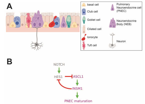
 

Figure 2. Pulmonary Neuroendocrine Cells (PNECs) in the upper airways. (A) Schematic depicting epithelial cell types found in the upper airways. Solitary PNECs and innervated Neuroendocrine Bodies (NEBs) are shown. (B) Diagram of signaling and transcription factor interactions that regulate PNEC differentiation.

Although the precise function of PNECs is not well-defined, their preservation across evolution -- similar cell types are found in fish gills and in all air-breathing vertebrates -- suggest an important physiological function (17–19). Furthermore, the position at airway bifurcation points of PNECs, their contact with the airway lumen, and innervation suggest a role in airway sensing. The bioactive compounds, hormones, and neuropeptides that PNECs secrete are known to affect oxygen sensing, pulmonary blood flow and bronchial tonus, and lung immune responses. The bioactive compounds secreted by PNECs include serotonin, calcitonin, calcitonin gene-related peptide (CGRP), gastrin-releasing peptide (GRP), chromogranin A, gamma aminobutyric acid (GABA), and synaptophysin, clearly suggesting an endocrine function for these cells (20–23). A summary of the hormones and neuropeptides expressed by PNECs and other NE cells discussed in this text is included in Table 3.


Table 3. NE Cell Expressed Hormones

Hormone

Associated

NE cell types

Reported function in

described NE cell types

Calcitonin

PNEC

pulmonary blood flow and bronchial tonus

Calcitonin gene-related peptide (CGRP)

PNEC

vasoregulation, bronchoprotection, immune cell recruitment

Gastrin releasing peptide (GRP)

PNEC

Regulates mucus and cytokine production upon inflammation or inflammation-associated processes

Gamma aminobutyric acid (GABA)

PNEC and pECs

Regulates mucus and cytokine production upon inflammation or inflammation-associated processes

Somatostatin (SST)

PNECs and GEP NE cells

 (D cells)

Inhibits secretion of insulin, glucagon, PYY, serotonin, and gastrin

Vasoactive intestinal polypeptide (VIP)

PNECs and GEP EECs

Acts as a neurotransmitter, an immune regulator, a vasodilator, and a secretagogue

Histamine

PNECs and Gastric EECs

(ECL cells)

Stimulates gastric acid secretion

Ghrelin (GHRL)

PNECs, Gastric EECs (X/A cell), and pECs (Epsilon cells)

Stimulates appetite, promotes gluconeogenesis and increased gastric acid secretion

Cholecystokinin (CCK)

PNECs and GI EECs

 (I cells)

Stimulates gallbladder contraction, pancreatic enzyme secretion, gut motility, satiety, and inhibits acid secretion

Serotonin (5-HT)

PNECs and GI EECs

(EC cells)

Regulates vasodilation and smooth muscle contraction

Secretin (SCT)

GI EECs

Stimulates the release of bicarbonate and water to neutralize gastric acid

Gastric inhibitory peptide (GIP)*

GI EECs (K cells)

Inhibits insulin secretion and to a lesser extent gastric acid secretion

Substance P (neuropeptide)

GI EECs (EC cells)

Regulates intestinal motility and mucosal permeability

Glucagon-like peptide 1 (GLP-1)

Intestinal EECs (L cells)

Stimulates insulin secretion and inhibits glucagon secretion

Neurotensin (NTS)

Intestinal EECs (N cells)

Inhibits gastric acid secretion

Glucagon (GCG)

pECs (alpha cells)

Increases blood glucose levels by stimulating glucose production and inhibiting glycogen storage by the liver

Insulin (INS)

pECs (beta cells)

Stimulates uptake of blood glucose by other tissues

Gastrin

pECs (G cells)

Stimulates gastric acid secretion

Pancreatic polypeptide (PPY)

pECs

(PP/gamma cells)

Inhibits glucagon and somatostatin

* Gastric inhibitory peptide (GIP) is also known as glucose-dependent insulinotropic polypeptide


PNECs in Development: Specification, Differentiation, and NEB Formation


PNECs are the first differentiated cell type to appear in the developing lung (15). The timing of PNEC differentiation in human lungs has not been comprehensively delineated but several studies have reported the appearance of PNECs in human fetal airways at 8 - 9 weeks of gestation (24). In mice, the fetal epithelial progenitor cells that give rise to mature PNECs appear for the first time at around embryonic day (E) 12.5 (25). These cells are defined by expression of the basic helix loop helix (bHLH) lineage transcription factor, ASCL1, which is required for their specification. Mice that carry null alleles of Ascl1 do not have PNECs (26,27).


PNEC lineage specification by expression of Ascl1 is followed by two key events that appear to happen in parallel: the formation of NEBs and the maturation of early Ascl1 expressing cells to fully differentiated PNECs. Using a lineage trace of Ascl1-positive cells in mouse embryonic lung, Kuo and Krasnow observed the first signs of PNEC clustering at around E13.5 to E14, followed by the appearance of bonafide NEBs, which contained mature PNECs at around E15.5 to E16 (25). In human fetal lungs PNEC clustering was first observed at about 9 to 10 weeks of gestation (28).


Although one might imagine that the formation of NEBs is likely achieved through proliferation of nascent PNECs, a different mechanism has been shown to be at play. Sparse lineage tracing of early fetal Ascl1-positive PNECs in mice using a multi-color lineage reporter showed that NEBs contained either different colored cells or a single labeled cell and multiple unlabeled cells, arguing that the PNECs in NEBs are not clonal (25). Live cell imaging of fetal mouse lung tissue showed that NEBs are formed through the migration and subsequent aggregation of PNECs at airway branchpoints (25,29). This process appears to be regulated by Slit-Roundabout (ROBO) signaling, a pathway more classically associated with axonal guidance (30). PNECs express the ROBO receptor and the lungs of mice where the Slit-ROBO pathway has been disrupted by mutation of either the Slit ligands or the ROBO receptor itself, have fewer NEBs and more solitary PNECs than the lungs of wildtype mice (21).


Concomitant with NEB formation, the transcriptional events initiated by expression of Ascl1 culminate in the emergence of fully differentiated, functional PNECs. In particular, expression of Ascl1 in lung progenitors induces expression of the zinc finger transcription factor, INSM1. PNECs in mice carrying mutant alleles of Insm1 fail to express the mature PNEC markers, CGRP and UCHL1 (ubiquitin C-terminal hydrolase L1, a.k.a. PGP9.5). While the INSM1 targets that mediate the PNEC maturation process have not been delineated, INSM1 has been shown to directly repress the bHLH transcription factor and Notch target gene, Hes1 (Figure 2B) (31).


HES1 and other Notch signaling components play crucial roles in repressing the differentiation and specification of PNECs and, thereby, in mediating the NE versus non-NE cell fate choice. The lungs of mice in which Hes1 has been conditionally deleted in early lung progenitors have more ASCL1-positive cells and, in particular, fewer solitary PNECs, showing instead more and larger NEBs compared to the lungs of wildtype mice (29). The increased size of NEBs in Hes1-deficient lungs suggests a mechanism of cell fate specification through Notch-mediated lateral inhibition whereby Notch is activated in PNEC neighboring cells through binding of Notch receptors to the Notch ligands expressed on the surface of PNECs themselves. The activated Notch signaling in these PNEC neighboring cells induces expression of Hes1, which in turn represses the PNEC fate.


PNECs express the Notch ligands Dll1, Dll4, Jag1, and Jag2 shortly after their specification during lung development and the cells surrounding PNECs in NEBs express Notch receptors (32). Genetic loss of either all three Notch receptors or Dll1 and Dll4 Notch ligands in the developing mouse lung leads to a dramatic increase in the number and size of NEBs, phenocopying conditional deletion of Hes1 (32,33). In cultures of human airway cells derived from induced pluripotent stem cells, inhibition of Notch signaling leads to increased numbers of PNECs (34,35).


As we will discuss in other parts of this text, two other bHLH transcription factors, NEUROG3 and NEUROD1 have been shown to play central roles in the differentiation of EECs and pancreatic islet cells. In contrast, as of yet, there is little evidence that these transcription factors are decisive for PNEC specification or differentiation. To date, Neurog3 expression has not been described in PNECs. While Neurod1 expression has been observed in some PNECs in both fetal and adult mouse lung, a limited number of studies have investigated its specific role in the PNEC lineage (27,36). Neurod1-null mouse lungs from mice less than 2 weeks old showed decreased numbers of solitary PNECs and more NEBs compared to lungs from age-matched wild type mice. However, this difference normalized once mutant mice were 6 weeks old (36). As will be discussed later on in this text, NEUROD1 is a marker of a subtype of the high-grade lung NEN, small cell lung cancer (SCLC), suggesting it might also play a role in normal PNEC biology.


Reactive PNEC Proliferations: PNECs in the Response to Lung Injury


Multiple studies have shown that PNEC numbers and NEB size are altered in several human disease conditions. Increased numbers of PNECs have been observed in the lungs of patients with COPD, asthma, cystic fibrosis, and some forms of pneumonia. Other pathological conditions associated with increased PNECs include sudden infant death syndrome (SIDS), bronchopulmonary dysplasia (BPD), and congenital diaphragmatic hernias (CDH) (15). Studies in animal models and PNEC culture systems provide experimental evidence that PNECs respond to environmental stimuli by both proliferation and/or secretion of bioactive compounds.


The early observation that murine PNECs proliferate in response to a common form of experimental lung injury, naphthalene administration, and that this proliferation precedes epithelial lung regeneration, led to the hypothesis that PNECs are multipotent stem cells that aid in lung regeneration (37). Indeed, lineage tracing studies of PNECs following naphthalene induced lung injury showed that a rare subpopulation of PNECs, termed NEstem, can function as stem cells in this context (38). In response to naphthalene, NEstem cells proliferate and sometimes migrate to the site of injury where they dedifferentiate (lose NE identity) and take on other lung cell fates. The process of dedifferentiation and reprogramming was shown to be mediated by Notch signaling, recalling the role of this pathway in PNEC fate specification, and by EZH2 (38,39). Nonetheless, NEstem cells are not solely responsible for regenerating the lung after injury, as they were shown to contribute only to a small portion of the regenerated surface epithelium (38). Furthermore, genetic ablation of PNECs does not abrogate lung regeneration following naphthalene injury (23,39). 


In thinking about PNECs as components of a diffuse hormonal system, two questions arise from the studies of PNECs in the context of naphthalene lung injury. The first is, how do PNECs detect injury? Club cells, which express cytochrome P450 2F2 (Cyp2f2), metabolize naphthalene to a toxic metabolite and the accumulation of this toxic metabolite leads to cell death specifically in these cells (40). Given that PNECs proliferate at time points shortly after peak Club cell injury, it is likely that they are responding to the Club cell injury and not to the naphthalene itself. Consistent with this hypothesis, selective ablation of Club cells using genetic ablation techniques also resulted in PNEC proliferation (41). Nonetheless, injury associated signals that are specific to PNECs and their responses have not been identified.


The second question that arises is, besides functioning as stem cells, do PNECs have an endocrine or paracrine/autocrine function in the context of lung injury? Although this question has not been explored in the naphthalene injury model, evidence from other model systems and from human diseases suggest that PNECs respond to some forms of lung injury or disease through the secretion of bioactive compounds. Cigarette smoke, a common culprit of lung injury, provides a good example. Bronchoalveolar lavage (BAL) fluid from smokers has increased levels of peptides secreted by PNECs, implicating these cells in the cellular response to cigarette smoke in humans (42). The PNEC-secreted bioactive compounds associated with this response include GABA and GRP, and both of these molecules have been implicated in inflammation and inflammation-associated processes (42,43). It is likely that PNEC proliferation is also involved in the response to cigarette smoke and its primary component, nicotine. Increased PNECs have been observed in rats exposed to cigarette smoke pre- and postnatally and in rhesus monkeys exposed to nicotine prenatally (44,45).


Pointing to a critical role for PNECs in oxygen sensing in the lung, hypoxia and hypoxia-mimicking genetic modifications have been shown to result in higher numbers of PNECs in mice, rats, rabbits, and guinea pigs (22,46–49). Shortly after showing that the distinct dense cored vesicles of PNECs carried serotonin, Lauweryns and Cokelaere went on to show that this serotonin was secreted by PNECs upon exposure to hypoxia (14,49). This finding was further refined and shown to be dependent on changes in intracellular Ca2+ concentrations using cultured rabbit and hamster lung slices (50,51). Serotonin release by PNECs is likely a physiologically relevant functional response to hypoxia as serotonin has been shown to induce vasoconstriction of pulmonary arteries (52).


Increased expression in PNECs of the neuropeptide, CGRP, has also been linked to hypoxia (46,53). CGRP has been implicated in promoting alveolar regeneration and in mediating immune cell responses in the lung (21,54). Importantly, results from a study by Shivaraju et al. linked the expression of CGRP by PNECs to the hypoxia-induced regenerative response of epithelial cells in the trachea. When the authors ablated PNECs and exposed mice to hypoxia, they observed a defective regenerative response that could be rescued by intranasal administration of CGRP (46).


PNECs appear to also respond to hyperoxia-induced lung injury. Patients with BPD, a chronic lung disease associated with oxygen supplementation of premature infants, have increased numbers of GRP-expressing PNECs (55). In a baboon model of BPD, some of the lung defects associated with the disease could be prevented by treatment of the animals with a GRP blocking antibody, demonstrating that GRP is directly linked to the disease phenotype (56). GRP also plays a role in the lung’s response to viral pneumonia and in the fibrotic response to radiation therapy (57,58).


There is clear evidence for a close interplay between PNECs and immune cells. In particular, the effector molecules secreted by PNECs can recruit and activate different populations of immune cells. In one of the first studies to show this, researchers developed a mouse model of CDH, a birth defect that results in pulmonary hypoplasia and pulmonary hypertension (21). CDH is associated with both a heightened immune response and increased numbers of PNECs (59). To study CDH, since point mutations in SLIT and ROBO genes are associated with the disease, Branchfield et al. generated mice with lung-specific deletions of the roundabout receptors, Robo1 and Robo2. When Robo1 and Robo2 were deleted in the entire lung epithelium the authors noted elevated immune cell infiltration in the lung, thus mimicking one of the features of CDH. When Robo1 and Robo2 were deleted only in PNECs, the mice displayed the same phenotype, directly linking the defect to PNECs. Interestingly, ROBO1- and ROBO2-deficient PNECs have increased levels of CGRP and knockout of the gene encoding CGRP partly reversed the immune and lung phenotypes of mice deficient for ROBO1 and ROBO2 in the lung epithelium.  


A potential immune regulatory role for PNECs is also suggested by the observation that PNEC numbers are elevated in patients with asthma and that more chromogranin A-positive PNECs are seen in guinea pigs after allergen sensitization and challenge (60). Mice deficient of PNECs due to deletion of Ascl1 in the lung epithelium, show a dampened response to allergen challenge -- reduced goblet cell hyperplasia and reduced immune cell infiltration -- and this was tied directly to reduced levels of PNEC-derived GABA and CGRP, respectively (61).


Diseases of Primary PNEC Hyperplasia: NEHI and DIPNECH


Up to now we have highlighted instances of increased PNEC number or NEB size that appear to be consequent to or at least associated with some forms of acute or underlying lung injury. These examples are instructive in that they point to a role for PNECs and the molecules they secrete in mediating the response to external stimuli and injury in the lung. Save for lung NENs, which will be discussed in further detail later in this text, there are two notable clinical instances of primary -- as opposed to reactive -- PNEC proliferation that have no known etiology and are not associated with common pathogenic triggers: neuroendocrine cell hyperplasia of infancy (NEHI) and diffuse idiopathic neuroendocrine hyperplasia (DIPNECH).


NEHI is a rare pediatric lung disease consisting histologically of hyperplastic GRP-positive and serotonin-positive PNECs in the distal lung epithelium of otherwise normal lung tissue. Symptoms are usually first noted between 6 to 8 months of life and include tachypnea, retractions, crackles and hypoxemia (62). In some cases, patients with NEHI show an inconspicuous, patchy pattern of inflammation or fibrosis, generally assumed to be a consequence of the increased PNEC numbers rather than its cause (63). Interestingly, despite increased PNEC numbers in the lungs of patients with NEHI, from a study on a small patient cohort (5 patients), it appeared that PNECs were not actively proliferating in the lungs of these patients as no Ki67 and GRP double positivity was observed (63). Unfortunately, treatment for patients with NEHI are currently limited to supportive oxygen supplementation and, in some cases, additional nutritional support. The majority of NEHI patients show gradual improvement of symptoms and the disease is not associated with mortality. Nonetheless, recent reports show that some patients experience abnormal lung function persisting into adulthood (62,64,65). While the etiology of this disease remains unknown, there are indications of a genetic basis for the disease. One study identified four families with multiple members diagnosed with NEHI and showing an autosomal dominant pattern of inheritance (66). Another study identified a heterozygous mutation in the NKX2.1 gene in members of a family with a history of childhood lung disease consistent with NEHI (67).


DIPNECH is a rare syndrome with adult onset consisting histologically of increased PNECs in the small bronchi and bronchioles and confined to the basement membrane, appearing as scattered PNECs, small nodules, or a linear proliferation of PNECs (68). These features are often seen in concomitance with what are referred to as tumorlets, PNEC proliferations that extend beyond the basement membrane but are less than 5 mm in diameter (69). Other histological features include fibrosis, chronic inflammatory cell infiltrate, and constrictive obliterative bronchiolitis. The majority of patients with DIPNECH are women and the disease is not associated with smoking or other lung diseases. Patients diagnosed with DIPNECH often present with symptoms including cough, exertional dyspnea and an obstructive or mixed obstructive/restrictive defect on pulmonary function test. A small number of patients with DIPNECH are diagnosed due to incidental findings (69).


DIPNECH was first recognized and formally defined in 1992 by Aguayo et al., who described the symptoms and histological features of 6 DIPNECH patients (42). While DIPNECH is considered a disease of primary rather than reactive PNEC proliferation, cases associated with parathyroid gland hyperplasia, acromegaly and pituitary adenoma, multiple endocrine neoplasia type I syndrome, and pulmonary adenocarcinoma have been reported (70–72). The World Health Organization (WHO) classifies DIPNECH as a preinvasive, possibly preneoplastic condition (73). Most patients with DIPNECH have multiple PNEC nodules, sometimes including both tumorlets and frank low grade lung NET (carcinoid) tumors (70). In contrast to NEHI where Ki67-positive PNECs were not observed, the PNEC proliferations in DIPNECH patients contain some Ki67-positive cells (63,74). While patients with DIPNECH most often follow a clinical course showing stability or slowly progressing functional decline, a small subset of patients have rapidly progressive disease including progression to respiratory failure or metastatic carcinoid tumors (70,75). To date, there is no standard of care for DIPNECH and the most effective treatment strategy for patients with DIPNECH are somatostatin analogues (SSAs), which have shown effectiveness in improving symptoms of cough and dyspnea in some patients (76–78). Considering that it has been well-established that SSAs inhibit the secretion of bioactive compounds from gastrointestinal NETs, the effectiveness of SSAs in treating cough and dyspnea in patients with DIPNECH suggests these symptoms are caused by the secretion of bioactive compounds by DIPNECH PNECs (79).


Lung NENs


Lung NENs account for 20-25% of all lung cancers and for 25-30% of NENs from all tissue sites (80,81). As is the case for NENs in general, lung NENs comprise both low grade, well-differentiated NETs and high grade, poorly differentiated NECs. Lung NENs can thus be subdivided into the high-grade carcinomas, small cell lung cancer (SCLC) and large cell neuroendocrine carcinomas (LCNEC), and the low-grade tumors, atypical carcinoids (AC), classified as intermediate grade, and typical carcinoids (TC), classified as low grade.   


SMALL CELL LUNG CANCER (SCLC)


The most common lung NEN, SCLC, accounts for 79% of all lung NENs and 30% of all lung cancers and is also the best studied among lung NENs (82). Consistent with its classification as NEC, SCLC is a highly aggressive tumor with a high rate of metastasis and a 10-year survival rate of only 1-2% (83). Among patients with SCLC, 97% have a history of smoking (18). While rare, patients with SCLC sometimes experience paraneoplastic endocrine syndromes, most commonly a syndrome of inappropriate antidiuretic hormone (SIADH) and ectopic Cushing’s syndrome (84). Studies of SCLC biology have been aided by a collection of tumor-derived cell lines, several patient-derived xenograft (PDX) models, and genetically engineered mouse models (GEMMs) of the disease (85–87). These preclinical model systems have allowed scientists to address questions in two key areas: the cell of origin of SCLC and molecular signatures predictive of therapeutic vulnerabilities.


Genetically, SCLC is a relatively homogeneous disease -- RB1 and TP53 are both almost universally lost in patient tumors (88). Conditional simultaneous genetic deletion of Rb1 and p53 in the mouse epithelium results in tumors that recapitulate many of the key features of the human disease at both the histological and molecular levels (87,89–91). Targeting the deletion of Rb1 and p53 to specific epithelial cell types in the lung provided definitive evidence that Cgrp-expressing PNECs are a cell of origin for tumors in this model (23,92). However, PNECs are not the only epithelial lung cell type that can be a cell of origin for mouse SCLC. A separate study showed that an, as of yet, unidentified CGRP-negative cell that is also negative for the canonical markers of two other common lung epithelial cell types gives rise to mouse SCLC lesions that are molecularly distinct from those initiated in CGRP-positive PNECs (93).


Genomic analysis of human and mouse SCLC primary tumors and cell lines has revealed commonly mutated genes and pathways, most notably loss of PTEN, NOTCH, and histone modification genes, and amplification of MYC family oncogenes (88,90,91). Several studies focused on metastasis in mouse SCLC have highlighted the role of NFIB in driving progression of some of these tumors, and data from human patients with SCLC support the clinical relevance of these findings (94–96). These studies, in combination with preclinical testing in mouse models and cell lines have suggested some degree of patient stratification. In particular, high expression of MYC is associated with tumor sensitivity to Aurora Kinase inhibitors (97).


The standard treatment regimen for patients with SCLC is a combination therapy of a platinum agent combined with etoposide (82). Despite a clinical response to these therapies in the majority of patients, almost all patients will then experience tumor recurrence (83). The analysis of the transcriptomes of both mouse and human SCLC tumors and cell lines has identified 4 molecular subtypes of SCLC, defined by their expression (or lack of expression) of 3 lineage-specific transcription factors: ASCL1 high, NEUROD1 high, POU2F3 high, and a fourth subtype that has low expression of NE transcription factors and has been proposed to be defined by expression of YAP1. A more recent study suggests a classification in which this fourth subtype is defined by expression of immune checkpoint genes and human leukocyte antigens (98,99). It has been hypothesized that the cell of origin for the POU2F3 high subtype of SCLC might be the pulmonary tuft cell, another chemosensory cell type in the lung distinct from PNECs (100,101).


Importantly, several studies using preclinical models of SCLC suggest that these molecular classifications can be used to stratify patients according to potential therapeutic vulnerabilities (102). Adding complexity to this schema, single cell RNAseq studies of mouse SCLC and of xenografts derived from circulating tumor cells from SCLC patients suggest that different molecular subtypes might represent different stages of progression where tumors begin in an ASCL1-high state and progress towards a non-NE state and that individual tumors might comprise cells belonging to different subtypes (101,103). Several studies have also shown other forms of intratumor heterogeneity in SCLC that have implications for patient therapy (104–106). Other new therapies suggested for SCLC include tricyclic antidepressants, therapies that target specific metabolic vulnerabilities, and therapies targeting the GNAS/ PKA/PP2A signaling axis (107–109).


LARGE CELL NEUROENDOCRINE CARCINOMA (LCNEC)


Pulmonary LCNEC is less common than SCLC, accounting for 16% of all lung NENs (82). Like SCLC, pulmonary LCNECs are highly metastatic and are associated with smoking history and with an overall 5-year survival rate ranging from 15% to 25% (110). In contrast to SCLC, however, there are relatively few preclinical models for LCNEC and we know much less about the basic biology of this disease. This might partly explain why guidelines for treating patients with LCNEC are rather rudimentary (82,111). 


Pathohistological analysis of tumors from GEMMs of SCLC found that a portion of the mouse tumors in these models had a histological pattern consistent with LCNEC. While these LCNEC tumors only accounted for 10% of the tumors from the GEMM in which only Rb1 and p53 were conditionally deleted in the lung epithelium, they were much more prominent in the GEMM in which Rb1, p53, and Pten were conditionally deleted specifically in CGRP-expressing PNECs (87). A different GEMM, based on loss of Rb1 and expression of mutant p53 alleles, also develops both SCLC and LCNEC mouse tumors (112). These GEMMs have, thus far, not been used to explore the biology specifically of LCNEC and doing so might present some technical challenges. Recently, the first GEMM specifically for LCNEC was reported. In this model, Rb1, p53, Pten, and Rbl1 were simultaneously deleted in the mouse lung epithelium, resulting in a tumor spectrum consisting primarily of LCNEC and low-grade NETs (113).   


The majority of insights into LCNEC have been provided by molecular analysis of primary patient tumor samples. The most comprehensive analysis, consisting of whole exome sequencing of 60 LCNEC tumors and RNA-sequencing expression analysis of 69 LCNEC tumors, highlighted the existence of two major molecular subtypes of LCNEC (114). Type I LCNECs had a higher rate of alterations in TP53 and STK11/KEAP1 and an NE expression profile defined by high expression of ASCL1 and DLL3 and low expression of NOTCH. Type II LCNECs had frequent mutations in RB1and TP53, therefore resembling SCLC at the genomic level. The expression profile of type II LCNECs, however, was distinct from SCLC and instead was defined as NE low, with low expression of ASCL1 and DLL3 but high expression of NOTCH.


The description of these two molecular subtypes for LCNEC highlights a clinical conundrum relating to the treatment of patients with LCNEC: should they be treated with SCLC chemotherapy regimens or with chemotherapy regimens for non-NE non-small cell lung cancer (NSCLC) (111)? The report of an SCLC-like subtype of LCNEC (type I) and a NSCLC-like subtype of LCNEC (type II), might suggest a way to stratify patients for different chemotherapy regimens. In line with this idea, a retrospective analysis of LCNEC cases found that patients whose tumors either had wildtype RB1 or showed expression of RB1 protein had a better outcome when treated with a NSCLC chemotherapy regimen as opposed to a SCLC chemotherapy regimen (115).   


Other therapies beyond traditional chemotherapy regimens are also being explored for patients with LCNEC. One example that relates to patient stratification according to LCNEC subtype, which was defined in part by differential patterns of DLL3 expression, involves therapeutic strategies that use DLL3 to target tumor cells. Given that DLL3 is also expressed by some SCLC tumors, this also represents a potential therapeutic opportunity in SCLC. Although a DLL3-antibody conjugated to the DNA-damaging pyrrolobenzodiazepine dimer toxin did not provide a survival benefit in 2 phase 3 clinical trials, other DLL3 targeting approaches are being developed (116). In addition, several studies have uncovered potentially targetable molecular alterations in some LCNEC tumors, including activating EGFR mutations, FGFR1 amplifications, activating BRAF mutations, ALK rearrangements, and mutations affecting BDNF/TrkB signaling (114,117–119). Given that the majority of these targetable mutations have been identified in LCNEC tumors with wildtype RB1, the question remains as to how best to treat patients with RB1 mutant LCNEC (120).


LUNG NETs: TYPICAL AND ATYPICAL CARCINOIDS


Lung NETs comprise low grade typical carcinoids (TC) and intermediate grade atypical carcinoids (AC), accounting for 5% and 0.5% of all lung NENs, respectively (82). TC and AC tend to present in younger patients than LCNEC and SCLC, and the majority of patients are women and non-smokers (121). Although the majority of lung NETs are sporadic and non-functional, a small percentage of patients with Lung NETs, 5% of TC and < 2% of AC, present with paraneoplastic syndromes including those associated with adrenocorticotropic hormone (ACTH), growth hormone releasing hormone (GHRH), histamine, and serotonin (122,123). Some of these are more commonly associated with metastatic lesions of TC or AC (123). Approximately 5% of lung NETs are associated with the familial cancer syndrome caused by germline mutations in multiple endocrine neoplasia gene type I (MEN1). Interestingly, 5% to 10% of lung NETs are also associated with tumor multiplicity, a feature which might suggest a connection with either an unappreciated familial predisposition syndrome or with premalignant conditions such as DIPNECH (80,121).


The overall 10-year survival rates for stage I TC and AC are comparable ranging from 98% to 91%, respectively. In the case of stage IV tumors, 10-year survival for TC patients is 49% but is only 18% for patients with AC (124). A distinguishing feature of TC and AC is their relatively slow growth. Indeed, the pathological criteria for diagnosing carcinoids are the number of mitoses per mm2 and the presence of absence of necrosis: < 2 mitoses per mm2 and no necrosis for a diagnosis of TC, 2 to 10 mitoses per mm2 and demonstration of necrosis for a diagnosis of AC (82). Morphologically, carcinoids typically contain small cells that show nested, rosette, and trabecular growth patterns with peripheral palisading (125).


Complete surgical resection is the most common treatment for patients with TC and AC, and for the majority of these patients’ surgery is associated with a favorable survival prognosis. Unfortunately, however, a fraction of carcinoid tumors metastasizes, and tumor recurrence (even after apparent curative resection) has been reported in 1 to 6% and 14 to 29% of patients with TC and AC, respectively. Due to a highly variable time to relapse for patients with recurrence (0.2 to 12 years), the recommended follow-up period is 15 years (121,126–129). The reported incidence for lymph node metastasis for TC and AC is variable with rates ranging from 12% to 17% for patients with TC and from 35% to 64% for patients with atypical carcinoid (80,130). The incidence of distant metastases for both TC and AC is 3% and 21%, respectively (129,131).


The incidence of tumor recurrence and metastasis calls attention to a clinical need for systemic treatment options for patients with TC and AC tumors that are unresectable, as well as for the need for effective adjuvant therapy options that can be offered to patients after surgery. Unfortunately, standard chemotherapy and radiotherapy regimens have proven to be mostly ineffective in this patient population (80). The only treatment option shown to improve progression-free survival in patients with advanced and progressive TC and AC is the mTOR inhibitor everolimus (132).


Other therapeutic options for patients with TC and AC that are not currently considered standard of care due to limited clinical trials, include somatostatin (SST) analogues and peptide receptor radionuclide therapy (PRRT), and temozolomide with or without capecitabine. Given that pulmonary carcinoids can express SST receptors, patients with these tumors can be potentially considered for palliative treatment with unlabeled or radiolabeled SST analogues.


The lack of clarity regarding standard of care for patients with unresectable, metastatic, or recurrent TC and AC, points to an unmet need for not only new and effective systemic treatment strategies for these patients, but also for clear patient stratification criteria for predicting the probability of a response of a given patient tumor to specific therapeutic options. Furthermore, given the broad range of tumor malignancy for TC and AC, biomarkers that can predict the potential for tumor progression and metastasis or recurrence are also needed. Efforts to address these clinical needs have been hindered by both difficulties in performing molecular characterization of these tumors and by a dearth of preclinical models representative of the disease. Only a handful of cell lines exist for TC and AC and only one GEMM for TC and AC has been reported (133,134).


In contrast to SCLC and LCNEC, pulmonary carcinoids have a low tumor mutational burden, have very few recurrent or characteristic mutations, and rarely contain “driver” mutations in known oncogenes. The most commonly mutated gene in pulmonary carcinoids is MEN1, and up to 5 to 13% of patients with germline mutations of this gene are diagnosed with pulmonary carcinoids (135–137). The most commonly mutated class of genes in pulmonary carcinoids are chromatin remodeling genes, a category that includes MEN1, PSIP1, and ARID1A. Though prevalent in SCLC and LCNEC, mutations in RB1 and TP53 are rare in pulmonary carcinoids (135,136). Recurrent copy number alterations have also been identified in pulmonary carcinoids, including in genes that would imply targetable therapeutic vulnerabilities such as, EGFR, MET, PDGFRB, AKT1/PKB, PIK3CA, FRAP1, RICTOR, KRAS, and SRC (136,138).


Transcriptional and methylation analysis of primary pulmonary carcinoids has also revealed distinct subclasses of these tumors. Using multi-omics factor analysis (MOFA), Alcala et al. identified 3 molecular clusters, termed A1, A2, and B (139). While most of the tumors in clusters A1 and A2 were TC, tumors in cluster B were primarily classified as ACs. Tumors in cluster B had high expression of ANGPTL3 and ERBB4, were enriched for mutations in MEN1, and were associated with a worse overall survival. Consistent with a worse prognosis for patients with tumors in cluster B, tumors in this cluster also showed universal downregulation of the orthopedia homeobox protein gene, OTP, whose expression has previously been associated with an improved prognosis in patients with pulmonary carcinoids (126). A separate study performed a similar multi-omic analysis of an independent set of pulmonary carcinoids and also identified 3 molecular subtypes that they termed LC1, LC2, and LC3 (140). The concordance between the molecular subtypes identified in these two studies was shown through integration of the two datasets, further validating the use of these molecular classifications for pulmonary carcinoids (141). 


The molecular analysis of pulmonary carcinoids has provided evidence that supports the idea that a fraction of lung NENs may actually fall into a category that lies between G2 ACs and G3 NECs in terms of malignancy. While such a category is recognized in GEP-NENs and is termed well-differentiated G3 NET, its existence has only recently been suggested for lung NENs (142). The study by Alcala et al. identified a subgroup of ACs, termed “supra-carcinoids,” that showed the morphologic characteristics of pulmonary carcinoids, but whose transcriptional profile was closer to that of LCNECs (139). In their analysis of the transcriptional profiles of a series of LCNECs and ACs, Simbolo et al. identified 3 molecular clusters, C1, enriched for LCNECs, C3 enriched for ACs, and C2, which was mixed in terms of number of ACs and LCNECs and which showed intermediate molecular features (143). Finally, an earlier study by Rektman et al. had identified 2 examples of what they referred to as “carcinoid-like” LCNEC tumors -- tumors that showed a clear carcinoid-like morphology and a molecular profile consistent with ACs (low tumor mutational burden and mutation in MEN1) but that had been classified as LCNEC due to a high proliferation rate (118). 


The supra-carcinoids in the Alcala et al. study showed a higher expression of MKI67 than other carcinoids in the series, supporting an idea that has been purported in the literature concerning a potential role for percent Ki67 positivity in identifying pulmonary carcinoids more likely to be associated with a poor prognosis (144–146). Typically, ACs show a Ki67 positivity rate of less than 20%. However, some tumors diagnosed as AC show rates between 20 and 50% (147,148). Likewise, as indicated by the “carcinoid-like” LCNEC tumors in the Rekhtman et al. study, some tumors that would otherwise be considered ACs, are diagnosed instead as LCNEC due to having a high proliferation rate (118,149). Furthermore, the comparison of proliferation rates between matched primary stage IV pulmonary carcinoids and metastases indicated an increased proliferation rate in 35% of the metastases, suggesting increased proliferation as a feature of progression (148). This idea is further supported by the observation that Ki67 positivity was heterogeneous in the analyzed tumors with some regions of the tumors showing hot-spots of increased proliferation compared to the rest of the tumor. Beyond Ki67 positivity, a list of defining features of supra-carcinoids or borderline pulmonary carcinoids/neuroendocrine carcinomas has yet to be established.


ENDOCRINE CELLS IN THE GASTROENTEROPANCREATIC TRACT


Together, the organs connected throughout the mouth to the anus are known as the gastrointestinal (GI) tract, and when the pancreas is included, these organs are collectively referred to as the gastroenteropancreatic (GEP) tract. Throughout the GEP tract endocrine cells can be found as either solitary cells, as is the case in the GI tract, or as innervated clusters, as is the case in the pancreas.


Throughout the gastrointestinal (GI) tract the solitary endocrine cells, which have a slender, elongated shape, are referred to as enteroendocrine cells (EECs). This classification helps to distinguish them from endocrine cells of other organs e.g., lung and pancreas. Despite representing only 1% of the gut epithelial cells, the large size of the intestinal epithelium makes it the body’s largest endocrine organ (150,151).


Compared to the slender EECs of the GI tract, the pancreatic endocrine cells, dispersed as clusters (known as islets of Langerhans) throughout the organ, have a more pyramidal or round-oval shaped appearance. An adult human has millions of islets, which collectively correspond to roughly 2% of the pancreatic epithelium (152,153). These islets are highly vascularized, a feature that enables pancreatic hormones to travel via the bloodstream to reach their target organs. In fact, the pancreatic hormones act both locally and systemically, eliciting responses throughout the body, consequently affecting the overall metabolic state of the organism. The sections below will provide an overview of the endocrine cells of the GEP tract as components of both the diffuse neuroendocrine system and the body’s diffuse hormonal systems. 


GASTRIC ENDOCRINE CELLS


The first major organ of the GI tract is the stomach, which, in humans, can be divided into 4 functionally distinct compartments. From proximal to distal; the cardia is the connective region between the esophagus and the stomach, the fundus stores undigested food and gases, the corpus is the largest compartment and performs the digestive action of the stomach, and, finally, the pylorus regulates gastric emptying (Figure 3A) (154,155).
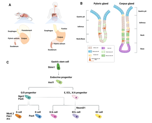
Figure 3. Gastric enteroendocrine cells. (A) Schematic showing the anatomical differences between the murine and human stomach (B) Schematic depicting epithelial cell types found in the gastric pylorus and corpus glands. Solitary gastric enteroendocrine cells are shown in orange. (C) Diagram of signaling and transcription factor interactions that regulate gastric enteroendocrine cell differentiation.

Histologically, the stomach comprises tubular-shaped mucosal invaginations containing a pit region of primarily surface mucous cells, and a gland region. The latter is further subdivided into the isthmus, neck, and base (Figure 3B). The primary differentiated cell types of the stomach are: mucus-producing pit cells, chief cells, which secrete digestive enzymes, acid-secreting parietal cells, gastric tuft cells, whose function is ill defined, and gastric EECs. These cells are continuously formed throughout life, albeit at different rates, by progenitor cells located in the isthmus of the gland region (156,157). With the exception of the cardia, which primarily contains pit cells and scattered parietal cells, gastric EECs can be found in all of the compartments of the stomach. In the corpus and fundus EECs are located in the lower third of the glands. EECs in the pylorus are located in the neck region (158).                                                  


Gastric EECs are divided into the following 5 main subtypes defined by their predominantly expressed hormone: G cells (gastrin), D cells (somatostatin), enterochromaffin (EC) (serotonin), EC-like (ECL) cells (histamine), and X/A cells (ghrelin) (Figure 3C) (159). While some gastric EEC subtypes overlap with those found in the intestine, comparison of duodenal EECs and gastric EECs by single cell RNA sequencing (scRNA-seq) suggested a distinct gastric EEC expression profile. These differences are most likely reflective of the tissue specific microenvironment of these EECs, the stimuli they are exposed to, and their different functions (160).     


As will be outlined in subsequent parts of this text, differentiation of intestinal EECs requires expression of the master bHLH transcription factor, NEUROG3. While NEUROG3 is important for gastric EECs, its role appears to be EEC subtype specific. Most gastric EECs are also dependent on ASCL1, the same transcription factor that initiates PNEC specification. Studies from two different groups, which independently generated Neurog3 null mice showed that while these mice lacked D-cells and G-cells and had decreased numbers of EC cells, ECL and X/A cells were unaffected (161,162). Ascl1 null mice displayed a similar but not identical phenotype, showing lack of D, G and EC cells and severely decreased numbers of X/A cells (163). ECL cells were not examined in Ascl1 null mice. Thus, some gastric EECs, such as D- and G- cells are dependent on both NEUROG3 and ASCL1, while others, such as X/A cells, are dependent on ASCL1 but not NEUROG3. EC cells appear to be entirely dependent on ASCL1, and only partially dependent on NEUROG3 (163). Ascl1 is not expressed in intestinal EECs from the mouse. Nonetheless, scRNA-seq of human intestinal EECs identified expression of ASCL1, suggesting a role for this transcription factor not only in gastric EECs but also in human intestinal EECs (164).


The EEC hormone most clearly associated with gastric function is gastrin, secreted by G-cells located in the pyloric compartment of the stomach. These cells are also found in the duodenum, but their function has been best studied in the stomach (160,164). Gastrin secreted into the bloodstream by G-cells binds to its receptor expressed by ECL cells in the corpus thereby stimulating them to secrete histamine, which, in turn, stimulates neighboring parietal cells to secrete gastric acid. Given that parietal cells themselves also express the gastrin receptor, gastrin release by G-cells can also directly stimulate parietal cells to secrete gastric acid (165–167). The other hormones produced by the gastric EECs are also secreted by other gastroenteropancreatic endocrine cells (GEP-ECs) and will be discussed in later sections.


INTESTINAL AND COLONIC ENDOCRINE CELLS


Architecture and Cell Types of the Intestine


The gut can be divided into the small and large intestine (also known as the colon). The two primary functions performed by the small intestinal epithelium are 1) to form a barrier against the continuous chemical and mechanical insults induced by the undigested food, microorganisms, and toxins present in the intestinal lumen, and 2) to absorb nutrients from ingested food (150). This latter process occurs with an exceptionally high efficiency made possible by the large surface area generated by the intestinal epithelium’s folded structure. Protrusions known as villi contain differentiated non-mitotic cells, while invaginations known as crypts contain proliferative, self-renewing stem cells and their epithelial niche cells, Paneth cells. The colon lacks villi and its primary function is water absorption and movement of the stool. The continuous damage experienced by the intestinal epithelium necessitates a high cellular turnover to maintain organ function. The intestinal stem cells, marked by the expression of leucine-rich G-protein-coupled receptor 5 (LGR5), continuously replace lost cells via rapid division which regenerates the epithelium within 4-5 days (150).


The differentiated cells that originate from the LGR5+ stem cells can be divided into two main functional categories, absorptive (enterocytes and microfold cells) and secretory (EECs, goblet, Paneth, and tuft cells) (Figure 4A). Intestinal EECs secrete hormones in response to stimuli such as nutrients from digested food and metabolites produced by the gut microbiota (168,169). The stimuli that EECs respond to can be both mechanical and chemical, and the hormones they produce are secreted both locally and into the bloodstream, thereby allowing them to act not just locally but also systemically. Gut hormones regulate important functions such as digestion, nutrient absorption, appetite, and gastric as well as gut motility (170).
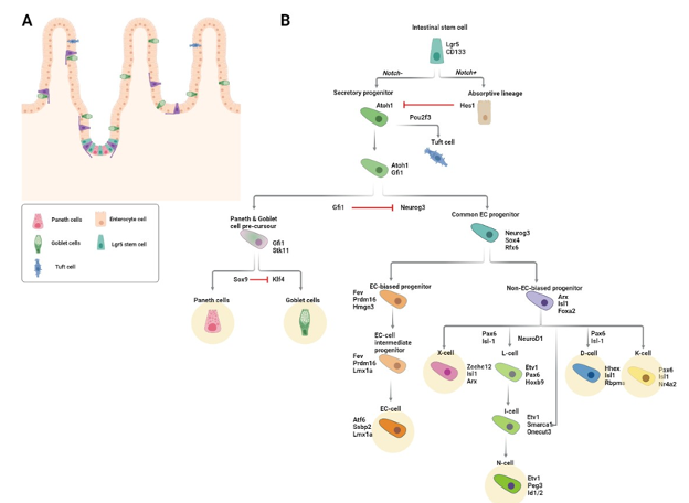
 

Figure 4. Intestinal enteroendocrine lineage specification. (A) Schematic of the intestinal epithelium. Solitary enteroendocrine cells (EECs) are depicted in purple. (B) Diagram of signaling and transcription factor interactions that regulate intestinal enteroendocrine cell differentiation. Differentiated EEC subtypes are highlighted with yellow circles.

Factors Regulating Commitment to the Secretory Lineage  


The rapid division of LGR5-positive stem cells gives rise to progenitor cells, the majority of which differentiate as they migrate upwards along the crypt-villus axis. The following section describes the differentiation of intestinal EECs and highlights some of the essential regulatory factors and signaling pathways that direct this process.


NOTCH SIGNALING  


The first step in becoming an EEC is commitment to the secretory lineage, a process that is initiated by the transcription factor, ATOH1 (Protein atonal homolog 1). At the crypt bottom, active Notch signaling prevents the differentiation of stem and progenitor cells. Cell-cell contact between Notch ligand (Dll1 and Dll4) expressing Paneth cells and stem/progenitor cells results in the expression of the Notch target gene, HES1, which in turn represses the secretory cell fate by repressing ATOH1 (171,172). Hence, commitment to the secretory lineage requires inactivation of Notch signaling.


Inactivation of Notch signaling is concomitant with loss of contact with Paneth cells. Due to the high ratio of progenitor to Paneth cells in the crypt, not all progenitors can simultaneously be in touch with a Paneth cell and this results in stochastic loss of Paneth cell-stem cell contact. Additionally, as new progenitors are continuously generated by the stem cells in the crypt, older progenitors are pushed upward along the crypt-villus axis, causing them to lose contact with the Notch ligand-presenting Paneth cells. The resultant loss of active Notch in these cells enables expression of ATOH1 and commitment to the secretory lineage (173,174). Mice lacking Atoh1 do not have any secretory cells (173). In contrast, mice with null alleles of Hes1 have excessive numbers of secretory cells.


Factors Regulating Commitment to the EEC Lineage         


Transient expression of NEUROG3 commits Atoh1-expressing secretory progenitors to the EEC fate. Whereas Neurog3 knockout mice completely lack EECs, overexpression of Neurog3 leads to increased numbers of EECs and decreased numbers of goblet cells (162,175,176). Homozygous NEUROG3 mutations have been identified in children with generalized malabsorption and reduced numbers of intestinal EECs (177). Downstream targets of NEUROG3 include other transcription factors important for EEC differentiation such as NEUROD1, PAX4/6, NKX2.2, INSM1, and PDX1 (162,178–181). NEUROG3 is also implicated in cell cycle control: Neurog3 expression in mouse pancreatic endocrine progenitors leads to upregulation of the cell cycle inhibitor, Cdkn1a, and consequent cell cycle exit (182). Consistent with the idea that cell cycle exit biases secretory progenitors to the EEC lineage, inhibition of either epidermal growth factor receptor (EGFR) or mitogen activated protein kinase (MAPK) signaling induced quiescence of intestinal stem cells in organoid culture. When this quiescence was reversed by reactivation of these pathways, the resulting organoids had an increased proportion of EECs (183).


Specification of the Different EEC Lineages  


While most progenitors generated in the crypt immediately begin migrating upwards, those primed to become EECs remain in the crypt for anywhere between 48h and 60h (184). During this time, these cells become committed to one of several divergent differentiation trajectories, each of which results in a different EEC subtype. Prior to leaving the crypt, EEC-committed progenitors have already started to express and secrete their lineage-defining hormones. The time required to produce a specific hormone varies between the different EEC lineages and this may explain why some EEC-committed progenitors remain in the crypt longer than others (184). 


Altogether, intestinal EECs produce more than 20 different hormones. The earliest classification of EECs was based on immunostainings and consisted of the following 8 EEC lineages as defined by the main hormone they were found to express: enterochromaffin (EC) cells that secrete serotonin (5-hydroxytryptamine, 5-HT), I cells that secrete cholecystokinin (CCK), K cells that secrete gastric inhibitory peptide (GIP), L cells that secrete glucagon-like peptide 1 (GLP-1), X cells that secrete ghrelin (GHRL), S cells that secrete secretin (SCT), D cells that secrete somatostatin (SST), and N cells that secrete neurotensin (NTS) (185).


At the time that this classification was first proposed, it was believed that EECs belonging to a given subtype predominantly expressed the hormone that defined that EEC subtype. Thus, for example, it was believed that EC cells only predominantly expressed serotonin, and L cells only predominantly expressed GLP-1. However, new techniques for performing hormone co-stainings using multiple antibodies, fluorescent hormone reporter mice, and transcriptome-based sequencing of EECs have all led to the observation that some EECs express multiple subtype-defining hormones (186–190). The question thus arose, did these multihormonal EECs represent previously unidentified and distinct EEC subtypes? Or were they simply cells caught in a transition state along the EEC subtype differentiation trajectory? The latter would suggest that EECs are capable of hormonal plasticity and are therefore capable of transitioning from expression of one lineage defining hormone to another.


RESOLVING LINEAGE IDENTITY AND HORMONE SWITCHING  


One of the first lines of evidence that EECs might undergo hormone switching was the observation that, while serotonin-producing EC cells were rapidly labeled after a single pulse of radioactive thymidine, secretin-producing S cells were labeled much later and only after multiple injections of the isotope (191,192). Thus, it was concluded that serotonin-expressing cells but not secretin-expressing cells had the ability to self-renew and that secretin cells did not differentiate before reaching the villus. Based on this data, one could postulate that serotonin-expressing cells might become secretin-expressing cells once they reach the villus.


A lineage relationship between EECs localized in the crypt and EECs localized in the villus was first suggested in 1990 by Roth and Gordon based on an immunohistochemical study in which it was observed that cells expressing substance P (encoded by the Tac1 gene) but not secretin were found in the crypt, cells expressing both substance P and secretin were found in the bottom of the villus, and cells expressing secretin but not substance P were found exclusively at the top of the villus. The majority of substance P-expressing and secretin-expressing cells co-expressed serotonin. The authors thus concluded that these hormones were sequentially expressed along the crypt villus axis (186). The fact that substance P-expressing cells were labeled by the thymidine analogue, BrdU, faster than secretin-producing cells further supported this idea of sequential expression of substance P and secretin by the same EEC (193). Functional evidence for dynamic hormone-switching in EECs was provided first by cell ablation studies showing that ablation of one EEC subtype led to decreased numbers of other EEC subtypes (194). Subsequently the generation of a novel mouse Neurog3 reporter allele, Neurog3Chrono, enabled more definitive delineation of dynamic hormone-expression patterns of single EECs (184).


In the Neurog3Chrono mouse, two fluorescent reporter proteins, an unstable mNeonGreen and highly-stable tdTomato, are expressed concurrently with endogenous Neurog3. As a result, the ratio of red to green fluorescence of a given EEC provides real-time information about the age of that cell relative to when it expressed Neurog3 (184). ScRNAseq of EECs from Neurog3Chrono mice provided definitive evidence that many EECs switch the hormone they produce throughout the course of their lives and furthermore suggested a more simplified EEC subtype classification consisting of five mature EEC lineages. One of these five lineages was the one proposed by Roth and Gordon of substance P expressing cells that give rise to serotonin-expressing cells and then to secretin-expressing cells. This lineage was also confirmed independently by lineage tracing of Tac1-expressing cells in the mouse intestinal epithelium (195). 


Two key observations from the Neurog3Chrono study substantiated a simplified EEC lineage classification. First, all EEC lineages except for SST-expressing D-cells began to express secretin upon entering the villus, thus rendering the S-cell lineage obsolete. Second, L-, I- and N- cells were shown to belong to a single lineage. Prior to this, observations from a mouse model of L-cell ablation had led the Schwartz lab to propose that L- and N- cells were part of the same lineage (194). The Neurog3Chrono study showed that, when located at the bottom of the crypt, L-cells secrete GLP-1 but, upon reaching the upper regions of the crypt, begin to express the I-cell defining hormone, CCK, and, finally, upon reaching the villus region, they begin to express the N-cell defining hormone, NTS. Thus, the EEC lineages or subtypes could be reduced to the following 5 main lineages: enterochromaffin (EC) cells that secrete Serotonin, K cells that secrete GIP, X cells that secrete GHRL, D cells that secrete SST, and LIN cells that secrete GLP-1, CCK and NTS (Figure 4B) (184).


ENTEROCHROMAFFIN (EC) CELLS


EC cells are the most prevalent EEC subtype and they can be found in all regions of the intestine (196,197). They are slender, triangularly-shaped cells that can have protrusions extending towards the luminal surface of the intestine (198,199). EC cells are defined by their expression of both serotonin and tryptophan hydroxylase 1 (TPH1), an enzyme that catalyzes the rate limiting step in the biosynthesis of serotonin (189). Serotonin in EC cells is stored in pleomorphic granules and is released in response to chemical and mechanical stimuli. Although commonly associated with brain development and regulation of mood and stress, 95% of the body’s serotonin is produced by the intestine where it regulates functions such as motility, fluid secretion, and vasodilation (200–202). The response of EC cells to different types of stimuli is mediated, in part, through their expression of various receptors, including the olfactory receptor, OLFR558, which acts as a sensor for microbial metabolites, and the transient receptor potential A1 (TRPA1), a receptor-operated ion channel that detects dietary irritants (203). A subset of EC cells expresses the mechanosensitive channel Piezo2, which converts mechanical forces to secretion of fluids and serotonin (204).


Given the important functions exerted by serotonin, it is not surprising that EC cells are implicated in several GI pathologies (202). Consistent with the observation that most SI-NETs express serotonin, intestinal EC cells are thought to be the cell of origin for these tumors. An early hint that SI-NETs might indeed arise from EC cells was provided by a Immunohistochemical study in which serial sections of an entire ileal SI-NET tumor were stained for several EEC markers, including serotonin. The authors observed aggregates of proliferating EC cells within crypts in close proximity to the tumor, which the authors speculated were indicative of where the tumor had originated (205). A later study made the same observation in a larger cohort of SI-NET samples from eight different patients, supporting the idea that aberrant proliferation of EC cells within the crypt is associated with SI-NET formation (206).

More recently, it has been suggested that the cell of origin of SI-NETs is not necessarily a fully differentiated EC cell, but rather an EC cell that expresses not only the EC cell markers, TPH1 and CHGA, but also markers of reserve stem cells (207). As we will discuss later in this text (see, EECs can act as reserve niche and stem cells), lineage tracing of both progenitor cells and of cells expressing mature EC cell markers in the mouse intestinal epithelium has shown that EC cells can adopt a stem cell fate upon injury (208). This observation suggests a plausible scenario whereby, if an EC cell were to acquire a genomic (or other) aberration capable of driving NET genesis, it could be long lived enough to indeed give rise to a tumor. Consistent with this hypothesis, Sei et al. identified human EC cells co-expressing the EC cell marker TPH1, and markers associated with both canonical and reserve stem cells, within crypt EC cell microtumors in tissue sections from patients with familial SI-NETs (209,210).


WHAT CAUSES HORMONE SWITCHING?  


Growth factor gradients change from high WNT, high EGF, high NOTCH, and low BMP, at the crypt bottom, to increasingly high BMP, low WNT, low EGF, and low NOTCH along the villus. Thus, upon leaving the crypt, EEC progenitors are exposed to increasingly different signaling environments. It was therefore attractive to speculate that these signaling gradients could induce hormone switching in EECs.


Adult stem cell (ASC) derived mouse and human intestinal organoids lack mesenchymal cells and are therefore not exposed to growth factor gradients but instead experience a constant environment determined by the media composition (211). Consequently, the ASC-derived intestinal organoid system provides researchers with a controlled, in vitro setting in which the signaling environment that intestinal cells experience can be modulated. Under expansion conditions that mimic the crypt environment and are optimized to promote stem cell maintenance, the EECs in mouse small intestinal organoids display a crypt hormone profile. However, when BMP4 was added to the culture media to mimic the villus environment, the EECs in the organoids expressed Secretin suggesting they had taken on a villus-like profile (195). The molecular mechanisms governing the hormone switching from GLP-1 to CCK to NTS observed in the L-I-N lineage remain to be unraveled but are likely to similarly involve cellular signals that differ along the crypt-villus axis.


Non-Neoplastic EEC Hyperplasia  


The previous sections described the formation of intestinal EECs. These cells and the hormones they secrete play a central role in regulating processes that are important for maintaining organismal function and energy homeostasis. It is therefore not surprising that EECs are implicated in a number of human disease conditions. Increased plasma level of EEC hormones and, in some cases, direct evidence of increased numbers of specific intestinal EEC subtypes have been reported in inflammatory bowel disease (IBD), irritable bowel syndrome (IBS), lymphocytic colitis, Celiac disease, H. pylori infection, and Giardia infection (212–215). A genome-wide association study (GWAS) identified a strong association between a single nucleotide polymorphism (SNP) in the promoter of the EEC transcription factor, PHOX2B, and Crohn’s Disease (CD), a form of IBD (216). The mechanism leading to intestinal EEC hyperplasia in these conditions is not clear, though there is some evidence that inflammatory cytokines are involved (213). Consistent with this idea, treatment of mice with IFN and TNF led to increased numbers of Chromogranin A-positive colonic EECs (217). Likewise, IL-13 has been linked to the response of EC cells to enteric parasite infection (218).


It is plausible that the observed changes in EEC numbers in these conditions are not necessarily or not only a result of the disease pathology, but are also mediators of the pathology itself. There is clear evidence for interplay between EECs, the immune system, sensory neurons, and commensal bacteria (168,219). Together with goblet cells, EECs have been shown to secrete the cytokine IL-17C in patients with IBD (220). EEC hormones have been shown to have immunomodulatory functions (219). Some EECs are directly innervated and one study showed that serotonin-expressing intestinal ECs form synapses with nerve fibers through which they can modulate nerve fiber activity (203). EECs express functional toll-like receptors (TLR) and can thereby interact with commensal bacteria by responding to the metabolites they produce (221). Furthermore, as EEC hormones are known to also act systemically, the consequences of disease associated alterations in their abundance or function are not limited to the GI tract. Most notably, GLP-1 and GIP, which amplify glucose stimulated insulin secretion, are less effective in patients with type 2 diabetes (T2D) and GLP-1 receptor agonists are currently used to treat T2D and obesity (185).


The most well-documented examples of non-neoplastic EEC hyperplasia are hyperplasias of gastric and duodenal EECs. These include: ECL cell hyperplasia and G-cell hyperplasia. ECL-cell hyperplasia is associated with chronic excessive gastrin production, hypergastrinemia, resulting most commonly from achlorhydria due to chronic atrophic gastritis (CAG), gastrin-producing tumors in Zollinger-Ellison syndrome (ZES), and long-term proton-pump inhibitor (PPI) treatment (222). Lineage tracing of gastrin/cholecystokinin-2 receptor (CCK2R)-expressing cells in mice showed that CCK2R-expressing ECL cells in the isthmus but not the base of the stomach proliferated in response to PPI-induced hypergastrinemia (223). ECL cell hyperplasia only rarely progresses to neoplastic gastric ECL NETs. G-cell hyperplasia is most commonly observed as a secondary change associated with CAG in patients with pernicious anemia. In rare cases, it has also been observed in patients with peptic ulcer disease in conjunction with decreased numbers of a different gastric EEC, the SST producing D-cell. G-cell hyperplastic lesions do sometimes progress to G-cell NETs, gastrinomas (222).


EECs CAN ACT AS A RESERVE NICHE AND STEM CELLS


The microenvironment in which LGR5-positive intestinal stem cells reside is known as the stem cell niche and includes Paneth cells. The stem cell niche presents the stem cells with cellular signals that prevent them from differentiating and help preserve their self-renewal capacity. Loss of Paneth cells or severe injury such as irradiation, chemotherapy treatment, or surgery can cause loss of intestinal stem cells. In these situations, several different epithelial cell populations have been shown to act as reserve stem cells (224–228). The first studies indicating that EECs could act as reserve stem cells followed the fate of secretory progenitors (224–228). As this progenitor population also contained progenitor cells fated to become Paneth or goblet cells, the contribution specifically of EECs could not be explicitly determined. A later study that used lineage tracing of a population of EEC committed secretory progenitors expressing markers of differentiated EECs showed that these cells can act as reserve stem cells, capable of both contributing to intestinal regeneration after irradiation and generating organoids in vitro (228).


Lineage tracing of mouse intestinal cells expressing either NEUROD1 or Tryptophan hydroxylase (TPH1; the rate limiting enzyme in Serotonin biosynthesis and considered an EC cell marker) showed that these cells, similar to the EEC committed secretory progenitors mentioned above, had the ability to contribute to homeostasis and to regeneration following irradiation induced injury (208). Given that the EEC-committed progenitors in the earlier study expressed Neurod1, and that a subset of these cells also expressed Tph1, it is unclear whether the Tph1 lineage-traced cells with regenerative capacity in this study were indeed mature differentiated EECs or the same multi-potent EEC committed secretory progenitors described before. More recently, it was shown that, upon genetic Paneth cell ablation, mature EECs replaced the ablated Paneth cells, serving as Notch-presenting niche cells for intestinal stem cells (229).


PANCREATIC ENDOCRINE CELLS


Located in the upper left abdomen behind the stomach, the adult pancreas, although being one anatomical entity, originates from two individual buds that arise from either side of the distal foregut endoderm during early embryonic development. As development progresses, these buds fuse to form the final glandular composite organ consisting of two compartments, one exocrine and one endocrine, which contain functionally and morphologically distinct cell types. Acinar cells secrete digestive enzymes which are transported to the duodenum by mucin-secreting ductal cells, and together these two cell types comprise the exocrine compartment. The endocrine compartment comprises 5 different pancreatic endocrine cell (pEC) types, each of which secretes a specific hormone: alpha (glucagon), beta (insulin), gamma/ PP (pancreatic polypeptide), delta (SST) and epsilon cells (ghrelin). As is the case for EEC-derived hormones, pancreatic hormones act both locally and systemically. Indeed, some pancreatic hormones are transported throughout the body via the bloodstream and instruct other organs to, among other things, release or store glucose from the blood. Hence, the pECs play a key role in nutrient metabolism, digestion, and glucose homeostasis (230).


Development and Differentiation of Pancreatic Endocrine Cells


In contrast to the GI tract where EECs are generated throughout the life of the organism, the pECs are almost exclusively generated during the fetal development of the organ (231). This process is orchestrated by an interplay of regulatory transcription factors. Whereas some of these transcription factors are expressed constitutively, others play a transient yet essential role in mediating pEC lineage commitment and differentiation (see table 4). Murine models have made invaluable contributions to our understanding of pEC generation. In rodents, pancreatic development is divided into 3 transition phases, each outlined below (Figure 5).


Table 4. NE Cell Lineage Transcription Factors

Transcription Factor

Associated NE cell types

Role (specifically in NE cells)

ASCL1

PNECs and gastric EECs

Lineage specification (26,27,163)

NEUROG3

GEP NE cells

Lineage specification (transiently expressed) (161,162,175,176,232,233)

NEUROD1

PNECs*, Intestinal EECs (L-I-N lineage), and pECs (beta cells)

Specification of some NE cell subtypes (not defined in lung, secretin-expressing cells and I-cells in the intestine, beta cells in the pancreas) (27,36,255,407,408)

ATOH1

(a.k.a. MATH1)

Intestinal EEC precursors

Specification of the intestinal secretory cell fate (precedes EEC lineage commitment) (173)

INSM1

PNECs and GEP EECs**

NE cell maturation (31,160,180)

GFI1

PNECs and Intestinal EECs

Maturation of CGRP+ PNECs (409); lineage specification (via inhibition of EEC fate in intestinal secretory progenitors) (410,411)

PDX1

GEP EECs (G cells, some intestinal EECs, and pECs

Patterning and specification of gastric and duodenal EECs (G cells, SST- and GIP-expressing duodenal EECs) (412,413); Maintenance of beta cell maturity (414,415)

ISL1

GEP EECs (some gastric EECs**, non EC intestinal EECs, and pECs)

Specification of non-EC intestinal EECs (416); maturation and survival of pECs in development (417,418); maintenance of adult beta cell function (419)

RFX3

pECs

pEC differentiation during development (420); differentiation and maintenance of mature beta cells in the adult (421)

RFX6

GEP EECs (gastric EECs**, some intestinal EECs, and pECs)

Differentiation of intestinal EECs (K, L, X, I, and EC cells) (184,422); pEC specification and differentiation in development (423,424); Regulation of insulin expression in beta-cells (425)

PAX4

GEP EECs (some gastric EECs, duodenal EECs, some pECs)

Differentiation of EECs in the duodenum, of EC cells and D cells in the stomach (178), and of beta and delta cells in the pancreas (426,427)

PAX6

GEP EECs (some gastric EECs, some duodenal EECs, some pECs)

Differentiation of D and G cells in the stomach, of K cells in the duodenum (178), and of alpha and epsilon cells in the pancreas (428,429)

NKX2.2

Some intestinal EECs and some pECs

Regulation of cell fate within the intestinal EEC population (promotes EC, L-I-N, and K lineages) (179) and within the pEC population (promotes beta cell, alpha cell, and PP cell lineages) (252)

NKX6.1

Some gastric EECs and some pECs

Differentiation of EC cells in the stomach**(430) and of beta cells in the pancreas (253)

NKX6.2

Some pECs

Differentiation of alpha, beta, and PP cells in the pancreas (431,432)

NKX6.3

Some gastric EECs

Differentiation of G cells in the stomach (433)

MAFA

Some pECs

Maintenance of beta cell identity in the adult pancreas (257,434)

MAFB

Some pECs

Differentiation of alpha and beta cells in the developing pancreas (246,247); Maturation of alpha and beta cells in the adult pancreas (247,256)

ARX

GEP NE cells (some gastric EECs, some intestinal EECs, and some pECs)

Differentiation of G cells in the stomach (435), of L-, I-, and K cells in the intestine (435), and of alpha cells in the developing pancreas (244,245); Maintenance of alpha cell identity in the adult pancreas (436)

FOXA2

Some intestinal EECs and some pECs

Differentiation of L cells and D cells in the intestine (437); Maintenance of adult beta cell maturation in the pancreas (438,439); Maturation of alpha cells in the developing pancreas (440)

HHEX

Some intestinal EECs and some pECs

Differentiation of and maintenance of delta cell identity in the pancreas (262)(implicated in delta cells of the intestine (184,195))

* Role in PNECs no well defined

** Role in gastric EECs implicated by expression pattern
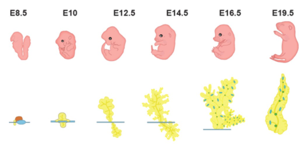
 

Figure 5. Rodent pancreas development. Schematic depicting the development of the murine pancreas. Islets of Langerhans containing the hormone-secreting endocrine cells are shown in blue and green.

PRIMARY TRANSITION


The first transition phase starts at around embryonic day (E) 9.0-9.5 with the formation of the pancreatic buds and ends at around E12.5 with the start of branching morphogenesis. During this period, the dorsal bud gives rise to the first pancreatic endocrine progenitor cells. As is the case for intestinal EECs, the formation of pECs depends on the expression of the bHLH transcription factor NEUROG3, which is transiently expressed at a relatively low level starting at ~E9.0-E11.0. Mice with targeted disruption of Neurog3 lack pECs and die postnatally of diabetes (232). Consistent with this data in mice, human induced pluripotent stem cells (iPSCs) with null alleles of NEUROG3 fail to differentiate into pECs (233). Following this first pulse of Neurog3 expression, a limited number of pancreatic progenitors becomes committed to the pEC lineage, and some of these cells begin to express glucagon (234).


SECONDARY TRANSITION


Branching morphogenesis continues throughout the first half of the second transition, (~E12.5-E15.5), during which the plexus expands and segregates into tip and trunk domains. From E16.5-E18.5 the epithelial plexus is remodeled into a ductal network with a tree-like structure. Inter- and intralobular ducts branch off from the main pancreatic duct, which connects the pancreas to the common bile duct. During this period, multipotent pancreatic progenitor cells, which make up the majority of cells at the beginning of this transition phase, become either unipotent, fated to become acinar cells at the tip, or bipotent, with the potential to form endocrine and ductal cells. These lineage commitments coincide with a second, more pronounced wave of Neurog3 expression that peaks at around E14.5-E15.5, and then rapidly decreases at around E17.5(235). This second wave of Neurog3 expression commits progenitors to the pEC fate and primes them towards one of the five pEC types.


TERTIARY TRANSITION


During the tertiary transition, individual progenitor cells that have now become fated to the pEC fate by the second, higher pulse of Neurog3 expression delaminate from the ductal network through what is thought to be asymmetric cell division and/or epithelial to mesenchymal transition (EMT). These pECs then migrate throughout the mesenchyme until they encounter other pEC-fated cells, aggregate with the help of Neural cell adhesion molecule (NCAM), and form oval shaped proto-islets (236,237). This process is reminiscent of the way, during lung development, PNECs form NEBs. The formation of fully mature islets requires their vascularization by endothelial cells and their innervation by neurons, both processes that take place from E16.5 onwards, and during the first weeks postnatally.


POSTNATAL


At birth the majority (more than 80%) of the pECs originates from endocrine progenitors and the remainder results from proliferation of preexisting pECs within the islets.


DIFFERENCES IN PANCREATIC ENDOCRINE CELL EVELOPMENT BETWEEN RODENTS AND HUMANS


Although studies of human pEC development are limited, they have uncovered a few main differences compared to rodents. First, there seems to be only one pulse of NEUROG3 during the formation of human pECs (compared to two in the mouse), which occurs at around 8 weeks of gestation. Second, the premature proto-islet structures described above are formed earlier in human development (at 12 weeks post conception) (238,239). Finally, the mature islet architecture differs between rodents and humans (Figure 6). Rodent islets have a central core of beta cells, which are also the most common of all the islet cells (60–80%). The remainder of the endocrine cells, alpha cells (15–20% of the cells of the islet), delta cells (<10% of islet cells), and gamma/ PP cells (<1% of cells) surround the beta cells in a circular structure known as the mantle (240). Human islets do not display the same organized structure as rodent islets. Instead, there is a salt and pepper pattern where the different endocrine cells are randomly scattered within the islets. The proportion of the different pECs also differs as human islets have proportionally less beta but more alpha cells compared to rodent islets, consisting of circa 30% alpha cells, 60% beta cells, about 10% delta cells, and rare <1% gamma/PP cells and epsilon cells (241,242).
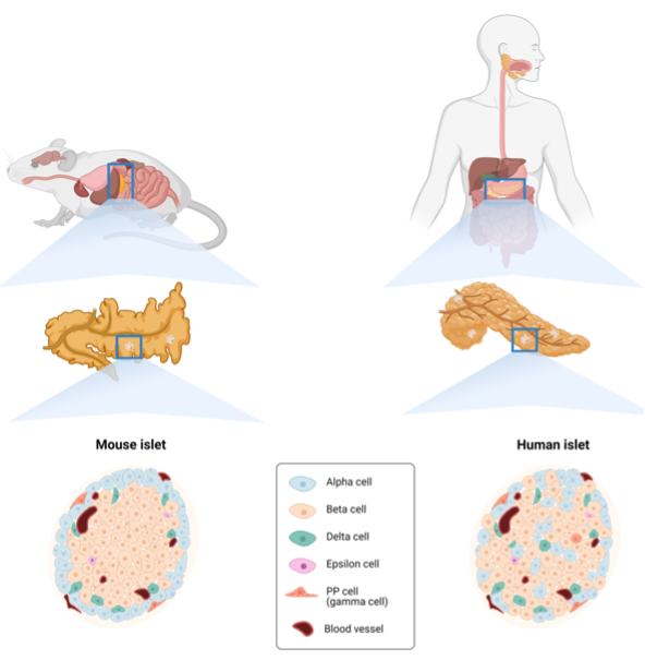
 

Figure 6. Differences between the murine and human pancreas. Schematic showing the anatomical differences between the murine and human pancreas in terms of both organ and islet architecture.


Pancreatic Islet Cells and Hormones – All with Their Own Function


This section describes the molecular and cellular characteristics of the different subtypes of pECs and their specific hormones and function during adult homeostasis (Figure 7).
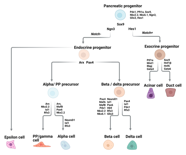
Figure 7. Pancreatic endocrine cells. Diagram of signaling and transcription factor interactions that regulate pancreatic endocrine cell differentiation.

ALPHA CELLS, GLUCAGON


Alpha cells secrete glucagon and are the second most common pEC subtype. Glucagon contributes to maintaining homeostatic blood glucose levels by stimulating glucose production and inhibiting glycogen storage by the liver. The glucagon receptor, GCGR, is widely expressed by multiple organs besides the liver, and glucagon mediates multiple other physiological processes including amino acid metabolism, glomerular filtration by the kidney, lipolysis, and gastric motility (243).


The key factor promoting commitment of pEC progenitors to alpha cells is the transcription factor ARX. Deletion of Arxin Pdx1-expressing progenitors in the mouse leads to a complete loss of alpha cells, accompanied by a compensatory increase in the number of beta and delta cells, which results in the same total numbers of pECs in mutant and wildtype mice (244). Similarly, overexpression of Arx in the embryonic mouse pancreas or in developing islet cells results in an increase in the number of alpha and PP cells while diminishing the number of beta and delta cells (245). While ARX is required for alpha cell specification, the transcription factor, MAFB, is required for their final maturation and hormone expression. Mice with null alleles of MafB show a 50% reduction in insulin and glucagon positive cells (246). Moreover, adult mice in which MafB has been conditionally deleted in either Neurog3-expressing pEC progenitors or in mature pECs had reduced numbers of glucagon-positive cells (247).


BETA CELLS, INSULIN


The most well-known and abundant cell type of the Islets of Langerhans is the beta cell. Beta cells produce insulin which lowers the blood glucose levels via direct and indirect effects on target tissues. Binding of insulin to its receptor facilitates glycolysis in liver hepatocytes and skeletal muscle cells and promotes lipogenesis in the liver and white adipose tissue. Additionally, insulin inhibits hepatic gluconeogenesis and glucagon production by alpha cells (248–251).


Some of the most important transcription factors involved in the differentiation of beta cells are NKX2.2, NKX6.1, NEUROD1, MAFA, and MAFB. In mice lacking the homeodomain transcription factor NKX2.2, beta cell precursors fail to fully mature or produce insulin and, as a consequence, the mutant mice develop hyperglycemia and die shortly after birth (252). NKX2.6 lies downstream of NKX2.2, and in mice lacking Nkx6.1 beta cell neogenesis during the second transition is blocked, and the development of fully differentiated beta cells is prevented (253). NEUROD1 is also required for beta cell maturation (254). Neurod1 null mice have reduced beta cell numbers and fail to develop mature islets (255). Differentiating beta cells express both MafA and MafB, but fully mature beta cells in the mouse only express MafA. Conditional deletion of MafB in the mouse pancreas delayed beta cell maturation (247). Notably, MAFB is expressed in adult human beta cells and while human pluripotent stem cells with MAFB knockout are capable of pEC differentiation, they favor delta cell and PP cell specification at the expense of beta cells (256). MAFA is not essential for beta cell development but is required for insulin secretion and for maintenance of the beta cell identity (257).


DELTA CELLS, SOMATOSTATIN


Pancreatic delta cells, which make up ~5-6% of all islet cells, are responsible for the production of SST. Like insulin and glucagon, SST is a peptide hormone and cleavage of the precursor pro-hormone gives rise to two active isoforms, a long form that acts primarily in the central nervous system and a short form that acts on the organs of the GEP system. Delta cells predominantly secrete the short isoform of SST in response to a variety of stimuli including acetylcholine, glutamate, GLP-1, and urocortin3 produced by beta cells, ghrelin produced by epsilon cells, and high blood glucose levels (258). Within the islets, SST binds to one of its five different receptors on the surface of beta cells and alpha cells, thereby inhibiting the secretion of insulin and glucagon (259,260). SST also acts on cholangiocytes in the liver, inhibiting their secretion of fluids and thereby mediating bile flow (261).


The delta cell-specific transcription factor, haematopoietically expressed homeobox (HHEX), is required for both differentiation of delta cells during embryogenesis and maintenance of their identity in the adult. Deletion of Hhex in mouse pEC progenitors during development led to loss of delta cells by E16.5. Likewise, deletion of Hhex in adult mouse pancreatic delta cells led to a reduced number of delta cells and reduced secretion of SST (262). The phenotype of mice lacking HHEX in the pancreas speaks to the hormonal interplay between pECs: reduced SST secretion in mutant mice led to increased hormone secretion from alpha and beta cells in response to different stimuli. Indeed, despite not being expressed in beta cells, HHEX has been repeatedly identified through GWAS studies as a locus conferring susceptibility to T2D (263).


EPSILON CELLS, GHRELIN  


Epsilon cells, described for the first time in 2002, were the last islet cell type to be discovered. The number of pancreatic epsilon cells is highest during embryogenesis, comprising up to 10% of islet cells at mid-gestation, but their numbers then decrease such that they make up only a bit more than 1% of islet cells in the adult pancreas (264). Scattered throughout the islets, epsilon cells produce the growth hormone secretagogue ghrelin. The name of this hormone comes from the Proto-Indo-European root “ghre-”, meaning "to grow," which seems appropriate for a hormone that was discovered in efforts to identify the ligand for the growth hormone secretagogue receptor (GHS-R) (265). Also known as the hunger hormone, ghrelin is a 28 amino acid peptide that needs to be post-translationally acetylated before it can bind to its receptor, GHS-R, and exert its function on its target cells in an expanding list of target organs (266).


Consistent with its stimulatory effect on food intake, ghrelin concentrations are highest during fasting. This function is primarily mediated by ghrelin secreted by gastric X cells. The other hallmark functions of ghrelin are stimulating fat deposition and stimulating growth hormone release from the pituitary. The list of functions attributed to ghrelin is still growing and includes regulating glucose and energy homeostasis, cardioprotection, muscle atrophy, and bone metabolism (267). Secretion of ghrelin is stimulated by glucagon and is inhibited by glucose, insulin, leptin, and GLP-2(268–270). In the pancreas, ghrelin secreted by epsilon cells binds to the GHS-R on delta cells, thereby inducing them to secrete SST, which in turn inhibits the secretion of insulin by beta cells (271,272). There is also some evidence that ghrelin promotes the survival and proliferation of beta cells (264).


A complex interaction between NKX2.2 and NEUROD1 appears to be involved in epsilon cell specification during development (264). However, the transcription factor most closely linked specifically to epsilon cells is PAX6, which appears to inhibit epsilon cell formation. Ghrelin-expressing cells are increased in the developing pancreas of Pax6 knock-out mice at the cost of alpha cells (273). Likewise, when Pax6 was deleted in the adult pancreas of mice, the same increase in epsilon cell numbers was observed, concomitant with loss of beta cells, alpha cells, and delta cells (274). A later study demonstrated that, upon loss of Pax6 expression, beta and alpha cells began to express ghrelin (275).


PP (GAMMA) CELLS, PANCREATIC POLYPEPTIDE  


PP cells comprise less than 1-2% of the islets and the majority are located in the head of the pancreas. Perhaps due to their scarcity in the islets, not much is known about the genetic determinants of PP cell specification and maturation. Signals from the vagus nerve, enteric neurons, and arginine following meal intake stimulate PP cells to release pancreatic polypeptide (PP) (276). This hormone has the opposite effect of ghrelin and is referred to as the satiety hormone (277). Transgenic mice that overexpress PP show reduced weight gain and decreased fat mass, and long-acting PP analogues are being explored for the treatment of human obesity (278,279). Within the pancreatic islet, PP indirectly affects insulin secretion by inhibiting glucagon and SST secretion via a receptor expressed on alpha and delta cells, respectively (280,281).


Other members belonging to the same peptide hormone family as PP are neuropeptide Y (NPY) and peptide YY (PYY) which are produced in the GI tract. PYY is produced by L cells in the intestine and colon and its function mimics that of PP in the pancreas (282).


Pancreatic Endocrine Cells in Injury Repair


Injury to the pancreatic epithelium in the form of surgery, inflammation or metabolic trauma disrupts homeostasis by causing extensive cell loss. Failure in injury repair can result in organ dysfunction and cause clinical symptoms.


The youthful pancreas has a high potential to regenerate damaged tissue following injury (283,284). In contrast to the GI tract, however, where this capacity to regenerate is maintained throughout life, the regenerative capacity of pECs is limited in adult tissue (285,286). Through observations made in rodent injury models, mostly looking at beta cells, it has become clear that the endocrine pancreas employs multiple strategies to regain homeostasis following injury.


REPLICATION OF PRE-EXISTING BETA CELLS  


The ability of pre-existing beta cells to replicate in response to injury was first shown by the Melton lab. Using a beta-cell-specific lineage trace they found that the newly generated beta cells that arose following partial pancreatectomy originated from pre-existing ones (287). Using the same injury model, but an unbiased thymidine analogue based labelling technique, a different group came to the same conclusion (288).


NEOGENESIS OF BETA CELLS FROM A STEM/PROGENITOR CELL    


An alternative hypothesis is that beta cell regeneration in the adult pancreas involves a process called neogenesis, which involves the formation of de novo beta cells derived from a stem or progenitor cell expressing Neurog3. Initial studies of neogenesis were based on in vivo pulse labelling with the thymidine analogue, BrdU. In one study, IFN-gamma induced beta cell depletion resulted in the appearance of budding duct/islet-like structures containing proliferative ductal cells as well as newly formed acinar and endocrine cells (289). Similar observations were made in two different models of pancreatic injury in rats, where regeneration involved marked proliferation of ductal cells, some of which expressed ductal and endocrine markers, followed by the formation of new pECs and islet structures (290,291). Researchers from the Heimberg lab found that, subsequent to pancreatitis induced by pancreatic duct ligation in mice, the region of the pancreas undergoing regeneration contained NEUROG3-positive cells, a portion of which also expressed ductal markers (292). Finally, a lineage trace of ductal cells in mice showed that, following injury, ductal, acinar, and endocrine cells all contained the lineage trace (293). Altogether, these studies suggest that, following injury, new pECs can be derived from a progenitor cell in the pancreas ductal epithelium, whose differentiation to the pEC fate is dependent on transient expression of Neurog3.


TRANS-DIFFERENTIATION OF NON-BETA CELLS INTO BETA CELLS        


Finally, a landmark study from the lab of Pedro L. Herrera, provided evidence for yet another strategy employed by the pancreatic endocrine compartment to regenerate.  Prior to inducing genetic ablation of adult beta cells, the authors induced a lineage trace of glucagon-producing alpha cells. In the initial months following beta cell ablation, mice required supplementation with insulin. However, at 6 months post-ablation, mice no longer required supplemental insulin and their pancreata showed increased beta cell mass and beta cells that expressed both insulin and glucagon. At early time points following ablation, the majority of the regenerated beta cells in these mice contained the alpha cell lineage trace, arguing that the beta cells had resulted from trans-differentiation of the alpha cells (294). This provided new evidence of endocrine cell plasticity.


Pancreatic Endocrine Cell Hyperplasia


In the previous section we discussed observations relating to pEC regeneration in the context of conditions that lead to direct loss of pECs. Given defining pEC characteristics such as direct innervation and the role of pECs in mediating a number of physiological processes, it is likely that pECs also respond to stimuli produced in the context of other pathological conditions. As we have discussed in previous sections of this text, EEC and PNEC hyperplasia have each been observed in the context of inflammation or injury of their respective tissue sites. Likewise, an increase in the number pECs has been observed as a response to some pathological conditions relating to the pancreas (295,296). Moreover, focal endocrine hyperplasias have been observed incidentally in the pancreas of up to 10% of screened adults at autopsy (297).


The pEC mass is normally 1% and 3% of the total pancreatic mass in adults and infants, respectively. If this increases to more than 2% or 10% of the total pancreatic mass in adults or infants, respectively, it is defined as pEC hyperplasia (222,297,298). Some instances of pEC hyperplasia involve a general increase in the size of pancreatic islets that results in an increase in overall islet cell numbers but not in a change in the relative frequencies of one pEC subtype versus another. However, the majority of pEC hyperplasia are associated with an increase in the number of a specific subtype of pEC, most commonly alpha and beta cells. Hyperplasia of other pEC subtypes including the rare gamma/PP cells have also been reported. Morphologically, pEC hyperplasia either appears as large islets (larger than 250 mm in diameter) or as budding structures protruding from the ductal epithelium (297). The latter budding structures are reminiscent of the structures described above in mouse models of pEC injury and are suggestive of pEC neogenesis.


BETA CELL HYPERPLASIA


Beta cell hyperplasia is commonly observed in patients with insulin resistance and early T2D and is likely a physiological response to these conditions. Other clinical conditions in which beta cell hyperplasia is implicated include persistent hyperinsulinemic hypoglycemia of infancy (PHHI) and non-insulinoma pancreatogenous hypoglycemia syndrome (NIPHS) (299–302). These conditions are associated with dysregulated insulin secretion and hypoglycemia in infants and neonates or in adults, respectively.


The pancreas of patients with PHHI is characterized by the presence of either focal beta cell hyperplasia, resulting in a focal increase in islet size or, more commonly, of diffuse beta cell hyperplasia in which enlarged islets and small, irregularly shaped endocrine cell clusters are found throughout the pancreas (297). The percentage of beta cells present within the islets of patients with PHHI is increased such that they account for 70-90% of the islet (222). Increased proliferation of not just beta cells but also of ductal and centroacinar cells has been reported in the pancreas of patients with PHHI (303). PHHI is caused by mutations in ABCC8 and KCNJ11, which encode for subunits of the ATP-sensitive potassium channel involved in insulin secretion, as well as by mutations in genes affecting beta cell metabolism such as glucokinase (GCK), glutamate dehydrogenase (GLUD1), and short chain fatty acid hyroxyacyl dehydrogenase (SCHAD) (304).


NIPH is defined by postprandial hypoglycemia and, unlike PHHI, the genetic cause has not been clearly identified. The pancreas of patients with NIPH exhibits an increase in both number and size of the islets, and contains endocrine cells budding from the ductal epithelium (305,306). Symptoms of both PHHI and NIPH can be resolved through either partial or near total pancreatectomy.


ALPHA CELL HYPERPLASIA


Alpha cell hyperplasia (ACH) is a rare condition most commonly caused by mutations in the gene that encodes for the glucagon receptor, GCGR (307,308). The number of islets in patients with ACH is increased and the islets vary in size, are often larger, and contain a high proportion of alpha cells (309). Patients with ACH often, though not always, also present with hyperglucagonemia, and multiple pancreatic NETs. The multifocality of ACH and pancreatic NET lesions in these patients, and the observed presence of large islets showing signs of morphological transition from ACH to glucagonomas suggests that ACH lesions can progress to frank glucagonomas (308,309). Interestingly, these patients do not display features of glucagonoma syndrome due to their GCGR mutations. Mice with germline null mutations in Gcgr, or with liver-specific deletion of Gcgr also develop ACH that can progress to glucagonomas. Pharmacologic interruption of glucagon signaling in mice also leads to ACH. In these models, ACH appears to be primarily driven by alpha cell proliferation, though it is possible that transdifferentiation of other pECs or of ductal cells is also involved. It is interesting to note that the clear link between GCGR function and ACH implies a signaling feedback loop that regulates both the number of glucagon-producing alpha cells and their secretion of glucagon. One of the signals that is likely to contribute to ACH is amino acids. Serum amino acid levels are increased in patients with ACH and transcriptomic analysis of mouse models of ACH have shown altered expression patterns for genes involved in amino acid catabolism and transport (310).


GAMMA/PPCELL HYPERPLASIA


Compared to alpha and beta cell hyperplasia even less is known about PP cell hyperplasia, which occurs very rarely. As with alpha and beta cell hyperplasia the number and size of the pancreatic islets in patients with PP cell hyperplasia is increased and contain a high proportion of PP cells (311–313). In 50% of the reported PP hyperplasia cases, the patients had suffered from gastrinoma or ZES. In addition, there is no correlation between PP cell number and PP serum levels. It is possible that PP hyperplasia arises as an effect of gastrinomas (297). There are also no genetic changes directly related to the onset of this condition.


GEP NEUROENDOCRINE NEOPLASMS (GEP-NENs)


GEP-NENs encompass all NENs that arise along the GEP tract and account for 55 to 70% of NENs from all tissue sites (82,314,315). GEP-NENs comprise both well-differentiated NETs and poorly differentiated NECs. In addition, some GEP-NENs occur as GEP-mixed neuroendocrine/non-neuroendocrine neoplasms (GEP-MiNEN) that can be either well- or poorly- differentiated and are characterized by their mixed morphology showing endocrine and non-endocrine features. Based on proliferation (measured by mitotic count and Ki67 index) GEP-NETs are further subdivided into low (G1), intermediate (G2), and high (G3) grade NETs (82). G3 well-differentiated NETs are associated with a poor prognosis and show a decidedly more aggressive clinical behavior than G1 and G2 GEP-NETs. G3 well-differentiated NETs are more commonly observed in the pancreas than in other GEP tissue sites. GEP-NECs can be subdivided into small cell NEC and large cell NECs.


GEP-NECs


GEP-NECs comprise 10-20% of all NENs, with roughly 38% arising in the GI tract (colon, anus, rectum) and 23% in the pancreas (316). GEP-NETs and GEP-NECs display different mutational profiles and are therefore considered separate disease entities. Indeed, the most common site for NECs is the large bowel, whereas the most common site for NETs is the ileum (316). In addition, the observations that some GI-NECs show features resembling adenocarcinomas or squamous cell carcinomas and that up to 40% of NECs show non-endocrine features, have led some researchers to hypothesize that GEP-NECs are more closely related to non-endocrine tumor types than to high grade (G3) NETs (82,317,318).


The importance of the two genes RB1 and TP53 in the genesis of NECs is highlighted by the fact that these genes are commonly mutated in both lung-NECs and GEP-NECs. TP53 mutations have been identified in 20-73% of all GEP-NECs (319–323) while RB1 mutations have been identified in 44-86% of all GEP-NECs (322,324–326). The limited number of studies that have performed genomic analyses of GEP-NECs have also identified other genes that are commonly mutated in GEP-NECs, including KRAS, SMAD4 and APC (318,327,328). In addition, GEP-NECs are characterized by frequent and severe chromosomal abnormalities (79).

As discussed previously, generating a GEMM of SCLC was achieved through conditional tissue-specific deletion of Rb1 and p53 in the mouse lung epithelium (89). In contrast, attempts to generate GEMMs of RB1; TP53 mutant GEP-NECs by deletion of Rb1 and p53 in targeted mouse GEP tissues have proven to be less straightforward and have given mixed results (329). Conditional deletion of Rb1 and p53 in renin-expressing mouse pECs led to the generation of highly aggressive, metastatic, glucagon-producing tumors (330). Given the expression of glucagon in combination with the aggressive course of the tumors developed by this GEMM, however, it is unclear whether they are a more suitable model for sporadic glucagonomas or for pancreatic NECs. In human patients, pancreatic NECs are almost exclusively non-functional, i.e., they do not secrete symptom-inducing hormones (331). In the RIP-Tag2 GEMM, instead, SV40 T-antigen is expressed in beta cells, thereby effectively abrogating the Rb1 and p53 pathways in these pECs. RIP-Tag2 mice primarily develop aggressive insulinomas and, to a lesser extent, poorly differentiated pancreatic NECs (332). The RIP-Tag2 model has been instrumental in delineating stepwise aspects of pancreatic NEN tumorigenesis and in identifying therapeutic strategies for these tumors in patients (329). Nonetheless, whether this GEMM can be used as a reliable model for pancreatic NECs is unclear. 


In addition to GEMMs, cell lines and, more recently, GEP-NEC patient-derived tumor organoid lines have been generated (333,334). Patient-derived tumor organoids, 3D long-term cultures of tumor cells, can be expanded long-term, can be cryopreserved, and have been shown to be representative of the patient tumor tissue from which they were derived at both the genetic and phenotypic levels (335). Recently, a genetically engineered organoid model of GEP-NECs was generated by CRISPR/Cas9 mediated compound knock-out of RB1 and TP53 combined with overexpression of 6 transcription factors in otherwise normal colon organoids (333). Interestingly, in the absence of transcription factor overexpression, compound knock-out of RB1 and TP53 was not sufficient to generate GEP-NECs from these cells.   


Together with genomic analyses of GEP-NECs, studies using preclinical models of GEP-NECs have been informative. Nonetheless, to date, strategies for stratifying patients in terms of the molecular characteristics of their tumors and the likely response of their tumors to specific therapies are lacking. Currently, the primary treatment strategies for GEP-NECs use platinum agents combined with etoposide, based on the relative effectiveness of this approach in treating SCLC, which is a more common and better studied NEC subtype (336–338). Overall, the response rate for GEP-NECs to first line therapy is 40-60%. Upon relapse or the tumors becoming refractory, there are no well-established second-line therapies. This is reflected in a median survival of 38 months in patients with localized disease and only 5 months in patients with metastatic disease (339). Other potential treatment strategies for patients with GEP-NECs that are currently undergoing clinical trials are the mTOR inhibitor everolimus and some forms of immunotherapy (336–338).                                               


GEP-NETs


GEP-NETs represent 80-90% of all GEP-NENs and comprise many different tumor types. GEP-NETs are classified as functional or nonfunctional depending on whether they secrete symptom-causing hormone peptides. Functional GEP-NETs, which can arise throughout the GEP-tract, include gastrinomas and insulinomas (10). GEP-NETs are highly heterogeneous with regards to their biological behavior and clinical presentation, course, and prognosis. The most common tissue sites of primary GEP-NETs are the small intestine, the rectum, the colon, the pancreas (12.1%), and the appendix (315). In general, G1 and G2 GEP-NETs are associated with high 5-year survival rates ranging from 75 to 79% and from 62 to 74%, respectively. The 5-year survival rate for patients with G3 well-differentiation NETs, on the other hand, shows more variability between NETs of the intestine (40%) and NETs of the pancreas (7%) (340). Notably, the recognition of the category of G3 well-differentiated pancreatic NET by the WHO in 2017 has had important implications for patients and clinicians, as it highlighted the fact that some pancreatic NETs, despite showing morphological features and differentiation more commonly associated with low-grade NETs, show aggressive behavior more similar to that of NECs (82,331).


The most prevalent sites of origin for GEP-NETs show regional differences that are likely reflective of differences in both environmental and genetic factors. GI-NETs arising from the small intestine or colon are most common in the USA, small intestinal or pancreatic NETs are more common in Europe, while in Asia gastric and rectal NETs are most prevalent (341). The most well studied GEP-NETs are pancreatic NETs and SI-NETs.


Gastric NETs (G-NETs) are relatively rare and only account for 4 to 6% of NENs (342,343). These tumors are classified into one of four categories according to their clinical characteristics. Most G-NETs are type I tumors, which are associated with CAG and arise as multiple small nodules. These tumors rarely metastasize. Type II G-NETs are very similar to type I tumors but are commonly associated with MEN1 syndrome (in conjunction with gastrin producing pancreatic NETs) or ZES. Type I and type II G-NETs arise as a consequence of excessive gastrin. Type III G-NETs are sporadic tumors that are not associated with other gastric conditions and present as large, solitary lesions. Finally, type IV tumors, G-NECs, are both the very rare and the most malignant. These tumors arise sporadically and are poorly differentiated. While type I, II, and III G-NETs are ECL cell tumors, type IV G-NECs arise from other endocrine cell types (344).


Small intestinal NETs (SI-NETs) are the most common neoplasm arising in the small intestine (345). They include NETs arising in the jejunum and ileum, with the ileum being the major site of incidence (346). A unique feature of ileal SI-NETs is that they are multifocal in 10-20% of cases, with the different tumors arising independently (347). Moreover, ileal SI-NETs have a high rate of metastasis, with >50% of ileal SI-NET patients presenting with metastases at the time of diagnosis (348). Finally, the majority of SI-NETs produce serotonin, causing many of these to induce carcinoid syndrome, the most common functional hormonal syndrome of patients with NETs (349). Clinical symptoms of carcinoid syndrome include watery diarrhea, flushing, hypotension, breathlessness, wheezing, and loss of appetite, all attributable to not just increased serum levels of serotonin but also of prostaglandins, histamine, bradykinin, and tachykinins (9,350). Carcinoid syndrome is most often observed when NETs, having metastasized to the liver, secrete their biologically active compounds directly into the systemic circulation (351). Carcinoid syndrome can cause carcinoid heart disease in which elevated serum serotonin levels cause fibrosis in the heart. Symptoms of carcinoid syndrome attributable to serotonin can be ameliorated through the administration of serotonin-inhibiting therapies (340,352).


Of note, duodenal NETs are sometimes considered separately from SI-NETs as they more closely resemble gastric and pancreatic NETs in terms of their mutational profile (353). Whereas the serotonin producing EC cells are thought to be the cell of origin of SI-NETS, duodenal NETs more commonly express gastrin and somatostatin (described in more detail later in this chapter) and are therefore thought to arise from G and D cells (354,355).


Pancreatic NETs (PanNETs) are the best studied subtype of GEP-NET and are the only subtype for which the category of high grade G3 well-differentiated NET has been officially recognized (356). While some well-differentiated PanNETs are functional and consist of a single hormone-producing cell type, the majority of PanNETs are non-functional and contain a mixture of cells expressing markers for the different pEC types (357,358). PanNETs are thought to arise from differentiated endocrine cells of the islets of Langerhans. However, a recent study based on next generation DNA methylation analysis suggests that they may also arise from the exocrine pancreas (359).


Familial GEP-NETS


While the majority of the GEP-NETs arise sporadically, approximately 5% occur as part of a hereditary cancer predisposition syndrome (360). The most common of these syndromes are associated with the development of duodenal and PanNETs and are caused by germline mutation in one of five different genes: MEN1, tuberous sclerosis complex 1 (TSC1), tuberous sclerosis complex 2 (TSC2), Von Hippel–Lindau (VHL), and neurofibromatosis type 1 (NF1) (361). Of these, MEN1 is the gene most strongly implicated in PanNETs. MEN1 encodes for the protein menin, which acts as a scaffold for both transcription factors and chromatin-modifying enzymes (362–364). Thus, although its exact function is yet to be determined, menin has been suggested to play a role in multiple processes including DNA damage repair, cell cycle regulation, histone methylation, and mTOR pathway activity (365).


Familial SI-NETs are far rarer and have been associated with germline mutations in CDKN1B, inositol polyphosphate multikinase (IPMK), and MutY DNA glycosylase gene (MUTYH) (366–369). Notably, germline mutations in CDKN1B have been shown to be causative of the MEN4 familial cancer syndrome in which patients develop parathyroid, pituitary, and, more rarely, SI-NETs (323). Finally, there is one example of a single consanguineous family in which several family members were affected by hypergastrinemia and consequent gastric ECL NETs. These familial G-NETs were shown to be caused by germline inactivating mutations in the gene for a proton pump expressed by parietal cells and involved in gastric secretion, ATP4a (370).


Genetics of Sporadic GEP-NETs


Although rare, familial cancer syndromes associated with NETs have pointed to genes and pathways that are also important for the genesis of sporadic NETs. Genetic studies have revealed that somatic inactivation by mutation, chromosomal alteration, or epigenetic silencing of genes such as MEN1, TSC2, and VHL, each associated with a familial NET syndrome, are found in approximately 40%, 35%, and 25% of sporadic pancreatic NETs (371,372). Familial SI-NET syndromes are rarer, and of the causative genes for these syndromes, only mutations in CDKN1Bhave also been identified in 9% of sporadic SI-NETs (373). Of note, the largest whole genome analysis of pancreatic NETs to date, uncovered germline mutations in CDKN1B and MUTYH in pancreatic NETs from patients that had no family history of the disease, therefore implicating alterations in these genes also in the genesis of pancreatic NETs and, perhaps, of GEP-NETs in general (371,372). Incidentally, germline variants in pancreatic NET samples in this cohort were also identified in checkpoint kinase 2 (CHEK2), and BRCA2 (371,372).    


Molecular analysis of pancreatic NETs also uncovered alterations in genes that are not implicated in familial NET syndromes. In particular, Jiao et al. identified recurrent mutations in PTEN and PIK3CA in pancreatic NETs. This study also identified recurrent mutations in the chromatin remodeling enzymes DAXX (death domain associated protein) and ATRX (α thalassemia/mental retardation syndrome X-linked), in 25% and 18% of pancreatic NETs (374). DAXX and ATRX function together in a complex that deposits histone H3.3 at different sites including telomeres and mutations in ATRX/DAXX were mutually exclusive in pancreatic NETs (372). ATRX/DAXX mutations in pancreatic NETs were correlated with chromosomal instability and the activation of a telomerase independent telomere maintenance mechanism, alternative lengthening of telomeres (ALT) (375,376). Perhaps it is thus not entirely surprising that ATRX/DAXX mutations are more often found in G3 pancreatic NETs than G1/G2 pancreatic NETs and are associated with reduced patient survival (323,375). Other mutations found in G3 pancreatic NETs and associated with shorter survival times are mutations in ARID1A and CDKN2a (377,378).


Whereas several driver mutations have been identified in pancreatic NETs, identifying a clear genetic driver of SI-NETs has been more difficult. Instead, the development of sporadic SI-NETs seems to be dependent on chromosomal aberrations. Loss of chromosome (chr) 18 is observed in more than 60% of SI-NETs. While the functional importance of this chromosomal loss in SI-NETs has not been determined, one study identified allelic loss of BCL2, CDH19, DCC, and SMAD4 (all on chr 18) in 44% of SI-NETs (379). Loss of chr 9 and 16 or gain of chr 4, 5, 7, 14 or 20 are also recurrently observed in SI-NETs, albeit at lower frequencies (353,380–383). Targeted mutational and copy number analysis of metastatic SI-NETs has identified recurrent mutations in APC, CDKN2C, BRAF, KRAS, PIK3CA, and TP53, though at relatively low frequencies (ranging from 4 to 10%) (328,379,384).


Although genomic studies demonstrate that SI-NETs and pancreatic NETs have distinct genomic profiles, a common pathway alteration stands out -- activation of mTOR/PI3K signaling. In pancreatic NETs the pathway is recurrently activated by frequent loss of negative regulators of the pathway (PTEN, TSC1/2) and by recurrent activating mutations in PIK3CA. Likewise, this pathway is implicated in SI-NETs by the observation of recurrent activating mutations in KRAS, BRAF, and PIK3CA as well as, more commonly, of frequent copy number gains in components of this pathway, including SRC and mTOR itself (323). Frequent alteration of CDKN family genes also appears to be a common feature of both GEP-NET subtypes, suggesting deregulation of the cell cycle is also important for NETs. Finally, though not discussed in this text, aberrant methylation patterns are likely to contribute to GEP-NETs in general and some studies have suggested that methylation patterns can be used to stratify GEP-NETs with implications for patient prognosis (385–387).


Treatments for GEP-NETs


The main alternatives used to treat GEP-NETs for which surgical resection is not possible are hormone analogs, PRRT, the mTOR inhibitor everolimus, and for pancreatic NETs, sunitinib. Chemotherapy mainly targets proliferative cells and is therefore predominantly used on NECs and G2 pancreatic NETs.


Somatostatin analogues (SSAs) such as octreotide and lanreotide bind to the somatostatin receptors (SSTR1-5) and mimic the natural hormone’s ability to exert an inhibitory function on the cell’s hormone secretion. To prolong their effect, these analogues have been synthetically engineered to have an increased half-life compared to the endogenous hormone. Administration of SSAs is common in the treatment of functional GEP-NETs as they prevent the tumors from secreting excessive amounts of hormones and thus alleviate clinical symptoms (388,389). Although GEP-NETs can express all five somatostatin receptors, the majority express SSTR2. Binding of SSAs to SSTR2 has been shown to decrease proliferation and lead to disease stabilization in GEP-NET patients (390–392). In addition, SSAs can also be coupled to radioactive isotopes and used for targeted radiological therapy. The internalization of the radioactive isotope by the cancer cells causes DNA damage and cell death (393,394).


As discussed previously, the mTOR-AKT pathway plays an important role in GEP-NETs. Consistent with this, inhibitors of this pathway such as everolimus have been shown to have a positive effect (132,395,396). Cell type-specific drugs have also been used in the treatment of pancreatic NETs and one of the oldest drugs is streptozotocin, a beta cell specific cytotoxic agent that has been used for almost four decades (397). Streptozotocin selectively enters beta cells via the glucose transporter, GLUT2, causing DNA damage resulting in cell death (398). Finally, inhibitors of receptor tyrosine kinases, vascular endothelial growth factor and its receptor have been effective in the treatment of GEP-NETs, most notably, pancreatic NETs as they are often highly vascularized (399,400).


CONCLUSIONS


When comparing neuroendocrine cells from different tissues, multiple recurrent themes can be identified. For one, their development relies on the expression of bHLH transcription factors ASCL1 and NEUROG3. Animal models have highlighted the differential roles played by these two transcription factors in the formation and differentiation of NE cells of the diffuse NE system. Whereas ASCL1 drives PNEC lineage commitment in the lung, NEUROG3 is essential for the formation of the intestinal EECs and pECs. Both ASCL1 and NEUROG3 appear to be important for the development of gastric EECs. Another interesting difference can be seen in the expression pattern of these transcription factors. Whereas NEUROG3 is transiently expressed during the formation of GEP endocrine cells, ASCL1 is constitutively expressed in lung progenitor and mature PNECs. The mechanistic differences and biological reason behind these differential dependencies on either ASCL1 or NEUROG3 remain to be determined.


Second, NE cells of the diffuse NE system appear to be involved in the response to some forms of injury, as indicated by their increased numbers in some disease conditions. Importantly, comparing NE cells during development or homeostasis to NE cells in disease conditions has the potential to uncover aberrations in common pathways and regulatory factors that contribute to or mediate the pathological state. This kind of analysis is likely to provide candidate targets for the development of new (targeted) therapies.


Another recurrent theme in the biology of neuroendocrine cells from different tissues, is the crosstalk between NE cells and their environment. This crosstalk can be seen both in the response of NE cells to environmental stimuli and in the direct influence they can exert both locally and systemically through the bioactive compounds they secrete. Finally, the ideas of NE cell plasticity and NE cell heterogeneity are ones that have not been fully explored but might be important to their function. As an example of the former, although EECs are named after their predominantly expressed hormone, they can also be multi-hormonal and even change their hormone expression depending on their location within the tissue. While EEC hormonal switching is just starting to be discovered within the intestinal tract, it is likely that a similar degree of plasticity in hormone expression exists for other NE cell types. Indeed, some hints of plasticity between endocrine cells can also be seen in pancreatic endocrine cells, where some studies imply trans-differentiation of alpha cells to beta cells under certain conditions. Regarding NE cell heterogeneity, it is interesting to note that, whereas the initial studies of NE cells were driven by observations about their shared features, with the advancement of single-cell sequencing technologies, the more recent era of NE cell research has highlighted a previously underappreciated heterogeneity of different tissue-specific NE cell populations (34,35,164,183,401,402). NE cell heterogeneity highlights the plasticity and dynamic nature of these cells as they respond to external stimuli and microenvironmental signals in a context-specific manner.


With regards to NENs, next generation sequencing efforts have started to characterize the mutational landscape of NENs. So far, these have been mostly focused on pancreatic and lung NENs. Similar studies of NENs originating from other organs would be of value as they could provide new insights into NEN biology. Importantly, candidates discovered in genome-wide genomic studies need to be validated to determine whether they represent drivers or passengers throughout disease progression. Additionally, their exact mechanistic function is of importance if they are to become targets for drug development. For NENs with a lower mutational burden and few recurrent mutations, epigenome, non-coding or protein level changes may play a more significant role in disease initiation and progression. These possibilities are just starting to be explored.


A significant challenge in studying NENs is the development of model systems that can be used to study both basic as well as translational NEN biology. Currently, the most commonly used models include GEMMs, cell lines, and patient derived xenografts (PDX). Although these model systems have provided invaluable knowledge about NEN biology they also have their limitations. GEMMs have been instrumental in the study of SCLC and some very specific subtypes of pancreatic NETs (RIP-Tag2 mice and Men1 mutant mice). Nonetheless, the current GEMMs for different NENs do not cover the entire range of different tumor types encompassed by NENs. Furthermore, species differences require comparisons and/or end stage studies in human derived model systems.


Cell lines, which while being robust and relatively cheap to culture, suffer from genetic changes occurring over time and may not recapitulate the full tumor heterogeneity.


PDX models circumvent those limitations while providing analysis in an in vivo environment, but have a very low engraftment rate of <10% and tedious logistics (403). Organoids derived from healthy tissue provide researchers with the means to study normal NE cell differentiation via the establishment of differentiation protocols (183,195) and offer the potential to model NEN disease progression by stepwise genome engineering, as has been achieved for other tumor types (333,404–406). Moreover, in vitro drug screens on patient-derived tumor organoids have the potential to aid personalized treatment. However, to date only a handful of NEN organoid lines have been established. Of note, most of those organoid lines represent NECs, rather than NETs (333,334).


Although NEN biology is starting to be explored in more detail, much remains to be discovered. A combination of both basic and translational research will be needed to provide the biological insights necessary to significantly be able to improve or establish novel clinical treatment options for NENs.


ACKNOWLEDGEMENTS


We thank Lisanne den Hartigh for help with making figure 5 and Joep Beumer for helpful discussions. Figures 1 - 4 and 6-7 were created with BioRender.com. This work was supported by an EMBO long-term fellowship (AALTF 332-2018 to A.A.R.) and funding from the Neuroendocrine Tumor Research Foundation under a Petersen Accelerator Award (H.C. and T.D.). 


REFERENCES


  1. Tischler AS. The dispersed neuroendocrine cells: the structure, function, regulation and effects of xenobiotics on this system. Toxicol. Pathol. 1989;17(2):307–316.
  2. Røseth A, Chapman R, Ramachandran R, Sheehan C. Chapter 9.16 - Gastrointestinal Tract. In: Wild D, ed. The Immunoassay Handbook (Fourth Edition). Oxford: Elsevier; 2013:891–900.
  3. Rosai J. The origin of neuroendocrine tumors and the neural crest saga. Mod. Pathol. 2011;24 Suppl 2:S53–7.
  4. Rindi G, Leiter AB, Kopin AS, Bordi C, Solcia E. The “normal” endocrine cell of the gut: changing concepts and new evidences. Ann. N. Y. Acad. Sci. 2004;1014:1–12.
  5. Lechago J. The endocrine cells of the digestive tract. General concepts and historic perspective. Am. J. Surg. Pathol. 1987;11 Suppl 1:63–70.
  6. Modlin IM, Champaneria MC, Bornschein J, Kidd M. Evolution of the diffuse neuroendocrine system--clear cells and cloudy origins. Neuroendocrinology 2006;84(2):69–82.
  7. de Herder WW, Rehfeld JF, Kidd M, Modlin IM. A short history of neuroendocrine tumours and their peptide hormones. Best Pract. Res. Clin. Endocrinol. Metab. 2016;30(1):3–17.
  8. Day R, Salzet M. The neuroendocrine phenotype, cellular plasticity, and the search for genetic switches: Redefi ning the diffuse neuroendocrine system. Oct-Dec 2002;2.
  9. Champaneria MC, Modlin IM, Kidd M, Eick GN. Friedrich Feyrter: a precise intellect in a diffuse system. Neuroendocrinology 2006;83(5-6):394–404.
  10. Hofland J, Kaltsas G, de Herder WW. Advances in the Diagnosis and Management of Well-Differentiated Neuroendocrine Neoplasms. Endocr. Rev. 2020;41(2). doi:10.1210/endrev/bnz004.
  11. Linnoila RI. Functional facets of the pulmonary neuroendocrine system. Lab. Invest. 2006;86(5):425–444.
  12. Frohlich F. The Light Cell Of the Bronchial Mucosa and Its Relationship to the Problem Of Chemoreceptors. Frankf. Z. Pathol. 1949;60(3-4):517–59, illust.
  13. Bensch KG, Corrin B, Pariente R, Spencer H. Oat-cell carcinoma of the lung. Its origin and relationship to bronchial carcinoid. Cancer 1968;22(6):1163–1172.
  14. Lauweryns JM, Cokelaere J, Theunynck P. Serotonin producing neuroepithelial bodies in rabbit respiratory mucosa. Science 1973;180(4084):410–413.
  15. Garg A, Sui P, Verheyden JM, Young LR, Sun X. Consider the lung as a sensory organ: A tip from pulmonary neuroendocrine cells. Curr. Top. Dev. Biol. 2019;132:67–89.
  16. Lauweryns JM, Peuskens JC. Neuro-epithelial bodies (neuroreceptor or secretory organs?) in human infant bronchial and bronchiolar epithelium. Anat. Rec. 1972;172(3):471–481.
  17. Van Lommel A, Bollé T, Fannes W, Lauweryns JM. The pulmonary neuroendocrine system: the past decade. Arch. Histol. Cytol. 1999;62(1):1–16.
  18. Noguchi M, Furukawa KT, Morimoto M. Pulmonary neuroendocrine cells: physiology, tissue homeostasis and disease. Dis. Model. Mech. 2020;13(12). doi:10.1242/dmm.046920.
  19. Xu J, Yu H, Sun X. Less Is More: Rare Pulmonary Neuroendocrine Cells Function as Critical Sensors in Lung. Dev. Cell 2020;55(2):123–132.
  20. Van Lommel A. Pulmonary neuroendocrine cells (PNEC) and neuroepithelial bodies (NEB): chemoreceptors and regulators of lung development. Paediatr. Respir. Rev. 2001;2(2):171–176.
  21. Branchfield K, Nantie L, Verheyden JM, Sui P, Wienhold MD, Sun X. Pulmonary neuroendocrine cells function as airway sensors to control lung immune response. Science 2016;351(6274):707–710.
  22. Cutz E, Pan J, Yeger H, Domnik NJ, Fisher JT. Recent advances and contraversies on the role of pulmonary neuroepithelial bodies as airway sensors. Semin. Cell Dev. Biol. 2013;24(1):40–50.
  23. Song H, Yao E, Lin C, Gacayan R, Chen M-H, Chuang P-T. Functional characterization of pulmonary neuroendocrine cells in lung development, injury, and tumorigenesis. Proceedings of the National Academy of Sciences 2012;109(43):17531–17536.
  24. Stahlman MT, Kasselberg AG, Orth DN, Gray ME. Ontogeny of neuroendocrine cells in human fetal lung. II. An immunohistochemical study. Lab. Invest. 1985;52(1):52–60.
  25. Kuo CS, Krasnow MA. Formation of a Neurosensory Organ by Epithelial Cell Slithering. Cell 2015;163(2):394–405.
  26. Borges M, Linnoila RI, van de Velde HJ, Chen H, Nelkin BD, Mabry M, Baylin SB, Ball DW. An achaete-scute homologue essential for neuroendocrine differentiation in the lung. Nature 1997;386(6627):852–855.
  27. Ito T, Udaka N, Yazawa T, Okudela K, Hayashi H, Sudo T, Guillemot F, Kageyama R, Kitamura H. Basic helix-loop-helix transcription factors regulate the neuroendocrine differentiation of fetal mouse pulmonary epithelium. Development 2000;127(18):3913–3921.
  28. Sunday ME. Pulmonary Neuroendocrine Cells and Lung Development. Endocr. Pathol. 1996;7(3):173–201.
  29. Noguchi M, Sumiyama K, Morimoto M. Directed Migration of Pulmonary Neuroendocrine Cells toward Airway Branches Organizes the Stereotypic Location of Neuroepithelial Bodies. CellReports 2015;13(12):2679–2686.
  30. Domyan ET, Branchfield K, Gibson DA, Naiche LA, Lewandoski M, Tessier-Lavigne M, Ma L, Sun X. Roundabout receptors are critical for foregut separation from the body wall. Dev. Cell 2013;24(1):52–63.
  31. Jia S, Wildner H, Birchmeier C. Insm1 controls the differentiation of pulmonary neuroendocrine cells by repressing Hes1. Dev. Biol. 2015;408(1):90–98.
  32. Stupnikov MR, Yang Y, Mori M, Lu J, Cardoso WV. Jagged and Delta-like ligands control distinct events during airway progenitor cell differentiation. Elife 2019;8. doi:10.7554/eLife.50487.
  33. Morimoto M, Nishinakamura R, Saga Y, Kopan R. Different assemblies of Notch receptors coordinate the distribution of the major bronchial Clara, ciliated and neuroendocrine cells. Development 2012;139(23):4365–4373.
  34. Hor P, Punj V, Calvert BA, Castaldi A, Miller AJ, Carraro G, Stripp BR, Brody SL, Spence JR, Ichida JK, Ryan Firth AL, Borok Z. Efficient Generation and Transcriptomic Profiling of Human iPSC-Derived Pulmonary Neuroendocrine Cells. iScience 2020;23(5):101083.
  35. Chen HJ, Poran A, Unni AM, Huang SX, Elemento O, Snoeck H-W, Varmus H. Generation of pulmonary neuro-endocrine cells and tumors resembling small cell lung cancers from human embryonic stem cells. Cold Spring Harbor Laboratory 2018:261461.
  36. Neptune ER, Podowski M, Calvi C, Cho J-H, Garcia JGN, Tuder R, Linnoila RI, Tsai M-J, Dietz HC. Targeted disruption of NeuroD, a proneural basic helix-loop-helix factor, impairs distal lung formation and neuroendocrine morphology in the neonatal lung. J. Biol. Chem. 2008;283(30):21160–21169.
  37. Reynolds SD, Giangreco A, Power JH, Stripp BR. Neuroepithelial bodies of pulmonary airways serve as a reservoir of progenitor cells capable of epithelial regeneration. Am. J. Pathol. 2000;156(1):269–278.
  38. Ouadah Y, Rojas ER, Riordan DP, Capostagno S, Kuo CS, Krasnow MA. Rare Pulmonary Neuroendocrine Cells Are Stem Cells Regulated by Rb, p53, and Notch. Cell 2019;179(2):403–416.e23.
  39. Yao E, Lin C, Wu Q, Zhang K, Song H, Chuang P-T. Notch Signaling Controls Transdifferentiation of Pulmonary Neuroendocrine Cells in Response to Lung Injury. Stem Cells 2018;36(3):377–391.
  40. Van Winkle LS, Gunderson AD, Shimizu JA, Baker GL, Brown CD. Gender differences in naphthalene metabolism and naphthalene-induced acute lung injury. Am. J. Physiol. Lung Cell. Mol. Physiol. 2002;282(5):L1122–34.
  41. Reynolds SD, Hong KU, Giangreco A, Mango GW, Guron C, Morimoto Y, Stripp BR. Conditional clara cell ablation reveals a self-renewing progenitor function of pulmonary neuroendocrine cells. American Journal of Physiology - Lung Cellular and Molecular Physiology 2000;278(6):L1256–63.
  42. Aguayo SM, Kane MA, King TE Jr, Schwarz MI, Grauer L, Miller YE. Increased levels of bombesin-like peptides in the lower respiratory tract of asymptomatic cigarette smokers. J. Clin. Invest. 1989;84(4):1105–1113.
  43. Wang G, Wang R, Ferris B, Salit J, Strulovici-Barel Y, Hackett NR, Crystal RG. Smoking-mediated up-regulation of GAD67 expression in the human airway epithelium. Respir. Res. 2010;11:150.
  44. Joad JP, Ji C, Kott KS, Bric JM, Pinkerton KE. In utero and postnatal effects of sidestream cigarette smoke exposure on lung function, hyperresponsiveness, and neuroendocrine cells in rats. Toxicol. Appl. Pharmacol. 1995;132(1):63–71.
  45. Fu XW, Spindel ER. Recruitment of GABA(A) receptors in chemoreceptor pulmonary neuroepithelial bodies by prenatal nicotine exposure in monkey lung. Adv. Exp. Med. Biol. 2009;648:439–445.
  46. Shivaraju M, Chitta UK, Grange RMH, Jain IH, Capen D, Liao L, Xu J, Ichinose F, Zapol WM, Mootha VK, Rajagopal J. Airway stem cells sense hypoxia and differentiate into protective solitary neuroendocrine cells. Science 2021;371(6524):52–57.
  47. Pan J, Yeger H, Ratcliffe P, Bishop T, Cutz E. Hyperplasia of pulmonary neuroepithelial bodies (NEB) in lungs of prolyl hydroxylase -1(PHD-1) deficient mice. Adv. Exp. Med. Biol. 2012;758:149–155.
  48. Ito T, Udaka N, Okudela K, Yazawa T, Kitamura H. Mechanisms of neuroendocrine differentiation in pulmonary neuroendocrine cells and small cell carcinoma. Endocr. Pathol. 2003;14(2):133–139.
  49. Lauweryns JM, Cokelaere M. Intrapulmonary neuro-epithelial bodies: hypoxia-sensitive neuro(chemo-)receptors. Experientia 1973;29(11):1384–1386.
  50. Fu XW, Nurse CA, Wong V, Cutz E. Hypoxia-induced secretion of serotonin from intact pulmonary neuroepithelial bodies in neonatal rabbit. J. Physiol. 2002;539(Pt 2):503–510.
  51. Livermore S, Zhou Y, Pan J, Yeger H, Nurse CA, Cutz E. Pulmonary neuroepithelial bodies are polymodal airway sensors: Evidence for CO 2/H +sensing. American Journal of Physiology - Lung Cellular and Molecular Physiology 2015;308(8):L807–L815.
  52. MacLean MR, Clayton RA, Templeton AG, Morecroft I. Evidence for 5-HT1-like receptor-mediated vasoconstriction in human pulmonary artery. Br. J. Pharmacol. 1996;119(2):277–282.
  53. Springall DR, Collina G, Barer G, Suggett AJ, Bee D, Polak JM. Increased intracellular levels of calcitonin gene-related peptide-like immunoreactivity in pulmonary endocrine cells of hypoxic rats. J. Pathol. 1988;155(3):259–267.
  54. Kawanami Y, Morimoto Y, Kim H, Nakamura T, Machida K, Kido T, Asonuma E, Yatera K, Yoshii C, Kido M. Calcitonin gene-related peptide stimulates proliferation of alveolar epithelial cells. Respir. Res. 2009;10:8.
  55. Sunday ME. Oxygen, gastrin-releasing Peptide, and pediatric lung disease: life in the balance. Front Pediatr 2014;2:72.
  56. Subramaniam M, Bausch C, Twomey A, Andreeva S, Yoder BA, Chang L, Crapo JD, Pierce RA, Cuttitta F, Sunday ME. Bombesin-like peptides modulate alveolarization and angiogenesis in bronchopulmonary dysplasia. Am. J. Respir. Crit. Care Med. 2007;176(9):902–912.
  57. Shirey KA, Sunday ME, Lai W, Patel MC, Blanco JCG, Cuttitta F, Vogel SN. Novel role of gastrin releasing peptide-mediated signaling in the host response to influenza infection. Mucosal Immunol. 2019;12(1):223–231.
  58. Tighe RM, Heck K, Soderblom E, Zhou S, Birukova A, Young K, Rouse D, Vidas J, Komforti MK, Toomey CB, Cuttitta F, Sunday ME. Immediate Release of Gastrin-Releasing Peptide Mediates Delayed Radiation-Induced Pulmonary Fibrosis. Am. J. Pathol. 2019;189(5):1029–1040.
  59. Kardon G, Ackerman KG, McCulley DJ, Shen Y, Wynn J, Shang L, Bogenschutz E, Sun X, Chung WK. Congenital diaphragmatic hernias: from genes to mechanisms to therapies. Dis. Model. Mech. 2017;10(8):955–970.
  60. Bousbaa H, Poron F, Fleury-Feith J. Changes in chromogranin A-immunoreactive guinea-pig pulmonary neuroendocrine cells after sensitization and challenge with ovalbumin. Cell Tissue Res. 1994;275(1):195–199.
  61. Sui P, Wiesner DL, Xu J, Zhang Y, Lee J, Van Dyken S, Lashua A, Yu C, Klein BS, Locksley RM, Deutsch G, Sun X. Pulmonary neuroendocrine cells amplify allergic asthma responses. Science 2018;360(6393). Available at: http://eutils.ncbi.nlm.nih.gov/entrez/eutils/elink.fcgi?dbfrom=pubmed&id=29599193&retmode=ref&cmd=prlinks.
  62. Nevel RJ, Garnett ET, Schaudies DA, Young LR. Growth trajectories and oxygen use in neuroendocrine cell hyperplasia of infancy. Pediatr. Pulmonol. 2018;53(5):656–663.
  63. Young LR, Brody AS, Inge TH, Acton JD, Bokulic RE, Langston C, Deutsch GH. Neuroendocrine cell distribution and frequency distinguish neuroendocrine cell hyperplasia of infancy from other pulmonary disorders. Chest 2011;139(5):1060–1071.
  64. Nevel RJ, Garnett ET, Worrell JA, Morton RL, Nogee LM, Blackwell TS, Young LR. Persistent Lung Disease in Adults with NKX2.1 Mutation and Familial Neuroendocrine Cell Hyperplasia of Infancy. Ann. Am. Thorac. Soc. 2016;13(8):1299–1304.
  65. Deterding RR, Pye C, Fan LL, Langston C. Persistent tachypnea of infancy is associated with neuroendocrine cell hyperplasia. Pediatr. Pulmonol. 2005;40(2):157–165.
  66. Popler J, Gower WA, Mogayzel PJ Jr, Nogee LM, Langston C, Wilson AC, Hay TC, Deterding RR. Familial neuroendocrine cell hyperplasia of infancy. Pediatr. Pulmonol. 2010;45(8):749–755.
  67. Young LR, Deutsch GH, Bokulic RE, Brody AS, Nogee LM. A mutation in TTF1/NKX2.1 is associated with familial neuroendocrine cell hyperplasia of infancy. Chest 2013;144(4):1199–1206.
  68. Davies SJ, Gosney JR, Hansell DM, Wells AU, du Bois RM, Burke MM, Sheppard MN, Nicholson AG. Diffuse idiopathic pulmonary neuroendocrine cell hyperplasia: an under-recognised spectrum of disease. Thorax 2007;62(3):248–252.
  69. Nassar AA, Jaroszewski DE, Helmers RA, Colby TV, Patel BM, Mookadam F. Diffuse idiopathic pulmonary neuroendocrine cell hyperplasia: a systematic overview. Am. J. Respir. Crit. Care Med. 2011;184(1):8–16.
  70. Rossi G, Cavazza A, Spagnolo P, Sverzellati N, Longo L, Jukna A, Montanari G, Carbonelli C, Vincenzi G, Bogina G, Franco R, Tiseo M, Cottin V, Colby TV. Diffuse idiopathic pulmonary neuroendocrine cell hyperplasia syndrome. Eur. Respir. J. 2016;47(6):1829–1841.
  71. Warth A, Herpel E, Schmähl A, Storz K, Schnabel PA. Diffuse idiopathic pulmonary neuroendocrine cell hyperplasia (DIPNECH) in association with an adenocarcinoma: a case report. J. Med. Case Rep. 2008;2:21.
  72. Mireskandari M, Abdirad A, Zhang Q, Dietel M, Petersen I. Association of small foci of diffuse idiopathic pulmonary neuroendocrine cell hyperplasia (DIPNECH) with adenocarcinoma of the lung. Pathol. Res. Pract. 2013;209(9):578–584.
  73. Escudero AG, Zarco ER, Arjona JCG, Moreno MJR, Rodríguez KG, Benítez AV, Cámpora RG. Expression of developing neural transcription factors in diffuse idiopathic pulmonary neuroendocrine cell hyperplasia (DIPNECH). Virchows Arch. 2016;469(3):357–363.
  74. Gosney JR, Williams IJ, Dodson AR, Foster CS. Morphology and antigen expression profile of pulmonary neuroendocrine cells in reactive proliferations and diffuse idiopathic pulmonary neuroendocrine cell hyperplasia (DIPNECH). Histopathology 2011;59(4):751–762.
  75. Flint K, Ye C, Henry TL. Diffuse Idiopathic Pulmonary Neuroendocrine Cell Hyperplasia (DIPNECH) with liver metastases. BMJ Case Rep. 2019;12(6). doi:10.1136/bcr-2018-228536.
  76. Carr LL, Chung JH, Duarte Achcar R, Lesic Z, Rho JY, Yagihashi K, Tate RM, Swigris JJ, Kern JA. The clinical course of diffuse idiopathic pulmonary neuroendocrine cell hyperplasia. Chest 2015;147(2):415–422.
  77. Chauhan A, Ramirez RA. Diffuse Idiopathic Pulmonary Neuroendocrine Cell Hyperplasia (DIPNECH) and the Role of Somatostatin analogs: A Case Series. Lung 2015;193(5):653–657.
  78. Al-Toubah T, Strosberg J, Halfdanarson TR, Oleinikov K, Gross DJ, Haider M, Sonbol MB, Almquist D, Grozinsky-Glasberg S. Somatostatin Analogs Improve Respiratory Symptoms in Patients With Diffuse Idiopathic Neuroendocrine Cell Hyperplasia. Chest 2020;158(1):401–405.
  79. Rindi G, Wiedenmann B. Neuroendocrine neoplasms of the gut and pancreas: New insights. Nature Reviews Endocrinology 2012;8(1):54–64.
  80. Pusceddu S, Vitali M, Haspinger E, Tavecchio L, Giovannetti R, Bille A, Concas L, Garassino M, Milione M, Braud F de, Buzzoni R. Update on therapeutic strategy in lung carcinoids. J. Cancer Ther. 2013;04(10):1466–1471.
  81. Travis WD. Advances in neuroendocrine lung tumors. Ann. Oncol. 2010;21(Supplement 7):vii65–vii71.
  82. Rindi G, Klimstra DS, Abedi-Ardekani B, Asa SL, Bosman FT, Brambilla E, Busam KJ, Krijger RR, Dietel M, El-Naggar AK, Fernandez-Cuesta L, Klöppel G, McCluggage WG, Moch H, Ohgaki H, Rakha EA, Reed NS, Rous BA, Sasano H, Scarpa A, Scoazec J-Y, Travis WD, Tallini G, Trouillas J, Krieken JH, Cree IA. A common classification framework for neuroendocrine neoplasms: an International Agency for Research on Cancer (IARC) and World Health Organization (WHO) expert consensus proposal. Mod. Pathol. 2018:1–17.
  83. Rossi G, Bertero L, Marchiò C, Papotti M. Molecular alterations of neuroendocrine tumours of the lung. Histopathology 2018;72(1):142–152.
  84. Soomro Z, Youssef M, Yust-Katz S, Jalali A, Patel AJ, Mandel J. Paraneoplastic syndromes in small cell lung cancer. J. Thorac. Dis. 2020;12(10):6253–6263.
  85. Gazdar AF, Girard L, Lockwood WW, Lam WL, Minna JD. Lung cancer cell lines as tools for biomedical discovery and research. J. Natl. Cancer Inst. 2010;102(17):1310–1321.
  86. Drapkin BJ, George J, Christensen CL, Mino-Kenudson M, Dries R, Sundaresan T, Phat S, Myers DT, Zhong J, Igo P, Hazar-Rethinam MH, Licausi JA, Gomez-Caraballo M, Kem M, Jani KN, Azimi R, Abedpour N, Menon R, Lakis S, Heist RS, Büttner R, Haas S, Sequist LV, Shaw AT, Wong K-K, Hata AN, Toner M, Maheswaran S, Haber DA, Peifer M, Dyson N, Thomas RK, Farago AF. Genomic and Functional Fidelity of Small Cell Lung Cancer Patient-Derived Xenografts. Cancer Discov. 2018;8(5):600–615.
  87. Gazdar AF, Savage TK, Johnson JE, Berns A, Sage J, Linnoila RI, MacPherson D, McFadden DG, Farago A, Jacks T, Travis WD, Brambilla E. The comparative pathology of genetically engineered mouse models for neuroendocrine carcinomas of the lung. J. Thorac. Oncol. 2015;10(4):553–564.
  88. George J, Lim JS, Jang SJ, Cun Y, Ozretić L, Kong G, Leenders F, Lu X, Fernández-Cuesta L, Bosco G, Müller C, Dahmen I, Jahchan NS, Park K-S, Yang D, Karnezis AN, Vaka D, Torres A, Wang MS, Korbel JO, Menon R, Chun S-M, Kim D, Wilkerson M, Hayes N, Engelmann D, Pützer B, Bos M, Michels S, Vlasic I, Seidel D, Pinther B, Schaub P, Becker C, Altmüller J, Yokota J, Kohno T, Iwakawa R, Tsuta K, Noguchi M, Muley T, Hoffmann H, Schnabel PA, Petersen I, Chen Y, Soltermann A, Tischler V, Choi C-M, Kim Y-H, Massion PP, Zou Y, Jovanovic D, Kontic M, Wright GM, Russell PA, Solomon B, Koch I, Lindner M, Muscarella LA, la Torre A, Field JK, Jakopovic M, Knezevic J, Castaños-Vélez E, Roz L, Pastorino U, Brustugun O-T, Lund-Iversen M, Thunnissen E, Köhler J, Schuler M, Botling J, Sandelin M, Sanchez-Cespedes M, Salvesen HB, Achter V, Lang U, Bogus M, Schneider PM, Zander T, Ansén S, Hallek M, Wolf J, Vingron M, Yatabe Y, Travis WD, Nürnberg P, Reinhardt C, Perner S, Heukamp L, Büttner R, Haas SA, Brambilla E, Peifer M, Sage J, Thomas RK. Comprehensive genomic profiles of small cell lung cancer. Nature 2015;524(7563):47–53.
  89. Meuwissen R, Linn SC, Linnoila RI, Zevenhoven J, Mooi WJ, Berns A. Induction of small cell lung cancer by somatic inactivation of both Trp53 and Rb1 in a conditional mouse model. Cancer Cell 2003;4(3):181–189.
  90. Peifer M, Fernandez-Cuesta L, Sos ML, George J, Seidel D, Kasper LH, Plenker D, Leenders F, Sun R, Zander T, Menon R, Koker M, Dahmen I, Müller C, Di Cerbo V, Schildhaus H-U, Altmüller J, Baessmann I, Becker C, de Wilde B, Vandesompele J, Böhm D, Ansén S, Gabler F, Wilkening I, Heynck S, Heuckmann JM, Lu X, Carter SL, Cibulskis K, Banerji S, Getz G, Park K-S, Rauh D, Grütter C, Fischer M, Pasqualucci L, Wright G, Wainer Z, Russell P, Petersen I, Chen Y, Stoelben E, Ludwig C, Schnabel P, Hoffmann H, Muley T, Brockmann M, Engel-Riedel W, Muscarella LA, Fazio VM, Groen H, Timens W, Sietsma H, Thunnissen E, Smit E, Heideman DAM, Snijders PJF, Cappuzzo F, Ligorio C, Damiani S, Field J, Solberg S, Brustugun OT, Lund-Iversen M, Sänger J, Clement JH, Soltermann A, Moch H, Weder W, Solomon B, Soria J-C, Validire P, Besse B, Brambilla E, Brambilla C, Lantuejoul S, Lorimier P, Schneider PM, Hallek M, Pao W, Meyerson M, Sage J, Shendure J, Schneider R, Büttner R, Wolf J, Nürnberg P, Perner S, Heukamp LC, Brindle PK, Haas S, Thomas RK. Integrative genome analyses identify key somatic driver mutations of small-cell lung cancer. Nat. Genet. 2012;44(10):1104–1110.
  91. McFadden DG, Papagiannakopoulos T, Taylor-Weiner A, Stewart C, Carter SL, Cibulskis K, Bhutkar A, McKenna A, Dooley A, Vernon A, Sougnez C, Malstrom S, Heimann M, Park J, Chen F, Farago AF, Dayton T, Shefler E, Gabriel S, Getz G, Jacks T. Genetic and clonal dissection of murine small cell lung carcinoma progression by genome sequencing. Cell 2014;156(6):1298–1311.
  92. Sutherland KD, Proost N, Brouns I, Adriaensen D, Song J-Y, Berns A. Cell of Origin of Small Cell Lung Cancer: Inactivation of Trp53 and Rb1 in Distinct Cell Types of Adult Mouse Lung. Cancer Cell 2011;19(6):754–764.
  93. Yang D, Denny SK, Greenside PG, Chaikovsky AC, Brady JJ, Ouadah Y, Granja JM, Jahchan NS, Lim JS, Kwok S, Kong CS, Berghoff AS, Schmitt A, Reinhardt HC, Park K-S, Preusser M, Kundaje A, Greenleaf WJ, Sage J, Winslow MM. Intertumoral Heterogeneity in SCLC Is Influenced by the Cell Type of Origin. Cancer Discov. 2018;8(10):1316–1331.
  94. Dooley AL, Winslow MM, Chiang DY, Banerji S, Stransky N, Dayton TL, Snyder EL, Senna S, Whittaker CA, Bronson RT, Crowley D, Barretina J, Garraway L, Meyerson M, Jacks T. Nuclear factor I/B is an oncogene in small cell lung cancer. Genes Dev. 2011;25(14):1470–1475.
  95. Denny SK, Yang D, Chuang C-H, Brady JJ, Lim JS, Grüner BM, Chiou S-H, Schep AN, Baral J, Hamard C, Antoine M, Wislez M, Kong CS, Connolly AJ, Park K-S, Sage J, Greenleaf WJ, Winslow MM. Nfib Promotes Metastasis through a Widespread Increase in Chromatin Accessibility. Cell 2016;166(2):328–342.
  96. Semenova EA, Kwon M-C, Monkhorst K, Song J-Y, Bhaskaran R, Krijgsman O, Kuilman T, Peters D, Buikhuisen WA, Smit EF, Pritchard C, Cozijnsen M, van der Vliet J, Zevenhoven J, Lambooij J-P, Proost N, van Montfort E, Velds A, Huijbers IJ, Berns A. Transcription Factor NFIB Is a Driver of Small Cell Lung Cancer Progression in Mice and Marks Metastatic Disease in Patients. CellReports 2016;16(3):631–643.
  97. Mollaoglu G, Guthrie MR, Böhm S, Brägelmann J, Can I, Ballieu PM, Marx A, George J, Heinen C, Chalishazar MD, Cheng H, Ireland AS, Denning KE, Mukhopadhyay A, Vahrenkamp JM, Berrett KC, Mosbruger TL, Wang J, Kohan JL, Salama ME, Witt BL, Peifer M, Thomas RK, Gertz J, Johnson JE, Gazdar AF, Wechsler-Reya RJ, Sos ML, Oliver TG. MYC Drives Progression of Small Cell Lung Cancer to a Variant Neuroendocrine Subtype with Vulnerability to Aurora Kinase Inhibition. Cancer Cell 2016:1–17.
  98. Rudin CM, Poirier JT, Byers LA, Dive C, Dowlati A, George J, Heymach JV, Johnson JE, Lehman JM, MacPherson D, Massion PP, Minna JD, Oliver TG, Quaranta V, Sage J, Thomas RK, Vakoc CR, Gazdar AF. Molecular subtypes of small cell lung cancer: a synthesis of human and mouse model data. Nat. Rev. Cancer 2019;19(5):289–297.
  99. Gay CM, Stewart CA, Park EM, Diao L, Groves SM, Heeke S, Nabet BY, Fujimoto J, Solis LM, Lu W, Xi Y, Cardnell RJ, Wang Q, Fabbri G, Cargill KR, Vokes NI, Ramkumar K, Zhang B, Della Corte CM, Robson P, Swisher SG, Roth JA, Glisson BS, Shames DS, Wistuba II, Wang J, Quaranta V, Minna J, Heymach JV, Byers LA. Patterns of transcription factor programs and immune pathway activation define four major subtypes of SCLC with distinct therapeutic vulnerabilities. Cancer Cell 2021. doi:10.1016/j.ccell.2020.12.014.
  100. Huang Y-H, Klingbeil O, He X-Y, Wu XS, Arun G, Lu B, Somerville TDD, Milazzo JP, Wilkinson JE, Demerdash OE, Spector DL, Egeblad M, Shi J, Vakoc CR. POU2F3 is a master regulator of a tuft cell-like variant of small cell lung cancer. Genes Dev. 2018;32(13-14):915–928.
  101. Ireland AS, Micinski AM, Kastner DW, Guo B, Wait SJ, Spainhower KB, Conley CC, Chen OS, Guthrie MR, Soltero D, Qiao Y, Huang X, Tarapcsák S, Devarakonda S, Chalishazar MD, Gertz J, Moser JC, Marth G, Puri S, Witt BL, Spike BT, Oliver TG. MYC Drives Temporal Evolution of Small Cell Lung Cancer Subtypes by Reprogramming Neuroendocrine Fate. Cancer Cell 2020;38(1):60–78.e12.
  102. Poirier JT, George J, Owonikoko TK, Berns A, Brambilla E, Byers LA, Carbone D, Chen HJ, Christensen CL, Dive C, Farago AF, Govindan R, Hann C, Hellmann MD, Horn L, Johnson JE, Ju YS, Kang S, Krasnow M, Lee J, Lee S-H, Lehman J, Lok B, Lovly C, MacPherson D, McFadden D, Minna J, Oser M, Park K, Park K-S, Pommier Y, Quaranta V, Ready N, Sage J, Scagliotti G, Sos ML, Sutherland KD, Travis WD, Vakoc CR, Wait SJ, Wistuba I, Wong KK, Zhang H, Daigneault J, Wiens J, Rudin CM, Oliver TG. New Approaches to SCLC Therapy: From the Laboratory to the Clinic. J. Thorac. Oncol. 2020;15(4):520–540.
  103. Stewart CA, Gay CM, Xi Y, Sivajothi S, Sivakamasundari V, Fujimoto J, Bolisetty M, Hartsfield PM, Balasubramaniyan V, Chalishazar MD, Moran C, Kalhor N, Stewart J, Tran H, Swisher SG, Roth JA, Zhang J, de Groot J, Glisson B, Oliver TG, Heymach JV, Wistuba I, Robson P, Wang J, Byers LA. Single-cell analyses reveal increased intratumoral heterogeneity after the onset of therapy resistance in small-cell lung cancer. Nat Cancer 2020;1:423–436.
  104. Calbo J, van Montfort E, Proost N, van Drunen E, Beverloo HB, Meuwissen R, Berns A. A functional role for tumor cell heterogeneity in a mouse model of small cell lung cancer. Cancer Cell 2011;19(2):244–256.
  105. Böttger F, Semenova EA, Song J-Y, Ferone G, van der Vliet J, Cozijnsen M, Bhaskaran R, Bombardelli L, Piersma SR, Pham TV, Jimenez CR, Berns A. Tumor Heterogeneity Underlies Differential Cisplatin Sensitivity in Mouse Models of Small-Cell Lung Cancer. Cell Rep. 2019;27(11):3345–3358.e4.
  106. Lim JS, Ibaseta A, Fischer MM, Cancilla B, O’Young G, Cristea S, Luca VC, Yang D, Jahchan NS, Hamard C, Antoine M, Wislez M, Kong C, Cain J, Liu Y-W, Kapoun AM, Garcia KC, Hoey T, Murriel CL, Sage J. Intratumoural heterogeneity generated by Notch signalling promotes small-cell lung cancer. Nature 2017:1–22.
  107. Jahchan NS, Dudley JT, Mazur PK, Flores N, Yang D, Palmerton A, Zmoos A-F, Vaka D, Tran KQT, Zhou M, Krasinska K, Riess JW, Neal JW, Khatri P, Park KS, Butte AJ, Sage J. A drug repositioning approach identifies tricyclic antidepressants as inhibitors of small cell lung cancer and other neuroendocrine tumors. Cancer Discov. 2013;3(12):1364–1377.
  108. Li L, Ng SR, Colón CI, Drapkin BJ, Hsu PP, Li Z, Nabel CS, Lewis CA, Romero R, Mercer KL, Bhutkar A, Phat S, Myers DT, Muzumdar MD, Westcott PMK, Beytagh MC, Farago AF, Vander Heiden MG, Dyson NJ, Jacks T. Identification of DHODH as a therapeutic target in small cell lung cancer. Sci. Transl. Med. 2019;11(517):eaaw7852.
  109. Coles GL, Cristea S, Webber JT, Levin RS, Moss SM, He A, Sangodkar J, Hwang YC, Arand J, Drainas AP, Mooney NA, Demeter J, Spradlin JN, Mauch B, Le V, Shue YT, Ko JH, Lee MC, Kong C, Nomura DK, Ohlmeyer M, Swaney DL, Krogan NJ, Jackson PK, Narla G, Gordan JD, Shokat KM, Sage J. Unbiased Proteomic Profiling Uncovers a Targetable GNAS/PKA/PP2A Axis in Small Cell Lung Cancer Stem Cells. Cancer Cell 2020;38(1):129–143.e7.
  110. Lantuejoul S, Fernandez-Cuesta L, Damiola F, Girard N, McLeer A. New molecular classification of large cell neuroendocrine carcinoma and small cell lung carcinoma with potential therapeutic impacts. Transl Lung Cancer Res 2020;9(5):2233–2244.
  111. Derks JL, van Suylen RJ, Thunnissen E, den Bakker MA, Groen HJ, Smit EF, Damhuis RA, van den Broek EC, Speel E-JM, Dingemans A-MC, PALGA group. Chemotherapy for pulmonary large cell neuroendocrine carcinomas: does the regimen matter? Eur. Respir. J. 2017;49(6). doi:10.1183/13993003.01838-2016.
  112. Akeno N, Reece AL, Callahan M, Miller AL, Kim RG, He D, Lane A, Moulton JS, Wikenheiser-Brokamp KA. TRP53 Mutants Drive Neuroendocrine Lung Cancer Through Loss-of-Function Mechanisms with Gain-of-Function Effects on Chemotherapy Response. Mol. Cancer Ther. 2017;16(12):2913–2926.
  113. Lázaro S, Pérez-Crespo M, Lorz C, Bernardini A, Oteo M, Enguita AB, Romero E, Hernández P, Tomás L, Morcillo MÁ, Paramio JM, Santos M. Differential development of large-cell neuroendocrine or small-cell lung carcinoma upon inactivation of 4 tumor suppressor genes. Proc. Natl. Acad. Sci. U. S. A. 2019;116(44):22300–22306.
  114. George J, Walter V, Peifer M, Alexandrov LB, Seidel D, Leenders F, Maas L, Müller C, Dahmen I, Delhomme TM, Ardin M, Leblay N, Byrnes G, Sun R, Reynies A, McLeer-Florin A, Bosco G, Malchers F, Menon R, Altmüller J, Becker C, Nürnberg P, Achter V, Lang U, Schneider PM, Bogus M, Soloway MG, Wilkerson MD, Cun Y, McKay JD, Moro-Sibilot D, Brambilla CG, Lantuejoul S, Lemaitre N, Soltermann A, Weder W, Tischler V, Brustugun OT, Lund-Iversen M, Helland Å, Solberg S, Ansén S, Wright G, Solomon B, Roz L, Pastorino U, Petersen I, Clement JH, Sänger J, Wolf J, Vingron M, Zander T, Perner S, Travis WD, Haas SA, Olivier M, Foll M, Büttner R, Hayes DN, Brambilla E, Fernandez-Cuesta L, Thomas RK. Integrative genomic profiling of large-cell neuroendocrine carcinomas reveals distinct subtypes of high-grade neuroendocrine lung tumors. Nat. Commun. 2018:1–13.
  115. Derks JL, Leblay N, Thunnissen E, van Suylen RJ, den Bakker M, Groen HJM, Smit EF, Damhuis R, van den Broek EC, Charbrier A, Foll M, McKay JD, Fernandez-Cuesta L, Speel E-JM, Dingemans A-MC, PALGA-Group. Molecular Subtypes of Pulmonary Large-cell Neuroendocrine Carcinoma Predict Chemotherapy Treatment Outcome. Clin. Cancer Res. 2018;24(1):33–42.
  116. Hermans BCM, Derks JL, Thunnissen E, van Suylen RJ, den Bakker MA, Groen HJM, Smit EF, Damhuis RA, van den Broek EC, PALGA-group, Ruland A, Speel EJM, Dingemans AMC. DLL3 expression in large cell neuroendocrine carcinoma (LCNEC) and association with molecular subtypes and neuroendocrine profile. Lung Cancer 2019;138:102–108.
  117. Miyoshi T, Umemura S, Matsumura Y, Mimaki S, Tada S, Makinoshima H, Ishii G, Udagawa H, Matsumoto S, Yoh K, Niho S, Ohmatsu H, Aokage K, Hishida T, Yoshida J, Nagai K, Goto K, Tsuboi M, Tsuchihara K. Genomic Profiling of Large-Cell Neuroendocrine Carcinoma of the Lung. Clin. Cancer Res. 2017;23(3):757–765.
  118. Rekhtman N, Pietanza MC, Hellmann MD, Naidoo J, Arora A, Won H, Halpenny DF, Wang H, Tian SK, Litvak AM, Paik PK, Drilon AE, Socci N, Poirier JT, Shen R, Berger MF, Moreira AL, Travis WD, Rudin CM, Ladanyi M. Next-Generation Sequencing of Pulmonary Large Cell Neuroendocrine Carcinoma Reveals Small Cell Carcinoma–like and Non–Small Cell Carcinoma–like Subsets. Clin. Cancer Res. 2016;22(14):3618–3629.
  119. Lou G, Yu X, Song Z. Molecular Profiling and Survival of Completely Resected Primary Pulmonary Neuroendocrine Carcinoma. Clin. Lung Cancer 2017;18(3):e197–e201.
  120. Derks JL, Leblay N, Lantuejoul S, Dingemans A-MC, Speel E-JM, Fernandez-Cuesta L. New Insights into the Molecular Characteristics of Pulmonary Carcinoids and Large Cell Neuroendocrine Carcinomas, and the Impact on Their Clinical Management. J. Thorac. Oncol. 2018;13(6):752–766.
  121. Modlin IM, Kidd M, Filosso PL, Roffinella M, Lewczuk A, Cwikla J, Bodei L, Kolasinska-Cwikla A, Chung K-M, Tesselaar ME, Drozdov IA. Molecular strategies in the management of bronchopulmonary and thymic neuroendocrine neoplasms. J. Thorac. Dis. 2017;9(S15):S1458–S1473.
  122. Metovic J, Barella M, Bianchi F, Hofman P, Hofman V, Remmelink M, Kern I, Carvalho L, Pattini L, Sonzogni A, Veronesi G, Harari S, Forest F, Papotti M, Pelosi G. Morphologic and molecular classification of lung neuroendocrine neoplasms. Virchows Arch. 2021;478(1):5–19.
  123. Baudin E, Hayes AR, Scoazec J-Y, Filosso PL, Lim E, Kaltsas G, Frilling A, Chen J, Kos-Kudła B, Gorbunova V, Wiedenmann B, Nieveen van Dijkum E, Ćwikła JB, Falkerby J, Valle JW, Kulke MH, Caplin ME, ENETS 2016 Munich Advisory Board Participants, ENETS 2016 Munich Advisory Board Participants. Unmet Medical Needs in Pulmonary Neuroendocrine (Carcinoid) Neoplasms. Neuroendocrinology 2019;108(1):7–17.
  124. Terra Md SBSP, Xie Md PhD H, Boland Md JM, Mansfield Md AS, Molina Md PhD JR, Roden Md AC. Loss of ATRX expression predicts worse prognosis in pulmonary carcinoid tumors. Hum. Pathol. 2019;94:78–85.
  125. Baine MK, Rekhtman N. Multiple faces of pulmonary large cell neuroendocrine carcinoma: update with a focus on practical approach to diagnosis. Transl Lung Cancer Res 2020;9(3):860–878.
  126. Moonen L, Derks J, Dingemans A-M, Speel E-J. Orthopedia Homeobox (OTP) in Pulmonary Neuroendocrine Tumors: The Diagnostic Value and Possible Molecular Interactions. Cancers2019;11(10). doi:10.3390/cancers11101508.
  127. Lou F, Sarkaria I, Pietanza C, Travis W, Roh MS, Sica G, Healy D, Rusch V, Huang J. Recurrence of pulmonary carcinoid tumors after resection: implications for postoperative surveillance. Ann. Thorac. Surg. 2013;96(4):1156–1162.
  128. Cañizares MA, Matilla JM, Cueto A, Algar J, Muguruza I, Moreno-Mata N, Moreno-Balsalobre R, Guijarro R, Arrabal R, Garcia-Fontan E, Gonzalez-Piñeiro A, Garcia-Yuste M, EMETNE-SEPAR Members. Atypical carcinoid tumours of the lung: prognostic factors and patterns of recurrence. Thorax 2014;69(7):648–653.
  129. Gridelli C, Rossi A, Airoma G, Bianco R, Costanzo R, Daniele B, Chiara GD, Grimaldi G, Irtelli L, Maione P, Morabito A, Piantedosi FV, Riccardi F. Treatment of pulmonary neuroendocrine tumours: state of the art and future developments. Cancer Treat. Rev. 2013;39(5):466–472.
  130. Kneuertz PJ, Kamel MK, Stiles BM, Lee BE, Rahouma M, Harrison SW, Altorki NK, Port JL. Incidence and Prognostic Significance of Carcinoid Lymph Node Metastases. Ann. Thorac. Surg. 2018;106(4):981–988.
  131. Steuer CE, Behera M, Kim S, Chen Z, Saba NF, Pillai RN, Owonikoko TK, Khuri FR, Ramalingam SS. Atypical carcinoid tumor of the lung: a surveillance, epidemiology, and end results database analysis. J. Thorac. Oncol. 2015;10(3):479–485.
  132. Yao JC, Fazio N, Singh S, Buzzoni R, Carnaghi C, Wolin E, Tomasek J, Raderer M, Lahner H, Voi M, Pacaud LB, Rouyrre N, Sachs C, Valle JW, Fave GD, Van Cutsem E, Tesselaar M, Shimada Y, Oh D-Y, Strosberg J, Kulke MH, Pavel ME, RAD001 in Advanced Neuroendocrine Tumours, Fourth Trial (RADIANT-4) Study Group. Everolimus for the treatment of advanced, non-functional neuroendocrine tumours of the lung or gastrointestinal tract (RADIANT-4): a randomised, placebo-controlled, phase 3 study. Lancet 2016;387(10022):968–977.
  133. Lorz C, Oteo M, Santos M. Neuroendocrine Lung Cancer Mouse Models: An Overview. Cancers2020;13(1). doi:10.3390/cancers13010014.
  134. Lázaro S, Pérez-Crespo M, Enguita AB, Hernández P, Martínez-Palacio J, Oteo M, Sage J, Paramio JM, Santos M. Ablating all three retinoblastoma family members in mouse lung leads to neuroendocrine tumor formation. Oncotarget 2017;8(3):4373–4386.
  135. Fernandez-Cuesta L, Peifer M, Lu X, Sun R, Cacute LO, Seidel D, Zander T, Leenders F, George J, Ller CMU, Dahmen I, Pinther B, Bosco G, Konrad K, Ller JAU, Rnberg PNU, Achter V, Lang U, Schneider PM, Bogus M, Soltermann A, Brustugun OT, Helland AS, Solberg S, Lund-Iversen M, N SAE, Stoelben E, Wright GM, Russell P, Wainer Z, Solomon B, Field JK, Hyde R, Davies MPA, Heukamp LC, Petersen I, Perner S, Lovly CM, Cappuzzo F, Travis WD, Wolf JUR, Vingron M, Brambilla E, Haas SA, Buettner R, Thomas RK. Frequent mutations in chromatin-remodelling genes in pulmonary carcinoids. Nat. Commun. 2014;5:1–7.
  136. Simbolo M, Mafficini A, Sikora KO, Fassan M, Barbi S, Corbo V, Mastracci L, Rusev B, Grillo F, Vicentini C, Ferrara R, Pilotto S, Davini F, Pelosi G, Lawlor RT, Chilosi M, Tortora G, Bria E, Fontanini G, Volante M, Scarpa A. Lung neuroendocrine tumours: deep sequencing of the four World Health Organization histotypes reveals chromatin-remodelling genes as major players and a prognostic role for TERT, RB1, MEN1 and KMT2D. J. Pathol. 2017;241(4):488–500.
  137. de Laat JM, Pieterman CR, van den Broek MF, Twisk JW, Hermus AR, Dekkers OM, de Herder WW, van der Horst-Schrivers AN, Drent ML, Bisschop PH, Havekes B, Vriens MR, Valk GD. Natural course and survival of neuroendocrine tumors of thymus and lung in MEN1 patients. J. Clin. Endocrinol. Metab. 2014;99(9):3325–3333.
  138. Voortman J, Lee J-H, Killian JK, Suuriniemi M, Wang Y, Lucchi M, Smith WI Jr, Meltzer P, Wang Y, Giaccone G. Array comparative genomic hybridization-based characterization of genetic alterations in pulmonary neuroendocrine tumors. Proc. Natl. Acad. Sci. U. S. A. 2010;107(29):13040–13045.
  139. Alcala N, Leblay N, Gabriel AAG, Mangiante L, Hervas D, Giffon T, Sertier AS, Ferrari A, Derks J, Ghantous A, Delhomme TM, Chabrier A, Cuenin C, Abedi-Ardekani B, Boland A, Olaso R, Meyer V, Altmuller J, Calvez-Kelm F, Durand G, Voegele C, Boyault S, Moonen L, Lemaitre N, Lorimier P, Toffart AC, Soltermann A, Clement JH, Saenger J, Field JK, Brevet M, Blanc-Fournier C, Galateau-Salle F, Le Stang N, Russell PA, Wright G, Sozzi G, Pastorino U, Lacomme S, Vignaud JM, Hofman V, Hofman P, Brustugun OT, Lund-Iversen M, de Montpreville VT, Muscarella LA, Graziano P, Popper H, Stojsic J, Deleuze JF, Herceg Z, Viari A, Nuernberg P, Pelosi G, Dingemans AMC, Milione M, Roz L, Brcic L, Volante M, Papotti MG, Caux C, Sandoval J, Hernandez-Vargas H, Brambilla E, Speel EJM, Girard N, Lantuejoul S, McKay JD, Foll M, Fernandez-Cuesta L. Integrative and comparative genomic analyses identify clinically relevant pulmonary carcinoid groups and unveil the supra-carcinoids. Nat. Commun. 2019:1–21.
  140. Laddha SV, da Silva EM, Robzyk K, Untch BR, Ke H, Rekhtman N, Poirier JT, Travis WD, Tang LH, Chan CS. Integrative Genomic Characterization Identifies Molecular Subtypes of Lung Carcinoids. Cancer Res. 2019;79(17):4339–4347.
  141. Gabriel AAG, Mathian E, Mangiante L, Voegele C, Cahais V, Ghantous A, McKay JD, Alcala N, Fernandez-Cuesta L, Foll M. A molecular map of lung neuroendocrine neoplasms. Gigascience 2020;9(11). doi:10.1093/gigascience/giaa112.
  142. Fernandez-Cuesta L, Foll M. Molecular studies of lung neuroendocrine neoplasms uncover new concepts and entities. Transl Lung Cancer Res 2019;8(Suppl 4):S430–S434.
  143. Simbolo M, Barbi S, Fassan M, Mafficini A, Ali G, Vicentini C, Sperandio N, Corbo V, Rusev B, Mastracci L, Grillo F, Pilotto S, Pelosi G, Pelliccioni S, Lawlor RT, Tortora G, Fontanini G, Volante M, Scarpa A, Bria E. Gene Expression Profiling of Lung Atypical Carcinoids and Large Cell Neuroendocrine Carcinomas Identifies Three Transcriptomic Subtypes with Specific Genomic Alterations. J. Thorac. Oncol. 2019;14(9):1651–1661.
  144. Marchiò C, Gatti G, Massa F, Bertero L, Filosso P, Pelosi G, Cassoni P, Volante M, Papotti M. Distinctive pathological and clinical features of lung carcinoids with high proliferation index. Virchows Arch. 2017;471(6):713–720.
  145. Rindi G, Klersy C, Inzani F, Fellegara G, Ampollini L, Ardizzoni A, Campanini N, Carbognani P, De Pas TM, Galetta D, Granone PL, Righi L, Rusca M, Spaggiari L, Tiseo M, Viale G, Volante M, Papotti M, Pelosi G. Grading the neuroendocrine tumors of the lung: an evidence-based proposal. Endocr. Relat. Cancer 2014;21(1):1–16.
  146. Pelosi G, Papotti M, Rindi G, Scarpa A. Unraveling tumor grading and genomic landscape in lung neuroendocrine tumors. Endocr. Pathol. 2014;25(2):151–164.
  147. Quinn AM, Chaturvedi A, Nonaka D. High-grade Neuroendocrine Carcinoma of the Lung With Carcinoid Morphology: A Study of 12 Cases. Am. J. Surg. Pathol. 2017;41(2):263–270.
  148. Rekhtman N, Desmeules P, Litvak AM, Pietanza MC, Santos-Zabala ML, Ni A, Montecalvo J, Chang JC, Beras A, Preeshagul IR, Sabari JK, Rudin CM, Ladanyi M, Klimstra DS, Travis WD, Lai W-C. Stage IV lung carcinoids: spectrum and evolution of proliferation rate, focusing on variants with elevated proliferation indices. Mod. Pathol. 2019;32(8):1106–1122.
  149. Megyesi M, Berta M, Khoor A. Endobronchial large cell neuroendocrine carcinoma. Pathol. Oncol. Res. 2003;9(3):198–200.
  150. van der Flier LG, Clevers H. Stem cells, self-renewal, and differentiation in the intestinal epithelium. Annu. Rev. Physiol. 2009;71:241–260.
  151. Ahlman H, Nilsson. The gut as the largest endocrine organ in the body. Ann. Oncol. 2001;12 Suppl 2:S63–8.
  152. Baeyens L, Lemper M, Staels W, De Groef S, De Leu N, Heremans Y, German MS, Heimberg H. (re)generating human beta cells: Status, pitfalls, and perspectives. Physiol. Rev. 2018;98(3):1143–1167.
  153. Ionescu-Tirgoviste C, Gagniuc PA, Gubceac E, Mardare L, Popescu I, Dima S, Militaru M. A 3D map of the islet routes throughout the healthy human pancreas. Sci. Rep. 2015;5:14634.
  154. Schepers A, Clevers H. Wnt signaling, stem cells, and cancer of the gastrointestinal tract. Cold Spring Harb. Perspect. Biol. 2012;4(4):a007989.
  155. Soybel DI. Anatomy and physiology of the stomach. Surg. Clin. North Am. 2005;85(5):875–94, v.
  156. Kim T-H, Shivdasani RA. Stomach development, stem cells and disease. Development 2016;143(4):554–565.
  157. Han S, Fink J, Jörg DJ, Lee E, Yum MK, Chatzeli L, Merker SR, Josserand M, Trendafilova T, Andersson-Rolf A, Dabrowska C, Kim H, Naumann R, Lee J-H, Sasaki N, Mort RL, Basak O, Clevers H, Stange DE, Philpott A, Kim JK, Simons BD, Koo B-K. Defining the Identity and Dynamics of Adult Gastric Isthmus Stem Cells. Cell Stem Cell 2019;25(3):342–356.e7.
  158. Fakhry J, Stebbing MJ, Hunne B, Bayguinov Y, Ward SM, Sasse KC, Callaghan B, McQuade RM, Furness JB. Relationships of endocrine cells to each other and to other cell types in the human gastric fundus and corpus. Cell Tissue Res. 2019;376(1):37–49.
  159. Solcia E, Rindi G, Buffa R, Fiocca R, Capella C. Gastric endocrine cells: types, function and growth. Regul. Pept. 2000;93(1-3):31–35.
  160. Busslinger GA, Weusten BLA, Bogte A, Begthel H, Brosens LAA, Clevers H. Human gastrointestinal epithelia of the esophagus, stomach, and duodenum resolved at single-cell resolution. Cell Rep. 2021;34(10):108819.
  161. Lee CS, Perreault N, Brestelli JE, Kaestner KH. Neurogenin 3 is essential for the proper specification of gastric enteroendocrine cells and the maintenance of gastric epithelial cell identity. Genes Dev. 2002;16(12):1488–1497.
  162. Jenny M, Uhl C, Roche C, Duluc I, Guillermin V, Guillemot F, Jensen J, Kedinger M, Gradwohl G. Neurogenin3 is differentially required for endocrine cell fate specification in the intestinal and gastric epithelium. EMBO J. 2002;21(23):6338–6347.
  163. Kokubu H, Ohtsuka T, Kageyama R. Mash1 is required for neuroendocrine cell development in the glandular stomach. Genes Cells 2008;13(1):41–51.
  164. Beumer J, Puschhof J, Bauzá-Martinez J, Martínez-Silgado A, Elmentaite R, James KR, Ross A, Hendriks D, Artegiani B, Busslinger GA, Ponsioen B, Andersson-Rolf A, Saftien A, Boot C, Kretzschmar K, Geurts MH, Bar-Ephraim YE, Pleguezuelos-Manzano C, Post Y, Begthel H, van der Linden F, Lopez-Iglesias C, van de Wetering WJ, van der Linden R, Peters PJ, Heck AJR, Goedhart J, Snippert H, Zilbauer M, Teichmann SA, Wu W, Clevers H. High-Resolution mRNA and Secretome Atlas of Human Enteroendocrine Cells. Cell 2020;181(6):1291–1306.e19.
  165. Gregory RA, Tracy HJ. THE CONSTITUTION AND PROPERTIES OF TWO GASTRINS EXTRACTED FROM HOG ANTRAL MUCOSA. Gut 1964;5:103–114.
  166. Grossman MI, Robertson CR, Ivy AC. Proof of a hormonal mechanism for gastric secretion; the humoral transmission of the distention stimulus. Am. J. Physiol. 1948;153(1):1–9.
  167. Engevik AC, Kaji I, Goldenring JR. The Physiology of the Gastric Parietal Cell. Physiol. Rev. 2020;100(2):573–602.
  168. Liddle RA. Interactions of Gut Endocrine Cells with Epithelium and Neurons. Compr. Physiol. 2018;8(3):1019–1030.
  169. Gribble FM, Reimann F. Function and mechanisms of enteroendocrine cells and gut hormones in metabolism. Nature Reviews Endocrinology 2019;15(4):226–237.
  170. Thomas RP, Hellmich MR, Townsend CM Jr, Evers BM. Role of gastrointestinal hormones in the proliferation of normal and neoplastic tissues. Endocr. Rev. 2003;24(5):571–599.
  171. Akazawa C, Ishibashi M, Shimizu C, Nakanishi S, Kageyama R. A Mammalian Helix-Loop-Helix Factor Structurally Related to the Product of Drosophila Proneural Gene atonal Is a Positive Transcriptional Regulator Expressed in the Developing Nervous System*. J. Biol. Chem. 1995;270(15):8730–8738.
  172. Jensen J, Pedersen EE, Galante P, Hald J, Heller RS, Ishibashi M, Kageyama R, Guillemot F, Serup P, Madsen OD. Control of endodermal endocrine development by Hes-1. Nat. Genet. 2000;24(1):36–44.
  173. Shroyer NF, Helmrath MA, Wang VY-C, Antalffy B, Henning SJ, Zoghbi HY. Intestine-specific ablation of mouse atonal homolog 1 (Math1) reveals a role in cellular homeostasis. Gastroenterology 2007;132(7):2478–2488.
  174. VanDussen KL, Carulli AJ, Keeley TM, Patel SR, Puthoff BJ, Magness ST, Tran IT, Maillard I, Siebel C, Kolterud Å, Grosse AS, Gumucio DL, Ernst SA, Tsai Y-H, Dempsey PJ, Samuelson LC. Notch signaling modulates proliferation and differentiation of intestinal crypt base columnar stem cells. Development 2012;139(3):488–497.
  175. Mellitzer G, Beucher A, Lobstein V, Michel P, Robine S, Kedinger M, Gradwohl G. Loss of enteroendocrine cells in mice alters lipid absorption and glucose homeostasis and impairs postnatal survival. J. Clin. Invest. 2010;120(5):1708–1721.
  176. López-Díaz L, Jain RN, Keeley TM, VanDussen KL, Brunkan CS, Gumucio DL, Samuelson LC. Intestinal Neurogenin 3 directs differentiation of a bipotential secretory progenitor to endocrine cell rather than goblet cell fate. Dev. Biol. 2007;309(2):298–305.
  177. Wang J, Cortina G, Wu SV, Tran R, Cho J-H, Tsai M-J, Bailey TJ, Jamrich M, Ament ME, Treem WR, Hill ID, Vargas JH, Gershman G, Farmer DG, Reyen L, Martín MG. Mutant neurogenin-3 in congenital malabsorptive diarrhea. N. Engl. J. Med. 2006;355(3):270–280.
  178. Larsson LI, St-Onge L, Hougaard DM, Sosa-Pineda B, Gruss P. Pax 4 and 6 regulate gastrointestinal endocrine cell development. Mech. Dev. 1998;79(1-2):153–159.
  179. Desai S, Loomis Z, Pugh-Bernard A, Schrunk J, Doyle MJ, Minic A, McCoy E, Sussel L. Nkx2.2 regulates cell fate choice in the enteroendocrine cell lineages of the intestine. Dev. Biol. 2008;313(1):58–66.
  180. Gierl MS, Karoulias N, Wende H, Strehle M, Birchmeier C. The zinc-finger factor Insm1 (IA-1) is essential for the development of pancreatic beta cells and intestinal endocrine cells. Genes Dev. 2006;20(17):2465–2478.
  181. Offield MF, Jetton TL, Labosky PA, Ray M, Stein RW, Magnuson MA, Hogan BL, Wright CV. PDX-1 is required for pancreatic outgrowth and differentiation of the rostral duodenum. Development 1996;122(3):983–995.
  182. Miyatsuka T, Kosaka Y, Kim H, German MS. Neurogenin3 inhibits proliferation in endocrine progenitors by inducing Cdkn1a. Proc. Natl. Acad. Sci. U. S. A. 2011;108(1):185–190.
  183. Basak O, Beumer J, Wiebrands K, Seno H, van Oudenaarden A, Clevers H. Induced Quiescence of Lgr5+ Stem Cells in Intestinal Organoids Enables Differentiation of Hormone-Producing Enteroendocrine Cells. Cell Stem Cell 2017;20(2):177–190.e4.
  184. Gehart H, van Es JH, Hamer K, Beumer J, Kretzschmar K, Dekkers JF, Rios A, Clevers H. Identification of Enteroendocrine Regulators by Real-Time Single-Cell Differentiation Mapping. Cell 2019;176(5):1158–1173.e16.
  185. Gribble FM, Reimann F. Enteroendocrine Cells: Chemosensors in the Intestinal Epithelium. Annu. Rev. Physiol. 2016;78:277–299.
  186. Roth KA, Gordon JI. Spatial differentiation of the intestinal epithelium: analysis of enteroendocrine cells containing immunoreactive serotonin, secretin, and substance P in normal and transgenic mice. Proc. Natl. Acad. Sci. U. S. A. 1990;87(16):6408–6412.
  187. Haber AL, Biton M, Rogel N, Herbst RH, Shekhar K, Smillie C, Burgin G, Delorey TM, Howitt MR, Katz Y, Tirosh I, Beyaz S, Dionne D, Zhang M, Raychowdhury R, Garrett WS, Rozenblatt-Rosen O, Shi HN, Yilmaz O, Xavier RJ, Regev A. A single-cell survey of the small intestinal epithelium. Nature 2017;551(7680):333–339.
  188. Habib AM, Richards P, Cairns LS, Rogers GJ, Bannon CAM, Parker HE, Morley TCE, Yeo GSH, Reimann F, Gribble FM. Overlap of endocrine hormone expression in the mouse intestine revealed by transcriptional profiling and flow cytometry. Endocrinology 2012;153(7):3054–3065.
  189. Diwakarla S, Fothergill LJ, Fakhry J, Callaghan B, Furness JB. Heterogeneity of enterochromaffin cells within the gastrointestinal tract. Neurogastroenterol. Motil. 2017;29(6). doi:10.1111/nmo.13101.
  190. Fothergill LJ, Callaghan B, Hunne B, Bravo DM, Furness JB. Costorage of Enteroendocrine Hormones Evaluated at the Cell and Subcellular Levels in Male Mice. Endocrinology 2017;158(7):2113–2123.
  191. Inokuchi H, Azuma T, Kawai K, Takeuchi Y, Sano Y. Serotonin immunohistochemistry reveals immature EC cells. Histochemistry 1984;80(5):517–518.
  192. Inokuchi H, Fujimoto S, Hattori T, Kawai K. Tritiated thymidine radioautographic study on the origin and renewal of secretin cells in the rat duodenum. Gastroenterology 1985;89(5):1014–1020.
  193. Aiken KD, Roth KA. Temporal differentiation and migration of substance P, serotonin, and secretin immunoreactive enteroendocrine cells in the mouse proximal small intestine. Dev. Dyn. 1992;194(4):303–310.
  194. Grunddal KV, Ratner CF, Svendsen B, Sommer F, Engelstoft MS, Madsen AN, Pedersen J, Nøhr MK, Egerod KL, Nawrocki AR, Kowalski T, Howard AD, Poulsen SS, Offermanns S, Bäckhed F, Holst JJ, Holst B, Schwartz TW. Neurotensin Is Coexpressed, Coreleased, and Acts Together With GLP-1 and PYY in Enteroendocrine Control of Metabolism. Endocrinology 2016;157(1):176–194.
  195. Beumer J, Artegiani B, Post Y, Reimann F, Gribble F, Nguyen TN, Zeng H, Van den Born M, Van Es JH, Clevers H. Enteroendocrine cells switch hormone expression along the crypt-to-villus BMP signalling gradient. Nat. Cell Biol. 2018;20(8):909–916.
  196. Sjölund K, Sandén G, Håkanson R, Sundler F. Endocrine cells in human intestine: an immunocytochemical study. Gastroenterology 1983;85(5):1120–1130.
  197. Cristina ML, Lehy T, Zeitoun P, Dufougeray F. Fine structural classification and comparative distribution of endocrine cells in normal human large intestine. Gastroenterology 1978;75(1):20–28.
  198. Gunawardene AR, Corfe BM, Staton CA. Classification and functions of enteroendocrine cells of the lower gastrointestinal tract. Int. J. Exp. Pathol. 2011;92(4):219–231.
  199. Modlin IM, Kidd M, Pfragner R, Eick GN, Champaneria MC. The functional characterization of normal and neoplastic human enterochromaffin cells. J. Clin. Endocrinol. Metab. 2006;91(6):2340–2348.
  200. Brummelte S, Mc Glanaghy E, Bonnin A, Oberlander TF. Developmental changes in serotonin signaling: Implications for early brain function, behavior and adaptation. Neuroscience 2017;342:212–231.
  201. Terry N, Margolis KG. Serotonergic Mechanisms Regulating the GI Tract: Experimental Evidence and Therapeutic Relevance. Handb. Exp. Pharmacol. 2017;239:319–342.
  202. Mawe GM, Hoffman JM. Serotonin signalling in the gut--functions, dysfunctions and therapeutic targets. Nat. Rev. Gastroenterol. Hepatol. 2013;10(8):473–486.
  203. Bellono NW, Bayrer JR, Leitch DB, Castro J, Zhang C, O’Donnell TA, Brierley SM, Ingraham HA, Julius D. Enterochromaffin Cells Are Gut Chemosensors that Couple to Sensory Neural Pathways. Cell 2017;170(1):185–198.e16.
  204. Alcaino C, Knutson KR, Treichel AJ, Yildiz G, Strege PR, Linden DR, Li JH, Leiter AB, Szurszewski JH, Farrugia G, Beyder A. A population of gut epithelial enterochromaffin cells is mechanosensitive and requires Piezo2 to convert force into serotonin release. Proc. Natl. Acad. Sci. U. S. A. 2018;115(32):E7632–E7641.
  205. Lundqvist M, Wilander E. A study of the histopathogenesis of carcinoid tumors of the small intestine and appendix. Cancer 1987;60(2):201–206.
  206. Moyana TN, Satkunam N. A comparative immunohistochemical study of jejunoileal and appendiceal carcinoids. Implications for histogenesis and pathogenesis. Cancer 1992;70(5):1081–1088.
  207. Sei Y, Feng J, Zhao X, Wank SA. Role of an active reserve stem cell subset of enteroendocrine cells in intestinal stem cell dynamics and the genesis of small intestinal neuroendocrine tumors. Am. J. Physiol. Gastrointest. Liver Physiol. 2020;319(4):G494–G501.
  208. Sei Y, Feng J, Samsel L, White A, Zhao X, Yun S, Citrin D, McCoy JP, Sundaresan S, Hayes MM, Merchant JL, Leiter A, Wank SA. Mature enteroendocrine cells contribute to basal and pathological stem cell dynamics in the small intestine. Am. J. Physiol. Gastrointest. Liver Physiol. 2018;315(4):G495–G510.
  209. Sei Y, Feng J, Zhao X, Forbes J, Tang D, Nagashima K, Hanson J, Quezado MM, Hughes MS, Wank SA. Polyclonal Crypt Genesis and Development of Familial Small Intestinal Neuroendocrine Tumors. Gastroenterology 2016;151(1):140–151.
  210. Muñoz J, Stange DE, Schepers AG, van de Wetering M, Koo B-K, Itzkovitz S, Volckmann R, Kung KS, Koster J, Radulescu S, Myant K, Versteeg R, Sansom OJ, van Es JH, Barker N, van Oudenaarden A, Mohammed S, Heck AJR, Clevers H. The Lgr5 intestinal stem cell signature: robust expression of proposed quiescent “+4” cell markers. EMBO J. 2012;31(14):3079–3091.
  211. Beumer J, Clevers H. Regulation and plasticity of intestinal stem cells during homeostasis and regeneration. Development 2016;143(20):3639–3649.
  212. Worthington JJ, Reimann F, Gribble FM. Enteroendocrine cells-sensory sentinels of the intestinal environment and orchestrators of mucosal immunity. Mucosal Immunol. 2018;11(1):3–20.
  213. Sciola V, Massironi S, Conte D, Caprioli F, Ferrero S, Ciafardini C, Peracchi M, Bardella MT, Piodi L. Plasma chromogranin a in patients with inflammatory bowel disease. Inflamm. Bowel Dis. 2009;15(6):867–871.
  214. El-Salhy M, Gundersen D, Hatlebakk JG, Hausken T. Chromogranin A cell density as a diagnostic marker for lymphocytic colitis. Dig. Dis. Sci. 2012;57(12):3154–3159.
  215. El-Salhy M, Gilja OH. Abnormalities in ileal stem, neurogenin 3, and enteroendocrine cells in patients with irritable bowel syndrome. BMC Gastroenterol. 2017;17(1):90.
  216. Rioux JD, Xavier RJ, Taylor KD, Silverberg MS, Goyette P, Huett A, Green T, Kuballa P, Barmada MM, Datta LW, Shugart YY, Griffiths AM, Targan SR, Ippoliti AF, Bernard E-J, Mei L, Nicolae DL, Regueiro M, Schumm LP, Steinhart AH, Rotter JI, Duerr RH, Cho JH, Daly MJ, Brant SR. Genome-wide association study identifies new susceptibility loci for Crohn disease and implicates autophagy in disease pathogenesis. Nat. Genet. 2007;39(5):596–604.
  217. Hernández-Trejo JA, Suárez-Pérez D, Gutiérrez-Martínez IZ, Fernandez-Vargas OE, Serrano C, Candelario-Martínez AA, Meraz-Ríos MA, Citalán-Madrid AF, Hernández-Ruíz M, Reyes-Maldonado E, Valle-Rios R, Feintuch-Unger JH, Schnoor M, Villegas-Sepúlveda N, Medina-Contreras O, Nava P. The pro-inflammatory cytokines IFNγ/TNFα increase chromogranin A-positive neuroendocrine cells in the colonic epithelium. Biochem. J 2016;473(21):3805–3818.
  218. Manocha M, Shajib MS, Rahman MM, Wang H, Rengasamy P, Bogunovic M, Jordana M, Mayer L, Khan WI. IL-13-mediated immunological control of enterochromaffin cell hyperplasia and serotonin production in the gut. Mucosal Immunol. 2013;6(1):146–155.
  219. Worthington JJ. The intestinal immunoendocrine axis: novel cross-talk between enteroendocrine cells and the immune system during infection and inflammatory disease. Biochem. Soc. Trans. 2015;43(4):727–733.
  220. Friedrich M, Diegelmann J, Schauber J, Auernhammer CJ, Brand S. Intestinal neuroendocrine cells and goblet cells are mediators of IL-17A-amplified epithelial IL-17C production in human inflammatory bowel disease. Mucosal Immunol. 2015;8(4):943–958.
  221. Bogunovic M, Davé SH, Tilstra JS, Chang DTW, Harpaz N, Xiong H, Mayer LF, Plevy SE. Enteroendocrine cells express functional Toll-like receptors. Am. J. Physiol. Gastrointest. Liver Physiol. 2007;292(6):G1770–83.
  222. Rindi G, Solcia E. Endocrine hyperplasia and dysplasia in the pathogenesis of gastrointestinal and pancreatic endocrine tumors. Gastroenterol. Clin. North Am. 2007;36(4):851–65, vi.
  223. Sheng W, Malagola E, Nienhüser H, Zhang Z, Kim W, Zamechek L, Sepulveda A, Hata M, Hayakawa Y, Zhao C-M, Chen D, Wang TC. Hypergastrinemia Expands Gastric ECL Cells Through CCK2R+ Progenitor Cells via ERK Activation. Cell Mol Gastroenterol Hepatol 2020;10(2):434–449.e1.
  224. van Es JH, Sato T, van de Wetering M, Lyubimova A, Yee Nee AN, Gregorieff A, Sasaki N, Zeinstra L, van den Born M, Korving J, Martens ACM, Barker N, van Oudenaarden A, Clevers H. Dll1+ secretory progenitor cells revert to stem cells upon crypt damage. Nat. Cell Biol. 2012;14(10):1099–1104.
  225. Yan KS, Chia LA, Li X, Ootani A, Su J, Lee JY, Su N, Luo Y, Heilshorn SC, Amieva MR, Sangiorgi E, Capecchi MR, Kuo CJ. The intestinal stem cell markers Bmi1 and Lgr5 identify two functionally distinct populations. Proc. Natl. Acad. Sci. U. S. A. 2012;109(2):466–471.
  226. Buczacki SJA, Zecchini HI, Nicholson AM, Russell R, Vermeulen L, Kemp R, Winton DJ. Intestinal label-retaining cells are secretory precursors expressing Lgr5. Nature 2013;495(7439):65–69.
  227. Tetteh PW, Basak O, Farin HF, Wiebrands K, Kretzschmar K, Begthel H, van den Born M, Korving J, de Sauvage F, van Es JH, van Oudenaarden A, Clevers H. Replacement of Lost Lgr5-Positive Stem Cells through Plasticity of Their Enterocyte-Lineage Daughters. Cell Stem Cell 2016;18(2):203–213.
  228. Yan KS, Gevaert O, Zheng GXY, Anchang B, Probert CS, Larkin KA, Davies PS, Cheng Z-F, Kaddis JS, Han A, Roelf K, Calderon RI, Cynn E, Hu X, Mandleywala K, Wilhelmy J, Grimes SM, Corney DC, Boutet SC, Terry JM, Belgrader P, Ziraldo SB, Mikkelsen TS, Wang F, von Furstenberg RJ, Smith NR, Chandrakesan P, May R, Chrissy MAS, Jain R, Cartwright CA, Niland JC, Hong Y-K, Carrington J, Breault DT, Epstein J, Houchen CW, Lynch JP, Martin MG, Plevritis SK, Curtis C, Ji HP, Li L, Henning SJ, Wong MH, Kuo CJ. Intestinal Enteroendocrine Lineage Cells Possess Homeostatic and Injury-Inducible Stem Cell Activity. Cell Stem Cell 2017;21(1):78–90.e6.
  229. van Es JH, Wiebrands K, López-Iglesias C, van de Wetering M, Zeinstra L, van den Born M, Korving J, Sasaki N, Peters PJ, van Oudenaarden A, Clevers H. Enteroendocrine and tuft cells support Lgr5 stem cells on Paneth cell depletion. Proc. Natl. Acad. Sci. U. S. A. 2019. doi:10.1073/pnas.1801888117.
  230. Röder PV, Wu B, Liu Y, Han W. Pancreatic regulation of glucose homeostasis. Experimental & molecular medicine 2016;48(3):e219.
  231. Deltour L, Leduque P, Paldi A, Ripoche MA, Dubois P, Jami J. Polyclonal origin of pancreatic islets in aggregation mouse chimaeras. Development 1991;112(4):1115–1121.
  232. Gradwohl G, Dierich A, LeMeur M, Guillemot F. neurogenin3 is required for the development of the four endocrine cell lineages of the pancreas. Proc. Natl. Acad. Sci. U. S. A. 2000;97(4):1607–1611.
  233. McGrath PS, Watson CL, Ingram C, Helmrath MA, Wells JM. The Basic Helix-Loop-Helix Transcription Factor NEUROG3 Is Required for Development of the Human Endocrine Pancreas. Diabetes 2015;64(7):2497–2505.
  234. Rukstalis JM, Habener JF. Neurogenin3: a master regulator of pancreatic islet differentiation and regeneration. Islets 2009;1(3):177–184.
  235. Villasenor A, Chong DC, Cleaver O. Biphasic Ngn3 expression in the developing pancreas. Dev. Dyn. 2008;237(11):3270–3279.
  236. Cirulli V, Crisa L, Beattie GM, Mally MI, Lopez AD, Fannon A, Ptasznik A, Inverardi L, Ricordi C, Deerinck T, Ellisman M, Reisfeld RA, Hayek A. KSA Antigen Ep-CAM Mediates Cell–Cell Adhesion of Pancreatic Epithelial Cells: Morphoregulatory Roles in Pancreatic Islet Development. J. Cell Biol. 1998;140(6):1519–1534.
  237. Cirulli V, Baetens D, Rutishauser U, Halban PA, Orci L, Rouiller DG. Expression of neural cell adhesion molecule (N-CAM) in rat islets and its role in islet cell type segregation. J. Cell Sci. 1994;107 ( Pt 6):1429–1436.
  238. Pan FC, Brissova M. Pancreas development in humans. Curr. Opin. Endocrinol. Diabetes Obes. 2014;21(2):77–82.
  239. Jennings RE, Berry AA, Kirkwood-Wilson R, Roberts NA, Hearn T, Salisbury RJ, Blaylock J, Piper Hanley K, Hanley NA. Development of the human pancreas from foregut to endocrine commitment. Diabetes 2013;62(10):3514–3522.
  240. Steiner DJ, Kim A, Miller K, Hara M. Pancreatic islet plasticity: interspecies comparison of islet architecture and composition. Islets 2010;2(3):135–145.
  241. Cabrera O, Berman DM, Kenyon NS, Ricordi C, Berggren P-O, Caicedo A. The unique cytoarchitecture of human pancreatic islets has implications for islet cell function. Proc. Natl. Acad. Sci. U. S. A. 2006;103(7):2334–2339.
  242. Brissova M, Fowler MJ, Nicholson WE, Chu A, Hirshberg B, Harlan DM, Powers AC. Assessment of human pancreatic islet architecture and composition by laser scanning confocal microscopy. J. Histochem. Cytochem. 2005;53(9):1087–1097.
  243. Hædersdal S, Lund A, Knop FK, Vilsbøll T. The Role of Glucagon in the Pathophysiology and Treatment of Type 2 Diabetes. Mayo Clin. Proc. 2018;93(2):217–239.
  244. Hancock AS, Du A, Liu J, Miller M, May CL. Glucagon deficiency reduces hepatic glucose production and improves glucose tolerance in adult mice. Mol. Endocrinol. 2010;24(8):1605–1614.
  245. Collombat P, Hecksher-Sørensen J, Krull J, Berger J, Riedel D, Herrera PL, Serup P, Mansouri A. Embryonic endocrine pancreas and mature beta cells acquire alpha and PP cell phenotypes upon Arx misexpression. J. Clin. Invest. 2007;117(4):961–970.
  246. Artner I, Blanchi B, Raum JC, Guo M, Kaneko T, Cordes S, Sieweke M, Stein R. MafB is required for islet beta cell maturation. Proc. Natl. Acad. Sci. U. S. A. 2007;104(10):3853–3858.
  247. Katoh MC, Jung Y, Ugboma CM, Shimbo M, Kuno A, Basha WA, Kudo T, Oishi H, Takahashi S. MafB Is Critical for Glucagon Production and Secretion in Mouse Pancreatic α Cells In Vivo. Mol. Cell. Biol. 2018;38(8). doi:10.1128/MCB.00504-17.
  248. Jung S-H, Jung C-H, Reaven GM, Kim SH. Adapting to insulin resistance in obesity: role of insulin secretion and clearance. Diabetologia 2018;61(3):681–687.
  249. Iozzo P, Hallsten K, Oikonen V, Virtanen KA, Kemppainen J, Solin O, Ferrannini E, Knuuti J, Nuutila P. Insulin-mediated hepatic glucose uptake is impaired in type 2 diabetes: evidence for a relationship with glycemic control. J. Clin. Endocrinol. Metab. 2003;88(5):2055–2060.
  250. Chadt A, Al-Hasani H. Glucose transporters in adipose tissue, liver, and skeletal muscle in metabolic health and disease. Pflugers Arch. 2020;472(9):1273–1298.
  251. Petersen MC, Shulman GI. Mechanisms of Insulin Action and Insulin Resistance. Physiol. Rev. 2018;98(4):2133–2223.
  252. Sussel L, Kalamaras J, Hartigan-O’Connor DJ, Meneses JJ, Pedersen RA, Rubenstein JL, German MS. Mice lacking the homeodomain transcription factor Nkx2.2 have diabetes due to arrested differentiation of pancreatic beta cells. Development 1998;125(12):2213–2221.
  253. Sander M, Sussel L, Conners J, Scheel D, Kalamaras J, Dela Cruz F, Schwitzgebel V, Hayes-Jordan A, German M. Homeobox gene Nkx6.1 lies downstream of Nkx2.2 in the major pathway of beta-cell formation in the pancreas. Development 2000;127(24):5533–5540.
  254. Gasa R, Mrejen C, Lynn FC, Skewes-Cox P, Sanchez L, Yang KY, Lin C-H, Gomis R, German MS. Induction of pancreatic islet cell differentiation by the neurogenin-neuroD cascade. Differentiation 2008;76(4):381–391.
  255. Naya FJ, Huang HP, Qiu Y, Mutoh H, DeMayo FJ, Leiter AB, Tsai MJ. Diabetes, defective pancreatic morphogenesis, and abnormal enteroendocrine differentiation in BETA2/neuroD-deficient mice. Genes Dev. 1997;11(18):2323–2334.
  256. Russell R, Carnese PP, Hennings TG, Walker EM, Russ HA, Liu JS, Giacometti S, Stein R, Hebrok M. Loss of the transcription factor MAFB limits β-cell derivation from human PSCs. Nat. Commun. 2020;11(1):2742.
  257. Zhang C, Moriguchi T, Kajihara M, Esaki R, Harada A, Shimohata H, Oishi H, Hamada M, Morito N, Hasegawa K, Kudo T, Engel JD, Yamamoto M, Takahashi S. MafA is a key regulator of glucose-stimulated insulin secretion. Mol. Cell. Biol. 2005;25(12):4969–4976.
  258. Arrojo E Drigo R, Jacob S, García-Prieto CF, Zheng X, Fukuda M, Nhu HTT, Stelmashenko O, Peçanha FLM, Rodriguez-Diaz R, Bushong E, Deerinck T, Phan S, Ali Y, Leibiger I, Chua M, Boudier T, Song S-H, Graf M, Augustine GJ, Ellisman MH, Berggren P-O. Structural basis for delta cell paracrine regulation in pancreatic islets. Nat. Commun. 2019;10(1):3700.
  259. Moss CE, Marsh WJ, Parker HE, Ogunnowo-Bada E, Riches CH, Habib AM, Evans ML, Gribble FM, Reimann F. Somatostatin receptor 5 and cannabinoid receptor 1 activation inhibit secretion of glucose-dependent insulinotropic polypeptide from intestinal K cells in rodents. Diabetologia 2012;55(11):3094–3103.
  260. Vergari E, Knudsen JG, Ramracheya R, Salehi A, Zhang Q, Adam J, Asterholm IW, Benrick A, Briant LJB, Chibalina MV, Gribble FM, Hamilton A, Hastoy B, Reimann F, Rorsman NJG, Spiliotis II, Tarasov A, Wu Y, Ashcroft FM, Rorsman P. Insulin inhibits glucagon release by SGLT2-induced stimulation of somatostatin secretion. Nat. Commun. 2019;10(1):139.
  261. Gong A-Y, Tietz PS, Muff MA, Splinter PL, Huebert RC, Strowski MZ, Chen X-M, LaRusso NF. Somatostatin stimulates ductal bile absorption and inhibits ductal bile secretion in mice via SSTR2 on cholangiocytes. Am. J. Physiol. Cell Physiol. 2003;284(5):C1205–14.
  262. Zhang J, McKenna LB, Bogue CW, Kaestner KH. The diabetes gene Hhex maintains δ-cell differentiation and islet function. Genes Dev. 2014;28(8):829–834.
  263. Billings LK, Florez JC. The genetics of type 2 diabetes: what have we learned from GWAS? Ann. N. Y. Acad. Sci. 2010;1212:59–77.
  264. Sakata N, Yoshimatsu G, Kodama S. Development and Characteristics of Pancreatic Epsilon Cells. Int. J. Mol. Sci. 2019;20(8). doi:10.3390/ijms20081867.
  265. Kojima M. The discovery of ghrelin--a personal memory. Regul. Pept. 2008;145(1-3):2–6.
  266. Koutouratsas T, Kalli T, Karamanolis G, Gazouli M. Contribution of ghrelin to functional gastrointestinal disorders’ pathogenesis. World J. Gastroenterol. 2019;25(5):539–551.
  267. Pradhan G, Samson SL, Sun Y. Ghrelin: much more than a hunger hormone. Curr. Opin. Clin. Nutr. Metab. Care 2013;16(6):619–624.
  268. Broglio F, Gottero C, Prodam F, Destefanis S, Gauna C, Me E, Riganti F, Vivenza D, Rapa A, Martina V, Arvat E, Bona G, van der Lely AJ, Ghigo E. Ghrelin secretion is inhibited by glucose load and insulin-induced hypoglycaemia but unaffected by glucagon and arginine in humans. Clinical Endocrinology 2004;61(4):503–509.
  269. Arafat MA, Otto B, Rochlitz H, Tschöp M, Bähr V, Möhlig M, Diederich S, Spranger J, Pfeiffer AFH. Glucagon inhibits ghrelin secretion in humans. Eur. J. Endocrinol. 2005;153(3):397–402.
  270. Banasch M, Bulut K, Hagemann D, Schrader H, Holst JJ, Schmidt WE, Meier JJ. Glucagon-like peptide 2 inhibits ghrelin secretion in humans. Regul. Pept. 2006;137(3):173–178.
  271. DiGruccio MR, Mawla AM, Donaldson CJ, Noguchi GM, Vaughan J, Cowing-Zitron C, van der Meulen T, Huising MO. Comprehensive alpha, beta and delta cell transcriptomes reveal that ghrelin selectively activates delta cells and promotes somatostatin release from pancreatic islets. Mol Metab 2016;5(7):449–458.
  272. Adriaenssens AE, Svendsen B, Lam BYH, Yeo GSH, Holst JJ, Reimann F, Gribble FM. Transcriptomic profiling of pancreatic alpha, beta and delta cell populations identifies delta cells as a principal target for ghrelin in mouse islets. Diabetologia 2016;59(10):2156–2165.
  273. Heller RS, Jenny M, Collombat P, Mansouri A, Tomasetto C, Madsen OD, Mellitzer G, Gradwohl G, Serup P. Genetic determinants of pancreatic epsilon-cell development. Dev. Biol. 2005;286(1):217–224.
  274. Hart AW, Mella S, Mendrychowski J, van Heyningen V, Kleinjan DA. The developmental regulator Pax6 is essential for maintenance of islet cell function in the adult mouse pancreas. PLoS One 2013;8(1):e54173.
  275. Ahmad Z, Rafeeq M, Collombat P, Mansouri A. Pax6 Inactivation in the Adult Pancreas Reveals Ghrelin as Endocrine Cell Maturation Marker. PLoS One 2015;10(12):e0144597.
  276. Brereton MF, Vergari E, Zhang Q, Clark A. Alpha-, Delta- and PP-cells: Are They the Architectural Cornerstones of Islet Structure and Co-ordination? J. Histochem. Cytochem. 2015;63(8):575–591.
  277. Batterham RL, Le Roux CW, Cohen MA, Park AJ, Ellis SM, Patterson M, Frost GS, Ghatei MA, Bloom SR. Pancreatic polypeptide reduces appetite and food intake in humans. J. Clin. Endocrinol. Metab. 2003;88(8):3989–3992.
  278. Ueno N, Inui A, Iwamoto M, Kaga T, Asakawa A, Okita M, Fujimiya M, Nakajima Y, Ohmoto Y, Ohnaka M, Nakaya Y, Miyazaki JI, Kasuga M. Decreased food intake and body weight in pancreatic polypeptide-overexpressing mice. Gastroenterology 1999;117(6):1427–1432.
  279. Batterham RL, Cohen MA, Ellis SM, Le Roux CW, Withers DJ, Frost GS, Ghatei MA, Bloom SR. Inhibition of Food Intake in Obese Subjects by Peptide YY3–36. N. Engl. J. Med. 2003;349(10):941–948.
  280. Aragón F, Karaca M, Novials A, Maldonado R, Maechler P, Rubí B. Pancreatic polypeptide regulates glucagon release through PPYR1 receptors expressed in mouse and human alpha-cells. Biochim. Biophys. Acta 2015;1850(2):343–351.
  281. Kim W, Fiori JL, Shin YK, Okun E, Kim JS, Rapp PR, Egan JM. Pancreatic polypeptide inhibits somatostatin secretion. FEBS Lett. 2014;588(17):3233–3239.
  282. Hyland NP, Pittman QJ, Sharkey KA. Peptide YY containing enteroendocrine cells and peripheral tissue sensitivity to PYY and PYY(3-36) are maintained in diet-induced obese and diet-resistant rats. Peptides 2007;28(6):1185–1190.
  283. Berrocal T, Luque AA, Pinilla I, Lassaletta L. Pancreatic regeneration after near-total pancreatectomy in children with nesidioblastosis. Pediatr. Radiol. 2005;35(11):1066–1070.
  284. Lehv M, Fitzgerald PJ. Pancreatic acinar cell regeneration. IV. Regeneration after resection. Am. J. Pathol. 1968;53(4):513–535.
  285. Watanabe H, Saito H, Rychahou PG, Uchida T, Evers BM. Aging is associated with decreased pancreatic acinar cell regeneration and phosphatidylinositol 3-kinase/Akt activation. Gastroenterology 2005;128(5):1391–1404.
  286. Menge BA, Tannapfel A, Belyaev O, Drescher R, Müller C, Uhl W, Schmidt WE, Meier JJ. Partial pancreatectomy in adult humans does not provoke beta-cell regeneration. Diabetes 2008;57(1):142–149.
  287. Dor Y, Brown J, Martinez OI, Melton DA. Adult pancreatic beta-cells are formed by self-duplication rather than stem-cell differentiation. Nature 2004;429(6987):41–46.
  288. Teta M, Rankin MM, Long SY, Stein GM, Kushner JA. Growth and Regeneration of Adult β Cells Does Not Involve Specialized Progenitors. Dev. Cell 2007;12(5):817–826.
  289. Gu D, Sarvetnick N. Epithelial cell proliferation and islet neogenesis in IFN-g transgenic mice. Development 1993;118(1):33–46.
  290. Wang RN, Klöppel G, Bouwens L. Duct- to islet-cell differentiation and islet growth in the pancreas of duct-ligated adult rats. Diabetologia 1995;38(12):1405–1411.
  291. Bonner-Weir S, Baxter LA, Schuppin GT, Smith FE. A Second Pathway for Regeneration of Adult Exocrine and Endocrine Pancreas A Possible Recapitulation of Embryonic Development.; 1993:1715–1735.
  292. Xu X, D’Hoker J, Stangé G, Bonné S, De Leu N, Xiao X, Van De Casteele M, Mellitzer G, Ling Z, Pipeleers D, Bouwens L, Scharfmann R, Gradwohl G, Heimberg H. β Cells Can Be Generated from Endogenous Progenitors in Injured Adult Mouse Pancreas. Cell 2008;132(2):197–207.
  293. Inada A, Nienaber C, Katsuta H, Fujitani Y, Levine J, Morita R, Sharma A, Bonner-Weir S. Carbonic anhydrase II-positive pancreatic cells are progenitors for both endocrine and exocrine pancreas after birth.; 2008. Available at: www.pnas.org/cgi/content/full/.
  294. Thorel F, Népote V, Avril I, Kohno K, Desgraz R, Chera S, Herrera PL. Conversion of adult pancreatic α-cells to β-cells after extreme β-cell loss. Nature 2010;464(7292):1149–1154.
  295. Heit JJ, Karnik SK, Kim SK. Intrinsic regulators of pancreatic beta-cell proliferation. Annu. Rev. Cell Dev. Biol. 2006;22:311–338.
  296. Ackermann AM, Gannon M. Molecular regulation of pancreatic beta-cell mass development, maintenance, and expansion. J. Mol. Endocrinol. 2007;38(1-2):193–206.
  297. Ouyang D, Dhall D, Yu R. Pathologic pancreatic endocrine cell hyperplasia. World J. Gastroenterol. 2011;17(2):137–143.
  298. Rahier J, Wallon J, Henquin JC. Cell populations in the endocrine pancreas of human neonates and infants. Diabetologia 1981;20(5):540–546.
  299. Klöppel G, Reinecke-Lüthge A, Koschoreck F. Focal and Diffuse Beta Cell Changes in Persistent Hyperinsulinemic Hypoglycemia of Infancy. Endocr. Pathol. 1999;10(4):299–304.
  300. Rahier J, Guiot Y, Sempoux C. Persistent hyperinsulinaemic hypoglycaemia of infancy: a heterogeneous syndrome unrelated to nesidioblastosis. Arch. Dis. Child. Fetal Neonatal Ed. 2000;82(2):F108–12.
  301. Thompson GB, Service FJ, Andrews JC, Lloyd RV, Natt N, van Heerden JA, Grant CS. Noninsulinoma pancreatogenous hypoglycemia syndrome: an update in 10 surgically treated patients. Surgery 2000;128(6):937–44;discussion 944–5.
  302. Won JGS, Tseng H-S, Yang A-H, Tang K-T, Jap T-S, Lee CH, Lin H-D, Burcus N, Pittenger G, Vinik A. Clinical features and morphological characterization of 10 patients with noninsulinoma pancreatogenous hypoglycaemia syndrome (NIPHS). Clin. Endocrinol.2006;65(5):566–578.
  303. Jack MM, Walker RM, Thomsett MJ, Cotterill AM, Bell JR. Histologic findings in persistent hyperinsulinemic hypoglycemia of infancy: Australian experience. Pediatr. Dev. Pathol. 2000;3(6):532–547.
  304. Maguire DJ, Bruley DF, Harrison DK, eds. Oxygen Transport to Tissue XXVIII. Springer, Boston, MA; 2008.
  305. Anlauf M, Wieben D, Perren A, Sipos B, Komminoth P, Raffel A, Kruse ML, Fottner C, Knoefel WT, Mönig H, Heitz PU, Klöppel G. Persistent hyperinsulinemic hypoglycemia in 15 adults with diffuse nesidioblastosis: diagnostic criteria, incidence, and characterization of beta-cell changes. Am. J. Surg. Pathol. 2005;29(4):524–533.
  306. Service FJ, Natt N, Thompson GB, Grant CS, van Heerden JA, Andrews JC, Lorenz E, Terzic A, Lloyd RV. Noninsulinoma pancreatogenous hypoglycemia: a novel syndrome of hyperinsulinemic hypoglycemia in adults independent of mutations in Kir6.2 and SUR1 genes. J. Clin. Endocrinol. Metab. 1999;84(5):1582–1589.
  307. Sipos B, Sperveslage J, Anlauf M, Hoffmeister M, Henopp T, Buch S, Hampe J, Weber A, Hammel P, Couvelard A, Höbling W, Lieb W, Boehm BO, Klöppel G. Glucagon cell hyperplasia and neoplasia with and without glucagon receptor mutations. J. Clin. Endocrinol. Metab. 2015;100(5):E783–8.
  308. Zhou C, Dhall D, Nissen NN, Chen C-R, Yu R. Homozygous P86S mutation of the human glucagon receptor is associated with hyperglucagonemia, alpha cell hyperplasia, and islet cell tumor. Pancreas 2009;38(8):941–946.
  309. Henopp T, Anlauf M, Schmitt A, Schlenger R, Zalatnai A, Couvelard A, Ruszniewski P, Schaps K-P, Jonkers YMH, Speel E-JM, Pellegata NS, Heitz PU, Komminoth P, Perren A, Klöppel G. Glucagon cell adenomatosis: a newly recognized disease of the endocrine pancreas. J. Clin. Endocrinol. Metab. 2009;94(1):213–217.
  310. Dean ED. A Primary Role for α-Cells as Amino Acid Sensors. Diabetes 2020;69(4):542–549.
  311. Albazaz R, Da Costa PE, Verbeke CS. Pancreatic polypeptide cell hyperplasia of the pancreas. J. Clin. Pathol. 2006;59(10):1087–1090.
  312. Martella EM, Ferraro G, Azzoni C, Marignani M, Bordi C. Pancreatic-polypeptide cell hyperplasia associated with pancreatic or duodenal gastrinomas. Hum. Pathol. 1997;28(2):149–153.
  313. Tomita T, Kimmel JR, Friesen SR, Mantz FA Jr. Pancreatic polypeptide cell hyperplasia with and without watery diarrhea syndrome. J. Surg. Oncol. 1980;14(1):11–20.
  314. Lee MR, Harris C, Baeg KJ, Aronson A, Wisnivesky JP, Kim MK. Incidence Trends of Gastroenteropancreatic Neuroendocrine Tumors in the United States. Clin. Gastroenterol. Hepatol. 2019;17(11):2212–2217.e1.
  315. Dasari A, Shen C, Halperin D, Zhao B, Zhou S, Xu Y, Shih T, Yao JC. Trends in the Incidence, Prevalence, and Survival Outcomes in Patients With Neuroendocrine Tumors in the United States. JAMA Oncol 2017;3(10):1335–1342.
  316. Sorbye H, Strosberg J, Baudin E, Klimstra DS, Yao JC. Gastroenteropancreatic high-grade neuroendocrine carcinoma. Cancer 2014;120(18):2814–2823.
  317. Nagtegaal ID, Odze RD, Klimstra D, Paradis V, Rugge M, Schirmacher P, Washington KM, Carneiro F, Cree IA, WHO Classification of Tumours Editorial Board. The 2019 WHO classification of tumours of the digestive system. Histopathology 2020;76(2):182–188.
  318. Konukiewitz B, Jesinghaus M, Steiger K, Schlitter AM, Kasajima A, Sipos B, Zamboni G, Weichert W, Pfarr N, Klöppel G. Pancreatic neuroendocrine carcinomas reveal a closer relationship to ductal adenocarcinomas than to neuroendocrine tumors G3. Hum. Pathol. 2018;77:70–79.
  319. Neychev V, Steinberg SM, Cottle-Delisle C, Merkel R. Mutation-targeted therapy with sunitinib or everolimus in patients with advanced low-grade or intermediate-grade neuroendocrine tumours of the gastrointestinal tract …. BMJ Open 2015. Available at: https://bmjopen.bmj.com/content/5/5/e008248.short.
  320. Nishikura K, Watanabe H, Iwafuchi M, Fujiwara T, Kojima K, Ajioka Y. Carcinogenesis of gastric endocrine cell carcinoma: analysis of histopathology and p53 gene alteration. Gastric Cancer 2003;6(4):203–209.
  321. Makuuchi R, Terashima M, Kusuhara M, Nakajima T, Serizawa M, Hatakeyama K, Ohshima K, Urakami K, Yamaguchi K. Comprehensive analysis of gene mutation and expression profiles in neuroendocrine carcinomas of the stomach. Biomed. Res. 2017;38(1):19–27.
  322. Takizawa N, Ohishi Y, Hirahashi M, Takahashi S, Nakamura K, Tanaka M, Oki E, Takayanagi R, Oda Y. Molecular characteristics of colorectal neuroendocrine carcinoma; similarities with adenocarcinoma rather than neuroendocrine tumor. Hum. Pathol. 2015;46(12):1890–1900.
  323. Mafficini A, Scarpa A. Genetics and Epigenetics of Gastroenteropancreatic Neuroendocrine Neoplasms. Endocr. Rev. 2019;40(2):506–536.
  324. Nassar H, Albores-Saavedra J, Klimstra DS. High-grade neuroendocrine carcinoma of the ampulla of vater: a clinicopathologic and immunohistochemical analysis of 14 cases. Am. J. Surg. Pathol. 2005;29(5):588–594.
  325. Pizzi S, Azzoni C, Bassi D, Bottarelli L, Milione M, Bordi C. Genetic alterations in poorly differentiated endocrine carcinomas of the gastrointestinal tract. Cancer 2003;98(6):1273–1282.
  326. Sahan EK, Erdogan N, Ulusoy I, Samet E, Igdem AA, Gonullu D. P53, KI-67, CD117 expression in gastrointestinal and pancreatic neuroendocrine tumours and evaluation of their correlation with clinicopathological and prognostic parameters. The Turkish Journal of Gastroenterology 2015;26(2):104–111.
  327. Woischke C, Schaaf CW, Yang H-M, Vieth M, Veits L, Geddert H, Märkl B, Stömmer P, Schaeffer DF, Frölich M, Blum H, Vosberg S, Greif PA, Jung A, Kirchner T, Horst D. In-depth mutational analyses of colorectal neuroendocrine carcinomas with adenoma or adenocarcinoma components. Mod. Pathol. 2017;30(1):95–103.
  328. Puccini A, Poorman K, Salem ME, Soldato D, Seeber A, Goldberg RM, Shields AF, Xiu J, Battaglin F, Berger MD, Tokunaga R, Naseem M, Barzi A, Iqbal S, Zhang W, Soni S, Hwang JJ, Philip PA, Sciallero S, Korn WM, Marshall JL, Lenz H-J. Comprehensive Genomic Profiling of Gastroenteropancreatic Neuroendocrine Neoplasms (GEP-NENs). Clin. Cancer Res. 2020;26(22):5943–5951.
  329. Detjen K, Hammerich L, Özdirik B, Demir M, Wiedenmann B, Tacke F, Jann H, Roderburg C. Models of Gastroenteropancreatic Neuroendocrine Neoplasms: Current Status and Future Directions. Neuroendocrinology 2021;111(3):217–236.
  330. Glenn ST, Jones CA, Sexton S, LeVea CM, Caraker SM, Hajduczok G, Gross KW. Conditional deletion of p53 and Rb in the renin-expressing compartment of the pancreas leads to a highly penetrant metastatic pancreatic neuroendocrine carcinoma. Oncogene 2014;33(50):5706–5715.
  331. Choe J, Kim KW, Kim HJ, Kim DW, Kim KP, Hong S-M, Ryu J-S, Tirumani SH, Krajewski K, Ramaiya N. What Is New in the 2017 World Health Organization Classification and 8th American Joint Committee on Cancer Staging System for Pancreatic Neuroendocrine Neoplasms? Korean J. Radiol. 2019;20(1):5–17.
  332. Hunter KE, Quick ML, Sadanandam A, Hanahan D, Joyce JA. Identification and characterization of poorly differentiated invasive carcinomas in a mouse model of pancreatic neuroendocrine tumorigenesis. PLoS One 2013;8(5):e64472.
  333. Kawasaki K, Toshimitsu K, Matano M, Fujita M, Fujii M, Togasaki K, Ebisudani T, Shimokawa M, Takano A, Takahashi S, Ohta Y, Nanki K, Igarashi R, Ishimaru K, Ishida H, Sukawa Y, Sugimoto S, Saito Y, Maejima K, Sasagawa S, Lee H, Kim H-G, Ha K, Hamamoto J, Fukunaga K, Maekawa A, Tanabe M, Ishihara S, Hamamoto Y, Yasuda H, Sekine S, Kudo A, Kitagawa Y, Kanai T, Nakagawa H, Sato T. An Organoid Biobank of Neuroendocrine Neoplasms Enables Genotype-Phenotype Mapping. Cell 2020;183(5):1420–1435.e21.
  334. Dijkstra KK, van den Berg JG, Weeber F, van de Haar J, Velds A, Kaing S, Peters DDGC, Eskens FALM, de Groot D-JA, Tesselaar MET, Voest EE. Patient-Derived Organoid Models of Human Neuroendocrine Carcinoma. Front. Endocrinol.2021;12:627819.
  335. Sachs N, Clevers H. Organoid cultures for the analysis of cancer phenotypes. Curr. Opin. Genet. Dev. 2014;24:68–73.
  336. Mitry E, Baudin E, Ducreux M, Sabourin JC, Rufié P, Aparicio T, Aparicio T, Lasser P, Elias D, Duvillard P, Schlumberger M, Rougier P. Treatment of poorly differentiated neuroendocrine tumours with etoposide and cisplatin. Br. J. Cancer 1999;81(8):1351–1355.
  337. Sorbye H, Baudin E, Borbath I, Caplin M, Chen J, Cwikla JB, Frilling A, Grossman A, Kaltsas G, Scarpa A, Welin S, Garcia-Carbonero R, ENETS 2016 Munich Advisory Board Participants. Unmet Needs in High-Grade Gastroenteropancreatic Neuroendocrine Neoplasms (WHO G3). Neuroendocrinology 2019;108(1):54–62.
  338. Kawasaki K, Fujii M, Sato T. Gastroenteropancreatic neuroendocrine neoplasms: genes, therapies and models. Dis. Model. Mech. 2018;11(2). doi:10.1242/dmm.029595.
  339. McNamara MG, Scoazec J-Y, Walter T. Extrapulmonary poorly differentiated NECs, including molecular and immune aspects. Endocr. Relat. Cancer 2020;27(7):R219–R238.
  340. Cives M, Strosberg JR. Gastroenteropancreatic Neuroendocrine Tumors. CA Cancer J. Clin. 2018;68(6):471–487.
  341. Das S, Dasari A. Epidemiology, Incidence, and Prevalence of Neuroendocrine Neoplasms: Are There Global Differences? Curr. Oncol. Rep. 2021;23(4):43.
  342. Mastracci L, Rindi G, Grillo F, Solcia E, Campora M, Fassan M, Parente P, Vanoli A, La Rosa S. Neuroendocrine neoplasms of the esophagus and stomach. Pathologica 2021;113(1):5–11.
  343. Tan H. Advances in the diagnosis and treatment of gastric neuroendocrine neoplasms. Transl Gastroenterol Hepatol 2016;1:87.
  344. Li T-T, Qiu F, Qian ZR, Wan J, Qi X-K, Wu B-Y. Classification, clinicopathologic features and treatment of gastric neuroendocrine tumors. World J. Gastroenterol. 2014;20(1):118–125.
  345. Modlin IM, Lye KD, Kidd M. A 5-decade analysis of 13,715 carcinoid tumors. Cancer 2003;97(4):934–959.
  346. Maggard MA, O’Connell JB, Ko CY. Updated population-based review of carcinoid tumors. Ann. Surg. 2004;240(1):117–122.
  347. Zhang Z, Mäkinen N, Kasai Y, Kim GE, Diosdado B, Nakakura E, Meyerson M. Patterns of chromosome 18 loss of heterozygosity in multifocal ileal neuroendocrine tumors. Genes Chromosomes Cancer 2020;59(9):535–539.
  348. Norlén O, Stålberg P, Öberg K, Eriksson J, Hedberg J, Hessman O, Janson ET, Hellman P, Åkerström G. Long-term results of surgery for small intestinal neuroendocrine tumors at a tertiary referral center. World J. Surg. 2012;36(6):1419–1431.
  349. Fanciulli G, Ruggeri RM, Grossrubatscher E, Calzo FL, Wood TD, Faggiano A, Isidori A, Colao A, NIKE. Serotonin pathway in carcinoid syndrome: Clinical, diagnostic, prognostic and therapeutic implications. Rev. Endocr. Metab. Disord. 2020;21(4):599–612.
  350. Robertson JI. Carcinoid syndrome and serotonin: therapeutic effects of ketanserin. Cardiovasc. Drugs Ther. 1990;4 Suppl 1:53–58.
  351. Pandit S, Annamaraju P, Bhusal K. Carcinoid Syndrome. In: StatPearls. Treasure Island (FL): StatPearls Publishing; 2021.
  352. Strosberg J, Gardner N, Kvols L. Survival and prognostic factor analysis of 146 metastatic neuroendocrine tumors of the mid-gut. Neuroendocrinology 2009;89(4):471–476.
  353. Banck MS, Kanwar R, Kulkarni AA, Boora GK, Metge F, Kipp BR, Zhang L, Thorland EC, Minn KT, Tentu R, Eckloff BW, Wieben ED, Wu Y, Cunningham JM, Nagorney DM, Gilbert JA, Ames MM, Beutler AS. The genomic landscape of small intestine neuroendocrine tumors. J. Clin. Invest. 2013;123(6):2502–2508.
  354. Sato Y, Hashimoto S, Mizuno K-I, Takeuchi M, Terai S. Management of gastric and duodenal neuroendocrine tumors. World J. Gastroenterol. 2016;22(30):6817–6828.
  355. Garbrecht N, Anlauf M, Schmitt A, Henopp T, Sipos B, Raffel A, Eisenberger CF, Knoefel WT, Pavel M, Fottner C, Musholt TJ, Rinke A, Arnold R, Berndt U, Plöckinger U, Wiedenmann B, Moch H, Heitz PU, Komminoth P, Perren A, Klöppel G. Somatostatin-producing neuroendocrine tumors of the duodenum and pancreas: incidence, types, biological behavior, association with inherited syndromes, and functional activity. Endocr. Relat. Cancer 2008;15(1):229–241.
  356. Tang LH, Basturk O, Sue JJ, Klimstra DS. A Practical Approach to the Classification of WHO Grade 3 (G3) Well-differentiated Neuroendocrine Tumor (WD-NET) and Poorly Differentiated Neuroendocrine Carcinoma (PD-NEC) of the Pancreas. Am. J. Surg. Pathol. 2016;40(9):1192–1202.
  357. Halfdanarson TR, Rabe KG, Rubin J, Petersen GM. Pancreatic neuroendocrine tumors (PNETs): incidence, prognosis and recent trend toward improved survival. Ann. Oncol. 2008;19(10):1727–1733.
  358. Cloyd JM, Poultsides GA. Non-functional neuroendocrine tumors of the pancreas: Advances in diagnosis and management. World J. Gastroenterol. 2015;21(32):9512–9525.
  359. Simon T, Riemer P, Detjen K, Di Domenico A, Bormann F, Menne A, Khouja S, Monjé N, Childs LH, Lenze D, Leser U, Jarosch A, Rossner F, Morkel M, Blüthgen N, Pavel M, Horst D, Capper D, Marinoni I, Perren A, Mamlouk S, Sers C. DNA methylation reveals distinct cells of origin for pancreatic neuroendocrine carcinomas (PanNECs) and pancreatic neuroendocrine tumors (PanNETs). Cold Spring Harbor Laboratory 2021:2020.06.12.146811.
  360. Pavel M, Öberg K, Falconi M, Krenning EP, Sundin A, Perren A, Berruti A, ESMO Guidelines Committee. Electronic address: clinicalguidelines@esmo.org. Gastroenteropancreatic neuroendocrine neoplasms: ESMO Clinical Practice Guidelines for diagnosis, treatment and follow-up. Ann. Oncol. 2020;31(7):844–860.
  361. Crona J, Skogseid B. GEP- NETS UPDATE: Genetics of neuroendocrine tumors. Eur. J. Endocrinol. 2016;174(6):R275–90.
  362. Lemmens I, Van de Ven WJ, Kas K, Zhang CX, Giraud S, Wautot V, Buisson N, De Witte K, Salandre J, Lenoir G, Pugeat M, Calender A, Parente F, Quincey D, Gaudray P, De Wit MJ, Lips CJ, Höppener JW, Khodaei S, Grant AL, Weber G, Kytölä S, Teh BT, Farnebo F, Thakker RV. Identification of the multiple endocrine neoplasia type 1 (MEN1) gene. The European Consortium on MEN1. Hum. Mol. Genet. 1997;6(7):1177–1183.
  363. Chandrasekharappa SC, Guru SC, Manickam P, Olufemi SE, Collins FS, Emmert-Buck MR, Debelenko LV, Zhuang Z, Lubensky IA, Liotta LA, Crabtree JS, Wang Y, Roe BA, Weisemann J, Boguski MS, Agarwal SK, Kester MB, Kim YS, Heppner C, Dong Q, Spiegel AM, Burns AL, Marx SJ. Positional cloning of the gene for multiple endocrine neoplasia-type 1. Science 1997;276(5311):404–407.
  364. Guru SC, Goldsmith PK, Burns AL, Marx SJ, Spiegel AM, Collins FS, Chandrasekharappa SC. Menin, the product of the MEN1 gene, is a nuclear protein. Proc. Natl. Acad. Sci. U. S. A. 1998;95(4):1630–1634.
  365. Balogh K, Rácz K, Patócs A, Hunyady L. Menin and its interacting proteins: elucidation of menin function. Trends Endocrinol. Metab. 2006;17(9):357–364.
  366. Alrezk R, Hannah-Shmouni F, Stratakis CA. MEN4 and CDKN1B mutations: the latest of the MEN syndromes. Endocr. Relat. Cancer 2017;24(10):T195–T208.
  367. Chasseloup F, Pankratz N, Lane J, Faucz FR, Keil MF, Chittiboina P, Kay DM, Tayeb TH, Stratakis CA, Mills JL, Hernández-Ramírez LC. Germline CDKN1B Loss-of-Function Variants Cause Pediatric Cushing’s Disease With or Without an MEN4 Phenotype. The Journal of Clinical Endocrinology & Metabolism 2020;105(6):1983–2005.
  368. Sei Y, Zhao X, Forbes J, Szymczak S, Li Q, Trivedi A, Voellinger M, Joy G, Feng J, Whatley M, Jones MS, Harper UL, Marx SJ, Venkatesan AM, Chandrasekharappa SC, Raffeld M, Quezado MM, Louie A, Chen CC, Lim RM, Agarwala R, Schäffer AA, Hughes MS, Bailey-Wilson JE, Wank SA. A Hereditary Form of Small Intestinal Carcinoid Associated With a Germline Mutation in Inositol Polyphosphate Multikinase. Gastroenterology 2015;149(1):67–78.
  369. Dumanski JP, Rasi C, Björklund P, Davies H, Ali AS, Grönberg M, Welin S, Sorbye H, Grønbæk H, Cunningham JL, Forsberg LA, Lind L, Ingelsson E, Stålberg P, Hellman P, Tiensuu Janson E. A MUTYH germline mutation is associated with small intestinal neuroendocrine tumors. Endocr. Relat. Cancer 2017;24(8):427–443.
  370. Calvete O, Reyes J, Zuñiga S, Paumard-Hernández B, Fernández V, Bujanda L, Rodriguez-Pinilla MS, Palacios J, Heine-Suñer D, Banka S, Newman WG, Cañamero M, Pritchard DM, Benítez J. Exome sequencing identifies ATP4A gene as responsible of an atypical familial type I gastric neuroendocrine tumour. Hum. Mol. Genet. 2015;24(10):2914–2922.
  371. Scarpa A, Chang DK, Nones K, Corbo V, Patch AM, Bailey P, Lawlor RT, Johns AL, Miller DK, Mafficini A, Rusev B, Scardoni M, Antonello D, Barbi S, Sikora KO, Cingarlini S, Vicentini C, McKay S, Quinn MCJ, Bruxner TJC, Christ AN, Harliwong I, Idrisoglu S, McLean S, Nourse C, Nourbakhsh E, Wilson PJ, Anderson MJ, Fink JL, Newell F, Waddell N, Holmes O, Kazakoff SH, Leonard C, Wood S, Xu Q, Nagaraj SH, Amato E, Dalai I, Bersani S, Cataldo I, Dei Tos AP, Capelli P, Dav\`i MV, Landoni L, Malpaga A, Miotto M, Whitehall VLJ, Leggett BA, Harris JL, Harris J, Jones MD, Humphris J, Chantrill LA, Chin V, Nagrial AM, Pajic M, Scarlett CJ, Pinho A, Rooman I, Toon C, Wu J, Pinese M, Cowley M, Barbour A, Mawson A, Humphrey ES, Colvin EK, Chou A, Lovell JA, Jamieson NB, Duthie F, Gingras MC, Fisher WE, Dagg RA, Lau LMS, Lee M, Pickett HA, Reddel RR, Samra JS, Kench JG, Merrett ND, Epari K, Nguyen NQ, Zeps N, Falconi M, Simbolo M, Butturini G, Van Buren G, Partelli S, Fassan M, Khanna KK, Gill AJ, Wheeler DA, Gibbs RA, Musgrove EA, Bassi C, Tortora G, Pederzoli P, Pearson JV, Waddell N, Biankin AV, Grimmond SM. Whole-genome landscape of pancreatic neuroendocrine tumours. Nature 2017;543(7643):65–71.
  372. Scarpa A. The landscape of molecular alterations in pancreatic and small intestinal neuroendocrine tumours. Ann. Endocrinol.2019;80(3):153–158.
  373. Francis JM, Kiezun A, Ramos AH, Serra S, Pedamallu CS, Qian ZR, Banck MS, Kanwar R, Kulkarni AA, Karpathakis A, Manzo V, Contractor T, Philips J, Nickerson E, Pho N, Hooshmand SM, Brais LK, Lawrence MS, Pugh T, McKenna A, Sivachenko A, Cibulskis K, Carter SL, Ojesina AI, Freeman S, Jones RT, Voet D, Saksena G, Auclair D, Onofrio R, Shefler E, Sougnez C, Grimsby J, Green L, Lennon N, Meyer T, Caplin M, Chung DC, Beutler AS, Ogino S, Thirlwell C, Shivdasani R, Asa SL, Harris CR, Getz G, Kulke M, Meyerson M. Somatic mutation of CDKN1B in small intestine neuroendocrine tumors. Nat. Genet. 2013;45(12):1483–1486.
  374. Jiao Y, Shi C, Edil BH, de Wilde RF, Klimstra DS, Maitra A, Schulick RD, Tang LH, Wolfgang CL, Choti MA, Velculescu VE, Diaz LA Jr, Vogelstein B, Kinzler KW, Hruban RH, Papadopoulos N. DAXX/ATRX, MEN1, and mTOR pathway genes are frequently altered in pancreatic neuroendocrine tumors. Science 2011;331(6021):1199–1203.
  375. Marinoni I, Kurrer AS, Vassella E, Dettmer M, Rudolph T, Banz V, Hunger F, Pasquinelli S, Speel E, Perren A. Loss of DAXX and ATRX Are Associated With Chromosome Instability and Reduced Survival of Patients With Pancreatic Neuroendocrine Tumors. Gastroenterology 2014;146(2):453–460.e5.
  376. Heaphy CM, De Wilde RF, Jiao Y, Klein AP, Edil BH, Shi C, Bettegowda C, Rodriguez FJ, Eberhart CG, Hebbar S, Offerhaus GJ, McLendon R, Rasheed BA, He Y, Yan H, Bigner DD, Oba-Shinjo SM, Marie SKN, Riggins GJ, Kinzler KW, Vogelstein B, Hruban RH, Maitra A, Papadopoulos N, Meeker AK. Altered telomeres in tumors with ATRX and DAXX mutations. Science 2011;333(6041):425.
  377. Han X, Chen W, Chen P, Zhou W, Rong Y, Lv Y, Li J-A, Ji Y, Chen W, Lou W, Xu X. Aberration of ARID1A Is Associated With the Tumorigenesis and Prognosis of Sporadic Nonfunctional Pancreatic Neuroendocrine Tumors. Pancreas 2020;49(4):514–523.
  378. Roy S, LaFramboise WA, Liu T-C, Cao D, Luvison A, Miller C, Lyons MA, O’Sullivan RJ, Zureikat AH, Hogg ME, Tsung A, Lee KK, Bahary N, Brand RE, Chennat JS, Fasanella KE, McGrath K, Nikiforova MN, Papachristou GI, Slivka A, Zeh HJ, Singhi AD. Loss of Chromatin-Remodeling Proteins and/or CDKN2A Associates With Metastasis of Pancreatic Neuroendocrine Tumors and Reduced Patient Survival Times. Gastroenterology 2018;154(8):2060–2063.e8.
  379. Simbolo M, Vicentini C, Mafficini A, Fassan M, Pedron S, Corbo V, Mastracci L, Rusev B, Pedrazzani C, Landoni L, Grillo F, Cingarlini S, Rindi G, Luchini C, Scarpa A, Lawlor RT. Mutational and copy number asset of primary sporadic neuroendocrine tumors of the small intestine. Virchows Arch. 2018;473(6):709–717.
  380. Kulke MH, Freed E, Chiang DY, Philips J, Zahrieh D, Glickman JN, Shivdasani RA. High-resolution analysis of genetic alterations in small bowel carcinoid tumors reveals areas of recurrent amplification and loss. Genes Chromosomes Cancer 2008;47(7):591–603.
  381. Andersson E, Swärd C, Stenman G, Ahlman H, Nilsson O. High-resolution genomic profiling reveals gain of chromosome 14 as a predictor of poor outcome in ileal carcinoids. Endocr. Relat. Cancer 2009;16(3):953–966.
  382. Samsom KG, van Veenendaal LM, Valk GD, Vriens MR, Tesselaar MET, van den Berg JG. Molecular prognostic factors in small-intestinal neuroendocrine tumours. Endocr Connect 2019;8(7):906–922.
  383. Yao J, Garg A, Chen D, Capdevila J, Engstrom P, Pommier R, Van Cutsem E, Singh S, Fazio N, He W, Riester M, Patel P, Voi M, Morrissey M, Pavel M, Kulke MH. Genomic profiling of NETs: a comprehensive analysis of the RADIANT trials. Endocr. Relat. Cancer 2019;26(4):391–403.
  384. Bottarelli L, Azzoni C, Pizzi S, D’Adda T, Silini EM, Bordi C, Rindi G. Adenomatous polyposis coli gene involvement in ileal enterochromaffin cell neuroendocrine neoplasms. Hum. Pathol. 2013;44(12):2736–2742.
  385. Karpathakis A, Dibra H, Pipinikas C, Feber A, Morris T, Francis J, Oukrif D, Mandair D, Pericleous M, Mohmaduvesh M, Serra S, Ogunbiyi O, Novelli M, Luong T, Asa SL, Kulke M, Toumpanakis C, Meyer T, Caplin M, Meyerson M, Beck S, Thirlwell C. Prognostic Impact of Novel Molecular Subtypes of Small Intestinal Neuroendocrine Tumor. Clin. Cancer Res. 2016;22(1):250–258.
  386. Karpathakis A, Dibra H, Thirlwell C. Neuroendocrine tumours: Cracking the epigenetic code. Endocrine-Related Cancer 2013;20(3). doi:10.1530/ERC-12-0338.
  387. Chan CS, Laddha SV, Lewis PW, Koletsky MS, Robzyk K, Da Silva E, Torres PJ, Untch BR, Li J, Bose P, Chan TA, Klimstra DS, Allis CD, Tang LH. ATRX, DAXX or MEN1 mutant pancreatic neuroendocrine tumors are a distinct alpha-cell signature subgroup. Nat. Commun. 2018;9(1):4158.
  388. Baldelli R, Barnabei A, Rizza L, Isidori AM, Rota F, Di Giacinto P, Paoloni A, Torino F, Corsello SM, Lenzi A, Appetecchia M. Somatostatin analogs therapy in gastroenteropancreatic neuroendocrine tumors: current aspects and new perspectives. Front. Endocrinol.2014;5:7.
  389. Rinke A, Müller H-H, Schade-Brittinger C, Klose K-J, Barth P, Wied M, Mayer C, Aminossadati B, Pape U-F, Bläker M, Harder J, Arnold C, Gress T, Arnold R, PROMID Study Group. Placebo-controlled, double-blind, prospective, randomized study on the effect of octreotide LAR in the control of tumor growth in patients with metastatic neuroendocrine midgut tumors: a report from the PROMID Study Group. J. Clin. Oncol. 2009;27(28):4656–4663.
  390. Susini C, Buscail L. Rationale for the use of somatostatin analogs as antitumor agents. Ann. Oncol. 2006;17(12):1733–1742.
  391. Palazzo M, Lombard-Bohas C, Cadiot G, Matysiak-Budnik T, Rebours V, Vullierme M-P, Couvelard A, Hentic O, Ruszniewski P. Ki67 proliferation index, hepatic tumor load, and pretreatment tumor growth predict the antitumoral efficacy of lanreotide in patients with malignant digestive neuroendocrine tumors. Eur. J. Gastroenterol. Hepatol. 2013;25(2):232.
  392. Caplin ME, Pavel M, Ćwikła JB, Phan AT, Raderer M, Sedláčková E, Cadiot G, Wolin EM, Capdevila J, Wall L, Rindi G, Langley A, Martinez S, Blumberg J, Ruszniewski P. Lanreotide in Metastatic Enteropancreatic Neuroendocrine Tumors. N. Engl. J. Med. 2014;371(3):224–233.
  393. Sansovini M, Severi S, Ambrosetti A, Monti M, Nanni O, Sarnelli A, Bodei L, Garaboldi L, Bartolomei M, Paganelli G. Treatment with the Radiolabelled Somatostatin Analog 177Lu-DOTATATE for Advanced Pancreatic Neuroendocrine Tumors. Neuroendocrinology 2013;97(4):347–354.
  394. Strosberg J, El-Haddad G, Wolin E, Hendifar A, Yao J, Chasen B, Mittra E, Kunz PL, Kulke MH, Jacene H, Bushnell D, O’Dorisio TM, Baum RP, Kulkarni HR, Caplin M, Lebtahi R, Hobday T, Delpassand E, Van Cutsem E, Benson A, Srirajaskanthan R, Pavel M, Mora J, Berlin J, Grande E, Reed N, Seregni E, Öberg K, Lopera Sierra M, Santoro P, Thevenet T, Erion JL, Ruszniewski P, Kwekkeboom D, Krenning E, NETTER-1 Trial Investigators. Phase 3 Trial of 177Lu-Dotatate for Midgut Neuroendocrine Tumors. N. Engl. J. Med. 2017;376(2):125–135.
  395. Pusceddu S, Verzoni E, Prinzi N, Mennitto A, Femia D, Grassi P, Concas L, Vernieri C, Lo Russo G, Procopio G. Everolimus treatment for neuroendocrine tumors: latest results and clinical potential. Ther. Adv. Med. Oncol. 2017;9(3):183–188.
  396. Yao JC, Shah MH, Ito T, Bohas CL, Wolin EM, Van Cutsem E, Hobday TJ, Okusaka T, Capdevila J, de Vries EGE, Tomassetti P, Pavel ME, Hoosen S, Haas T, Lincy J, Lebwohl D, Öberg K, RAD001 in Advanced Neuroendocrine Tumors, Third Trial (RADIANT-3) Study Group. Everolimus for advanced pancreatic neuroendocrine tumors. N. Engl. J. Med. 2011;364(6):514–523.
  397. Weatherstone K, Meyer T. Streptozocin-based chemotherapy is not history in neuroendocrine tumours. Target. Oncol. 2012;7(3):161–168.
  398. Szkudelski T. The mechanism of alloxan and streptozotocin action in B cells of the rat pancreas. Physiol. Res. 2001;50(6):537–546.
  399. Maggio I, Mollica V, Brighi N, Lamberti G, Manuzzi L, Ricci AD, Campana D. The functioning side of the pancreas: a review on insulinomas. J. Endocrinol. Invest. 2020;43(2):139–148.
  400. Raymond E, Dahan L, Raoul J-L, Bang Y-J, Borbath I, Lombard-Bohas C, Valle J, Metrakos P, Smith D, Vinik A, Chen J-S, Hörsch D, Hammel P, Wiedenmann B, Van Cutsem E, Patyna S, Lu DR, Blanckmeister C, Chao R, Ruszniewski P. Sunitinib malate for the treatment of pancreatic neuroendocrine tumors. N. Engl. J. Med. 2011;364(6):501–513.
  401. Wang YJ, Kaestner KH. Single-Cell RNA-Seq of the Pancreatic Islets--a Promise Not yet Fulfilled? Cell Metab. 2019;29(3):539–544.
  402. Muraro MJ, Dharmadhikari G, Grün D, Groen N, Dielen T, Jansen E, van Gurp L, Engelse MA, Carlotti F, de Koning EJP, van Oudenaarden A. A Single-Cell Transcriptome Atlas of the Human Pancreas. Cell Systems 2016;3(4):385–394.e3.
  403. Yang Z, Zhang L, Serra S, Law C, Wei A, Stockley TL, Ezzat S, Asa SL. Establishment and Characterization of a Human Neuroendocrine Tumor Xenograft. Endocr. Pathol. 2016;27(2):97–103.
  404. Drost J, van Jaarsveld RH, Ponsioen B, Zimberlin C, van Boxtel R, Buijs A, Sachs N, Overmeer RM, Offerhaus GJ, Begthel H, Korving J, van de Wetering M, Schwank G, Logtenberg M, Cuppen E, Snippert HJ, Medema JP, Kops GJPL, Clevers H. Sequential cancer mutations in cultured human intestinal stem cells. Nature 2015;521(7550):43–47.
  405. Drost J, van Boxtel R, Blokzijl F, Mizutani T, Sasaki N, Sasselli V, de Ligt J, Behjati S, Grolleman JE, van Wezel T, Nik-Zainal S, Kuiper RP, Cuppen E, Clevers H. Use of CRISPR-modified human stem cell organoids to study the origin of mutational signatures in cancer. Science 2017;358(6360):234–238.
  406. Matano M, Date S, Shimokawa M, Takano A, Fujii M, Ohta Y, Watanabe T, Kanai T, Sato T. Modeling colorectal cancer using CRISPR-Cas9-mediated engineering of human intestinal organoids. Nat. Med. 2015;21(3):256–262.
  407. Mutoh H, Fung BP, Naya FJ, Tsai MJ, Nishitani J, Leiter AB. The basic helix-loop-helix transcription factor BETA2/NeuroD is expressed in mammalian enteroendocrine cells and activates secretin gene expression. Proc. Natl. Acad. Sci. U. S. A. 1997;94(8):3560–3564.
  408. Gu C, Stein GH, Pan N, Goebbels S, Hörnberg H, Nave K-A, Herrera P, White P, Kaestner KH, Sussel L, Lee JE. Pancreatic beta cells require NeuroD to achieve and maintain functional maturity. Cell Metab. 2010;11(4):298–310.
  409. Kazanjian A, Wallis D, Au N, Nigam R, Venken KJT, Cagle PT, Dickey BF, Bellen HJ, Gilks CB, Grimes HL. Growth factor independence-1 is expressed in primary human neuroendocrine lung carcinomas and mediates the differentiation of murine pulmonary neuroendocrine cells. Cancer Res. 2004;64(19):6874–6882.
  410. Bjerknes M, Cheng H. Cell Lineage metastability in Gfi1-deficient mouse intestinal epithelium. Dev. Biol. 2010;345(1):49–63.
  411. Shroyer NF, Wallis D, Venken KJT, Bellen HJ, Zoghbi HY. Gfi1 functions downstream of Math1 to control intestinal secretory cell subtype allocation and differentiation. Genes Dev. 2005;19(20):2412–2417.
  412. Chen C, Fang R, Davis C, Maravelias C, Sibley E. Pdx1 inactivation restricted to the intestinal epithelium in mice alters duodenal gene expression in enterocytes and enteroendocrine cells. Am. J. Physiol. Gastrointest. Liver Physiol. 2009;297(6):G1126–37.
  413. Larsson LI, Madsen OD, Serup P, Jonsson J, Edlund H. Pancreatic-duodenal homeobox 1 -role in gastric endocrine patterning. Mech. Dev. 1996;60(2):175–184.
  414. Holland AM, Hale MA, Kagami H, Hammer RE, MacDonald RJ. Experimental control of pancreatic development and maintenance. Proc. Natl. Acad. Sci. U. S. A. 2002;99(19):12236–12241.
  415. Ahlgren U, Jonsson J, Jonsson L, Simu K, Edlund H. beta-cell-specific inactivation of the mouse Ipf1/Pdx1 gene results in loss of the beta-cell phenotype and maturity onset diabetes. Genes Dev. 1998;12(12):1763–1768.
  416. Terry NA, Walp ER, Lee RA, Kaestner KH, May CL. Impaired enteroendocrine development in intestinal-specific Islet1 mouse mutants causes impaired glucose homeostasis. Am. J. Physiol. Gastrointest. Liver Physiol. 2014;307(10):G979–91.
  417. Du A, Hunter CS, Murray J, Noble D, Cai C-L, Evans SM, Stein R, May CL. Islet-1 is required for the maturation, proliferation, and survival of the endocrine pancreas. Diabetes 2009;58(9):2059–2069.
  418. Liu J, Hunter CS, Du A, Ediger B, Walp E, Murray J, Stein R, May CL. Islet-1 regulates Arx transcription during pancreatic islet alpha-cell development. J. Biol. Chem. 2011;286(17):15352–15360.
  419. Ediger BN, Du A, Liu J, Hunter CS, Walp ER, Schug J, Kaestner KH, Stein R, Stoffers DA, May CL. Islet-1 Is essential for pancreatic β-cell function. Diabetes 2014;63(12):4206–4217.
  420. Ait-Lounis A, Baas D, Barras E, Benadiba C, Charollais A, Nlend Nlend R, Liègeois D, Meda P, Durand B, Reith W. Novel function of the ciliogenic transcription factor RFX3 in development of the endocrine pancreas. Diabetes 2007;56(4):950–959.
  421. Ait-Lounis A, Bonal C, Seguín-Estévez Q, Schmid CD, Bucher P, Herrera PL, Durand B, Meda P, Reith W. The transcription factor Rfx3 regulates beta-cell differentiation, function, and glucokinase expression. Diabetes 2010;59(7):1674–1685.
  422. Piccand J, Vagne C, Blot F, Meunier A, Beucher A, Strasser P, Lund ML, Ghimire S, Nivlet L, Lapp C, Petersen N, Engelstoft MS, Thibault-Carpentier C, Keime C, Correa SJ, Schreiber V, Molina N, Schwartz TW, De Arcangelis A, Gradwohl G. Rfx6 promotes the differentiation of peptide-secreting enteroendocrine cells while repressing genetic programs controlling serotonin production. Mol Metab 2019;29:24–39.
  423. Smith SB, Qu H-Q, Taleb N, Kishimoto NY, Scheel DW, Lu Y, Patch A-M, Grabs R, Wang J, Lynn FC, Miyatsuka T, Mitchell J, Seerke R, Désir J, Vanden Eijnden S, Abramowicz M, Kacet N, Weill J, Renard M-E, Gentile M, Hansen I, Dewar K, Hattersley AT, Wang R, Wilson ME, Johnson JD, Polychronakos C, German MS. Rfx6 directs islet formation and insulin production in mice and humans. Nature 2010;463(7282):775–780.
  424. Soyer J, Flasse L, Raffelsberger W, Beucher A, Orvain C, Peers B, Ravassard P, Vermot J, Voz ML, Mellitzer G, Gradwohl G. Rfx6 is an Ngn3-dependent winged helix transcription factor required for pancreatic islet cell development. Development 2010;137(2):203–212.
  425. Piccand J, Strasser P, Hodson DJ, Meunier A, Ye T, Keime C, Birling M-C, Rutter GA, Gradwohl G. Rfx6 maintains the functional identity of adult pancreatic β cells. Cell Rep. 2014;9(6):2219–2232.
  426. Sosa-Pineda B, Chowdhury K, Torres M, Oliver G, Gruss P. The Pax4 gene is essential for differentiation of insulin-producing beta cells in the mammalian pancreas. Nature 1997;386(6623):399–402.
  427. Prado CL, Pugh-Bernard AE, Elghazi L, Sosa-Pineda B, Sussel L. Ghrelin cells replace insulin-producing beta cells in two mouse models of pancreas development. Proc. Natl. Acad. Sci. U. S. A. 2004;101(9):2924–2929.
  428. St-Onge L, Sosa-Pineda B, Chowdhury K, Mansouri A, Gruss P. Pax6 is required for differentiation of glucagon-producing alpha-cells in mouse pancreas. Nature 1997;387(6631):406–409.
  429. Sander M, Neubüser A, Kalamaras J, Ee HC, Martin GR, German MS. Genetic analysis reveals that PAX6 is required for normal transcription of pancreatic hormone genes and islet development. Genes Dev. 1997;11(13):1662–1673.
  430. Oster A, Jensen J, Edlund H, Larsson LI. Homeobox gene product Nkx 6.1 immunoreactivity in nuclei of endocrine cells of rat and mouse stomach. J. Histochem. Cytochem. 1998;46(6):717–721.
  431. Henseleit KD, Nelson SB, Kuhlbrodt K, Hennings JC, Ericson J, Sander M. NKX6 transcription factor activity is required for alpha- and beta-cell development in the pancreas. Development 2005;132(13):3139–3149.
  432. Nelson SB, Schaffer AE, Sander M. The transcription factors Nkx6.1 and Nkx6.2 possess equivalent activities in promoting beta-cell fate specification in Pdx1+ pancreatic progenitor cells. Development 2007;134(13):2491–2500.
  433. Choi MY, Romer AI, Wang Y, Wu MP, Ito S, Leiter AB, Shivdasani RA. Requirement of the tissue-restricted homeodomain transcription factor Nkx6.3 in differentiation of gastrin-producing G cells in the stomach antrum. Mol. Cell. Biol. 2008;28(10):3208–3218.
  434. Hang Y, Yamamoto T, Benninger RKP, Brissova M, Guo M, Bush W, Piston DW, Powers AC, Magnuson M, Thurmond DC, Stein R. The MafA transcription factor becomes essential to islet β-cells soon after birth. Diabetes 2014;63(6):1994–2005.
  435. Du A, McCracken KW, Walp ER, Terry NA, Klein TJ, Han A, Wells JM, May CL. Arx is required for normal enteroendocrine cell development in mice and humans. Dev. Biol. 2012;365(1):175–188.
  436. Wilcox CL, Terry NA, Walp ER, Lee RA, May CL. Pancreatic α-Cell Specific Deletion of Mouse Arx Leads to α-Cell Identity Loss. PLoS One 2013;8(6):e66214.
  437. Ye DZ, Kaestner KH. Foxa1 and Foxa2 control the differentiation of goblet and enteroendocrine L- and D-cells in mice. Gastroenterology 2009;137(6):2052–2062.
  438. Sund NJ, Vatamaniuk MZ, Casey M, Ang SL, Magnuson MA, Stoffers DA, Matschinsky FM, Kaestner KH. Tissue-specific deletion of Foxa2 in pancreatic beta cells results in hyperinsulinemic hypoglycemia. Genes Dev. 2001;15(13):1706–1715.
  439. Elsayed AK, Younis I, Ali G, Hussain K, Abdelalim EM. Aberrant development of pancreatic beta cells derived from human iPSCs with FOXA2 deficiency. Cell Death Dis. 2021;12(1):103.
  440. Lee CS, Sund NJ, Behr R, Herrera PL, Kaestner KH. Foxa2 is required for the differentiation of pancreatic alpha-cells. Dev. Biol. 2005;278(2):484–495.
  441. Fernandez CJ, Agarwal M, Pottakkat B, Haroon NN, George AS, Pappachan JM. Gastroenteropancreatic neuroendocrine neoplasms: A clinical snapshot. World J. Gastrointest. Surg. 2021;13(3):231–255.

Hypertension in Diabetes

ABSTRACT


The coexistence of diabetes and hypertension is known to have a multiplicative effect on adverse clinical outcomes with respect to both microvascular and macrovascular disease. Effective management of diabetes should therefore include a multifaceted approach combining optimal control of blood pressure and lipids with appropriate glycemic control. The pathophysiology of hypertension in diabetes involves maladaptive changes in the autonomic nervous system, vascular endothelial dysfunction, enhanced activation of the renin-angiotensin-aldosterone system, immune function alterations, and harmful environmental factors. Multiple high-quality randomized controlled trials have shown improvement in morbidity with lowering of elevated blood pressure in people with diabetes. Attention must be paid to individual risk factors and co-morbidities with a goal of less than 130/80 mm Hg in most patients with diabetes who are at higher risk of cardiovascular disease (CVD) than those without diabetes.  Good glycemic control, optimizing weight, and promotion of exercise as well as lessening harmful environment factors such as air pollution exposure are integral components of the approach to blood pressure control in these patients. Judicious selection of therapy and consideration of relevant side-effect profiles is paramount. The potential for both beneficial and detrimental drug interactions must be kept in mind and drug combinations should be chosen after due deliberation. Angiotensin converting enzyme inhibitors and angiotensin receptor blockers remain preferred agents for initiation of antihypertensive therapy, while combined use of these agents is not recommended due to poor renal outcomes. With the advent of newer antidiabetic agents such as SGLT inhibitors and GLP1 receptor agonists, consideration should be given to their antihypertensive, renal, and cardiovascular disease lowering properties when initiating therapy for glycemic control.


INTRODUCTION


Statistics from the Centers for Disease Control and Prevention (CDC) and National Health and Nutritional Examination Survey (NHANES) database show that the incidence of Type 2 diabetes mellitus (T2DM) has risen steeply in the last few decades. It is estimated that diabetes affects 34.2 million people in US 10.5% of US population. 73.6% of individuals with diabetes aged 18 years or more have hypertension. The coexistence of hypertension and diabetes in a large population of patients is not coincidental; individuals with T2DM often display a constellation of metabolic derangements termed the cardiometabolic or cardiorenal metabolic syndrome (1). This syndrome comprises a cluster of CVD risk factors including T2DM, hypertension, dyslipidemia, central obesity, and chronic kidney disease. The coexistence of hypertension and diabetes in these individuals substantially increases the risk for cardiovascular disease (CVD), cerebrovascular accident (CVA), retinopathy, and nephropathy (2). The rising prevalence of obesity and sedentary lifestyles in the US are the major driver of both diabetes and hypertension and the resulting health care costs are a serious public health concern. Increasingly, the role of environmental factors such as food deserts and environmental pollution in the promotion of diabetes, hypertension, and CVD is being appreciated. These harmful environmental factors especially affect minorities and other disadvantaged populations.


The increasing prevalence of T2DM in the general population has expectedly been paralleled by a rise in microvascular and macrovascular complications. Despite major advances in healthcare delivery, diabetes mellitus continues to be the leading cause of blindness, end stage renal disease (ESRD), and non-traumatic lower limb amputations in the US as well as the seventh leading cause of death as of 2017 (1). While optimal glycemic control remains paramount in the prevention of microvascular complications (retinopathy, nephropathy, and neuropathy), concurrent cardiometabolic derangements such as hypertension and dyslipidemia play a pivotal role in the initiation and progression of macrovascular disease (ischemic heart disease, stroke, and peripheral vascular disease) (3). Effective management of diabetes should therefore include a multifaceted approach combining optimal control of blood pressure and lipids with appropriate glycemic control (4). This chapter will focus on the management of hypertension in patients with diabetes.


PATHOPHYSIOLOGY OF HYPERTENSION IN DIABETES


The pathophysiology of hypertension in diabetes can be traced to maladaptive changes and complex interactions between the autonomic nervous system, a maladaptive immune system, enhanced activation of the renin-angiotensin-aldosterone system (RAAS) as well as adverse environmental factors. The factors listed below play a major role in the pathogenesis of hypertension and have been targeted for therapeutic interventions (2,5).


Sedentary Lifestyle, Excessive Caloric Intake and Insulin Resistance


Sedentary lifestyle and excessive caloric intake can lead to increased adiposity which has been associated with increased risk of worsening insulin resistance. Insulin resistance has been linked in turn to an increased vascular oxidative stress, inflammation, and endothelial dysfunction characterized by diminished vascular nitric oxide bioactivity, all of which promote vascular stiffness resulting in a persistent elevation of blood pressure and the promotion of CVD (6,7).


Elevated Intravascular Volume


Intravascular volume is strongly influenced by total body sodium content. Sodium is the major extracellular cation in human beings, and possesses osmotic activity which helps determine effective arterial blood volume. A mismatch between sodium intake and sodium loss can result in a positive sodium balance. The ensuing increase in intravascular sodium concentration stimulates an influx of water along the osmotic gradient, thus raising intravascular volume. Elevated intravascular volume consequently increases venous return to the heart boosting cardiac output in accordance with the Frank Starling Law, and this process eventually leads to elevated arterial pressure (8). There is also increasing evidence that increased activation of sodium inward transport in endothelial cells contribute to increased vascular stiffness and elevated blood pressure in states of obesity and insulin resistance as exists in most patients with T2DM (7).


Increased blood pressure (BP) from intravascular volume expansion is typically corrected by a rise in glomerular filtration and compensatory urinary salt excretion. This phenomenon of increased salt excretion in a state of elevated blood pressure has been termed pressure natriuresis. Unfortunately, this mechanism alone cannot correct persistently elevated blood pressure, principally because of secondary changes within the kidney microvasculature and maladaptive changes within the glomerular apparatus itself that lower glomerular filtration and stimulate sodium reabsorption. These changes are most apparent in overt chronic kidney disease (CKD) and end stage renal disease (ESRD), both of which are characterized by concurrent volume overload and sustained hypertension. Hypertension in CKD/ESRD is often difficult to control and requires restoration of normal vascular volume, which can be achieved by means of diuretics or dialysis (8,9).


Premature Vascular Aging


Changes in vessel lumen elasticity affect the ease with which blood can flow through arteries. Minimal reductions in lumen diameter can lead to exponentially increased resistance to blood flow. Patients with hypertension often demonstrate structural and functional changes that adversely alter the lumen of small arteries and arterioles. The vascular remodeling, low grade inflammation, vascular fibrosis and stiffening seen with hypertension in individuals with diabetes can arise as a response to elevated BP. Patients with diabetes thus manifest accelerated premature vascular aging characterized by impaired endothelial mediated relaxation, enhanced vascular smooth muscle contraction and resistance as well as vascular stiffness (7). These maladaptive vascular changes both contribute to the development of hypertension and accelerate the harmful effects of hypertension on vessel integrity (8,10).


Autonomic Nervous System Dysregulation


The autonomic nervous system is an important determinant of BP. Both the sympathetic and the parasympathetic systems are involved in the regulation of BP. Increased sympathetic activity leads to an increase in heart rate, force of contraction of ventricles, peripheral vascular resistance, and fluid retention. These physiological actions combine to promote BP elevation. Decreased parasympathetic outflow also results in increased heart rate and relative sympathetic hyperactivity thus contributing to an elevation in BP. Dysregulation of these pathways is seen with central obesity, insulin resistance, and sleep apnea. Hypertension associated with these disorders is often accompanied by increased sympathetic activity, an activated RAAS, and resistant hypertension. Furthermore, activation of the sympathetic nervous system also promotes insulin resistance and risk of T2DM. The autonomic dysfunction seen with T2DM can also contribute to these changes and thus worsen hypertension. The relevance of these pathways in the pathogenesis of hypertension and diabetes is demonstrated by the observation that interruption of the central sympathetic outflow by renal denervation is associated with improved insulin sensitivity, better glycemic control, and reductions in BP (2,8).


Renin Angiotensin Aldosterone System (RAAS)


The RAAS pathway plays a central role in maintaining normal BP. RAAS activation is closely linked to the pathogenesis of hypertension via the cardiovascular and renal effects of elevations, particularly of plasma aldosterone level.  Angiotensin II is a potent vasoconstrictor and acts directly to increase vascular smooth muscle tone. Angiotensin II also stimulates secretion of aldosterone, which promotes sodium and water retention, leading to elevated blood pressure through volume expansion.  Obesity is associated with elevated plasma aldosterone levels, even in the absence of elevation of angiotensin levels. This elevation is thought to be related, in part, to secretion of aldosterone releasing factors from the expanded adipose tissue (7). Understanding the physiology of RAAS is essential as it is the principal target for angiotensin converting enzyme (ACE) inhibitors, angiotensin II receptor blockers (ARBs), and increasingly mineralocorticoid receptor antagonists, which are cornerstones of BP management in individuals with diabetes (8).


Renin is a proteolytic enzyme secreted by the juxtaglomerular cells in the kidney. Renin cleaves circulating angiotensinogen to angiotensin I. ACE within the lung capillaries then converts angiotensin I into angiotensin II. Production and release of renin is tightly regulated by many interdependent factors such as renal perfusion pressure, sodium chloride concentration in distal tubule of nephron, and stimulation of renin secreting cells by the sympathetic nervous system.


Physiologic activation of RAAS is seen with renal hypoperfusion due to hypovolemia. Release of renin from the juxtaglomerular apparatus results in a cascade of events, culminating in increased production of angiotensin II. Angiotensin II then raises blood pressure through direct vasoconstriction and by stimulation of aldosterone secretion leading to sodium and water retention and restoration of intravascular volume (8).


Obesity and insulin resistance are associated with inappropriate activation of RAAS and the sympathetic nervous system. Increased adiposity has been linked with high levels of plasma aldosterone suggesting that RAAS may be chronically overactive in obesity (7). Angiotensin II and aldosterone have been shown to inhibit insulin metabolic signaling in classical insulin sensitive tissues and this likely plays a role in impaired endothelial-mediated vascular relaxation and the development of hypertension. Angiotensin II and aldosterone may also promote insulin resistance through non-genomic mechanisms such as activation of serine kinases and increased serine phosphorylation of insulin receptor substrate 1, reduced phosphatidylinositol 3-kinase engagement and protein kinase B stimulation, diminished insulin metabolic signaling, and impaired nitric oxide mediated vascular relaxation. (11,12). Increasingly it is recognized that elevated aldosterone in conjunction with hyperinsulinemia, as often exists in obesity and insulin resistance, promote vascular stiffness and associated increases in hypertension and CVD (7).


Renal Dysfunction


Renal dysfunction appears to share a reciprocal relationship with hypertension in diabetic individuals. While hypertension itself is recognized as a risk factor for chronic kidney disease in the setting of diabetes, it is important to note that diabetic nephropathy also contributes to development of hypertension. This reciprocal relationship is most obvious in type 1 diabetics without pre-existing hypertension. Longitudinal studies have shown that microalbuminuria precedes hypertension in this population, and the prevalence of hypertension rises progressively with worsening kidney disease, approaching 90% in type 1 diabetics with end stage renal disease. Proposed mechanisms include volume expansion secondary to increased renal sodium reabsorption, peripheral vasoconstriction arising from endothelial dysfunction, dysregulated activation of the RAAS, upregulation of endothelin1, and downregulation of nitric oxide (13).


Role of Innate and Adaptive Immunity


There is emerging evidence that innate immunity and acquired immunity are involved in angiotensin II and aldosterone-induced hypertension and vascular disease (6). Animal studies suggest that intact T cell function is required for full expression of these adverse effects and that T cells and macrophages mediate the oxidative injury associated with these effects. On the other hand, the protective properties of T regulatory cells in animal models suggests a potential therapeutic role for these cells, although at this time such interventions are limited to the research setting.


Environmental and Socioeconomic Factors


There are marked disparities in hypertension between White and Black Americans. This disparity is increasing despite higher levels of awareness and treatment of their hypertension amongst Black Americans as compared to their White counterparts (14). This disparity has been magnified with the recent Covid-19 pandemic with disproportionate levels of morbidity and mortality amongst communities of color. Some have suggested that this disparity in Covid outcomes are related to similar environmental, economic, and social inequities as those that promote hypertension, obesity, and diabetes (15).


Foods that are traditionally considered healthy and promoted as components of the DASH diet (16) are often unavailable to people living in these communities due to either lack of access or reasons of affordability. Instead, they become consumers of cheap high salt and high caloric foods, a process that naturally leads to obesity and hypertension (15). Furthermore, lack of safe outdoor spaces discourages exercise and targeted advertising increases poor health decisions such as smoking. These effects are further reinforced by a study of Black and White Americans living in the same environmental setting (long term integrated neighborhoods).  In the Exploring Health Disparities in Integrated Communities-South Western Baltimore (EHDIC-SWB) study it was found that although the odds ratio for hypertension was higher in Blacks in the sample population, it was decreased by roughly 30% as compared to NHANES data. The authors concluded that social and environmental exposure explained a substantial proportion of race differences in persons with hypertension and diabetes (17).


BLOOD PRESSURE MEASUREMENT AND MONITORING


Accurate measurement of BP is key for both diagnosis and effective management of hypertension. BP measurement is most often conducted in the medical office, where it can be performed either through the auscultatory technique of listening to Korotkoff sounds or the oscillometric technique employed in automated devices. Use of oscillometric devices has largely replaced the auscultatory method primarily for reasons of convenience and concerns over inter-observer variability with manual measurements. However, it is important to remember that even automated measurements can be erroneous if certain precautions are not taken. Measurements should be made in the seated position after the patient has rested for 3-5 minutes, and preferably with an empty bladder. No exertion, physical exercise, eating, smoking or exposure to stress for at least 30 minutes before BP reading. Three readings within a period of 2 weeks will be ideal. The device used should be calibrated regularly to ensure reliable readings. Improper cuff size is a common source of erroneous readings. It is recommended that cuff bladder length be equal to the patient’s arm circumference measured at the midpoint of acromion and olecranon process and the width be equal to about one-half of the arm circumference. Use of a cuff that is too small is more common because of the rising incidence of obesity and results in overestimation of blood pressure. Despite using all these precautions, there can be significant variability between individual readings and American Heart Association (AHA) recommends obtaining at least two readings during each clinic visit (18).


Ambulatory blood pressure monitoring (ABPM) is a fully-automated non-invasive modality that involves placement of a blood pressure cuff on the non-dominant arm with measurements every 15 to 30 minutes over the course of a 24-hour period.  Compared to in-office blood pressure measurement, ABPM has higher prognostic value for target organ damage and cardiovascular outcomes (19). The primary advantage of ABPM lies in its comprehensive nature unlike office monitoring that relies on single measurements. This format permits detection of distinct blood pressure patterns such as sustained, white-coat, masked, and nocturnal hypertension, as well as non-dipping or reverse-dipping patterns that cannot be detected with office measurements alone. These patterns are associated with varying cardiovascular outcomes and must therefore be managed quite differently. White-coat hypertension denotes a situation wherein office measurements are in the hypertensive range but ABPM readings are consistently normal. This phenomenon is attributed to the effect of an observer at the time of measurement; it is associated with minimal cardiovascular risk and is not an indication for antihypertensive therapy. It should be noted however that individuals with white-coat are at elevated risk for developing sustained hypertension and should therefore be monitored periodically. On the other hand, masked hypertension refers to a situation where office measurements are normal but ABPM shows readings in the hypertensive range. This phenomenon is associated with increased cardiovascular risk comparable to that seen with sustained hypertension. Importantly, masked hypertension is more common in diabetic individuals and obese patients. It is assumed that these patients benefit from aggressive antihypertensive therapy although no randomized controlled trials have been performed to confirm such expectations (20).


Blood pressure normally displays a physiologic circadian rhythm, dipping by more than 10% during the night relative to daytime readings.  Patients in whom blood pressure drops by less than 10% are said to have a non-dipping pattern. This non-dipping pattern is more prevalent in diabetic individuals and has been associated with cardiovascular autonomic neuropathy. Its contribution to progression of diabetic complications is more controversial. Hyperglycemia itself can influence the normal nocturnal dip through its effect on circulating plasma volume, blood flow distribution and renal hemodynamics (21).


BLOOD PRESSURE TARGETS IN PATIENTS WITH DIABETES


The importance of rigorous blood pressure control in prevention of diabetes-related morbidity cannot be overemphasized. This holds true for macrovascular as well as microvascular complications and is supported by a mounting body of evidence. The United Kingdom Prospective Diabetes Study (UKPDS), showed 44, 32, and 34 percent reductions in risks for stroke, diabetes related deaths, and retinopathy respectively with blood pressure reduction (target blood pressure <150/85 mm Hg). A linear relationship between systolic blood pressure reduction and adverse outcomes was seen in readings as low as 120 mm Hg (22,23).


The Hypertension On Target (HOT) trial showed a reduction in CVD with lowering of diastolic blood pressure. Interestingly however, this benefit was only seen in patients with diabetes, suggesting the need for establishing a different and perhaps more aggressive blood pressure target in this population subgroup (24).


The Action in Diabetes and Vascular Disease: Preterax and Diamicron MR Controlled Evaluation (ADVANCE) trial was the first study designed specifically to address blood pressure control in subjects with diabetes. The results were impressive, showing significant reduction in microvascular events, cardiovascular deaths, and all-cause mortality with aggressive reduction in both systolic and diastolic blood pressure (mean achieved blood pressure of 134/74 mm Hg versus 140/76 mm Hg) (25).


Major medical societies including the American Diabetes Association (ADA) recommend a target blood pressure of less than 130/80 mm Hg for patients with diabetes. The first trial to seek justification for this recommendation was the Normotensive Appropriate Blood Pressure Control in Diabetes (ABCD) trial. Although no specific blood pressure target was pursued, the mean attained blood pressure of 128/75 mm Hg in the intensive treatment group, was under the systolic target of 130 mm Hg. Over a follow up of five years, no significant difference was seen in creatinine clearance (primary outcome) or cardiovascular events when compared to the placebo group (mean blood pressure 137/81). The intensive treatment group did manifest significant reductions in progression of retinopathy, albuminuria, and absolute risk of stroke (26,27).


The notion of a systolic blood pressure goal of less than 130 mm Hg was challenged by the ACCORD blood pressure trial. This large randomized control trial compared a systolic target of <120 mm Hg (intensive therapy) to a systolic target of <140 mm Hg (standard therapy). With more than 4500 patients and a mean follow up of 4.7 years, no significant difference was seen between the two groups in terms of combined CVD outcomes (heart attack, stroke, and cardiovascular death). Importantly, similar to the results of the ABCD trial, a 40 percent reduction was seen in stroke risk (28). This study was confounded by factors that do not allow for recommendations based on the outcomes of this study.


The most recent large-scale randomized control trial that examined a lower systolic blood pressure goal was the Systolic Blood Pressure Intervention Trial (SPRINT). This trial compared the benefit of treatment to a systolic blood pressure target of less than 120 mm Hg (intensive-treatment group) with the treatment to a target of less than 140 mm Hg (standard-treatment group). At 1 year, the intensive-treatment group had a mean systolic blood pressure of 121.4 mm Hg versus the standard-treatment group with a mean systolic blood pressure of 136.2 mm Hg. The results showed significantly lower rates of fatal and nonfatal cardiovascular events and death from any cause in the intensive-treatment group. Serious adverse events possibly or definitely related to the intervention were statistically more frequent in the intensive-treatment group with a hazard ratio of 1.88 (P<0.001). This study included 9361 participants with a median follow up of 3.26 years; however, patients with diabetes were excluded. The SPRINT trial therefore supports a lower goal but cannot be applied directly to the diabetic population because of its study design (29).


Some experts have suggested that the ACCORD trial was underpowered to show a significant difference for the primary endpoint. A recently pooled analysis merged the data from the SPRINT and ACCORD trials and looked at the same primary endpoint that was used in SPRINT. The primary endpoint differed from the ACCORD trial in that it included unstable angina and acute decompensated heart failure in addition to myocardial infarction, stroke and CVD death. The final analysis showed a significant favorable effect for the intensive treatment group in both patients with and without diabetes. This suggests that there may not be a differential beneficial effect of intensive blood pressure lowering (i.e., to less than 130/80 mm Hg) in patients with T2DM (30). It must also be noted that both the SPRINT and ACCORD trials involved BP measurements under strictly controlled conditions that would be expected to yield lower readings compared to conventional clinic settings. This observation raises questions about whether more liberal targets might be used in real world settings to achieve comparable cardiovascular benefits.


In conclusion, multiple high-quality randomized controlled trials have shown improvement in morbidity with correction of elevated BP in people with diabetes. Patients with T2DM appear to be particularly susceptible to the deleterious effects of hypertension in initiation and progression of CVD. In the treatment of hypertension in patients with diabetes attention must be paid to individual risk factors, co-morbidities, and patient preferences when considering lower treatment targets. A lower blood pressure target, for instance, might be more appropriate for a young person who would likely benefit from a reduction in stroke risk and reduced progression of retinopathy without experiencing unwanted side effects of hypotension, syncope, and hyperkalemia that are encountered more commonly in the older population and those with multiple co-morbidities.


Key outcome studies and results are summarized in table 1.


Table 1. Key Outcome Studies and Results

Outcome Study

Intervention

Results

United Kingdom Prospective Diabetes Study (UKPDS)

Blood pressure reduction

(< 150/85 mmHg)


44 % risk reduction in stroke


32 % risk reduction in

diabetes related deaths


34 % risk of retinopathy


Hypertension On Target Trial (HOT)

Lower diastolic blood pressure


Reduction in CVD


Action in Diabetes and Vascular Disease: Preterax and Diamicron MR Controlled Evaluation trial (ADVANCE)

Reduced systolic and diastolic blood pressure     

(134/74mmHg vs 140/76mmHg)


Reduction in microvascular events, cardiovascular deaths, and all-cause mortality


Normotensive Appropriate Blood Pressure Control in Diabetes Trial (ABCD)

Intensive blood pressure control

(128/75mmHg vs 137/81mmHg)


Reduction in progression of retinopathy, albuminuria, and absolute risk of stroke


No difference in creatinine clearance or cardiovascular events


ACCORD Study Group Trial

Intensive blood pressure control

(Systolic target <120mmHg vs <140mmHg)


40% risk reduction for stroke


No difference for combined CVD outcomes (heart attack, stroke, and cardiovascular death)


Systolic Blood Pressure Intervention Trial (SPRINT)

Intensive blood pressure control

(Systolic target <120mmHg vs <140mmHg)


Achieved mean blood pressure 121.4mmHg vs 136.2mmHg


Reduced rates of fatal and nonfatal cardiovascular evens and death


Increased adverse events related to intensive group


TREATMENT OF HYPERTENSION


Treatment of hypertension in patients with diabetes is challenging as these patients can develop resistant hypertension. Moreover, individuals with diabetes have a higher incidence of cardiac and renal comorbidities that can lower tolerance to aggressive antihypertensive therapy. An effective treatment regimen must therefore address all aspects of the complex metabolic derangements seen in this population group (4).


This section will focus chiefly on the treatment of hypertension in association with T2DM. We will examine treatment strategies by drug class, critically reviewing the advantages and disadvantages of each. The importance of accurately measuring BP and using proper techniques needs to be emphasized, especially considering the lifelong implications for the patient. Once a diagnosis of hypertension has been established in a patient with diabetes, it is imperative that aggressive treatment be initiated in a timely manner. It is also worth noting that with some exceptions, the degree of blood pressure reduction achieved is of greater importance than the class of antihypertensive employed.


The various classes of antihypertensive drug that are commonly employed in diabetic individuals are summarized in table 2. The overall approach to hypertension in a diabetic patient is outlined in figure 1.

Figure 1. Approach to hypertension in the diabetic patient


Table 2. Summary of Antihypertensive Agents with Emphasis on Patients with Diabetes

Class with representative examples

Preferred use

Notable side effects

Contraindications

Effect on insulin resistance and/or glycemic control

ACE inhibitor*

·     Lisinopril

·     Ramipril

·     Benazepril

Diabetics

Also preferred in:

·     Proteinuric CKD

·     HFrEF

·     Established CAD

·     Hyperkalemia

·     Acute kidney injury (up to 25% rise in creatinine is expected)

·     Angioedema

·     Cough

·     Teratogenicity

·    Pregnancy

·    Avoid concomitant use with aliskiren or ARB

Improved

ARB

·     Telmisartan

·     Valsartan

·     Losartan

·     Irbesartan

·     Candesartan

Diabetics who are intolerant of ACE inhibitors

Also preferred in:

·     Proteinuric CKD

·     HFrEF

·     Established CAD

·     Hyperkalemia

·     Acute kidney injury (up to 25% rise in creatinine is expected)

·     Teratogenicity

·     Pregnancy

·     Avoid concomitant use with aliskiren or ACE inhibitor

Improved

Direct renin inhibitor**

·     Aliskiren

Diabetics with proteinuric CKD who are intolerant of both ACE inhibitors and ARBs

·     Hyperkalemia

·     Acute kidney injury

·     Teratogenicity

·     Pregnancy

·     Avoid concomitant use with ACE inhibitor or ARB

Unknown

Thiazide-like diuretic

·     Chlorthalidone

·     Indapamide

·     HCTZ

Hypervolemic or edematous patients

Must be used before diagnosing “resistant hypertension”

·     Photosensitivity

·     Hyponatremia

·     Hypokalemia

·     Hypomagnesemia

·     Hyperuricemia

·     Orthostatic hypotension

·     Pregnancy

·     Use with caution in cirrhotic patients (risk of hyponatremia)

·     Ineffective in advanced CKD-GFR<30

Worsened with HCTZ

Indapamide has positive effect

Dihydropyridine calcium channel blocker*

·     Nicardipine

·     Amlodipine

Patients who are already on preferred agents but not at target blood pressure

·     Peripheral edema

·     None but should not be initiated until other preferred agents have been started

Neutral

Beta adrenergic blocker

·     Carvedilol

·     Nebivolol

·     Metoprolol

Preferred in:

·     History of myocardial infarction

·     HFrEF

·     Orthostatic hypotension

·     Acute decompensation of heart failure

·     Bronchospasm

·     Hypoglycemia unawareness

·     Depression

·     Impotence

·     Avoid in active bronchospasm, vasospastic disorders

·     Avoid if pheochromocytoma suspected (until adequate alpha blockade)

·     Use with caution in PVD

Worsened with non-vasodilating agents like metoprolol and not with Carvedilol and nebivolol

Mineralocorticoid receptor blocker

·     Spironolactone

·     Eplerenone

·     Finerenone

Preferred in:

·     HFpEF and HFrEF

·     Resistant hypertension

·     Primary aldosteronism

·     Hyperkalemia

·     Gynecomastia (with spironolactone)

·     Avoid in pregnancy

·     Caution if using with ACE, ARB or renin inhibitors

Improved with spironolactone, unknown with other agents

Preferred agents within each class are bolded. Preference is based on available evidence from randomized control trials.

*All agents in this class are considered equivalent

**Only agent currently approved in this class

Abbreviations: ACE: Angiotensin converting enzyme; ARB: Angiotensin receptor blocker; CAD: Coronary artery disease; CKD: Chronic kidney disease; HCTZ: Hydrochlorothiazide; HFpEF: Heart failure with preserved ejection fraction; HFrEF: Heart failure with reduced ejection fraction; PVD: Peripheral vascular disease. GFR: Glomerular Filtration rate


Lifestyle Modification


Lifestyle modification is a very important and often overlooked aspect of treatment of diabetes and hypertension. Changes to lifestyle that appear to have health benefits include:


  • Reducing salt intake to less than 1.5 g/day
  • Increasing consumption of fruits and vegetables (8-10 servings per day)
  • Increasing consumption of low- fat dairy products (2-3 servings per day)
  • Increasing activity levels/ engaging in regular aerobic physical activity (e.g., brisk walking 30 min/day)
  • Losing excess weight
  • Avoiding excessive alcohol consumption (less than 2 drinks (30 ml ethanol)/day for men and less than 1 drink (15ml of ehanol)/day for women)


Lifestyle modification may be used as a sole treatment modality in patients with BP <140/90, but ideally should be combined with pharmacotherapy in patients with systolic blood pressure (SBP) ≥ 140 and/or diastolic blood pressure (DBP) ≥ 90 mm Hg. It is generally agreed that lifestyle modification has modest antihypertensive effects, yielding an effective blood pressure reduction of 5-10 mm Hg. Nevertheless, ancillary benefits of improved cardiovascular fitness, reduced adiposity, and the possibility of future reduction in medication doses make such interventions an indispensable part of the management of these patients.


Angiotensin Converting Enzyme Inhibitors


ACE inhibitors inhibit the angiotensin converting enzyme and thus prevent conversion of angiotensin 1 to angiotensin II. This along with other mechanisms leads to decreased peripheral resistance and lowering of BP. ACE inhibitors selectively dilate the efferent renal arterioles and therefore lower intraglomerular pressure. This hemodynamic effect is reno-protective in patients with diabetic kidney disease. An acute rise in serum creatinine may occur at the onset of ACE inhibitor therapy. Elevation of serum creatinine by up to 30% above baseline is acceptable and does not mandate stopping therapy but does underscore the need for careful monitoring. The beneficial effects of ACE inhibitors on renal and cardiac function are widely recognized (31,32,33) and these agents are prescribed almost reflexively as initial antihypertensive treatment in patients with concomitant diabetes and hypertension (34). However, it must be noted that the primary advantage of ACE inhibitors over other classes of antihypertensive agents, lies in their proven ability to slow the progression of proteinuria.


ACE inhibitors possess a favorable side effect profile and are well-tolerated in general. Use of these agents is not associated with adverse alterations in lipid profile, glucose levels, and uric acid levels, such as those seen with other antihypertensive agents. As noted above creatinine elevation is frequently observed and should not require cessation of therapy unless excessive. On the other hand, dry persistent cough, another common side effect, is a reasonable cause for discontinuation of therapy. Patients with long standing diabetes, diabetic nephropathy, and hyporeninemic hypoaldosteronism/ type 4 renal tubular acidosis can develop hyperkalemia with these drugs. Angioedema – a severe hypersensitivity reaction more commonly observed in the African American population, is also associated with ACE inhibitor use and the drug should be permanently discontinued in such patients. Further, ACE inhibitors have teratogenic potential by interfering with fetal kidney development and caution must be exercised while using ACE inhibitors in females of child bearing age (33).


Due to their potential benefits and favorable risk benefit profile, ACE inhibitors have been established as the benchmark by which newer classes of antihypertensive agents are judged, especially in patients with diabetes and diabetic kidney disease.


Angiotensin Receptor Blockers (ARBs)


ARBs exert similar salutary effects as ACE inhibitors, by displacing angiotensin II from its receptor. The main advantage of ARBs over ACE inhibitors is the lower incidence of cough and angioedema with their use. The ONTARGET trial compared the ARB telmisartan to the ACE inhibitor Ramipril and the combined use of these drugs. This trial established general non-inferiority of telmisartan compared to ramipril with regards to BP control as judged by outcomes such as cardiovascular deaths, myocardial infarction, stroke, and hospitalization for heart failure. Additionally, the telmisartan arm had substantially lower rates of cough and angioedema. Data from the ONTARGET trial also showed that although both telmisartan and ramipril offered equivalent renal protection, the combined use of these two drugs led to inferior renal outcomes (35). A combination of ACE inhibitors and ARBs is therefore not recommended at this time. As with ACE inhibitors, hyperkalemia remains a potential adverse effect. The risk of hyperkalemia can be attenuated by combining these agents with other medications like thiazide or loop diuretics which promote urinary potassium loss. ARBs used to cost substantially more than ACE inhibitors but the advent of generic ARB’s have addressed this concern. Today ARBs are a popular choice for treatment of hypertension, and for prevention of renal complications in patients with diabetes, and are the preferred treatment in patients who develop a cough with ACE inhibitors (34).


Diuretics


Diuretics transiently decrease blood pressure by boosting renal sodium excretion and consequently lowering plasma volume. Overtime, these changes in volume status revert back to normal, but the antihypertensive effect persists due to a decrease in peripheral vascular resistance. Hydrochlorothiazide (HCTZ) and related sulfonamide compounds (chlorthalidone) are effective for blood pressure management in patients with mild to moderate hypertension and eGFR >50. In patients with eGFR <30, loop diuretics or a combination of loop diuretics and thiazides are more efficacious (34).


Data from the Swedish Trial in Old Patients with Hypertension-2 (STOP Hypertension-2) trial demonstrated that diuretics were as efficacious as ACE inhibitors or calcium channel blockers (CCBs) in lowering BP and reducing cardiovascular mortality in patients with diabetes (36).


Use of diuretics is associated with metabolic derangements like hypokalemia, hyperglycemia, and hyperuricemia. Once again, the risk of hypokalemia associated with diuretic use can be mitigated by combining a diuretic with medications, like an ACE inhibitor, ARB, potassium-sparing diuretic, or aldosterone antagonist (37). Patients with T2DM and concomitant hypertension also demonstrate impaired nocturnal BP dipping compared to patients without diabetes. Chlorthalidone, with its longer duration of action and higher potency might be a better choice to treat hypertension in this subgroup of patients (38).


Calcium Channel Blockers (CCBs)


CCBs are sub-classified as Dihydropyridines (DHPs) (amlodipine, felodipine, isradipine, nicardipine, nifedipine) and non-DHPs (NDHPs) (verapamil, diltiazem). DHPs exert their antihypertensive activity through peripheral vasodilatation, without significantly affecting cardiac conduction and contractility. NDHPs also have a modest antihypertensive effect, but they affect cardiac automaticity and conduction, and hence are primarily used for management of arrhythmias (34).


The strongest evidence for CCB use over other classes of antihypertensive drugs comes from the Avoiding Cardiovascular Events through Combination Therapy in Patients Living with Systolic Hypertension (ACCOMPLISH) trial. This trial was designed to compare benazepril plus amlodipine to benazepril plus hydrochlorothiazide in subjects with hypertension and a high risk of cardiovascular events, and showed fewer cardiovascular events in the CCB/ACE combination arm when compared to the ACE/Diuretic combination arm (39). These results are not in line with those of ALLHAT trial which found that ACE inhibitors, CCBs and alpha-blockers were not superior to thiazide diuretics for either BP control or improvement of cardiovascular or renal outcomes (40). Regardless of these conflicting results, the available evidence positions calcium channel blockers in line with ACE/ARBs and thiazides for treatment of hypertension in patients at high risk for cardiovascular events. CCB’s are well tolerated by most patients. Common side effects include headache, peripheral edema, and flushing (41).


Adrenergic Receptor Antagonist


The adrenergic receptor antagonists have been sub-classified into three categories: beta-blockers, alpha-blockers, and combined alpha and beta-blockers. Alpha-beta blockers like carvedilol and labetalol produce greater reductions in BP compared to pure beta blockers (34).


Beta-blockers have gained popularity due to mortality benefits in patients with heart failure and in patients who have sustained a myocardial infarction. Despite lack of robust evidence, beta- blockers are widely used for primary prevention of myocardial infarction as well. Use of beta-blockers can be associated with precipitation of bronchospasm, worsening peripheral arterial disease, sexual dysfunction, and worsening of glycemic control. Of particular concern is the decreased perception of hypoglycemia symptoms in patients with diabetes (42).


Beta-blockers are also known to alter insulin resistance and lipid metabolism – properties that are especially relevant in diabetic individuals. However, these effects vary across individual drugs and are more often seen with older non-vasodilatory beta-blockers such as atenolol, metoprolol and propranolol. For instance, a randomized control trial comparing metoprolol and carvedilol in patients with T2DM demonstrated that metoprolol was associated with worsened glycemic control compared to carvedilol at doses titrated to achieve comparable BP control (43). The same study also revealed that carvedilol had beneficial impacts on lipid profile with lowering of total cholesterol, triglycerides and non-HDL cholesterol. In contrast, metoprolol use was associated with increased need for lipid lowering therapy with statins (44). Similarly, labetalol and nebivolol, highly selective beta-1-blockers with nitric oxide dependent vasodilatory properties have been shown to improve insulin resistance (45). Unopposed activation of the alpha-adrenergic system has been proposed as a putative mechanism (46). Nebivolol also decreases cellular stiffness and stimulates endothelial cell growth causing improved endothelial function (47).


Mineralocorticoid Receptor Antagonists 


Steroidal mineralocorticoid receptor (MR) antagonists (spironolactone and eplerenone) and new non-steroidal antagonists such as finerenone are particularly efficacious in those with resistant hypertension, which is more common in persons with obesity and diabetes (7). They also lower mortality in patients with heart failure by blocking the deleterious effects of aldosterone on cardiac remodeling. Addition of finerenone to patients receiving ACE inhibitors or ARB reduced urinary albumin excretion compared to placebo. The FIDELIO-DKD trial also showed improved cardiovascular outcomes and reduced progression of kidney disease with finerenone (48).


Hyperkalemia is a common side effect of steroidal MR antagonists, and monitoring for hyperkalemia is of particular importance, as MR antagonists are often added to an ACE inhibitor or an ARB. This is less of a problem with the newer non-steroidal MR antagonists (49). Gynecomastia and menstrual irregularities are other potential adverse effects seen with spironolactone. Eplerenone is a more selective aldosterone antagonist and it seldom causes anti-androgenic effects. It is likely that the newer non-steroidal MR antagonist will negate many of these concerns, and they will likely be increasingly used for treatment of hypertension in patients with diabetes.


Direct Renin Inhibitors 


Aliskiren, a first in class direct renin inhibitor was approved by FDA in 2007. It is an effective antihypertensive agent and provides end-organ protection, but its exact place in the hypertension treatment algorithm remains uncertain. Aliskiren improves left ventricular hypertrophy, and shows synergism when used in combination with ARB. Its side effect profile is similar to ARBs and monitoring of potassium levels is recommended (34). The Aliskiren Trial in Type 2 Diabetes Using Cardiovascular and Renal Disease Endpoints (ALTITUDE) trial was a randomized control trial evaluating the efficacy of Aliskiren in combination with ACE inhibitors or ARBs in patients with T2DM. It was prematurely halted because of increased cardiovascular events and safety concerns. Additionally, there was more hyperkalemia and hypotension with the combination (50). It is possible that these adverse events were related to use of combination therapy analogous to the ONTARGET trial. At this time, Aliskiren should not be used in combination with ACE inhibitors or ARBs for management of hypertension in patients with T2DM. Aliskiren may be used for its antiproteinuric effect in patients who are intolerant of both ACE inhibitors and ARBs.


DIABETES MEDICATIONS WITH ANTIHYPERTENSIVE EFFECTS


Several anti-diabetic medications possess modest antihypertensive properties. These should be kept in mind especially in patients concurrently receiving antihypertensive drugs who may experience hypotensive symptoms if caution is not exercised. On the other hand, several of these drugs provide cardiovascular protection, likely in part from their antihypertensive effects that make them attractive options for patients at increased cardiovascular risk. Anti-diabetic medications with these properties include thiazolidinediones, dipeptidyl diphosphatase (DPP-4) inhibitors, glucagon-like peptide 1 (GLP-1) receptor agonists, and sodium glucose cotransporter 2 (SGLT 2) inhibitors. Of these classes, GLP-1 receptor agonists appear to exert the largest effect on blood pressure (51).


In a metanalysis of 16 randomized control trials comparing the GLP-1 agonists exenatide and liraglutide to placebo as well as other antihyperglycemic agents, BP reduction was seen. Against placebo, exenatide lowered systolic BP by approximately 6 mm Hg. Similarly, a mean reduction of about 5 mm Hg in systolic BP was seen with liraglutide versus placebo (52). A randomized control trial studying the hemodynamic effects of dulaglutide also showed a reduction in systolic BP regardless of baseline readings (53). The other classes of anti-hyperglycemic medications have shown reductions in systolic BP of less than 5 mm Hg (51).


Due to their sodium and volume lowering properties, the SGLT2 inhibitors were looked at early on for effects on BP. In phase 2 and 3 trials, canagliflozin and dapagliflozin showed modest reductions in BP, just under 4 mm Hg (54,55). A recent meta-analysis confirmed this finding across all other major SGLT2 inhibitors currently on the market with a mean reduction of 3.6/1.7 mm Hg in blood pressure as compared to placebo. This reduction is comparable to that seen with low dose hydrochlorothiazide (56). The exact mechanism of BP reduction is not completely understood but is postulated to be mediated by osmotic diuresis, natriuresis, and weight loss (57,58). Importantly, these agents have been shown to have CVD and renal disease reducing properties in patients with diabetes (54,59,60).


IMPACT OF COMORBIDITIES ON CHOICE OF ANTIHYPERTENSIVE REGIMEN


Despite advances in diagnosis and management, a significant proportion of diabetic individuals develop microvascular and macrovascular complications throughout their lifetime. Indeed, many patients present with advanced complications at diagnosis. These comorbidities must be considered when choosing an antihypertensive regimen because of ancillary benefits and potential for harm. Proteinuria and chronic kidney disease are most responsive to ACE inhibitors and ARBs and these agents are considered standard of care for such patients. On the other hand, beta-blockers have demonstrated benefit in the settings of established coronary artery disease and heart failure with reduced ejection fraction (HFrEF) but have no proven mortality benefit in their absence. Their use should therefore be restricted to the appropriate settings. Beta-blockers may exacerbate peripheral arterial disease due to reflex vasoconstriction and are best avoided in such patients. Beta-blockers should also be avoided in patients with a history of brittle diabetes and frequent hypoglycemia because of their ability to mask symptoms of hypoglycemia and thus contribute to hypoglycemia unawareness. Mineralocorticoid antagonists have shown proven benefits in HFrEF and should be included in antihypertensive regimens for diabetics with heart failure.


RESISTANT HYPERTENSION


Resistant hypertension is defined as BP greater than 140/90 mm Hg despite a therapeutic strategy that includes appropriate lifestyle modifications along with a diuretic and two other antihypertensive drugs from different classes, administered at optimal doses. It poses a special therapeutic challenge for endocrinologists. It is important to keep in mind that a number of other conditions need to be excluded before diagnosing resistant hypertension. Medication non-adherence must always be ruled out and barriers such as cost and side effects should be addressed. White coat hypertension can be remarkably resistant to therapy or alternatively be associated with intolerable side effects at home, leading to medication non-adherence and can be assessed by means of ABPM. Finally, secondary causes of hypertension should be looked for. The list of causes for secondary hypertension is extensive and includes such diverse disorders as renal artery stenosis, hyperaldosteronism, obstructive sleep apnea, and illicit drug use. Of special interest to the consulting endocrinologist are the various endocrine disorders that manifest with hypertension including primary hyperaldosteronism, pheochromocytomas and paragangliomas, Cushing’s syndrome, and hypo- and hyperthyroidism. Many of these disorders are characterized by distinct clinical presentations, and an exhaustive and expensive evaluation should be discouraged in the absence of supportive signs and symptoms. Obstructive sleep apnea deserves special mention because of its close association with diabetes and obesity and must always be considered in patients with resistant hypertension. Once diagnosed, secondary hypertension is often amenable to specific therapies with immediate improvement in BP.


After confirming a diagnosis of resistant hypertension and excluding possible secondary causes, pharmacological therapy with addition of mineralocorticoid receptor antagonists is typically the most effective intervention. These agents are effective in patients with T2DM when added to existing treatment with an ACE inhibitor or ARB, diuretic and calcium channel blocker. Mineralocorticoid receptor antagonists also reduce proteinuria and have additional cardiovascular benefits as noted above. However, adding a mineralocorticoid receptor antagonist to a regimen that already includes an ACE inhibitor or ARB increases the risk for hyperkalemia. Therefore, these patients need regular monitoring of serum creatinine and potassium.


SPECIAL CONSIDERATIONS IN TYPE 1 DIABETES


Patients with type 1 diabetes currently make up about 5% to 8% of the total diabetes population in the US (1). In contrast to patients with T2DM, patients with type 1 diabetes typically develop renal disease before developing hypertension. Longitudinal studies of type 1 diabetics consistently show development of proteinuria prior to onset of hypertension (13). However, once hypertension has developed, it accelerates the course of microvascular and macrovascular disease similar to patients with T2DM. Unfortunately, there is limited data in type 1 diabetics. A randomized trial has demonstrated that an ACE inhibitor protects against deterioration in renal function in insulin-dependent diabetic nephropathy and is significantly more effective than blood-pressure control alone (69). Therefore, guidelines for antihypertensive therapy in these patients are extrapolated from patients with T2DM, such as a preference for therapy with an ACE inhibitor or ARB. Furthermore, as tight glycemic control with insulin is the cornerstone of management of these patients, beta-blockers should be avoided because of their propensity to promote hypoglycemia and their ability to mask symptoms of hypoglycemia (61).


Perhaps the most distinctive aspect of hypertension in type 1 diabetes relates to the role of glycemic control in its prevention. Data from the Diabetes Control and Complications Trial (DCCT) and the Epidemiology of Diabetes Intervention and Complications (EDIC) trials showed that intensive therapy reduced incident hypertension by 24% over a 15 year follow up period (62). Interestingly the reduction in incident hypertension was not seen while the subjects were actually on intensive control but only appeared years later, suggesting that the connection between hyperglycemia and hypertension is not direct but rather is mediated through chronic complications of diabetes such as diabetic nephropathy.


COVID-19 AND ANTIHYPERTENSIVE THERAPY IN INDIVIDUALS WITH DIABETES


The ongoing novel SARS-CoV-2 coronavirus (COVID-19) pandemic has disproportionately affected individuals with multiple medical comorbidities. For instance, a large observational study from China showed that up to 23.7% of patients with severe infection had hypertension and 16.2% had diabetes compared to just 13.4% and 5.7% respectively, of patients with non-severe infection (63). The higher incidence of adverse outcomes seen with COVID-19 in patients with hypertension and diabetes is now believed to be related not only to the direct immunosuppressive effects of these comorbidities but also to common underlying socioeconomic themes such as lack of access to quality healthcare and healthy foods (64).


Further research on COVID-19 infection in this subset of patients led to questions on the role of ACE inhibitors and ARBs in its pathogenesis. Specifically, the observation that the novel coronavirus binds to human cells via the angiotensin converting enzyme 2 raised concerns that medications like ACE inhibitors and ARBs that increase levels of this enzyme might accelerate infection with the novel coronavirus. However, at this time there are no clinical data to support this hypothesis and the European Society of Cardiology Council on Hypertension, the American College of Cardiology (ACC)/ American Heart Association (AHA)/ Heart Failure Society of America (HFSA) and the American Society of Hypertension have all released policy statements strongly recommending that patients continue treatment with their usual antihypertensive regimen. Therefore, at this time, recognizing the multiple benefits obtained with these classes of medications in patients with diabetes or hypertension, it is not advisable to discontinue therapy simply because of COVID-19 infection (65).


RECENT GUIDELINES


The high blood pressure clinical practice guidelines released by the ACC/AHA Task Force in 2017 redefined hypertension as a blood pressure greater than 130/80 mm Hg and eliminated the category of pre-hypertension altogether. By lowering the threshold for diagnosis, this new definition immediately reclassifies a large proportion of individuals with diabetes as hypertensive, and consequently raises the incidence and prevalence of hypertension in the diabetic population. These guidelines recommend that pharmacologic therapy be initiated in patients with diabetes who have a blood pressure of greater than 130/80 mm Hg as it is assumed that they have an increased risk of cardiovascular disease. In the general population it is recommended that the 10-year atherosclerotic cardiovascular disease (ASCVD) risk be calculated. Pharmacotherapy should be initiated in those with an ASCVD risk of greater than ten percent when the blood pressure is greater than 130/80 mm Hg while the remainder can be treated with lifestyle modification alone (66).


The position statement on cardiovascular risk management in diabetes released by the American Diabetes Association (ADA) in 2021 retains the traditional cut off of 140/90 mm Hg for diagnosis of hypertension among individuals with diabetes. Just like the ACC/AHA guidelines, the ADA guidelines incorporate the ASCVD risk calculator in their treatment algorithm. The ADA guidelines differ however, in that the score is used to determine the target blood pressure. Thus, individuals with diabetes who have a score below fifteen percent have a target blood pressure of less than 140/90 mm Hg while individuals with a score greater than fifteen percent should aim for less than 130/80 mm Hg if such a goal can be safely achieved. This approached is based on observations from the SPRINT and other trials that the absolute benefit from BP reduction correlated with absolute baseline cardiovascular risk. These guidelines also emphasize the importance of individualized treatment targets and considering patient preferences and provider judgement when setting blood pressure goals (67).


Both the ACC/AHA guidelines and the ADA guidelines recommend pharmacologic therapy with two drugs belonging to different classes in patients with stage 2 hypertension, defined as a blood pressure greater than 160/100 mm Hg. This recommendation is based on evidence from multiple trials showing that combination therapy is safe and more efficacious than monotherapy in achieving blood pressure control. Combination therapy also leads to faster lowering of blood pressure and accelerates achievement of target levels, minimizing target organ damage in patients with stage 2 hypertension. The ADA guidelines also support use of single pill fixed dose combinations to maximize patient adherence. However, it should be noted that single pill combinations are often difficult to titrate, leading to suboptimal dosing of one component because of intolerance to maximal dosing of the other. This is especially relevant for ACE-inhibitors, ARBs and beta-blockers that show dose dependent benefits and should always be up titrated to maximally tolerated doses.


SUMMARY


Adequate treatment of hypertension in patients with diabetes is critical for prevention of end-organ damage and limiting the massive socioeconomic burden imposed by these disorders.

However, despite an abundance of evidence supporting tight control of blood pressure in diabetic individuals, it is sobering to note that BP targets are not met in the majority. Indeed, a larger retrospective registry-based study showed that as recently as 2018 only 48% of adults with diabetes were able to achieve a blood pressure of less than 130/80 mm Hg (68). Barriers to achieving good control include poor access to quality healthcare, lack of awareness among patients and providers, and concerns about side effects of tight control especially among older and frail individuals.


Judicious selection of therapy and consideration of relevant side-effect profiles is paramount. The potential for both beneficial and detrimental drug interactions should be kept in mind and drug combinations should be chosen after due deliberation. ACE inhibitors and ARBs continue to enjoy a special place in the management of hypertension in patients with diabetes and remain the preferred agents in this population subgroup. Combined use of these agents, however, is not recommended due to poor renal outcomes and hyperkalemia. The ancillary antihypertensive effects of antidiabetic medications should also be considered when designing an optimal regimen.


Goal blood pressure in patients with diabetes remains a subject of active discussion. This is reflected in the divergent recommendations offered by major organizations as noted above. While the evidence for lowering of blood pressure to a target of 140/90 mm Hg is unequivocal, the benefits of further intensification of therapy are less clear and must be balanced against the risk of adverse events such as falls, electrolyte abnormalities, and renal failure. Moreover, BP measurement protocols applied in trial settings can yield lower readings than comparable measurements in real world clinic settings, raising questions of whether such tight control is truly needed. A nuanced approach based on cardiovascular risk factors, comorbidities and patient preferences is encouraged.


REFERENCES


  1. Centers for Disease Control and Prevention. National Diabetes Statistics Report, 2020. Atlanta, GA: Centers for Disease Control and Prevention, U.S. Dept of Health and Human Services; 2020.
  2. Oparil S, Zaman MA, Calhoun DA. Pathogenesis of hypertension. Ann Intern Med. 2003; 139(9):761-776.
  3. The effect of intensive treatment of diabetes on the development and progression of long-term complications in insulin-dependent diabetes mellitus. The Diabetes Control and Complications Trial Research Group. N Engl J Med. 1993;329:977-86.
  4. Gaede P, Vedel P, Larsen N, et al. Multifactorial intervention and cardiovascular disease in patients with type 2 diabetes. N Engl J Med 2003; 348:383.
  5. Sowers JR, Epstein M, Frohlich ED.Diabetes, hypertension, and cardiovascular disease: an update. Hypertension. 2001;37(4):1053-9.
  6. Sowers JR. Diabetes mellitus and vascular disease. Hypertension. 2013;61(5): 943-7.
  7. Hill M, Yanag Y,Zhang L, Sun Z, jia G, Sowers J Parrish A, Insulin resistance , cardiovascular stiffening, cardiovascular disease. Metabolism Clinical Experimental 119(2021) 154766.
  8. Victor RG. Braunwald’s heart disease: A textbook of cardiovascular Medicine. 9th Edition. Philadelphia PA:Elsevier, Sauders. 2012 Chapter 45
  9. Van Buren PN, Toto RD. The pathogenesis and management of hypertension in diabetic kidney disease. Med Clin North Am. 2013;97(1):31-51.
  10. Victor RG, Goldman's Cecil Medicine 24 th Eds: L. Goldman, AI Schafer. Vol. 1. 2012. Saunders, An Imprint of Elsevier.
  11. Whaley-Connell A, Sowers JR. Aldosterone and risk for insulin resistance. Hypertension. 2011;58(6):998-1000.
  12. Sowers JR, Whaley-Connell A, Epstein M. Narrative Review: The emerging clinical implications of the role of aldosterone in the metabolic syndrome and resistant hypertension. Ann Intern Med. 2009;150:776-83.
  13. Van Buren PN, Toto R. Hypertension in diabetic nephropathy: epidemiology, mechanisms, and management. Adv Chronic Kidney Dis. 2011 Jan;18(1):28-41.
  14. Hertz, R, Unger, A, Cornell, J, Saunders, E. Racial Disparities in Hypertension prevalence, Awareness, and Management. Arch Intern Med, 2005;165(18):2098-104.
  15. Belanger MJ, Hill MA, Angelidi AM, Dalamaga M, Sowers JR, Mantzoros CS. Covid-19 and Disparities in Nutrition and Obesity. N Engl J Med. 2020 Sep 10;383(11):e69.
  16. Appel LJ, Brands MW, Daniels SR, Karanja N, Elmer PJ, Sacks FM; American Heart Association. Dietary approaches to prevent and treat hypertension: a scientific statement from the American Heart Association. Hypertension. 2006 Feb;47(2):296-308.
  17. Thrope, R, Brandon, D, LaVeist, T. Social context as an explanation for race disparities in hypertension: Findings from the Exploring Health Disparities in Integrated Communities (EHDIC) Study Social Science & Medicine 2008;67(10): 1604-161.
  18. Muntner P, Shimbo D, Carey RM, Charleston JB, Gaillard T, Misra S, Myers MG, Ogedegbe G, Schwartz JE, Townsend RR, Urbina EM, Viera AJ, White WB, Wright JT Jr. Measurement of Blood Pressure in Humans: A Scientific Statement From the American Heart Association. Hypertension. 2019 May;73(5):e35-e66.
  19. Cohen JB, Cohen DL. Integrating Out-of-Office Blood Pressure in the Diagnosis and Management of Hypertension. Curr Cardiol Rep. 2016;18:112.
  20. Leitao CB, Canani LH, Silveiro SP, Gross JL. Ambulatory Blood Pressure Monitoring and Type 2 Diabetes Mellitus. Arq Bras Cardiol. 2007;88(2):315-321.
  21. Parati G, Bilo G. Should 24-h ambulatory blood pressure monitoring be done in every patient with diabetes? Diabetes Care. 2009 Nov;32 Suppl 2(Suppl 2):S298-304.
  22. Intensive blood-glucose control with sulphonylureas or insulin compared with conventional treatment and risk of complications in patients with type 2 diabetes (UKPDS 33). UK Prospective Diabetes Study (UKPDS) Group. Lancet. 1998; 352:837-53.
  23. Tight blood pressure control and risk of macrovascular and microvascular complications in type 2 diabetes: UKPDS 38. UK Prospective Diabetes Study Group. BMJ. 1998;317:703-13.
  24. Hansson L, Zanchetti A, Carruthers SG, Dahlöf B, Elmfeldt D, Julius S, Ménard J, Rahn KH, Wedel H, Westerling S. Effects of intensive blood-pressure lowering and low-dose aspirin in patients with hypertension: principal results of the Hypertension Optimal Treatment (HOT) randomised trial. HOT Study Group. Lancet. 1998; 351:1755-62.
  25. Patel A; ADVANCE Collaborative Group, MacMahon S, Chalmers J, Neal B, Woodward M, Billot L, Harrap S, Poulter N, Marre M, Cooper M, Glasziou P, Grobbee DE, Hamet P, Heller S, Liu LS, Mancia G, Mogensen CE, Pan CY, Rodgers A, Williams B. Effects of a fixed combination of perindopril and indapamide on macrovascular and microvascular outcomes in patients with type 2 diabetes mellitus (the ADVANCE trial): a randomised controlled trial. Lancet. 2007; 370:829-40.
  26. Bakris GL, Sowers JR; American Society of Hypertension Writing Group. ASH position paper: treatment of hypertension in patients with diabetes-an update. J Clin Hypertens (Greenwich). 2008;10:707-13.
  27. Schrier RW, Estacio RO, Esler A, Mehler P. Effects of aggressive blood pressure control in normotensive type 2 diabetic patients on albuminuria, retinopathy and strokes. Kidney Int. 2002; 61:1086-97.
  28. ACCORD Study Group, Cushman WC, Evans GW, Byington RP, Goff DC Jr, Grimm RH Jr, Cutler JA, Simons-Morton DG, Basile JN, Corson MA, Probstfield JL, Katz L, Peterson KA, Friedewald WT, Buse JB, Bigger JT, Gerstein HC, Ismail-Beigi F. Effects of intensive blood-pressure control in type 2 diabetes mellitus. N Engl J Med. 2010;362:1575-85.
  29. The SPRINT Research Group. A Randomized Trial of Intensive versus Standard Blood-Pressure Control. N Engl J Med 2015; 373:2103-2116.
  30. Brouwer TF, Vehmeijer JT, Kalkman DN, Berger WR, van den Born BH, Peters RJ, Knops RE. Intensive Blood Pressure Lowering in Patients With and Patients Without Type 2 Diabetes: A Pooled Analysis From Two Randomized Trials. Diabetes Care. 2018 Jun;41(6):1142-1148.
  31. Chaturvedi N, Sjolie AK, Stephenson JM, Abrahamian H, Keipes M, Castellarin A, Rogulja-Pepeonik Z, Fuller JH. Effect of lisinopril on progression of retinopathy in normotensive people with type 1 diabetes. The EUCLID Study Group. EURODIAB Controlled Trial of Lisinopril in Insulin-Dependent Diabetes Mellitus. Lancet. 1998; 351:28-31.
  32. Wing LM, Reid CM, Ryan P, Beilin LJ, Brown MA, Jennings GL, Johnston CI, McNeil JJ, Macdonald GJ, Marley JE, Morgan TO, West MJ; Second Australian National Blood Pressure Study Group. A comparison of outcomes with angiotensin-converting enzyme inhibitors and diuretics for hypertension in the elderly. N Eng J Med. 2003;348:583-92.
  33. Pfeffer MA, McMurray JJ, Velazquez EJ, Rouleau JL, Køber L, Maggioni AP, Solomon SD, Swedberg K, Van de Werf F, White H, Leimberger JD, Henis M, Edwards S, Zelenkofske S, Sellers MA, Califf RM; Valsartan in Acute Myocardial Infarction Trial Investigators. Valsartan, captopril, or both in myocardial infarction complicated by heart failure, left ventricular dysfunction, or both. N Eng J Med. 2003;349:1893-906.
  34. Kaplan NM. In Braunwald’s Heart Disease: A textbook of Cardiovascular Medicine. 9th Edition. Philadelphia, PA: Elsevier, Saunders. 2012. Chapter 46. Systemic Hypertension: Therapy; p. 955.
  35. ONTARGET Investigators, Yusuf S, Teo KK, Pogue J, Dyal L, Copland I, Schumacher H, Dagenais G, Sleight P, Anderson C. Telmisartan, ramipril, or both in patients at high risk for vascular events. N Eng J Med. 2008;358:1547-59.
  36. Lindholm LH, Hansson L, Ekbom T, Dahlöf B, Lanke J, Linjer E, Scherstén B, Wester PO, Hedner T, de Faire U. Comparison of antihypertensive treatments in preventing cardiovascular events in elderly diabetic patients: results from the Swedish Trial in Old Patients with Hypertension-2. STOP Hypetension-2 Study Group. J Hypertens. 2000; 18:1671-5.
  37. Beckett NS, Peters R, Fletcher AE, Staessen JA, Liu L, Dumitrascu D, Stoyanovsky V, Antikainen RL, Nikitin Y, Anderson C, Belhani A, Forette F, Rajkumar C, Thijs L, Banya W, Bulpitt CJ; HYVET Study Group. Treatment of hypertension in patients 80 years of age or older. N Eng J Med. 2008;358:1887-98.
  38. Ernst ME, Carter BL, Goerdt CJ, Steffensmeier JJ, Phillips BB, Zimmerman MB, Bergus GR. Comparative antihypertensive effects of hydrochlorothiazide and chlorthalidone on ambulatory and office blood pressure. Hypertension. 2006;47:352-8.
  39. Jamerson K, Weber MA, Bakris GL, Dahlöf B, Pitt B, Shi V, Hester A, Gupte J, Gatlin M, Velazquez EJ; ACCOMPLISH Trial Investigators. Benazepril plus amlodipine or hydrochlorothiazide for hypertension in high-risk patients. N Eng J Med. 2008;359:2417-28.
  40. Barzilay JI, Davis BR, Cutler JA, Pressel SL, Whelton PK, Basile J, Margolis KL, Ong ST, Sadler LS, Summerson J; ALLHAT Collaborative Research Group. Fasting glucose levels and incident diabetes mellitus in older nondiabetic adults randomized to receive 3 different classes of antihypertensive treatment: a report from the Antihypertensive and Lipid-Lowering Treatment to Prevent Heart Attack Trial (ALLHAT). Arch Intern Med. 2006;166(20):2191-201.
  41. Hansson L, Hedner T, Lund-Johansen P, Kjeldsen SE, Lindholm LH, Syvertsen JO, Lanke J, de Faire U, Dahlöf B, Karlberg BE. Randomised trial of effects of calcium antagonists compared with diuretics and beta-blockers on cardiovascular morbidity and mortality in hypertension: the Nordic Diltiazem (NORDIL) study. Lancet. 2000; 356:359-65.
  42. Efficacy of atenolol and captopril in reducing risk of macrovascular and microvascular complications in type 2 diabetes: UKPDS 39. UK Prospective Diabetes Study Group. BMJ. 1998;317:713-20.
  43. Bakris GL, Fonseca V, Katholi RE, McGill JB, Messerli FH, Phillips RA, Raskin P, Wright JT Jr, Oakes R, Lukas MA, Anderson KM, Bell DS; GEMINI Investigators. Metabolic effects of carvedilol vs metoprolol in patients with type 2 diabetes mellitus and hypertension: a randomized controlled trial. JAMA. 2004 Nov 10;292(18):2227-36.
  44. Bell DS, Bakris GL, McGill JB. Comparison of carvedilol and metoprolol on serum lipid concentration in diabetic hypertensive patients. Diabetes Obes Metab. 2009 Mar;11(3):234-8.
  45. Fonseca VA. Effects of beta-blockers on glucose and lipid metabolism. Curr Med Res Opin. 2010 Mar;26(3):615-29.
  46. Marketou M, Gupta Y, Jain S, Vardas P. Differential Metabolic Effects of Beta-Blockers: an Updated Systematic Review of Nebivolol. Curr Hypertens Rep. 2017 Mar;19(3):22.
  47. Hillebrand U, Lang D, Telgmann RG, Hagedorn C, Reuter S, Kliche K, Stock CM, Oberleithner H, Pavenstädt H, Büssemaker E, Hausberg M. Nebivolol decreases endothelial cell stiffness via the estrogen receptor beta: a nano-imaging study. J Hypertens. 2009;27:517-26.
  48. Bakris GL, Agarwal R, Anker SD, Pitt B, Ruilope LM, Rossing P, Kolkhof P, Nowack C, Schloemer P, Joseph A, Filippatos G; FIDELIO-DKD Investigators. Effect of Finerenone on Chronic Kidney Disease Outcomes in Type 2 Diabetes. N Engl J Med. 2020 Dec 3;383(23):2219-2229.
  49. Bakris G,Aggrawal R, Anker S, Pitt B et al Effect of Finerenone on Chronic kidney disease outcomes in Type 2 Diabetes . N Engl J Med. 2020;383;2219-29
  50. Parving HH, Brenner BM, McMurray JJ, de Zeeuw D, Haffner SM, Solomon SD, Chaturvedi N, Persson F, Desai AS, Nicolaides M, Richard A, Xiang Z, Brunel P, Pfeffer MA; ALTITUDE Investigators. Cardiorenal end points in a trial of aliskiren for type 2 diabetes. N Engl J Med. 2012;367(23):2204-13.
  51. Darshan Khangura, L. Romayne Kurukulasuriya & James R. Sowers (2016) Treatment of hypertension in diabetes: a contemporary approach with a focus on improving cardiovascular outcomes, Expert Review of Endocrinology & Metabolism, 11:1, 41-50.
  52. Wang B, Zhong J, Lin H, Zhao Z, Yan Z, He H, Ni Y, Liu D, Zhu Z. Blood pressure-lowering effects of GLP-1 receptor agonists exenatide and liraglutide: a meta-analysis of clinical trials. Diabetes Obes Metab. 2013;15(8):737-49.
  53. Ferdinand KC, White WB, Calhoun DA, Lonn EM, Sager PT, Brunelle R, Jiang HH, Threlkeld RJ, Robertson KE, Geiger MJ. Effects of the once-weekly glucagon-like peptide-1 receptor agonist dulaglutide on ambulatory blood pressure and heart rate in patients with type 2 diabetes mellitus. Hypertension. 2014;64(4):731-7.
  54. Neal B, Perkovic V, Mahaffey KW, de Zeeuw D, Fulcher G, Erondu N, Shaw W, Law G, Desai M, Matthews DR; CANVAS Program Collaborative Group. Canagliflozin and Cardiovascular and Renal Events in Type 2 Diabetes. N Engl J Med. 2017;377(7):644-657.
  55. Sjöström CD, Johansson P, Ptaszynska A, List J, Johnsson E. Dapagliflozin lowers blood pressure in hypertensive and non-hypertensive patients with type 2 diabetes. Diab Vasc Dis Res. 2015;12(5):352-8.
  56. Panagiotis, G, Agarwal, R. SGLT-2 Inhibitors and Ambulatory BP Reduction. Diabetes Care. 2019;42:693-700.
  57. Xiang B, Zhao X, Zhou X. Cardiovascular benefits of sodium-glucose cotransporter 2 inhibitors in diabetic and nondiabetic patients. Cardiovasc Diabetol. 2021;20(1):78.
  58. Majewski C, Bakris GL. Blood pressure reduction: an added benefit of sodium-glucose cotransporter 2 inhibitors in patients with type 2 diabetes. Diabetes Care. 2015;38(3):429-30.
  59. Zinman B,Wanner C,Lachen J, Fitchett D et al. Empagliflozin, cardiovascular outcomes, and mortality in Type 2 Diabetes. The EMPARA-REG outcome investigators N Eng J Med 2015;373;2117-2128.
  60. Wheeler DC, Stefanson BV,Jongs N, Chertow GM et al. DAPA-CKD Trial committees and investigators. Effects of dapagliflozin on major adverse kidney and cardiovascular events in patients with diabetic and non-diabetic chronic kidney disease. Lancet Diabetes and Endocrinology 2021;9(1) 22-31.
  61. Sowers JR. Treatment of hypertension in patients with diabetes. Arch Intern Med. 2004 Sep 27;164(17):1850-7.
  62. de Boer IH, Kestenbaum B, Rue TC, Steffes MW, Cleary PA, Molitch ME, Lachin JM, Weiss NS, Brunzell JD; Diabetes Control and Complications Trial (DCCT)/Epidemiology of Diabetes Interventions and Complications (EDIC) Study Research Group. Insulin therapy, hyperglycemia, and hypertension in type 1 diabetes mellitus. Arch Intern Med. 2008 Sep 22;168(17):1867-73.
  63. Guan WJ, Ni ZY, Hu Y, Liang WH, Ou CQ, He JX, Liu L, Shan H, Lei CL, Hui DSC, Du B, Li LJ, Zeng G, Yuen KY, Chen RC, Tang CL, Wang T, Chen PY, Xiang J, Li SY, Wang JL, Liang ZJ, Peng YX, Wei L, Liu Y, Hu YH, Peng P, Wang JM, Liu JY, Chen Z, Li G, Zheng ZJ, Qiu SQ, Luo J, Ye CJ, Zhu SY, Zhong NS; China Medical Treatment Expert Group for Covid-19. Clinical Characteristics of Coronavirus Disease 2019 in China. N Engl J Med. 2020 Apr 30;382(18):1708-1720.
  64. Mueller, M, Purnell, T, Mensah, G, Cooper, L. Reducing Racial and Ethnic Disparities in Hypertension Prevention and Control: What Will It Take to Translate Research into Practice and Policy? American Journal of Hypertension,2015; 28( 6): 699–716.
  65. Hill MA, Mantzoros C, Sowers JR. Commentary: COVID-19 in patients with diabetes. Metabolism. 2020 Jun;107:154217
  66. Whelton PK, Carey RM, Aronow WS, Casey DE Jr, Collins KJ, Dennison Himmelfarb C, DePalma SM, Gidding S, Jamerson KA, Jones DW, MacLaughlin EJ, Muntner P, Ovbiagele B, Smith SC Jr, Spencer CC, Stafford RS, Taler SJ, Thomas RJ, Williams KA Sr, Williamson JD, Wright JT Jr. 2017 ACC/AHA/AAPA/ABC/ACPM/AGS/APhA/ASH/ASPC/NMA/PCNA Guideline for the Prevention, Detection, Evaluation, and Management of High Blood Pressure in Adults: A Report of the American College of Cardiology/American Heart Association Task Force on Clinical Practice Guidelines. Hypertension. 2018 Jun;71(6):e13-e115.
  67. American Diabetes Association. 10. Cardiovascular Disease and Risk Management: Standards of Medical Care in Diabetes-2021. Diabetes Care 2021 ; 44 (Supplement Jan 1): S125-50.
  68. Wang L, Li X, Wang Z, Bancks MP, Carnethon MR, Greenland P, Feng YQ, Wang H, Zhong VW. Trends in Prevalence of Diabetes and Control of Risk Factors in DiabetesAmong US Adults, 1999-2018. JAMA. 2021 Jun 25.
  69. Lewis EJ, Hunsicker LG, Bain RP, Rohde RD. The effect of angiotensin-converting-enzyme inhibition on diabetic nephropathy. The Collaborative Study Group. N Engl J Med

         1993 Nov 11;329(20

Current Pharmacologic Treatment of Overweight and Obesity in Adults

ABSTRACT


Obesity pharmacotherapy has evolved significantly over the past 60 years. Today, six anti-obesity medications (AOMs) are approved by the Federal Drug Administration (FDA) for the long-term treatment of obesity. Similar in approach to other chronic diseases, AOMs are indicated in combination with lifestyle modification for the management of overweight and obesity. Current guidelines recommend that individuals who have attempted lifestyle improvements and continue to have a body mass index (BMI) of ≥ 30 kg/m2 or ≥ 27 kg/m2 with an obesity-related comorbidity are eligible for weight loss medication treatment. The AOMs reviewed in this chapter include the FDA-approved medicines for chronic weight management, FDA-approved medicines for short-term use of weight management, and off-label use of medicines that have demonstrated benefits for weight control.


INTRODUCTION


Obesity is recognized as a major pandemic of the 21st century, contributing to increased morbidity, mortality, and the burden of healthcare costs (1). Overweight and obesity are defined by the World Health Organization (WHO) as a BMI of 25-29.9 kg/m2 and a BMI ≥ 30 kg/m2, respectively (2). In the United States, the prevalence of obesity had risen to 42.4% in 2017-2018 (3) and predictive models now suggest that the prevalence will grow to one in two adults by 2030 (4). Internationally, one in five adults now have obesity (5). The Global Burden of Disease study reports that overweight and obesity are the fourth leading risk for global deaths, and more than 4.7 million adults die each year as a result of overweight or obesity (6). Obesity is a major risk factor in the development of cardiovascular disease (CVD), type 2 diabetes (T2D), musculoskeletal disorders, and several cancers (2). In certain ethnic populations (i.e., East Asian or South Asian), these comorbidities can develop at lower BMIs (7).


The associations between obesity, central obesity (increased waist circumference, especially intra-abdominal/visceral fat) and the risks for cardiometabolic diseases as well as obstructive sleep apnea, asthma and nonalcoholic fatty liver disease (NAFLD) are well established (8,9). Cytokines secreted from visceral adipocytes, including interleukin-6, tumor necrosis factor alpha, resistin, and plasminogen activation inhibitor-1, have been implicated in the pathogenesis of these diseases, in part by promoting local and systemic states of inflammation and thrombosis (10-12).  A reduction in body weight of 5-10% significantly lowers inflammatory and pro-thrombotic makers, as well as chronic disease incidence (13,14).


OBESITY PHARMACOTHERAPY

Principles of Obesity Pharmacotherapy


As with other chronic diseases, the initial management of overweight and obesity emphasizes sustainable nutritional, physical activity, and behavioral changes that have been shown to reduce weight and lower cardiometabolic risk. However, lifestyle interventions that include caloric restriction and/or portion control alone are insufficient in achieving long-term weight loss maintenance in most patients, with one-third to two-thirds of lost weight regained within one-year following end of treatment, and > 95% weight regained within 5 years (15).


For patients who have failed to achieve clinically significant weight loss, defined as ≥ 5% of baseline weight (16) after 6 months of lifestyle interventions (16-19), professional organizations including The Obesity Society, the Endocrine Society, and the American Association of Clinical Endocrinologists recommend AOMs for individuals with BMI ≥ 30 kg/m2 or BMI ≥ 27 kg/m2 with comorbidities.


For health care professionals using pharmacotherapy for weight management, the following basic principles can be kept in mind:


  • Lifelong treatment: Because obesity is a chronic disease, pharmacotherapy should be prescribed with the intent of lifelong use and as part of a comprehensive management plan that includes nutrition, physical activity, and behavioral counseling. Discontinuation of an AOM often leads to weight regain.
  • AOMs affect pathophysiological pathways that lead to obesity: Current obesity pharmacotherapy targets the underlying neurohormonal dysregulations that cause weight gain and prevent sustained weight loss. Changes in hormones in response to diet-induced weight loss, such as reduction in the anorexigenic hormone leptin and increase in the orexigenic hormone ghrelin, create a physiologic environment conducive to the body returning to its previously established, higher body weight set point (20,21). Additional adaptation responses to diet-induced weight loss affecting energy expenditure, including reductions in basal metabolic rate, also challenge weight loss maintenance (22,23).
  • Treatments benefit both weight and comorbidities: The goals of obesity treatment are primary, secondary, and tertiary prevention (17); that is, to prevent the development or exacerbation of obesity and its complications. For example, improvements in cardiometabolic risk factors and reduced diabetes risk have been consistently reported in the Phase 3 trials for AOM’s.
  • Expect heterogeneity in weight loss response: Phase 3 trials have consistently demonstrated that AOMs achieve significantly greater weight loss than placebo when combined with lifestyle modifications (24-30) The average efficacy in these studies ranges from 5-10% total body weight loss. However, as with any medical therapy, significant inter-individual response variability (31,32) has been reported, including the possibility of no weight loss (non-responders) to 20% or greater weight loss.


History of Anti-Obesity Medications


The development of AOMs dates as far back as the 1940s, predating the standard FDA rules and regulations that are familiar today. Drug approval in the 1940s necessitated only proof of efficacy beyond placebo; evaluation of benefit versus risk with controlled investigations was not a requirement until passage of the Kefauver-Harris amendment in 1962. Approval of the first AOM, desoxyephedrine, in 1947 led to the development of a number of amphetamine derivatives for weight loss that have all since been removed from the market due to this amendment (33). Since the FDA’s adoption of stricter regulations and proof of clinical efficacy, only a couple of the recently approved AOMs have been removed from the U.S. market for safety concerns (Table 1).


Table 1. Recently Recalled Anti-Obesity Medications

Name (Trade Name)

Years Approved

Reason for Removal

Sibutramine (Meridia)

1997-2010

Patients at high risk for CVD were found to have elevated risk of CVD events when given sibutramine (34)

Lorcaserin (Belviq)

2012-2020

Re-analysis of a safety clinical trial showed an increased incidence of certain cancers (35)


Only two AOMs have been removed from the market in recent history. The administration of sibutramine to individuals at high risk of CVD in the SCOUT trial was widely criticized by the medical community as it did not reflect real-life clinical practice; subgroup analysis of patients with T2D without CVD in SCOUT actually showed no increase in CVD events and a decrease in mortality with sibutramine compared to placebo (36). The voluntary recall of lorcaserin in 2020 occurred among significant confusion, as long-term data from the CAMELLIA-TIMI 61 trial did not demonstrate an imbalance in adverse events between treatment groups (37,38). In a recent communication, the FDA has clarified their findings that led to this withdrawal recommendation. When all post-randomization adverse events were considered, not just those that occurred “on treatment” (i.e., those that occurred within 30 days of drug discontinuation) as analyzed in CAMELLIA-TIMI 61 (35), even though similar numbers of patients experienced cancers (n=462 out of 6000 on lorcaserin and n=423 out of 6000 on placebo), a greater number of participants who received lorcaserin compared to placebo were reported with multiple primary cancers (n=20 vs. 8), total cancers (n=520 vs. 470), metastases (n=34 vs. 19), and cancer deaths (n=52 vs. 33). The latency period to reach significance for differences in all cancers between the treatment groups was a little over 2 years, and although the overall cancer rates were low, the FDA felt that benefits of lorcaserin could not yet be judged to outweigh this adverse risk.


FDA-Approved Medications for Weight Management


Today, nine FDA-approved AOMs remain on the market, with six approved for long-term weight loss, of which one is indicated for specific monogenic obesity mutations, and one “device” that functions as a medication (Table 2).


Table 2. FDA Approved Anti-Obesity Medications

Name (Trade Names)

Year Approved

Mechanism of Action / Clinical Effect

Average placebo-subtracted weight loss (%)

Achieved ≥5% Weight Loss, Intervention vs. placebo (%)

Approved for short-term use*

Phentermine (Adipex, Lomaira) (39)

1959

Sympathomimetic / Suppresses appetite


4.4 at 28 wks

49 vs.16 at 28 wks

Diethylpropion (40)

197 1979

Sympathomimetic / Suppresses appetite

6.6 at 6 months

67.6 vs. 25.0

Approved for long-term use

Orlistat (Alli, Xenical) (41)

1999

Intestinal lipase inhibitor / Reduces fat absorption by up to 30%


3.8

50.5 vs. 30.7

Phentermine-topiramate (Qsymia) (26)

2012

Combination sympathomimetic and carbonic anhydrase inhibitor / Decreases appetite and binge eating behaviors


8.6

70 vs. 21

Bupropion-naltrexone (Contrave) (42)

2014

Combination of a dopamine and norepinephrine re-uptake inhibitor and mu-opioid receptor antagonist / Decreases appetite and cravings


4.8

48 vs. 16

Liraglutide 3.0mg (Saxenda) (28)

2014

GLP-1 receptor agonist / Decreases appetite, increases fullness, increases satiety


5.4

63.2 vs. 27.1

Gelesis100 (Plenity) (43)

2019

Superabsorbent hydrogel particles of a cellulose-citric acid matrix / Increases fullness. Considered a medical device but functions as a medication.

2.0 at 6 months

58.6 vs. 42.2

Setmelanotide (Imciveree)

2020

Melanocortin-4-receptor agonist / Decreases appetite

Not applicable

12.5-25.6

Not applicable

64-90

Semaglutide 2.4 mg (Wegovy)

2021

GLP-1 receptor agonist / Decreases appetite, increases fullness, increases satiety

12.4

86.4 vs. 31.5

Weight loss outcomes reported are based on intention-to-treat or intention-to-treat last observation carried forward analyses from RCTs using the maximum doses of medications for 56 weeks unless otherwise stated (17). GLP-1, glucagon-like peptide-1. *Short-term use is generally accepted as 3 months. Range of weight loss observed in single-arm trial (not placebo-controlled) depended on genetic mutation.


PHENTERMINE AND DIETHYLPROPION


Phentermine (trade name Adipex) was among the first FDA-approved short-term medications for weight loss and remains available today.  Phentermine is a sympathomimetic anorexigenic agent.  A study from 1968 is the only longer-term controlled trial of phentermine (44).  In this 36-week study, 64 patients were randomized to placebo, phentermine 30 mg daily, or intermittent phentermine 30 mg daily (4 weeks on, 4 weeks off). Both phentermine groups lost approximately 13% of their initial weight, while the placebo group lost only 5%.  As discussed below, phentermine in combination with topiramate has been approved for long-term use.


Diethylpropion (trade name Tenuate), another sympathomimetic and derivative of bupropion, is also an approved short-term drug for treating obesity. It acts through modulation of norepinephrine action. A 6-month double-blinded placebo-controlled RCT followed by an open-label 6-month extension in 69 adults with obesity demonstrated diethylpropion 50 mg twice a day resulted in average weight loss of 9.8% at 6 months vs. 3.2% with placebo (40).


Phentermine’s and diethylpropion’s main side effects are related to their sympathomimetic properties, including elevation in blood pressure and pulse, insomnia, constipation, and dry mouth (45). Sympathomimetic agents are contraindicated in individuals with uncontrolled hypertension, known CVD (e.g., coronary artery disease, stroke, arrhythmias, congestive heart failure), hyperthyroidism, glaucoma, or exposure to monoamine oxidase inhibitors during or within 14 days of administration. Caution should be used in patients with pulmonary hypertension.


ORLISTAT


Orlistat (trade name Xenical) is approved for adult and adolescent obesity (ages 12 to 16) (46). It promotes weight loss by inhibiting gastrointestinal lipases, thereby decreasing the absorption of fat from the gastrointestinal tract. On average, 120 mg of orlistat taken three times per day will decrease fat absorption by 30% (47). Orlistat has been found to be more effective in inhibiting the digestion of fat in solid foods, as opposed to liquids (48). Orlistat at a lower dose of 60 mg 3 times daily (trade name Alli) is approved for over-the-counter use in the United States (49).


Efficacy


Several trials support orlistat’s efficacy for weight loss and maintenance. Rossner et al. found that subjects receiving orlistat lost significantly more weight in the first year of treatment, and fewer regained weight during the second year of treatment, than those taking placebo (50).  Subjects taking orlistat had significantly lower serum levels of vitamins D, E, and B-carotene.  However, these nutritional deficiencies are easily treated with oral multivitamin supplementation. Trials in Europe demonstrated similar results over a two-year period. Subjects in the orlistat group lost significantly more weight in the first year (10.2 vs. 6.1%) and regained half as much weight during the second year of treatment, as compared to the placebo group (51).


Effect on Metabolic Profile


In addition to promoting weight loss and maintaining lost weight, orlistat has been shown to improve insulin sensitivity and lower serum glucose levels. In a 2-year trial, Davidson et al. reported less weight regain rates and lower levels of serum glucose and insulin in patients maintained on a 120 mg three times per day dose of orlistat, as compared to those on placebo (52). In the 4-year XENDOS study conducted in Sweden, the cumulative incidence of T2D was 9.0% in the placebo plus diet and lifestyle group and 6.2% in the subjects receiving orlistat (24), corresponding to a risk reduction in development of T2D of 37.3%.


In patients with obesity and T2D with or without insulin treatment, orlistat resulted in improved glycemic control, determined via serum blood glucose levels and hemoglobin A1c (HbA1c) measurements, and reduced total cholesterol, low density lipoprotein (LDL) cholesterol, triglyceride, and apolipoprotein B levels (53,54).  In subjects with obesity and T2D, hypercholesterolemia, or hypertension, orlistat treatment also led to greater weight loss and reductions in HbA1c, LDL, and total cholesterol (55).


Safety and Side-Effects


The gastrointestinal side effects of orlistat, including fatty/oily stool, fecal urgency, oily spotting, increased defecation, fecal incontinence, flatus with discharge, and oily evacuation (46), are the main reasons for discontinuation of therapy.  These symptoms are usually mild to moderate and decrease in frequency the longer the medication is continued. Administration of orlistat with psyllium mucilloid reduced the incidence of GI side effects to 29% with psyllium vs. 71% without psyllium (56). Orlistat may reduce the absorption of fat-soluble vitamins A, D, E, and K, which can be mitigated with separate administration of vitamin supplementation.


PHENTERMINE/TOPIRAMATE


The controlled-release, single-tablet combination phentermine plus topiramate (trade name Qsymia) was approved by the FDA in 2012 as a long-term treatment for obesity for adults with BMI ≥ 30 kg/m2 or BMI ≥27 kg/m2 with at least one weight-related comorbidity. Phentermine is thought to promote weight loss by increasing norepinephrine release and decreasing its uptake in hypothalamic nuclei, leading to a decrease in food intake (57). It also acts as an adrenergic agonist that activates the sympathetic nervous system (58) to possibly increase energy expenditure. Topiramate is an FDA-approved medicine for epilepsy and migraine prophylaxis that has been shown to reduce body weight by promoting taste aversion and decreasing caloric intake (59). A carbonic-anhydrase inhibitor, topiramate was found to stimulate lipolysis in preclinical studies (60). Phentermine/topiramate is available in 4 doses: 3.75/23 mg (starting dose), 7.5/46 mg (lowest treatment dose), 11.25/69 mg or 15/92 mg (maximum treatment dose) daily.


Efficacy


Multiple Phase 1, 2, and 3 studies including more than 5000 subjects have evaluated the efficacy and safety of phentermine/topiramate combination therapy. The one-year EQUIP trial, a phase three 56-week RCT enrolled 1267 patients with obesity (mean BMI of 42.0 kg/m2) and showed 3.5% weight loss in the starting dose group (3.75 mg/23 mg) and 9.3% placebo-subtracted weight loss in the top treatment dose (15 mg/92 mg) group (27). The 52-week CONQUER trial randomized 2487 patients with obesity and a comorbidity (e.g. hypertension, dyslipidemia, prediabetes, diabetes, or abdominal obesity) to placebo, mid-dose treatment dose (7.5mg/46 mg), or maximum treatment dose (15/92 mg) and found 6.6% and 8.6% placebo-subtracted weight loss in the mid and maximum dose arms, respectively (26). A two-year extension of the CONQUER trial was published (SEQUEL) demonstrating mean placebo-subtracted weight loss of 7.5% in the mid-dose group and 8.7% in the maximum-dose group and (61).


Effect on Metabolic Profile


Improvements in systolic and diastolic blood pressure, triglycerides, and high density lipoprotein (HDL) cholesterol were seen in subjects treated with phentermine plus topiramate compared with placebo in both EQUIP and CONQUER (26,27).  Improvements in fasting glucose and insulin levels were seen in the SEQUEL study, and a 54% and 76% reduction in progression to T2D in the two treatment groups was noted in subjects without diabetes at baseline (61).


Safety and Side Effects


Phentermine-topiramate is not recommended for patients with significant cardiac history such as coronary disease and uncontrolled hypertension (62). However, in individuals without coronary disease and with well-controlled hypertension, it is considered safe to use this drug along with regular blood pressure monitoring. Phentermine/topiramate exposure carries an increased risk of cleft lip/palate in infants exposed to the combination drug during the first trimester of pregnancy. Women of child-bearing age should have a pregnancy test prior to starting the medicine and be using contraception while taking it. Clinicians who prescribe phentermine-topiramate and pharmacists who dispense it should enroll in a Risk Evaluation and Mitigation Strategy (REMS), which includes education on prescribing information, monitoring during treatment, and side effects. This medication is also contraindicated in patients with hyperthyroidism, glaucoma, and in patients who have taken monoamine oxidase (MAO) inhibitors within 14 days. Topiramate can increase the risk of acidosis and renal stones so should be used cautiously in patients who have had stones previously (63).


In order to mitigate side effects, which include paresthesias, dizziness, dry mouth, constipation, dysgeusia, insomnia, and anxiety, a step-wise dosage titration is recommended. Phentermine-topiramate is initiated at the 3.75/23 mg dose daily for 14 days, followed by 7.5/46 mg daily thereafter. If after 12 weeks, a 3 percent loss in baseline bodyweight is not achieved, the dose can be increased to 11.25/69 mg for 14 days, and then to 15/92 mg daily. If an individual does not lose 5 percent of body weight after 12 weeks on the highest dose, phentermine-topiramate should be discontinued due to lack of response. Discontinuation should be performed gradually because rapid withdrawal of topiramate may provoke seizures.


BUPROPION/NALTREXONE 


The combination tablet of bupropion and naltrexone (trade name Contrave) was FDA-approved for weight loss in September 2014. Bupropion is a reuptake inhibitor of dopamine and norepinephrine that promotes activation of the central melanocortin pathways (64). Naltrexone is an opioid receptor antagonist that diminishes the mu-opioid receptor auto-inhibitory feedback loop on anorexigenic hypothalamic neurons activated by bupropion, thereby allowing for sustained weight loss (65). Bupropion/naltrexone comes in tablets containing 90 mg of bupropion HCl sustained-release and 8 mg of naltrexone HCl. The recommended starting dose is 1 tablet daily and increasing by 1 tablet each week until a total dose of 2 tabs twice daily is reached (total daily dose: bupropion 360 mg/naltrexone 32 mg). 


Efficacy


Four 56-week multicenter, double-blind, placebo-controlled trials (CONTRAVE Obesity Research: COR-I, COR-II, COR-BMOD, and COR-Diabetes) were conducted to evaluate the effect of bupropion/naltrexone in conjunction with lifestyle modification compared to a placebo-controlled cohort of 4536 patients.  The COR-I, COR-II, and COR-BMOD trials enrolled patients with BMI ≥ 30 kg/m² or BMI ≥ 27 kg/m² with at least one comorbidity (25,30,42). The COR-Diabetes trial enrolled patients with BMI greater than 27 kg/m² with T2D with or without hypertension or dyslipidemia (66). The primary endpoints were percent change from baseline body weight and the proportion of patients achieving at least a 5% reduction in body weight. In the 56-week COR-I trial, significantly greater mean weight loss (6.1%) occurred in patients assigned to naltrexone 32 mg/bupropion 360 mg dose compared with the placebo group (1.3%), and 48% of active treatment group achieved ≥5% weight loss compared to only 16% of placebo group (42). Similar weight loss efficacy was reported in COR-II (25) and COR-Diabetes (66) trials. Bupropion/naltrexone can be combined with intensive behavioral therapy (IBT) to achieve even greater weight loss (5.2% with placebo and 9.3% with bupropion/naltrexone) (30).


Effect on Metabolic Profile


In all of the COR trials, secondary cardiovascular risk endpoints were met, including statistically significant greater improvements in waist circumference (WC), visceral fat, HDL cholesterol, and triglyceride levels in the participants treated with the bupropion 360 mg/naltrexone 32 mg dose compared with placebo-treated participants (25,30,42,66). Participants with diabetes in the COR-Diabetes trial using bupropion/naltrexone also showed a significantly greater 0.6% reduction in HbA1c from baseline, compared to a 0.1% reduction in placebo (66).


Safety and Side Effects


The most common side effects of bupropion/naltrexone include nausea/vomiting, constipation, headache, dizziness, insomnia, and dry mouth. Medication interactions include MAO inhibitors (use during or within 14 days of administration), opioids and opioid agonists (including partial agonists) that are inactive in the presence of naltrexone, and abrupt discontinuation of alcohol, benzodiazepines, barbiturates, or antiepileptic drugs that can increase risk for seizure.  Bupropion/naltrexone should be avoided in patients with uncontrolled hypertension, history of seizures, history of bulimia or anorexia nervosa, and in individuals taking narcotics for pain control (67).


The FDA recommends monitoring patients for worsening or emergence of suicidal thoughts or behaviors. Women of child-bearing age should have a pregnancy test prior to starting the medicine and be using contraception while taking it.


LIRAGLUTIDE 3.0


Liraglutide 3.0 mg (trade name Saxenda) was approved by the FDA in December 2014 for adult obesity and has proven efficacy in adolescents age 12 to <18 years of age (68). Liraglutide is a glucagon-like peptide-1 (GLP-1) analogue that activates the GLP-1 receptor. In animal studies, peripheral administration of liraglutide results in uptake in specific brain regions regulating appetite, including the hypothalamus and brainstem (69). A short-term study (5 weeks) involving individuals with obesity and without diabetes demonstrated that liraglutide 3.0 mg/d suppressed acute food intake, subjective hunger, and delayed gastric emptying (70). Energy expenditure in subjects treated with liraglutide 3.0 mg/d decreased, even when corrected for weight loss (70), which may reflect metabolic adaptation to weight loss.


Efficacy


SCALE Obesity and Prediabetes (n=3731) and SCALE Diabetes (n=846) evaluated the effect of liraglutide 3.0 mg on overweight and obesity with normoglycemia, prediabetes, and diabetes respectively (28,71).  Both 56-week, randomized, placebo-controlled, double-blind clinical trials demonstrated significantly greater mean weight loss than placebo (8% vs. 2.6% in SCALE Obesity and Prediabetes (28) and 6.0% vs. 2% in SCALE Diabetes (71). The efficacy of liraglutide 3.0 in maintaining weight loss was examined in the SCALE Maintenance study. Four hundred and twenty-two subjects who lost ≥ 5% of their initial body weight on a low-calorie diet were randomly assigned to liraglutide 3.0 mg daily or placebo for 56 weeks. Mean weight loss on the initial diet was 6.0%.  By the end of the study, participants in the liraglutide 3.0 group lost an additional 6.2% compared to 0.2% with placebo (72).


Effect on Metabolic Profile


Secondary endpoints in the SCALE Obesity and Prediabetes included waist circumference, lipids, HbA1c, and blood pressure, all of which showed significantly greater improvement than placebo (28). Systolic blood pressure dropped by 4.2 mmHg vs. 1.5 mmHg in the liraglutide 3.0 mg vs. placebo groups. Diastolic blood pressured was reduced by 2.6 mm Hg vs. 1.9 mm Hg. The most significant change in lipid profile was in the triglycerides that were reduced by 13.0 mg/dl in the liraglutide 3.0 mg group vs. 5.5 mg/dl in the placebo group. Participants assigned to liraglutide 3.0 had a lower frequency of prediabetes and were less likely to develop T2D than those assigned to placebo (28), an outcome that persisted in a 3-year extension analysis (73). For participants with obesity and moderate/severe obstructive sleep apnea, liraglutide 3.0 mg treatment resulted in significantly greater reductions than placebo in apnea-hypopnea index, body weight, systolic blood pressure, and HbA1c levels (74).


In the SCALE Diabetes study, HbA1c levels were 0.93% lower in the liraglutide 3.0 vs. placebo treated group, and similar significant benefits on triglyceride (lower) and HDL cholesterol (higher) as in the SCALE Obesity study were reported (71).


Although liraglutide 3.0 mg was not evaluated in a cardiovascular outcomes trial (CVOT), the lower dose liraglutide 1.8 mg (Victoza), approved for T2D, was assessed in the LEADER trial (75). The primary outcome was a composite of major adverse cardiovascular events (MACE) including CVD death, nonfatal myocardial infarction (MI), and nonfatal stroke. Adults with T2D and baseline average BMI 32.5 kg/m2 were randomized to liraglutide 1.8 mg vs. placebo. Approximately 81% of participants had established CVD. After a median of 3.8 years, individuals on liraglutide 1.8 mg demonstrated a 13% risk reduction in 3-point MACE compared to placebo. Analysis of additional outcomes showed a 22% reduction in CVD death and a 15% reduction in all-cause deaths. This risk reduction was driven primarily by a reduction in death from CV causes (p=0.01 for superiority) and all-cause mortality was reduced by 15%.  Statistical significance was not achieved with individual endpoints of nonfatal MI or nonfatal stroke. Liraglutide 1.8 mg is now FDA-approved for secondary CV prevention in adults with T2D (76).


Safety and Side Effects


Gastrointestinal symptoms, such as nausea, vomiting and abdominal pain were the most common reason subjects withdrew from the SCALE trials. In a secondary analysis of these trials, treatment with liraglutide 3.0 resulted in dose-independent, reversible increases in amylase/lipase activity (7% for amylase and 31% for lipase) (77). Thirteen subjects (0.4%) in the liraglutide 3.0 group compared to one (0.1%) with placebo developed pancreatitis, but nearly half of these had evidence for gallstones as well (77).  Even though liraglutide treatment showed improvements in blood pressure and lipids, it was found to increase heart rate by an average of 2 beats/min in SCALE Diabetes (71).  Animal studies with liraglutide showing an association with medullary thyroid cancer have led to FDA label warnings. Even though the relevance of this observation to humans has not been determined, a personal or family history of medullary thyroid cancer or multiple endocrine neoplasia type 2 (MEN 2) is considered a contraindication for treatment with this medication (78).


Women of child-bearing age should have a pregnancy test prior to starting the medicine and be using contraception while taking it.


SETMELANOTIDE


Setmelanotide (trade name Imcivree) is a melanocortin-4-receptor (MC4R) agonist that was FDA-approved in November 2020 for the treatment of monogenic obesity due to pro-opiomelanocortin (POMC), proprotein convertase subtilisin/kexin type 1 (PCSK1), or leptin receptor (LEPR) deficiency in individuals ages 6 or older (79). Binding of leptin to its receptor causes intracellular PCSK1 to cleave the POMC peptide into alpha-melanocyte stimulating hormone (ɑMSH), which is the endogenous agonist of MC4R (80). Deficiencies in this pathway manifest clinically as hyperphagia, impaired pubertal development, obesity, and insulin resistance with individuals who are homozygous or compound heterozygous for deleterious mutations in POMC also presenting with adrenal insufficiency and hypopigmentation. Setmelanotide is administered as a once daily subcutaneous injection starting at 2 mg daily in patients age 12 or older and 1 mg daily in patients age 6 to less than 12 years. Dose may be titrated up to a maximum of 3 mg daily depending on tolerance and efficacy.


Efficacy


A single-arm, open-label, multicenter phase 3 trial of 21 participants aged 6 years and older evaluated the efficacy of setmelanotide for weight loss in patients with POMC deficiency (homozygous or compound heterozygous variants in POMC or PCSK1) or LEPR deficiency (81). After 12 weeks of treatment, those who lost at least 5 kg (or 5% if baseline weight was <100 kg) were then continued into an 8-week placebo-controlled withdrawal phase consisting of 4 weeks each of blinded setmelanotide or placebo treatment followed by an additional 32 weeks of open-label treatment. After approximately 1 year, mean weight loss was 25.6% among individuals with POMC deficiency and 12.5% among those with LEPR deficiency. Eight (80%) participants with POMC deficiency and 5 (45%) participants with LEPR deficiency achieved ≥ 10% weight loss.


Effect on Metabolic Profile


Individuals with POMC deficiency experienced an absolute reduction in HbA1c of -0.3%, and those with LEPR deficiency saw a reduction of -0.2%, neither of which were statistically significant. Lipid profiles improved among all participants: HDL increased by 45.0% and 19.6%, LDL decreased by -7.6% and -10.0%, and triglycerides decreased by -36.6% and -7.0% in POMC and LEPR deficiency groups, respectively.


Safety and Side Effects


The most common adverse events were injection site reactions, hyperpigmentation, and nausea. No clinically significant changes in heart rate or blood pressure were observed. Spontaneous penile erections in males have occurred (79). Though the manufacturer warns of suicidal ideation and depression, the phase 3 trial reported one case of suicidal ideation not present at baseline and no treatment-related worsening in depression (81).


SEMAGLUTIDE 2.4


Semaglutide 2.4mg (trade name Wegovy) was FDA-approved for the treatment of adult obesity in June 2021. Semaglutide is a long-acting GLP-1 analogue administered via weekly subcutaneous injection at doses of 0.25 mg, 0.5 mg, 1.0 mg, 1.7 mg, and 2.4 mg (82).  It promotes weight loss through multiple mechanisms including slowing gastric emptying, thereby reducing hunger and energy intake, in addition to direct anorexigenic effects on the brain leading to increased satiety (83).


Efficacy


Semaglutide Treatment Effect in People with obesity (STEP) trials 1-4 evaluated the effect of semaglutide 2.4mg once weekly on weight loss in patients with overweight or obesity, with and without T2D (84-87). STEP 1-4 are 68-week, phase 3, double-blind, randomized, multicenter trials.  STEP 1 (n=1961) was conducted in adult patients with obesity/overweight without T2D and demonstrated an average placebo-subtracted weight loss of 12.4% with 86.4% achieving ≥ 5% weight loss compared to 31.5% with placebo. STEP 2 (n=1210) was conducted in adults with obesity or overweight and T2D and found an average placebo-subtracted weight loss of 6.2%, with 68.8% achieving ≥ 5% weight loss compared to 28.5% with placebo.  STEP 3 (n=611) treated adults with obesity or overweight with semaglutide 2.4 mg as an adjunct to intensive behavioral therapy (IBT) and found an average placebo-subtracted weight loss of 10.3%, with 86.6% achieving ≥ 5% weight loss compared to 47.6% with placebo plus IBT. STEP 4 (n=902) examined the efficacy of semaglutide 2.4mg weekly in maintaining weight loss achieved after a 20-week run-in period (16 weeks of dose escalation; 4 weeks of maintenance dose). Among the 803 patients who completed the run-in period with a mean weight loss of 10.6%, those continued on semaglutide from week 20 to 68 achieved further average weight loss of 7.9% versus an average weight gain of 6.8% in those randomized to placebo after the run-in period.


Evidence for efficacy compared to similar agents is limited to a 52-week multicenter phase 2 RCT conducted in adults with obesity and without T2D, which demonstrated weight loss superiority of daily semaglutide in doses ranging from 0.2-0.4 mg/d as compared to liraglutide 3.0 mg/d or placebo (88). STEP 8 (NCT04074161) is a planned head-to-head RCT with liraglutide.


A cardiovascular outcome trial (CVOT) for semaglutide 2.4 mg is ongoing (SELECT, NCT03574597). However, CVOT results for semaglutide 1.0mg (tradename Ozempic), which is approved for T2D and often used off-label for obesity, have been published. In the CVOT SUSTAIN-6, adults with T2D were randomized to semaglutide 1.0 mg weekly vs. placebo to evaluate the primary outcome of 3-point MACE (CV death, nonfatal MI, nonfatal stroke). At baseline, average BMI was 32.8 kg/m2 and 83.0% had established CVD (89). After a median follow-up time of 2.1 years, the semaglutide group demonstrated a 26% reduction in risk of the primary outcome compared to placebo, which was driven by nonfatal MI and nonfatal stroke. There was no significant difference in the number of deaths from CV causes between semaglutide and placebo.


Effect on Metabolic Profile


Secondary endpoints in the STEP 1 trial included weight circumference, blood pressure, lipids, c-reactive protein, HbA1c, and physical functioning scores (SF-36, IWQOL-Lite-CT), all of which showed significantly greater improvement than placebo (133). Systolic blood pressure was reduced by -6.16 mmHg vs. -1.06 mmHg in the semaglutide 2.4 mg vs. placebo groups. Diastolic blood pressure decreased by -2.83 mmHg vs. -0.42 in the semaglutide 2.4 mg vs. placebo groups.  HbA1c decreased by -0.52% vs. -0.17% in semaglutide 2.4 vs. placebo groups, with 84.1% of participants achieving normoglycemia at 68 weeks on semaglutide 2.4 vs. 47.8% of patients on placebo. In the STEP 2 trial, HbA1c levels at 68 weeks were reduced by -1.6% in the semaglutide 2.4 vs. -1.5% in the semaglutide 1.0 vs. -0.4% in the placebo group, and 78.5%, 72.3%, and 26.5% achieved an HbA1c<7.0 (87). There were also significant improvements over placebo in systolic blood pressure, triglycerides, C-reactive protein and physical functioning scores.


Safety and Side Effects


The most common side effects in phase 3 RCTs of semaglutide 2.4mg were nausea, diarrhea, vomiting and constipation. In the STEP 1 trial, these gastrointestinal side effects occurred more often in those receiving semaglutide vs. placebo (74.2% vs. 47.9%).  However, most of these were mild-moderate in severity; serious adverse events occurred in 9.8% of those receiving semaglutide vs. 6.4% of those on placebo.  Serious adverse events included serious gastrointestinal disorders (1.4% with semaglutide vs. 0% with placebo), hepatobiliary disorders (1.3% with semaglutide vs. 0.2% with placebo), gallbladder disorders (2.6% with semaglutide vs. 1.2% with placebo), and mild acute pancreatitis (0.2% with semaglutide vs. 0% placebo). Across all RCTs, participants experienced an average increase in heart rate of 1-4 beats per minute (bpm); 26% of individuals on semaglutide vs. 16% of those on placebo had increased heart rates by 20 bpm or more (82). Among patients with T2D, hypoglycemia occurred in 6.2% of patients treated with semaglutide vs. 2.5% of patients on placebo (87).


Like liraglutide, semaglutide is contraindicated in the setting of a personal or family history of medullary thyroid carcinoma or Multiple Endocrine Neoplasia syndrome type 2. In rodents, semaglutide was found to cause thyroid C-cell tumors, but no human cases have been linked to semaglutide use. Women of child-bearing age should have a pregnancy test prior to starting the medicine and be using contraception while taking it. Semaglutide 2.4mg should be discontinued at least 2 months prior to conception per manufacturer’s recommendation (82). 


GELESIS 100


Gelesis100 (Plenity) is the first AOM FDA-approved for adults with overweight (BMI 25-40 kg/m2) with or without comorbidities. Gelesis100 is a hydrogel matrix composed of modified cellulose cross-linked with citric acid. Its mechanism of action is to absorb water to occupy about one-fourth of the average stomach volume, promoting fullness. Because it achieves its primary intended purpose through a mechanical mode of action, it is considered a device rather than a drug and has no systemic effects. One dose is three oral capsules (2.25 g/dose) that is ingested with 500 ml of water 20-30 min prior to lunch and dinner.


Efficacy


The efficacy of Gelesis100 was evaluated in the Gelesis Loss of Weight (GLOW) randomized double-blind placebo-control trial (90). Adults with overweight or obesity with or without comorbidities were randomized to Gelesis100 (n=223) or placebo (n=213) for 6 months, and completers who had lost ≥ 3% of baseline weight after 24 weeks were offered to continue in the 24-week open-label single cross-over extension trial GLOW-EX(90). At 6 months, weight loss was 6.4% vs. 4.4% (p=0.0007) in the Gelesis100 vs. placebo groups, respectively, and 58.6% vs. 42.2% of individuals lost ≥ 5% of baseline weight (p=0.0008). Gelesis100 was not significantly more effective in individuals with prediabetes or drug-naïve T2D with respect to mean percent change in body weight, which had been a notable observation in the pilot study First Loss of Weight (FLOW) (90). However, weight loss of ≥ 10% in this subgroup was achieved by 44% vs. 14% of those on Gelesis100 vs. placebo, respectively. In GLOW-EX (n=39), participants in the Gelesis100 group had achieved at mean of 7.1% weight loss at end of the GLOW trial, and continuation of Gelesis100 resulted in a mean weight loss of 7.6% at 48 weeks, demonstrating weight loss maintenance.


Effect on Metabolic Profile


Overall, there were no significant differences between Gelesis100 or placebo in cardiovascular risk factors of LDL-C, HDL-C, triglycerides, systolic BP, diastolic BP, or HOMA-IR. In a subgroup of individuals with elevated LDL-C, blood pressure, or HOMA-IR, there was a greater reduction in LDL-C, resolution of hypertension, and reduction in HOMA-IR in those treated with Gelesis100.


Safety and Side Effects


Side effects due to Gelesis100 are commonly gastrointestinal, including abdominal distension, infrequent bowel movements, or dyspepsia. There were no significant differences between groups with regards to serum vitamin levels. Gelesis100 is contraindicated in pregnancy or individuals with allergies to cellulose, citric acid, sodium stearyl fumarate, gelatin, or titanium oxide (43). It should be avoided in patients with esophageal anatomic anomalies, suspected strictures, or post-operative complications that affect gastrointestinal transit and motility. The manufacturer recommends caution in patients with active gastrointestinal reflux diseases. The impact of Gelesis100 on the absorption of other medications was investigated only with metformin. Concurrent administration of Gelesis100 with metformin in the fasting state reduced the median area-under-the-curve (AUC) for metformin but had no effect on metformin AUC when administered during a meal. It is recommended that Gelesis100 be considered “food” when counseling patients on administration of other medications that require ingestion “on an empty stomach” vs. “with food.”


NON-FDA APPROVED (OFF-LABEL) MEDICATIONS THAT CAUSE WEIGHT LOSS              


Several medications prescribed for conditions other than obesity have been found to be effective weight loss drugs in patients with obesity. If used for weight loss, the prescribed use of these medications would be off-label.


Bupropion     


Bupropion (trade name Wellbutrin or Zyban) is used for depression and smoking cessation and can cause weight loss as a side effect. While the mean weight loss seen with bupropion is small, it is a preferred alternative to most antidepressants, which commonly cause weight gain.


A 48-week placebo-controlled trial randomized individuals with obesity to placebo, 300 mg, or 400 mg of bupropion sustained release (SR). Percentage losses of initial body weight for subjects completing 24 weeks were 5.0%, 7.2%, and 10.1% for placebo, bupropion SR 300, and 400 mg/d, respectively (91).  In subjects with obesity and depressive symptoms, bupropion SR was more effective than placebo in achieving weight-loss when combined with a 500 kcal deficit diet (4.6% vs.1.8% loss of baseline body weight, P<0.001) (92). Bupropion is contraindicated in patients with seizures, current or prior diagnosis of bulimia or anorexia nervosa, and concurrent use with MAOs (93). Caution should be used in patients with hypertension, mania/hypomania, psychosis, and angle-closure glaucoma.


Metformin


Metformin (trade name Glucophage) is an antihyperglycemic agent that acts by suppressing gluconeogenesis and increasing peripheral insulin sensitivity (94). Potential weight loss mechanisms include:


  1. Activation of AMP-activated protein kinase (AMPK) to mimic an “energy deficient” state (95,96)
  2. Increasing anorexigenic hormones GLP-1 (97), growth/differentiation factor-15 (GDF-15) (98), neuropeptide Y (NPY), and agouti-related protein (AgRP) (99)
  3. Increasing leptin sensitivity (100)


In the landmark Diabetes Prevention Program (DPP), 3234 participants without T2D but with fasting and post-prandial hyperglycemia were randomized to intensive lifestyle intervention (ILI), metformin, or placebo (14). ILI consisted of a 7% weight loss goal, 150 minutes per week of physical activity, and a low-fat diet. The mean age was 51 years and mean BMI was 34.0 kg/m2. The metformin group was not offered ILI and was assigned to metformin 850 mg twice a day. After an average follow-up of 2.8 years, patients in the metformin group achieved greater weight loss than placebo but less than the ILI group. The average weight loss was 0.1 kg, 2.1 kg, and 5.6 kg in the placebo, metformin, and ILI groups, respectively (P<0.001, cross-group comparison) (14). The extended observational trial DPP Observation Study showed that the group on metformin maintained 3% weight reduction compared to placebo for 6-15 years after DPP ended (101). Short-term studies and meta-analyses in individuals with obesity and without prediabetes/diabetes consistently demonstrate ~2% weight loss beyond placebo, with a greater response in those with more insulin resistance (102). Metformin is therefore considered a first line drug in treating patients with type 2 diabetes and obesity. The most common side effects of metformin are nausea, flatulence, diarrhea, and bloating (103). The most serious side effect is lactic acidosis, but this is rare (<1/100,000) (104).  Monitoring for vitamin B12 deficiency is recommend as long-term use of metformin has been associated with low vitamin B12 levels and neuropathy (105).


Pramlintide


Pramlintide acetate (trade name Symlin) is an injectable agent that is FDA-approved for the treatment of type 1 and type 2 diabetes.  Pramlintide mimics the action of the pancreatic hormone amylin, which along with insulin regulates postprandial glucose control. Its effect on weight loss is thought to be mediated through central (brain) receptors (106)that improve appetite control (107). In a pooled, post-hoc analysis of overweight and obese insulin-treated patients with T2D, pramlintide-treated patients (receiving 120 g twice daily) had a body weight reduction of -1.8 kg (P<0.0001) compared with placebo-treated patients (108). In this study, pramlintide-treated patients experienced a 3-fold increase in successfully achieving a total body weight loss of ≥ 5%, when compared to those who received placebo. Subsequently, randomized trials combining pramlintide or placebo with a lifestyle intervention were undertaken in obese participants without diabetes. Treatment with pramlintide (up to 240 g three time daily) for 16 weeks resulted in a placebo-corrected reduction in body weight of 3.7% (P<0.001) and 31% of pramlintide-treated subjects achieved ≥5% weight loss vs. 2% with placebo (P<0.001) (109). In another study with one year follow-up, placebo-corrected weight loss in those taking 120 g three time daily and 360 g twice daily averaged 5.6% and 6.8% (110). Nausea is the most common adverse event with pramlintide treatment in these studies.


Sodium-Glucose Transporter-2 Inhibitors


Sodium-glucose transport-2 (SLGT2) inhibitors are a class of medications used for the reabsorption, resulting in glucosuria and improved plasma glucose levels. As of 2020, there are four SLGT2 inhibitors approved in the U.S.: canagliflozin (Invokana), dapagliflozin (Farxiga), ertugliflozin (Steglatro), and empagliflozin (Jardiance). Pooled analyses of four phase 3 trials in adults with T2D showed about 2-3% placebo-subtracted weight loss with canagliflozin 100-300 mg/d at 26 weeks (111). Dapagliflozin on a background of metformin was found to result in a placebo-subtracted weight loss of 2.42kg at 102 weeks in adults with T2D and obesity (112). In the landmark EMPA-REG CVOT, average placebo-subtracted weight loss of about 2 kg was maintained out to 220 weeks with empagliflozin 25 mg (113). The fourth SGLT2 inhibitor, ertugliflozin, also resulted in about 2kg weight loss over placebo in adults with T2D treated for 26 weeks (114). A meta-analysis of EMPA-REG, CANVAS (115,116), and DECLARE-TIMI 58 (117) found that SGLT2 inhibitors were associated with a 24% reduction in hospitalization for heart failure and CVD death in individuals with T2D and established CVD (118). This same meta-analysis concluded that SGLT2 inhibitors were also associated with nearly a 50% reduction in the composite outcome of end-stage renal disease, renal worsening, or renal failure in individuals with T2D and CVD or CVD risk factors. The reno-protective effect may be independent of baseline A1c given attenuated estimate glomerular filtration rate (eGFR) declines observed in CREDENCE and DAPA-HF trials with little change in A1c (119,120), suggesting a role of SGLT2 inhibitors in individuals with nephropathy without T2D. Dapagliflozin was recently approved by the FDA for the treatment of heart failure in individuals with or without T2D based on the results of the DAPA-HF trial (120). DAPA-CKD and EMPA-KIDNEY are ongoing trials evaluating the effect of SGLT2 inhibitors in the broader CKD population (121,122).


Due to the mechanism of action, all SGLT2 inhibitors may cause urinary tract infections, genital mycotic infections, and dehydration. They are contraindicated in severe renal impairment (eGFR < 30 ml/min/1.73m2), end-stage renal disease, and dialysis (123-126).


Topiramate


Topiramate (trade name Topamax) is an antiepileptic agent that has been found to reduce body weight in patients with a variety of disorders including epilepsy, bipolar disorder, and binge eating disorder (127). Randomized controlled trials have shown that topiramate is both tolerable and effective in promoting weight loss (59). In addition to use for epilepsy, topiramate has received FDA approval for the prevention of migraine headaches. Topiramate can cause paresthesias and cognitive side effects, such as word-finding difficulty and memory loss. Caution should be taken if used in patients predisposed to renal stones, acute angle glaucoma, or metabolic acidosis (128).


Zonisamide


Zonisamide (trade name Zonegran) is another antiepileptic medication that has also been found to reduce body weight in patients. Short (16 weeks) and longer (one year) randomized-controlled studies in patients with obesity have shown that 400 mg of zonisamide daily is effective in promoting modest weight loss (~5 kg placebo-subtracted weight) (129,130). The most commonly reported side effects compared to placebo were gastrointestinal (nausea/vomiting), nervous system (headaches), and cognitive (anxiety, impaired memory, language problems) (130).  Zonisamide should not be given to patients hypersensitive to sulfonamides (131).


MEDICATION-INDUCED OBESITY


The role of medications as a factor that can induce weight gain is often overlooked.  Several commonly prescribed medications as well as over-the-counter medications are associated with significant weight gain. This includes medications used to treat T2D, hypertension, depression, schizophrenia, and insomnia (132-134). When evaluating a patient with obesity for the first time, the clinician should perform a thorough review of all current prescription and over-the-counter medications to investigate for potential weight-gaining medications.  Whenever possible, the clinician should consider alternatives to medications known to cause weight gain (135), or should consider measures that would ameliorate the weight-gaining effect of the prescribed drug.


FUTURE DIRECTIONS FOR WEIGHT-LOSS MEDICATIONS


Medical providers, policy makers, and pharmaceutical industries have increasingly recognized the need for safe and effective pharmacotherapy for patients with overweight or obesity. A number of AOMs is currently in various stages of development. Dual gastrointestinal peptide modulators of GLP-1 and either glucose-dependent insulinotropic polypeptic (GIP) or glucagon receptors, like tirzepatide and cotadutide, have phase 2 results demonstrating effective weight loss in specific populations (136). Bimagrumab is a first-in-class novel AOM that is a monoclonal antibody against activin type 2 receptors on skeletal myoblasts; its phase 2 trial focused on the unique endpoint of fat mass loss rather than total body weight loss (137). With the advent of highly effective AOMs like semaglutide 2.4 mg and newer agents targeted specifically at fat mass loss, pharmacotherapy is likely to become more acceptable by society and the medical community to treat obesity as a disease.


IMPLICATIONS FOR PRACTICE


The plethora of on- and off-label AOMs creates the unique challenge for physicians to decide which medication may be most appropriate for the individual patient. Akin to management of other chronic diseases, selection of an AOM is often based on specific disease characteristics, contraindications, and AOM side effect profiles.


The following principles could serve as a guide the physician in choice of AOM:


  • Contraindications and side effect profiles: A patient with HTN and lower extremity edema may be better treated with a diuretic rather than amlodipine, which may have the side effect of leg swelling. Analogously, in a patient with obesity and HTN or anxiety, sympathomimetics like phentermine and bupropion/naltrexone should be avoided or used with caution due to potential side effects of these AOMs.


  • Dual indications: A patient with HTN and T2D complicated by microalbuminuria would be recommended for an angiotensin converting enzyme inhibitor (ACEi) or aldosterone receptor blocker (ARB) instead of a calcium channel blocker because of the dual benefits of ACEi’s or ARBs. In obesity and T2D, liraglutide is logical choice for AOM because of it is approved for obesity at the 3.0 mg dose and for T2D at the 1.8 mg dose.


  • Comprehensive care team: Since comorbidities are common in the obesity population, the choice of AOM is often made in conjunction with different specialists, such as the patient’s psychiatrist. A patient on a selective serotonin reuptake inhibitor (SSRI) for depression may be experiencing weight gain due to the antidepressant, and a discussion on cross-titration to a more weight-neutral medication like bupropion may be beneficial.


  • Combinations of AOMs: Combining medications with complementary mechanisms of action is a rational management strategy to target the pathophysiology of obesity and metabolic adaptation. For example, a patient who has lost weight with metformin and reached a weight loss plateau may experience increased hunger due to higher levels of ghrelin, a mechanism that has been reported after diet-induced weight loss; an appetite suppressant such as phentermine or phentermine/topiramate may be helpful to mitigate this compensatory mechanism. While some of these combinations have been investigated (138,139), most AOM permutations have not been tested in RCTs, and the “how” and “when” of AOM combinatorial approaches remains in the realm of clinical judgement and future research.


CONCLUSION


The obesity pandemic continues to grow at an alarming rate.  Because lifestyle modifications have been limited in their success in weight loss maintenance, pharmacotherapy plays an important role in achieving clinically significant weight loss and preventing the development or exacerbation of comorbid conditions. As society and the scientific community furthers our understanding of obesity, obesity management will evolve to match the standard of care of other chronic conditions, recognizing polypharmacotherapy as a vital component of comprehensive care.


REFERENCES


  1. Ogden CL, Carroll MD, Fryar CD, Flegal KM. Prevalence of Obesity Among Adults and Youth: United States, 2011-2014. NCHS Data Brief. 2015(219):1-8.
  2. World Health Organization (WHO). Overweight and Obesity. Vol 20202020.
  3. Centers for Disease Control and Prevention (CDC). Overweight & Obesity: Adult Obesity Facts. Vol 20212020.
  4. Ward ZJ, Bleich SN, Cradock AL, Barrett JL, Giles CM, Flax C, Long MW, Gortmaker SL. Projected U.S. state-level prevalence of adult obesity and severe obesity. N Engl J Med. 2019;381(25):2440-2450.
  5. Organisation for Economic Co-operation and Development (OECD). Obesity Update. 2017.
  6. GBD 2017 Risk Factor Collaborators. Global, regional, and national comparative risk assessment of 84 behavioural, environmental and occupational, and metabolic risks or clusters of risks for 195 countries and territories, 1990–2017: a systematic analysis for the Global Burden of Disease Study 2017. The Lancet. 2018;392(10159):1923-1994.
  7. Jayawardana R, Ranasinghe P, Sheriff MH, Matthews DR, Katulanda P. Waist to height ratio: a better anthropometric marker of diabetes and cardio-metabolic risks in South Asian adults. Diabetes Res Clin Pract. 2013;99(3):292-299.
  8. Bray GA. Medical consequences of obesity. Journal of Clinical Endocrinology and Metabolism. 2004;89(6):2583-2589.
  9. Stein CJ, Colditz GA. The epidemic of obesity. The Journal of clinical endocrinology and metabolism. 2004;89(6):2522-2525.
  10. Trayhurn P, Wood IS. Adipokines: inflammation and the pleiotropic role of white adipose tissue. British Journal of Nutrition. 2004;92(3):347-355.
  11. Silha JV, Krsek M, Skrha JV, Sucharda P, Nyomba BL, Murphy LJ. Plasma resistin, adiponectin and leptin levels in lean and obese subjects: correlations with insulin resistance. European journal of endocrinology / European Federation of Endocrine Societies. 2003;149(4):331-335.
  12. Schmidt MI, Duncan BB. Diabesity: an inflammatory metabolic condition. Clin Chem Lab Med. 2003;41(9):1120-1130.
  13. Després JP, Lemieux I, Prud'homme D. Treatment of obesity: need to focus on high risk abdominally obese patients. British Medical Journal. 2001;322(7288):716-720.
  14. Knowler WC, Barrett-Connor E, Fowler SE, Hamman RF, Lachin JM, Walker EA, Nathan DM, Diabetes Prevention Program Research G. Reduction in the incidence of type 2 diabetes with lifestyle intervention or metformin. N Engl J Med. 2002;346(6):393-403.
  15. Foster G. The behavioral approach to treating obesity. Am Heart J. 2006;151(3):625-627.
  16. Jensen MD, Ryan DH, Apovian CM, Ard JD, Comuzzie AG, Donato KA, Hu FB, Hubbard VS, Jakicic JM, Kushner RF, Loria CM, Millen BE, Nonas CA, Pi-Sunyer FX, Stevens J, Stevens VJ, Wadden TA, Wolfe BM, Yanovski SZ. 2013 AHA/ACC/TOS guideline for the management of overweight and obesity in adults: A report of the American College of Cardiology/American Heart Association Task Force on Practice Guidelines and The Obesity Society. Circulation. 2014;129(25 SUPPL. 1):102-138.
  17. Garvey WT, Mechanick JI, Brett EM, Garber AJ, Hurley DL, Jastreboff AM, Nadolsky K, Pessah-Pollack R, Plodkowski R. American Association of Clinical Endocrinologists (AACE) and American College of Endocrinology (ACE) Comprehensive Clinical Practice Guidelines for Medical Care of Patients with Obesity. Endocrine practice : official journal of the American College of Endocrinology and the American Association of Clinical Endocrinologists. 2016;22 Suppl 3:1-203.
  18. National Institute of Health (NIH). The Practical Guide to the Identification, Evaluation, and Treatment of Overweight and Obesity in Adults. National Heart Lung and Blood Institute Guidelines. 2000:1-94.
  19. Apovian CM, Aronne LJ, Bessesen DH, McDonnell ME, Murad MH, Pagotto U, Ryan DH, Still CD, Endocrine S. Pharmacological Management of Obesity: An Endocrine Society Clinical Practice Guideline. The Journal of clinical endocrinology and metabolism. 2015;100(2):342-362.
  20. Korner J, Aronne LJ. The emerging science of body weight regulation and its impact on obesity treatment. The Journal of clinical investigation. 2003;111(5):565-570.
  21. Lowe MR. Self-regulation of energy intake in the prevention and treatment of obesity: is it feasible? Obes Res. 2003;11 Suppl:44S-59S.
  22. Sumithran P, Prendergast LA, Delbridge E, Purcell K, Shulkes A, Kriketos A, Proietto J. Long-term persistence of hormonal adaptations to weight loss. N Engl J Med. 2011;365(17):1597-1604.
  23. Fothergill E, Guo J, Howard L, Kerns JC, Knuth ND, Brychta R, Chen KY, Skarulis MC, Walter M, Walter PJ, Hall KD. Persistent metabolic adaptation 6 years after "The Biggest Loser" competition. Obesity (Silver Spring, Md). 2016;24(8):1612-1619.
  24. Torgerson JS, Hauptman J, Boldrin MN, Sjostrom L. XENical in the prevention of diabetes in obese subjects (XENDOS) study: a randomized study of orlistat as an adjunct to lifestyle changes for the prevention of type 2 diabetes in obese patients. Diabetes Care. 2004;27(1):155-161.
  25. Apovian CM, Aronne L, Rubino D, Still C, Wyatt H, Burns C, Kim D, Dunayevich E, Group C-IS. A randomized, phase 3 trial of naltrexone SR/bupropion SR on weight and obesity-related risk factors (COR-II). Obesity (Silver Spring, Md). 2013;21(5):935-943.
  26. Gadde KM, Allison DB, Ryan DH, Peterson CA, Troupin B, Schwiers ML, Day WW. Effects of low-dose, controlled-release, phentermine plus topiramate combination on weight and associated comorbidities in overweight and obese adults (CONQUER): a randomised, placebo-controlled, phase 3 trial. Lancet. 2011;377(9774):1341-1352.
  27. Allison DB, Gadde KM, Garvey WT, Peterson CA, Schwiers ML, Najarian T, Tam PY, Troupin B, Day WW. Controlled-release phentermine/topiramate in severely obese adults: a randomized controlled trial (EQUIP). Obesity (Silver Spring, Md). 2012;20(2):330-342.
  28. Pi-Sunyer X, Astrup A, Fujioka K, Greenway F, Halpern A, Krempf M, Lau DCW, Le Roux CW, Ortiz RV, Jensen CB, Wilding JPH. A randomized, controlled trial of 3.0 mg of liraglutide in weight management. New England Journal of Medicine. 2015;373(1):11-22.
  29. Wadden TA, Tronieri JS, Sugimoto D, Lund MT, Auerbach P, Jensen C, Rubino D. Liraglutide 3.0 mg and Intensive Behavioral Therapy (IBT) for Obesity in Primary Care: The SCALE IBT Randomized Controlled Trial. Obesity (Silver Spring, Md). 2020;28(3):529-536.
  30. Wadden TA, Foreyt JP, Foster GD, Hill JO, Klein S, O'Neil PM, Perri MG, Pi-Sunyer FX, Rock CL, Erickson JS, Maier HN, Kim DD, Dunayevich E. Weight loss with naltrexone SR/bupropion SR combination therapy as an adjunct to behavior modification: the COR-BMOD trial. Obesity (Silver Spring, Md). 2011;19(1):110-120.
  31. Khera R, Murad MH, Chandar AK, Dulai PS, Wang Z, Prokop LJ, Loomba R, Camilleri M, Singh S. Association of Pharmacological Treatments for Obesity With Weight Loss and Adverse Events: A Systematic Review and Meta-analysis. Journal of the American Medical Association. 2016;315(22):2424-2434.
  32. Ryder JR, Kaizer AM, Jenkins TM, Kelly AS, Inge TH, Shaibi GQ. Heterogeneity in Response to Treatment of Adolescents with Severe Obesity: The Need for Precision Obesity Medicine. Obesity (Silver Spring, Md). 2019;27(2):288-294.
  33. Colman E. Anorectics on trial: a half century of federal regulation of prescription appetite suppressants. Ann Intern Med. 2005;143(5):380-385.
  34. James WP, Caterson ID, Coutinho W, Finer N, Van Gaal LF, Maggioni AP, Torp-Pedersen C, Sharma AM, Shepherd GM, Rode RA, Renz CL, Investigators S. Effect of sibutramine on cardiovascular outcomes in overweight and obese subjects. N Engl J Med. 2010;363(10):905-917.
  35. Sharretts J, Galescu O, Gomatam S, Andraca-Carrera E, Hampp C, Yanoff L. Cancer Risk Associated with Lorcaserin - The FDA's Review of the CAMELLIA-TIMI 61 Trial. N Engl J Med. 2020;383(11):1000-1002.
  36. Wright SM, Aronne LJ. Obesity in 2010: the future of obesity medicine: where do we go from here? Nat Rev Endocrinol. 2011;7(2):69-70.
  37. Bohula EA, Scirica BM, Inzucchi SE, McGuire DK, Keech AC, Smith SR, Kanevsky E, Murphy SA, Leiter LA, Dwyer JP, Corbalan R, Hamm C, Kaplan L, Nicolau JC, Ophuis TO, Ray KK, Ruda M, Spinar J, Patel T, Miao W, Perdomo C, Francis B, Dhadda S, Bonaca MP, Ruff CT, Sabatine MS, Wiviott SD, Investigators C-TSC. Effect of lorcaserin on prevention and remission of type 2 diabetes in overweight and obese patients (CAMELLIA-TIMI 61): a randomised, placebo-controlled trial. Lancet. 2018;392(10161):2269-2279.
  38. Kumar RB, Ryan DH. Lorcaserin Departs, Leaving More Questions than Answers. Obesity (Silver Spring, Md). 2020;28(7):1167.
  39. Aronne LJ, Wadden TA, Peterson C, Winslow D, Odeh S, Gadde KM. Evaluation of phentermine and topiramate versus phentermine/topiramate extended-release in obese adults (EQUATE). Obesity (Silver Spring, Md). 2013;21(11):2163-2171.
  40. Cercato C, Roizenblatt VA, Leança CC, Segal A, Lopes Filho AP, Mancini MC, Halpern A. A randomized double-blind placebo-controlled study of the long-term efficacy and safety of diethylpropion in the treatment of obese subjects. Int J Obes (Lond). 2009;33(8):857-865.
  41. Hauptman J, Lucas C, Boldrin MN, Collins H, Segal KR. Orlistat in the long-term treatment of obesity in primary care settings. Arch Fam Med. 2000;9(2):160-167.
  42. Greenway FL, Fujioka K, Plodkowski RA, Mudaliar S, Guttadauria M, Erickson J, Kim DD, Dunayevich E, Group C-IS. Effect of naltrexone plus bupropion on weight loss in overweight and obese adults (COR-I): a multicentre, randomised, double-blind, placebo-controlled, phase 3 trial. Lancet. 2010;376(9741):595-605.
  43. Plenity (Gelesis100) [package insert]. Boston, MA: Gelesis, Inc.; 2019.
  44. Munro JF, MacCuish AC, Wilson EM, Duncan LJ. Comparison of continuous and intermittent anorectic therapy in obesity. Br Med J. 1968;1(5588):352-354.
  45. Adipex-p (phentermine) [package insert]. Sellersville, PA: Teva Pharmaceuticals; 2012.
  46. Xenical (orlistat) [package insert]. San Francisco, CA: Genentech USA, Inc.; 1999.
  47. Zhi J, Melia AT, Guerciolini R, Chung J, Kinberg J, Hauptman JB, Patel IH. Retrospective population-based analysis of the dose-response (fecal fat excretion) relationship of orlistat in normal and obese volunteers. Clin Pharmacol Ther. 1994;56(1):82-85.
  48. Carrière F, Renou C, Ransac S, Lopez V, De Caro J, Ferrato F, De Caro A, Fleury A, Sanwald-Ducray P, Lengsfeld H, Beglinger C, Hadvary P, Verger R, Laugier R. Inhibition of gastrointestinal lipolysis by Orlistat during digestion of test meals in healthy volunteers. Am J Physiol Gastrointest Liver Physiol. 2001;281(1):G16-28.
  49. Williams G. Orlistat over the counter. British Medical Journal. 2007;335(7631):1163-1164.
  50. Rössner S, Sjöström L, Noack R, Meinders AE, Noseda G. Weight loss, weight maintenance, and improved cardiovascular risk factors after 2 years treatment with orlistat for obesity. European Orlistat Obesity Study Group. Obes Res. 2000;8(1):49-61.
  51. Sjostrom L, Rissanen A, Andersen T, Boldrin M, Golay A, Koppeschaar HP, Krempf M. Randomised placebo-controlled trial of orlistat for weight loss and prevention of weight regain in obese patients. European Multicentre Orlistat Study Group. Lancet. 1998;352(9123):167-172.
  52. Davidson MH, Hauptman J, DiGirolamo M, Foreyt JP, Halsted CH, Heber D, Heimburger DC, Lucas CP, Robbins DC, Chung J, Heymsfield SB. Weight control and risk factor reduction in obese subjects treated for 2 years with orlistat: a randomized controlled trial. Journal of the American Medical Association. 1999;281(3):235-242.
  53. Hollander PA, Elbein SC, Hirsch IB, Kelley D, McGill J, Taylor T, Weiss SR, Crockett SE, Kaplan RA, Comstock J, Lucas CP, Lodewick PA, Canovatchel W, Chung J, Hauptman J. Role of orlistat in the treatment of obese patients with type 2 diabetes. A 1-year randomized double-blind study. Diabetes Care. 1998;21(8):1288-1294.
  54. Kelley DE, Bray GA, Pi-Sunyer FX, Klein S, Hill J, Miles J, Hollander P. Clinical efficacy of orlistat therapy in overweight and obese patients with insulin-treated type 2 diabetes: A 1-year randomized controlled trial. Diabetes Care. 2002;25(6):1033-1041.
  55. Lindgärde F. The effect of orlistat on body weight and coronary heart disease risk profile in obese patients: the Swedish Multimorbidity Study. J Intern Med. 2000;248(3):245-254.
  56. Cavaliere H, Floriano I, Medeiros-Neto G. Gastrointestinal side effects of orlistat may be prevented by concomitant prescription of natural fibers (psyllium mucilloid). International journal of obesity and related metabolic disorders : journal of the International Association for the Study of Obesity. 2001;25(7):1095-1099.
  57. Nelson DL, Gehlert DR. Central nervous system biogenic amine targets for control of appetite and energy expenditure. Endocrine. 2006;29(1):49-60.
  58. Hirsch J, Mackintosh RM, Aronne LJ. The effects of drugs used to treat obesity on the autonomic nervous system. Obes Res. 2000;8(3):227-233.
  59. Wilding J, Van Gaal L, Rissanen A, Vercruysse F, Fitchet M, Group O-S. A randomized double-blind placebo-controlled study of the long-term efficacy and safety of topiramate in the treatment of obese subjects. International journal of obesity and related metabolic disorders : journal of the International Association for the Study of Obesity. 2004;28(11):1399-1410.
  60. Verrotti A, Scaparrotta A, Agostinelli S, Di Pillo S, Chiarelli F, Grosso S. Topiramate-induced weight loss: a review. Epilepsy Res. 2011;95(3):189-199.
  61. Garvey WT, Ryan DH, Look M, Gadde KM, Allison DB, Peterson CA, Schwiers M, Day WW, Bowden CH. Two-year sustained weight loss and metabolic benefits with controlled-release phentermine/topiramate in obese and overweight adults (SEQUEL): a randomized, placebo-controlled, phase 3 extension study. The American journal of clinical nutrition. 2012;95(2):297-308.
  62. Qsymia (phentermine and topiramate extended-release) [package insert]. Winchester, KY: VIVUS Inc.; 2012.
  63. Fujioka K. Safety and tolerability of medications approved for chronic weight management. Obesity (Silver Spring, Md). 2015;23 Suppl 1:S7-11.
  64. Greenway FL, Whitehouse MJ, Guttadauria M, Anderson JW, Atkinson RL, Fujioka K, Gadde KM, Gupta AK, O'Neil P, Schumacher D, Smith D, Dunayevich E, Tollefson GD, Weber E, Cowley MA. Rational design of a combination medication for the treatment of obesity. Obesity (Silver Spring, Md). 2009;17(1):30-39.
  65. Billes SK, Sinnayah P, Cowley MA. Naltrexone/bupropion for obesity: an investigational combination pharmacotherapy for weight loss. Pharmacol Res. 2014;84:1-11.
  66. Hollander P, Gupta AK, Plodkowski R, Greenway F, Bays H, Burns C, Klassen P, Fujioka K, Group CO-DS. Effects of naltrexone sustained-release/bupropion sustained-release combination therapy on body weight and glycemic parameters in overweight and obese patients with type 2 diabetes. Diabetes Care. 2013;36(12):4022-4029.
  67. Contrave (naltrexone HCl and bupropion HCl) [package insert]. San Diego, CA: Nalpropion Pharmaceuticals, Inc.; 2014.
  68. Kelly AS, Auerbach P, Barrientos-Perez M, Gies I, Hale PM, Marcus C, Mastrandrea LD, Prabhu N, Arslanian S, Investigators N-T. A Randomized, Controlled Trial of Liraglutide for Adolescents with Obesity. The New England journal of medicine. 2020;382(22):2117-2128.
  69. Kanoski SE, Hayes MR, Skibicka KP. GLP-1 and weight loss: unraveling the diverse neural circuitry. American journal of physiology Regulatory, integrative and comparative physiology. 2016;310(10):R885-895.
  70. van Can J, Sloth B, Jensen CB, Flint A, Blaak EE, Saris WHM. Effects of the once-daily GLP-1 analog liraglutide on gastric emptying, glycemic parameters, appetite and energy metabolism in obese, non-diabetic adults. International journal of obesity. 2014.
  71. Davies MJ, Bergenstal R, Bode B, Kushner RF, Lewin A, Skjøth TV, Andreasen AH, Jensen CB, DeFronzo RA, Valensi P, Levy M, Benabdallah S, Serusclat P, Courreges JP, Gouet D, Clavel S, Cariou B, Tyler K, Hanefeld M, Jordan R, Milek K, Rose L, Sauter J, Steindorf J, Wendish U, Rudofsky G, Erlinger R, Harman-Boehm I, Mosenzon O, Cohen J, Karasik A, Minuchin O, Snyman HH, Komati SM, Naiker P, Lombaard JJ, Podgorski G, Saleh MF, Muñoz M, Parreño LDT, García SD, Esteban BM, Fernández MR, Pérez AMS, Burguera B, Vázquez C, Hemmingsson JU, Eizyk E, Rautio A, Norrby A, Jasinska E, Comlekci A, Gokce C, Gul K, Mansell P, Johnson AB, Millward A, Bilous R, Collier DA, Hitman G, Maxwell T, Joseph F, Davis M, Holmes P, Thekkepay S, Park A, Capehorn M, Taheri S, Aroda VR, Blevins TC, Bode BW, Bressler P, Bristol PE, Cheung D, Bergenstal RM, Fitz-Patrick D, Furlong K, Gorman D, Hollander P, Huffman D, Kwon E, Lipetz RS, Lucas KJ, Pollock J, Rivera-Colon L, Rosenstock J, Salazar HA, Selam JL, Shelmet J, Simon HJ, Soler NG, Sugimoto D, Touger S, Wynne A, Leichter SB, Klein EJ, Kolettis EM, Lynn L, Lane JT, Bays HE, Granda-Rodriguez R, Busch RS, Cannon K, Chang A, Chappel CM, Dow JT, Earl JK, Elinoff V, Farmer MV, Woolley JH, Gonte WS, Klein S, Lane WS, Lang JA, Lerman S, Lubin B, Martin PA, McNeill RE, Mills RE, Murray AV, Myers L, Bao S, Orr R, Powell S, Reed JC, Rhudy J, Saway W, Sofley W, Turner M, Welch M, Wilson J, Zimmerman TS. Efficacy of liraglutide for weight loss among patients with type 2 diabetes: The SCALE diabetes randomized clinical trial. JAMA - Journal of the American Medical Association. 2015;314(7):687-699.
  72. Wadden TA, Hollander P, Klein S, Niswender K, Woo V, Hale PM, Aronne L, Investigators NN. Weight maintenance and additional weight loss with liraglutide after low-calorie-diet-induced weight loss: the SCALE Maintenance randomized study. Int J Obes (Lond). 2013;37(11):1443-1451.
  73. le Roux CW, Astrup AV, Fujioka K, Greenway F, Lau DCW, Van Gaal L, Ortiz RV, Wilding JPH, Skjøth TV, Manning LS, Pi-Sunyer X, Hamann A, Barakat A, Blüher M, Linn T, Mölle A, Segner A, Stübler P, Tosch-Sisting R, Pacini F, Santini F, Marchesini G, Rotella CM, Invitti C, Vettor R, Buscemi S, Raya PM, Freijoo FC, de Barbará RG, Carraro R, Bobillo ER, de la Cuesta C, Farsang C, Csaszar A, Zahorska-Markiewicz B, Pupek-Musialik D, Franek E, Ostrowska L, Olszanecka-Glinianowicz M, Lalic N, Micic D, Ludvik B, Paulweber B, Prager R, Scheen A, Hermansen K, Madsbad S, Rissanen A, Nieminen S, Savolainen M, Krempf M, Romon M, Laville M, Marre M, Mira R, Finucane F, Veenendaal A, van Berkum F, Johannsson-Vidarsdóttir S, Van de Walle V, Meesters E, Hjelmesæth J, Klemsdal TO, Kulseng B, Bach-Kliegel B, Laederach K, Villiger L, Golay A, Bilz S, Sathyapalan T, Bain S, Kumar S, Lean MEJ, McGowan B, Rehman T, Wilding J, Wittert G, Caterson I, Proietto J, Prins J, Neto BG, Gross JL, Chacra AR, Halpern A, de Almeida Suplicy H, Chow FCC, Thacker HP, Chadha M, Chandalia H, Unnikrishnan A, Kalra S, Deshpande N, Shunmugavelu M, Deshmukh VC, Maislos M, Lieberman GS, Shimon I, Stern N, Nabriski D, Karnieli E, Shehadeh N, Gonzalez-Galvez G, del Rosario Arechavaleta-Granell M, Ortiz RMV, Franco GM, Gurieva I, Suplotova LA, Troshina E, Ruyatkina LA, Voychik EA, Martsevich S, Startseva MA, Seeber ME, Badat A, Ellis G, Altuntas Y, Guler S, Ulgen E, Delibasi T, Chetty T, Hart R, Janzen J, Labonte I, Lau D, Liutkus J, O'Keefe D, Padwal R, Ransom TPP, Tytus R, Weisnagel SJ, Adler J, Aqua K, Aronoff SL, Bedel GW, Blevins TC, Blumenau J, Brockmyre AP, Call RS, Canadas R, Chaykin LB, Cohen K, Conrow JK, Davis MG, Downey HJ, Drosman SR, Duckor S, Farmer HF, Farrell J, Fehnel S, Finneran MP, Forbes R, Forker A, Fredrick M, Geller SA, Gill S, Glaser L, Greco SN, Greenway FL, Harper W, Herman L, Hoekstra J, Ingebretsen R, Ison R, Jain RK, Kaplan R, Kaster SR, Haase GA, Kerzner B, Kirstein JL, Koltun W, Krieger DR, Lewis CE, Madder R, Marple RN, McDermott EJ, Mello CJ, Miller AB, Mullen J, Nardandrea J, O'Neil P, Pi-Sunyer FX, Pucillo RM, Rhee C, Redrick S, Pardini A, Rothman J, Rubino DM, Sellers G, Smith T, Byars WD, Soufer J, Sussman AM, Patrick K, Schramm EL, Van Cleeff M, Berg SR, Wyatt HR, Simon JA. 3 Years of Liraglutide Versus Placebo for Type 2 Diabetes Risk Reduction and Weight Management in Individuals With Prediabetes: a Randomised, Double-Blind Trial. The Lancet. 2017;389(10077):1399-1409.
  74. Blackman A, Foster GD, Zammit G, Rosenberg R, Aronne L, Wadden T, Claudius B, Jensen CB, Mignot E. Effect of liraglutide 3.0 mg in individuals with obesity and moderate or severe obstructive sleep apnea: the SCALE Sleep Apnea randomized clinical trial. Int J Obes (Lond). 2016;40(8):1310-1319.
  75. Marso SP, Daniels GH, Brown-Frandsen K, Kristensen P, Mann JF, Nauck MA, Nissen SE, Pocock S, Poulter NR, Ravn LS, Steinberg WM, Stockner M, Zinman B, Bergenstal RM, Buse JB, Committee LS, Investigators LT. Liraglutide and Cardiovascular Outcomes in Type 2 Diabetes. N Engl J Med. 2016;375(4):311-322.
  76. Victoza (liraglutide 1.8mg) [package insert]. Bagsvaerd, Denmark: Novo Nordisk A/S; 2010.
  77. Steinberg WM, Rosenstock J, Wadden TA, Donsmark M, Jensen CB, DeVries JH. Impact of Liraglutide on Amylase, Lipase, and Acute Pancreatitis in Participants With Overweight/Obesity and Normoglycemia, Prediabetes, or Type 2 Diabetes: Secondary Analyses of Pooled Data From the SCALE Clinical Development Program. Diabetes Care. 2017;40(7):839-848.
  78. Saxenda (liraglutide 3.0mg) [package insert]. Plainsboro, NJ: Novo Nordisk; 2014.
  79. Rhythm Pharmaceuticals. Imcivree (setmelanotide) [package insert]. Boston, MA2021.
  80. Ayers KL, Glicksberg BS, Garfield AS, Longerich S, White JA, Yang P, Du L, Chittenden TW, Gulcher JR, Roy S, Fiedorek F, Gottesdiener K, Cohen S, North KE, Schadt EE, Li SD, Chen R, Van Der Ploeg LHT. Melanocortin 4 Receptor Pathway Dysfunction in Obesity: Patient Stratification Aimed at MC4R Agonist Treatment. The Journal of Clinical Endocrinology & Metabolism. 2018;103(7):2601-2612.
  81. Clément K, van den Akker E, Argente J, Bahm A, Chung WK, Connors H, De Waele K, Farooqi IS, Gonneau-Lejeune J, Gordon G, Kohlsdorf K, Poitou C, Puder L, Swain J, Stewart M, Yuan G, Wabitsch M, Kühnen P. Efficacy and safety of setmelanotide, an MC4R agonist, in individuals with severe obesity due to LEPR or POMC deficiency: single-arm, open-label, multicentre, phase 3 trials. Lancet Diabetes Endocrinol. 2020;8(12):960-970.
  82. Novo Nordisk. Wegovy (semaglutide 2.4mg) [package insert]. Plainsboro, NJ2021.
  83. Knudsen LB, Lau J. The Discovery and Development of Liraglutide and Semaglutide. Front Endocrinol (Lausanne). 2019;10:155.
  84. Wilding JPH, Batterham RL, Calanna S, Davies M, Van Gaal LF, Lingvay I, McGowan BM, Rosenstock J, Tran MTD, Wadden TA, Wharton S, Yokote K, Zeuthen N, Kushner RF. Once-Weekly Semaglutide in Adults with Overweight or Obesity. N Engl J Med. 2021;384(11):989.
  85. Wadden TA, Bailey TS, Billings LK, Davies M, Frias JP, Koroleva A, Lingvay I, O’Neil PM, Rubino DM, Skovgaard D, Wallenstein SOR, Garvey WT. Effect of Subcutaneous Semaglutide vs Placebo as an Adjunct to Intensive Behavioral Therapy on Body Weight in Adults With Overweight or Obesity. Journal of the American Medical Association. 2021;325(14):1403.
  86. Rubino D, Abrahamsson N, Davies M, Hesse D, Greenway FL, Jensen C, Lingvay I, Mosenzon O, Rosenstock J, Rubio MA, Rudofsky G, Tadayon S, Wadden TA, Dicker D. Effect of Continued Weekly Subcutaneous Semaglutide vs Placebo on Weight Loss Maintenance in Adults With Overweight or Obesity: The STEP 4 Randomized Clinical Trial. Journal of the American Medical Association. 2021.
  87. Davies M, Færch L, Jeppesen OK, Pakseresht A, Pedersen SD, Perreault L, Rosenstock J, Shimomura I, Viljoen A, Wadden TA, Lingvay I. Semaglutide 2·4 mg once a week in adults with overweight or obesity, and type 2 diabetes (STEP 2): a randomised, double-blind, double-dummy, placebo-controlled, phase 3 trial. Lancet. 2021;397(10278):971-984.
  88. O'Neil PM, Birkenfeld AL, McGowan B, Mosenzon O, Pedersen SD, Wharton S, Carson CG, Jepsen CH, Kabisch M, Wilding JPH. Efficacy and safety of semaglutide compared with liraglutide and placebo for weight loss in patients with obesity: a randomised, double-blind, placebo and active controlled, dose-ranging, phase 2 trial. Lancet. 2018;392(10148):637-649.
  89. Marso SP, Bain SC, Consoli A, Eliaschewitz FG, Jodar E, Leiter LA, Lingvay I, Rosenstock J, Seufert J, Warren ML, Woo V, Hansen O, Holst AG, Pettersson J, Vilsboll T, Investigators S-. Semaglutide and Cardiovascular Outcomes in Patients with Type 2 Diabetes. N Engl J Med. 2016;375(19):1834-1844.
  90. Greenway FL, Aronne LJ, Raben A, Astrup A, Apovian CM, Hill JO, Kaplan LM, Fujioka K, Matejkova E, Svacina S, Luzi L, Gnessi L, Navas-Carretero S, Alfredo Martinez J, Still CD, Sannino A, Saponaro C, Demitri C, Urban LE, Leider H, Chiquette E, Ron ES, Zohar Y, Heshmati HM. A Randomized, Double-Blind, Placebo-Controlled Study of Gelesis100: A Novel Nonsystemic Oral Hydrogel for Weight Loss. Obesity (Silver Spring, Md). 2019;27(2):205-216.
  91. Anderson JW, Greenway FL, Fujioka K, Gadde KM, McKenney J, O'Neil PM. Bupropion SR enhances weight loss: a 48-week double-blind, placebo- controlled trial. Obes Res. 2002;10(7):633-641.
  92. Jain AK, Kaplan RA, Gadde KM, Wadden TA, Allison DB, Brewer ER, Leadbetter RA, Richard N, Haight B, Jamerson BD, Buaron KS, Metz A. Bupropion SR vs. placebo for weight loss in obese patients with depressive symptoms. Obes Res. 2002;10(10):1049-1056.
  93. Wellbutrin (bupropion hydrochloride) [package insert]. Research Triangle Park, NC: GlaxoSmithKline; 1985.
  94. Rena G, Hardie DG, Pearson ER. The mechanisms of action of metformin. Diabetologia. 2017;60(9):1577-1585.
  95. Towler MC, Hardie DG. AMP-activated protein kinase in metabolic control and insulin signaling. Circ Res. 2007;100(3):328-341.
  96. Hawley SA, Gadalla AE, Olsen GS, Grahame Hardie D. The antidiabetic drug metformin activates the AMP-activated protein kinase cascade via an adenine nucleotide-independent mechanism. Diabetes. 2002;51(8):2420-2425.
  97. Preiss D, Dawed A, Welsh P, Heggie A, Jones AG, Dekker J, Koivula R, Hansen TH, Stewart C, Holman RR, Franks PW, Walker M, Pearson ER, Sattar N. Sustained influence of metformin therapy on circulating glucagon-like peptide-1 levels in individuals with and without type 2 diabetes. 2017(1463-1326 (Electronic)).
  98. Coll AP, Chen M, Taskar P, Rimmington D, Patel S, Tadross JA, Cimino I, Yang M, Welsh P, Virtue S, Goldspink DA, Miedzybrodzka EL, Konopka AR, Esponda RR, Huang JTJ, Tung YCL, Rodriguez-Cuenca S, Tomaz RA, Harding HP, Melvin A, Yeo GSH, Preiss D, Vidal-Puig A, Vallier L, Nair KS, Wareham NJ, Ron D, Gribble FM, Reimann F, Sattar N, Savage DB, Allan BB, O’Rahilly S. GDF15 mediates the effects of metformin on body weight and energy balance. Nature. 2020;578(2295):444-448.
  99. Aubert G, Mansuy V, Voirol MJ, Pellerin L, Pralong FP. The anorexigenic effects of metformin involve increases in hypothalamic leptin receptor expression. Metabolism. 2011;60(3):327-334.
  100. Kim YW, Kim JY, Park YH, Park SY, Won KC, Choi KH, Huh JY, Moon KH. Metformin restores leptin sensitivity in high-fat-fed obese rats with leptin resistance. Diabetes. 2006;55(3):716-724.
  101. Apolzan JW, Venditti EM, Edelstein SL, Knowler WC, Dabelea D, Boyko EJ, Pi-Sunyer X, Kalyani RR, Franks PW, Srikanthan P, Gadde KM, Diabetes Prevention Program Research G. Long-Term Weight Loss With Metformin or Lifestyle Intervention in the Diabetes Prevention Program (DPP) Outcomes Study (DPPOS). Ann Intern Med. 2019;170(10):682-690.
  102. Igel LI, Sinha A, Saunders KH, Apovian CM, Vojta D, Aronne LJ. Metformin: an Old Therapy that Deserves a New Indication for the Treatment of Obesity. Current Atherosclerosis Reports. 2016;18(4):16-16.
  103. Glucophage (metformin hydrochloride) [package insert]. Princeton, NJ: Bristol-Myers Squibb Company; 1995.
  104. Salpeter SR, Greyber E, Pasternak GA, Salpeter Posthumous EE. Risk of fatal and nonfatal lactic acidosis with metformin use in type 2 diabetes mellitus. Cochrane Database Syst Rev. 2010(1):CD002967.
  105. Aroda VR, Edelstein SL, Goldberg RB, Knowler WC, Marcovina SM, Orchard TJ, Bray GA, Schade DS, Temprosa MG, White NH, Crandall JP, Group DPPR. Long-term Metformin Use and Vitamin B12 Deficiency in the Diabetes Prevention Program Outcomes Study. The Journal of clinical endocrinology and metabolism. 2016;101(4):1754-1761.
  106. Lutz TA. The role of amylin in the control of energy homeostasis. American journal of physiology Regulatory, integrative and comparative physiology. 2010;298(6):R1475-1484.
  107. Smith SR, Blundell JE, Burns C, Ellero C, Schroeder BE, Kesty NC, Chen KS, Halseth AE, Lush CW, Weyer C. Pramlintide treatment reduces 24-h caloric intake and meal sizes and improves control of eating in obese subjects: a 6-wk translational research study. American journal of physiology Endocrinology and metabolism. 2007;293(2):E620-627.
  108. Hollander P, Maggs DG, Ruggles JA, Fineman M, Shen L, Kolterman OG, Weyer C. Effect of pramlintide on weight in overweight and obese insulin-treated type 2 diabetes patients. Obes Res. 2004;12(4):661-668.
  109. Aronne L, Fujioka K, Aroda V, Chen K, Halseth A, Kesty NC, Burns C, Lush CW, Weyer C. Progressive reduction in body weight after treatment with the amylin analog pramlintide in obese subjects: a phase 2, randomized, placebo-controlled, dose-escalation study. The Journal of clinical endocrinology and metabolism. 2007;92(8):2977-2983.
  110. Smith SR, Aronne LJ, Burns CM, Kesty NC, Halseth AE, Weyer C. Sustained weight loss following 12-month pramlintide treatment as an adjunct to lifestyle intervention in obesity. Diabetes Care. 2008;31(9):1816-1823.
  111. Cefalu WT, Stenlöf K, Leiter LA, Wilding JP, Blonde L, Polidori D, Xie J, Sullivan D, Usiskin K, Canovatchel W, Meininger G. Effects of canagliflozin on body weight and relationship to HbA1c and blood pressure changes in patients with type 2 diabetes. Diabetologia. 2015;58(6):1183-1187.
  112. Bolinder J, Ljunggren Ö, Johansson L, Wilding J, Langkilde AM, Sjöström CD, Sugg J, Parikh S. Dapagliflozin maintains glycaemic control while reducing weight and body fat mass over 2 years in patients with type 2 diabetes mellitus inadequately controlled on metformin. Diabetes, obesity & metabolism. 2014;16(2):159-169.
  113. Zinman B, Wanner C, Lachin JM, Fitchett D, Bluhmki E, Hantel S, Mattheus M, Devins T, Johansen OE, Woerle HJ, Broedl UC, Inzucchi SE, Investigators E-RO. Empagliflozin, Cardiovascular Outcomes, and Mortality in Type 2 Diabetes. N Engl J Med. 2015;373(22):2117-2128.
  114. Aronson R, Frias J, Goldman A, Darekar A, Lauring B, Terra SG. Long‐term efficacy and safety of ertugliflozin monotherapy in patients with inadequately controlled T2DM despite diet and exercise: VERTIS MONO extension study. Diabetes, obesity & metabolism. 2018;20(6):1453-1460.
  115. Radholm K, Figtree G, Perkovic V, Solomon SD, Mahaffey KW, de Zeeuw D, Fulcher G, Barrett TD, Shaw W, Desai M, Matthews DR, Neal B. Canagliflozin and Heart Failure in Type 2 Diabetes Mellitus: Results From the CANVAS Program. Circulation. 2018;138(5):458-468.
  116. Mahaffey KW, Neal B, Perkovic V, de Zeeuw D, Fulcher G, Erondu N, Shaw W, Fabbrini E, Sun T, Li Q, Desai M, Matthews DR, Group CPC. Canagliflozin for Primary and Secondary Prevention of Cardiovascular Events: Results From the CANVAS Program (Canagliflozin Cardiovascular Assessment Study). Circulation. 2018;137(4):323-334.
  117. Wiviott SD, Raz I, Bonaca MP, Mosenzon O, Kato ET, Cahn A, Silverman MG, Zelniker TA, Kuder JF, Murphy SA, Bhatt DL, Leiter LA, McGuire DK, Wilding JPH, Ruff CT, Gause-Nilsson IAM, Fredriksson M, Johansson PA, Langkilde AM, Sabatine MS, Investigators D-T. Dapagliflozin and Cardiovascular Outcomes in Type 2 Diabetes. N Engl J Med. 2019;380(4):347-357.
  118. Zelniker TA, Wiviott SD, Raz I, Im K, Goodrich EL, Bonaca MP, Mosenzon O, Kato ET, Cahn A, Furtado RHM, Bhatt DL, Leiter LA, McGuire DK, Wilding JPH, Sabatine MS. SGLT2 inhibitors for primary and secondary prevention of cardiovascular and renal outcomes in type 2 diabetes: a systematic review and meta-analysis of cardiovascular outcome trials. Lancet. 2019;393(10166):31-39.
  119. Cannon CP, Perkovic V, Agarwal R, Baldassarre J, Bakris G, Charytan DM, de Zeeuw D, Edwards R, Greene T, Heerspink HJL, Jardine MJ, Levin A, Li JW, Neal B, Pollock C, Wheeler DC, Zhang H, Zinman B, Mahaffey KW. Evaluating the Effects of Canagliflozin on Cardiovascular and Renal Events in Patients With Type 2 Diabetes Mellitus and Chronic Kidney Disease According to Baseline HbA1c, Including Those With HbA1c <7%: Results From the CREDENCE Trial. Circulation. 2020;141(5):407-410.
  120. McMurray JJV, Solomon SD, Inzucchi SE, Kober L, Kosiborod MN, Martinez FA, Ponikowski P, Sabatine MS, Anand IS, Belohlavek J, Bohm M, Chiang CE, Chopra VK, de Boer RA, Desai AS, Diez M, Drozdz J, Dukat A, Ge J, Howlett JG, Katova T, Kitakaze M, Ljungman CEA, Merkely B, Nicolau JC, O'Meara E, Petrie MC, Vinh PN, Schou M, Tereshchenko S, Verma S, Held C, DeMets DL, Docherty KF, Jhund PS, Bengtsson O, Sjostrand M, Langkilde AM, Committees D-HT, Investigators. Dapagliflozin in Patients with Heart Failure and Reduced Ejection Fraction. N Engl J Med. 2019;381(21):1995-2008.
  121. Heerspink HJL, Stefansson BV, Chertow GM, Correa-Rotter R, Greene T, Hou FF, Lindberg M, McMurray J, Rossing P, Toto R, Langkilde AM, Wheeler DC, Investigators D-C. Rationale and protocol of the Dapagliflozin And Prevention of Adverse outcomes in Chronic Kidney Disease (DAPA-CKD) randomized controlled trial. Nephrol Dial Transplant. 2020;35(2):274-282.
  122. Rhee JJ, Jardine MJ, Chertow GM, Mahaffey KW. Dedicated kidney disease-focused outcome trials with sodium-glucose cotransporter-2 inhibitors: Lessons from CREDENCE and expectations from DAPA-HF, DAPA-CKD, and EMPA-KIDNEY. Diabetes, obesity & metabolism. 2020;22 Suppl 1:46-54.
  123. Invokana (canagliflozin) [package insert]. Titusville, NJ: Janssen Pharmaceuticals Inc.; 2013.
  124. Farxiga (dapagliflozin) [package insert]. Wilmington, DE: AstraZeneca Pharmaceuticals LP; 2014.
  125. Steglatro (ertugliflozin) [package insert]. Whitehouse Station, NJ: Merck & Co. Inc.; 2017.
  126. Jardiance (empagliflozin) [package insert]. Indianapolis, IN: Eli Lilly and Company; 2014.
  127. Appolinario JC, Fontenelle LF, Papelbaum M, Bueno JR, Coutinho W. Topiramate use in obese patients with binge eating disorder: an open study. Can J Psychiatry. 2002;47(3):271-273.
  128. Topamax (topiramate) [package insert]. Titusville, NJ: Janssen Pharmaceuticals Inc.,; 1996.
  129. Gadde KM, Franciscy DM, Wagner HR, Krishnan KR. Zonisamide for weight loss in obese adults: a randomized controlled trial. Journal of the American Medical Association. 2003;289(14):1820-1825.
  130. Gadde KM, Kopping MF, Wagner HR, Yonish GM, Allison DB, Bray GA. Zonisamide for weight reduction in obese adults: a 1-year randomized controlled trial. Archives of internal medicine. 2012;172(20):1557-1564.
  131. Zonegran (zonisamide) [package insert]. Teaneck, NJ: Eisai Inc.; 2000.
  132. Saunders KH, Igel LI, Shukla AP, Aronne LJ. Drug-induced weight gain: Rethinking our choices. J Fam Pract. 2016;65(11):780-788.
  133. Verhaegen A, Van Gaal L. Drugs That Affect Body Weight, Body Fat Distribution, and Metabolism. 2000.
  134. Verhaegen AA, Van Gaal LF. Drugs That Affect Body Weight, Body Fat Distribution, and Metabolism. In: Feingold KR, Anawalt B, Boyce A, Chrousos G, Dungan K, Grossman A, Hershman JM, Kaltsas G, Koch C, Kopp P, Korbonits M, McLachlan R, Morley JE, New M, Perreault L, Purnell J, Rebar R, Singer F, Trence DL, Vinik A, Wilson DP, eds. Endotext. South Dartmouth (MA)2000.
  135. Igel LI, Kumar RB, Saunders KH, Aronne LJ. Practical Use of Pharmacotherapy for Obesity. Gastroenterology. 2017;152(7):1765-1779.
  136. Tchang BG, Tarazi MS, Aras M, Shukla AP. An update on pharmacotherapeutic strategies for obesity. Expert Opin Pharmacother. 2021:1-14.
  137. Heymsfield SB, Coleman LA, Miller R, Rooks DS, Laurent D, Petricoul O, Praestgaard J, Swan T, Wade T, Perry RG, Goodpaster BH, Roubenoff R. Effect of Bimagrumab vs Placebo on Body Fat Mass Among Adults With Type 2 Diabetes and Obesity. JAMA Network Open. 2021;4(1):e2033457.
  138. Tronieri JS, Wadden TA, Walsh OA, Berkowitz RI, Alamuddin N, Gruber K, Leonard S, Chao AM. Effects of liraglutide plus phentermine in adults with obesity following 1year of treatment by liraglutide alone: A randomized placebo-controlled pilot trial. Metabolism: clinical and experimental. 2019;96:83-91.
  139. Hollander P, Bays HE, Rosenstock J, Frustaci ME, Fung A, Vercruysse F, Erondu N. Coadministration of Canagliflozin and Phentermine for Weight Management in Overweight and Obese Individuals Without Diabetes: A Randomized Clinical Trial. Diabetes Care. 2017;40(5):632-639.


Empty Sella

ABSTRACT


Empty sella is a radiological finding of a flattened pituitary in a sellar space filled with cerebrospinal fluid. It may be primary or secondary consequent to various processes causing injury and shrinkage of the pituitary gland (postpartum hemorrhage, pituitary surgery, irradiation, apoplexy, infection, head trauma, hypophysitis, etc.). The mechanisms involved in the pathogenesis of the so called “primary empty sella” may range from continuously or intermittently increased intracranial pressure due to idiopathic benign intracranial hypertension, obesity, arterial hypertension, or multiple pregnancies in female patients with accompanying insufficiency of the sellar diaphragm and changes in pituitary gland volume (hyperplasia during pregnancy, lactation, menopause etc.). Primary empty sella can be an incidental radiological finding in an asymptomatic patient with preserved pituitary function. In symptomatic patients with the so called “empty sella syndrome” (headache, visual disturbances and hormonal dysfunction), the radiological finding of an empty sella is important in the differential diagnosis of other sellar lesions. Hypopituitarism, partial or complete, and hyperprolactinemia are not uncommon in these patients. The treatment of hypopituitarism and hyperprolactinemia is advocated in all patients with confirmatory results. In patients with the secondary empty sella, hypopituitarism is more common and more readily recognized due to damage caused by surgery, radiation therapy, or various pathological causes. Rarely, empty sella can be associated with hormonal hypersecretion from an “invisible” micro adenoma producing prolactin, growth hormone (acromegaly) or ACTH (Cushing’s disease). A wide range of radiological findings in patients with secondary and primary empty sella coupled with clinical data (important hints from the history and data on endocrine function) are presented for further illustration of this topic.


HISTORY


The term ‘empty sella’ was first used by Bush in 1951 (1) to describe a peculiar anatomical condition, observed in 40 of 788 human cadavers, particularly females, characterized by a sella turcica with an incomplete diaphragm sellae that forms only a small peripheral rim, with a pituitary gland not absent, but flattened in such a manner as to form a thin layer of tissue at the bottom of the sella turcica. Kaufman (2), in 1968, speculated that ‘…empty sella is a distinct anatomical and radiographic entity, function of an incompleteness of the diaphragma sellae and of the cerebrospinal fluid (CSF) pressure, normal or elevated’. The role of the normal fluctuations of CSF pressures and the effect of a superimposed prolonged increase in CSF pressure were related to the anatomic changes involving the bony wall of the empty sella.


DEFINITION, ETIOLOGY, AND PREVALENCE OF EMPTY SELLA


Empty sella is defined as herniation of subarachnoid space into the sella turcica (arachnoidocele). It is a term for the radiological finding of “empty sellar space” on magnetic resonance imaging (MRI) and computerized tomography (CT) with a flattened pituitary and elongated stalk. It can be partial if less than 50% of sellar space is filled with cerebro-spinal fluid (CSF), or complete if CSF fills more than 50% of space in the sella and gland thickness is less than 2mm (3,4).


Regarding its etiology, it can be primary if there is no pathological process in the sellar region preceding the pituitary damage or secondary if it is consequent to a specific pathological process. Primary empty sella (PES) can be an incidental finding or may arise during imaging for headache, endocrine disorders, neurological symptoms, visual disturbances, abnormal sella turcica radiograph, and other reasons.


Primary empty sella (PES) can be caused by intracranial hypertension and/or insufficiency of the sellar diaphragm in subjects with no previous history of pituitary disease. Insufficiency of the sellar diaphragm, a deflection of dura matter separating the suprasellar cistern from the pituitary fossa, allows unobstructed pulsatile movements of CSF from chiasmatic cistern causing flattening of the pituitary to the sellar floor. In extreme cases bone erosion of the sellar floor and CSF leak (rhinorrhea) may occur, increasing the risk of meningitis. Partial or complete absence of the sellar diaphragm has been demonstrated in patients with PES.


Intracranial pressure can be intermittently increased due to obesity, sleep apnea, arterial hypertension, pregnancy, and labor. Intracranial hypertension may be idiopathic or associated with other intracranial processes such as tumors, venous thrombosis, infections, or malformations. Idiopathic intracranial hypertension (IIH) or “pseudo-tumor cerebri” is a rare condition affecting 1 in 100,000 persons. It can be due to impaired CSF absorption, increased CSF secretion and/or increased capillary permeability (5). Impaired CSF dynamics and absorption have been found in up to 77% and 84% of patients with PES, respectively (6), The prevalence of PES is very high in patients with IIH ranging from 70-94% (4).


Changes in the pituitary gland volume may also be involved in the pathogenesis of empty sella syndrome including hyperplasia during pregnancy and lactation and pituitary involution after menopause accounting for the significantly higher prevalence of this condition in female patients (female to male ratio 5:1).


Factors involved in the pathogenesis of primary empty sella are shown in Figure 1.

Secondary empty sella is more common and is related to various pathological processes of the sellar region. Among many causes of pituitary tumor shrinkage occurring after medical treatment, surgery, radiotherapy, and apoplexy of a pituitary adenoma are frequent causes of secondary empty sella. Likewise, postpartum pituitary necrosis, pituitary infection, hypophysitis, and traumatic brain injury may lead to pituitary atrophy. The diagnosis of secondary empty sella is more difficult if there is no known underlying pathology involving the pituitary gland. In these cases, the sella is normal in size, and the function of the flattened pituitary gland may or may not be compromised. Such is the case in congenital causes of hypopituitarism both acquired and genetic, presenting with a hypoplastic pituitary gland and ectopic posterior lobe. Large intracranial tumors such as slow-growing meningiomas can also cause increased intracranial pressure and secondary empty sella in a significant number of patients. Pituitary MRI images of patients with secondary and primary empty sella are presented in the section dedicated to radiological appearance of an empty sella (Fig. 2-6). Associated risk factors for primary and secondary empty sella are shown in Table 1.


The reported prevalence of empty sella depends on techniques used for detection. In autopsy studies empty sella has been found in up to 5.5-12% cases (1,2), On imaging the overall incidence has been estimated at 12% (4). The prevalence of PES is very high in patients with IIH.


Table 1. Associated Risk Factors for Primary and Secondary Empty Sella

Primary Empty Sella (PES)

Secondary Empty Sella (SES)

Female sex

Multiple pregnancies

Obesity and sleep apnea

Arterial hypertension

Benign intracranial hypertension


Medical therapy

Pituitary surgery

Irradiation

Pituitary apoplexy

Sheehan’s syndrome

Traumatic brain injury

Congenital hypopituitarism


Figure 1. Factors involved in pathogenesis of primary empty sella including the upper-sellar factors, incompetence or incomplete formation of the sellar diaphragm, and pituitary factors associated with the variation in the pituitary volume. Modified from Bioscientifica Ltd., Chiloiro S et al, European Journal of Endocrinology (2017) 177, R275–R285

RADIOLOGICAL APPEARANCE OF EMPTY SELLA


Most commonly an empty sella is incidentally discovered during MRI or CT imaging or during evaluation for headache, endocrine, neurological or visual disturbances. Less commonly it is observed after additional imaging for abnormal sella turcica radiographs. Chronic intracranial hypertension can lead to sellar remodeling and enlargement with thinning of the sellar floor and rarely rhinorrhea.


On typical presentation CSF filling is in continuity with overlying subarachnoid spaces and the residual pituitary gland is flattened against the sellar floor of enlarged bony sella with pituitary volume usually less than 611.21 mm3 (7). The differential diagnosis with cystic lesions and congenital pituitary abnormalities may pose a challenge. The stalk is usually thinned and located in the midline. Asymmetry is a frequent sign of the secondary empty sella. In rare cases the chiasm can herniate into the sella in cases of both primary and secondary etiology. Indirect signs of intracranial hypertension may also be present such as flattening of the posterior sclera, prominent subarachnoid spaces along the optic nerves, vertical tortuosity of the optic nerve sheath complex, and increased width of the optic nerve sheaths (8).


Sagittal and coronal T1-weighted (T1W) contrast enhanced images and coronal T2-weighted (T2W) images are strongly indicated for MR studies because they show CSF within the sella. On FLAIR sequences the intrasellar fluid completely suppresses and it presents without restriction in DWI sequences. After contrast T1W images show normal enhancement of residual pituitary gland and the stalk in PES in contrast to scaring and distortion in SES.


In patients with congenital hypopituitarism an ectopic posterior pituitary, stalk duplication or absence may be present in combination with other midline defect (agenesis of corpus callosum, supraoptic dysplasia, etc.).


Figures 2, 3, 4, 5 represent various MRI findings of patients with secondary empty sella due to postpartum hemorrhage (Sheehan’s syndrome Fig. 2), lymphocytic hypophysitis (Fig. 3), shrinkage of a macroprolactinoma after successful treatment with cabergoline (Fig.4), and congenital hypopituitarism with empty sella (Fig.5)


Figure 2. Sagittal T1W images of two patients with empty sella and Sheehan’s syndrome
Left Panel (sagittal T1W image without contrast) and Right (sagittal T1W image after contrast enhancement) represent the MRI appearance of empty sella in two patients with Sheehan’s syndrome. Both patients presented with hyponatremic coma due to unrecognized panhypopituitarism and infection 3 and 20 years after delivery complicated by postpartum hemorrhage.

Figure 3. Contrast enhanced coronal and sagittal T1W images of lymphocytic hypophysitis spontaneous evolution from the presentation (panel A, B), after 4 (panel C) and 10 years (panel D) of follow-up resulting in secondary empty sella.

Figure 4. Coronal T1W images demonstrating a pituitary tumor (macroprolactinoma) shrinkage in a patient treated with the dopamine agonist cabergoline for 10 years. The patient presented with hyperprolactinemia causing galactorrhea-amenorrhea, secondary adrenal insufficiency and central hypothyroidism. Hyperprolactinemia and adrenal insufficiency completely recovered during follow up.

Figure 5. Sagittal T1W showing small pituitary gland found at the bottom of sella, thin stalk and ectopic posterior lobe in a young adult with isolated childhood-onset growth hormone deficiency (congenital).

Figure 6. Sagittal T1W images of pituitary stalk pressed against the dorsum sellae causing mild hyperprolactinemia in a patient with primary empty sella.

PRIMARY EMPTY SELLA (PES) AND EMPTY SELLA SYNDROME


Epidemiologically, this is associated with female sex (female to male ratio 5:1), multiple pregnancies, obesity, arterial hypertension and middle age. It may present with headaches, endocrine dysfunction, and visual disturbances due to pressure on the neighboring structures. 

A typical clinical picture consists of headache and obesity. Women in the reproductive age may be affected by menstrual irregularities, galactorrhea and sterility. Man can develop gynecomastia and sexual disturbances. Primary empty sella due to the syndrome of increased intracranial pressure can also be associated with symptoms of intracranial hypertension.


Pathogenesis of Primary Empty Sella (PES)


Pregnancy may trigger the onset of PES. It is associated with pituitary hyperplasia and CSF hypertension especially in multiple pregnancies. PES has also been associated with CSF hypertension related to obesity and arterial hypertension. In the largest study with 175 patients, multiple pregnancies were reported in 58.3% women with PES, while obesity and arterial hypertension were recorded in 49.5% and 27.3% of patients (8). In patients with benign intracranial hypertension, empty sella is a common finding.


Endocrine Dysfunction in PES


Hyperprolactinemia, usually mild (less than 50 ng/ml), is present in approximately 10% of patients (8). It is often due to increased pressure of the CSF on the pituitary stalk and diminished dopamine inhibitory effect. Prolactin dynamics in PES may be influenced by gonadal status, intracranial pressure, neurotransmitters, and stalk integrity. Rarely, pituitary microadenomas causing acromegaly and Cushing’s disease may be associated with empty sella. In the recent study by Himes et al. empty sella was associated with an increased rate of MRI negative Cushing’s disease (9). Pituitary compression causing a relative reduction in the volume of the pituitary for imaging is a plausible cause for not detecting the tumor mass with MRI (9).


The prevalence of hypopituitarism in patients with PES is variable. In a study by Guitelman et al. it was found in 28% of patients (9,10). Panhypopituitarism was present in 40% of these patients, while partial or isolated hormone deficiencies were diagnosed in 60% of hypopituitary patients (10). The most prevalent pituitary deficiency was growth hormone deficiency.


In a pooled meta-analysis, which included 4 studies, the frequency of hypopituitarism was 52% (11). Multiple pituitary hormone deficiencies were present in 30%, isolated in 21% of patients with PES. Growth hormone and the gonadotropins were most common isolated insufficiencies (11).


In the recent retrospective single-center study of 765 patients by Ekhzaimy et al. 79% of PES was diagnosed incidentally on MRI. The majority of patients were evaluated by general practitioners with suboptimal hormonal evaluation while only 20 % were referred to endocrinologists for hormonal evaluation (12).


Hormonal assessment is advocated in all patients with ES. In case of borderline results or suspected isolated or partial insufficiency stimulatory tests are recommended if clinically relevant for hormone replacement.


Other symptoms in patients with ES include: headache, visual and neurological disturbances. In patients with intracranial hypertension these symptoms are more common.


Neurological and Ophthalmic Dysfunction in PES


Headache is present in approximately 80% (3,6). In 20% of patients it may be accompanied by visual disturbances, even papilledema in intracranial hypertension (3,6).


Visual disturbances including worsening of visual acuity, blurred vision, diplopia, defects of oculomotor nerve, and optical neuritis were also reported in patients with PES. In case of benign intracranial hypertension ophthalmic echography and computerized visual field with evoked potentials should be performed in consultation with ophthalmologists. In case of chiasmal herniation into the empty sella with acute visual deterioration, new neurosurgical techniques of chiasmal transsphenoidal elevation are available (13, 14).


Study by Yilmaz et al. evaluated retinal optic nerve fiber thickness by optic coherence tomography in asymptomatic patients with empty sella and healthy controls (14, 16). Reduced values in asymptomatic patients provide valuable data for monitoring of these patients (15).


Neurological disturbances: dizziness, syncope, cranial nerve disorders, convulsions and depression were reported in approximately 40%of patients with PES (3, 6). Rhinorrhea imposes the risk of meningitis.


TREATMENT


In patients with increased idiopathic intracranial pressure osmotic diuretics or acetazolamide (Diamox) are advocated. Weight loss may be efficient in obese and overweight patients especially if accompanied by sleep apnea. Neurosurgical techniques may be indicated for rhinorrhea and some symptomatic secondary causes of empty sella syndrome with increased intracranial pressure and acute visual disturbances. If diagnosed hypopituitarism should be replaced following the current recommendations (16) and hyperprolactinemia treated with dopamine agonists.


CONCLUSION


Primary empty sella can be heterogeneous in origin and presentations range from an asymptomatic incidental radiological finding to endocrine and neuro-ophthalmological manifestations. Female sex, multiple pregnancies, obesity, and arterial hypertension are associated risk factors as well as the syndrome of benign intracranial hypertension.  Secondary empty sella is caused by various pathological processes resulting in shrinkage of the pituitary gland. Routine hormonal status assessment and regular follow-up are indicated in all patients since the prevalence of pituitary dysfunction is significant.


REFERENCES


  1. Busch W. Die Morphologie der Sella turcica und ihre Beziehungen zur Hypophyse. Virchows Archiv für Pathologische Anatomie und Physiologie und für klinische Medizin 1951; 320: 437–458.
  2. Kaufman B. The ‘empty’ sella turcica-a manifestation of the intrasellar subarachnoid space. Radiology 1968; 90: 931–941.
  3. De Marinis L, Bonadonna S, Bianchi A, Giulio M, Gustina A. Extensive clinical experience: primary empty sella. Journal of Clinical Endocrinology and Metabolism 2005:5471-5477.
  4. Chiloiro S, Giampietro A. Bianchi A, Tartaglione T, Capobianco A, Anile C, De Marinis L. Primary empty sella: a comprehensive review. European Journal of Endocrinology 2017; 177:6; R275-R285.
  5. Friedman DI, Jacobson DM. Diagnostic criteria for idiopathic intracranial hypertension. Neurology2002; 59: 1492–1495.
  6. Maira G, Anile C, Mangiola A. Primary empty sella syndrome in a series of 142 patients. Journal of Neurosurery2005; 103: 831–836.
  7. Hoffmann J, Schmidt C, Kunte H, Klingebiel R, Harms L, Huppertz HJ, Lüdemann L & Wiener E. Volumetric assessment of optic nerve sheath and hypophysis in idiopathic intracranial hypertension. American Journal of Neuroradiology2014;35: 513–518.
  8. Degnan AJ, Levy LM. Pseudotumor cerebri: brief review of clinical syndrome and imaging findings. American Journal of Neuroradiology 2012; doi 10.3174/ajnrA2.404
  9. Himes BT, Bhargav AG, Brown DA, Kaufmann TJ, Bancos I, Van Gompel JJ. Does pituitary compression/empty sella syndrome contribute to MRI-negative Cushing's disease? A single-institution experience. Neurosurg Focus. 2020 Jun;48(6):E3. doi: 10.3171/2020.3.FOCUS2084. PMID: 32480375.
  10. Guitelman M, Basalvibaso NG, Vitale M, Chervin A, Katz D, Miragaya K, Herrera J, Cornalo D, Servido M, Boero L, Manavela M, Danilowicz K, Alfieri A, Stalldecker G, Glerean M, Fainstein Day P, Ballarino C, Mallea Gil MS, Rogozinski A. Primary empty sella (PES): a review of 175 cases. Pituitary 2013, 16:270-274.
  11. Auer MK, MR Stieg, Crispin A, Sievers C, Stalla GK, Kopczak A. Primary empty sella syndrome and the prevalence of hormonal dysregulation. DeutschesArtzteblattInternational 2018; 115: 99-105.
  12. Ekhzaimy AA, Mujammami M, Tharkar S, Alansary MA, Al Otaibi D. Clinical presentation, evaluation and case management of primary empty sella syndrome: a retrospective analysis of 10-year single-center patient data. BMC Endocr Disord. 2020 Sep 17;20(1):142. doi: 10.1186/s12902-020-00621-5. PMID: 32943019; PMCID: PMC7495892.
  13. Barzaghi LR, Donofrio CA, Panni P, Losa M, Mortini P. Treatment of empty sella associated with visual impairment: a systematic review of chiasmapexy techniques. Pituitary. 2018 Feb;21(1):98-106. doi: 10.1007/s11102-017-0842-6. Review
  14. Guinto G, Nettel B, Hernández E, Gallardo D, Aréchiga N, Mercado M. Osseous Remodeling Technique of the Sella Turcica: A New Surgical Option for Primary Empty Sella Syndrome. World Neurosurg. 2019 Jun;126:e953-e958. doi:10.1016/j.wneu.2019.02.195. Epub 2019 Mar 12. PMID: 30877013.
  15. Yilmaz A, Gok M, Altas H, Yildirim T, Kaygisiz S, Isik HS. Retinal nerve fibre and ganglion cell inner plexiform layer analysis by optical coherence tomography in asymptomatic empty sella patients. Int J Neurosci. 2020 Jan;130(1):45-51. doi: 1080/00207454.2019.1660328. Epub 2019 Sep 13. PMID:31462116.
  16. Flesseriu M, Hashim IA, Karavitaki N, Melmed S, Murad MH, Salvatori R, Samuels MH: Hormonal replacement in hypoituitarism in adults: an endocrine society clinical practice guideline. Journal of Clinical Endocrinology and metabolism 2016; 101:3888-3921.

Calcium and Phosphate Metabolism and Related Disorders During Pregnancy and Lactation

ABSTRACT


Pregnancy and lactation require women to provide calcium to the fetus and neonate in amounts that may exceed their normal daily intake. Specific adaptations are invoked within each time period to meet the fetal, neonatal, and maternal calcium requirements. During pregnancy, intestinal calcium absorption more than doubles, and this appears to be the main adaptation to meet the fetal demand for mineral. During lactation, intestinal calcium absorption is normal. Instead, the maternal skeleton is resorbed through the processes of osteoclast-mediated bone resorption and osteocytic osteolysis, in order to provide most of the calcium content of breast milk. In women this lactational loss of bone mass and strength is not suppressed by higher dietary intakes of calcium. After weaning, the skeleton appears to be restored to its prior bone density and strength, together with concomitant increases in bone volumes and cross-sectional diameters that may offset any effect of failure to completely restore the trabecular microarchitecture. These maternal adaptations during pregnancy and lactation also influence the presentation, diagnosis, and management of disorders of calcium and bone metabolism such as primary hyperparathyroidism, hypoparathyroidism, and vitamin D deficiency. Pregnancy and lactation can also cause pseudohyperparathyroidism, a form of hypercalcemia that is mediated by parathyroid hormone-related protein, produced in the breasts or placenta during pregnancy, and by the breasts alone during lactation. Although some women may experience fragility fractures as a consequence of pregnancy or lactation, for most women parity and lactation do not affect the long-term risks of low bone density, osteoporosis, or fracture.


INTRODUCTION


By the end of full-term gestation, the average fetus accretes about 30 g of calcium, 20 g of phosphorus, and 0.8 g of magnesium to mineralize its skeleton and maintain normal physiological processes. The suckling neonate obtains more than this amount of calcium in breast milk during six months of exclusive lactation. The adaptations through which women meet these calcium demands differ between pregnancy and lactation (Figure 1). Although providing this extra calcium to the offspring could conceivably jeopardize the ability of the mother to maintain her own calcium homeostasis and skeletal mineralization, as this review will make clear, pregnancy and lactation normally do not cause any adverse long-term consequences to the maternal skeleton. The reader is referred to several comprehensive reviews for more details and extensive reference lists for the material covered in this chapter (1-7).

Figure 1. Schematic illustration contrasting calcium homeostasis in human pregnancy and lactation, as compared to normal. The thickness of arrows indicates a relative increase or decrease with respect to the normal and non-pregnant state. Although not illustrated, the serum (total) calcium is decreased during pregnancy, while the ionized calcium remains normal during both pregnancy and lactation. Adapted from ref. (8), © 1997, The Endocrine Society.

MINERAL PHYSIOLOGY DURING PREGNANCY


Calcium provided from the maternal decidua aids in fertilization of the egg and implantation of the blastocyst; from that point onward the rate of transfer from mother to offspring increases substantially. About 80% of the calcium and phosphate present in the fetal skeleton at the end of gestation crossed the placenta during the third trimester and is mostly derived from the maternal diet during pregnancy. Intestinal calcium and phosphate absorption doubles during pregnancy, driven by 1,25-dihydroxyvitamin D (calcitriol) and other factors, and this appears to be the main adaptation through which women meet the mineral demands of pregnancy.


Mineral Ions                                                                                             


There are several characteristic changes in maternal serum chemistries and calciotropic hormones during pregnancy (Figure 2), which can easily be mistaken as indicating the presence of a disorder of calcium and bone metabolism, especially since it is not common for clinicians to measure calcium, phosphate, and calciotropic hormones during pregnancy (1). The serum albumin and hemoglobin fall during pregnancy due to hemodilution; the albumin remains low until parturition. In turn that fall in albumin causes the total serum calcium to decline to values that can be well below the normal range. The total calcium includes albumin-bound, bicarbonate-and-citrate-complexed, and ionized or free fractions of calcium. The ionized calcium, the physiologically important fraction, remains constant during pregnancy, which confirms that the fall in total calcium is but an artifact that can usually be ignored. However, that artifactual decline in total calcium means that the serum calcium cannot be relied upon to detect hypercalcemia or hypocalcemia. The ionized calcium should be measured or the albumin-corrected total calcium should be calculated to resolve any uncertainty about what the true serum calcium level is in a pregnant woman. Serum phosphate and magnesium concentrations remain normal during pregnancy.

Figure 2. Schematic depiction of longitudinal changes in calcium, phosphorus, and calciotropic hormone levels during human pregnancy. Shaded regions depict the approximate normal ranges. PTH does not decline in women with low calcium or high phytate intakes, and may even rise above normal. Calcidiol (25OHD) values are not depicted; most longitudinal studies indicate that the levels are unchanged by pregnancy, but may vary due to seasonal variation in sunlight exposure and changes in vitamin D intake. FGF23 values cannot be plotted due to lack of data. Reproduced with permission from (1).

Parathyroid Hormone


Parathyroid hormone (PTH) was first measured with assays that reported high circulating levels during pregnancy. The finding of a low total serum calcium and an apparently elevated PTH led to the concept of “physiological secondary hyperparathyroidism in pregnancy.” This erroneous concept persists in some textbooks even today. Those early-generation PTH assays measured many biologically inactive fragments of PTH. When measured with 2-site “intact” assays or the more recent “bio-intact” PTH assays, PTH falls during pregnancy to the low-normal range (i.e., 0-30% of the mean non-pregnant value) during the first trimester, and may increase back to the mid-normal range by term. Most of these recent studies of PTH during pregnancy have examined women from North America and Europe who also consumed calcium-replete diets. In contrast, in women from Asia and Gambia who have very low dietary calcium intakes (and often high intakes of phytate that blocks dietary calcium absorption), the PTH level does not suppress during pregnancy and in some cases it has been found to increase above normal (1).


Vitamin D Metabolites


25-hydroxyvitamin D or calcifediol (25OHD) readily crosses the rodent hemochorial placenta (9) and appears to cross hemochorial human placentas just as easily because cord blood 25OHD levels generally range from 75% to near 100% of the maternal value (1,5). A common concern is that the placenta and fetus might deplete maternal 25OHD stores, but this does not appear to be the case. Even in severely vitamin D deficient women there was no significant change in maternal 25OHD levels during pregnancy (1,4,10,11).


Total calcitriol levels increase two to five-fold early in pregnancy and stay elevated until parturition, whereas measured free calcitriol levels were reported to be increased only in the third trimester (12). However, when the 20-40% increase in vitamin D binding protein and the decline in serum albumin during pregnancy are considered, that calculated free calcitriol should be increased in all three trimesters (11,13-16).There are several unusual aspects about this situation. PTH is normally the main stimulator of the renal 1α-hydroxylase; consequently, elevated calcitriol values are usually driven by high PTH concentrations. An exception to this is the ectopic expression of an autonomously functioning 1α-hydroxylase by such conditions as sarcoidosis and other granulomatous diseases. Another exception is pregnancy because the rise in calcitriol occurs when PTH levels are typically falling or quite low. Moreover, this increase in calcitriol occurs despite the ability of high levels of fibroblast growth factor-23 (FGF23) to suppress the synthesis and increase the catabolism of calcitriol, as shown in animal models of X-linked hypophosphatemic rickets (17-19). Evidence from additional animal models suggest that it is not PTH but other factors, such as PTH-related protein (PTHrP), estradiol, prolactin and placental lactogen, which drive the 1α-hydroxylase to synthesize calcitriol (1).


The placenta expresses 1α-hydroxylase and it is often assumed that autonomous placental production of calcitriol explains why the maternal calcitriol level doubles; other sources such as maternal decidua and the fetus itself could conceivably contribute to the maternal value. However, it appears that any contributions of placenta and other extra-renal sources to the maternal calcitriol level are trivial. Animal studies indicate that the maternal renal 1α-hydroxylase is markedly upregulated during pregnancy (20,21) and that placental expression of 1α-hydroxylase is many-fold less than in the maternal kidneys (17). Clinical studies have revealed that anephric women on dialysis have very low circulating calcitriol levels before and during pregnancy (1,22), confirming that maternal kidneys must be the main source of the normal 2 to 5-fold increase in calcitriol during normal pregnancy. Rodent studies, including pregnancies in mice that lack the 1α-hydroxylase, have confirmed that there is a small contribution of fetal or placental calcitriol to the maternal circulation (1,23,24). However, it is not enough to account for the marked increase in maternal calcitriol that normally occurs during pregnancy.


Calcitonin


Serum calcitonin levels are increased during pregnancy and may derive from maternal thyroid, breast, decidua, and placenta. The importance of these extrathyroidal sites of calcitonin synthesis has been shown by serum calcitonin levels rising from undetectable to normal values in totally thyroidectomized women who become pregnant (25). Whether calcitonin plays an important role in the physiological responses to the calcium demands of pregnancy is unknown. It has been proposed to protect the maternal skeleton against excessive resorption during times of increased calcium demand; however, there are no clinical studies that have addressed this question. Study of pregnant women who lack the gene for calcitonin or the calcitonin receptor would be informative, but no such women have been identified. On the other hand, mice that lack the gene for calcitonin have normal calcium and bone metabolism during pregnancy (26,27).


PTHrP concentrations steadily increase in the maternal circulation, reaching the highest levels in the third trimester (1,11). The assays most commonly used in these studies detected PTHrP peptides encompassing amino acids 1-86, but PTHrP is a prohormone. It is cleaved into multiple N-terminal, mid-molecule, and C-terminal peptides, which differ in their biological activities and specificities. None of these peptides have been systematically measured during pregnancy. The commonly available PTHrP1-86 assays do not measure PTHrP1-34, which is likely the most abundant of the active, PTH-like, N-terminal forms of this protein. Moreover, in many clinical studies and case reports it is evident that inappropriate blood samples were used for assaying PTHrP. Special collection and handling are required because PTHrP is rapidly cleaved and degraded in serum. Blood samples should be collected in tubes containing EDTA and aprotinin (a protease inhibitor), kept chilled, and then centrifuged, separated, and frozen within 15 minutes of sample collection. Even with these rigorous standards, PTHrP has been found to begin degrading by 15 minutes after sample collection (28). Many studies did not use this method of sample collection and preparation, but used sera that had been allowed to clot at room temperature for up to 60 minutes. This likely explains why such studies found undetectable serum concentrations of PTHrP, as compared to those that studied the plasma concentration of PTHrP during pregnancy. Individual case reports are also fraught with this problem, since standard blood collection protocols for hospital laboratories do not use the special handling described above.


PTHrP is produced by many tissues in the fetus and mother; consequently, it is uncertain which source(s) account for the rise in PTHrP in the maternal circulation. However, the placenta and breasts are likely the major sources of PTHrP. Whether circulating PTHrP has a role in maternal physiology during pregnancy is unclear, but its rise may stimulate the renal 1α-hydroxylase and contribute to the increase in calcitriol and, indirectly, the suppression of PTH. However, PTHrP appears less potent than PTH in stimulating the 1α-hydroxylase (29,30), which is why its contribution to the rise in calcitriol during pregnancy is uncertain. On the other hand, several case reports have clearly implicated breast- and placental-derived PTHrP as a cause of maternal hypercalcemia with elevated PTHrP and undetectable PTH, a condition called pseudohyperparathyroidism of pregnancy (see below). Since breasts and placenta were sources of excess PTHrP in these cases, those two tissues seem likely to be dominant sources of PTHrP during normal pregnancy. Moreover, since excess PTHrP impacted maternal calcium homeostasis to cause hypercalcemia in these cases, it is possible that the more modest elevations in circulating PTHrP seen during normal pregnancy also affect maternal calcium homeostasis.


A carboxyl-terminal form of PTHrP (so-called “osteostatin”) has been shown to inhibit osteoclastic bone resorption in vitro, and thus the notion arises that PTHrP may play a role in protecting the maternal skeleton from excessive resorption during pregnancy (31). Animal studies have shown that PTHrP has other roles during gestation such as regulating placental calcium transport in the fetus (1,32). Maternally produced PTHrP is not likely to regulate placental calcium transport since the protein should not be able to cross the placenta (1,5); instead, it is PTHrP produced within the fetus and placenta that is responsible for regulating placental calcium transport.


Fibroblast Growth Factor-23 (FGF23)


Intact FGF23 doubles its concentration in the mother’s circulation during rodent pregnancies (17-19), but whether such levels change during human pregnancy has not been reported. Within 24 hours after delivery, mean values in postpartum women were similar to non-pregnant women (33).


Other Hormones


This section has focused on changes in static concentrations of minerals and the known calciotropic hormones; there are no studies testing hormonal reserves or response to challenges such as hypocalcemia or hypophosphatemia. Pregnancy also induces significant changes in other hormones known to affect calcium and bone metabolism, including sex steroids, prolactin, placental lactogen, oxytocin, leptin, and IGF-1. Each of these – and possibly other hormones not normally associated with mineral and bone metabolism – may have direct or indirect effects on mineral homeostasis during pregnancy. However, this aspect of the physiology of pregnancy has been largely unexplored to date.


Prolactin and placental lactogen both increase during pregnancy and activate prolactin receptors. Osteoblasts express prolactin receptors, and prolactin receptor deficient mice show decreased bone formation (34). Suppressing the prolactin level with bromocriptine blunted a pregnancy-related gain in bone mineral content in rats (35). These data are consistent with the notion that prolactin or placental lactogen regulate skeletal metabolism during pregnancy. Furthermore, prolactin can indirectly affect skeletal metabolism by stimulating PTHrP synthesis and release from the breasts (36-38).


Circulating oxytocin levels also rise during pregnancy (39), and the oxytocin receptor is expressed by osteoclasts and osteoblasts (40). Male and female mice lacking oxytocin or its receptor have an osteoporotic phenotype with low bone formation (41). Oxytocin has been shown to stimulate osteoblast differentiation and function, stimulate osteoclast formation, but inhibit osteoclast function and skeletal resorption (41,42). Taken together, these data predict that oxytocin may regulate bone metabolism during pregnancy, but this has not been directly studied in vivo.


Intestinal Calcium and Phosphate Absorption


Intestinal absorption of calcium doubles as early as 12 weeks of human pregnancy, as shown by clinical studies that used stable isotopes of calcium, and by other calcium balance studies (1). This increase in calcium absorption appears to be the major maternal adaptation to meet the fetal need for calcium. It has been generally believed that the doubling or tripling of calcitriol levels explains the increased intestinal calcium absorption and concurrent increases in the intestinal expression of calbindin9k-D (S100G), TRPV6, Ca2+-ATPase (PMCA1), and other genes and proteins involved in calcium transport. However, intestinal calcium absorption doubles in the first trimester, well before the rise in free calcitriol levels during the third trimester. Animal studies have indicated that placental lactogen, prolactin, and other factors may stimulate intestinal calcium absorption (1) and that calcitriol or the vitamin D receptor are not required for intestinal calcium absorption to increase during pregnancy (1,23,43-45). 


The peak fetal demand for calcium does not occur until the third trimester, and so it is unclear why intestinal calcium absorption should be upregulated in the first trimester. It may allow the maternal skeleton to store calcium in advance of the peak demands for calcium that occur later in pregnancy and lactation; some studies in rodents have shown this to be the case with the bone mineral content rising significantly before term (17,26,45). Women have also been found to be in a positive calcium balance by mid-pregnancy (46), likely due to the effect of increased intestinal calcium absorption on skeletal mineralization.


Intestinal phosphate absorption also undergoes a doubling during rodent and other mammalian pregnancies (1), and presumably human pregnancy as well. However, no clinical studies have studied this.


Renal Handling of Calcium


The doubling of intestinal calcium absorption in the first trimester means that the extra calcium must be passed to the fetus, deposited in the maternal skeleton, or excreted in the urine. Renal calcium excretion is increased as early as the 12th week of gestation and 24-hour urine values (corrected for creatinine excretion) often exceed the normal range. Conversely, fasting urine calcium values are normal or low, confirming that this hypercalciuria is a consequence of the enhanced intestinal calcium absorption (1). This is absorptive hypercalciuria and will not be detected by spot or fasting urine samples that have been corrected for creatinine concentration. Absorptive hypercalciuria contributes to the increased risk of kidney stones during pregnancy.


This absorptive hypercalciuria also renders nomograms of fractional calcium excretion invalid for the diagnosis of familial hypocalciuric hypercalcemia during pregnancy (47,48).


Pharmacological doses of calcitonin promote renal calcium excretion, but whether the physiologically elevated levels of calcitonin during pregnancy promote renal calcium excretion is unknown.


Hypocalciuria during pregnancy has been associated with pre-eclampsia, pregnancy-induced hypertension, and low (equal to non-pregnant values) serum calcitriol (49-52). These changes appear largely secondary to disturbed renal function and reduced creatinine clearance, rather than being causes of the hypertension. However, calcium supplementation reduces the risk of pre-eclampsia in women within the lowest quintile of calcium intake (see section J. Low and High Calcium Intake, below), and so there is a pathophysiological link between calcium metabolism and pregnancy-induced hypertension (1).


Skeletal Calcium Metabolism and Bone Density/Bone Marker Changes


As mentioned earlier, some studies in rodents indicate that bone mineral content increases during pregnancy, and other studies have shown that histomorphometric parameters of bone turnover are increased at this time. Systematic studies of bone histomorphometry from pregnant women have not been done. However, one study of 15 women who electively terminated a pregnancy at 8-10 weeks found bone biopsy evidence of increased bone resorption, including increased resorption surface and increased numbers of resorption cavities (53). These findings were not present in biopsies obtained from 13 women at term, or in the non-pregnant controls. This study bears repeating but it does suggest that early pregnancy induces skeletal resorption.


Bone turnover markers – by-products of bone formation and resorption that can be measured in the serum or urine – have been systematically studied during pregnancy in multiple studies (1). In the non-pregnant adult with osteoporosis these bone markers are fraught with significant intra- and inter-individual variability which limit their utility on an individual basis. There are additional problems with the use of bone markers during pregnancy, including lack of pre-pregnancy baseline values; hemodilution; increased GFR; altered creatinine excretion; placental, uterine and fetal contributions; degradation and clearance by the placenta; and lack of diurnally timed or fasted specimens. Bone resorption has been assessed using urinary (deoxypyridinoline, pyridinoline, and hydroxyproline) and serum (C-telopeptide) markers, and the consistent finding is that bone resorption appears increased from early or mid-pregnancy (1). Conversely, bone formation has been assessed by serum markers (osteocalcin, procollagen I carboxypeptides, and bone specific alkaline phosphatase) that were generally not corrected for hemodilution or increased GFR. These bone formation markers are decreased in early or mid-pregnancy from pre-pregnancy or non-pregnant values and rise to normal or above before term (1). The lack of correction for hemodilution and increased GFR means that the apparent decline in bone formation markers may not indicate a true decline in bone formation; it could mask no change or even an increase in bone formation. It should be noted that total alkaline phosphatase rises early in pregnancy due to the placental fraction and is not a useful marker of bone formation during pregnancy.


Overall, the scant bone biopsy data and the results of bone turnover markers suggest that bone resorption is increased from as early as the 10th week of pregnancy, whereas bone formation may be suppressed (if the bone formation marker results are correct) or normal (if the bone formation markers are artifactually suppressed due to the aforementioned confounding factors) (1). Notably there is little maternal-fetal calcium transfer occurring in the first trimester, nor is there a marked increase in turnover markers during the third trimester when maternal-fetal calcium transfer is at a peak. These findings may simply underscore that resorption of the maternal skeleton is a minor contributor to calcium homeostasis during pregnancy, whereas the upregulation of intestinal calcium absorption is the main mechanism through which the fetal demand for calcium is met.


Another way of assessing whether the maternal skeleton contributes to calcium regulation during pregnancy is to measure bone mineral content or density. A few sequential areal bone density (aBMD) studies have been done using older techniques (single and/or dual-photon absorptiometry, i.e., SPA and DPA), and none with newer techniques (DXA or qCT) due to concerns about fetal radiation exposure. Studies of aBMD are known to be confounded by changes in body composition, weight and skeletal volumes, and all three of these factors change during normal pregnancy. The longitudinal studies used SPA or DPA and found no significant change in cortical or trabecular aBMD during pregnancy (1). Most recent studies examined 16 or fewer subjects with DXA prior to planned pregnancy (range 1-18 months prior, but not always stated) and after delivery (range 1-6 weeks postpartum) [studies reviewed in detail in (54)]. One study found no change in lumbar spine aBMD measurements obtained pre-conception and within 1-2 weeks post-delivery, whereas the other studies reported 4-5% decreases in lumbar aBMD with the postpartum measurement taken between 1-6 weeks post-delivery. A larger study from Denmark obtained DXA measurements of hip, spine, and radius at baseline (up to 8 months before pregnancy) and again within 15 days of delivery in 73 women (55). DXA of the radius was also obtained once each trimester. BMD decreased between pre-pregnancy and post-pregnancy by 1.8% at the lumbar spine, 3.2% at the total hip, 2.4% at the whole body, 4% at the ultradistal forearm, and 1% at the total forearm, whereas it increased by 0.5% at the proximal 1/3 forearm (55). All women went on to breastfeed, which means that the final BMD values were confounded by lactation-induced bone loss (see lactation section). These changes in BMD were statistically significant when compared to 57 non-pregnant controls who also had serial measurements done, but the magnitudes of change were small, and would not be considered statistically significant for an individual woman.


Ultrasound measurements of the os calcis and fingers have been examined in other longitudinal studies, which reported a progressive decrease in indices that correlate with volumetric BMD (1,54). Whether observed changes in the os calcis accurately indicate a true or clinically meaningful decrease in volumetric BMD, or imply that losses of BMD are occurring in the spine or hip during pregnancy, is not known. The reliability or relevance of data obtained from ultrasound is questionable since this technique failed to detect any change in volumetric BMD at the os calcis during lactation (56), even though substantial bone loss occurs at the spine and hip during lactation (see lactation section).


Overall, the existing studies have insufficient power to allow a firm conclusion as to the extent of bone loss that might occur during pregnancy, but it seems likely (especially when data from the Danish study are considered) that modest bone loss occurs, which would be difficult to discern on an individual basis. In the long term, pregnancy does not impair skeletal strength or lead to reduced bone density. Several dozen epidemiological studies of osteoporotic and osteopenic women have failed to find a significant association of parity with bone density or fracture risk (1,57), and many have shown a protective effect of parity (58-75).


DISORDERS OF CALCIUM AND BONE METABOLISM DURING PREGNANCY


Osteoporosis in Pregnancy


The occasional woman will present with a fragility fracture (most commonly a vertebral fracture, but appendicular fractures also occur) during the third trimester or puerperium, and low bone mineral density may be subsequently confirmed by DXA (76). In most cases a BMD value prior to pregnancy is not available because it was never indicated to be done in an otherwise healthy reproductive-age woman. Therefore, the extent of bone loss that occurred during pregnancy is unknown in most cases, and it is not possible to exclude that low bone density or skeletal fragility preceded pregnancy. In favor of a genetic predisposition is the report that among 35 women who presented with pregnancy associated osteoporosis, there was a higher than expected prevalence of fragility fractures in their mothers (77). It is conceivable that pregnancy may induce significant skeletal losses in some women and, thereby, predispose to fracture. The normal pregnancy-induced changes in mineral metabolism may cause excessive resorption of the skeleton in selected cases, and other factors such as low dietary calcium intake and vitamin D insufficiency may contribute to skeletal losses (76). A high rate of bone turnover is an independent risk factor for fragility fractures outside of pregnancy, and so the apparently increased bone resorption observed during pregnancy may increase fracture risk. In favor of pregnancy inducing fragility through excess skeletal losses is an observational study of 13 women with pregnancy-associated osteoporosis who were followed for up to eight years. Since the bone mineral density at the spine and hip increased significantly during follow-up in these women, the investigators concluded that significant bone loss must have occurred during the pregnancy (78). Taken together, fragility fractures in pregnancy or the puerperium may result from the combination of abnormal skeletal microarchitecture or fragility preceding pregnancy and increased bone resorption during pregnancy.


Osteoporosis in association with pregnancy is likely under-recognized and under-reported. Approximately 75% of vertebral compression fractures in older women are clinically silent and discovered only through radiological surveys; the same may be true for reproductive age women. Back pain may signal a vertebral fracture but it may be readily dismissed as a common symptom of normal pregnancy. The medical literature is biased toward reports of women who suffered a frightening cascade of multiple compression fractures in association with pregnancy (76,79), whereas how commonly a single vertebral compression fracture might occur during pregnancy is unknown. Although the literature has focused on vertebral compression fractures occurring with pregnancy, one report suggested that ankle and other lower limb fractures are more common (80).


Osteoporosis usually presents in a first pregnancy with many but not all reports suggesting that there is no increased risk in subsequent pregnancies or with higher parity (76-78,81-84).  This may indicate that reversible factors such as nutrition were corrected after the first pregnancy, or that all structurally compromised vertebrae collapsed under the load of the first pregnancy. About 60% of patients present with lower thoracic or lumbar pain that may be quite debilitating due to vertebral collapse (78,83,84). Most cases show normal serum chemistries and calciotropic hormone levels, but in a few, secondary causes of bone loss may be identified, including low calcium intake, anorexia nervosa, celiac disease, hyperparathyroidism, osteogenesis imperfecta, inactivating mutations in LRP5, premature ovarian failure, and corticosteroid or heparin therapy (76,77,83-87). For example, a woman’s habitual calcium intake of only 229 mg daily was not enough to meet maternal and fetal demands for calcium, and likely contributed to a cascade of vertebral compression fractures occurring late in pregnancy and the puerperium (76). Bone biopsies have only occasionally been done in woman having fractures associated with pregnancy. Most have confirmed osteoporosis and the absence of osteomalacia, while DXA as shown BMD Z-scores are often in the low bone mass or osteoporotic ranges (78,83,84).


Pain from vertebral compression fractures resolves spontaneously over several weeks in most cases while the bone density has been reported to substantially improve in most women following pregnancy. Fractures tend not to recur in subsequent pregnancies. Thus, although myriad medical treatments (bisphosphonates, estrogen, testosterone, calcitonin, teriparatide, denosumab) and surgical interventions (kyphoplasty, vertebroplasty, spinal fusion) have been used in individual cases of pregnancy-associated osteoporosis (76), the tendency for this condition to spontaneously improve may make pharmacological treatment unjustified except for the severest cases. At the least, it may be prudent to wait 12-18 months to determine the extent to which the BMD recovers on its own after a pregnancy-associated vertebral fracture (76).


A distinct condition is focal, transient osteoporosis of the hip (76). This is rare, self-limited, and probably not a manifestation of altered calciotropic hormone levels or mineral balance during pregnancy. Instead, it may be a consequence of local factors. A variety of theories have been offered to explain this condition, including femoral venous stasis due to pressure from the pregnant uterus, Sudeck’s atrophy or reflex sympathetic dystrophy (causalgia), ischemia, trauma, viral infections, marrow hypertrophy, immobilization, and fetal pressure on the obturator nerve. These patients present with unilateral or bilateral hip pain, limp and/or hip fracture in the third trimester or puerperium (76,88-90). Radiographs and DXA indicate radiolucency and reduced bone density of the symptomatic femoral head and neck, while MRI demonstrates increased water content of the femoral head and the marrow cavity; a joint effusion may also be present. The differential diagnosis of this condition includes inflammatory joint disorders, avascular necrosis of the hip, bone marrow edema, and reflex sympathetic dystrophy. It is a self-limiting condition with both symptoms and radiological appearance resolving within two to six months post-partum; conservative measures including bed rest are usually all that is required during the symptomatic phase (76). Of course, fractures of an involved femur require urgent arthroplasty or hip replacement. The condition recurs in about 40% of cases (not necessarily during pregnancy), unlike osteoporosis involving the spine, and this has prompted prophylactic hip arthroplasty to be done in a few cases where the opposite hip appears to be affected.


Vertebral compression fractures and transient osteoporosis of the hip are not always distinct entities; both have occurred in a few women in association with pregnancy (91-94).


TREATMENT CONSIDERATIONS


For fragility fractures occurring in association with pregnancy, treatment should include optimization of calcium and vitamin D intake, encouraging judicious weight-bearing physical activity, correction of nutritional deficiencies, and treatment of any reversible causes of bone loss or fragility. A supportive corset may provide short-term pain relief. Breastfeeding is not contraindicated but its relative safety should be discussed, since it will lead to progressive loss in BMD and a transient further increase in fracture risk (see lactation section, below). The potential to rush in with pharmacotherapy should be tempered by the realization that BMD normally increases 20-70% during the subsequent six to twelve months in women who fractured but received no interventions (77,78,95-99). Therefore, it seems prudent to delay any use of pharmacotherapy for 12-18 months until the extent of spontaneous recovery has been assessed.Documented pharmacotherapies have included calcitonin, bisphosphonates, denosumab, strontium ranelate, and teriparatide, using the same regimens as for post-menopausal osteoporosis but with treatment durations from 6 months to as much as 10 years (1,76,79,85,100-104). These reports are observational and lacked controls to determine whether any improvements in BMD exceeded what would have been observed with spontaneous recovery. Vertebroplasty and kyphoplasty have also been used to treat painful vertebral fractures during the post-partum, but their overall efficacy is uncertain, given that blinded randomized trials have found no superiority over sham surgery or medical approaches in older subjects (105).


For transient osteoporosis of the hip that has not yet resulted in a fracture, the main consideration is whether to prophylactically rod the affected femur(s), or to observe the patient with the expectation that full spontaneous recovery will occur.


Primary Hyperparathyroidism


This is an uncommon condition but there are no firm data available on its prevalence. Hypercalcemia, has been found in 0.03% of routinely screened reproductive age women, while two case series indicated that 1% of all parathyroidectomies were done during pregnancy (106,107). There are at least several hundred cases in the medical literature. The diagnosis will be obscured by the normal pregnancy-induced changes that lower the total serum calcium and suppress PTH; however, finding the ionized or albumin-corrected calcium to be increased, and PTH to be detectable, should indicate primary hyperparathyroidism in most cases (note the exception of FHH in the next section).


Physiological changes of pregnancy described earlier increase intestinal calcium absorption and bone resorption, and cause hypercalciuria. In turn these developments can worsen primary hyperparathyroidism and may lead to more severe hypercalcemia, pancreatitis, and kidney stones. The potential for worsening of hypercalcemia is also offset in part by active transfer of calcium across the placenta into the developing fetus.


Primary hyperparathyroidism during pregnancy has been reported to cause a variety of symptoms that are not specific to hypercalcemia, and cannot be distinguished from those occurring in normal pregnancy (nausea, vomiting, renal colic, malaise, muscle aches and pains, etc.). Conversely the literature associates primary hyperparathyroidism with an alarming rate of adverse outcomes in the fetus and neonate, including a 10-30% rate for each of spontaneous abortion, stillbirth, and perinatal death, and 30-50% incidence of neonatal tetany (107-111). These high rates were reported in older literature; more recent case series suggest that the rates of stillbirth and neonatal death are each about 2%, while neonatal tetany occurred in 15% (108). The adverse postnatal outcomes are thought to result from suppression of the fetal and neonatal parathyroid glands; this suppression may be prolonged after birth for 3-5 months (108) and in some cases it has been permanent (108,110,112).


To prevent these adverse outcomes, surgical correction of primary hyperparathyroidism during the second trimester has been almost universally recommended. Several case series have found elective surgery to be well tolerated, and to dramatically reduce the rate of adverse events when compared to the earlier cases reported in the literature. In a series of 109 mothers with hyperparathyroidism during pregnancy who were treated medically (N=70) or surgically (N=39), there was a 53% incidence of neonatal complications and 16% incidence of neonatal deaths among medically treated mothers, as opposed to a 12.5% neonatal complications and 2.5% neonatal deaths in mothers who underwent parathyroidectomy (107). Choosing the second trimester allows organogenesis to be complete in the fetus and to avoid the poorer surgical outcomes and risk of preterm birth associated with surgery during the third trimester(108,111,113,114).


Many women in the earliest published cases had a more severe form of primary hyperparathyroidism that is not often seen today (symptomatic, with nephrocalcinosis and renal insufficiency). While mild, asymptomatic primary hyperparathyroidism during pregnancy has been followed conservatively with successful outcomes, complications continue to occur, so that, in the absence of definitive data, surgery during the second trimester remains the most common recommendation (115). An analysis of 1,057 reproductive-aged women with primary hyperparathyroidism found that the rate of C-sections was doubled but there was no difference in the incidence of spontaneous abortions; no data were available on other pregnancy outcomes or neonatal complications (116). In another study of 134 pregnancies in women with primary hyperparathyroidism compared to 431 pregnancies in normocalcemic women, there were no differences in pregnancy-related complications or spontaneous abortions, but neonatal complications were not reported (117). Other recent cases are consistent with lower rates of still birth, neonatal death, and neonatal tetany as compared to the older literature. Therefore, it is reasonable that milder cases diagnosed during the third trimester may be observed until delivery; however, rapid and severe postpartum worsening of the hypercalcemia can occur (114,118-121). This postpartum “parathyroid crisis” occurs because the placental calcium outflow has been lost, while surging PTHrP production in the breasts means an additional factor stimulating bone resorption.


TREATMENT CONSIDERATIONS


The main consideration is whether to operate electively in the second trimester, or observe the patient in the hope that surgical intervention can be delayed until sometime after delivery. There are no definitive medical management guidelines for hyperparathyroidism during pregnancy apart from ensuring adequate hydration and correction of electrolyte abnormalities (115). Pharmacologic agents to treat hypercalcemia have not been adequately studied in pregnancy, and follow-up on the babies has been brief (if at all).  Calcitonin does not cross the placenta and has been used safely (115). Oral phosphate has also been used but is limited by diarrhea, hypokalemia, and risk of soft tissue calcifications. Bisphosphonates are relatively contraindicated because of their potential adverse effects on fetal endochondral bone development, although a review of 78 cases of bisphosphonate use in pregnancy found no obvious problems in most cases (122). Denosumab crosses the placenta and has been shown to cause an osteopetrotic-like phenotype in fetal cynomolgus monkeys and rats (123,124), and so it should be avoided in human pregnancy. High-dose magnesium has been proposed as a therapeutic alternative which should decreases serum PTH and calcium levels by activating the calcium sensing-receptor, but it has not been adequately studied for this purpose (125,126). The calcium receptor agonist cinacalcet, which is used to suppress PTH and calcium in nonpregnant subjects with primary or secondary hyperparathyroidism and parathyroid carcinoma, has also been tried in pregnancy (127-130). However, since the calcium receptor is expressed in the placenta and regulates fetal-placental calcium transfer (131), the possibility of adverse effects of cinacalcet on the fetus and neonate remain a concern. Heparin-free hemodialysis can lower the serum calcium before surgery (132).


In any case that was followed medically, parathyroidectomy is recommended to be done postpartum, with monitoring in place to detect a postpartum hypercalcemic crisis. Since these women are presenting young with primary hyperparathyroidism, genetic testing may be indicated to rule out inherited causes.


Familial Hypocalciuric Hypercalcemia (FHH)


Inactivating mutations in the calcium-sensing receptor cause this autosomal dominant condition which presents with hypercalcemia and hypocalciuria (133).  During pregnancy there will be persistent hypercalcemia with non-suppressed PTH, and the serum calcium may progressively rise across the trimesters. As noted above, fractional excretion of calcium is not reduced during pregnancy in this condition, because it is overridden by the physiological increase in intestinal calcium absorption that in turn causes hypercalciuria (47,48,134). Consequently, FHH presenting during pregnancy can be easily mistaken for primary hyperparathyroidism. Unfortunately, at least one pregnant woman with FHH was mistaken to have primary hyperparathyroidism because of worsening hypercalcemia and hypercalciuria, and underwent a three-and-a-half gland parathyroidectomy during the second trimester. FHH was only recognized when her hypercalcemia persisted and her neonate was found to be hypercalcemic too (47).


Pregnancy in women with familial hypocalciuric hypocalcemia should be uneventful for the mother, but the maternal hypercalcemia has caused fetal and neonatal parathyroid suppression with subsequent tetany in both normal and hemizygous children (5,135,136). A hemizygous neonate will later develop benign hypercalcemia, but if the baby has two inactivating mutations of the calcium receptor (most commonly from both parents being hemizygous for FHH), then the neonate may suffer a life-threatening hypercalcemic crisis (5).


TREATMENT CONSIDERATIONS


Hypercalcemia is a normal state of affairs for women with FHH and it should not be treated or mistaken for primary hyperparathyroidism. Instead, the newborn should be watched for postnatal hypocalcemia and for the later development of hypercalcemia as a sign that it inherited the mutation.


Hypoparathyroidism


Hypoparathyroidism during pregnancy usually presents as a pre-existing condition that the clinician is challenged to manage. The natural history of hypoparathyroidism during pregnancy is confusing due to seemingly conflicting case reports in the literature [reviewed in (1,3,137,138)].  Early in pregnancy, some hypoparathyroid women have fewer hypocalcemic symptoms and require less supplemental calcium. This is consistent with a limited role for PTH in the pregnant woman, and suggests that an increase in calcitriol and/or increased intestinal calcium absorption occurs in the absence of PTH. However, other case reports clearly indicate that some pregnant hypoparathyroid women required increased calcitriol replacement in order to avoid worsening hypocalcemia. Adding to the confusion is that in some case reports, it appears that the normal, artifactual decrease in total serum calcium during pregnancy was the parameter that led to treatment with increased calcium and calcitriol supplementation; fewer cases reported that dose increments in calcitriol and calcium were made because of maternal symptoms of hypocalcemia or tetany, or objective evidence of true hypocalcemia (low ionized or albumin-corrected calcium). I


Among more recent cases, it is clear that hypoparathyroidism may improve, worsen, or stay the same during pregnancy (139-142). It is not possible to know in advance who will improve and who will worsen during pregnancy; the task is to maintain the albumin-corrected serum calcium or ionized calcium in the normal range for the duration of pregnancy. Maternal hypocalcemia due to hypoparathyroidism must be avoided because it has been associated with intrauterine fetal hyperparathyroidism and fetal death. Conversely, overtreatment must be avoided because maternal hypercalcemia is associated with the fetal and neonatal complications described above under Primary Hyperparathyroidism. Calcitriol and 1α-calcidiol are recommended due to their shorter half-lives, lower risk of toxicity, and the clinical experience with these agents.


Late in pregnancy, hypercalcemia may occur in hypoparathyroid women unless the calcitriol dosage and supplemental calcium are substantially reduced or discontinued. This effect appears to be mediated by the increasing levels of PTHrP in the maternal circulation in late pregnancy. Conversely, one case report of hypoparathyroidism in pregnancy found that there was a transient interval of increased requirement for calcitriol immediately after delivery and before lactation was fully underway (139). This may be the result of loss of placental sources of PTHrP followed by a surge in production of PTHrP by the lactating breast (see lactation section, below).


TREATMENT CONSIDERATIONS


The albumin-corrected serum calcium should be maintained in the mid-normal range in order to insure adequate delivery of calcium to the fetus. This short-term recommendation differs from the common recommendation for non-pregnant adults of maintaining the albumin-corrected serum calcium near or just below the lower end of normal, which reduces the renal filtered load and may slow the progression of nephrocalcinosis over the long term. As discussed above, management during pregnancy may not require any change in pre-existing doses of calcium and calcitriol or 1α-calcidiol, or it may require increases or decreases in both the calcium and the active vitamin D analog.


Pseudohypoparathyroidism


Pseudohypoparathyroidism is a genetic disorder causing resistance to PTH and manifest by hypocalcemia, hypophosphatemia, and high PTH levels. The two main subtypes include type I, which has blunted PTH-induced phosphaturia and renal production of cyclic AMP, while type II has blunting of PTH-induced phosphaturia only. They are managed similarly to hypoparathyroidism.


The published experience with pseudohypoparathyroidism during pregnancy is similar to that of hypoparathyroidism, with a mix of cases that improved, worsened, or had no change. Type I pseudohypoparathyroidism improved during four pregnancies as shown by fewer hypocalcemic symptoms, achievement of normocalcemia, lowering of PTH to near-normal, calcitriol increasing several-fold, urinary calcium excretion normalizing, and supplemental vitamin D, calcitriol, or analogs no longer required (143). These findings are consistent with PTH-independent increases in intestinal calcium absorption and calcitriol synthesis occurring during pregnancy that in turn improve calcium homeostasis; endogenous serum calcitriol did double mid-pregnancy in two women in whom supplemental calcitriol had been discontinued. However, in seven other pregnancies in women with types I and II pseudohypoparathyroidism there was subjective worsening of hypocalcemia-like symptoms, or the apparent need to increase the doses of calcium, calcitriol, or 1α-calcidiol (1,144-147). A more recent case reported that no change in calcium or calcitriol dosages were required during pregnancy (148).


If maternal hypocalcemia persists during pregnancy, pseudohypoparathyroidism can lead to the same adverse fetal outcomes that have been associated with maternal hypoparathyroidism, including parathyroid hyperplasia, skeletal demineralization, and fractures (149,150). The maternal calcium concentration must be maintained in the normal range to avoid these fetal outcomes.


TREATMENT CONSIDERATIONS


Maintain the albumin-corrected serum calcium in the mid-normal range. As with hypoparathyroidism, this may not require any change in pre-existing doses of calcium and calcitriol or 1α-calcidiol, or it may require increases or decreases in both the calcium and active vitamin D analog.


Pseudohyperparathyroidism


As mentioned above, pseudohyperparathyroidism is hypercalcemia that is caused by physiological release of PTHrP driving increased skeletal resorption, akin to how PTHrP also causes hypercalcemia of malignancy. In one such case the breasts were the source of PTHrP because the hypercalcemia and elevated PTHrP did not abate until a bilateral reduction mammoplasty was carried out (151,152). It has occurred in women who simply have large breasts (153,154). In another case the hypercalcemia, elevated PTHrP, and suppressed PTH reversed within a few hours of an urgent C-section, thereby confirming the placenta as the source (155). In all cases of pseudohyperparathyroidism, it should be anticipated that the cord blood calcium will also be increased, and that the baby is at risk for fetal and neonatal hypoparathyroidism with hypocalcemic tetany.


TREATMENT CONSIDERATIONS


The diagnosis may not be clear until after delivery, when the serum calcium rapidly normalizes (indicating placental PTHrP was the cause) or stays elevated (indicating production of PTHrP by the breasts is the cause). Prior to delivery, medical management is similar to that for primary hyperparathyroidism.


Vitamin D Deficiency and Insufficiency


There are no comprehensive studies of the effects of vitamin D deficiency or insufficiency on human pregnancy, but the available data from small clinical trials of vitamin D supplementation, observational studies, and case reports suggest that, consistent with animal studies, vitamin D insufficiency and deficiency is not associated with any worsening of maternal calcium homeostasis (this topic is reviewed in detail in (1,4,7). Maternal hypocalcemia is milder with vitamin D deficiency due to the effects of secondary hyperparathyroidism to increase skeletal resorption and renal calcium reabsorption. Consequently, hypocalcemia due to vitamin D deficiency has not been clearly associated with the same adverse fetal outcomes that maternal hypoparathyroidism causes (reviewed in detail in (5,156)). The fetal effects of vitamin D deficiency, inability to form calcitriol, and absence of the vitamin D receptor have been examined across several animal species and all have indicated that the fetus will have a normal serum calcium and fully mineralized skeleton at term (reviewed in detail in (5,156)). Neonatal hypocalcemia and rickets can occur in infants born of mothers with severe vitamin D deficiency, but it is usually in the weeks to months after birth that this presents, after intestinal calcium absorption becomes dependent on calcitriol.


There has been much interest in studies that have inconsistently associated third-trimester measurements of 25OHD, or estimated vitamin D intakes during pregnancy or the first year after birth, with possible extraskeletal benefits in the mother (reduced bacterial vaginosis, pre-eclampsia, pre-term delivery) or in the offspring (lower incidence of type 1 diabetes, greater skeletal mineralization, etc.). These associational studies won’t be discussed in detail (some are cited in: (1,5,157)) because they are confounded by factors which contribute to lower 25OHD levels (maternal overweight/obesity, lower socioeconomic status, poor nutrition, lack of exercise, etc.). It is necessary to test these associations in randomized clinical trials that compare higher versus lower intakes of vitamin D during pregnancy. At present the results of the associational studies are insufficient to warrant prescribing higher intakes of vitamin D during pregnancy to prevent these postulated outcomes.


Among many clinical trials of vitamin D supplementation that have been carried out (1), only a few have included over a 100 study participants who were vitamin D deficient at entry, while other recent studies that gained press attention did not include many vitamin D deficient subjects at all.


Among the trials with over 100 participants (14,158-165), the two largest were from Bangladesh and UK with over 1,000 participants (164,165). Baseline maternal 25OHD levels were lowest (20-29 nmol/L) in trials from Bangladesh, UK, Iran, and UAE, and in the 40-60 nmol/L range in the remainder. Interventions consisted of placebo/no treatment versus low dose (400 IU/day) or high dose (1,000-5,000 IU/day) vitamin D supplementation, initiated before mid-pregnancy, and maintained until delivery.  For most trials the primary outcomes were simply maternal and neonatal-cord blood 25OHD and calcium. The most recent and largest study was from Bangladesh, and the primary outcome was pre-specified as infant length-for-age z-scores at 1 year of age (165). Offspring anthropometric parameters and/or bone mineral content were pre-specified only in a few of the remaining studies (161,163,164).


In all studies vitamin D supplementation increased maternal serum and cord blood 25OHD, but there was no overall effect on cord blood calcium. The largest achieved difference in a single study was 16 nmol/L (6.4 ng/mL) in the untreated and 168 nmol/L (67 ng/mL) in vitamin D-supplemented mothers at term; however, there was no obstetrical or fetal benefit (158). The incidence of neonatal hypocalcemia was reduced in offspring of vitamin-D treated mothers, reflecting the role of vitamin D/calcitriol to stimulate postnatal intestinal calcium absorption (158). In the large Bangladesh study, there were no significant differences in infant anthropometrics or any other fetal, neonatal or maternal outcomes (165). In one US-based study there was no benefit on mode of delivery, gestational age at delivery, and preterm birth (14), while in another there was no benefit on mode of delivery, C-section rates, adverse events, hypertension, infection, gestational diabetes, still birth, gestational age at delivery, or combinations of these outcomes (160). The UK MAVIDOS trial reported no obstetrical benefit, and no benefit to any of the primary (neonatal bone area, BMC, and BMD within the first 10-14 days after birth) or secondary outcomes (anthropometric and body composition parameters within 48 hours of birth). However, it received much publicity for a demonstrated increase in BMC and BMD in winter-born neonates of vitamin D-supplemented vs. placebo-treated mothers (164). Because the neonatal skeleton accretes 100 mg/day of mineral content after birth, this result may reflect improved intestinal mineral delivery over 14 days after birth, rather than a prenatal effect on skeletal mineralization (1,166,167). Curiously, autumn-born neonates of vitamin D-supplemented vs. placebo-treated mothers showed an adverse trend of similar magnitude on BMC and BMD, which suggests possible harm from vitamin D supplementation, or chance findings due to small numbers within the sub-groups (167). These sub-group analyses of treatment by season interaction were not specified outcomes in the trial registries (ISRCTN 82927713 and EUDRACT 2007-001716-23). In the UK study that achieved the greatest difference in 25OHD levels between untreated and vitamin D-treated mothers and babies, there was a trend for smaller for gestational age babies born to mothers who did not receive antenatal vitamin D supplementation (28% vs. 15%, p<0.1), but the study was not powered for this outcome (158). In studies from the UAE, and Iran there was also no benefit on obstetrical outcomes (variably, mode of delivery, C-section rates, adverse events, stillbirths, gestational age at delivery) or neonatal anthropometric measurements and bone mass measurements (159,161,163).


The lack of any beneficial effect on maternal, immediate fetal/neonatal and neonatal outcomes (anthropometrics and cord blood calcium), even in studies that included mothers with some of the lowest 25OHD levels (158,161,163,165), suggests that vitamin D supplementation during pregnancy confers no benefit to the neonate.  The most recent study was well-powered to demonstrate a beneficial effect on infant length and other fetal/neonatal outcomes, but did not yield any significant results, despite low vitamin D levels in the mothers at study entry (165).


Systematic reviews have used these and the results of smaller trials to examine the effect of vitamin D supplementation during pregnancy on maternal, fetal, and neonatal extra-skeletal outcomes (168-173). Vitamin D supplementation had no significant effect on pre-eclampsia in four (169,171-173) and a positive effect in two reviews (168,170), while combined vitamin D and calcium supplementation reduced the incidence of pre-eclampsia in three systematic reviews (168-170). No consistent effect was seen on other outcomes such as preterm birth, low birth weight, small for gestational age, infections, C-section rate, and newborn anthropometrics. A recent Cochrane analysis found possible effects of vitamin D supplementation to reduce the risk of preeclampsia, gestational diabetes, and low birthweight; however, these findings were based on only 2 or 3 trials out of some 90 clinical trials under consideration (174). Many trials did not record pregnancy outcomes, while trials that lacked a placebo arm were excluded, thereby eliminating studies that compared low dose to high dose vitamin D supplementation.


Overall, available data are insufficient from the individual clinical trials or these systematic reviews to conclude that vitamin D supplementation during pregnancy confers any proven obstetrical benefits, especially with respect to calcium and bone metabolism. However, much interest remains in determining whether vitamin D prevents adverse non-skeletal events in mother and baby. In the meantime, one should always ensure that any pregnant woman is vitamin D sufficient prior to pregnancy, or soon after her pregnancy is confirmed.


TREATMENT CONSIDERATIONS


Vitamin D supplementation is not harmful to the nonpregnant adult unless excessive doses are administered that cause hypervitaminosis D (typically in excess of 10,000 IU daily); however, the maximal level of maternal intake that is safe for the developing fetus has not been established. Clinical trials in pregnant women have safely administered doses ranging from 400 to 5,000 IU of vitamin D daily without obvious adverse effects to mother or offspring. All pregnant women should have their vitamin D intake optimized. This should prevent any nonskeletal outcomes that may be caused by vitamin D deficiency, and will also ensure that the newborn has sufficient vitamin D stores to be able to normalize mineral homeostasis in the hours to days after birth, when it switches from being dependent upon the placenta for mineral delivery to relying on its maturing intestines to absorb that mineral from milk.


Genetic Vitamin D Resistance Syndromes


Case reports and series have provided insight into the effect of pregnancy on genetic disorders of vitamin physiology. Pregnancies have generally been unremarkable in women with vitamin D-dependent rickets type 1 (VDDR-I) which is due to absence of Cyp27b1, and in women with VDDR-II that is due to absence of functional VDRs (175-177). In one such uneventful VDR-II pregnancy, the pre-pregnancy intake of supplemental calcium (800 mg) and high-dose calcitriol were maintained until her clinicians increased the dose of calcitriol later in pregnancy “because of the knowledge that the circulating 1,25-(OH)2D concentration normally rises during pregnancy,” and not because of any change in albumin-adjusted serum calcium (176). Consequently, it’s unclear that any change was needed. However, it is reasonable to increase the dose of calcitriol to mirror the increase that happens during normal pregnancy. In women with VDDR-I, the dose of calcitriol was unchanged in one-third of pregnancies but increased 1.5 to 2-fold in others (175).


TREATMENT CONSIDERATIONS


Maintain a normal albumin-corrected serum calcium with adjustments to oral calcium and calcitriol dosing as needed based on serial monitoring of blood chemistries.


24-Hydroxylase Deficiency


Loss of the catabolic effects of 24-hydroxylase causes high calcitriol and mild hypercalcemia in non-pregnant adults, which may be asymptomatic (178). But during pregnancy, the physiological 2 to 5-fold increase in calcitriol is unopposed by catabolism, which causes an exaggerated increase in calcitriol, followed by symptomatic hypercalcemia. Hypercalcemia can be quite marked, with suppressed or undetectable PTH, and calcitriol concentrations that exceed what is expected for pregnancy (179-181). Pregnant patients may also present with nephrolithiasis or acute pancreatitis (181,182).


TREATMENT CONSIDERATIONS


Treatment of the hypercalcemia is difficult because the agents that could be used are not approved for pregnancy. Increased intestinal calcium absorption is the direct cause, and so use of increased hydration and a modestly restricted calcium diet, combined with phosphate supplementation to bind dietary calcium, are relatively safe management approaches. If PTH increases above normal, then dietary calcium restriction should be lessened to prevent maternal bone resorption and fetal secondary hyperparathyroidism. Other pharmacologic therapy should be reserved for the most severe cases and used with caution. This includes oral glucocorticoids to suppress intestinal calcium absorption, loop diuretics, calcitonin, and bisphosphonates; denosumab should not be used because of teratogenic effects observed in cynomolgus monkeys and mice (123,124). Cinacalcet will not be useful because PTH will already be suppressed due to the combined effects of pregnancy and hypercalcemia.


Low or High Calcium Intake


Through the doubling of intestinal calcium absorption during pregnancy, women have the ability to adapt to wide ranges of calcium intakes and still meet the fetal demand for calcium. It is conceivable that extremely low maternal calcium intakes could impair maternal calcium homeostasis and fetal mineral accretion, but there are scant clinical data examining this possibility (183). Among women with low dietary calcium intake, there are differing results as to whether or not calcium supplementation during pregnancy improved maternal or neonatal bone density (184). There is short term evidence that bone turnover markers were reduced when 1.2 gm of supplemental calcium was given for 20 days to 31 Mexican woman at 25-30 weeks of gestation; their mean dietary calcium intake was 1 gm (185). In a double-blind study conducted in 256 pregnant women, 2 gm of calcium supplementation improved bone mineral content only in the infants of supplemented mothers who were in the lowest quintile of calcium intake (186). Among cases of fragility fractures presenting during pregnancy, some women had very low calcium intakes (<300 mg per day), and in such cases substantial maternal skeletal resorption must be invoked in order to meet the fetal calcium requirement and maintain the maternal serum calcium concentration (76).


Overall the physiological changes in calcium and bone metabolism that usually occur during pregnancy and lactation are likely to be sufficient for fetal bone growth and breast-milk production in women with reasonably sufficient calcium intake (187). However, the use of calcium supplementation for pregnant women with low calcium intake can be defended by the links between low calcium intake and both preeclampsia and hypertension in the offspring (183). Clinical trials and meta-analyses have also demonstrated the supplemental calcium will reduce the risk of preeclampsia in women with low dietary calcium intakes, but not in those with adequate intake (188-191).


High calcium intake, similar to primary hyperparathyroidism, can cause increased intestinal calcium absorption, maternal hypercalcemia, increased transplacental flow of calcium, and suppression of the fetal parathyroids. Cases of neonatal hypoparathyroidism have been reported wherein women consumed 3 to 6 grams of elemental calcium daily as antacids or antinauseants (1).


TREATMENT CONSIDERATIONS


Very low calcium intake must be avoided because it increases the risk of preeclampsia, maternal skeletal resorption, and inadequate mineralization of the fetal skeleton. Conversely, high calcium intake must be avoided because it increases the risk of maternal hypercalcemia and suppression of the fetal parathyroids. The Institute of Medicine advises that pregnant women require the same calcium intake as non-pregnant women, a value that ranges from 1,000 to 1,200 mg daily, depending on age (192).


Hypercalcemia of Malignancy


Hypercalcemia of malignancy is usually a terminal condition. When it has been diagnosed during pregnancy, in some cases the baby has been spared from chemotherapy, whereas in other cases the pregnancy was terminated (or ignored) so that chemotherapy could be administered in an attempt to prolong the woman’s life. Half of published case reports haven’t even mentioned the baby’s outcome. A baby born of a mother with humoral hypercalcemia of malignancy may have a high concentration of calcium in cord blood, and is at high risk for fetal and neonatal hypoparathyroidism with hypocalcemic tetany.


FGF-23 Disorders


X-linked hypophosphatemic rickets (XLH) is caused by inactivating mutations in the PHEX gene, which lead to high circulating levels of FGF23. In turn this causes hypophosphatemia with rickets or osteomalacia. Pregnancies were normal in a mouse model of XLH. In particular, despite very high circulating levels of FGF23, which normally downregulate calcitriol synthesis and increase its catabolism, maternal serum calcitriol increased to the high levels normally seen during pregnancy (19,193). This rise in calcitriol should contribute to increased intestinal calcium and phosphate absorption. Several case reports documented persistent hypophosphatemia during pregnancy in women with XLH, but no adverse outcomes (194,195). Nevertheless, it is generally recommended to supplement with calcitriol and phosphate to keep the serum phosphate near normal during pregnancy.


Hyperphosphatemic disorders due to loss of FGF23 action have not been studied during human pregnancy, and animal data are also lacking because these conditions are lethal before sexual maturity. Renal insufficiency or failure causes hyperphosphatemia, and both animal and human data indicate that such renal disorders increase the risks of gestational hypertension, pre-eclampsia, eclampsia, and maternal mortality. However, the extent to which the hyperphosphatemia contributes to these risks is unknown.


TREATMENT CONSIDERATIONS


For XLH and other FGF-23 mediated disorders that lead to hypophosphatemia, the serum phosphate should be kept near normal with the use of phosphate supplements and calcitriol if needed. Burosumab is a new anti-FGF23 antibody that corrects hypophosphatemia in various FGF-23 mediated disorders but it has not yet been studied during pregnancy. For hyperphosphatemic disorders due to inadequate FGF23 action, there are no data to guide potential treatment guidelines. However, judicious use of phosphate binders may be of value, with avoidance of any that may be harmful to the fetus.


MINERAL PHYSIOLOGY DURING LACTATION AND POST-WEANING


As lactation begins the mother is faced with another demand for calcium in order to make milk. The average daily loss of calcium into breast milk is 210 mg, although daily losses as great as 1000 mg calcium have been reported is some women nursing twins (1). Although women meet the calcium demands of pregnancy by upregulating intestinal calcium absorption and serum concentrations of calcitriol, a different adaptation occurs during lactation. A temporary resorption and demineralization of the maternal skeleton appears to be the main mechanism by which breastfeeding women meet these calcium requirements. This adaptation does not appear to require PTH or calcitriol, but is regulated by the combined effects of increased circulating concentrations of PTHrP and low estradiol levels. Characteristic changes in serum minerals are calciotropic hormones are depicted in Figure 3.

Figure 3. Schematic depiction of longitudinal changes in calcium, phosphorus, and calciotropic hormone levels during lactation and post-weaning skeletal recovery in women. Normal adult values are indicated by the shaded areas. PTH does not decline in women with low calcium or high phytate intakes, and may even rise above normal. Calcidiol (25OHD) values are not depicted; most longitudinal studies indicate that the levels are unchanged by lactation, but may vary due to seasonal variation in sunlight exposure and changes in vitamin D intake. PTHrP and prolactin surge with each suckling episode, and this is represented by upward spikes. FGF23 values cannot be plotted due to lack of data. Very limited data suggest that calcitriol and PTH may increase during post-weaning, and the lines are dashed to reflect the uncertainty. Reproduced with permission from (1).


Mineral Ions


The albumin-corrected serum calcium and ionized calcium are both normal during lactation, but longitudinal studies have shown that both are increased slightly over the non-pregnant values. Serum phosphate levels are also higher and may exceed the normal range. Since reabsorption of phosphate by the kidneys appears to be increased, the increased serum phosphate levels may, therefore, reflect the combined effects of increased flux of phosphate into the blood from diet and from skeletal resorption, in the setting of decreased renal phosphate excretion.


Parathyroid Hormone


PTH, as measured by 2-site “intact” or newer “bio-intact” assays, may be undetectable or in the lower quarter of the normal range during the first several months of lactation in women from North America and Europe who consume adequate calcium. PTH rises to normal by the time of weaning, and in two case series was found to rise above normal post-weaning. In contrast, and similar to findings during pregnancy, PTH did not suppress in several studies of women from Asia and Gambia who consumed diets that were low in calcium or high in phytate. The low PTH concentrations are an indication that PTH isn’t required for mineral homeostasis during lactation, and this is confirmed by hypoparathyroid and aparthyroid women in whom mineral and skeletal homeostasis normalize while they continue to breastfeed (see Hypoparathyroidism, below). The same is true of mice that lack the gene for parathyroid hormone. They are hypocalcemic and hyperphosphatemic when non-pregnant, but maintain normal serum calcium and phosphate concentrations while lactating and for a time during post-weaning (17).


Vitamin D Metabolites


A common concern has been that the suckling neonate will deplete maternal 25OHD stores, but this is not the case. 25OHD should not decline because it does not enter breast milk; conversely, although vitamin D can enter milk, it is present at very low concentrations because appreciable amounts exist in the maternal circulation for only a short postprandial interval. In observational studies and in the placebo arms of several clinical trials, there was either no change or at most a nonsignificant decline in maternal 25OHD levels during lactation, even in severely vitamin D deficient women (4). Calcitriol levels were twice normal during pregnancy but both free and bound calcitriol levels fall to normal within days of parturition and remain there in breastfeeding women (a single study found that women breastfeeding twins had higher calcitriol concentrations than women nursing singletons) (196). Animal studies show that severely vitamin D deficient rodents and mice lacking the vitamin D receptor are able to lactate and provide normal milk (4,45), thereby indicating that vitamin D and calcitriol are not required for lactation to proceed normally (at least in rodents). However, a more recent study found that mice lacking calcitriol produced milk with a lower calcium content (23).


Calcitonin


Calcitonin levels fall to normal during the first six weeks postpartum in women. Mice lacking the gene that encodes calcitonin lose twice the normal amount of bone mineral content during lactation, which indicates that physiological levels of calcitonin may protect the maternal skeleton from excessive resorption during this time period (26). Whether calcitonin plays a similar role in human physiology is unknown. Totally thyroidectomized women are not calcitonin deficient during lactation due to substantial production of calcitonin by the breasts, which in turn leads to systemic calcitonin concentrations that are the same as in women with intact thyroids (25). Consequently, study of totally thyroidectomized women is not the equivalent of studying a calcitonin-null state when they are breastfeeding.


PTHrP


Plasma PTHrP concentrations are significantly higher in lactating women than in non-pregnant controls. The source of PTHrP appears to be the breast, which secretes PTHrP into breast milk at concentrations that are 1,000 to 10,000 times the level found in the blood of patients with hypercalcemia of malignancy or in normal human controls. The circulating PTHrP concentration also increases after suckling (197,198). Additional evidence that the breasts are the source of PTHrP include that ablation of the PTHrP gene selectively from mammary tissue resulted in reduced circulating levels of PTHrP in lactating mice (199). PTHrP also has an intimate association with breast tissue: in animals it has been shown to regulate mammary development and blood flow, and the calcium and water content of milk in rodents, whereas in humans it is commonly expressed by breast cancers.


Furthermore, as described in more detail below, during lactation PTHrP reaches the maternal circulation from the lactating breast to cause resorption of calcium from the maternal skeleton, renal tubular reabsorption of calcium, and (indirectly) suppression of PTH. In support of this hypothesis, deletion of the PTHrP gene from mammary tissue at the onset of lactation resulted in more modest losses of bone mineral content during lactation in mice (199). In humans, PTHrP correlates with the amount of bone mineral density lost, negatively with serum PTH, and positively with the ionized calcium of lactating women (197,200,201). Lastly, clinical observations in hypoparathyroid and aparathyroid women demonstrate the physiological importance of PTHrP to regulate calcium and skeletal homeostasis during lactation (see Hypoparathyroidism, below).


Prolactin


Prolactin is persistently elevated during early lactation and spikes further upward with suckling. Later during lactation basal prolactin levels are normal but continue to spike with suckling. Prolactin is important for initiating and maintaining milk production (202), but it also alters bone metabolism by stimulating PTHrP production in lactating mammary tissue, inhibiting GnRH and ovarian function, and possibly (as noted earlier) through direct actions in osteoblasts that express the prolactin receptor.


Oxytocin


Oxytocin induces milk ejection by contracting myoepithelial cells within mammary tissue. If milk is not ejected, the pressure of milk stasis causes apoptosis of mammary cells, and lactation ceases. Oxytocin spikes in the maternal circulation within 10 minutes after the start of suckling (203). As noted earlier, the oxytocin receptor is expressed in osteoblasts and osteoclasts. But whether oxytocin plays a role in bone metabolism during lactation has proven difficult to determine because oxytocin null mice cannot lactate due to the lack of milk ejection (204).


Estradiol


In lactating women, estradiol levels fall and this stimulates RANKL and inhibits osteoprotegerin production by osteoblasts, thereby stimulating osteoclast proliferation, function, and bone resorption. Studies in mice have shown that increasing the serum estradiol concentration to 7 times the virgin level blunts the magnitude of bone loss during lactation (205), which confirms that estradiol deficiency plays a role in the skeletal resorption that occurs during lactation.


FGF23


FGF23 levels during lactation have not been reported. It is possible that FGF23 increases to compensate for the increased serum phosphate and low PTH that occur during lactation, but it’s also possible that FGF23 is low and contributing to the high serum phosphate.


Other Hormones


Serotonin appears to be involved in regulating PTHrP and its effect to resorb the maternal skeleton (206,207). Lactation induces changes in myriad other hormones, such as luteinizing and follicle stimulating hormone, progesterone, testosterone, inhibins, and activins. Whether these play roles in regulating skeletal metabolism during lactation has not been investigated.


Intestinal Absorption of Calcium and Phosphate


Although intestinal calcium absorption was upregulated during pregnancy, it quickly decreases post-partum to the non-pregnant rate. This also corresponds to the fall in calcitriol levels to normal. This differs from rodents which maintain increased intestinal calcium absorption during lactation; their large litters sizes mandate the need to provide some of the calcium for milk production through this route.


Intestinal phosphate absorption has not been measured during human lactation, whereas in rodents it remains increased.


Renal Handling of Calcium and Phosphate


Renal excretion of calcium is typically reduced to about 50 mg per 24 hours or lower, and the glomerular filtration rate is also decreased. These findings suggest that the tubular reabsorption of calcium must be increased to conserve calcium, perhaps through the actions of PTHrP.


Renal tubular phosphate reabsorption is increased during lactation. Despite this, urine phosphate excretion may be increased, likely due to the large efflux of phosphate from resorbed bone, which exceeds what is needed for milk production.


Skeletal Calcium Metabolism and Bone Density/Bone Marker Changes


Histomorphometric data from lactating animals have consistently shown increased bone turnover, and losses of 35% or more of bone mineral are achieved during 2-3 weeks of normal lactation in rodents [reviewed in (1)]. There are no histomorphometric data from lactating women; instead, biochemical markers of bone formation and resorption have been assessed in numerous cross-sectional and prospective studies. Confounding factors discussed earlier for pregnancy need to be considered when assessing bone turnover markers in lactating women; in particular, opposing changes from pregnancy include that the glomerular filtration rate is reduced and the intravascular volume is now contracted. Serum and urinary (24-hr collection) markers of bone resorption are elevated 2-3-fold during lactation and are higher than the levels attained in the third trimester. Serum markers of bone formation (not adjusted for hemoconcentration or reduced GFR) are generally high during lactation, and increased over the levels attained during the third trimester. The most marked increase is in the bone resorption markers, suggesting that bone turnover becomes negatively uncoupled, with bone resorption markedly exceeding bone formation, and thereby causing net bone loss. Total alkaline phosphatase falls immediately postpartum due to loss of the placental fraction, but may still remain above normal due to elevation of the bone-specific fraction. Overall, these bone marker results are compatible with significant increased bone resorption occurring during lactation.


Serial measurements of aBMD during lactation (by SPA, DPA or DXA) have shown that bone mineral content falls 3 to 10.0% in women after two to six months of lactation at trabecular sites (lumbar spine, hip, femur and distal radius), with smaller losses at cortical sites and whole body (1,57). These aBMD changes are in accord with studies in rats, mice, and primates in which the skeletal resorption has been shown to occur largely at trabecular surfaces and to a lesser degree in cortical bone, and as much as 25-30% of bone mass or aBMD is lost during three weeks of lactation in normal rodents. The loss in women occurs at a peak rate of 1-3% per month, far exceeding the 1-3% per year that can occur in postmenopausal women who are considered to be losing bone rapidly. This bone resorption is an obligate consequence of lactation and cannot be prevented by increasing the calcium intake in women. Several randomized trials and other studies have shown that calcium supplementation does not significantly reduce the amount of bone lost during lactation (208-211). Not surprisingly, the lactational decrease in bone mineral density correlates with the amount of calcium lost in the breast milk (212).


The skeletal losses are due in part to the low estradiol levels during lactation which stimulate osteoclast number and activity. However, low estradiol is not the sole cause of the accelerated bone resorption or other changes in calcium homeostasis that occur during lactation. It is worth noting what happens to reproductive-age women who have marked estrogen deficiency induced by GnRH agonist therapy in order to treat endometriosis, fibroids, or severe acne. Six months of GnRH-induced estrogen deficiency caused 1-4% losses in trabecular (but not cortical) aBMD, increased urinary calcium excretion, and suppression of calcitriol and PTH (Figure 4) [reviewed in (1,8)]. In contrast, during lactation women are not as estrogen deficient but lose more aBMD (at both trabecular and cortical sites), have normal (as opposed to low) calcitriol levels, and have reduced (as opposed to increased) urinary calcium excretion (Figure 4). The difference between isolated GnRH-induced estrogen deficiency and lactation appears to be explained by PTHrP. It stimulates osteoclast-mediated bone resorption and stimulates renal calcium reabsorption; by so doing, it complements the effects of low estradiol during lactation. Stimulated in part by suckling and high prolactin levels, PTHrP and estrogen deficiency combine to cause marked skeletal resorption during lactation (Figure 5).

Figure 4. Comparison of the effects of acute estrogen deficiency vs. lactation on calcium and bone metabolism. Acute estrogen deficiency (e.g., GnRH analog therapy) increases skeletal resorption and raises the blood calcium; in turn, PTH is suppressed and renal calcium losses are increased. During lactation, the combined effects of PTHrP (secreted by the breast) and estrogen deficiency increase skeletal resorption, reduce renal calcium losses, and raise the blood calcium, but calcium is directed into breast milk. Reprinted from ref. (8), © 1997, The Endocrine Society.

Figure 5. Brain-Breast-Bone Circuit. The breast is a central regulator of skeletal demineralization during lactation. Suckling and prolactin both inhibit the hypothalamic gonadotropin-releasing hormone (GnRH) pulse center, which in turn suppresses the gonadotropins (luteinizing hormone [LH] and follicle-stimulating hormone [FSH]), leading to low levels of the ovarian sex steroids (estradiol and progesterone). PTHrP production and release from the breast is controlled by several factors, including suckling, prolactin, and the calcium receptor. PTHrP enters the bloodstream and combines with systemically low estradiol levels to markedly upregulate bone resorption. Increased bone resorption releases calcium and phosphate into the blood stream, which then reaches the breast ducts and is actively pumped into the breast milk. PTHrP also passes into milk at high concentrations, but whether swallowed PTHrP plays a role in regulating calcium physiology of the neonate is unknown. Calcitonin (CT) may inhibit skeletal responsiveness to PTHrP and low estradiol. Not depicted are that direct effects of oxytocin and prolactin on bone cells are also possible. Adapted from ref. (26) © 2006, The Endocrine Society.

The mechanism through which the skeleton is resorbed has been shown in rodents to involve two processes, both osteoclast-mediated bone resorption (1) and osteocytic osteolysis, in which osteocytes function like osteoclasts to resorb the bone matrix that surrounds them (213). Both of these processes are dependent upon PTHrP. Conditional deletion of the PTHrP gene from mammary tissue reduced the amount of bone resorbed during lactation, whereas conditional deletion of the PTH/PTHrP receptor from osteocytes appeared to eliminate osteocytic osteolysis (214). Moreover, osteocyte-specific deletion of the PTH/PTHrP receptor resulted in a 50% blunting of the amount of BMD lost during lactation (214), which may indicate that osteocytic osteolysis and osteoclast-mediated bone resorption each contribute about half of the net bone loss achieved during lactation. To date no studies have examined whether osteocytic osteolysis occurs in lactating women.


The lactational bone density losses in women are substantially and completely reversed during six to twelve months following weaning (1,57,209). This corresponds to a gain in bone density of 0.5 to 2% per month in a woman who has weaned her infant. The mechanism for this restoration of bone density is unknown, but studies in mice have shown that it is not dependent upon calcitriol, calcitonin, PTH, or PTHrP (17,23,26,45,215,216); nor is it fully explained by restoration of estradiol levels to normal (1). The remarkable ability of the skeleton to recover is exemplified by mice lacking the gene that encodes calcitonin. They lose up to 55% of trabecular mineral content from the spine during lactation but completely restore it within 18 days after weaning (26).


Although aBMD appears to be completely restored after weaning in women and all animals that have been studied, more detailed examination of microarchitecture by µCT has shown variable completeness of recovery of microarchitecture by skeletal site. In rodents, the vertebrae recover completely while persistent loss of trabeculae is evident in the long bones (217). Studies in women have similarly shown that the trabecular content of the long bones also appears to be incompletely restored (1,57,209,218,219). However, in both women (74,219,220) and rodents (26,221,222) the cross-sectional diameters and volumes of the long bones may be significantly increased after post-weaning. Such structural changes potentially compensate for any reduction in strength that loss of trabecular microarchitecture might induce, because an increased cross-sectional diameter increases the ability of a hollow shaft to resist bending (cross-sectional moment of inertia) and torsional stress (polar moment of inertia). This is supported by the finding that the breaking strength of rodent bones returns to pre-pregnant values after weaning (1,215), and limited clinical studies that correlated the increased bone volumes achieved after reproductive cycles with increased bone strength (74,220). In women, the vast majority of several dozen epidemiologic studies of pre- and postmenopausal women have found no adverse effect of a history of lactation on peak bone mass, bone density, or hip fracture risk (1,7,54,57). In fact, multiple studies have suggested a protective effect of lactation on the future risk of low BMD or fragility fractures. Consequently, although lactational bone loss can transiently increase risk of fracture (see next section), it is likely unimportant in the long run for most women, in whom the skeleton is restored to its prior mineral content and strength.


DISORDERS OF CALCIUM AND BONE METABOLISM DURING LACTATION


Osteoporosis of Lactation


On occasion a woman will suffer one or more fragility fractures during lactation, and osteoporotic bone density will be found by DXA (76). As with osteoporosis presenting during pregnancy, this may represent a coincidental, unrelated disease; the woman may have had low bone density and abnormal skeletal microarchitecture prior to pregnancy. Alternatively, it is likely that some cases represent an exacerbation of the normal degree of skeletal demineralization that occurs during lactation, and a continuum from the changes in bone density and bone turnover that occurred during pregnancy. It may be somewhat artificial, therefore, to separate “osteoporosis of lactation” from “osteoporosis of pregnancy.” But since lactation normally causes a significant net loss of bone whereas pregnancy does not, it seems more likely for lactation to cause a subset of women to develop low-trauma fractures. For example, excessive PTHrP release from the lactating breast into the maternal circulation could conceivably cause excessive bone resorption, osteoporosis, and fractures. PTHrP levels were high in one case of lactational osteoporosis, and remained elevated for months after weaning (223).


The literature can be confusing because “pregnancy-associated osteoporosis” is the term often used for bone loss and fractures that present post-partum, but which are more likely to have resulted from the effects of lactation rather than pregnancy.


TREATMENT CONSIDERATIONS


The diagnostic and treatment considerations described earlier for osteoporosis of pregnancy also apply to women who are lactating (76). Case series have revealed that a spontaneous 20-70% increase in bone density occurs in women who fractured while breastfeeding (224). Therefore, pharmacological therapy may be best avoided for 12 to 18 months to determine the extent of spontaneous recovery, and then decide if additional treatment is necessary (76). The mechanism through which post-weaning recovery occurs is not established, but it a theoretical concern is that anti-remodeling agents such as bisphosphonates or denosumab might blunt spontaneous recovery since they suppress bone formation. Furthermore, none of the available pharmacotherapies are indicated for use in premenopausal women, and especially not in women who continue to breastfeed. Individual case reports have described marked increases in bone mass in association with pharmacotherapy use after lactation, but in each of these cases the magnitude of increases were in keeping with that achieved through spontaneous recovery. Consequently, it is unclear whether early use of pharmacotherapy after lactation-induced bone loss achieved any added benefit.


Primary Hyperparathyroidism


When surgical correctional of primary hyperparathyroidism is not possible or advisable during pregnancy, it is normally carried out in the postpartum interval. A hypercalcemic crisis is possible soon after delivery due in part to loss of the placental calcium infusion, which represented a drain on the serum calcium. If a woman with untreated primary hyperparathyroidism chooses to breastfeed, the serum calcium should be monitored closely for significant worsening due to the effects of secretion of PTHrP from the breasts being added to the high concentrations of PTH already in the circulation. The potential impact of this is even more evident in women with hypoparathyroidism, as discussed below.


TREATMENT CONSIDERATIONS


The albumin-corrected serum calcium may subside after pregnancy due to intestinal calcium absorption returning to normal after the pregnancy-induced increase. Consequently, there may be no urgency for parathyroidectomy to be done unless a parathyroid crisis occurs. If future pregnancies are planned, then it will be prudent for a neck exploration to be done to correct the condition in advance of another pregnancy. Breastfeeding may require that surgery and required localization procedures be delayed.


Familial Hypocalciuric Hypercalcemia


The calcium-sensing receptor is expressed in mammary epithelial ducts, and it modulates the production of PTHrP and calcium transport into milk during lactation in mice (225,226). Inactivating calcium-sensing receptor mutations increased mammary tissue production of PTHrP but decreased the calcium content of milk (226). These opposing changes meant that there was a further increase in bone resorption during lactation as compared to normal mice, and the serum calcium also became higher because of reduced output of calcium into milk. Conversely, a calcimimetic drug (similar to cinacalcet) caused increased milk calcium content (226). These data predict that women with FHH will have more marked skeletal resorption during lactation, lower milk calcium content, higher serum calcium, and a greater loss of BMD during lactation as compared to normal women. However, the effect of breastfeeding on mineral and skeletal homeostasis in women with FHH has not yet been described.


TREATMENT CONSIDERATIONS


Women with FHH can be expected to breastfeed normally and do not require any treatment. It would be of interest for observational studies to be done to clarify if they experience excess bone loss and produce milk with lower calcium content, as compared to women without FHH.


Hypoparathyroidism


As noted earlier, in the first day or two after parturition the requirement for supplemental calcium and calcitriol may transiently increase in hypoparathyroid women before secretion of PTHrP surges in the breast tissue (139). The onset of lactation induces an important change in skeletal metabolism because the breasts produce PTHrP at high levels, some of which escapes into the maternal circulation to stimulate bone resorption and raise the serum calcium level. In women who lack parathyroid glands, the release of PTHrP into the circulation during lactation can temporarily restore calcium and bone homeostasis to normal. Levels of calcitriol and calcium supplementation required for treatment of hypoparathyroid women fall early and markedly after the onset of lactation, and hypercalcemia can occur if the calcitriol dosage and calcium intake are not substantially reduced (227-230). This decreased need for calcium and calcitriol occurs at a time when circulating PTHrP levels are high in the maternal circulation (227,230,231). As illustrated in one case, this is consistent with PTHrP reaching the maternal circulation in amounts sufficient to allow stimulation of calcitriol synthesis, and maintenance of normal (or slightly increased) maternal serum calcium (231).


TREATMENT CONSIDERATIONS


Management of hypoparathyroidism during lactation requires monitoring the albumin-corrected calcium or ionized calcium, reducing or stopping the calcitriol and calcium as indicated, and planning to reinstitute both supplements in escalating doses as lactation wanes. However, production of PTHrP doesn’t necessarily promptly cease around the time of weaning. The author is aware of a woman with hypoparathyroidism who required no supplemental calcium or calcitriol at all for about a year after her baby had been weaned. She thought that her hypoparathyroidism had been permanently cured by breastfeeding, until the abrupt recurrence of symptomatic hypocalcemia, and the need for pre-pregnancy doses of calcium and calcitriol, signaled the end of PTHrP production by her breasts. In another woman, lactation appeared to permanently cure her hypoparathyroidism (232), likely because of persistent production of PTHrP by her breasts.


Pseudohypoparathyroidism


The management of pseudohypoparathyroidism during lactation has been less well documented. Since these patients are likely resistant to the renal actions of PTHrP, and the placental sources of calcitriol are lost at parturition, the calcitriol requirements might well increase and may require further adjustments during lactation. Conversely, these patients do not have skeletal resistance to PTH, and so it is possible that calcium and calcitriol requirements may decrease secondary to enhanced skeletal resorption caused by the combined effects of high PTH levels, PTHrP release from the breast, and lactation-induced estrogen deficiency. Thus, women with pseudohypoparathyroidism might lose more bone density than normal during lactation, but this has not been studied.


TREATMENT CONSIDERATIONS


In the absence of data, it would be best to monitor the albumin-corrected serum calcium to determine if any adjustments are needed in the doses of oral calcium and calcitriol.


Pseudohyperparathyroidism


Severe, PTHrP-mediated hypercalcemia during lactation was first noted to occur in women with large breasts, but it has also developed in women with average-sized breasts in whom milk let-down took place but the baby’s illness prevented breastfeeding (153). This represents an exaggeration of normal lactational physiology, which benefits hypoparathyroid women, but in some normal women can overwhelm the normal regulatory pathways and cause potentially severe hypercalcemia.


TREATMENT CONSIDERATIONS


Cessation of lactation should reverse the condition, aided by use of breast-binders, and bromocriptine or cabergoline to suppress prolactin. However, a reduction mammoplasty or mastectomy has proved necessary for recalcitrant hypercalcemia in some cases.


Vitamin D Deficiency and Insufficiency, and Genetic Vitamin D Disorders


The available data from small clinical trials, observational studies and case reports indicate that lactation proceeds normally regardless of vitamin D status, and breast milk calcium content is unaffected by vitamin D deficiency or supplementation in doses as high as 6,400 IU per day given to the mother, and achieved maternal 25OHD blood levels of 168 nmol/L (topic reviewed in detail in (1,4,5,7,156)). This is likely because maternal calcium homeostasis is dominated by skeletal resorption induced by estrogen deficiency and PTHrP, with vitamin D/calcitriol playing no substantial role in lactational mineral homeostasis. It is the neonate who will suffer the consequences of being born of a vitamin D deficient mother. This is especially true if the infant is exclusively breast fed, since both vitamin D and 25-hyroxyvitamin D are normally present at very low concentrations in breast milk.


The high-dose (6,400 IU) vitamin D supplementation strategy raises the maternal vitamin D concentration substantially for hours and, in turn, this increases the penetration of vitamin D into milk. Consequently, breastfed babies whose mothers consumed 6,400 IU per day achieved the same 25OHD level as babies who received a 300 IU dose of vitamin D directly (233). The potential advantage of this approach is that all of the neonate’s nutrition can then come from breast milk, rather than requiring that breastfed babies receive a vitamin D supplement. Further study is needed regarding the safety of this approach for the mothers and their babies.


A misconception about vitamin D and milk often arises because marketed forms of cow’s and goat’s milk contain approximately 100 IU of vitamin D per standard serving, but that is a synthetic vitamin D supplement which is added to the milk after the pasteurization stage. It is not put there by the cow or goat.


Given that vitamin D deficiency does not affect breast milk content in humans, it is likely that genetic absence of VDR or calcitriol also does not affect milk calcium, but this has not been studied.


Whether vitamin D deficiency impairs the ability of the maternal skeleton to recover post-weaning has not been examined in any clinical study. However, studies in mice lacking the vitamin D receptor or Cyp27b1 to synthesize calcitriol, indicate that these mice are able to fully remineralize their skeletons after lactation (23,45).


TREATMENT CONSIDERATIONS


Breastfeeding women have the same vitamin D intake requirements as non-pregnant and pregnant women (192). Therefore, no change in dose of any oral supplements is needed. Penetrance of vitamin D into breast milk is poor, and so breastfed babies need oral vitamin D supplementation to prevent vitamin D deficiency, until such time as vitamin D-supplemented infant nutrition is taken.


24-Hydroxylase Deficiency


Hereditary absence of Cyp24a1 reduces calcitriol catabolism, which can lead to very high calcitriol concentrations and marked maternal hypercalcemia during pregnancy. But calcitriol production falls to non-pregnant levels during normal lactation, and the same should be true in women with 24-hydroxylase deficiency. Consistent with this, in one affected woman who breastfed, calcitriol was normal and hypercalcemia was milder compared to pregnancy (179).


TREATMENT CONSIDERATIONS


Breastfeeding appears unlikely to require any intervention in women with 24-hydroxylase deficiency.


Low and High Calcium Intakes


The calcium content of milk appears to be largely derived from skeletal resorption during lactation, a process that cannot be suppressed in women by consuming greater amounts of calcium (however, it can be suppressed in rodents by high calcium intakes). It shouldn’t be surprising, therefore, that low calcium intake does not impair breast milk quality, nor does it accentuate maternal bone loss (187). Even in women with very low calcium intakes, the same amount of mineral was lost during lactation from the skeleton as compared to women who had supplemented calcium intakes, and the breast milk calcium content was unaffected by calcium intake or vitamin D status (234-236). Conversely, since randomized trials and cohort studies have shown that high calcium intakes do not affect the degree of skeletal demineralization that occurs during lactation (208-211), it is unlikely that increasing calcium supplementation well above normal would affect skeletal demineralization either.


There is a lingering concern that adolescent mothers with low calcium intakes may not achieve normal peak bone mass as a consequence of lactation-induced bone loss. In fact the adolescent skeleton appears to recover fully from lactation (237), and adolescent women who breastfed have higher BMD than those who did not breastfeed or had not been pregnant as adolescents (238). However, it remains reasonable to give a calcium supplement to adolescents who lactate in order to ensure that the needs of adolescent growth are met and that peak bone mass is achieved (187,237).


TREATMENT CONSIDERATIONS


A normal calcium intake of 1,000-1,200 mg daily, as suggested by the Institute of Medicine (192), is recommended for breastfeeding women. Low and high calcium intakes should be avoided. It is well established that breastfeeding women do not require increased calcium intake.


FGF23 Disorders


There is very limited information available about the effect of FGF23-related disorders on mineral homeostasis during lactation, and milk production. Lactation normally causes the serum phosphorus to rise due to the increased release of phosphate from the resorbing skeleton. Indeed in one case report, serum phosphorus increased into the normal range in a breastfeeding woman with XLH (194).  Curiously, the phosphate content of expressed milk was reduced to 50% of normal in two cases of XLH (194,195), which differs from findings in animal models of XLH in which milk phosphate content was normal (239).  Use of oral phosphate supplementation normalized the milk phosphate content in one case where this intervention was studied (194).


Milk phosphate content has not been studied in disorders in which FGF23 activity is reduced; however, given the findings with XLH, it is conceivable that milk phosphate content will be increased.


TREATMENT CONSIDERATIONS


The mineral content of milk is not normally analyzed. Given the limited findings from two cases of XLH, it may be prudent to recommend that oral phosphate supplementation be maintained while breastfeeding, even if the serum phosphorus has spontaneously normalized. If the baby develops hypophosphatemia, this may indicate inadequate milk phosphate content or that the baby inherited XLH.


IMPLICATIONS


During pregnancy and lactation, novel regulatory systems specific to these settings complement the usual regulators of mineral homeostasis. Intestinal calcium absorption more than doubles from early in pregnancy in order to meet the fetal demand for calcium. In comparison, skeletal calcium resorption is a dominant mechanism by which calcium is supplied to the breast milk, while renal calcium conservation is also apparent. Calcium supplementation during pregnancy will result in a woman absorbing more calcium, but it is clear from clinical trials and observational studies that calcium supplements have little or no impact on the amount of bone lost during lactation.


The skeleton appears to recover promptly from lactation to achieve the pre-pregnancy bone mass through mechanisms that remain unclear. The transient loss of bone mass during lactation can at least temporarily compromise skeletal strength and lead to fragility fractures in some women. Furthermore, full recovery of mineral content and bone strength may not always be achieved after weaning. But the majority of women can be assured that the changes in calcium and bone metabolism during pregnancy and lactation are normal, healthy, temporary, and without adverse consequences in the long-term.


REFERENCES


  1. Kovacs CS. Maternal Mineral and Bone Metabolism During Pregnancy, Lactation, and Post-Weaning Recovery. Physiol Rev 2016; 96:449-547
  2. Wysolmerski JJ. Conversations between breast and bone: physiological bone loss during lactation as evolutionary template for osteolysis in breast cancer and pathological bone loss after menopause. BoneKEy 2007; 4:209-225
  3. Kovacs CS. Calcium and bone metabolism disorders during pregnancy and lactation. Endocrinol Metab Clin North Am 2011; 40:795-826
  4. Kovacs CS. The role of vitamin D in pregnancy and lactation: insights from animal models and clinical studies. Annu Rev Nutr 2012; 32:97-123
  5. Kovacs CS. Bone development and mineral homeostasis in the fetus and neonate: roles of the calciotropic and phosphotropic hormones. Physiol Rev 2014; 94:1143-1218
  6. Kovacs CS. Physiology of calcium, phosphorus, and bone metabolism during pregnancy, lactation, and postweaning. In: Kovacs CS, Deal CL, eds. Maternal-Fetal and Neonatal Endocrinology: Physiology, Pathophysiology, and Clinical Management. San Diego: Academic Press; 2019:61-73.
  7. Kovacs CS, Chakhtoura M, El-Hajj Fuleihan G. Disorders of mineral and bone metabolism during pregnancy and lactation. In: Kovacs CS, Deal CL, eds. Maternal-Fetal and Neonatal Endocrinology: Physiology, Pathophysiology, and Clinical Management. San Diego: Academic Press; 2019:329-370.
  8. Kovacs CS, Kronenberg HM. Maternal-fetal calcium and bone metabolism during pregnancy, puerperium, and lactation. Endocr Rev 1997; 18:832-872
  9. Haddad JG, Jr., Boisseau V, Avioli LV. Placental transfer of vitamin D3 and 25-hydroxycholecalciferol in the rat. J Lab Clin Med 1971; 77:908-915
  10. Hillman LS, Slatopolsky E, Haddad JG. Perinatal vitamin D metabolism. IV. Maternal and cord serum 24,25-dihydroxyvitamin D concentrations. J Clin Endocrinol Metab 1978; 47:1073-1077
  11. Ardawi MS, Nasrat HA, HS BAA. Calcium-regulating hormones and parathyroid hormone-related peptide in normal human pregnancy and postpartum: a longitudinal study. Eur J Endocrinol 1997; 137:402-409
  12. Bikle DD, Gee E, Halloran B, Haddad JG. Free 1,25-dihydroxyvitamin D levels in serum from normal subjects, pregnant subjects, and subjects with liver disease. J Clin Invest 1984; 74:1966-1971
  13. Zhang JY, Lucey AJ, Horgan R, Kenny LC, Kiely M. Impact of pregnancy on vitamin D status: a longitudinal study. Br J Nutr 2014; 112:1081-1087
  14. Hollis BW, Johnson D, Hulsey TC, Ebeling M, Wagner CL. Vitamin D supplementation during pregnancy: double-blind, randomized clinical trial of safety and effectiveness. J Bone Miner Res 2011; 26:2341-2357
  15. Markestad T, Ulstein M, Aksnes L, Aarskog D. Serum concentrations of vitamin D metabolites in vitamin D supplemented pregnant women. A longitudinal study. Acta Obstet Gynecol Scand 1986; 65:63-67
  16. Ritchie LD, Fung EB, Halloran BP, Turnlund JR, Van Loan MD, Cann CE, King JC. A longitudinal study of calcium homeostasis during human pregnancy and lactation and after resumption of menses. Am J Clin Nutr 1998; 67:693-701
  17. Kirby BJ, Ma Y, Martin HM, Buckle Favaro KL, Karaplis AC, Kovacs CS. Upregulation of calcitriol during pregnancy and skeletal recovery after lactation do not require parathyroid hormone. J Bone Miner Res 2013; 28:1987-2000
  18. Cooke-Hubley S, Kirby BJ, Suppiah S, Simmonds CS, Karaplis AC, Kovacs CS. Circulating FGF23 is regulated by PTH and calcitriol during fetal development but low FGF23 does not significantly alter fetal phosphorus metabolism. IBMS BoneKEy 2013; 10:S95
  19. Ma Y, Samaraweera M, Cooke-Hubley S, Kirby BJ, Karaplis AC, Lanske B, Kovacs CS. Neither absence nor excess of FGF23 disturbs murine fetal-placental phosphorus homeostasis or prenatal skeletal development and mineralization. Endocrinology 2014; 155:1596-1605
  20. Fenton E, Britton HG. 25-hydroxycholecalciferol 1 alpha-hydroxylase activity in the kidney of the fetal, neonatal and adult guinea pig. Biol Neonate 1980; 37:254-256
  21. Kubota M, Ohno J, Shiina Y, Suda T. Vitamin D metabolism in pregnant rabbits: differences between the maternal and fetal response to administration of large amounts of vitamin D3. Endocrinology 1982; 110:1950-1956
  22. Turner M, Barre PE, Benjamin A, Goltzman D, Gascon-Barre M. Does the maternal kidney contribute to the increased circulating 1,25-dihydroxyvitamin D concentrations during pregnancy? Miner Electrolyte Metab 1988; 14:246-252
  23. Gillies BR, Ryan BA, Tonkin BA, Poulton IJ, Ma Y, Kirby BJ, St-Arnaud R, Sims NA, Kovacs CS. Absence of Calcitriol Causes Increased Lactational Bone Loss and Lower Milk Calcium but Does Not Impair Post-lactation Bone Recovery in Cyp27b1 Null Mice. J Bone Miner Res 2018; 33:16-26
  24. Ryan BA, Alhani K, Sellars KB, Kirby BJ, St-Arnaud R, Kaufmann M, Jones G, Kovacs CS. Mineral Homeostasis in Murine Fetuses Is Sensitive to Maternal Calcitriol but Not to Absence of Fetal Calcitriol. J Bone Miner Res 2019; 34:669-680
  25. Bucht E, Telenius-Berg M, Lundell G, Sjoberg HE. Immunoextracted calcitonin in milk and plasma from totally thyroidectomized women. Evidence of monomeric calcitonin in plasma during pregnancy and lactation. Acta Endocrinol (Copenh) 1986; 113:529-535
  26. Woodrow JP, Sharpe CJ, Fudge NJ, Hoff AO, Gagel RF, Kovacs CS. Calcitonin plays a critical role in regulating skeletal mineral metabolism during lactation. Endocrinology 2006; 147:4010-4021
  27. Woodrow JP. Calcitonin modulates skeletal mineral loss during lactation through interactions in mammary tissue and directly though osteoclasts in bone [PhD thesis] [PhD]. St. John's, Newfoundland: Biomedical Sciences, Faculty of Medicine, Memorial University of Newfoundland; 2009.
  28. Hutchesson AC, Hughes SV, Bowden SJ, Ratcliffe WA. In vitro stability of endogenous parathyroid hormone-related protein in blood and plasma. Ann Clin Biochem 1994; 31 ( Pt 1):35-39
  29. Horwitz MJ, Tedesco MB, Sereika SM, Hollis BW, Garcia-Ocana A, Stewart AF. Direct comparison of sustained infusion of human parathyroid hormone-related protein-(1-36). J Clin Endocrinol Metab 2003; 88:1603-1609
  30. Horwitz MJ, Tedesco MB, Sereika SM, Syed MA, Garcia-Ocana A, Bisello A, Hollis BW, Rosen CJ, Wysolmerski JJ, Dann P, Gundberg C, Stewart AF. Continuous PTH and PTHrP infusion causes suppression of bone formation and discordant effects on 1,25(OH)2 vitamin D. J Bone Miner Res 2005; 20:1792-1803
  31. Cornish J, Callon KE, Nicholson GC, Reid IR. Parathyroid hormone-related protein-(107-139) inhibits bone resorption in vivo. Endocrinology 1997; 138:1299-1304
  32. Kovacs CS, Lanske B, Hunzelman JL, Guo J, Karaplis AC, Kronenberg HM. Parathyroid hormone-related peptide (PTHrP) regulates fetal-placental calcium transport through a receptor distinct from the PTH/PTHrP receptor. Proc Natl Acad Sci U S A 1996; 93:15233-15238
  33. Ohata Y, Arahori H, Namba N, Kitaoka T, Hirai H, Wada K, Nakayama M, Michigami T, Imura A, Nabeshima Y, Yamazaki Y, Ozono K. Circulating levels of soluble alpha-Klotho are markedly elevated in human umbilical cord blood. J Clin Endocrinol Metab 2011; 96:E943-947
  34. Clement-Lacroix P, Ormandy C, Lepescheux L, Ammann P, Damotte D, Goffin V, Bouchard B, Amling M, Gaillard-Kelly M, Binart N, Baron R, Kelly PA. Osteoblasts are a new target for prolactin: analysis of bone formation in prolactin receptor knockout mice. Endocrinology 1999; 140:96-105
  35. Suntornsaratoon P, Wongdee K, Goswami S, Krishnamra N, Charoenphandhu N. Bone modeling in bromocriptine-treated pregnant and lactating rats: possible osteoregulatory role of prolactin in lactation. Am J Physiol Endocrinol Metab 2010; 299:E426-436
  36. Thiede MA. The mRNA encoding a parathyroid hormone-like peptide is produced in mammary tissue in response to elevations in serum prolactin. Mol Endocrinol 1989; 3:1443-1447
  37. Ratcliffe WA, Thompson GE, Care AD, Peaker M. Production of parathyroid hormone-related protein by the mammary gland of the goat. J Endocrinol 1992; 133:87-93
  38. Stiegler C, Leb G, Kleinert R, Warnkross H, Ramschak-Schwarzer S, Lipp R, Clarici G, Krejs GJ, Dobnig H. Plasma levels of parathyroid hormone-related peptide are elevated in hyperprolactinemia and correlated to bone density status. J Bone Miner Res 1995; 10:751-759
  39. Dawood MY, Ylikorkala O, Trivedi D, Fuchs F. Oxytocin in maternal circulation and amniotic fluid during pregnancy. J Clin Endocrinol Metab 1979; 49:429-434
  40. Colucci S, Colaianni G, Mori G, Grano M, Zallone A. Human osteoclasts express oxytocin receptor. Biochem Biophys Res Commun 2002; 297:442-445
  41. Tamma R, Colaianni G, Zhu LL, DiBenedetto A, Greco G, Montemurro G, Patano N, Strippoli M, Vergari R, Mancini L, Colucci S, Grano M, Faccio R, Liu X, Li J, Usmani S, Bachar M, Bab I, Nishimori K, Young LJ, Buettner C, Iqbal J, Sun L, Zaidi M, Zallone A. Oxytocin is an anabolic bone hormone. Proc Natl Acad Sci U S A 2009; 106:7149-7154
  42. Liu X, Shimono K, Zhu LL, Li J, Peng Y, Imam A, Iqbal J, Moonga S, Colaianni G, Su C, Lu Z, Iwamoto M, Pacifici M, Zallone A, Sun L, Zaidi M. Oxytocin deficiency impairs maternal skeletal remodeling. Biochem Biophys Res Commun 2009; 388:161-166
  43. Halloran BP, DeLuca HF. Calcium transport in small intestine during pregnancy and lactation. Am J Physiol 1980; 239:E64-68
  44. Brommage R, Baxter DC, Gierke LW. Vitamin D-independent intestinal calcium and phosphorus absorption during reproduction. Am J Physiol 1990; 259:G631-638
  45. Fudge NJ, Kovacs CS. Pregnancy up-regulates intestinal calcium absorption and skeletal mineralization independently of the vitamin D receptor. Endocrinology 2010; 151:886-895
  46. Heaney RP, Skillman TG. Calcium metabolism in normal human pregnancy. J Clin Endocrinol Metab 1971; 33:661-670
  47. Walker A, Fraile JJ, Hubbard JG. "Parathyroidectomy in pregnancy"-a single centre experience with review of evidence and proposal for treatment algorithim. Gland Surg 2014; 3:158-164
  48. Morton A. Altered calcium homeostasis during pregnancy may affect biochemical differentiation of hypercalcaemia. Intern Med J 2004; 34:655-656; author reply 656-657
  49. Pedersen EB, Johannesen P, Kristensen S, Rasmussen AB, Emmertsen K, Moller J, Lauritsen JG, Wohlert M. Calcium, parathyroid hormone and calcitonin in normal pregnancy and preeclampsia. Gynecol Obstet Invest 1984; 18:156-164
  50. Frenkel Y, Barkai G, Mashiach S, Dolev E, Zimlichman R, Weiss M. Hypocalciuria of preeclampsia is independent of parathyroid hormone level. Obstet Gynecol 1991; 77:689-691
  51. Seely EW, Wood RJ, Brown EM, Graves SW. Lower serum ionized calcium and abnormal calciotropic hormone levels in preeclampsia. J Clin Endocrinol Metab 1992; 74:1436-1440
  52. Lalau JD, Jans I, el Esper N, Bouillon R, Fournier A. Calcium metabolism, plasma parathyroid hormone, and calcitriol in transient hypertension of pregnancy. Am J Hypertens 1993; 6:522-527
  53. Purdie DW, Aaron JE, Selby PL. Bone histology and mineral homeostasis in human pregnancy. Br J Obstet Gynaecol 1988; 95:849-854
  54. Kovacs CS, Fuleihan Gel H. Calcium and bone disorders during pregnancy and lactation. Endocrinol Metab Clin North Am 2006; 35:21-51, v
  55. Moller UK, Vieth Streym S, Mosekilde L, Rejnmark L. Changes in bone mineral density and body composition during pregnancy and postpartum. A controlled cohort study. Osteoporos Int 2012; 23:1213-1223
  56. To WW, Wong MW. Changes in bone mineral density of the os calcis as measured by quantitative ultrasound during pregnancy and 24 months after delivery. Aust N Z J Obstet Gynaecol 2011; 51:166-171
  57. Sowers M. Pregnancy and lactation as risk factors for subsequent bone loss and osteoporosis. J Bone Miner Res 1996; 11:1052-1060
  58. Kreiger N, Kelsey JL, Holford TR, O'Connor T. An epidemiologic study of hip fracture in postmenopausal women. Am J Epidemiol 1982; 116:141-148
  59. Aloia JF, Vaswani AN, Yeh JK, Ross P, Ellis K, Cohn SH. Determinants of bone mass in postmenopausal women. Arch Intern Med 1983; 143:1700-1704
  60. Aloia JF, Cohn SH, Vaswani A, Yeh JK, Yuen K, Ellis K. Risk factors for postmenopausal osteoporosis. Am J Med 1985; 78:95-100
  61. Hreshchyshyn MM, Hopkins A, Zylstra S, Anbar M. Associations of parity, breast-feeding, and birth control pills with lumbar spine and femoral neck bone densities. Am J Obstet Gynecol 1988; 159:318-322
  62. Dequeker J, Tobing L, Rutten V, Geusens P. Relative risk factors for osteoporotic fracture: a pilot study of the MEDOS questionnaire. Clin Rheumatol 1991; 10:49-53
  63. Feldblum PJ, Zhang J, Rich LE, Fortney JA, Talmage RV. Lactation history and bone mineral density among perimenopausal women. Epidemiology 1992; 3:527-531
  64. Cumming RG, Klineberg RJ. Breastfeeding and other reproductive factors and the risk of hip fractures in elderly women. Int J Epidemiol 1993; 22:684-691
  65. Berning B, van Kuijk C, Schutte HE, Kuiper JW, Drogendijk AC, Fauser BC. Determinants of lumbar bone mineral density in normal weight, non-smoking women soon after menopause. A study using clinical data and quantitative computed tomography. Bone Miner 1993; 21:129-139
  66. Cure CC, Ramirez PC, Lopez-Jaramillo P. Osteoporosis, pregnancy, and lactation. Lancet 1998; 352:1227-1228
  67. Cure-Cure C, Cure-Ramirez P, Teran E, Lopez-Jaramillo P. Bone-mass peak in multiparity and reduced risk of bone-fractures in menopause. Int J Gynaecol Obstet 2002; 76:285-291
  68. Bjornerem A, Ahmed LA, Jorgensen L, Stormer J, Joakimsen RM. Breastfeeding protects against hip fracture in postmenopausal women: the Tromso study. J Bone Miner Res 2011; 26:2843-2850
  69. Chapman DJ. Longer cumulative breastfeeding duration associated with improved bone strength. Journal of human lactation : official journal of International Lactation Consultant Association 2012; 28:18-19
  70. Canal-Macias ML, Roncero-Martin R, Moran JM, Lavado-Garcia JM, Costa-Fernandez Mdel C, Pedrera-Zamorano JD. Increased bone mineral density is associated with breastfeeding history in premenopausal Spanish women. Archives of medical science : AMS 2013; 9:703-708
  71. Heidari B, Heidari P, Nourooddini HG, Hajian-Tilaki KO. Relationship between parity and bone mass in postmenopausal women according to number of parities and age. J Reprod Med 2013; 58:389-394
  72. Schnatz PF, Marakovits KA, O'Sullivan DM. Assessment of postmenopausal women and significant risk factors for osteoporosis. Obstet Gynecol Surv 2010; 65:591-596
  73. Schnatz PF, Barker KG, Marakovits KA, O'Sullivan DM. Effects of age at first pregnancy and breast-feeding on the development of postmenopausal osteoporosis. Menopause (New York, NY) 2010; 17:1161-1166
  74. Specker B, Binkley T. High parity is associated with increased bone size and strength. Osteoporos Int 2005; 16:1969-1974
  75. Crandall CJ, Liu J, Cauley J, Newcomb PA, Manson JE, Vitolins MZ, Jacobson LT, Rykman KK, Stefanick ML. Associations of Parity, Breastfeeding, and Fractures in the Women's Health Observational Study. Obstet Gynecol 2017; 130:171-180
  76. Kovacs CS, Ralston SH. Presentation and management of osteoporosis presenting in association with pregnancy or lactation. Osteoporos Int 2015; 26:2223-2241
  77. Dunne F, Walters B, Marshall T, Heath DA. Pregnancy associated osteoporosis. Clin Endocrinol (Oxf) 1993; 39:487-490
  78. Phillips AJ, Ostlere SJ, Smith R. Pregnancy-associated osteoporosis: does the skeleton recover? Osteoporos Int 2000; 11:449-454
  79. Laroche M, Talibart M, Cormier C, Roux C, Guggenbuhl P, Degboe Y. Pregnancy-related fractures: a retrospective study of a French cohort of 52 patients and review of the literature. Osteoporos Int 2017; 28:3135-3142
  80. Herath M, Wong P, Trinh A, Allan CA, Wallace EM, Ebeling PR, Fuller PJ, Milat F. Minimal-trauma ankle fractures predominate during pregnancy: a 17-year retrospective study. Archives of osteoporosis 2017; 12:86
  81. Dent CE, Friedman M. Pregnancy and idiopathic osteoporosis. Q J Med 1965; 34:341-357
  82. Nordin BE, Roper A. Post-pregnancy osteoporosis; a syndrome? Lancet 1955; 268:431-434
  83. Khovidhunkit W, Epstein S. Osteoporosis in pregnancy. Osteoporos Int 1996; 6:345-354
  84. Smith R, Athanasou NA, Ostlere SJ, Vipond SE. Pregnancy-associated osteoporosis. Quarterly Journal of Medicine 1995; 88:865-878
  85. Campos-Obando N, Oei L, Hoefsloot LH, Kiewiet RM, Klaver CC, Simon ME, Zillikens MC. Osteoporotic vertebral fractures during pregnancy: be aware of a potential underlying genetic cause. J Clin Endocrinol Metab 2014; 99:1107-1111
  86. Cook FJ, Mumm S, Whyte MP, Wenkert D. Pregnancy-associated osteoporosis with a heterozygous deactivating LDL receptor-related protein 5 (LRP5) mutation and a homozygous methylenetetrahydrofolate reductase (MTHFR) polymorphism. J Bone Miner Res 2014; 29:922-928
  87. Khastgir G, Studd JW, King H, Abdalla H, Jones J, Carter G, Alaghband-Zadeh J. Changes in bone density and biochemical markers of bone turnover in pregnancy-associated osteoporosis. Br J Obstet Gynaecol 1996; 103:716-718
  88. Lakhanpal S, Ginsburg WW, Luthra HS, Hunder GG. Transient regional osteoporosis. A study of 56 cases and review of the literature. Ann Intern Med 1987; 106:444-450
  89. Arayssi TK, Tawbi HA, Usta IM, Hourani MH. Calcitonin in the treatment of transient osteoporosis of the hip. Semin Arthritis Rheum 2003; 32:388-397
  90. Curtiss PH, Jr., Kincaid WE. Transitory demineralization of the hip in pregnancy. A report of three cases. The Journal of bone and joint surgery American volume 1959; 41-A:1327-1333
  91. Baki ME, Uygun H, Ari B, Aydin H. Bilateral femoral neck insufficiency fractures in pregnancy. Eklem hastaliklari ve cerrahisi = Joint diseases & related surgery 2014; 25:60-62
  92. Aynaci O, Kerimoglu S, Ozturk C, Saracoglu M. Bilateral non-traumatic acetabular and femoral neck fractures due to pregnancy-associated osteoporosis. Arch Orthop Trauma Surg 2008; 128:313-316
  93. Bonacker J, Janousek M, Krober M. Pregnancy-associated osteoporosis with eight fractures in the vertebral column treated with kyphoplasty and bracing: a case report. Arch Orthop Trauma Surg 2014; 134:173-179
  94. Pallavi P, Padma S, Vanitha Anna Selvi D. Transient osteoporosis of hip and lumbar spine in pregnancy. Journal of obstetrics and gynaecology of India 2012; 62:8-9
  95. Iwamoto J, Sato Y, Uzawa M, Matsumoto H. Five-year follow-up of a woman with pregnancy and lactation-associated osteoporosis and vertebral fractures. Therapeutics and clinical risk management 2012; 8:195-199
  96. Anai T, Tomiyasu T, Arima K, Miyakawa I. Pregnancy-associated osteoporosis with elevated levels of circulating parathyroid hormone-related protein: a report of two cases. The journal of obstetrics and gynaecology research 1999; 25:63-67
  97. Tran HA, Petrovsky N. Pregnancy-associated osteoporosis with hypercalcaemia. Intern Med J 2002; 32:481-485
  98. Segal E, Hochberg I, Weisman Y, Ish-Shalom S. Severe postpartum osteoporosis with increased PTHrP during lactation in a patient after total thyroidectomy and parathyroidectomy. Osteoporos Int 2011; 22:2907-2911
  99. Liel Y, Atar D, Ohana N. Pregnancy-associated osteoporosis: preliminary densitometric evidence of extremely rapid recovery of bone mineral density. South Med J 1998; 91:33-35
  100. Ijuin A, Yoshikata H, Asano R, Tsuburai T, Kikuchi R, Sakakibara H. Teriparatide and denosumab treatment for pregnancy and lactation-associated osteoporosis with multiple vertebral fractures: A case study. Taiwanese journal of obstetrics & gynecology 2017; 56:863-866
  101. Hong N, Kim JE, Lee SJ, Kim SH, Rhee Y. Changes in bone mineral density and bone turnover markers during treatment with teriparatide in pregnancy- and lactation-associated osteoporosis. Clin Endocrinol (Oxf) 2018; 88:652-658
  102. Grizzo FM, da Silva Martins J, Pinheiro MM, Jorgetti V, Carvalho MD, Pelloso SM. Pregnancy and Lactation-Associated Osteoporosis: Bone Histomorphometric Analysis and Response to Treatment with Zoledronic Acid. Calcif Tissue Int 2015; 97:421-425
  103. Chan B, Zacharin M. Maternal and infant outcome after pamidronate treatment of polyostotic fibrous dysplasia and osteogenesis imperfecta before conception: a report of four cases. J Clin Endocrinol Metab 2006; 91:2017-2020
  104. Tanriover MD, Oz SG, Sozen T. Ten-year follow-up in pregnancy and lactation-associated osteoporosis: sequential therapy with strontium ranelate and ibandronate. Spine J 2015; 15:1164-1165
  105. Rodriguez AJ, Fink HA, Mirigian L, Guanabens N, Eastell R, Akesson K, Bauer DC, Ebeling PR. Pain, Quality of Life, and Safety Outcomes of Kyphoplasty for Vertebral Compression Fractures: Report of a Task Force of the American Society for Bone and Mineral Research. J Bone Miner Res 2017; 32:1935-1944
  106. Kort KC, Schiller HJ, Numann PJ. Hyperparathyroidism and pregnancy. Am J Surg 1999; 177:66-68
  107. Kelly TR. Primary hyperparathyroidism during pregnancy. Surgery 1991; 110:1028-1033; discussion 1033-1034
  108. Shangold MM, Dor N, Welt SI, Fleischman AR, Crenshaw MC, Jr. Hyperparathyroidism and pregnancy: a review. Obstet Gynecol Surv 1982; 37:217-228
  109. Wagner G, Transhol L, Melchior JC. Hyperparathyroidism and pregnancy. Acta Endocrinol (Copenh) 1964; 47:549-564
  110. Ludwig GD. Hyperparathyroidism in relation to pregnancy. N Engl J Med 1962; 267:637-642
  111. Delmonico FL, Neer RM, Cosimi AB, Barnes AB, Russell PS. Hyperparathyroidism during pregnancy. Am J Surg 1976; 131:328-337
  112. Bruce J, Strong JA. Maternal hyperparathyroidism and parathyroid deficiency in the child, with account of effect of parathyroidectomy on renal function, and of attempt to transplant part of tumor. Q J Med 1955; 24:307-319
  113. Kristoffersson A, Dahlgren S, Lithner F, Jarhult J. Primary hyperparathyroidism in pregnancy. Surgery 1985; 97:326-330
  114. Carella MJ, Gossain VV. Hyperparathyroidism and pregnancy: case report and review. J Gen Intern Med 1992; 7:448-453
  115. Schnatz PF, Curry SL. Primary hyperparathyroidism in pregnancy: evidence-based management. Obstet Gynecol Surv 2002; 57:365-376
  116. Abood A, Vestergaard P. Pregnancy outcomes in women with primary hyperparathyroidism. Eur J Endocrinol 2014; 171:69-76
  117. Hirsch D, Kopel V, Nadler V, Levy S, Toledano Y, Tsvetov G. Pregnancy outcomes in women with primary hyperparathyroidism. J Clin Endocrinol Metab 2015; 100:2115-2122
  118. Matthias GS, Helliwell TR, Williams A. Postpartum hyperparathyroid crisis. Case report. Br J Obstet Gynaecol 1987; 94:807-810
  119. Clark D, Seeds JW, Cefalo RC. Hyperparathyroid crisis and pregnancy. Am J Obstet Gynecol 1981; 140:840-842
  120. Salem R, Taylor S. Hyperparathyroidism in pregnancy. Br J Surg 1979; 66:648-650
  121. Nilsson IL, Adner N, Reihner E, Palme-Kilander C, Edstrom G, Degerblad M. Primary hyperparathyroidism in pregnancy: a diagnostic and therapeutic challenge. J Womens Health (Larchmt) 2010; 19:1117-1121
  122. Stathopoulos IP, Liakou CG, Katsalira A, Trovas G, Lyritis GG, Papaioannou NA, Tournis S. The use of bisphosphonates in women prior to or during pregnancy and lactation. Hormones (Athens, Greece) 2011; 10:280-291
  123. Boyce RW, Varela A, Chouinard L, Bussiere JL, Chellman GJ, Ominsky MS, Pyrah IT. Infant cynomolgus monkeys exposed to denosumab in utero exhibit an osteoclast-poor osteopetrotic-like skeletal phenotype at birth and in the early postnatal period. Bone 2014; 64:314-325
  124. Okamatsu N, Sakai N, Karakawa A, Kouyama N, Sato Y, Inagaki K, Kiuchi Y, Oguchi K, Negishi-Koga T, Takami M. Biological effects of anti-RANKL antibody administration in pregnant mice and their newborns. Biochem Biophys Res Commun 2017; 491:614-621
  125. Rajala B, Abbasi RA, Hutchinson HT, Taylor T. Acute pancreatitis and primary hyperparathyroidism in pregnancy: treatment of hypercalcemia with magnesium sulfate. Obstet Gynecol 1987; 70:460-462
  126. Dahan M, Chang RJ. Pancreatitis secondary to hyperparathyroidism during pregnancy. Obstet Gynecol 2001; 98:923-925
  127. Horjus C, Groot I, Telting D, van Setten P, van Sorge A, Kovacs CS, Hermus A, de Boer H. Cinacalcet for hyperparathyroidism in pregnancy and puerperium. J Pediatr Endocrinol Metab 2009; 22:741-749
  128. Gonzalo Garcia I, Robles Fradejas M, Martin Macias MLA, Biain Ciganda A, Bustinza Beaskoetxea Z, Ruiz Perez E, Fernandez Matia G, Martinez Guisasola J. Primary hyperparathyroidism in pregnancy treated with cinacalcet: a case report. J Obstet Gynaecol 2018; 38:132-134
  129. Vera L, Oddo S, Di Iorgi N, Bentivoglio G, Giusti M. Primary hyperparathyroidism in pregnancy treated with cinacalcet: a case report and review of the literature. Journal of medical case reports 2016; 10:361
  130. Nadarasa K, Bailey M, Chahal H, Raja O, Bhat R, Gayle C, Grossman AB, Druce MR. The use of cinacalcet in pregnancy to treat a complex case of parathyroid carcinoma. Endocrinol Diabetes Metab Case Rep 2014; 2014:140056
  131. Kovacs CS, Ho-Pao CL, Hunzelman JL, Lanske B, Fox J, Seidman JG, Seidman CE, Kronenberg HM. Regulation of murine fetal-placental calcium metabolism by the calcium-sensing receptor. J Clin Invest 1998; 101:2812-2820
  132. Zeng H, Li Z, Zhang X, Wang N, Tian Y, Wang J. Anesthetic management of primary hyperparathyroidism during pregnancy: A case report. Medicine (Baltimore) 2017; 96:e9390
  133. Brown EM, Pollak M, Seidman CE, Seidman JG, Chou YH, Riccardi D, Hebert SC. Calcium-ion-sensing cell-surface receptors. N Engl J Med 1995; 333:234-240
  134. Jones AR, Hare MJ, Brown J, Yang J, Meyer C, Milat F, Allan CA. Familial Hypocalciuric Hypercalcemia in Pregnancy: Diagnostic Pitfalls. JBMR Plus 2020; 4:e10362
  135. Powell BR, Buist NR. Late presenting, prolonged hypocalcemia in an infant of a woman with hypocalciuric hypercalcemia. Clin Pediatr (Phila) 1990; 29:241-243
  136. Thomas BR, Bennett JD. Symptomatic hypocalcemia and hypoparathyroidism in two infants of mothers with hyperparathyroidism and familial benign hypercalcemia. J Perinatol 1995; 15:23-26
  137. Kovacs CS. Hypoparathyroidism during pregnancy, lactation, and fetal/neonatal development. In: Brandi ML, Brown EM, eds. Hypoparathyroidism. Milan: Springer; 2015:249-269.
  138. Khan AA, Clarke B, Rejnmark L, Brandi ML. MANAGEMENT OF ENDOCRINE DISEASE: Hypoparathyroidism in pregnancy: review and evidence-based recommendations for management. Eur J Endocrinol 2019; 180:R37-r44
  139. Sweeney LL, Malabanan AO, Rosen H. Decreased calcitriol requirement during pregnancy and lactation with a window of increased requirement immediately post partum. Endocr Pract 2010; 16:459-462
  140. Hatswell BL, Allan CA, Teng J, Wong P, Ebeling PR, Wallace EM, Fuller PJ, Milat F. Management of hypoparathyroidism in pregnancy and lactation - A report of 10 cases. Bone Rep 2015; 3:15-19
  141. Hartogsohn EAR, Khan AA, Kjaersulf LU, Sikjaer T, Hussain S, Rejnmark L. Changes in treatment needs of hypoparathyroidism during pregnancy and lactation: A case series. Clin Endocrinol (Oxf) 2020; 93:261-268
  142. Wang JJ, Wang O, Wang YB, Yang J, Song A, Jiang Y, Li M, Xia WB, Xing XP. Changes in serum calcium and treatment of hypoparathyroidism during pregnancy and lactation: A single-center case series. J Clin Endocrinol Metab 2021;
  143. Breslau NA, Zerwekh JE. Relationship of estrogen and pregnancy to calcium homeostasis in pseudohypoparathyroidism. J Clin Endocrinol Metab 1986; 62:45-51
  144. O'Donnell D, Costa J, Meyers AM. Management of pseudohypoparathyroidism in pregnancy. Case report. Br J Obstet Gynaecol 1985; 92:639-641
  145. Saito H, Saito M, Saito K, Terauchi A, Kobayashi T, Tominaga T, Hosoi E, Senoo M, Saito T. Subclinical pseudohypoparathyroidism type II: evidence for failure of physiologic adjustment in calcium metabolism during pregnancy. Am J Med Sci 1989; 297:247-250
  146. Seki K, Osada H, Yasuda T, Sekiya S. Pseudohypoparathyroidism type 1b in pregnancy. Gynecol Obstet Invest 1999; 47:278-280
  147. Ochiai D, Uchino H, Ikeda T, Yakubo K, Fukuiya T. Pseudohypoparathyroidism type 1a in pregnancy. J Obstet Gynaecol 2013; 33:900
  148. Guedes Ramallo P, Boix Carreno E. Management of pseudohypoparathyroidism in pregnancy: a case report. J Obstet Gynaecol 2017; 37:519-520
  149. Glass EJ, Barr DG. Transient neonatal hyperparathyroidism secondary to maternal pseudohypoparathyroidism. Arch Dis Child 1981; 56:565-568
  150. Vidailhet M, Monin P, Andre M, Suty Y, Marchal C, Vert P. [Neonatal hyperparathyroidism secondary to maternal hypoparathyroidism]. Arch Fr Pediatr 1980; 37:305-312
  151. Khosla S, van Heerden JA, Gharib H, Jackson IT, Danks J, Hayman JA, Martin TJ. Parathyroid hormone-related protein and hypercalcemia secondary to massive mammary hyperplasia. N Engl J Med 1990; 322:1157
  152. Jackson IT, Saleh J, van Heerden JA. Gigantic mammary hyperplasia in pregnancy associated with pseudohyperparathyroidism. Plastic and reconstructive surgery 1989; 84:806-810
  153. Sato K. Hypercalcemia during pregnancy, puerperium, and lactation: review and a case report of hypercalcemic crisis after delivery due to excessive production of PTH-related protein (PTHrP) without malignancy (humoral hypercalcemia of pregnancy). Endocr J 2008; 55:959-966
  154. Marx SJ, Zusman RM, Umiker WO. Benign breast dysplasia causing hypercalcemia. J Clin Endocrinol Metab 1977; 45:1049-1052
  155. Eller-Vainicher C, Ossola MW, Beck-Peccoz P, Chiodini I. PTHrP-associated hypercalcemia of pregnancy resolved after delivery: a case report. Eur J Endocrinol 2012; 166:753-756
  156. Kovacs CS, Ward LE. Disorders of calcium, phosphorus, and bone metabolism during fetal and neonatal development. In: Kovacs CS, Deal CL, eds. Maternal-Fetal and Neonatal Endocrinology: Physiology, Pathophysiology, and Clinical Management. San Diego: Academic Press; 2019:755-782.
  157. Rosen CJ, Adams JS, Bikle DD, Black DM, Demay MB, Manson JE, Murad MH, Kovacs CS. The nonskeletal effects of vitamin D: an Endocrine Society scientific statement. Endocr Rev 2012; 33:456-492
  158. Brooke OG, Brown IR, Bone CD, Carter ND, Cleeve HJ, Maxwell JD, Robinson VP, Winder SM. Vitamin D supplements in pregnant Asian women: effects on calcium status and fetal growth. Br Med J 1980; 280:751-754
  159. Roth DE, Al Mahmud A, Raqib R, Akhtar E, Perumal N, Pezzack B, Baqui AH. Randomized placebo-controlled trial of high-dose prenatal third-trimester vitamin D3 supplementation in Bangladesh: the AViDD trial. Nutrition journal 2013; 12:47
  160. Wagner CL, McNeil R, Hamilton SA, Winkler J, Rodriguez Cook C, Warner G, Bivens B, Davis DJ, Smith PG, Murphy M, Shary JR, Hollis BW. A randomized trial of vitamin D supplementation in 2 community health center networks in South Carolina. Am J Obstet Gynecol 2013; 208:137 e131-113
  161. Dawodu A, Saadi HF, Bekdache G, Javed Y, Altaye M, Hollis BW. Randomized controlled trial (RCT) of vitamin D supplementation in pregnancy in a population with endemic vitamin D deficiency. J Clin Endocrinol Metab 2013; 98:2337-2346
  162. Grant CC, Stewart AW, Scragg R, Milne T, Rowden J, Ekeroma A, Wall C, Mitchell EA, Crengle S, Trenholme A, Crane J, Camargo CA, Jr. Vitamin D during pregnancy and infancy and infant serum 25-hydroxyvitamin D concentration. Pediatrics 2014; 133:e143-153
  163. Vaziri F, Dabbaghmanesh MH, Samsami A, Nasiri S, Shirazi PT. Vitamin D supplementation during pregnancy on infant anthropometric measurements and bone mass of mother-infant pairs: A randomized placebo clinical trial. Early Hum Dev 2016; 103:61-68
  164. Cooper C, Harvey NC, Bishop NJ, Kennedy S, Papageorghiou AT, Schoenmakers I, Fraser R, Gandhi SV, Carr A, D'Angelo S, Crozier SR, Moon RJ, Arden NK, Dennison EM, Godfrey KM, Inskip HM, Prentice A, Mughal MZ, Eastell R, Reid DM, Javaid MK, Group MS. Maternal gestational vitamin D supplementation and offspring bone health (MAVIDOS): a multicentre, double-blind, randomised placebo-controlled trial. The Lancet Diabetes & Endocrinology 2016; 4:393-402
  165. Roth DE, Morris SK, Zlotkin S, Gernand AD, Ahmed T, Shanta SS, Papp E, Korsiak J, Shi J, Islam MM, Jahan I, Keya FK, Willan AR, Weksberg R, Mohsin M, Rahman QS, Shah PS, Murphy KE, Stimec J, Pell LG, Qamar H, Al Mahmud A. Vitamin D Supplementation in Pregnancy and Lactation and Infant Growth. N Engl J Med 2018; 379:535-546
  166. Kovacs CS, Ward LE. Physiology of calcium, phosphorus, and bone metabolism during fetal and neonatal development. In: Kovacs CS, Deal CL, eds. Maternal-Fetal and Neonatal Endocrinology: Physiology, Pathophysiology, and Clinical Management. San Diego: Academic Press; 2019:573-586.
  167. Kovacs CS. Analysis of the MAVIDOS trial. The Lancet Diabetes & Endocrinology 2017; 5:328
  168. Khaing W, Vallibhakara SA, Tantrakul V, Vallibhakara O, Rattanasiri S, McEvoy M, Attia J, Thakkinstian A. Calcium and Vitamin D Supplementation for Prevention of Preeclampsia: A Systematic Review and Network Meta-Analysis. Nutrients 2017; 9
  169. De-Regil LM, Palacios C, Lombardo LK, Pena-Rosas JP. Vitamin D supplementation for women during pregnancy. Cochrane Database Syst Rev 2016; 1:CD008873
  170. Hypponen E, Cavadino A, Williams D, Fraser A, Vereczkey A, Fraser WD, Banhidy F, Lawlor D, Czeizel AE. Vitamin D and pre-eclampsia: original data, systematic review and meta-analysis. Annals of nutrition & metabolism 2013; 63:331-340
  171. Thorne-Lyman A, Fawzi WW. Vitamin D during pregnancy and maternal, neonatal and infant health outcomes: a systematic review and meta-analysis. Paediatric and perinatal epidemiology 2012; 26 Suppl 1:75-90
  172. Roth DE, Leung M, Mesfin E, Qamar H, Watterworth J, Papp E. Vitamin D supplementation during pregnancy: state of the evidence from a systematic review of randomised trials. BMJ 2017; 359:j5237
  173. Harvey NC, Holroyd C, Ntani G, Javaid K, Cooper P, Moon R, Cole Z, Tinati T, Godfrey K, Dennison E, Bishop NJ, Baird J, Cooper C. Vitamin D supplementation in pregnancy: a systematic review. Health Technol Assess 2014; 18:1-190
  174. Palacios C, Kostiuk LK, Pena-Rosas JP. Vitamin D supplementation for women during pregnancy. Cochrane Database Syst Rev 2019; 7:Cd008873
  175. Edouard T, Alos N, Chabot G, Roughley P, Glorieux FH, Rauch F. Short- and long-term outcome of patients with pseudo-vitamin D deficiency rickets treated with calcitriol. J Clin Endocrinol Metab 2011; 96:82-89
  176. Marx SJ, Swart EG, Jr., Hamstra AJ, DeLuca HF. Normal intrauterine development of the fetus of a woman receiving extraordinarily high doses of 1,25-dihydroxyvitamin D3. J Clin Endocrinol Metab 1980; 51:1138-1142
  177. Malloy PJ, Tiosano D, Feldman D. Hereditary 1,25-Dihydroxyvitamin-D-Resistant Rickets. In: Feldman D, Pike JW, Adams JS, eds. Vitamin D, Third Edition. San Diego, CA: Academic Press; 2011:1197-1232.
  178. Carpenter TO. CYP24A1 loss of function: Clinical phenotype of monoallelic and biallelic mutations. J Steroid Biochem Mol Biol 2017; 173:337-340
  179. Shah AD, Hsiao EC, O'Donnell B, Salmeen K, Nussbaum R, Krebs M, Baumgartner-Parzer S, Kaufmann M, Jones G, Bikle DD, Wang Y, Mathew AS, Shoback D, Block-Kurbisch I. Maternal Hypercalcemia Due to Failure of 1,25-Dihydroxyvitamin-D3 Catabolism in a Patient With CYP24A1 Mutations. J Clin Endocrinol Metab 2015; 100:2832-2836
  180. Dinour D, Davidovits M, Aviner S, Ganon L, Michael L, Modan-Moses D, Vered I, Bibi H, Frishberg Y, Holtzman EJ. Maternal and infantile hypercalcemia caused by vitamin-D-hydroxylase mutations and vitamin D intake. Pediatr Nephrol 2015; 30:145-152
  181. Woods GN, Saitman A, Gao H, Clarke NJ, Fitzgerald RL, Chi NW. A Young Woman With Recurrent Gestational Hypercalcemia and Acute Pancreatitis Caused by CYP24A1 Deficiency. J Bone Miner Res 2016; 31:1841-1844
  182. Kwong WT, Fehmi SM. Hypercalcemic Pancreatitis Triggered by Pregnancy With a CYP24A1 Mutation. Pancreas 2016; 45:e31-32
  183. Prentice A. Micronutrients and the bone mineral content of the mother, fetus and newborn. J Nutr 2003; 133:1693S-1699S
  184. Prentice A. Pregnancy and lactation. In: Glorieux FH, Petifor JM, Jüppner H, eds. Pediatric bone: biology & diseases. New York: Academic Press; 2003:249-269.
  185. Janakiraman V, Ettinger A, Mercado-Garcia A, Hu H, Hernandez-Avila M. Calcium supplements and bone resorption in pregnancy: a randomized crossover trial. Am J Prev Med 2003; 24:260-264
  186. Koo WW, Walters JC, Esterlitz J, Levine RJ, Bush AJ, Sibai B. Maternal calcium supplementation and fetal bone mineralization. Obstet Gynecol 1999; 94:577-582
  187. Prentice A. Calcium in pregnancy and lactation. Annu Rev Nutr 2000; 20:249-272
  188. Hofmeyr GJ, Atallah AN, Duley L. Calcium supplementation during pregnancy for preventing hypertensive disorders and related problems. Cochrane Database Syst Rev 2006; 3:CD001059
  189. Kumar A, Devi SG, Batra S, Singh C, Shukla DK. Calcium supplementation for the prevention of pre-eclampsia. Int J Gynaecol Obstet 2009; 104:32-36
  190. Hiller JE, Crowther CA, Moore VA, Willson K, Robinson JS. Calcium supplementation in pregnancy and its impact on blood pressure in children and women: follow up of a randomised controlled trial. Aust N Z J Obstet Gynaecol 2007; 47:115-121
  191. Villar J, Abdel-Aleem H, Merialdi M, Mathai M, Ali MM, Zavaleta N, Purwar M, Hofmeyr J, Nguyen TN, Campodonico L, Landoulsi S, Carroli G, Lindheimer M. World Health Organization randomized trial of calcium supplementation among low calcium intake pregnant women. Am J Obstet Gynecol 2006; 194:639-649
  192. Ross AC, Abrams SA, Aloia JF, Brannon PM, Clinton SK, Durazo-Arvizu RA, Gallagher JC, Gallo RL, Jones G, Kovacs CS, Manson JE, Mayne ST, Rosen CJ, Shapses SA. Dietary Reference Intakes for Calcium and Vitamin D. Washington, DC: Institute of Medicine, 2011.
  193. Ohata Y, Yamazaki M, Kawai M, Tsugawa N, Tachikawa K, Koinuma T, Miyagawa K, Kimoto A, Nakayama M, Namba N, Yamamoto H, Okano T, Ozono K, Michigami T. Elevated fibroblast growth factor 23 exerts its effects on placenta and regulates vitamin D metabolism in pregnancy of Hyp mice. J Bone Miner Res 2014; 29:1627-1638
  194. Jonas AJ, Dominguez B. Low breast milk phosphorus concentration in familial hypophosphatemia. J Pediatr Gastroenterol Nutr 1989; 8:541-543
  195. Reade TM, Scriver CR. Hypophosphatemic rickets and breast milk. N Engl J Med 1979; 300:1397
  196. Greer FR, Lane J, Ho M. Elevated serum parathyroid hormone, calcitonin, and 1,25-dihydroxyvitamin D in lactating women nursing twins. Am J Clin Nutr 1984; 40:562-568
  197. Dobnig H, Kainer F, Stepan V, Winter R, Lipp R, Schaffer M, Kahr A, Nocnik S, Patterer G, Leb G. Elevated parathyroid hormone-related peptide levels after human gestation: relationship to changes in bone and mineral metabolism. J Clin Endocrinol Metab 1995; 80:3699-3707
  198. Yamamoto M, Duong LT, Fisher JE, Thiede MA, Caulfield MP, Rosenblatt M. Suckling-mediated increases in urinary phosphate and 3',5'-cyclic adenosine monophosphate excretion in lactating rats: possible systemic effects of parathyroid hormone-related protein. Endocrinology 1991; 129:2614-2622
  199. VanHouten JN, Dann P, Stewart AF, Watson CJ, Pollak M, Karaplis AC, Wysolmerski JJ. Mammary-specific deletion of parathyroid hormone-related protein preserves bone mass during lactation. J Clin Invest 2003; 112:1429-1436
  200. Kovacs CS, Chik CL. Hyperprolactinemia caused by lactation and pituitary adenomas is associated with altered serum calcium, phosphate, parathyroid hormone (PTH), and PTH-related peptide levels. J Clin Endocrinol Metab 1995; 80:3036-3042
  201. Sowers MF, Hollis BW, Shapiro B, Randolph J, Janney CA, Zhang D, Schork A, Crutchfield M, Stanczyk F, Russell-Aulet M. Elevated parathyroid hormone-related peptide associated with lactation and bone density loss. JAMA 1996; 276:549-554
  202. Kelly PA, Bachelot A, Kedzia C, Hennighausen L, Ormandy CJ, Kopchick JJ, Binart N. The role of prolactin and growth hormone in mammary gland development. Mol Cell Endocrinol 2002; 197:127-131
  203. Dawood MY, Khan-Dawood FS, Wahi RS, Fuchs F. Oxytocin release and plasma anterior pituitary and gonadal hormones in women during lactation. J Clin Endocrinol Metab 1981; 52:678-683
  204. Nishimori K, Young LJ, Guo Q, Wang Z, Insel TR, Matzuk MM. Oxytocin is required for nursing but is not essential for parturition or reproductive behavior. Proc Natl Acad Sci U S A 1996; 93:11699-11704
  205. VanHouten JN, Wysolmerski JJ. Low estrogen and high parathyroid hormone-related peptide levels contribute to accelerated bone resorption and bone loss in lactating mice. Endocrinology 2003; 144:5521-5529
  206. Hernandez LL, Gregerson KA, Horseman ND. Mammary gland serotonin regulates parathyroid hormone-related protein and other bone-related signals. Am J Physiol Endocrinol Metab 2012; 302:E1009-1015
  207. Horseman ND, Hernandez LL. New concepts of breast cell communication to bone. Trends Endocrinol Metab 2014; 25:34-41
  208. Kolthoff N, Eiken P, Kristensen B, Nielsen SP. Bone mineral changes during pregnancy and lactation: a longitudinal cohort study. Clin Sci (Lond) 1998; 94:405-412
  209. Polatti F, Capuzzo E, Viazzo F, Colleoni R, Klersy C. Bone mineral changes during and after lactation. Obstet Gynecol 1999; 94:52-56
  210. Kalkwarf HJ, Specker BL, Bianchi DC, Ranz J, Ho M. The effect of calcium supplementation on bone density during lactation and after weaning. N Engl J Med 1997; 337:523-528
  211. Cross NA, Hillman LS, Allen SH, Krause GF. Changes in bone mineral density and markers of bone remodeling during lactation and postweaning in women consuming high amounts of calcium. J Bone Miner Res 1995; 10:1312-1320
  212. Laskey MA, Prentice A, Hanratty LA, Jarjou LM, Dibba B, Beavan SR, Cole TJ. Bone changes after 3 mo of lactation: influence of calcium intake, breast-milk output, and vitamin D-receptor genotype. Am J Clin Nutr 1998; 67:685-692
  213. Teti A, Zallone A. Do osteocytes contribute to bone mineral homeostasis? Osteocytic osteolysis revisited. Bone 2009; 44:11-16
  214. Qing H, Ardeshirpour L, Pajevic PD, Dusevich V, Jahn K, Kato S, Wysolmerski J, Bonewald LF. Demonstration of osteocytic perilacunar/canalicular remodeling in mice during lactation. J Bone Miner Res 2012; 27:1018-1029
  215. Kirby BJ, Ardeshirpour L, Woodrow JP, Wysolmerski JJ, Sims NA, Karaplis AC, Kovacs CS. Skeletal recovery after weaning does not require PTHrP. J Bone Miner Res 2011; 26:1242-1251
  216. Collins JN, Kirby BJ, Woodrow JP, Gagel RF, Rosen CJ, Sims NA, Kovacs CS. Lactating Ctcgrp nulls lose twice the normal bone mineral content due to fewer osteoblasts and more osteoclasts, whereas bone mass is fully restored after weaning in association with up-regulation of Wnt signaling and other novel genes. Endocrinology 2013; 154:1400-1413
  217. Liu XS, Ardeshirpour L, VanHouten JN, Shane E, Wysolmerski JJ. Site-specific changes in bone microarchitecture, mineralization, and stiffness during lactation and after weaning in mice. J Bone Miner Res 2012; 27:865-875
  218. Bjornerem A, Ghasem-Zadeh A, Wang X, Bui M, Walker SP, Zebaze R, Seeman E. Irreversible Deterioration of Cortical and Trabecular Microstructure Associated With Breastfeeding. J Bone Miner Res 2017; 32:681-687
  219. Brembeck P, Lorentzon M, Ohlsson C, Winkvist A, Augustin H. Changes in cortical volumetric bone mineral density and thickness, and trabecular thickness in lactating women postpartum. J Clin Endocrinol Metab 2015; 100:535-543
  220. Wiklund PK, Xu L, Wang Q, Mikkola T, Lyytikainen A, Volgyi E, Munukka E, Cheng SM, Alen M, Keinanen-Kiukaanniemi S, Cheng S. Lactation is associated with greater maternal bone size and bone strength later in life. Osteoporos Int 2012; 23:1939-1945
  221. Bowman BM, Miller SC. Skeletal adaptations during mammalian reproduction. J Musculoskelet Neuronal Interact 2001; 1:347-355
  222. Vajda EG, Bowman BM, Miller SC. Cancellous and cortical bone mechanical properties and tissue dynamics during pregnancy, lactation, and postlactation in the rat. Biol Reprod 2001; 65:689-695
  223. Reid IR, Wattie DJ, Evans MC, Budayr AA. Post-pregnancy osteoporosis associated with hypercalcaemia. Clin Endocrinol (Oxf) 1992; 37:298-303
  224. Ghaznavi SA, Saad NM, Donovan LE. The Biochemical Profile of Familial Hypocalciuric Hypercalcemia and Primary Hyperparathyroidism during Pregnancy and Lactation: Two Case Reports and Review of the Literature. Case reports in endocrinology 2016; 2016:2725486
  225. VanHouten J, Dann P, McGeoch G, Brown EM, Krapcho K, Neville M, Wysolmerski JJ. The calcium-sensing receptor regulates mammary gland parathyroid hormone-related protein production and calcium transport. J Clin Invest 2004; 113:598-608
  226. Ardeshirpour L, Dann P, Pollak M, Wysolmerski J, VanHouten J. The calcium-sensing receptor regulates PTHrP production and calcium transport in the lactating mammary gland. Bone 2006; 38:787-793
  227. Caplan RH, Beguin EA. Hypercalcemia in a calcitriol-treated hypoparathyroid woman during lactation. Obstet Gynecol 1990; 76:485-489
  228. Salle BL, Berthezene F, Glorieux FH, Delvin EE, Berland M, David L, Varenne JP, Putet G. Hypoparathyroidism during pregnancy: treatment with calcitriol. J Clin Endocrinol Metab 1981; 52:810-813
  229. Sadeghi-Nejad A, Wolfsdorf JI, Senior B. Hypoparathyroidism and pregnancy. Treatment with calcitriol. JAMA 1980; 243:254-255
  230. Shomali ME, Ross DS. Hypercalcemia in a woman with hypoparathyroidism associated with increased parathyroid hormone-related protein during lactation. Endocr Pract 1999; 5:198-200
  231. Mather KJ, Chik CL, Corenblum B. Maintenance of serum calcium by parathyroid hormone-related peptide during lactation in a hypoparathyroid patient. J Clin Endocrinol Metab 1999; 84:424-427
  232. Rideout KL. When endocrine disorders disrupt pregnancy: perspectives of affected mothers. In: Kovacs CS, Deal CL, eds. Maternal-Fetal and Neonatal Endocrinology: Physiology, Pathophysiology, and Clinical Management. San Diego: Academic Press; 2019:xxxvii-xli.
  233. Wagner CL, Hulsey TC, Fanning D, Ebeling M, Hollis BW. High-dose vitamin D3 supplementation in a cohort of breastfeeding mothers and their infants: a 6-month follow-up pilot study. Breastfeed Med 2006; 1:59-70
  234. Prentice A, Jarjou LM, Cole TJ, Stirling DM, Dibba B, Fairweather-Tait S. Calcium requirements of lactating Gambian mothers: effects of a calcium supplement on breast-milk calcium concentration, maternal bone mineral content, and urinary calcium excretion. Am J Clin Nutr 1995; 62:58-67
  235. Prentice A, Jarjou LM, Stirling DM, Buffenstein R, Fairweather-Tait S. Biochemical markers of calcium and bone metabolism during 18 months of lactation in Gambian women accustomed to a low calcium intake and in those consuming a calcium supplement. J Clin Endocrinol Metab 1998; 83:1059-1066
  236. Prentice A, Yan L, Jarjou LM, Dibba B, Laskey MA, Stirling DM, Fairweather-Tait S. Vitamin D status does not influence the breast-milk calcium concentration of lactating mothers accustomed to a low calcium intake. Acta Paediatr 1997; 86:1006-1008
  237. Bezerra FF, Mendonca LM, Lobato EC, O'Brien KO, Donangelo CM. Bone mass is recovered from lactation to postweaning in adolescent mothers with low calcium intakes. Am J Clin Nutr 2004; 80:1322-1326
  238. Chantry CJ, Auinger P, Byrd RS. Lactation among adolescent mothers and subsequent bone mineral density. Arch Pediatr Adolesc Med 2004; 158:650-656
  239. Delzer PR, Meyer RA, Jr. Normal milk composition in lactating X-linked hypophosphatemic mice despite continued hypophosphatemia. Calcif Tissue Int 1983; 35:750-754


Pregestational Diabetes Mellitus

ABSTRACT


The physiologic changes which occur during the reproductive cycle are vast and involve every body system.  In this chapter, we will review the metabolic changes that occur during normal pregnancies and those affected by pre-gestational diabetes.  Due to the significant overlap in maternal and perinatal risks secondary to pre-gestational diabetes and obesity, we will review the risks of maternal obesity and hyperglycemia on maternal, fetal, and neonatal outcomes. Management of preexisting diabetes in pregnancy will be reviewed in detail including up to date medications and diabetes technologies. Postpartum issues including changes in insulin sensitivity, breastfeeding and contraception for women with preexisting diabetes will be discussed. 


ROLE OF PRECONCEPTION AND INTERPREGNANCY COUNSELING/CARE


In recent years, increasing focus has been placed on improving preconception and inter-pregnancy care for reproductive aged individuals (1,2).  Obstetric and perinatal outcomes are improved when an individual with pre-gestational diabetes enters the pregnancy in a medically-optimized state (3–6).  Since roughly 50% of pregnancies are unplanned, it is in the patient’s best interest if their team begins discussing contraception and family planning during adolescence and early adulthood, as recommended by the ADA (7).  Among those with pre-gestational diabetes, emphasis on importance of strict glycemic control, folic acid supplementation, discontinuation of potentially harmful medications (such as statins, angiotensin converting enzyme [ACE] inhibitors), encouraging weight loss in overweight/obese women and optimization of associated medical conditions, including complications of diabetes, are all important components of preconception care.


Hyperglycemia in the months leading up to conception and through the first trimester confers a significant “dose-dependent” risk of congenital anomalies, including cardiac and skeletal defects, as well as miscarriage (8–10).  A hemoglobin A1c value ≤6.0% around the time of conception is associated with a risk for congenital anomalies of 1-3%, similar to the baseline population risk (9).  Furthermore, if diabetes is poorly controlled or sequelae such as renal and cardiac disease are present at the time of conception, obstetric risks of preeclampsia, preterm delivery, and stillbirth are also increased (11,12).


We encourage health care providers to view every encounter with an individual of reproductive age as a pre-conception visit (figure 1). Socio-economic barriers including poor health literacy, smoking, being unmarried, lower family income, and poor relationship with their provider are associated with an absence of pre-pregnancy care, so increased efforts must be made to provide avenues to discuss family planning among these patients (13).  Some suggested solutions include app-based platforms to engage patients and provide education on diet and lifestyle as well as pharmacy-based surveys to identify individuals who require folic acid supplements or other medication adjustments (14,15).

Figure 1. Adapted from Wilkie, G. & Leftwich, H. (2020). Optimizing Care Preconception for Women With Diabetes and Obesity. Clinical Obstetrics and Gynecology.

NORMAL GLUCOSE LEVELS IN PREGNANCY


Understanding normal glucose levels in pregnancy is important for setting glycemic targets in women with diabetes. The first change that happens is a fall in fasting glucose levels, which occurs early in the first trimester. In the second and third trimesters, glucose levels rise slightly due to insulin resistance. A careful review of the literature including all available trials using continuous glucose monitors (CGM), plasma glucose samples, and self-monitored blood glucose (SMBG) demonstrated that pregnant women without diabetes (body mass index (BMI) 22-28 kg/m2) during the 3rd trimester (~34 weeks) have on average a fasting blood glucose (FBG) of 71 mg/dl; a 1 hour postprandial (PP) glucose of 109 mg/dl; and a 2 hour value of 99 mg/dl, much lower than the current targets for glycemic control for women with diabetes during pregnancy (16). (See figure 2). Increasing gestational age and maternal BMI affect "normal" glucose levels. A longitudinal study of 32 healthy, normal weight women between 16 weeks’ gestation to 6 weeks postpartum demonstrated a rise in mean glucose levels from 16 weeks (4.57 mmol/l (82.3 mg/dl) to 36 weeks (5.22 mmol/l (94.0 mg/dl) which was maintained at 6 weeks postpartum (5.20 mmol/l (93.7 mg/dl)) using CGM (17). Two-hour postprandial levels were increased rising from 95.7 mg/dl at 16 weeks to a peak of 110.6 mg/dl at 36 weeks. Although fasting blood levels are lower in pregnancy, postprandial glucose levels are slightly elevated which is likely related to the many impaired insulin actions; altered β cell secretion, hepatic gluconeogenesis and placenta derived circulating hormones (18).


Figure 2. Glucose Levels During Pregnancy

REDUCING RISK OF CONGENITAL ANOMALIES


Hyperglycemia is a known teratogen whether occurring from T1DM or T2DM and can result in complex cardiac defects, CNS anomalies such as anencephaly and spina bifida, skeletal malformations, and genitourinary abnormalities (19,20). A systematic review of 13 observational studies of women with T1DM and T2DM demonstrated that poor glycemic control resulted in a pooled odds ratio of 3.44 (95%CI 2.3-5.15) of a congenital anomaly, 3.23 (CI 1.64- 6.36) of spontaneous loss, and 3.03 (1.87-4.92) of perinatal mortality compared to women with optimal glycemic control (21). Women with a normal A1c at conception and during the first trimester have no increased risk while women with a A1c of 10-12% or a fasting blood glucose >260 mg/dl have up to a 25% risk of major malformations (22,23). A recent analysis of 1,676 deliveries to women with preexisting diabetes between 2009-2018 found a similar significant rate of anomaly especially with increasing A1c taken at the first prenatal visit: women with A1c of 10% had a major congenital anomaly rate of 10% while women with A1c of 13% had a 20% major anomaly rate. The overall anomaly rate was 8% in this modern cohort. Most women in the cohort had T2DM (n=1,449) and fewer had T1DM (n=149), however more women with T1DM had infants with anomalies. The offspring of women with T1DM have higher prevalence of neonatal death (RR 4.56 [95% CI 3.42, 6.07], p < 0.0001) as well as infant death (RR 1.86 [95% CI 1.00, 3.46], p = 0.046) compared to offspring of women without diabetes (113). Periconception A1c >6.6% (adjusted odds ratio [aOR] 1.02 [95% CI 1.00, 1.04], p = 0.01), preconception retinopathy (aOR 2.05 [95% CI 1.04, 4.05], p = 0.04), and lack of preconception folic acid supplementation (aOR 2.52 [95% CI 1.12, 5.65], p = 0.03) were all independently associated with risk of neonatal and infant death (24). Most organizations recommend women achieve an A1c of less than 6.5% prior to conception (25,26). For women with hypoglycemia unawareness, less stringent glycemic targets may need to be used such as an A1c <7.0%. The A1C falls in pregnancy and if it is possible without significant hypoglycemia, an A1c of less than 6% is recommended.


The mechanism of glucose-induced congenital anomalies has not been fully elucidated (27). It has been shown that diabetes-induced fetal abnormalities may be mediated by a number of metabolic disturbances including elevated superoxide dismutase activity, reduced levels of myoinositol and arachidonic acid, and inhibition of the pentose phosphate shunt pathway. Oxidative stress appears to be involved in the etiology of fetal dysmorphogenesis and neural tube defects in the embryos of diabetic mice are also associated with altered expression of genes which control development of the neural tube (28).


Women with T2DM are more likely to be treated for dyslipidemia and hypertension. Chronic hypertension occurs in 13-19% of women with T2DM and many of these will be prescribed an ACE-inhibitor or Angiotensin receptor blocker (ARB) (29). The data on risk for first trimester exposure to ACE inhibitors is conflicting (see nephropathy section). Depending on the indication for use, an informed discussion on the benefits and risks of stopping these agents before pregnancy must occur but they should certainly be stopped as soon as a missed period occurs. The data on teratogenicity of statins for treatment of hypercholesterolemia is also conflicting and is based on animal, not human, studies (30).  Pravastatin has had favorable effects on vascular endothelial growth factor in animal studies (31–33). A small multicenter pilot study examining pravastatin in prevention of preeclampsia in high-risk women found that pravastatin was safe when started between 12-16 weeks gestation. There is a large randomized clinical trial of 1,550 women evaluating pravastatin to prevent preeclampsia ongoing currently (ClinicalTrials.gov ID NCT03944512).  At this time, current guidelines recommend that statins be stopped prior to pregnancy, but definitely at diagnosis of pregnancy.


INFLUENCE OF METABOLIC CHANGES IN PREGNANCY


Pregnancy is a complex metabolic state that involves dramatic alterations in the hormonal milieu in addition to changes in adipocytes and inflammatory cytokines. There are high levels of estrogen, progesterone, prolactin, cortisol, human chorionic gonadotropin, placental growth hormone, human chorionic somatomammotropin (human placental lactogen), leptin, TNFα, and oxidative stress biomarkers. In addition, decreases in adiponectin worsen maternal insulin resistance in the second trimester, in order to facilitate fuel utilization by the conceptus (34).


Metabolically, the first trimester is characterized by increased insulin sensitivity, which promotes adipose tissue accretion in early pregnancy. What mediates this increased insulin sensitivity remains unclear. Women are at increased risk for hypoglycemia, especially if accompanied by nausea and vomiting in pregnancy. Although most women show an increase in insulin sensitivity between 6-20 weeks’ gestation of pregnancy and report more frequent episodes of hypoglycemia, especially at night, there is a transient increase in insulin resistance very early in pregnancy (prior to 10 weeks), usually followed by increased insulin sensitivity up until 14-20 weeks (35).


In the fasting state, pregnant women deplete their glycogen stores quickly due to the fetoplacental glucose demands, and switch from carbohydrate to fat metabolism within 12 hours, resulting in increased lipolysis and ketone production (36–38).  In women without diabetes, the second and third trimesters are characterized by insulin resistance with a 200-300% increase in the insulin response to glucose (39).  This serves to meet the metabolic demands of the fetus, which requires 80% of its energy as glucose, while maintaining euglycemia in the mother. The placental and fetal demands for glucose are considerable and approach the equivalent of ~150 grams per day of glucose in the third trimester (37).  In addition, the maternal metabolic rate increases by ~150-300 kcal/day in the third trimester, depending on the amount of gestational weight gain in pregnancy. These increased nutritional needs place the mother at risk for ketosis, which occurs much earlier than usual without adequate oral or intravenous nutrients, frequently referred to as "accelerated starvation of pregnancy" (36).  See “Diabetic Ketoacidosis in Pregnancy” section for further details.


DIABETES COMPLICATIONS AND TREATMENT OPTIONS IN WOMEN WITH PREGESTATIONAL DIABETES AND THE ROLE OF PRECONCEPTION COUNSELING


Although historically, T1DM has been more prevalent than T2DM in women of child-bearing age, this is changing with increased obesity rates worldwide. The prevalence of prediabetes and diabetes is a burgeoning global epidemic (40,41). In the United States, the prevalence of diabetes among adults between 1980 and 2020 has quadrupled with an estimated 21.9 million adults living with diabetes, including reproductive aged women (41). There was higher prevalence of diabetes among non- Hispanic blacks and Mexican Americans (42).  In Canada, the number of women with pre- existing diabetes has increased 50% between 1996 and 2001 with T2DM representing a growing proportion (43). There is limited data on the burden of diabetes during pregnancy in low and middle income countries around the world (44).


Both women with T1DM and T2DM are at increased risk of poor obstetrical outcomes, and both can have improved outcomes with optimized care (5,45).  The White Classification (Table 1) was developed decades ago by Priscilla White at the Joslin Clinic to stratify risk of adverse pregnancy outcomes in women with T1DM according to the age of the patient, duration of diabetes, and presence of vascular complications of diabetes. Although recent evidence suggests that the classification does not predict adverse pregnancy outcomes better than taking into account the increased risk of micro- and macrovascular disease (e.g. retinopathy, nephropathy, hypertension, coronary artery disease, etc.), it is still often used in the U.S. to indicate level of risk for adverse pregnancy outcomes (46).  Although it was developed to use in women with T1DM rather than T2DM, given the very low prevalence of T2DM in women of childbearing age decades ago when it was first established in 1949, many also apply it to this group of women. ACOG further modified it in 1986 and GDM was added to the classification and designated as A1 (controlled by diet alone) and A2 (controlled by medication). Women with T2DM are at least as high of a risk of pregnancy complications as women with T1DM. The reasons for this may include older age, a higher incidence of obesity, a lower rate of preconception counseling, disadvantaged socioeconomic backgrounds, and the co-existence of the metabolic syndrome including hyperlipidemia, hypertension, and chronic inflammation

 [29]. Furthermore, the causes of pregnancy loss appear to differ in women with T1DM versus T2DM. In one series comparing outcomes, >75% of pregnancy losses in women with T1DM were due to major congenital anomalies or prematurity (47).  In women with T2DM, >75% were attributable to stillbirth or chorioamnionitis, suggesting that obesity may play a role.


Table 1. Modified White Classification of Pregnant Diabetic Women

Class

Diabetes onset age (year)

Duration (year)

Type of Vascular

Disease

Medication Need

Gestational Diabetes (GDM)

A1

Any

Pregnancy

None

None

A2

Any

Pregnancy

None

Yes

Pre-gestational Diabetes

B

20

<10

None

Yes

C

10-19 OR

10-19

None

Yes

D

<10 OR

20

Benign

Retinopathy

Yes

F

Any

Any

Nephropathy

Yes

R

Any

Any

ProliferativeRetinopathy

Yes

T

Any

Any

Renal Transplant

Yes

H


Any

Any

Coronary Artery

Disease

Yes


MANAGEMENT OF PRE-EXISTING DIABETES DURING PREGNANCY


Treatment Options in Achieving Glycemic Control


All women with T1DM and T2DM should target an A1c of <6.5% preconception when possible. For women with T2DM on oral or noninsulin injectable agents, consider switching to insulin prior to pregnancy, even in women with goal glycemic control.


ORAL AND NON-INSULIN INJECTABLE GLYCEMIC LOWERING AGENTS


No oral hypoglycemics are approved for pre-existing diabetes in pregnancy although glyburide and metformin have been used in multiple RCTs for GDM. (Please see Endotext Gestational Diabetes chapter). There is no evidence that exposure to glyburide or metformin in first trimester are teratogenic, but both do cross the placenta, metformin substantially more than glyburide (48–50).  There appear to be no metformin receptors in the embryo but there are metformin receptors in the fetus. There is minimal data on thiazolidinediones or meglitinides and no data on incretin-based therapies (dipeptidyl peptidase [DPP]-4 inhibitors and glucagon-like peptide [GLP]-1 analogues). It is recommended that women with T2DM who are actively trying to become pregnant should be switched from oral or noninsulin injectable hypoglycemic agents to insulin prior to conception if possible. This rationale is based on the fact that it may take some time to determine the ideal insulin dose prior to the critical time of embryogenesis. Furthermore, oral hypoglycemic agents alone are likely to fail to control glucoses during pregnancy given the insulin resistance of pregnancy. However, women who conceive on any oral agents should not stop them until they can be switched effectively to insulin because hyperglycemia is potentially much more dangerous than any of the current available therapies to treat diabetes (51). There are potential concerns for sodium-glucose cotransporter-2 (SGLT2) inhibitors in pregnancy based on a case of profound polyuria in a pregnant patient with familial renal glycosuria (mutation in gene encoding SGLT2 transporter) (52).  Furthermore, pregnancy causes polyuria and glycosuria normally due to increased glomerular filtration rate, and it is unknown whether SGLT2-inhibitors would provide additional efficacy. Metformin is sometimes used preconception and throughout the first trimester in women with polycystic ovary syndrome (PCOS) not for glycemic control but to improve fertility and prevent early miscarriage. Recent guidelines do not recommend metformin as a first line agent for ovulation induction in women with PCOS and infertility, but rather letrozole. There is no known teratogenic effect of metformin when used in women with PCOS (50,53,54).


Oral agents may be considered if insulin is not a possible treatment in special situations, such as patient declining insulin therapy due to injection phobia, history of intravenous drug use, or cost. For women with preexisting diabetes who decline insulin therapy, oral agents are preferable to no therapy. Their use is unlikely to result in adverse outcomes from the agents themselves unless hyperglycemia is inadequately controlled. There are few studies looking at metformin use among pregnant women with T2DM (55,56).  One study of 106 women with T2DM receiving metformin during pregnancy compared to insulin alone treatment found a large failure rate of metformin monotherapy (84.9% of metformin only group required addition of insulin), less neonatal hypoglycemia (p=<0.01), less NICU stay >24 hours (p=o.o1), less maternal weight gain (p<0.01), less gestational hypertension (p=0.029); however, SGA infants were more common in the metformin group compared to the insulin only group (p<-0.01) (55).  In a small randomized pilot study of 19 pregnant women with T2DM (8 receiving metformin and 11 receiving insulin), there was no significant difference in glycemic control, NICU stay, cesarean section, need for neonatal dextrose between the two groups (56). The Metformin in Women with Type 2 Diabetes in Pregnancy Trial (MiTy) is currently enrolling pregnant women with T2DM who are on insulin, randomizing them to metformin or placebo, and should be a very helpful in evaluation of maternal and neonatal outcomes (57).


INSULIN USE IN PREGNANCY


Given lack of long-term safety data on metformin use in pregnancy, the American Diabetes Association (ADA) and American College of Obstetricians and Gynecologists (ACOG) recommend insulin as the first line agent for treatment of diabetes in pregnancy, including preexisting diabetes and GDM (25,26).  Insulin therapy must be individualized for women with preexisting diabetes in pregnancy.


Basal Insulin


Basal insulin is given 1-2 times daily or via a continuous insulin infusion pump. Although there are less safety data on the use of long-acting insulin analogues in pregnancy, they do appear to be safe. There were early concerns that glargine may have a more pronounced mitogenic effect due to the higher affinity to the IGF-1 receptor. A recent meta-analysis did not demonstrate any difference in maternal or fetal outcomes in pregnancies exposed to glargine vs. NPH (58).  Early case reports raised concern over progression of retinopathy with glargine, however recent studies in the non-pregnant population have not shown this. The efficacy and safety of detemir has been confirmed in a multinational RCT involving 371 women, approximately half of whom were enrolled prior to pregnancy (59,60).  There were no differences in any of the maternal or neonatal outcomes. Overall glycemic control was slightly better with detemir compared to NPH, with lower fasting glucose, less risk of maternal hypoglycemia and slightly reduced A1c levels. There have been no studies looking at safety of newer basal insulins such as degludec (Tresiba), glargine U300 (Toujeo), and the biosimilar glargine (Basaglar). Basal insulin may be provided as two doses of NPH or with one of the long-acting analogues - detemir preferred over glargine. The absence of a peak with glargine and detemir may result in inadequate control of fasting glucoses, which can often be ameliorated by the use of NPH before bedtime to take advantage of its 8-hour peak. The evening dose of NPH usually needs to be moved to bedtime to avoid nocturnal hypoglycemia and prevent fasting hyperglycemia.  A recent study evaluating detemir vs NPH in 108 women with type 2 diabetes during pregnancy found improved neonatal and maternal outcomes with use of detemir. Women using detemir compared to NPH had improved composite outcome of adverse neonatal outcomes, shoulder dystocia, large for gestational age, neonatal intensive care unit stay, respiratory distress, or hypoglycemia as well as improved maternal outcomes, including less hypoglycemia and fewer hypertensive disorders.  Although women with T2DM may sometimes achieve adequate glycemic control with twice-daily injections, perinatal outcomes were better with four times daily compared to twice-daily regimens in both women with T2DM and GDM in a randomized study (61).


Bolus Insulin


Bolus insulin dosing is provided with short acting insulin with doses calculated based on pre-meal glucose and carbohydrate intake using a correction factor and insulin to carbohydrate ratio (62). For women that do not know how to count carbohydrates, fixed meal-time insulin dosing can be prescribed. Lispro and aspart have been used in multiple trials in pregnancy, and their safety and efficacy are well-established. Lispro and aspart are preferred  to regular insulin due to improvement in postprandial glycemia and reduced hypoglycemia, while fetal outcomes were similar (63,64). Patient satisfaction has also been higher for patients using lispro or aspart compared to regular insulin. There are no data on short acting insulin glulisine, ultra-fast acting aspart, nor the recently FDA approved ultra-fast acting insulin lispro in pregnancy. Lispro or aspart insulin may be especially helpful in women with hyperemesis or gastroparesis because they can be dosed after a successful meal and still be effective. It has been demonstrated that rapid acting insulins may take longer to reach maximal concentrations (49 [37-55] vs 71 [52-108] min) in late gestation (65).  Thus, for some women it may be necessary to take mealtime insulin 15-30 minutes prior to the start of a meal (termed pre-bolusing).


CONTINUOUS SUBCUTANEOUS INSULIN INFUSION (CSII) OR INSULIN PUMP THERAPY


Many patients with T1DM or long-standing T2DM require multiple daily injections (4-5 injections per day) or an insulin pump to achieve optimal glycemic control during pregnancy. Many women with T1DM use continuous insulin infusion pumps and CGM during pregnancy (66).  CGM will be reviewed in detail below. There have been several studies showing insulin pump use is safe in pregnancy. In a large multicenter trial of women with T1DM during pregnancy, there was improved A1c both in the first trimester as well as in the third trimester and no difference in rates of diabetic ketoacidosis (DKA) or severe hypoglycemia compared to women with T1DM treated with multiple daily injections during pregnancy (67).  Most studies have shown improvement in glycemic control, but not all (68–70).  Most studies have shown similar maternal and perinatal outcomes (71). An analysis of data from 248 women with T1DM enrolled in the Continuous Glucose Monitoring in Women With Type 1 Diabetes in Pregnancy Trial (CONCEPTT) showed that women using MDI therapy versus insulin pump therapy had similar first trimester glycemia but MDI users had lower glycemia at 34 weeks than insulin pump users and were more likely to achieve target A1c. MDI users were more likely to achieve target A1c (p = 0.009 and p = 0.001, respectively).  In this analysis, pump users had lower hypoglycemia fear but increased NICU admissions, neonatal hypoglycemia, and hypertensive disorders compared to MDI users. There are no definitive studies favoring continuous subcutaneous insulin infusion (insulin pump) over multiple daily injections (70,72,73).  RCTs of multiple daily injections versus the insulin pump generally showed equivalent glycemic control and perinatal outcomes. Insulin pumps can be especially useful for patients with nocturnal hypoglycemia, gastroparesis, or a prominent dawn phenomenon(72). 


Disadvantages of insulin pump therapy include cost and the risk for marked hyperglycemia or DKA as a consequence of insulin delivery failure from a kinked catheter or from infusion site problems, although rare (74). Patients should be educated on how to quickly recognize and manage insulin pump failure. Therefore, it may be optimal to begin pump therapy before pregnancy due to the steep learning curve involved with its use and the need to continually adjust basal and bolus settings due to the changing insulin resistance in pregnancy. However, in motivated pregnant patients with a multidisciplinary team of diabetes education specialists and pump trainers, insulin pump initiation is safe in pregnancy. Several studies demonstrate the significant changes in bolus more than basal insulin requirements during pregnancy which should be understood to achieve optimal glycemic control (71,75). 


A recently published paper on the use of a closed loop pump in pregnancy was very favorable but a pregnancy–specific algorithm was required (76).  There are currently two FDA-approved hybrid closed loop systems on the market in the United States, the Medtronic 670G with recent upgrade to the 770G and the Tandem T-slim X2 with Control IQ technology. Neither semi closed loop system is approved for use in pregnancy, partly due to the lack of large safety trials and that each has an algorithm that fixes the target glucose at higher levels than goal glycemia in pregnancy. With the rise in popularity of these automated insulin pump systems, women are becoming pregnant using these automated insulin pumps and using off label in pregnancy. A discussion needs to occur with their diabetes care team about continuing to use the pump off-label in pregnancy or switching to manual (or non-automated) mode. A case series of three women using the Medtronic 670G in pregnancy in auto mode at varying times and durations has shown some improvement in time in range when in auto mode although the women did not achieve the A1c target in pregnancy (<6%). This case series is too small to evaluate obstetric or neonatal outcomes and highlights the need for larger trials on automated insulin delivery systems in pregnancy. Well-designed randomized clinical trials with appropriate glycemic control outcomes (using CGM) and adequate power to examine obstetric and neonatal outcomes are needed to make strong recommendations regarding the benefits of hybrid closed loop insulin pump therapy in pregnancy. However, recruitment to such trials is challenging. The next generation of closed loop pumps that allow for customized lower targets for pregnancy and are responsive to the fairly rapid changes in insulin sensitivity from week to week are anticipated in the near future (77). There have been few studies evaluating hybrid closed loop insulin delivery systems in pregnancy. A randomized study evaluating hybrid closed loop therapy in women with T1DM during pregnancy vs sensor-augmented pump therapy (SAPT) showed that hybrid closed loop insulin pump therapy achieved a higher percentage of glucose time in range compared to patients using SAPT alone (76). This study included a subset of women who continued closed loop therapy through labor and delivery, and these women achieved a high percentage of glucose in range during hospitalization and delivery. In this study, there were no significant differences in time in hypoglycemic range or adverse outcomes between the hybrid closed loop group and SAPT group. In one study using intermittent blinded CGM in which the information was used by the health care team to adjust insulin treatment, there was improved glycemic control in the third trimester and a reduction in macrosomia rates (78). In another study of intermittent use of real time CGM (where glucose results are simultaneously displayed) there was no improvement of glycemic control or macrosomia (79).


With the burgeoning technology options for management of diabetes during pregnancy, research on hybrid closed loop insulin pump technologies during pregnancy is needed. There are currently a couple of studies of hybrid closed loop systems in pregnancy, the Pregnancy Intervention with a Closed Loop System and the Automated Insulin Delivery Among Pregnant women with T1D.


Importance of Glycemic Control


Failure to achieve optimal control in early pregnancy may have teratogenic effects in the first 3- 10 weeks of gestation or lead to early fetal loss. Poor glycemic control later in pregnancy increases the risk of intrauterine fetal demise, macrosomia, cardiac septal enlargement in the fetus, perinatal death, and metabolic complications such as hypoglycemia in the newborn. Target glucose values for fasting and postprandial times should be discussed with the patient. Current guidelines are that fasting and premeal blood glucose should be <95 mg/dl, the 1 hour postprandial glucose <130-140mg/dl and the 2-hour postprandial glucose <120mg/dl (80).


Although a review of the literature suggests that the mean FPG, 1-hour PP, and 2-hour PP +/- 1 SD glucoses are significantly lower in normal weight women in the 3rd trimester (FPG ~71 +/- 8 mg/dl; 1-hour PP ~109 +/- 13 mg/dl; 2-hour PP 99 +/- 10 mg) than current therapeutic targets, (19), no RCTs have been completed to determine whether lowering the therapeutic targets results in more favorable pregnancy outcomes or decreases large for gestation age (LGA). A prospective study in pregnant women with T1DM showed less preeclampsia with glucose targets of fasting <5.1 mmol/L (92 mg/dl), preprandial <6.0 mmol/L (108 mg/dl) and 1 hour postprandial <7.8 mmol/L (140 mg/dl) (81). An A1c should be done at the first visit and every 1-3 months thereafter depending on if at target or not (<6% if possible with minimal hypoglycemia) (25,51).  Additional labs and exams recommended for women with preexisting diabetes during pregnancy are summarized in Table 2.


Table 2. Evaluation of Pregnant Women with Preexisting Diabetes

A1c

Initially and every 1 – 3 months

TSH

TSH every trimester if + TPO antibodies

Triglycerides

Repeat if borderline due to doubling in pregnancy

ALT; AST

For evaluation for non-alcoholic fatty liver disease and as baseline

preeclampsia labs

Cr; Urine albumin or protein

If abnormal, obtain 24-hour urine for protein and estimated CrCl Repeat Prot/Cr ratio or 24-hour urine every 1 – 3 months if significant proteinuria or hypertension

Ferritin, B12

Obtain for anemia or abnormal MCV, especially B12 if T1DM DM

Baseline preeclampsia labs

Consider Uric Acid; Obtain CBC with platelet count in addition to AST, ALT, BUN, Cr, 24-hour urine for protein, Cr

EKG

For women ≥35 years or CV risk factors; Consider further evaluation if indicated

Dilated Retinal Exam

Within 3 months of pregnancy or first trimester and repeat evaluation according to risk of progression


The risk of maternal hypoglycemia needs to be weighed with the risk of maternal hyperglycemia. Maternal hypoglycemia is common and often severe in pregnancy in women with T1DM.  During the first trimester, before the placenta increases the production of hormones, nausea and increased insulin sensitivity may place the mother at risk for hypoglycemia. Women must be counseled that their insulin requirements in the first trimester are likely to decrease by 10-20% (82). This is especially true at night when prolonged fasting and continuous fetal-placental glucose utilization places the woman at even a higher risk for hypoglycemia. One of the highest risk periods for severe hypoglycemia is between midnight and 8:00 a.m. Pregnant women with diabetes complicated by gastroparesis or hyperemesis gravidarum are at the greatest risk for daytime hypoglycemia. In a series of 84 pregnant women with T1DM, hypoglycemia requiring assistance from another person occurred in 71% of patients with a peak incidence at 10-15 weeks gestation (83).  One third of subjects had a least one severe episode resulting in seizures, loss of consciousness, or injury. There are also data to suggest that the counterregulatory hormonal responses to hypoglycemia, particularly growth hormone and epinephrine, are diminished in pregnancy (84,85). This risk of hypoglycemia may be ameliorated if efforts are made to achieve good glycemic control preconception, by the use of analogue insulins, and with the use of CGM (86,87) Insulin pumps with or without CGM may help achieve glycemic targets without increasing hypoglycemia (67,76,88). 


Use of CGM especially with real-time sensor glucose data shared with a partner or loved one has been shown to reduce fear of hypoglycemia in pregnancy. The risk of hypoglycemia is also present in pregnant women with T2DM, but tends to be less so than in women with T1DM (89). The risk of hypoglycemia to the fetus is difficult to study but animal studies indicate that hypoglycemia is potentially teratogenic during organogenesis, which would translate into a gestational age between 3-10 weeks in the human (90). Exposure to hypoglycemia in utero may have long-term effects on the offspring including neuropsychological defects, so intensive efforts must be made to avoid it (90). To help reduce risk of nocturnal hypoglycemia, women with T1DM especially if using NPH insulin may need a small bedtime snack and/or reduce overnight basal insulin doses. The patient should have a glucagon emergency kit and carry easily absorbed carbohydrate with her at all times. Education of patients and care providers to avoid hypoglycemia can reduce the incidence of hypoglycemia unawareness. The incidence of severe hypoglycemia in pregnant women with T1DM can be reduced often without significantly increasing A1c levels and is a priority given hypoglycemic unawareness worsens with repeated episodes and can result in maternal seizures and rarely maternal death (91).


By 18-20 weeks of gestation, peripheral insulin resistance increases resulting in increasing insulin requirements so that it is not unusual for a pregnant woman to require 2-3 times as much insulin as she did prior to pregnancy depending on baseline insulin resistance, carbohydrate intake, and body mass index. In a study of 27 women with T1DM on an insulin pump, the carbohydrate-to-insulin ratio intensified 4-fold from early to late pregnancy (e.g. 1 unit for every 20 grams to 1 unit for every 5 grams), and the basal insulin rates increased 50% (71).


Glucose Monitoring Timing and Frequency


Pregnant women with diabetes must frequently self-monitor their glucose in order to achieve tight glycemic control. Since fetal macrosomia (overgrowth) is related to both the fasting and postprandial glucose excursions, pregnant women with diabetes need to monitor their post-meal and fasting glucoses regularly and women with T1DM or T2DM using a flexible intensive insulin regimen also need to monitor their pre-meal glucoses (92).


Postprandial glucose measurements determine if the insulin to carbohydrate ratios are effective in meeting glycemic targets as optimal control is associated with less macrosomia, metabolic complications in the fetus, and possibly preeclampsia (81,93).  Due to the increased risk of nocturnal hypoglycemia with any intensive insulin therapy, glucose monitoring during the night is often necessary given the frequent occurrence of recurrent hypoglycemia and resulting hypoglycemic unawareness with the achievement of tight glycemic control.


CONTINUOUS GLUCOSE MONITORING


CGM may help identify periods of hyper- or hypoglycemia and certainly confirm glycemic patterns and variability (78,94).  The ADA and ACOG recommend that CGM may be used as adjunctive therapy with self-monitored blood glucose (SMBG) values in pregnancy especially in those with frequent or severe hypoglycemia. In pregnancy, the mean sensor glucose may be better at estimating glycemic control than A1c. For women with T1DM, the International Consensus on Time in Range recommends increasing time in range in pregnancy quickly and safely with a pregnancy goal sensor glucose range of 63 to 140 mg/dl (3.5-7.8 mmol/L) with >70% time in range, <25% time above range (>140 mg/dl [>7.8 mol/L], <4% of time below 63 mg/dl (<3.5 mmol/L), and <1% time below 54 mg/dl (<3 mmol/L). The expert guidance did not recommend time in range goals for patients with T2DM or gestational diabetes due to lack of clear evidence in these populations.


CGM has been an advancing technology with tremendous improvements in accuracy, comfort, longer duration, convenience, and insurance coverage over the past decade. Some newer CGM devices are factory calibrated and do not require fingerstick glucose calibrations. There are also flash CGM systems on the market which require scanning of the sensor with a receiver to display the sensor glucose. Although CGM is currently not approved for use during pregnancy, many women with T1DM and some with T2DM use the technology preconception and continue to do so during pregnancy, or are started on CGM during pregnancy off-label. Pregnant women with diabetes may use CGM either in conjunction with an insulin pump or with MDI therapy to help achieve glycemic control. Sensor-augmented pump therapy (SAPT) and hybrid closed loop pump therapy have growing data in pregnancy. The Continuous glucose monitoring in pregnant women with type 1 diabetes (CONCEPTT) trial was a large multicenter trial examined CGM use in women planning pregnancy as well as pregnant women with T1DM using either MDI or insulin pump therapy (94). This study found statistically significantly lower incidence of large for gestational age (LGA) infants, less neonatal intensive care unit stays more than 24 hours, and less neonatal hypoglycemia. This study found a small difference in A1c among the pregnant women using CGM, less time spent in hyperglycemia range, and more time spent in range. Importantly this was the first study to show improvement in non-glycemic outcomes for CGM use in pregnancy (94). A follow-up study to the CONCEPTT trial found that pregnant women using real time CGM compared to capillary glucose monitoring were more likely to achieve ADA and NICE (National Institute of Clinical Excellence) guidelines for A1c targets by 34 weeks gestation.


Sensor glucose values from CGM may not be as accurate at extremes of hypo- or hyperglycemia or with rapid changes in glucose, so patients should always check fingerstick glucose if the patient feels the glucose is different than the displayed sensor glucose. CGM values may have a lag time behind actual plasma glucose values.


DIABETES MICROVASCULAR AND MACROVASCULAR COMPLICATIONS


Women should be up-to-date on screening for complications of diabetes prior to conceiving. Diabetes care providers should discuss risk of progression of complications during pregnancy especially in women with retinopathy and nephropathy.


Retinopathy


Diabetic retinopathy may progress during pregnancy and throughout the first year postpartum. However, pregnancy does not cause permanent worsening in mild retinopathy (95,96). The cause for progression in moderate and especially severe proliferative retinopathy is likely due to a combined effect of the rapid institution of tight glycemic control, increased plasma volume, anemia, placental angiogenic growth factors, and the hypercoagulable state of pregnancy (97,98). In 179 pregnancies in women with T1DM DM who were followed prospectively, progression of retinopathy occurred in 5% of women. Risk factors for progression were duration of diabetes >10 years (10% versus 0% in the <10-year duration of diabetes group), moderate to severe background retinopathy (30% versus 3.7% in the no or background diabetic retinopathy group), and a trend for those women who had the greatest fall in A1c (97). The risk of progression of retinopathy is most pronounced in women with more severe pre-existing proliferative retinopathy, chronic hypertension, preeclampsia, development of hypertension during pregnancy, and poor glycemic control prior to pregnancy and dilated retinal exams during pregnancy are indicated (99). Proliferative retinopathy may also progress during pregnancy, especially in women with hypertension or poor glycemic control early in pregnancy (100).  Pregnancy can also contribute to macular edema, which is often reversible following delivery (101).


Therefore, women with T1DM and T2DM should have ophthalmological assessments before conception. All guidelines recommend that women have a comprehensive eye exam or fundus photography before pregnancy and in the first trimester. Laser photocoagulation for severe non-proliferative or proliferative retinopathy prior to pregnancy reduces the risk of vision loss in pregnancy and should be done prior to pregnancy (26). Women with low-risk eye disease should be followed by an ophthalmologist during pregnancy, but significant vision-threatening progression of retinopathy is rare in these individuals. For vision-threatening retinopathy, laser photocoagulation can be used during pregnancy (101).  Safety of bevacizumab injection during pregnancy is not clear with some case reports of normal pregnancy after bevacizumab injections for macular edema in pregnancy, and other early pregnancy loss following bevacizumab injection. In women with severe untreated proliferative retinopathy, vaginal delivery with the Valsalva maneuver has been associated with retinal and vitreous hemorrhage. Little data exist to guide mode of delivery in women with advanced retinal disease and some experts have suggested avoiding significant Valsalva maneuvers—instead offering assisted second-stage delivery or cesarean delivery (23).


Diabetic Nephropathy/Chronic Kidney Disease


Microalbuminuria and overt nephropathy are associated with increased risk of maternal and fetal complications (102–105). Although proteinuria increases during pregnancy in women with preexisting nephropathy, those with a normal glomerular filtration rate (GFR) rarely have a permanent deterioration in renal function provided blood pressure and blood glucose are well controlled (106–108). Those with more severe renal insufficiency (creatinine >1.5 mg/dl) have a 30-50% risk of a permanent pregnancy-related decline in GFR (109). Among pregnant women with diabetes, nephropathy significantly increases the risk of preeclampsia which is seen in 35-64% of women with baseline nephropathy and 9-17% of women with diabetes without nephropathy.  Factors which may contribute to worsening nephropathy in pregnancy include the hyperfiltration of pregnancy, increase in protein intake, hypertension, and withdrawal of ACE Inhibitors or ARBs. More stringent control of blood pressure in pregnancy may reduce the likelihood of increasing protein excretion and reduced GFR. In a series of 36 women with T1DM and nephropathy, maternal and obstetric outcomes were strongly dependent on the degree of maternal renal function (110). In women with a creatinine clearance of >80 cc/min, the prematurity rate was 19% and the mean birth weight was 2670 grams in comparison to women with a creatinine clearance of 30-80 cc/min in whom 60% of the infants were premature and the mean birth weight was only 1640 grams. Overall, ~50% of the patients developed nephrotic range proteinuria, 97% of the patients required antihypertensive treatment, and 20% of the children had neurodevelopmental delays. In normal pregnancy, urinary albumin excretion increases up to 30 mg/day and total protein excretion increases up to 300 mg/day. Women with pre-existing proteinuria often have a significant progressive increase in protein excretion, frequently into the nephrotic range, in part due to the 30-50% increase in GFR that occurs during pregnancy.


Prior to conception, women should be screened for chronic kidney disease. Dipstick methods are unreliable and random urine protein/creatinine ratios are convenient but not as accurate as methods to carefully quantify proteinuria using 24-hour urine excretions in pregnancy. There have not been studies looking at spot urine albumin to creatinine ratio versus 24-hour urine protein assessment in pregnant women with diabetes. In hypertensive pregnant women, one study found that the spot urine albumin to creatinine ratio had higher diagnostic accuracy than 24-hour urine protein assessment (111). It is reasonable to collect a spot urine albumin to creatinine ratio in patients who have not followed through with collection of 24-hour urine specimens.


There is conflicting information on whether first-trimester exposure to ACE inhibitors and ARBs is associated with an increased risk of congenital malformations. A meta-analysis, limited by small study size (786 exposed infants), demonstrated a significant risk ratio (relative risk [RR] 1.78, 95% confidence interval [CI] 1.07–2.94) for increased anomalies in infants exposed to first-trimester ACE inhibitors and ARBs compared to the normal population (112). However, the increased risk of congenital anomalies appears to be more related to hypertension itself, rather than drug exposure. There was no statistically significant difference (RR 1.41, 95% confidence interval (CI) 0.66–3.04) when ACE inhibitor and ARB exposed pregnancies were compared to other hypertensive pregnancies. A large cohort study of women with chronic hypertension including over 4100 pregnant women exposed to ACE inhibitors during the first trimester of pregnancy found no significant increase in major congenital anomalies (113).  Exposure in the second and third trimesters is clearly associated with a fetal renin-angiotensin system blockade syndrome, which includes anuria in the 2nd and 3rd trimester, which may be irreversible. However, one recent case report of a pregnant women with anhydramnios who had ARB exposure at 30 weeks’ gestation had normalization of amniotic fluid volume after cessation of the medication. Furthermore, there were no apparent renal abnormalities at birth or 2 year follow up (114). Women who are taking ACE-inhibitors or ARBs should be counseled that these agents are contraindicated in the 2nd and 3rd trimester of pregnancy. Women who are actively trying to get pregnant should be switched to calcium channel blockers (such as nifedipine or diltiazem), methyldopa, hydralazine, or selected B-adrenergic blockers such as labetalol.


Women who are considering pregnancy but not likely to become pregnant in a short time and who are receiving renal protection from ACE inhibitors or ARBs due to significant underlying renal disease can be counseled to continue these agents. However, they should closely monitor their menstrual cycles and stop these agents as soon as pregnancy is confirmed.


Women with severe renal insufficiency should be counseled that their chances for a favorable obstetric outcome may be higher with a successful renal transplant. Women with good function of their renal allografts who have only mild hypertension, do not require high doses of immunosuppressive agents, and are 1-2 years out from their renal transplant have a much better prognosis than women with severe renal insufficiency and who are likely to require dialysis during pregnancy. Successful pregnancy outcomes have been reported in 89% of these successful renal transplant patients (115). Timing of conception in relation to transplant is controversial and should be individualized. Pre-pregnancy graft function can help predict risk of adverse pregnancy outcomes, especially preeclampsia, and postpartum graft function (116).


Cardiovascular Disease


Although infrequent, cardiovascular disease (CVD) can occur in women of reproductive age with diabetes. The increasing prevalence of T2DM with associated hyperlipidemia, hypertension, obesity, advanced maternal age, and inflammation is further increasing the prevalence of CVD. CVD most often occurs in women with long-standing diabetes, hypertension, and nephropathy (117). Because of the high morbidity and mortality of coronary artery disease in pregnancy, women with pre-existing diabetes and cardiac risk factors such as hyperlipidemia, hypertension, smoking, advanced maternal age (>35), or a strong family history should have their cardiac status assessed with functional testing prior to conception (51,118). There are limited case reports of coronary artery disease events during pregnancy, but with the increased oxygen demand from increased cardiac output, events do occur and need to be treated similarly to outside of pregnancy, trying to minimize radiation exposure to the fetus (117,119,120).  In a recent study of 79 women with history of coronary artery disease prior to pregnancy of which 22.8% had preexisting diabetes, there were low rates of cardiac events during pregnancy in all women with and without diabetes but more frequent poor obstetric and neonatal outcomes including small for gestational age, preeclampsia, and preterm delivery. This study confirms the need for preconception counseling and cardiac testing for women with risk of or known coronary artery disease.


Due to the increased cardiac output of pregnancy, decrease in systemic vascular resistance, and increase in oxygen consumption, the risk of myocardial ischemia is higher in pregnancy. Myocardial oxygen demands are even higher at labor and delivery, and activation of catecholamines and stress hormones can cause myocardial ischemia. Coronary artery dissection is also more common in pregnancy and typical chest pain should be appropriately evaluated. An EKG should be considered preconception for any woman with diabetes older than 35 years (23). Women with longstanding diabetes and especially those with other risk factors for coronary artery disease (hyperlipidemia or hypertension) should be evaluated for asymptomatic coronary artery disease before becoming pregnant. Women with atypical chest pain, significant dyspnea, or an abnormal resting EKG should also have a cardiology consultation for consideration of a functional cardiac stress test before pregnancy. Statins should be discontinued before conception since there is inadequate data about their safety during pregnancy. However, if a woman has severe hypertriglyceridemia with random TG >1000 or fasting >400, placing her at high risk for pancreatitis, it may be necessary to continue fibrate therapy if a low-fat diet, fish oil, or niacin therapy is not effective or tolerated. Triglycerides typically double to quadruple in pregnancy placing women at high risk for this condition. There is inadequate data on the use of ezetimibe in pregnancy. (See Endotext chapter on Lipid and Lipoproteins during Pregnancy).


Neuropathy


There are limited data on diabetic neuropathy during pregnancy. Neuropathy may manifest as peripheral neuropathy, gastroparesis, and cardiac autonomic neuropathy. Gastroparesis may present as intractable nausea and vomiting, and it can be particularly difficult to control both the symptoms and glucoses in women with gastroparesis during pregnancy. For patients with gastroparesis, timing of insulin delivery in relation to the meal needs to carefully be weighed against the risk of hypoglycemia as discussed previously.


DIABETES AND AUTOIMMUNE DISORDERS


Associated Autoimmune Thyroid Disease


Up to 30-40% of young women with T1DM have accompanying thyroid disease, and women with T1DM have a 5-10% risk of developing autoimmune thyroid disease first diagnosed in pregnancy (most commonly Hashimoto's thyroiditis) (121). TSH should be checked prior to pregnancy since the fetus is completely dependent on maternal thyroid hormone in the first trimester (122,123). Women with positive TPO antibodies should have their TSH checked each trimester (Table 2) since the demands of pregnancy can unmask decreased thyroid reserve from Hashimoto’s thyroiditis. Thyroid hormone requirements increase by 30-50% in most pregnant women, often early in pregnancy due to increase in thyroid binding globulin stimulated by estrogen. For most women on thyroid hormone replacement prior to pregnancy, the American Thyroid Association (ATA) and ACOG recommend TSH be within the trimester-specific reference range for pregnancy at a particular lab, or if not provided, preconception and first trimester TSH <2.5 mU/L and second and third trimester TSH goals <3 mU/L, and thyroid hormone replacement should be adjusted to achieve these goals (124,125). For diagnosis of hypothyroidism during pregnancy, recent recommendations from the ATA recommend new reference ranges for TSH during pregnancy and screening in women with history of T1DM each trimester with reference range being 0.4 from the lower limit of the nonpregnant TSH reference range and 0.5 from the upper non-pregnant range which results in a new TSH range of ~0.1-4mUl/L(124,126). This recommendation is based on the TSH range in pregnant women in the Maternal Fetal Medicine Units Network, and there was no benefit in treating women with levothyroxine with TSH <4.


Other Autoimmune Conditions


Other autoimmune conditions are also more common among women with T1DM compared to women without T1DM. Celiac disease has been estimated to have a prevalence of 3-9% in individuals with T1DM and is more common among females than males (127,128). This can often lead to vitamin D deficiency and iron deficiency and it is reasonable to screen women with T1DM for vitamin D deficiency in pregnancy if they have not been previously screened.  Autoimmune gastritis and pernicious anemia are also more common among individuals with T1DM than patients without diabetes with prevalence approximating 5-10% and 1-3%, respectively (129). Addison’s disease is also seen in 0.5-1% of patients with T1DM (129).


DIABETIC KETOACIDOSIS IN PREGNANCY


Pregnancy predisposes the mother to accelerated starvation with enhanced lipolysis, which can result in ketonuria after an overnight fast. DKA may therefore occur at lower glucose levels (~200 mg/dl or ~11 mmol/l), often referred to as "euglycemic DKA" of pregnancy, and may develop more rapidly than it does in non-pregnant individuals (130,131). Up to 30% of episodes of DKA in pregnant women with diabetes have occurred with glucose values <250 mg/dl (13.9 mmol/L).  Women also have a lower buffering capacity due to the progesterone-induced respiratory alkalosis resulting in a compensatory metabolic acidosis. Furthermore, euglycemic DKA is not uncommon in pregnancy due to increased propensity to ketosis in pregnant women and glomerular hyperfiltration in pregnancy which causes glycosuria at lower serum glucoses. Any pregnant woman with T1DM with unexplained weight loss or unable to keep down food or fluids should check urine ketones at home and if positive, a chemistry panel should be ordered to rule out an anion gap even if the maternal glucose is <200 mg/dl. It should also be recommended to check urine ketones with any glucose >200 mg/dL.


Maternal DKA is associated with significant risk to the fetus and poor neonatal outcomes including morbidity and mortality.  Cardiotography of the fetus during maternal DKA suggests fetal distress (as evidenced by late decelerations) and in one study improved with correction of maternal acidosis. In a study of 20 consecutive cases of DKA, only 65% of fetuses were alive on admission to the hospital (131). Once the patient was hospitalized and treated, the risk of fetal loss declined dramatically. Risk factors for fetal loss included DKA presenting later in pregnancy (mean gestational age 31 weeks versus 24 weeks); glucose > 800 mg/dl; BUN > 20 mg/dl; osmolality > 300 mmol/L; high insulin requirements; and longer duration until resolution of DKA. The fetal heart rate must be monitored continuously until the acidosis has resolved. There was no maternal mortality in this small series. In another case series of DKA in pregnancy, almost all women presented with nausea and vomiting (97%) and the majority had improvement of hyperglycemia to <200 mg/dL within 6 hours of admission and resolution of acidosis within 12 hours (132). Causes of DKA in pregnancy vary widely with infection less common as a precipitant (133). Of the infectious causes, pyelonephritis was the most common. However, there is often no precipitant other than emesis in the pregnant woman who can develop starvation ketosis very quickly. In a series of 37 pregnant women with DKA, emesis alone accounted for 42% of the cases (60% of these women had gastroparesis), and 17% were non- compliant with prescribed insulin dosing. Beta agonist therapy, insulin pump failure, infection, undiagnosed pregnancy, and new onset diabetes each accounted for 8% of the cases.


Prolonged fasting is a common precipitant for DKA and it has been shown that even women with GDM can become severely ketotic if they are given B-mimetic tocolytic medications or betamethasone (to accelerate fetal lung maturity) in the face of prolonged fasting (134). It is imperative to remember that the pregnant woman unable to take glucose orally requires an additional 100-150 grams of intravenous glucose to meet the metabolic demands of the pregnancy in the 2nd and 3rd trimester. Without adequate carbohydrate (often a D10 glucose solution is needed), fat will be burned for fuel and the patient in DKA will remain ketotic. Diabetic ketoacidosis carries the highest risk of fetal mortality in the third trimester thought in part due to the extreme insulin resistance in these patients and insulin requirements to treat DKA that are nearly twice as high as in the second trimester (131).


HYPERTENSIVE DISORDERS IN PREGNANCY


Individuals with pregestational diabetes are at increased risk of complications of pregnancy secondary to hypertensive disease (51,135).  Serum creatinine, AST, ALT, and platelets as well as proteinuria (24-hour collection or random protein to creatinine ratio) should be collected as early as possible in pregnancy to establish a baseline and provide counseling on risks associated with significant proteinuria or renal failure. The updated ACC/AHA categorization of normal and abnormal blood pressure ranges outside of pregnancy have not been adopted in the obstetric population (136).  Normal blood pressure values in pregnancy are defined as <140/90 mmHg; blood pressures ≥160/110 mmHg are considered severely elevated and warrant prompt treatment for maternal stroke prevention (137).


Although outside of pregnancy achieving a BP < 120/80 is renal-protective, there are no prospective trials that have demonstrated that achieving this goal improves pregnancy outcomes. Emerging evidence does suggest some elevated risk of preeclampsia, gestational diabetes, low birth weight, and preterm birth with increasing blood pressures >140/90 mmHg (138,139).   Establishing blood pressure thresholds at which treatment should be initiated remains challenging due to the competing interests of the mother and fetus (137,140).  Relative hypotension increases the risk for poor uteroplacental perfusion and fetal growth restriction, while poorly controlled hypertension is associated with increased risk of stroke, placental abruption and preterm delivery (141–143). 


Among individuals with pregestational diabetes and pregestational (chronic or essential) hypertension, blood pressure treatment should be continued or initiated and titrated with a goal value of ~135/85 mmHg (108).  A lower goal of 120/80 mmHg should be achieved in the setting of diabetic nephropathy (108).  Women with diabetic nephropathy are at extremely high risk of developing preeclampsia which often leads to intrauterine growth restriction and prematurity. Even women with microalbuminuria are at a higher risk of preeclampsia than women without microalbuminuria.  Blood pressure control is imperative to try to minimize the deterioration of renal function. Preferred anti-hypertensive agents in pregnancy include calcium channel blockers (Nifedipine, Amlodipine), select beta-blockers (Labetalol), and alpha-2 agonists (Methyldopa) (137).  ACE-inhibitors and ARBs are contraindicated in all trimesters of pregnancy and diuretics are reserved for the treatment of pulmonary edema due to concerns that further decreasing the intravascular volume with diuretics could further compromise tissue and placental perfusion. All classes of hypertensive agents are safe in lactating mothers in the postpartum period (144).


After 20-24 weeks gestation, elevated blood pressure should prompt evaluation for preeclampsia. The etiology and pathophysiology of preeclampsia continues to be incompletely characterized, though evidence strongly suggests the microvascular disease may begin early in pregnancy at the time of implantation and manifest in the second or third trimesters (137,145). As a result, treatment of elevated blood pressure has not been shown to prevent preeclampsia. Since 2014, the US Preventative Task Force recommends low dose aspirin (81 mg) daily after 12 weeks’ gestation for those at high risk of preeclampsia who do not have a risk or contraindication to aspirin use (137,146,147). High risk factors include pregestational diabetes, chronic hypertension, history of preeclampsia, renal disease and should prompt low-dose aspirin initiation in the second trimester; ≥2 moderate risk factors such as nulliparity, obesity, age ≥35, family history of preeclampsia, or personal socioeconomic or poor obstetric history should also prompt use of low-dose aspirin (146,147). 


FETAL SURVEILLANCE


Maternal hyperglycemia has temporal effects on the developing pregnancy based on gestational age at exposure (6,51,148).  Hyperglycemia around the time of conception and early pregnancy is associated with increased risk of miscarriage, congenital anomalies, with cardiac malformations being most common, as well as placental dysfunction related to “end-organ damage” which could lead to growth-restricted fetuses (6). An early dating ultrasound in the first trimester is recommended to confirm gestational age of the fetus and to coordinate detailed anatomic survey at 18-20 weeks gestation. A fetal echocardiogram should be offered at 20-22 weeks if the A1c was elevated (>6.5-7.0) during the first trimester (148).


Later in pregnancy, hyperglycemia is associated with excessive weight gain in the fetus, with abdominal circumference and shoulder girth primarily measuring larger than expected for gestational age (149–151).  Consideration can be made for serial ultrasound evaluation of fetal growth if there is suspicion of abnormal growth, though at minimum, a growth ultrasound in the third trimester should be performed (51).  Serial ultrasounds are used to monitor growth and if the estimated fetal weight is less than the 10th percentile (small for gestational age (SGA)), umbilical artery Doppler velocimetry as an adjunct antenatal test is recommended to estimate the degree of uteroplacental insufficiency, predict poor obstetric outcome and assist in determining the optimal timing of delivery (152).


The association of pregestational diabetes and increased risk for stillbirth was documented as early as the 1950s, leading to a historical practice of intense monitoring with weekly contraction stress test and fetal lung maturity testing prior to delivery (153). Data emerged identifying congenital anomalies as a key factor in stillbirth; with increased focus on improved glycemic control in early pregnancy, stillbirth rates were reduced significantly (154,155).  Contemporary practice typically consists of non-stress testing 1-2 times per week, with or without biophysical profile testing, with initiation around 32 weeks gestation (156).  However, due to the increased risk of uteroplacental insufficiency and intrauterine fetal demise in patients with longstanding T1 DM, especially in those women with microvascular disease, diabetic nephropathy, hypertension, or evidence of poor intrauterine growth, fetal surveillance may be recommended earlier. While comorbidities such as poor glycemic control, vascular complications, hypertension, or nephropathy have a summative effect on risk for perinatal complications, antenatal testing is recommended for all patients with pregestational diabetes (156).  A positive correlation between HbA1c and stillbirth is observed- the higher the HbA1c >6.5%, the higher the risk (157,158). Fetal hypoxia and cardiac dysfunction secondary to poor glycemic control are probably the most important pathogenic factors in stillbirths among pregnant women with diabetes (159).


LABOR AND DELIVERY


Delivery management and the timing of delivery is made according to maternal well-being, the degree of glycemiccontrol, the presence of diabetic complications, growth of the fetus, evidence of uteroplacental insufficiency, and the results of fetal surveillance (160). A third trimester anesthesia consultation should be considered in the setting of concerns about cardiac dysfunction or ischemic heart disease, pulmonary hypertension from sleep apnea, hypertension, thromboembolic risks, potential desaturation while laying supine in women with severe obesity, or the possibility of difficult epidural placement or intubations.


Optimal delivery timing in the setting of pregestational diabetes requires a balance of perinatal risks, typically stillbirth versus risks of prematurity. In general, patients with reassuring fetal monitoring can continue a pregnancy until 39 weeks gestation, though expectant management beyond the estimated due date is not advised (161). Concurrent medical complications of mother or fetus may take precedent and require consideration for delivery prior to 39 weeks (161).  When late preterm delivery is necessary, it should not be delayed for administration of corticosteroids for fetal lung maturity, as this practice has not been evaluated in pregnancies complicated by pregestational diabetes, and neonatal hypoglycemia may result (162). 


With regards to route of delivery, a vaginal delivery is preferred for women with diabetes due to the increased maternal morbidity of cesarean delivery such as infection, thromboembolic disease, and longer recovery time. Nevertheless, when the estimated fetal weight is >4500g in the setting of diabetes, an elective cesarean delivery may be offered (163).


The target range for glycemic control during labor and at the time of delivery is 70-125 mg/dL; maintenance in this physiologic range aims to reduce to risk of neonatal hypoglycemia (164).  To achieve this goal, most women with preexisting diabetes require management with an insulin drip and a dextrose infusion, though laboring individuals can eat and continue their home insulin regimen prior to admission. Ideally scheduled cesarean deliveries will occur in the morning, so that patients can simply reduce their morning long-acting insulin dosing by half on the day of surgery, though consideration can be made to skip it in a patient with well controlled T2DM who hadn’t required medication prior to pregnancy. An individual being admitted for scheduled induction of labor can be instructed to reduce long-acting insulin dosing for both the night before and morning of the induction (165).  Once the patient is eating, the insulin drip can be discontinued and subcutaneous insulin resumed. Alternative management options include ongoing use of insulin pump or subcutaneous insulin, though both often pose logistic challenges due to the unpredictable length of labor. Prevention of neonatal hypoglycemia must be weighed against risk of maternal hypoglycemia during labor.


With the delivery of the placenta, insulin requirements drop in an acute and dramatic fashion, with most womenneeding roughly 10-30% less than their pre-pregnancy insulin doses or 1/2 to 1/3 of their third trimester insulindosages; some women require no insulin for the first 24-48 hours (164). A glucose goal of 100-180 mg/dl postpartum seems prudent to avoid hypoglycemia given the high demands in caring for an infant and especially in nursing women as the increased caloric demands of lactation are known to reduce insulin requirements.


Immediate Risks to Newborn


The immediate neonatal period is characterized by the transition from in-utero to independent physiology, with unique risks in neonates born to individuals with diabetes.  Glycemic control throughout the entire gestation as well as in the hours before birth both influence this transition. As previously described, hyperglycemia early in pregnancy may result in congenital anomalies, such as cardiac anomalies, which complicate the transition to post-natal circulation. Glycemic control in the second and third trimester may result in a macrosomic infant with increased adiposity in the shoulders and abdomen. And finally, hyperglycemia during labor exacerbates the adjustment of the neonatal pancreas when glucose delivery via the placenta abruptly ceases.


Even with aggressive management of diabetes, the incidence of neonatal complications ranges from 12-75% (166) In a large analysis of nearly 200,000 neonates, severe neonatal morbidity was increased in neonates born to women with pregestational diabetes compared to those with gestational diabetes or no diabetes at an odds ratio of 2.27 and 1.96 respectively (167).  Driving this relationship was the increased risks of respiratory distress syndrome, mechanical ventilation, and neonatal death (167).  Additionally, neonates were more likely to be large for gestational age and require neonatal intensive care unit admission (167).  In the setting of poor glycemic control, respiratory distress syndrome may occur in up to 31% of infants due to known insulin antagonism of cortisol on fetal pneumocytes and surfactant production (168). The estimated odds ratio between pregestational diabetes and neonatal respiratory distress syndrome is 2.66 (169). With extremely poor glucose control, there is also an increased risk of fetal mortality due to fetal acidemia and hypoxia. One study found higher rates of neonatal hypoglycemia in women managed with continuous insulin infusion pump during pregnancy compared to multiple daily injection therapy, although confounders including early maternal BMI and duration of an insulin infusion play a role (170).


Macrosomia places the mother at increased risk of requiring a cesarean section and the infant at increased risk for shoulder dystocia. Shoulder dystocia can result in Erb’s palsy, Klumpke palsy, clavicular and humeral fractures, and hypoxic ischemic encephalopathy, with overall neonatal injury rate of 5.2% (171,172). Shoulder dystocia occurs nearly 20% of the time when a 4500-gram infant is delivered vaginally. Nevertheless, shoulder dystocia remains challenging to predict, with 60% of shoulder dystocias occur in neonates weighing <4000g (173). There are a number of conflicting studies regarding induction versus cesarean section for suspected macrosomia (174–176).  A large RCT performed in France, Switzerland, and Belgium compared induction of labor at 39 weeks gestation to expectant management among individuals with LGA fetuses, though insulin-dependent diabetes was an exclusion factor (176).  Induction of labor was associated with a significant reduction in the composite primary outcome (significant shoulder dystocia, fracture of the clavicle or long bone, brachial plexus injury, intracranial hemorrhage, or neonatal death), with a RR of 0.32 (95% CI 0.15-0.71)(176). While a small but significant difference in spontaneous vaginal delivery was noted between groups, rates of operative vaginal delivery and cesarean deliveries were not significantly different (176). Current guidelines from ACOG do not recommend delivery prior to 39 weeks for suspected macrosomia (177).


POSTPARTUM CARE AND CONCERNS FOR PREGESTATIONAL DIABETES


The postpartum care for mothers with diabetes should include counseling on a number of critical issues including maintenance of glycemic control, diet, exercise, weight loss, blood pressure management, breastfeeding, contraception/future pregnancy planning, and postpartum thyroiditis (for T1 DM). It has been demonstrated that the majority of women with pre-existing diabetes, even those who have been extremely adherent and who have had optimal glycemic control during pregnancy, have a dramatic worsening of their glucose control after the birth of their infant (178,179). Unfortunately, individuals utilizing public insurance often lose access as early as 6 weeks postpartum. Historically, the postpartum period has been relatively neglected, as both the new mother and her physician relax their vigilance. However, this period offers a unique opportunity to institute health habits that could have highly beneficial effects on the quality of life of both the mother and her infant and potentially achieve optimal glycemic control prior to a subsequent pregnancy.


Home glucose monitoring should be continued vigilantly in the postpartum period because insulin requirements drop almost immediately and often dramatically at this time, increasing the risk of hypoglycemia. Women with T1DM often need to decrease their third trimester insulin dosages by at least 50%, often to less than pre-pregnancy doses, immediately after delivery; they may have a "honeymoon" period for several days in which their insulin requirements are minimal. Some estimates of insulin requirements postpartum suggest that women may require as little as 60% of their pre-pregnancy doses, and requirements continue to be less than pre- pregnancy doses while breastfeeding (180). For women on an insulin pump, the postpartum basal rates can be discussed and preprogrammed prior to delivery to allow a seamless transition to the lower doses following delivery (181). If well controlled prior to pregnancy, pre- pregnancy insulin delivery settings can serve as an excellent starting point for the postpartum period, with an expected decrease in basal rates by 14% and increase in carb ratios by 10% (181).


Women with T1DM have been reported to have a between 3-25% incidence of postpartum thyroiditis (182). Hyperthyroidism can occur in the 2–4-month postpartum period and hypothyroidism may present in the 4–8-month period. Given the significance of this disorder, a TSH measurement should be offered at 3 and 6 months postpartum and before this time if a patient has symptoms (124).


Breastfeeding


Both the benefits of breastfeeding- and conversely, the risks of failing to do so- are profound and well documented for both mother and child (183).  Pregestational diabetes and obesity have been identified as an independent risk factors for low milk supply, raising the question whether the metabolic milieu during lactogenesis I in mid-pregnancy or during the transition to lactogenesis II and III after delivery may be contributing (184).  Additional challenges emerge at the time of delivery, with considerable separation of mothers and infants due to NICU admission and treatment for prematurity, respiratory distress syndrome, and hyperglycemia (167).  Dyads can be set up for success with policies and procedures that encourage antenatal colostrum collection, early initiation of pumping if unable to directly breastfeed, and ample lactation consultant support. Women with both T1 DM and T2DM have lower rates of breastfeeding despite good intentions (185,186).  When women have stopped breastfeeding, most stop due to low milk supply rather than diabetes specific reasons (187).


When individuals with diabetes are successful in breastfeeding their infants, benefits include reduction in postpartum weight retention and reduced risk for obesity and insulin resistance in offspring(188). Conflicting data exists on the relationship between breastfeeding and the incidence of type 1 diabetes in offspring of women with pregestational diabetes, though breastmilk induction at the time of complementary food introduction is linked to reduced risk of islet autoimmunity and type 1 diabetes (189,190).


Additional considerations must be made for individuals with Type 2 diabetes as they consider oral agents in the postpartum period. Acceptable levels of metformin have been identified in breastmilk, rendering it a safe medication for lactating women (191,192). A small study suggested that glyburide and glipizide do not appreciably cross into breast mild and may be safe (193). There are no adequate data on the use of thiazolidinediones, meglitinides, or incretin therapy in nursing mothers.


Statins should not be started if the woman is nursing due to inadequate studies in breastfeeding mothers. Women who are candidates for an ACE-inhibitor can be started on one of these agents at this time as they have not been shown to appear significantly in breast milk (144).


CONTRACEPTION


Starting at puberty, it is recommended to provide women with diabetes preconception counseling including discussion of options for contraceptive use based on the Medical Eligibility Criteria (MEC) according to WHO and CDC (7,194).  Counseling on contraceptive choices should be patient-centered and focused on the short- and long-term reproductive goals of the patient, taking into consideration the alternative-no contraception- and associated individualized risks of carrying a pregnancy to term. A meta-analysis found that low-income women with diabetes had low rates of postpartum birth control and more often were offered permanent contraception rather than reversible options (195).


Taken in isolation, a diagnosis of diabetes without vascular complications is compatible with all hormonal and non-hormonal contraception options: copper intrauterine device, levonorgestrel-releasing IUD, progestin implant, depo medroxyprogesterone acetate, progestin only pills, and combined estrogen-progestin methods (194). Evidence of vascular disease is a contraindication to combined hormonal contraception and depo medroxyprogesterone acetate (194).  A large study recently found an overall low risk of venous thromboembolism among women with T1DM and T2DM (196). Concurrent conditions and habits such as poorly controlled hypertension, hypertriglyceridemia, or smoking increase the risk of venous thromboembolic events (197). Systematic reviews failed to find sufficient evidence to assess whether progestogen-only and combined contraceptives differ from non-hormonal contraceptives in diabetes control, lipid metabolism, and complications in women with pre-existing diabetes (198,199).


Long-acting reversible contraception (LARC) methods lasting 3-10 years include copper and hormonal intrauterine devices as well as progestin implants. There is no increase in pelvic inflammatory disease with the use of intrauterine devices in women with well controlled T1DM or T2DM after the post-insertion period. Immediate postpartum implants and IUDs are becoming increasingly available to patients who desire LARCs, and are effective in spacing pregnancies in high risk populations (200).  For women who have completed childbearing and desire permanent sterilization, laparoscopic methods are safe and effective (201).


OBESITY IN PREGNANCY


Obesity alone or accompanied by Type 1 diabetes (T1DM), Type 2 diabetes (T2DM) or gestational diabetes (GDM) carries significant risks to both the mother and the infant, and obesity is the leading health concern in pregnant women (202–204). By the most recent NHANES statistics in women over age 20, 57% of black women, 44% of Hispanic or Mexican American women, and 40% of white women are obese (205).  Independent of preexisting diabetes or GDM, obesity increases the maternal risks of hypertensive disorders, non-alcoholic fatty liver disease (NAFLD), proteinuria, gall bladder disease, aspiration pneumonia, thromboembolism, sleep apnea, cardiomyopathy, and pulmonary edema (203,206). In addition, it increases the risk of induction of labor, failed induction of labor, cesarean delivery, multiple anesthesia complications, postoperative infections including endometritis, wound dehiscence, postpartum hemorrhage, venous thromboembolism, postpartum depression, and lactation failure. Maternal obesity independently increases the risk of first trimester loss, stillbirth, recurrent pregnancy losses, and congenital malformations including central nervous system (CNS), cardiac, and gastrointestinal defects and cleft palate, shoulder dystocia, meconium aspiration, and impaired fetal growth including macrosomia. Most significantly, obesity increases the risk of perinatal mortality (202). Because so many women with T2DM are also obese, all of these complications increase the risk of poor pregnancy outcomes in this population. The majority (50-60%) of women who are overweight or obese prior to pregnancy gain more than the recommended amount of gestational weight by the Institute of Medicine guidelines (207,208). This results in higher weight retention postpartum and higher pre-pregnancy weight for subsequent pregnancies.


Obesity is an independent risk factor for congenital anomalies including spina bifida, neural tube defects, cardiac defects, cleft lip and palate, and limb reduction anomalies (209).  Several reports have demonstrated an association of maternal BMI with neural tube defects and possibly other congenital anomalies (210). One study concluded that for every unit increase in BMI the relative risk of a neural tube defect increased 7% (210). In addition to an increased anomaly risk with maternal obesity, it is well known that detection of fetal anomalies in first and second trimester isreduced by 20% due to difficulty in adequate visualization in the setting of maternal obesity (211,212). There is conflicting evidence on the role of folic acid in these obesity-associated congenital anomalies (213–216).


Obese women with normal glucose tolerance on a controlled diet have higher glycemic patterns throughout the day and night by CGM compared to normal weight women both early and late in pregnancy (217, 218,219) The glucose area under the curve (AUC) was higher in the obese women both early and late in pregnancy on a controlled diet as were all glycemic values throughout the day and night. The mean 1-hour postprandial glucose during late pregnancy by CGM was 115 versus 102 mg/dl in the obese and normal weight women respectively and the mean 2-hour postprandial values were 107 mg/dl versus 96 mg/dl, respectively, both still much lower than current therapeutic targets (<140 mg/dl at 1 hour; < 120 mg/dl at 2 hours).


Women with Class III obesity (BMI>40) actually have improved pregnancy outcomes if they undergo bariatric surgery before becoming pregnant given such surgery decreases insulin resistance resulting in less diabetes, hypertension, and macrosomia compared to those who have not had the surgery (218,219). In any woman who has had prior bariatric surgery, it has been shown in systematic review to reduce the rate of gestational diabetes and preeclampsiain future pregnancies, however many studies are confounded given 80% of patients post bariatric surgery remain obese (220),. Following bariatric surgery, pregnancy should not be considered for 12-18 months post-operatively and after the rapid weight loss phase has been completed. Close attention to nutritional deficiencies must be maintained, especially with fat soluble vitamins D and K as well as folate, iron, thiamine, and B12. In a study of a cohort of infants born to obese women who had bariatric surgery, the offspring had improved fasting insulin levels and reduced measures of insulin resistance compared to siblings born prior to bariatric surgery (221). 


MEDICAL NUTRITION THERAPY, EXERCISE AND WEIGHT GAIN RECOMMENDATIONS FOR WOMEN WITH DIABETES OR OBESITY


Currently, there is no consensus on the ideal macronutrient prescription for pregnant women or women with GDM, and there is concern that significant restriction of carbohydrate (33- 40% of total calories) leads to increased fat intake given protein intake is usually fairly constant at 15-20% (26,80,222). It is also important to asses intake along with energy requirements which is known to increase in pregnancy by approximately 200, 300, and 400 kcal/d in the first, second, and third trimesters, respectively, but these values vary depending on BMI, total energy expenditure, and physical activity (223). Women with pre-existing diabetes and GDM should receive individualized medical nutrition therapy (MNT) as needed to achieve treatment goals.


Pregravid BMI should be assessed and gestational weight gain (GWG) recommendations

should be consistent with the current Institute of Medicine (IOM) weight gain guidelines (See Table 3) due to adverse maternal, fetal and neonatal outcomes (224). However, there are many trials which support no weight gain for women with a BMI of ≥30 kg/m2 with improved pregnancy outcomes and the lack of weight gain or even modest weight loss, did not increase the risk for small for gestational age (SGA) infants in the obese cohort. Further, targeting GWG to the lower range of the IOM guidelines (~11 kg or 25 lbs. for normal weight women; ~7 kg or 15 lbs. for overweight women; and 5 kg (11 lbs.) for women with Class 1 obesity (BMI 30-34 kg/m2).9 kg/m2) has been shown in many trials to decrease the risk of preeclampsia, cesarean delivery, GDM, and postpartum weight retention (225). This is an increasing public health concern given risks of excessive weight gain (greater than IOM recommendations) including cesarean deliveries, post-partum weight retention for the mother, large for gestational age infants, macrosomia, and childhood overweight or obesity for the offspring (223). Obese women are at increased risk of venous thromboembolism postpartum, and this risk is augmented in those who have had a cesarean section, resulting in ACOG’s recommendation for pneumatic sequential compression devices for those who have had cesarean section (226–228). 


TABLE 3. Institute of Medicine Weight Gain Recommendations in Singleton Pregnancy

BMI

Total weight gain

(lbs.)

2nd/3rd trimester rate of weight gain

(kg/week)

Low (<19.8 kg/m2)

28-40

1.0 (1-1.3 lb./week)

Normal (19.8-26 kg/m2)

25-35

1.0 (0.8-1 lb./week)

High (>26-29 kg/m2)

15-25

0.66 (0.5-0.7 lb./week)

Obese (>29 kg/m2)

11-20

0.5 (0.4-0.6 lb./week)


There is also increasing evidence that overweight or obese women with GDM may have improved pregnancy outcomes with less need for insulin if they gain weight less than the IOM recommendations without appreciably increasing the risk of SGA (229–231). For obese women, ~25 kcal/kg rather than 30 kcal/kg is currently recommended (232).  However, other investigators would argue for a lower caloric intake (1600-1800 calories/day), which does not appear to increase ketone production (233).


The diet should be culturally appropriate and women should consume at least 150 grams of carbohydrate, primarily as complex carbohydrate and limit simple carbohydrates, especially those with high glycemic indices (80). Protein intake should be at least 1.1 g/kg/day (15-20% of total calories) unless patients have severe renal disease. Patients should be taught to control fat intake and to limit saturated fat to <10-15% of energy intake, trans fats to the minimal amount possible, and encourage consumption of the n-3 unsaturated fatty acids that supply a DHA intake of at least 200 mg/day (22). Diets high in saturated fat have been shown to worsen insulin resistance, provide excess TGs and FFAs for fetal fat accretion, increase inflammation, and have been implicated in adverse fetal programming effects on the offspring (see risk to offspring above). A fiber intake of at least 28 g/day is advised and the use of artificial sweeteners, other than saccharin, are deemed safe in pregnancy when used in moderation (23).


For normal weight women with T1DM with appropriate gestational weight gain, carbohydrate and calorie restriction may not be necessary as long as it is appropriately covered by insulin. Emphasizing consistent timing of meals with atleast a bedtime snack to minimize hypoglycemia in proper relation to insulin doses is important.  Many patients who dose prandial insulin based on an insulin to carbohydrate ratio are skilled at carbohydrate counting. 


Exercise is an important component of healthy lifestyle and is recommended in pregnancy by ACOG, the ADA, and Society of Obstetricians and Gynecologists of Canada (25,234). The U.S. Department of Health and Human Services issued physical activity guidelines for Americans and recommend healthy pregnant and postpartum women receive at least 150 minutes per week of moderate-intensity aerobic activity (i.e., equivalent to brisk walking) (235).  A large meta-analysis of all RCTs on diet and physical activity, which evaluated RCTs (using diet only n=13, physical activity n=18 or both n=13) concluded that dietary therapy was more effective in decreasing excess GWG and adverse pregnancy outcomes compared to physical activity (236). However, there was data suggesting that physical activity may decrease the risk of LGA infants (LGA, >90th percentile). There was no increase in SGA infants (SGA; <10thpercentile) with physical activity. Submaximal exertion (≤70% maximal aerobic activity) does not appear to affect the fetal heart rate and although high intensity at maximal exertion has not been linked to adverse pregnancy outcomes, transient fetal bradycardia and shunting of blood flow away from the placenta and to exercising muscles has been observed with maximal exertion. Observational studies of women who exercise during pregnancy have shown benefits such as decreased GDM, cesarean and operative vaginal delivery and postpartum recovery time, although evidence from RCTs is limited (237,238).


Some data suggest that women who continued endurance exercise until term gained less weight and delivered slightly earlier than women who stopped at 28 weeks but they had a lower incidence of cesarean deliveries, shorter active labors, and fewer fetuses with intolerance of labor (239).  Babies weighing less were born to women who continued endurance exercise during pregnancy compared with a group of women who reduced their exercise after the 20th week (3.39 kg versus 3.81 kg). Contraindications for a controlled exercise program include women at risk for preterm labor or delivery or any obstetric or medical conditions predisposing to growth restriction.


RISK TO OFFSPRING FROM AN INTRAUTERINE ENVIRONMENT CHARACTERIZED BY DIABETES OR OBESITY


Early Life Origins of Metabolic Diseases


Given the strong associations between maternal diabetes and obesity and the risk of childhood obesity and glucose intolerance, the metabolic milieu of the intrauterine environment is a critical risk factor for the genesis of adult diabetes and cardiovascular disease (203,240–243).  The evidence of this fetal programming and its contribution to the developmental origins of human disease (DoHAD) is one of the most compelling reasons why optimizing maternal glycemic control, identifying other nutrients contributing to excess fetal fat accretion, emphasizing weight loss efforts before pregnancy, ingesting a healthy low-fat diet, and avoiding excessive weight gain are so critical and carry long term health implications to both the mother and her offspring. The emerging field of epigenetics has clearly shown in animal models and non-human primates that the intrauterine environment, as a result of maternal metabolism and nutrient exposure, can modify fetal gene expression (244,245).


Maternal hyperglycemia in early pregnancy has been associated with childhood leptin levels at 5 years of age, even when adjusted for maternal BMI and other confounders (β=0.09 ± 0.04, p=0.03) (246). In this study, higher maternal glucose levels post-75-gram glucose tolerance test in the second trimester were associated with greater total body fat percentage as measured by DXA in the children at 5 years of age.


There are data, especially in animal and non-human primate models, to support that a maternal high fat diet andobesity can influence mesenchymal stems cells to differentiate along adipocyte rather than osteocyte pathways, invoke changes in the serotonergic system resulting in increased anxiety in non-human primate offspring, affect neural pathways involved with appetite regulation, promote lipotoxicity, regulate gluconeogenic enzymes in the fetal liver generating histology consistent with NAFLD, alter mitochondrial function in skeletal muscle, and program beta cell mass in the pancreas (242,247–254).  These epigenetic changes are being substantiated in human studies with evidence of differential adipokine methylation and gene expression in adult offspring of women with diabetes in pregnancy and through alterations in fetal placental DNA methylation of the lipoprotein lipase gene which are associated with the anthropometric profile in children at 5 years of age (255). These findings further support the concept of fetal metabolic programming through epigenetic changes (256). As a result, the intrauterine metabolic environment may have a transgenerational influence on obesity and diabetes risk in the offspring, influencing appetite regulation, beta cell mass, liver dysfunction, adipocyte metabolism, and mitochondrial function.


Offspring of mothers with type 2 diabetes and gestational diabetes have higher risk of childhood obesity, young adult or adolescent insulin resistance and diabetes, nonalcoholic fatty liver disease, hypertension, and cardiovascular disease (257–262) . The risk of youth onset diabetes is higher in offspring of mothers born with pregestational type 2 diabetes than with gestational diabetes (14-fold compared to 4-fold risk) (263). These epigenetic changes are not isolated to maternal BMI alone but it has also been demonstrated that paternal factors impact offspring risk of obesity and diabetes (264,265). Offspring of women with T1DM have a risk of developing T1DM of about 1-3%. The risk is higher in the offspring if the father has T1DM rather than the mother (~3-6%) and if both parents have T1DM, the risk is ~20% (266,267).


CONCLUSION


The obstetric outlook for pregnancy in women with pre-existing diabetes has improved over the last century and has the potential to continue to improve as rapid advances in diabetes management, fetal surveillance, and neonatal care emerge. However, the greatest challenge health care providers face is the growing number of women developing GDM and T2DM as the obesity epidemic increases affecting women prior to pregnancy. In addition, the prevalence of T1DM is increasing globally. Furthermore, obesity-related complications exert a further deleterious effect on pregnancy outcomes. The development of T2DM in women with a history of GDM as well as obesity and glucose intolerance in the offspring of women with preexisting DM or GDM set the stage for a perpetuating cycle that must be aggressively addressed with effective primary prevention strategies that begin in-utero. Pregnancy is clearly a unique opportunity to implement strategies to improve the mother’s lifetime risk for CVD in addition to that of her offspring and offers the potential to decrease the intergenerational risk of obesity, diabetes, and other metabolic derangements.


REFERENCES


  1. Louis JM, Bryant A, Ramos D, Stuebe A, Blackwell SC. Interpregnancy Care. Am J Obstet Gynecol. 2019 Jan 1;220(1):B2–18.
  2. Practice AS for RM& AC of O and GC on G. Prepregnancy counseling: Committee Opinion No. 762. Fertil Steril [Internet]. 2019;111(1):P32-42. Available from: https://www.fertstert.org/article/S0015-0282(18)32252-0/abstract
  3. Buschur EO, Polsky S. Type 1 Diabetes: Management in Women From Preconception to Postpartum. J Clin Endocrinol Metab [Internet]. 2021;106(4):952–67. Available from: https://doi.org/10.1210/clinem/dgaa931
  4. Wahabi HA, Fayed A, Esmaeil S, Elmorshedy H, Titi MA, Amer YS, et al. Systematic review and meta-analysis of the effectiveness of pre-pregnancy care for women with diabetes for improving maternal and perinatal outcomes. PLoS One [Internet]. 2020;1–32. Available from: https://doi.org/10.1371/journal.pone.0237571
  5. Temple R. Preconception care for women with diabetes: Is it effective and who should provide it? Best Pract Res Clin Obstet Gynaecol [Internet]. 2011;25(1):3–14. Available from: https://www.sciencedirect.com/science/article/pii/S1521693410001240
  6. Ray JG, O’brien TE, Chan WS. Preconception care and the risk of congenital anomalies in the offspring of women with diabetes mellitus: A meta-analysis. QJM - Mon J Assoc Physicians [Internet]. 2001;94(8):435–44. Available from: https://academic.oup.com/qjmed/article/94/8/435/1506863?login=true
  7. Management of diabetes in pregnancy: Standards of medical care in diabetes-2020. Diabetes Care. 2020;
  8. Suhonen L, Hiilesmaa V, Teramo K. Glycaemic control during early pregnancy and fetal malformations in women with type I diabetes mellitus. Diabetologia. 2000;43(1):79–82.
  9. Ludvigsson JF, Neovius M, Söderling J, Gudbjörnsdottir S, Svensson AM, Franzén S, et al. Periconception glycaemic control in women with type 1 diabetes and risk of major birth defects: Population based cohort study in Sweden. BMJ. 2018;362(26):k2638.
  10. Jovanovic L, Knopp RH, Kim H, Cefalu WT, Zhu XD, Young JL, et al. Elevated pregnancy losses at high and low extremes of maternal glucose in early normal and diabetic pregnancy: Evidence for a protective adaptation in diabetes. Diabetes Care. 2005;28(5):1113–7.
  11. Bramham K, Parnell B, Nelson-Piercy C, Seed PT, Poston L, Chappell LC. Chronic hypertension and pregnancy outcomes: Systematic review and meta-analysis. BMJ. 2014;348:g2301.
  12. Gonzalez Suarez ML, Kattah A, Grande JP, Garovic V. Renal Disorders in Pregnancy: Core Curriculum 2019. Am J Kidney Dis. 2019;73(1):119–30.
  13. Holing E V. Preconception care of women with diabetes: The unrevealed obstacles. J Matern Neonatal Med. 2000;9(1):10–3.
  14. Reidenbach M, Bade L, Bright D, DiPietro Mager N, Ellis A. Preconception care needs among female patients of childbearing age in an urban community pharmacy setting. J Am Pharm Assoc. 2019;59(4S):S52–6.
  15. Nwolise CH, Carey N, Shawe J. Exploring the acceptability and feasibility of a preconception and diabetes information app for women with pregestational diabetes: A mixed-methods study protocol. Digit Heal. 2017;3:1–11.
  16. Hernandez TL, Friedman JE, Van Pelt RE, Barbour LA. Patterns of glycemia in normal pregnancy: Should the current therapeutic targets be challenged? Vol. 34, Diabetes Care. 2011.
  17. Siegmund T, Rad NT, Ritterath C, Siebert G, Henrich W, Buhling KJ. Longitudinal changes in the continuous glucose profile measured by the CGMS ® in healthy pregnant women and determination of cut-off values. Eur J Obstet Gynecol Reprod Biol. 2008;139(1).
  18. Angueira AR, Ludvik AE, Reddy TE, Wicksteed B, Lowe WL, Layden BT. New insights into gestational glucose metabolism: Lessons learned from 21st century approaches. Diabetes. 2015;64(2).
  19. Yang J, Cummings EA, O’Connell C, Jangaard K. Fetal and neonatal outcomes of diabetic pregnancies. Obstet Gynecol. 2006;108(3).
  20. Temple RC, Aldridge VJ, Murphy HR. Prepregnancy care and pregnancy outcomes in women with type 1 diabetes. Diabetes Care. 2006;29(8).
  21. Inkster ME, Fahey TP, Donnan PT, Leese GP, Mires GJ, Murphy DJ. Poor glycated haemoglobin control and adverse pregnancy outcomes in type 1 and type 2 diabetes mellitus: Systematic review of observational studies. Vol. 6, BMC Pregnancy and Childbirth. 2006.
  22. Makrides M. Is there a dietary requirement for DHA in pregnancy? Prostaglandins Leukot Essent Fat Acids. 2009;81(2–3).
  23. Kitzmiller JL, Block JM, Brown FM, Catalano PM, Conway DL, Coustan DR, et al. Managing preexisting diabetes for pregnancy: Summary of evidence and consensus recommendations for care. Vol. 31, Diabetes Care. 2008.
  24. Tennant PWG, Glinianaia S V., Bilous RW, Rankin J, Bell R. Pre-existing diabetes, maternal glycated haemoglobin, and the risks of fetal and infant death: A population-based study. Diabetologia. 2014;57(2).
  25. American Diabetes Association. 2. Classification and diagnosis of diabetes: Standards of medical care in diabetes-2021. Diabetes Care [Internet]. 2021 Jan 1 [cited 2021 May 24];44(Supplement 1):S15–33. Available from: https://doi.org/10.2337/dc21-S002
  26. Committee on Practice Bulletins—Obstetrics. ACOG Practice Bulletin No. 190: Gestational Diabetes Mellitus. Obstet Gynecol. 2018;131(2):e49–64.
  27. Zabihi S, Loeken MR. Understanding diabetic teratogenesis: where are we now and where are we going? Vol. 88, Birth defects research. Part A, Clinical and molecular teratology. 2010.
  28. Dheen S, Tay S, Boran J, Ting L, Kumar S, Fu J, et al. Recent Studies on Neural Tube Defects in Embryos of Diabetic Pregnancy: An Overview. Curr Med Chem. 2009;16(18).
  29. Gonzalez-Gonzalez NL, Ramirez O, Mozas J, Melchor J, Armas H, Garcia-Hernandez JA, et al. Factors influencing pregnancy outcome in women with type 2 versus type 1 diabetes mellitus. Acta Obstet Gynecol Scand. 2008;87(1).
  30. Kazmin A, Garcia-Bournissen F, Koren G. Motherisk Rounds: Risks of Statin Use During Pregnancy: A Systematic Review. J Obstet Gynaecol Canada. 2007;29(11).
  31. Cudmore M, Ahmad S, Al-Ani B, Fujisawa T, Coxall H, Chudasama K, et al. Negative regulation of soluble Flt-1 and soluble endoglin release by heme oxygenase-1. Circulation. 2007;115(13).
  32. Costantine MM, Cleary K. Pravastatin for the prevention of preeclampsia in high-risk pregnant women. Vol. 121, Obstetrics and Gynecology. 2013.
  33. Costantine MM, Tamayo E, Lu F, Bytautiene E, Longo M, Hankins GDV, et al. Using pravastatin to improve the vascular reactivity in a mouse model of soluble Fms-like tyrosine kinase-1-induced preeclampsia. Obstet Gynecol. 2010;116(1).
  34. Barbour LA, McCurdy CE, Hernandez TL, Kirwan JP, Catalano PM, Friedman JE. Cellular mechanisms for insulin resistance in normal pregnancy and gestational diabetes. Diabetes Care [Internet]. 2007 Jul 1 [cited 2021 May 17];30(SUPPL. 2):S112–9. Available from: https://care.diabetesjournals.org/content/30/Supplement_2/S112
  35. García-Patterson A, Gich I, Amini SB, Catalano PM, De Leiva A, Corcoy R. Insulin requirements throughout pregnancy in women with type 1 diabetes mellitus: Three changes of direction. Diabetologia. 2010;53(3).
  36. de Veciana M. Diabetes ketoacidosis in pregnancy. Semin Perinatol [Internet]. 2013;37(4):267–73. Available from: https://www.sciencedirect.com/science/article/pii/S0146000513000529
  37. Lain KY, Catalano PM. Metabolic changes in pregnancy. Clin Obstet Gynecol [Internet]. 2007 Dec [cited 2021 May 17];50(4):938–48. Available from: https://journals.lww.com/clinicalobgyn/Fulltext/2007/12000/Metabolic_Changes_in_Pregnancy.12.aspx
  38. Diderholm B, Stridsberg M, Ewald U, Lindeberg-Nordén S, Gustafsson J. Increased lipolysis in non-obese pregnant women studied in the third trimester. BJOG An Int J Obstet Gynaecol. 2005;112(6).
  39. Catalano PM, Huston L, Amini SB, Kalhan SC. Longitudinal changes in glucose metabolism during pregnancy in obese women with normal glucose tolerance and gestational diabetes mellitus. Am J Obstet Gynecol. 1999;180(4):903–16.
  40. Centers for Disease Control and Prevention UD of H and HS. National Diabetes Statistics Report, 2017. Estimates of Diabetes and Its Burden in the United States Background. Div Diabetes Transl. 2017;
  41. CDC. National Diabetes Statistics Report 2020. Estimates of diabetes and its burden in the United States. 2020.
  42. Selvin E, Parrinello CM, Sacks DB, Coresh J. Trends in prevalence and control of diabetes in the United States, 1988-1994 and 1999-2010. Ann Intern Med. 2014;160(8).
  43. Feig DS, Razzaq A, Sykora K, Hux JE, Anderson GM. Trends in deliveries, prenatal care, and obstetrical complications in women with pregestational diabetes: A population-based study in Ontario, Canada, 1996-2001. Diabetes Care. 2006;29(2).
  44. Kanguru L, Bezawada N, Hussein J, Bell J. The burden of diabetes mellitus during pregnancy in low- and middle-income countries: A systematic review. Vol. 7, Global Health Action. 2014.
  45. Macintosh MCM, Fleming KM, Bailey JA, Doyle P, Modder J, Acolet D, et al. Perinatal mortality and congenital anomalies in babies of women with type 1 or type 2 diabetes in England, Wales, and Northern Ireland: Population based study. Br Med J. 2006;333(7560).
  46. Cormier CM, Martinez CA, Refuerzo JS, Monga M, Ramin SM, Saade G, et al. White’s classification of diabetes in pregnancy in the 21st century: Is it still valid? Am J Perinatol. 2010;27(5).
  47. Cundy TIM, Gamble G, Neale L, Elder R, McPherson P, Henley P, et al. Differing causes of pregnancy loss in type 1 and type 2 diabetes. Diabetes Care. 2007;30(10).
  48. Paglia MJ, Coustan DR. The use of oral antidiabetic medications in gestational diabetes mellitus. Vol. 9, Current Diabetes Reports. 2009.
  49. Gutzin SJ, Kozer E, Magee LA, Feig DS, Koren G. The safety of oral hypoglycemic agents in the first trimester of pregnancy: A meta-analysis. Can J Clin Pharmacol. 2003;10(4).
  50. Lautatzis ME, Goulis DG, Vrontakis M. Efficacy and safety of metformin during pregnancy in women with gestational diabetes mellitus or polycystic ovary syndrome: A systematic review. Vol. 62, Metabolism: Clinical and Experimental. 2013.
  51. American College of Obstetricians and Gynecologists’ Committee on Practice Bulletins—Obstetrics. ACOG Practice Bulletin No. 201: Pregestational Diabetes Mellitus. Obstet Gynecol. 2018;132(6):2228–48.
  52. Toka HR, Yang J, Zera CA, Duffield JS, Pollak MR, Mount DB. Pregnancy-associated polyuria in familial renal glycosuria. Am J Kidney Dis. 2013;62(6).
  53. Penzias A, Bendikson K, Butts S, Coutifaris C, Falcone T, Fossum G, et al. Role of metformin for ovulation induction in infertile patients with polycystic ovary syndrome (PCOS): a guideline. Fertil Steril. 2017;108(3).
  54. Glueck CJ, Wang P, Goldenberg N, Sieve-Smith L. Pregnancy outcomes among women with polycystic ovary syndrome treated with metformin. Hum Reprod. 2002;17(11).
  55. Ainuddin JA, Karim N, Zaheer S, Ali SS, Hasan AA. Metformin treatment in type 2 diabetes in pregnancy: An active controlled, parallel-group, randomized, open label study in patients with type 2 diabetes in pregnancy. J Diabetes Res. 2015;2015.
  56. Refuerzo JS, Gowen R, Pedroza C, Hutchinson M, Blackwell SC, Ramin S. A pilot randomized, controlled trial of metformin versus insulin in women with type 2 diabetes mellitus during pregnancy. Am J Perinatol. 2015;30(2).
  57. Feig DS, Murphy K, Asztalos E, Tomlinson G, Sanchez J, Zinman B, et al. Metformin in women with type 2 diabetes in pregnancy (MiTy): A multi-center randomized controlled trial. BMC Pregnancy Childbirth. 2016;16(1).
  58. Lepercq J, Lin J, Hall GC, Wang E, Dain M-P, Riddle MC, et al. Meta-Analysis of Maternal and Neonatal Outcomes Associated with the Use of Insulin Glargine versus NPH Insulin during Pregnancy. Obstet Gynecol Int. 2012;2012.
  59. Mathiesen ER, Hod M, Ivanisevic M, Garcia SD, Brøndsted L, Jovanovič L, et al. Maternal efficacy and safety outcomes in a randomized, controlled trial comparing insulin detemir with NPH insulin in 310 pregnant women with type 1 diabetes. Diabetes Care. 2012;35(10).
  60. Hod M, Mathiesen ER, Jovanovič L, McCance DR, Ivanisevic M, Durán-Garcia S, et al. A randomized trial comparing perinatal outcomes using insulin detemir or neutral protamine Hagedorn in type 1 diabetes. J Matern Neonatal Med. 2014;27(1).
  61. Nachum Z, Ben-Shlomo I, Weiner E, Shalev E. Twice daily versus four times daily insulin dose regimens for diabetes in pregnancy: Randomised controlled trial. Br Med J. 1999;319(7219).
  62. Gabbe SG, Carpenter LB, Garrison EA. New strategies for glucose control in patients with type 1 and type 2 diabetes mellitus in pregnancy. Clin Obstet Gynecol. 2007;50(4).
  63. Hod M, Damm P, Kaaja R, Visser GHA, Dunne F, Demidova I, et al. Fetal and perinatal outcomes in type 1 diabetes pregnancy: a randomized study comparing insulin aspart with human insulin in 322 subjects. Am J Obstet Gynecol. 2008;198(2).
  64. Mathiesen ER, Kinsley B, Amiel SA, Heller S, McCance D, Duran S, et al. Maternal glycemic control and hypoglycemia in type 1 diabetic pregnancy: A randomized trial of insulin aspart versus human insulin in 322 pregnant women. Diabetes Care. 2007;30(4).
  65. Murphy HR, Elleri D, Allen JM, Harris J, Simmons D, Rayman G, et al. Pathophysiology of postprandial hyperglycaemia in women with type 1 diabetes during pregnancy. Diabetologia. 2012;55(2).
  66. Hadar E, Stewart ZA, Hod M, Murphy HR. Technology and Pregnancy. Vol. 19, Diabetes technology & therapeutics. 2017.
  67. Kallas-Koeman MM, Kong JM, Klinke JA, Butalia S, Lodha AK, Lim KI, et al. Insulin pump use in pregnancy is associated with lower HbA1c without increasing the rate of severe hypoglycaemia or diabetic ketoacidosis in women with type 1 diabetes. Diabetologia. 2014;57(4).
  68. Kekäläinen P, Juuti M, Walle T, Laatikainen T. Continuous Subcutaneous Insulin Infusion during Pregnancy in Women with Complicated Type 1 Diabetes is Associated with Better Glycemic Control but Not with Improvement in Pregnancy Outcomes. Diabetes Technol Ther. 2016;18(3).
  69. Gabbe SG, Holing E, Temple P. Benefits, risks, costs, and patient satisfaction associated with insulin pump therapy for the pregnancy complicated by type 1 diabetes mellitus. Am J Obstet Gynecol. 2000;182(6).
  70. Farrar D, Tuffnell DJ, West J, West HM. Continuous subcutaneous insulin infusion versus multiple daily injections of insulin for pregnant women with diabetes. Cochrane database Syst Rev. 2016;(6).
  71. Mathiesen JM, Secher AL, Ringholm L, Norgaard K, Hommel E, Andersen HU, et al. Changes in basal rates and bolus calculator settings in insulin pumps during pregnancy in women with type 1 diabetes. J Matern Neonatal Med. 2014;27(7).
  72. Castorino K, Paband R, Zisser H, Jovanovič L. Insulin pumps in pregnancy: Using technology to achieve normoglycemia in women with diabetes. Curr Diab Rep. 2012;12(1).
  73. Abell SK, Suen M, Pease A, Boyle JA, Soldatos G, Regan J, et al. Pregnancy Outcomes and Insulin Requirements in Women with Type 1 Diabetes Treated with Continuous Subcutaneous Insulin Infusion and Multiple Daily Injections: Cohort Study. Diabetes Technol Ther. 2017;19(5).
  74. Mukhopadhyay A, Farrell T, Fraser RB, Ola B. Continuous subcutaneous insulin infusion vs intensive conventional insulin therapy in pregnant diabetic women: a systematic review and metaanalysis of randomized, controlled trials. Vol. 197, American Journal of Obstetrics and Gynecology. 2007.
  75. Qiu L, Weng J, Zheng X, Luo S, Yang D, Xu W, et al. Insulin dose analysis during pregnancy in type 1 diabetic patients treated with insulin pump therapy. Natl Med J China. 2017;97(8).
  76. Stewart ZA, Wilinska ME, Hartnell S, Temple RC, Rayman G, Stanley KP, et al. Closed-Loop Insulin Delivery during Pregnancy in Women with Type 1 Diabetes. N Engl J Med. 2016;375(7).
  77. Stewart ZA, Murphy HR. To Pump or Not to Pump in Pregnancy? Vol. 19, Diabetes Technology and Therapeutics. 2017.
  78. Murphy HR, Rayman G, Lewis K, Kelly S, Johal B, Duffield K, et al. Effectiveness of continuous glucose monitoring in pregnant women with diabetes: Randomised clinical trial. BMJ [Internet]. 2008 Oct 18 [cited 2020 Sep 24];337(7675):907–10. Available from: http://www.bmj.com/
  79. Secher AL, Ringholm L, Andersen HU, Damm P, Mathiesen ER. The effect of real-time continuous glucose monitoring in pregnant women with diabetes A randomized controlled trial. Diabetes Care [Internet]. 2013 Jul 1 [cited 2020 Sep 24];36(7):1877–83. Available from: http://care.diabetesjournals.org/lookup/suppl/doi:10
  80. Metzger BE, Buchanan TA, Coustan DR, De Leiva A, Dunger DB, Hadden DR, et al. Summary and recommendations of the Fifth International Workshop-Conference on Gestational Diabetes Mellitus. Diabetes Care. 2007;30(SUPPL. 2).
  81. Manderson JG, Patterson CC, Hadden DR, Traub AI, Ennis C, McCance DR. Preprandial versus postprandial blood glucose monitoring in type 1 diabetic pregnancy: a randomized controlled clinical trial. Am J Obstet Gynecol. 2003;189(2).
  82. Jovanovic L, Knopp RH, Brown Z, Conley MR, Park E, Mills JL, et al. Declining insulin requirement in the late first trimester of diabetic pregnancy. Diabetes Care. 2001;24(7).
  83. Rosenn BM, Miodovnik M, Holcberg G, Khoury JC, Siddiqi TA. Hypoglycemia: The price of intensive insulin therapy for pregnant women with insulin-dependent diabetes mellitus. Obstet Gynecol. 1995;85(3).
  84. Rosenn BM, Miodovnik M, Khoury JC, Siddiqi TA. Counterregulatory hormonal responses to hypoglycemia during pregnancy. Obstet Gynecol. 1996;87(4).
  85. Björklund A, Adamson U, Andréasson K, Carlström K, Hennen G, Igout A, et al. Hormonal counterregulation and subjective symptoms during induced hypoglycemia in insulin-dependent diabetes mellitus patients during and after pregnancy. Acta Obstet Gynecol Scand. 1998;77(6).
  86. Heller S, Damm P, Mersebach H, Skjøth TV, Kaaja R, Hod M, et al. Hypoglycemia in type 1 diabetic pregnancy: Role of preconception insulin aspart treatment in a randomized study. Diabetes Care. 2010;33(3).
  87. Negrato CA, Rafacho A, Negrato G, Teixeira MF, Araújo CAR, Vieira L, et al. Glargine vs. NPH insulin therapy in pregnancies complicated by diabetes: An observational cohort study. Diabetes Res Clin Pract. 2010;89(1).
  88. Gómez AM, Marín Carrillo LF, Arévalo Correa CM, Muñoz Velandia OM, Rondón Sepúlveda MA, Silva Herrera JL, et al. Maternal-Fetal Outcomes in 34 Pregnant Women with Type 1 Diabetes in Sensor-Augmented Insulin Pump Therapy. Diabetes Technol Ther. 2017;19(7).
  89. Murphy HR, Rayman G, Duffield K, Lewis KS, Kelly S, Johal B, et al. Changes in the glycemic profiles of women with type 1 and type 2 diabetes during pregnancy. Diabetes Care [Internet]. 2007 Nov 1 [cited 2020 Sep 24];30(11):2785–91. Available from: http://care.diabetesjournals.org
  90. Ter Braak EWMT, Evers IM, Erkelens DW, Visser GHA. Maternal hypoglycemia during pregnancy in type 1 diabetes: Maternal and fetal consequences. Vol. 18, Diabetes/Metabolism Research and Reviews. 2002.
  91. Ringholm L, Secher AL, Pedersen-Bjergaard U, Thorsteinsson B, Andersen HU, Damm P, et al. The incidence of severe hypoglycaemia in pregnant women with type 1 diabetes mellitus can be reduced with unchanged HbA1c levels and pregnancy outcomes in a routine care setting. Diabetes Res Clin Pract. 2013;101(2).
  92. de Veciana M, Major CA, Morgan MA, Asrat T, Toohey JS, Lien JM, et al. Postprandial versus Preprandial Blood Glucose Monitoring in Women with Gestational Diabetes Mellitus Requiring Insulin Therapy. N Engl J Med [Internet]. 1995 Nov 9 [cited 2021 Jun 3];333(19):1237–41. Available from: https://pubmed.ncbi.nlm.nih.gov/7565999/
  93. Jovanovic-Peterson L, Peterson CM, Reed GF, Metzger BE, Mills JL, Knopp RH, et al. Maternal postprandial glucose levels and infant birth weight: the Diabetes in Early Pregnancy Study. The National Institute of Child Health and Human Development--Diabetes in Early Pregnancy Study. Am J Obstet Gynecol. 1991;164(1 Pt 1).
  94. Feig DS, Donovan LE, Corcoy R, Murphy KE, Amiel SA, Hunt KF, et al. Continuous glucose monitoring in pregnant women with type 1 diabetes (CONCEPTT): a multicentre international randomised controlled trial. Lancet. 2017;
  95. Rahman W, Rahman FZ, Yassin S, Al-Suleiman SA, Rahman J. Progression of retinopathy during pregnancy in type 1 diabetes mellitus. Clin Exp Ophthalmol. 2007;35(3).
  96. Chew EY, Mills JL, Metzger BE, Remaley NA, Jovanovic-Peterson L, Knopp RH, et al. Metabolic control and progression of retinopathy. The Diabetes in Early Pregnancy Study. National Institute of Child Health and Human Development Diabetes in Early Pregnancy Study. Diabetes Care. 1995;18(5).
  97. Temple RC, Aldridge VA, Sampson MJ, Greenwood RH, Heyburn PJ, Glenn A. Impact of pregnancy on the progression of diabetic retinopathy in Type 1 diabetes. Diabet Med. 2001;18(7).
  98. Egan AM, McVicker L, Heerey A, Carmody L, Harney F, Dunne FP. Diabetic retinopathy in pregnancy: A population-based study of women with pregestational diabetes. J Diabetes Res. 2015;2015.
  99. Toda J, Kato S, Sanaka M, Kitano S. The effect of pregnancy on the progression of diabetic retinopathy. Jpn J Ophthalmol. 2016;60(6).
  100. Ringholm L, Vestgaard M, Laugesen CS, Juul A, Damm P, Mathiesen ER. Pregnancy-induced increase in circulating IGF-I is associated with progression of diabetic retinopathy in women with type 1 diabetes. Growth Horm IGF Res. 2011;21(1).
  101. Vestgaard M, Ringholm L, Laugesen CS, Rasmussen KL, Damm P, Mathiesen ER. Pregnancy-induced sight-threatening diabetic retinopathy in women with Type 1 diabetes. Diabet Med. 2010;27(4).
  102. Ekbom P, Damm P, Feldt-Rasmussen B, Feldt-Rasmussen U, Molvig J, Mathiesen ER. Pregnancy Outcome in Type 1 Diabetic Women With Microalbuminuria. Diabetes Care. 2001 Oct 1;24(10).
  103. Dunne FP, Chowdhury TA, Hartland A, Smith T, Brydon PA, McConkey C, et al. Pregnancy outcome in women with insulin-dependent diabetes mellitus complicated by nephropathy. QJM - Mon J Assoc Physicians. 1999;92(8).
  104. Rossing K, Jacobsen P, Hommel E, Mathiesen E, Svenningsen A, Rossing P, et al. Pregnancy and progression of diabetic nephropathy. Diabetologia. 2002;45(1).
  105. Jensen DM, Damm P, Ovesen PER, Mølsted-Pedersen L, Beck-Nielsen H, Westergaard JG, et al. Microalbuminuria, preeclampsia, and preterm delivery in pregnant women with type 1 diabetes: Results from a nationwide Danish study. Diabetes Care. 2010;33(1).
  106. Reece EA, Leguizamon G, Homko C. Stringent controls in diabetic nephropathy associated with optimization of pregnancy outcomes. J Matern Fetal Med. 1998;7(4).
  107. Leguizamon G, Reece EA. Effect of medical therapy on progressive nephropathy: Influence of pregnancy, diabetes and hypertension. J Matern Neonatal Med. 2000;9(1).
  108. Ringholm Nielsen L, Damm P, Mathiesen ER. Improved Pregnancy Outcome in Type 1 Diabetic Women With Microalbuminuria or Diabetic Nephropathy: Effect of intensified antihypertensive therapy? Diabetes Care. 2009;32(1):38–44.
  109. Gordon M, Landon MB, Samuels P, Hissrich S, Gabbe SG. Perinatal outcome and long-term follow-up associated with modern management of diabetic nephropathy. Obstet Gynecol. 1996;87(3).
  110. Kimmerle R, Za RP, Cupisti S, Somville T, Bender R, Pawlowski B, et al. Pregnancies in women with diabetic nephropathy: long-term outcome for mother and child. Diabetologia. 1995;38(2).
  111. Waugh J, Hooper R, Lamb E, Robson S, Shennan A, Milne F, et al. Spot protein-creatinine ratio and spot albumin-creatinine ratio in the assessment of pre-eclampsia: A diagnostic accuracy study with decision-analytic model-based economic evaluation and acceptability analysis. Health Technol Assess (Rockv). 2017;21(61).
  112. Li DK, Yang C, Andrade S, Tavares V, Ferber JR. Maternal exposure to angiotensin converting enzyme inhibitors in the first trimester and risk of malformations in offspring: A retrospective cohort study. BMJ. 2011;343(7829).
  113. Bateman BT, Patorno E, Desai RJ, Seely EW, Mogun H, Dejene SZ, et al. Angiotensin-Converting Enzyme Inhibitors and the Risk of Congenital Malformations. In: Obstetrics and Gynecology. 2017.
  114. Saar T, Levitt L, Amsalem H. Reversible Fetal Renal Impairment following Angiotensin Receptor Blocking Treatment during Third Trimester of Pregnancy: Case Report and Review of the Literature. Case Rep Obstet Gynecol. 2016;2016.
  115. Bar J, Ben-Rafael Z, Padoa A, Orvieto R, Boner G, Hod M. Prediction of pregnancy outcome in subgroups of women with renal disease. Clin Nephrol. 2000;53(6).
  116. Mohammadi FA, Borg M, Gulyani A, McDonald SP, Jesudason S. Pregnancy outcomes and impact of pregnancy on graft function in women after kidney transplantation. Clin Transplant. 2017;31(10).
  117. Gordon MC, Landon MB, Boyle J, Stewart KS, Gabbe SG. Coronary artery disease in insulin-dependent diabetes mellitus of pregnancy (class H): A review of the literature. Obstet Gynecol Surv. 1996;51(7).
  118. Jones TB, Savasan ZA, Johnson Q, Bahado-Singh R. Management of Pregnant Patients with Diabetes with Ischemic Heart Disease. Vol. 33, Clinics in Laboratory Medicine. 2013.
  119. Wilson JD, Moore G. Successful Pregnancy in the Didmoad Syndrome (Diabetes Insipidus, Diabetes Mellitus, Optic Atrophy, Deafness). Aust New Zeal J Obstet Gynaecol. 1995;35(1).
  120. Pombar X, Strassner HT, Penner PC. Pregnancy in a woman with class H diabetes mellitus and previous coronary artery bypass graft: A case report and review of the literature. Obstet Gynecol. 1995;85(5).
  121. Umpierrez GE, Latif KA, Murphy MB, Lambeth HC, Stentz F, Bush A, et al. Thyroid dysfunction in patients with type 1 diabetes: A longitudinal study. Diabetes Care. 2003;26(4).
  122. De Groot L, Abalovich M, Alexander EK, Amino N, Barbour L, Cobin RH, et al. Management of thyroid dysfunction during pregnancy and postpartum: An endocrine society clinical practice guideline. Vol. 97, Journal of Clinical Endocrinology and Metabolism. 2012.
  123. Stagnaro-Green A, Abalovich M, Alexander E, Azizi F, Mestman J, Negro R, et al. Guidelines of the American Thyroid Association for the Diagnosis and Management of Thyroid Disease During Pregnancy and Postpartum. Thyroid. 2011 Oct;21(10).
  124. Alexander EK, Pearce EN, Brent GA, Brown RS, Chen H, Dosiou C, et al. 2017 Guidelines of the American Thyroid Association for the Diagnosis and Management of Thyroid Disease during Pregnancy and the Postpartum. Thyroid. 2017;27(3):315–89.
  125. ACOG Practice Bulletin, Number 223: Thyroid Disease in Pregnancy: Obstet Gynecol. 2020;135(6).
  126. Casey BM, Thom EA, Peaceman AM, Varner MW, Sorokin Y, Hirtz DG, et al. Treatment of Subclinical Hypothyroidism or Hypothyroxinemia in Pregnancy. N Engl J Med. 2017;376(9).
  127. Pham-Short A, Donaghue KC, Ambler G, Phelan H, Twigg S, Craig ME. Screening for celiac disease in type 1 diabetes: A systematic review. Pediatrics. 2015;136(1).
  128. Craig ME, Prinz N, Boyle CT, Campbell FM, Jones TW, Hofer SE, et al. Prevalence of celiac disease in 52,721 youth with type-1 diabetes: International comparison across three continents. Diabetes Care 2017;40:1034-1040. Vol. 40, Diabetes Care. 2017.
  129. Kahaly GJ, Hansen MP. Type 1 diabetes associated autoimmunity. Vol. 15, Autoimmunity Reviews. 2016.
  130. Parker JA, Conway DL. Diabetic ketoacidosis in pregnancy. Obstet Gynecol Clin. 2007 Sep 1;34(3):533–43.
  131. Montoro MN, Myers VP, Mestman JH, Yunhua X, Anderson BG, Golde SH. Outcome of Pregnancy in Diabetic Ketoacidosis. Am J Perinatol. 1993;10(1).
  132. Bryant SN, Herrera CL, Nelson DB, Cunningham FG. Diabetic ketoacidosis complicating pregnancy. J Neonatal Perinatal Med. 2017;10(1).
  133. Rodgers BD, Rodgers DE. Clinical variables associated with diabetic ketoacidosis during pregnancy. J Reprod Med Obstet Gynecol. 1991;36(11).
  134. Mahoney CA. Extreme Gestational Starvation Ketoacidosis: Case Report and Review of Pathophysiology. Am J Kidney Dis. 1992;20(3).
  135. Colatrella A, Loguercio V, Mattei L, Trappolini M, Festa C, Stoppo M, et al. Hypertension in diabetic pregnancy: Impact and long-term outlook. Best Pract Res Clin Endocrinol Metab. 2010;24(4):635–51.
  136. Whelton PK, Carey RM, Aronow WS, Casey DE, Collins KJ, Dennison Himmelfarb C, et al. 2017 ACC/AHA/AAPA/ABC/ACPM/AGS/APhA/ASH/ASPC/NMA/PCNA Guideline for the Prevention, Detection, Evaluation, and Management of High Blood Pressure in Adults: A Report of the American College of Cardiology/American Heart Association Task Force on Clinical Pr. J Am Coll Cardiol. 2018;71(19):e127–248.
  137. Committee on Practice Bulletins—Obstetrics. Gestational Hypertension and Preeclampsia: ACOG Practice Bulletin, Number 222. Obstet Gynecol. 2020;135(6):e237–60.
  138. Sutton EF, Hauspurg A, Caritis SN, Powers RW, Catov JM. Maternal outcomes associated with lower range stage 1 hypertension. Obstet Gynecol. 2018;132(4):843–9.
  139. Wu D-D, Gao L, Huang O, Ullah K, Guo M-X, Liu Y, et al. Increased Adverse Pregnancy Outcomes Associated With Stage 1 Hypertension in a Low-Risk Cohort: Evidence From 47 874 Cases. Hypertens (Dallas, Tex1979) [Internet]. 2020 Feb 3;75(3):772–80. Available from: http://www.ncbi.nlm.nih.gov/pubmed/32008433
  140. Nabhan AF, Elsedawy MM. Tight control of mild-moderate pre-existing or non-proteinuric gestational hypertension. Cochrane Database Syst Rev. 2011;(7):1465–858.
  141. ACOG. Screening for Hepatitis C Infection [Internet]. Practice Advisory. 2020 [cited 2020 Mar 12]. Available from: https://www.acog.org/clinical/clinical-guidance/practice-advisory/articles/2020/04/screening-for-hepatitis-c-virus-infection
  142. Von Dadelszen P, Ornstein MP, Bull SB, Logan AG, Koren G, Magee LA. Fail in mean arterial pressure and fetal growth restriction in pregnancy hypertension: A meta-analysis. Lancet. 2000;355(9198):87–92.
  143. Seely EW, Ecker J. Chronic hypertension in pregnancy. Circulation. 2014;129(11):1254–61.
  144. Anderson PO. Treating Hypertension during Breastfeeding. Breastfeed Med. 2018;13(2):95–6.
  145. Stevens DU, de Nobrega Teixeira JA, Spaanderman MEA, Bulten J, van Vugt JMG, Al-Nasiry S. Understanding decidual vasculopathy and the link to preeclampsia: A review. Placenta [Internet]. 2020;97:95–100. Available from: https://www.sciencedirect.com/science/article/pii/S0143400420301880
  146. LeFevre ML. Low-dose aspirin use for the prevention of morbidity and mortality from preeclampsia: U.S. Preventive Services Task Force recommendation statement. Ann Intern Med. 2014;161(11):819–26.
  147. ACOG. Low-Dose Aspirin Use During Pregnancy: Committee Opinion No 743. Obstet Gynecol. 2018;132:e44–52.
  148. Starikov R, Bohrer J, Goh W, Kuwahara M, Chien EK, Lopes V, et al. Hemoglobin A1c in pregestational diabetic gravidas and the risk of congenital heart disease in the fetus. Pediatr Cardiol [Internet]. 2013;34:1716–22. Available from: https://link.springer.com/article/10.1007/s00246-013-0704-6
  149. Landon MB, Mintz MC, Gabbe SG. Sonographic evaluation of fetal abdominal growth: Predictor of the large-for-gestational-age infant in pregnancies complicated by diabetes mellitus. Am J Obstet Gynecol [Internet]. 1989;160(1):115–21. Available from: https://www.sciencedirect.com/science/article/abs/pii/0002937889901014
  150. Herranz L, Pallardo LF, Hillman N, Martin-Vaquero P, Villarroel A, Fernandez A. Maternal third trimester hyperglycaemic excursions predict large-for-gestational-age infants in type 1 diabetic pregnancy. Diabetes Res Clin Pract [Internet]. 2007;75(1):42–6. Available from: https://www.sciencedirect.com/science/article/pii/S0168822706002361
  151. Nelson LT, Wharton B, Grobman WA. Prediction of large for gestational age birth weights in diabetic mothers based on early third-trimester sonography. J Ultrasound Med. 2011;30:1625–8.
  152. American College of Obstetricians and Gynecologists’ Committee on Practice Bulletins—Obstetrics. ACOG Practice Bulletin No. 204: Fetal Growth Restriction. Obstet Gynecol. 2019;133(2):e97–109.
  153. Jackson WP, Woolf N. Maternal prediabetes as a cause of the unexplained stillbirth. Diabetes. 1958;7(6):446–8.
  154. Lagrew DC, Pircon RA, Towers C V., Dorchester W, Freeman RK. Antepartum fetal surveillance in patients with diabetes: When to start? Am J Obstet Gynecol. 1993;168(6):1825–6.
  155. Brecher A, Tharakan T, Williams A, Baxi L. Perinatal mortality in diabetic patients undergoing antepartum fetal evaluation: a case–control study. J Matern Neonatal Med. 2002;12(6):423–7.
  156. American College of Obstetricians and Gynecologists’ Committee on Practice Bulletins—Obstetrics. Practice bulletin no. 145: Antepartum fetal surveillance. Obstet Gynecol. 2014;124(1):182–92.
  157. Finneran MM, Kiefer MK, Ware CA, Buschur EO, Thung SF, Landon MB, et al. The use of longitudinal hemoglobin A1c values to predict adverse obstetric and neonatal outcomes in pregnancies complicated by pregestational diabetes. Am J Obstet Gynecol MFM. 2020;2(1):1–6.
  158. Damm P, Mersebach H, Råstam J, Kaaja R, Hod M, McCance DR, et al. Poor pregnancy outcome in women with type 1 diabetes is predicted by elevated HbA1c and spikes of high glucose values in the third trimester. J Matern Neonatal Med. 2014;27(2):149–54.
  159. Mathiesen ER, Ringholm L, Damm P. Stillbirth in diabetic pregnancies. Best Pract Res Clin Obstet Gynaecol [Internet]. 2011;25(1):105–11. Available from: https://www.sciencedirect.com/science/article/pii/S1521693410001410
  160. Thung SF, Landon MB. Fetal surveillance and timing of delivery in pregnancy complicated by diabetes mellitus. Clin Obstet Gynecol. 2013;56(4):837–43.
  161. American College of Obstetricians and Gynecologists’. Medically Indicated Late-Preterm and Early-Term Deliveries: ACOG Committee Opinion, Number 818. Obstet Gynecol. 2021;137(4):559–758.
  162. Gyamfi-Bannerman C, Thom EA, Blackwell SC, Tita ATN, Reddy UM, Saade GR, et al. Antenatal Betamethasone for Women at Risk for Late Preterm Delivery. N Engl J Med. 2016;374(14):1311–20.
  163. American College of Obstetricians and Gynecologists’ Committee on Practice Bulletins—Obstetrics. Macrosomia: ACOG Practice Bulletin, Number 216. Obstet Gynecol. 2020;135(1):e18–35.
  164. Landon MB, Galan HL, Jauniaux ERM, Driscoll DA, Berghella V, Grobman WA, et al. Gabbe’s Obstetrics: Normal and Problem Pregnancies. 8th ed. Philadelphia: Elsevier; 2021. 45:871-907.
  165. Kitzmiller JL, Gavin L. Manual of Endocrinology and Metabolism. 3rd ed. Lavin N, editor. Lippincott Williams & Wilkins; 2002.
  166. Landon MB. Obstetric management of pregnancies complicated by diabetes mellitus. Clin Obstet Gynecol [Internet]. 2000;43(1):65–74. Available from: https://journals.lww.com/clinicalobgyn/Fulltext/2000/03000/The_Molecular_Biology_of_Endometrial.7.aspx
  167. Battarbee AN, Venkatesh KK, Aliaga S, Boggess KA. The association of pregestational and gestational diabetes with severe neonatal morbidity and mortality. J Perinatol. 2020;40:232–9.
  168. Robert MF, Neff RK, Hubbell JP, Taeusch HW, Avery ME. Association between Maternal Diabetes and the Respiratory-Distress Syndrome in the Newborn. N Engl J Med. 1976;294(7):357–60.
  169. Li Y, Wang W, Zhang D. Maternal diabetes mellitus and risk of neonatal respiratory distress syndrome: a meta-analysis. Acta Diabetol. 2019;56:729–470.
  170. Sargent JA, Roeder HA, Ward KK, Moore TR, Ramos GA. Continuous Subcutaneous Insulin Infusion versus Multiple Daily Injections of Insulin for the Management of Type 1 Diabetes Mellitus in Pregnancy: Association with Neonatal Chemical Hypoglycemia. Am J Perinatol. 2015;32(14):1324–30.
  171. Hoffman MK, Bailit JL, Branch DW, Burkman RT, Van Veldhusien P, Lu L, et al. A comparison of obstetric maneuvers for the acute management of shoulder dystocia. Obstet Gynecol. 2011;117(6):1272–8.
  172. Acker DS, Sachs BP, Friedman EA. Risk factors for shoulder dystocia. Obstet Gynecol. 1985;66(6):762–8.
  173. Mendez-Figueroa H, Hoffman MK, Grantz KL, Blackwell SC, Reddy UM, Chauhan SP. Shoulder Dystocia and Composite Adverse Outcomes for the Maternal-Neonatal Dyad. Am J Obstet Gynecol MFM. 2021;
  174. American College of Obstetricians and Gynecologists’ Committee on Practice Bulletins—Obstetrics. Practice Bulletin No 178: Shoulder Dystocia. Obstet Gynecol. 2017;129(5):e123–33.
  175. Witkop CT, Neale D, Wilson LM, Bass EB, Nicholson WK. Active compared with expectant delivery management in women with gestational diabetes: A systematic review. Obstet Gynecol. 2009;113(1):206--217.
  176. Boulvain M, Senat MV, Perrotin F, Winer N, Beucher G, Subtil D, et al. Induction of labour versus expectant management for large-for-date fetuses: A randomised controlled trial. Lancet. 2015 Jun 27;385(9987):2600–5.
  177. Committee on Practice Bulletins—Obstetrics. Macrosomia: ACOG Practice Bulletin, Number 216. Obstet Gynecol [Internet]. 2020 Jan 1 [cited 2021 Jul 4];135(1):E18–35. Available from: https://journals.lww.com/greenjournal/Fulltext/2020/01000/Macrosomia__ACOG_Practice_Bulletin,_Number_216.50.aspx
  178. Gold AE, Reilly C, Walker JD. Transient improvement in glycemic control: The impact of pregnancy in women with IDDM. Diabetes Care. 1998;21(3):374–8.
  179. Riskin-Mashiah S, Almog R. Missed opportunities for appropriate postpartum care in women with pregestational diabetes. J Matern Neonatal Med. 2016;29(11):1715–9.
  180. Ringholm L, Mathiesen ER, Kelstrup L, Damm P. Managing type 1 diabetes mellitus in pregnancy - From planning to breastfeeding. Nat Rev Endocrinol. 2012;8(11):659–67.
  181. Nørgaard SK, Nørgaard K, Roskjær AB, Mathiesen ER, Ringholm L. Insulin Pump Settings during Breastfeeding in Women with Type 1 Diabetes. Diabetes Technol Ther. 2020;22(4):314–20.
  182. Stagnaro-Green A. Approach to the patient with postpartum thyroiditis. J Clin Endocrinol Metab. 2012;97(2):334–42.
  183. Stuebe A. The risks of not breastfeeding for mothers and infants. Rev Obstet Gynecol. 2009;2(4):222–31.
  184. Riddle SW, Nommsen-Rivers LA. A Case Control Study of Diabetes during Pregnancy and Low Milk Supply. Breastfeed Med. 2016;11(2):80–5.
  185. Cordero L, Thung S, Landon MB, Nankervis CA. Breast-feeding initiation in women with pregestational diabetes mellitus. Clin Pediatr (Phila). 2014;53(1):18–25.
  186. Finkelstein SA, Keely E, Feig DS, Tu X, Yasseen AS, Walker M. Breastfeeding in women with diabetes: Lower rates despite greater rewards. A population-based study. Diabet Med. 2013;30(9):1094–101.
  187. Stage E, Nørgård H, Damm P, Mathiesen E. Long-term breast-feeding in women with type 1 diabetes. Diabetes Care. 2006;29(4):771–4.
  188. Feig DS, Lipscombe LL, Tomlinson G, Blumer I. Breastfeeding predicts the risk of childhood obesity in a multi-ethnic cohort of women with diabetes. J Matern Neonatal Med. 2011;24(3):511–5.
  189. Frederiksen B, Kroehl M, Lamb MM, Seifert J, Barriga K, Eisenbarth GS, et al. Infant exposures and development of type 1 diabetes mellitus: The Diabetes Autoimmunity Study in the Young (DAISY). JAMA Pediatr. 2013;167(9):808–15.
  190. Rewers M, Ludvigsson J. Environmental risk factors for type 1 diabetes. Lancet. 2016;387(10035):2340–8.
  191. Gardiner SJ, Kirkpatrick CMJ, Begg EJ, Zhang M, Peter Moore M, Saville DJ. Transfer of metformin into human milk. Clin Pharmacol Ther. 2003;73(1):71–7.
  192. Hale T, Kristensen J, Hackett L, Kohan R, Ilett K. Transfer of metformin into human milk. Diabetologia. 2002;45:1509–14.
  193. Feig DS, Briggs GG, Kraemer JM, Ambrose PJ, Moskovitz DN, Nageotte M, et al. Transfer of glyburide and glipizide into breast milk. Diabetes Care. 2005;28(8):1851–5.
  194. Curtis KM, Tepper NK, Jatlaoui TC, Berry-Bibee E, Horton LG, Zapata LB, et al. U.S. medical eligibility criteria for contraceptive use, 2016. MMWR Recomm Reports. 2016;65(3):1–103.
  195. Schwarz EB, Braughton MY, Riedel JC, Cohen S, Logan J, Howell M, et al. Postpartum care and contraception provided to women with gestational and preconception diabetes in California’s Medicaid program. Contraception. 2017;96(6):432–8.
  196. O’Brien SH, Koch T, Vesely SK, Schwarz EB. Hormonal contraception and risk of thromboembolism in women with diabetes. Diabetes Care. 2017;
  197. Gourdy P. Diabetes and oral contraception. Best Pract Res Clin Endocrinol Metab. 2013;27(1):67–76.
  198. Visser J, Snel M, Van Vliet HAAM. Hormonal versus non-hormonal contraceptives in women with diabetes mellitus type 1 and 2. Cochrane Database Syst Rev. 2013;(3).
  199. Lopez LM, Grimes DA, Schulz KF. Steroidal contraceptives: effect on carbohydrate metabolism in women without diabetes mellitus. Cochrane database Syst Rev. 2014;(4).
  200. Salinas A, Merino PM, Giraudo F, Codner E. Long-acting contraception in adolescents and young women with type 1 and type 2 diabetes. Pediatr Diabetes. 2020;21:1074–82.
  201. American College of Obstetricians and Gynecologists’ Committee on Practice Bulletins. ACOG Practice Bulletin No. 208: Benefits and Risks of Sterilization. Obstet Gynecol. 2019;133(3):e194–207.
  202. Committee on Practice Bulletins- Obetetrics. ACOG Practice Bulletin 230: Obesity in Pregnancy. Obstet Gynecol. 2021 Jun;137(6).
  203. Dutton H, Borengasser SJ, Gaudet LM, Barbour LA, Keely EJ. Obesity in Pregnancy: Optimizing Outcomes for Mom and Baby. Vol. 102, Medical Clinics of North America. 2018.
  204. Davies G AL, Cynthia Maxwell KO, Lynne McLeod TO, Gagnon R, Melanie Basso MQ, Hayley Bos VB, et al. SOGC CLINICAL PRACTICE GUIDELINES Obesity in Pregnancy☆. Int J Gynecol Obstet. 2010;110.
  205. Ogden CL, Carroll MD, Fryar CD, Flegal KM. Prevalence of Obesity Among Adults and Youth: United States, 2011-2014. NCHS Data Brief. 2015;(219).
  206. Yogev Y, Catalano PM. Pregnancy and Obesity. Obstet Gynecol Clin North Am. 2009 Jun;36(2).
  207. Gunderson EP. Childbearing and Obesity in Women: Weight Before, During, and After Pregnancy. Vol. 36, Obstetrics and Gynecology Clinics of North America. 2009.
  208. Nohr EA, Vaeth M, Baker JL, Sørensen TIA, Olsen J, Rasmussen KM. Combined associations of prepregnancy body mass index and gestational weight gain with the outcome of pregnancy. Am J Clin Nutr. 2008;87(6).
  209. Stothard KJ, Tennant PWG, Bell R, Rankin J. Maternal overweight and obesity and the risk of congenital anomalies: A systematic review and meta-analysis. Vol. 301, JAMA - Journal of the American Medical Association. 2009.
  210. Watkins ML, Rasmussen SA, Honein MA, Botto LD, Moore CA. Maternal obesity and risk for birth defects. Pediatrics. 2003;111(5 II).
  211. Reddy UM, Abuhamad AZ, Levine D, Saade GR. Fetal imaging: Executive summary of a joint Eunice Kennedy Shriver National Institute of Child Health and Human Development, society for maternal-fetal medicine, American Institute of ultrasound in medicine, American College of Obstetricians and gynecologists, American College of radiology, society for pediatric radiology, and society of radiologists in ultrasound fetal imaging workshop. J Ultrasound Med. 2014;33(5).
  212. Dashe JS, McIntire DD, Twickler DM. Maternal obesity limits the ultrasound evaluation of fetal anatomy. J Ultrasound Med. 2009;28(8).
  213. Mojtabai R. Body mass index and serum folate in childbearing age women. Eur J Epidemiol. 2004;19(11).
  214. Laraia BA, Bodnar LM, Siega-Riz AM. Pregravid body mass index is negatively associated with diet quality during pregnancy. Public Health Nutr. 2007;10(9).
  215. Parker SE, Yazdy MM, Tinker SC, Mitchell AA, Werler MM. The impact of folic acid intake on the association among diabetes mellitus, obesity, and spina bifida. Am J Obstet Gynecol. 2013;209(3).
  216. Correa A, Marcinkevage J. Prepregnancy obesity and the risk of birth defects: An update. Nutr Rev. 2013;71(SUPPL1).
  217. Harmon KA, Gerard L, Jensen DR, Kealey EH, Hernandez TL, Reece MS, et al. Continuous glucose profiles in obese and normal-weight pregnant women on a controlled diet: Metabolic determinants of fetal growth. Diabetes Care. 2011;34(10).
  218. Beard JH, Bell RL, Duffy AJ. Reproductive considerations and pregnancy after bariatric surgery: Current evidence and recommendations. Vol. 18, Obesity Surgery. 2008.
  219. Guelinckx I, Devlieger R, Vansant G. Reproductive outcome after bariatric surgery: A critical review. Vol. 15, Human Reproduction Update. 2009.
  220. Wax JR, Cartin A, Pinette MG. Promoting preconception, pregnancy, and postpartum care following bariatric surgery: A best practice planning toolkit for patients and their physicians. J Reprod Med. 2014;59(6).
  221. Guénard F, Deshaies Y, Cianflone K, Kral JG, Marceau P, Vohl MC. Differential methylation in glucoregulatory genes of offspring born before vs. after maternal gastrointestinal bypass surgery. Proc Natl Acad Sci U S A. 2013;110(28).
  222. Hernandez TL, Anderson MA, Chartier-Logan C, Friedman JE, Barbour LA. Strategies in the nutritional management of gestational diabetes. Clin Obstet Gynecol [Internet]. 2013 Dec [cited 2021 Jun 3];56(4):803–15. Available from: https://pubmed.ncbi.nlm.nih.gov/24047934/
  223. Kominiarek MA, Peaceman AM. Gestational weight gain. Am J Obstet Gynecol. 2017 Dec;217(6).
  224. Committee to Reexamine IOM Pregnancy Weight Guidelines, Food and Nutrition Board, Board on Children Youth and Families. Weight Gain During Pregnancy. Reexamining the Guidelines [Internet]. Rasmussen KM, Yaktine AL, editors. Washington, DC: The National Academies Press; 2009. Available from: https://www.ncbi.nlm.nih.gov/books/NBK32813
  225. Kominiarek MA, Seligman NS, Dolin C, Gao W, Berghella V, Hoffman M, et al. Gestational weight gain and obesity: Is 20 pounds too much? In: American Journal of Obstetrics and Gynecology. 2013.
  226. Duhl AJ, Paidas MJ, Ural SH, Branch W, Casele H, Cox-Gill J, et al. Antithrombotic therapy and pregnancy: consensus report and recommendations for prevention and treatment of venous thromboembolism and adverse pregnancy outcomes. Vol. 197, American Journal of Obstetrics and Gynecology. 2007.
  227. Larsen TB, Sørensen HT, Gislum M, Johnsen SP. Maternal smoking, obesity, and risk of venous thromboembolism during pregnancy and the puerperium: A population-based nested case-control study. Thromb Res. 2007;120(4).
  228. Committee on Practice Bulletins- Obstetrics. ACOG Practice Bulletin No. 196: Thromboembolism in Pregnancy. Obstet Gynecol. 2018 Jul;132(1).
  229. Katon J, Reiber G, Williams MA, Yanez D, Miller E. Weight loss after diagnosis with gestational diabetes and birth weight among overweight and obese women. Matern Child Health J [Internet]. 2013 [cited 2021 Jun 3];17(2):374–83. Available from: https://pubmed.ncbi.nlm.nih.gov/22692470/
  230. Park JE, Park S, Daily JW, Kim SH. Low gestational weight gain improves infant and maternal pregnancy outcomes in overweight and obese Korean women with gestational diabetes mellitus. Gynecol Endocrinol [Internet]. 2011 Oct [cited 2021 Jun 3];27(10):775–81. Available from: https://pubmed.ncbi.nlm.nih.gov/21190417/
  231. Cheng YW, Chung JH, Kurbisch-Block I, Inturrisi M, Shafer S, Caughey AB. Gestational weight gain and gestational diabetes mellitus: Perinatal outcomes. Obstet Gynecol [Internet]. 2008 Nov [cited 2021 Jun 3];112(5):1015–22. Available from: https://pubmed.ncbi.nlm.nih.gov/18978100/
  232. Uplinger N. The controversy continues: Nutritional management of the pregnancy complicated by diabetes. Curr Diab Rep [Internet]. 2009 [cited 2021 Jun 3];9(4):291–5. Available from: https://pubmed.ncbi.nlm.nih.gov/19640342/
  233. Knopp RH, Magee MS, Raisys V, Benedetti T. Metabolic effects of hypocaloric diets in management of gestational diabetes. In: Diabetes. 1991.
  234. ACOG Committee Opinion 804: Physical Activity and Exercise During Pregnancy and the Postpartum Period. Obstet Gynecol. 2020 Apr;135(4).
  235. U.S. Department of Health and Human Services. Physical Activity Guidelines Advisory Committee Report 2008. Washingt DC US. 2008;67(2).
  236. Thangaratinam S, Rogozińska E, Jolly K, Glinkowski S, Roseboom T, Tomlinson JW, et al. Effects of interventions in pregnancy on maternal weight and obstetric outcomes: Meta-analysis of randomised evidence. BMJ. 2012;344(7858).
  237. Gregg VH, Ferguson JE. Exercise in Pregnancy. Clin Sports Med. 2017 Oct;36(4).
  238. Carpenter MW. The Role of Exercise in Pregnant Women With Diabetes Mellitus. Clin Obstet Gynecol [Internet]. 2000;43(1):56–64. Available from: https://journals.lww.com/clinicalobgyn/Fulltext/2000/03000/The_Role_of_Exercise_in_Pregnant_Women_With.6.aspx
  239. Clapp JF. The course of labor after endurance exercise during pregnancy. Am J Obstet Gynecol. 1990;163(6 PART 1).
  240. Catalano PM, Thomas A, Huston-Presley L, Amini SB. Phenotype of infants of mothers with gestational diabetes. Diabetes Care. 2007;30(SUPPL. 2).
  241. Simeoni U, Barker DJ. Offspring of diabetic pregnancy: Long-term outcomes. Semin Fetal Neonatal Med [Internet]. 2009 Apr [cited 2021 Jun 3];14(2):119–24. Available from: https://pubmed.ncbi.nlm.nih.gov/19208505/
  242. Friedman, Jacob E. Obesity and Gestational Diabetes Mellitus Pathways for Programming in Mouse, Monkey, and Man. Diabetes Care. 2015;38(August).
  243. Catalano PM, Farrell K, Thomas A, Huston-Presley L, Mencin P, De Mouzon SH, et al. Perinatal risk factors for childhood obesity and metabolic dysregulation. Am J Clin Nutr. 2009;90(5).
  244. Heerwagen MJR, Miller MR, Barbour LA, Friedman JE. Maternal obesity and fetal metabolic programming: A fertile epigenetic soil. Vol. 299, American Journal of Physiology - Regulatory Integrative and Comparative Physiology. 2010.
  245. Pinney SE, Simmons RA. Metabolic programming, Epigenetics, and gestational diabetes mellitus. Curr Diab Rep. 2012;12(1).
  246. Blais K, Arguin M, Allard C, Doyon M, Dolinsky VW, Bouchard L, et al. Maternal glucose in pregnancy is associated with child’s adiposity and leptin at 5 years of age. Pediatr Obes. 2021;
  247. Chen J, Zhang J, Lazarenko OP, Kang P, Blackburn ML, Ronis MJJ, et al. Inhibition of fetal bone development through epigenetic down‐regulation of HoxA10 in obese rats fed high‐fat diet. FASEB J. 2012;26(3).
  248. Boyle KE, Patinkin ZW, Shapiro ALB, Baker PR, Dabelea D, Friedman JE. Mesenchymal stem cells from infants born to obese mothers exhibit greater potential for adipogenesis: The healthy start babybump project. Diabetes. 2016;65(3).
  249. Sullivan EL, Grayson B, Takahashi D, Robertson N, Maier A, Bethea CL, et al. Chronic consumption of a high-fat diet during pregnancy causes perturbations in the serotonergic system and increased anxiety-like behavior in nonhuman primate offspring. J Neurosci. 2010;30(10).
  250. McCurdy CE, Bishop JM, Williams SM, Grayson BE, Smith MS, Friedman JE, et al. Maternal high-fat diet triggers lipotoxicity in the fetal livers of nonhuman primates. J Clin Invest. 2009;119(2).
  251. Suter MA, Chen A, Burdine MS, Choudhury M, Harris RA, Lane RH, et al. A maternal high-fat diet modulates fetal SIRT1 histone and protein deacetylase activity in nonhuman primates. FASEB J. 2012;26(12).
  252. Portha B, Chavey A, Movassat J. Early-life origins of type 2 diabetes: Fetal programming of the beta-cell mass. Vol. 2011, Experimental Diabetes Research. 2011.
  253. Nolan CJ, Damm P, Prentki M. Type 2 diabetes across generations: From pathophysiology to prevention and management. In: The Lancet. 2011.
  254. Vrachnis N, Antonakopoulos N, Iliodromiti Z, Dafopoulos K, Siristatidis C, Pappa KI, et al. Impact of maternal diabetes on epigenetic modifications leading to diseases in the offspring. Vol. 2012, Experimental Diabetes Research. 2012.
  255. Houshmand-Oeregaard A, Hansen NS, Hjort L, Kelstrup L, Broholm C, Mathiesen ER, et al. Differential adipokine DNA methylation and gene expression in subcutaneous adipose tissue from adult offspring of women with diabetes in pregnancy. Clin Epigenetics. 2017;9(1).
  256. Gagné-Ouellet V, Houde AA, Guay SP, Perron P, Gaudet D, Guérin R, et al. Placental lipoprotein lipase DNA methylation alterations are associated with gestational diabetes and body composition at 5 years of age. Epigenetics. 2017;12(8).
  257. Nelson RG, Morgenstern H, Bennett PH. Intrauterine diabetes exposure and the risk of renal disease in diabetic Pima Indians. Diabetes. 1998;47(9).
  258. Pavkov ME, Knowler WC, Hanson RL, Williams DE, Lemley K V., Myers BD, et al. Comparison of serum cystatin C, serum creatinine, measured GFR, and estimated GFR to assess the risk of kidney failure in American Indians with diabetic nephropathy. Am J Kidney Dis. 2013;62(1).
  259. Jaiswal M, Fufaa GD, Martin CL, Pop-Busui R, Nelson RG, Feldman EL. Burden of diabetic peripheral neuropathy in pima indians with type 2 diabetes. Vol. 39, Diabetes Care. 2016.
  260. Sellers EAC, Dean HJ, Shafer LA, Martens PJ, Phillips-Beck W, Heaman M, et al. Exposure to gestational diabetes mellitus: Impact on the development of early-onset type 2 diabetes in Canadian first nations and non-first nations offspring. Diabetes Care. 2016;39(12).
  261. Bunt JC, Antonio Tataranni P, Salbe AD. Intrauterine exposure to diabetes is a determinant of hemoglobin A1c and systolic blood pressure in pima indian children. J Clin Endocrinol Metab. 2005;90(6).
  262. Dabelea D, Mayer-Davis EJ, Lamichhane AP, D’Agostino RB, Liese AD, Vehik KS, et al. Association of intrauterine exposure to maternal diabetes and obesity with type 2 diabetes in youth: The SEARCH case-control study. Diabetes Care. 2008;31(7).
  263. Kue Young T, Martens PJ, Taback SP, Sellers EAC, Dean HJ, Cheang M, et al. Type 2 diabetes mellitus in children: Prenatal and early infancy risk factors among Native Canadians. Arch Pediatr Adolesc Med. 2002;156(7).
  264. Dunford AR, Sangster JM. Maternal and paternal periconceptional nutrition as an indicator of offspring metabolic syndrome risk in later life through epigenetic imprinting: A systematic review. Vol. 11, Diabetes and Metabolic Syndrome: Clinical Research and Reviews. 2017.
  265. Sharp GC, Lawlor DA. Paternal impact on the life course development of obesity and type 2 diabetes in the offspring. Vol. 62, Diabetologia. 2019.
  266. Warram JH, Krolewski AS, Gottlieb MS, Kahn CR. Differences in risk of insulin-dependent diabetes in offspring of diabetic mothers and diabetic fathers. Obstet Gynecol Surv. 1985;40(3).
  267. Harjutsalo V, Reunanen A, Tuomilehto J. Differential transmission of type 1 diabetes from diabetic fathers and mothers to their offspring. Diabetes. 2006;55(5).


Gestational Diabetes

ABSTRACT


Gestational diabetes (GDM) is characterized by glucose intolerance first manifesting during pregnancy and is an important driver for fetal macrosomia, birth injury, and neonatal metabolic alterations--particularly when persistent maternal hyperglycemia exists. Individuals with GDM face short term complications such as cesarean section and hypertensive disorders, and significantly increased risk for type 2 diabetes over the subsequent 10-20 years. Many of these concerns may be moderated with lifestyle and pharmacotherapeutic interventions to reduce maternal hyperglycemia and thereby improve maternal and neonatal health.


PREVALANCE AND PATHOPHYSIOLOGY


The prevalence of GDM is rapidly rising, ranging from 9 to 26% of pregnancies throughout the world, and is highest in ethnic groups that have a higher incidence of Type 2 diabetes mellitus (T2DM): Hispanic, African American, Native American, and Asian or Pacific Islander individuals (1). The prevalence of GDM doubled in the past 15-20 years due to the obesity epidemic (2–4).


Insulin resistance is paired with increased insulin secretion during pregnancy; however, among individuals with GDM, inadequate β-cell compensation relative to the level of insulin resistance results in failure to maintain euglycemia (5). GDM is caused by carbohydrate intolerance due to abnormalities in at least 3 aspects of fuel metabolism: insulin resistance, impaired insulin secretion, and increased hepatic glucose production (6,7).


Although insulin resistance is a universal finding in pregnancy in GDM, the cellular mechanisms for this type of insulin resistance are multi-factorial and just beginning to be understood. Insulin binding to its receptor is unchanged in pregnant and GDM subjects, and in skeletal muscle, GLUT4 is unchanged as well (8). Pregnancy reduces the capacity for insulin-stimulated glucose transport independent of obesity, due in part to a tissue-specific decrease in insulin receptor phosphorylation and decreased expression of Insulin Receptor Substrate-1 (IRS-1), a major docking protein in skeletal muscle (9). In addition to these mechanisms, in muscles from GDM subjects, IRS-1 is further decreased and there are reciprocal and inverse changes in the degree of serine and tyrosine phosphorylation of the insulin receptor (IR) and IRS-1, further inhibiting insulin signaling (10). GDM subjects also tend to have higher circulating FFA and reduced PPAR expression in adipose tissue, a target for thiazolidinediones (11). There is evidence for a decrease in the number of glucose transporters (GLUT-4) in adipocytes in GDM subjects and an abnormal translocation of these transporters that results in the reduced ability of insulin to recruit them to the cell surface, which contributes to the overall insulin resistance of GDM (12).   In GDM individuals, serum adiponectin levels have been shown to be decreased and leptin, IL-6, and TNFα were increased (13).


Although diabetes usually remits after pregnancy, up to 70% of individuals diagnosed with GDM go on to develop T2DM later in life, particularly if obesity is present (14,15). Both GDM and T2DM are aggravated by increasing obesity and age. Thus, pregnancy is a “stress test” for the eventual non-pregnancy development of glucose intolerance and GDM may represent an unmasking of the genetic predisposition of T2DM induced by the hormonal changes of pregnancy (16,17).


DATA TO SUPPORT THE SCREENING, DIAGNOSIS, and TREATMENT OF GESTATIONAL DIABETES


As early as the 1940s, glucose intolerance and GDM separate from pregestational DM were recognized to have adverse maternal and perinatal outcomes (18).  Over the course of the following 7 decades, candidates for screening or testing as well as the diagnostic criteria of GDM were debated (19,20). Early on, value was placed on recognition of GDM in order to identify individuals at risk for T2DM (21).  More recently, robust studies have demonstrated the more immediate obstetric and perinatal benefits of screening, diagnosing, and treating GDM.


The first was a landmark trial conducted in Austria and New Zealand referred to as the ACHOIS trial (Australian Carbohydrate Intolerance Study in Pregnant Women), which demonstrated reduced serious perinatal outcomes with intervention/treatment of GDM versus no intervention (1% versus 4%) (22). This RCT enrolled 1000 women with either ≥1 risk factor for GDM or positive 1-hour 50 gm glucose challenge test (GCT) (≥140mg/dL) after completion of blinded 75 gm glucose tolerance test (GTT) at 24-34 weeks gestation demonstrated no severe glucose impairment.  Individuals were randomized to receive dietary advice, SMBG, and insulin therapy as needed to achieve fasting glucose <99 mg/dL and 2-hour postprandial values <126 mg/dL versus routine care. The primary outcome included fetal or neonatal death, shoulder dystocia, bone fracture, and nerve palsy was reduced in the intervention/treatment group (1% versus 4%). Although the induction of labor rate was higher in the intervention group, the cesarean delivery rate was not different.


A second landmark RCT, the National Institute of Child Health and Human Development Maternal-Fetal Medicine Units Network study (NICHD MFMU Network), demonstrated significant differences in meaningful secondary outcomes (mean birth weight, neonatal fat mass, frequency of large for gestational age infants, birth weight >4000 g, shoulder dystocia, cesarean delivery, and hypertensive disorders of pregnancy) by treating mild GDM (23). A total of 958 women who met criteria for mild GDM between 24-31 weeks were randomly assigned to usual prenatal care (control) or dietary interventions, SMBG, and insulin therapy if necessary (treatment group). Although there was no significant difference in groups in the frequency of the composite outcome and no perinatal deaths in this population with very mild GDM, there were significant reductions with treatment in several pre-specified secondary outcomes.  Furthermore, treatment of mild GDM was also associated with reduced rates for preeclampsia and gestational hypertension.


There is also compelling data that the risk of adverse maternal-fetal outcomes from maternal carbohydrate intolerance is along a graded continuum (24,25). The Hyperglycemia and Adverse Outcomes (HAPO) trial enrolled 25,505 pregnant women at 15 centers in nine countries, with participants completing a 2-hour 75 gm GTT at 24-32 weeks’ gestation (25). Data remained blinded if the fasting glucose ≤105 mg/dL and the 2-hour plasma glucose was ≤200 mg/dL. This trial demonstrated that a fasting blood glucose (FBG) ≥92 mg/dl, a 1-hour value ≥180 mg/dl, or a 2-hour value of ≥ 153 mg/dl increased the risk by 1.75-fold for large for gestational age (LGA) and an elevated cord-blood C-peptide consistent with fetal hyperinsulinemia. Furthermore, the fasting glucose was more strongly predictive of these outcomes than the 1-hour or 2-hour value stressing the importance of fasting glucose levels in predicting poor perinatal outcomes. The results also indicated a strong and continuous association with these outcomes and maternal glucose levels below those considered diagnostic of GDM.


DIAGNOSIS OF GESTATIONAL DIABETES


The implications of diabetes recognized for the first time in pregnancy on both maternal and perinatal outcomes have been known for nearly a century (18,26).  Nevertheless, substantial controversy persists when considering which individuals warrant screening or outright diagnostic testing, at which gestational age this should occur, and the laboratory cutoffs which should confirm the diagnosis and prompt possible intervention. 


Evaluation for what we currently define as GDM, for “early GDM” or for T2DM first diagnosed in pregnancy may take place in a risk-based or universal manner. In general, the basic tenants of screening as defined by the WHO are met: GDM and DM are significant health problems with significant consequences if left untreated, a suitable test exists, with benefits of screening outweighing the risks (27). The lack of consensus generally results from concerns about cost-effectiveness of differing strategies and psychological and public health burdens with increased prevalence of the condition. 


Unfortunately, the historical definition of GDM as a glucose-intolerant state with onset or first recognition during pregnancy allows for inclusion of both unrecognized, pregestational, overt diabetes in addition to “true” gestational diabetes resulting from the physiologic and hormonal changes of pregnancy.  Since individuals with undiagnosed pregestational diabetes are at increased risk for both maternal and fetal complications, including major malformations if their A1c is ≥ 6.5% in the first trimester, the IADPSG recommends that GDM be only diagnosed if the glucose intolerance was identified in pregnancy AND women did not qualify for pre-existing (overt) diabetes (28). The details and nuanced considerations of the current understanding of gestational diabetes as well as screening and testing modalities are described in the literature (29–31).


In the United States, the USPSTF and ACOG recommend universal screening for all pregnant individuals at 24-28 weeks gestation since prior use of historic factors alone failed to identify 50% of patients with GDM.  ACOG further supports a two-step process involving a 50 gm,1-hour glucose challenge test (GCT) followed by a 100 gm, 3-hour oral glucose tolerance test (GTT) (1).  Internationally, the one-step IADPSG approach is utilized more frequently.  The ADA continues to recognizes that there is no clear evidence which supports IADPSG versus traditional ACOG two-step screening approach (32).


Table 1. Gestational Diabetes. Screening and Diagnostic Criteria, performed at 24-28 weeks gestation for individuals without evidence of pregestational diabetesα

One-step approach

Perform a 75 gm GTT, with plasma glucose measurement when patient is fasting, at 1 and 2 hours.  Test should occur in the morning following ≥8 hour fast

Diagnosis of GDM is made with ≥1 of the following:

Fasting ≥92mg/dL

1 hour ≥180mg/dL

2 hour ≥153mg/dL


Benefits: Single diagnostic test

Disadvantages: Must be fasting, increased prevalence of GDM without clear perinatal or long-term differences in outcomes compared to two-step approach, increased healthcare costs

Supported by: IADPSG, ADA

Two-step approach

Perform a non-fasting 50 gm GCT, with plasma glucose measurement at 1 hour

If glucose level measured 1 hour after the load is ≥130, 135, or 140 mg/dL β, proceed to a 100 gm GTT.

Perform a 100 gm GTT, with plasma glucose measurement when patient is fasting, at 1, 2 & 3 hours.  Test should occur in the morning following ≥8 hour fast

The diagnosis is made when ≥2¥ of the following (Carpenter/Coustan criteria):

Fasting ≥95mg/dL

1 hour ≥180mg/dL

2 hour ≥155mg/dL

3 hour ≥140mg/dL

The diagnosis is made when ≥2¥ of the following (National Diabetes Data Group criteria):

Fasting ≥105mg/dL

1 hour ≥190mg/dL

2 hour ≥165mg/dL

3 hour ≥145mg/dL

Benefits: Increased compliance with “milder” initial test, no fasting required on GCT

Disadvantages: No consensus on cutoffs for GCT

Supported by: ACOG, ADA

α: Note that while most international and national guidelines have moved toward universal testing, the United Kingdom’s National Institute for Health and Care Excellence continues to recommend risk-based GDM testing (33)

β: Cutoff of 140mg/dL results in sensitivity of 70–88% and specificity of 69–89% and requires GTT in ~15% patients; cutoff of 130mg/dL results in sensitivity of 88–99% and specificity of 66–77% specific and requires GTT in ~25% patients.

¥: ACOG allows for consideration of diagnosis with ≥1 criteria met


The controversy with the diagnosis of GDM relies heavily on the outcomes studied. The IADPSG recommendations are based on the HAPO trial, which showed that a single value on a 75 gm 2 hour-GTT resulted in a 1.75-fold increased risk of LGA and thus should be the basis for the diagnosis; however, critics disagree. Critics complained that a 2.0-fold increase in LGA risk instead of 1.75 could have been chosen which would not have appreciably increased the prevalence of GDM over the ACOG criteria (34,35). Nearly 90% of all of the women who met criteria for GDM using the 75 gm 2-hour GTT were diagnosed based on the FBG and 1-hour values, raising the question of whether the 2-hour value is worth the extra time and cost (36). Some countries are considering making the diagnostic criteria for GDM be based only on a FBG and 1-hour value, which would decrease subject burden and possibly cost.


Currently there is no consensus about the adoption of the IADPSG criteria over the ACOG criteria. The NIH held a Consensus Conference in March of 2013 (37).  They acknowledged that the HAPO study was the first to demonstrate that glycemic thresholds currently lower than the ACOG diagnostic criteria thresholds correlated with LGA and adopting the 75 gm GTT globally would be beneficial in standardizing diagnostic criteria internationally. However, they concluded that there were insufficient data from RCTs demonstrating that adopting the lower glucose thresholds would significantly benefit the much larger population of women who make the diagnostic criteria for GDM based on the IADPSG criteria and such adoption could markedly increase cost of treatment. Further, there was a concern that adopting the IADPSG criteria could triple the prevalence of GDM, potentially outstripping the resources to treat it. A recent meta-analysis proved this to be true, with implementation associated with 75% increase in number of individuals diagnosed with GDM (38).


At the consensus conference, it was also argued that it is not clear how much the increased risk of LGA at lower glucose thresholds observed in the HAPO trial on which it was based was due to maternal obesity or mild hyperglycemia. A retrospective review of nearly 10,000 women who were diagnosed with GDM using the IADPSG criteria showed an overall GDM prevalence of 24%. After excluding women who required treatment for GDM, 75% of GDM women were overweight or obese. Although GDM nearly doubled the risk of LGA over obesity alone (22.3% versus 12.7% respectively), in women without GDM, 21.6% of LGA was attributable to being overweight and obese. The combination of GDM in addition to being overweight or obese did not add much to the attributable risk for LGA and accounted for 23.3% of LGA infants (39).


Hillier et al published the results of their pragmatic, randomized trial comparing one-step screening with two-step screening in which nearly 24,000 individuals were randomized (40).  The diagnosis of GDM was roughly doubled using the one step approach versus the two-step approach, at rates of 16.5% and 8.5% respectively, (unadjusted relative risk, 1.94; 97.5% confidence interval [CI], 1.79 to 2.11) (40).  Importantly, there were no significant differences between diagnostic approaches among the primary outcomes (LGA infants, perinatal composite, gestational hypertension or preeclampsia, and primary cesarean delivery). Although this study was quite large, criticisms include a sample size underpowered to detect differences in the groups, treatment of individuals with a single elevated GTT value, and suboptimal adherence to protocols (31).


Screening Prior to 24 Weeks Gestation- Who, Why, and How?


Several factors have contributed to newer recommendations to consider early screening for diabetes:  the rising incidence of Type 2 diabetes (T2DM) as a result of the obesity epidemic, in addition to a better understanding of the risks associated with hyperglycemia early in pregnancy, such as congenital anomalies and miscarriage (41). In an obstetric setting, the best time to screen for and diagnose pregestational diabetes is as early as possible in the pregnancy, ideally in the first trimester before the placental effects causing insulin resistance have begun. There is not a recognized lower gestational age cutoff at which insulin resistance can be attributed to placental effects, though this likely occurs prior to the GDM screening range of 24-28 weeks.


The 2021 ADA guidelines recommend screening individuals at the first prenatal visit if BMI >25 kg/m2 (or >23 kg/m2 in Asian Americans) and one or more risk factors (Table 2). The IADPSG/ADA recommends that women diagnosed for the first time in pregnancy should be considered as having overt diabetes following the same criteria for diabetes outside of pregnancy (Table 3) (28,32).


Table 2. Risk Factors for Early Diabetes Screening

ADA 2021 (32)

·       Family history of diabetes in a first-degree relative

·       High risk ethnicity (African American, Latino, Native American, Asian American, Pacific Islander)

·       History of CVD

·       Hypertension

·       HDL <35mg/dL, and/or TG >250mg/dL

·       Polycystic ovarian disease (PCOS)

·       Physical inactivity

·       Obesity with BMI >40 kg/m2 or acanthosis nigricans

·       A1C greater than or equal to 5.7%, impaired glucose tolerance, or impaired fasting glucose on previous testing

Additional risk factors from ACOG (1)

·       Have previously given birth to an infant weighing 4,000g (approximately 9 lbs.) or more

·       Previous gestational diabetes mellitus


Table 3. Early Screening: Identification of Prediabetes and Diabetes

Risk-based testing performed at the first prenatal visit using standard diagnostic criteria

Diagnostic for diabetes (any one of the following):

Fasting: ≥126mg/dL

75 gm GTT with 2-hour value ≥200mg/dL

HbA1c ≥6.5%

Random plasma glucose ≥200 mg/dL in the setting of classic symptoms of hyperglycemia or hyperglycemic crisis

If diagnostic criteria for diabetes are met, no additional GDM testing required

Diagnostic for prediabetes (any one of the following):

Fasting:100-125mg/dL

75 gm GTT with 2-hour value 140-199mg/dL

HbA1c ≥5.7-6.4%

Alternative early two step approach (1)

Perform a non-fasting 50 gm GCT, with plasma glucose measurement at 1 hour

If glucose level measured 1 hour after the load is ≥130, 135, or 140 mg/dL, proceed to a 100 gm GTT.

Perform a 100 gm GTT, with plasma glucose measurement when patient is fasting, at 1, 2 & 3 hours.  Test should occur in the morning following ≥8 hour fast

The diagnosis is made when ≥1 of the following (Carpenter/Coustan criteria):

Fasting ≥95mg/dL

1 hour ≥180mg/dL

2 hour ≥155mg/dL

3 hour ≥140mg/dL


ACOG recommends that high risk individuals be screened on their first prenatal visit with a 50g glucose load and if the value at 1 hour exceeds 130-140 mg/dl, a diagnostic 3-hour 100g OGTT be performed. The 1-hour test does not have to be performed during a fasting state but a serum sample must be drawn exactly 1-hour after administering the oral glucose.


The options given by the ADA to diagnose overt diabetes in early pregnancy has resulted in some opponentsunderscoring that some high-risk women with only impaired glucose tolerance (by a GTT) will be missed early using the IADPSG criteria since a practitioner can choose whether to obtain an A1c, fasting glucose, or 75 gm 2-hour GTT early in pregnancy. Some practitioners are recommending that an A1c of 5.7-6.4% be used to diagnose “early GDM” since this level diagnoses prediabetes outside of pregnancy (42,43). However, an A1c of 5.7-6.4% or greater was not given as an optional criterion by either IADPSG or the ADA to diagnose GDM but a recommendation to screen. Further, studies outside of pregnancy have demonstrated that the A1c is the least sensitive test to diagnose either prediabetes or diabetes, especially given that anemia is common in pregnancy and the A1c will be falsely low in conditions of high red blood cell turnover (44,45). Further, it has been demonstrated that the FBG is less sensitive than the post glucose load value on a 75 gm 2-hour GTT for diagnosing prediabetes or diabetes, especially for Asian women who have been shown to typically have normal FBGs. There is a profound difference amongst different ethnic populations studied in the HAPO trial in regards to the sensitivity of a FBG versus a 1- or 2-hour 75 gm glucose value in diagnosing GDM (36).  In Hong Kong, of all of the women in the HAPO trial who were diagnosed as having GDM using the new criteria, only 26% had an abnormal FBG and the remainder were diagnosed by either a 1-hour post glucose value (45%) or abnormal 2-hour value (29%) (25). Thus, guidelines recommended lower BMI criteria/stricter criteria for screening in the Asian American population. Both ACOG and the ADA agree that if initial testing does not result in a diagnosis of diabetes, routine screening for GDM should occur at 24 to 28 weeks gestation (1,32).


RISKS TO THE MOTHER AND INFANT WITH GESTATIONAL DIABETES


The pregnancy-associated risks to the individual with GDM are an increased incidence of cesarean delivery (~25%), preeclampsia (~20%), and polyhydramnios (~20%) (46–50). The long- term risks are related to recurrent GDM pregnancies and the substantial risk of developing T2DM. Individuals with GDM represent a group with an extremely high risk (~50-70%) of developing T2DM in the subsequent 5-30 years (1,51). Individuals with fasting hyperglycemia, obesity, those belonging to an ethnic group with a high prevalence of T2DM (especially Latin-American women), or who demonstrate impaired glucose tolerance or fasting glucose at 6 weeks postpartum, have the highest risk of developing T2DM (1,51,52). Counseling with regard to diet, weight loss, and exercise is essential and is likely to improve insulin sensitivity.


Thiazolididiones, metformin, and lifestyle modifications have all been demonstrated to decrease the risk of developing T2DM in GDM women who have impaired fasting glucose or glucose intolerance postpartum (53–55).


The potential risks to the infant from GDM are similar to those in pregnancies complicated by T1DM or T2DM with suboptimal control in the second half of pregnancy (stillbirth, macrosomia, shoulder dystocia, premature delivery, neonatal hypoglycemia, hyperbilirubinemia, and NICU admission). Notably congenital malformations and miscarriage risks would not be observed with later-onset insulin resistance with GDM (1,56–58).  Like pregestational DM, the degree of hyperglycemia correlates with perinatal risk. Among diet-controlled GDM pregnancies the risk of stillbirth is not increased compared to the general population (59).  In addition to immediate postnatal risks, infants of GDM mothers are at increased risk for childhood and adult-onset obesity and diabetes.


MEDICAL NUTRITION MANAGEMENT AND EXERCISE


Individuals with GDM should be taught home glucose monitoring to ensure that their glycemic goals are being met throughout the duration of pregnancy. The same goals are utilized in GDM pregnancies as in pregestational DM: a fasting glucose <95mg/dL, 1-hour postprandial <140mg/dL and 2-hour postprandial <120mg/dL (1,32).  The best therapy for GDM depends entirely on the severity of the glucose intolerance, the individual‘s response to non-pharmacologic or pharmacologic treatment, as well as effects on fetal growth. Diet and exercise alone will maintain the fasting and postprandial blood glucose values within the target range in at least 50% of individual with GDM. A 2018 meta-analysis evaluating diet modifications illustrated improvements in fasting and postprandial glucose values and lower need for medical treatment when compared to controls (60).  Furthermore, diet modifications were also associated with lower infant birth weight and less macrosomia (60).


Nevertheless, data are limited regarding the optimal GDM diet to achieve euglycemia and avoid perinatal complications of GDM.  The current recommended diet for GDM includes consumption of at least 175 gm of carbohydrate, with complex carbohydrates favored over simple carbohydrates, a minimum of 71 gm of protein, and 28 gm of fiber to provide adequate macronutrients and avoid ketosis (32).  There is little evidence to support one dietary approach over another but common practice is three meals and 2-3 snacks daily to distribute carbohydrate intake andreduce postprandial hyperglycemia. The caloric intake and weight gain recommendations are also consistent with what is recommended in individuals with obesity or T2DM. We recommend multidisciplinary care with a dietician or nurse educator familiar with GDM to individualize a dietary plan.


In 2009, the Institute of Medicine (name changed to National Academy of Medicine in 2015), published recommended gestational weight gain (GWG) based on pre-pregnancy BMI, with less GWG recommended for overweight and obese individuals compared to those with normal BMI (61).  Nevertheless, follow up studies demonstrated a large proportion of pregnant individuals worldwide had GWG above (27.8% and below (39.4%) the IOM guidelines, with highest mean GWG and pre-pregnancy BMI observed in North America (62).  Furthermore, a variety of adverse outcomes have been observed in the setting of excess GWG, including LGA and macrosomic infants in the GDM population (63–66).  As a result, the question has been raised whether weight gain less than the IOM guidelines in GDM pregnancies could improve outcomes. There are studies suggesting that weight gain less than IOM recommendations for overweight GDM women may decrease insulin requirements, cesarean delivery, and improve pregnancy outcomes without appreciably increasing SGA (67–69). Further, a third study suggesting that slight weight loss (mean of 1.4 kg) in overweight GDM women decreased birth weight without increasing small for gestational age (SGA) (70).  Larger meta-analyses confirmed these findings, but failed to identify an ideal weight gain range for optimal outcomes (71).


Since postprandial glucose levels have been strongly associated with the risk of macrosomia it has been suggested that carbohydrate restriction to ~33-40% of total calories may be helpful to blunt the postprandial glucose excursions, in addition to preventing excessive weight gain (72). However, the actual dietary composition that optimized perinatal outcomes is unknown. There is also growing concern that women are substituting fat for carbohydrates which has been associated with adverse fetal programming including oxidative stress as well as an insulin resistant phenotype (73,74). Although a low carbohydrate, higher fat diet has been conventionally recommended to minimize postprandial hyperglycemia, a review of the few randomized controlled trials examining nutritional management in 250 GDM women suggested that a diet higher in complex carbohydrate and fiber, low in simple sugar and lower in saturated fat may be effective in blunting postprandial hyperglycemia, preventing worsened insulin resistance, and excess fetal growth (75). A more recent trial challenged the traditional low-carb/higher-fat diet and demonstrated that a diet with higher complex carbohydrates and lower-fat reduced fasting blood glucose and infant adiposity (76). Given these trials, a diet of complex carbohydrates is recommended over simple carbohydrates primarily due to slower digestion time which prevents rapid increases in blood glucose.


The role of exercise in GDM may be even more important than in individuals with preexisting diabetes given exercise in some individuals may lessen the need for medical therapy. This idea is similar to the evidence in non-pregnant patients with diabetes which supports weight training due to increases in lean muscle and increased tissue sensitivity to insulin. A 2013 review showed that in individuals with GDM, five of seven (~70%) activity-based interventions showed improvement in glycemic control or limiting insulin use (77). In most successful studies (3 times/week), insulin needs decrease by 2-3-fold, and overweight or obese women benefited the most with a longer delay from diagnosis to initiation of insulin therapy. Moderate exercise is well tolerated and has been shown in several trials in GDM women to lower maternal glucose levels (78–80). Using exercise after a meal in the form of a brisk walk may blunt thepostprandial glucose excursions sufficiently in some women that medical therapy might be avoided.


Establishing a regular routine of modest exercise during pregnancy, per ACOG of 30 minutes of moderate-intensity aerobic activity at least 5 days/week, may also have long lasting benefits for the GDM patient who clearly has an appreciable risk of developing T2DM in the future (1).


MEDICAL TREATMENT OPTIONS


Once the diagnosis of gestational diabetes is confirmed and glycemic control exceeds target ranges despite dietary education and lifestyle changes, pharmacotherapy must be considered.  Prior to the 21st century, insulin was the sole medical option for GDM. After the introduction of glyburide and metformin, initiation of oral agents became the preferred first-line therapy, with addition or transition to insulin therapy if glycemic control remained suboptimal. In 2018, ACOG joined the ADA in endorsing insulin as first-line therapy, while the Society of Maternal-Fetal Medicine (SMFM) released a statement recommending metformin as a reasonable first-choice therapy.


Although there are few data from randomized controlled trials to determine the optimal therapeutic glycemic targets, the standard of care is that women who have fasting blood glucose levels >95 mg/dl, 1-hour postprandial glucose levels >140 mg/dl or 2-hour postprandial glucose levels >120 mg/dl be started on medical therapy. In 5 randomized trials it was demonstrated that if insulin therapy is started in women with GDM whose maternal glucoses are at target levels on diet alone but whose fetuses demonstrate excessive growth by an increased AC relative to the biparietal diameter (BPD) i.e. body to head disproportion, the rate of fetal macrosomia can be decreased (81). This fetal based strategy using ultrasound at 29-33 weeks to measure the AC in order dictate the aggressiveness of maternal glycemic control has been recommended by the Fifth International Workshop-Conference on Gestational Diabetes and the IADPSG (28,82,83). GDM can often be treated with twice daily injections of intermediate or long-acting insulin (NPH, glargine, detemir) and mealtime injections of lispro or aspart as necessary for postprandial hyperglycemia. Short acting insulin (lispro or aspart) is preferred over regular insulin due to time of onset and duration to better control postprandial glycemic excursions. See Endotext chapter “Pregestational Diabetes” for details regarding antepartum, intrapartum, and postpartum insulin dosing regimens.


Metformin


One of the largest experiences with metformin in the setting of GDM was with metformin initiated later in pregnancy (84). In this randomized, controlled Metformin in Gestation (MIG) trial, 751 individuals with GDM were randomized to metformin versus insulin. Individuals that did not get adequate glycemic control on metformin received insulin. There was no difference in both groups concerning the primary composite outcome (neonatal hypoglycemia, respiratory distress, need for phototherapy, birth trauma, 5-minute APGAR <7), or premature birth. As such, metformin did not appear to increase any adverse outcomes, although it was associated with a slight increase in preterm birth; however, this did not appear to be clinically relevant.


Importantly, 46% of the women in the metformin group required supplemental insulin to achieve adequate glycemic control. This study demonstrated interesting metformin benefits including: reduced maternal weight gain, improved patient satisfaction, and reduced incidence of gestational hypertension. In a smaller RCT, Ijas et al demonstrated metformin had a 32% failure rate. They also noted metformin failures were more likely to be obese, have higher fasting blood glucose levels, and initiated pharmacotherapy earlier (85). Spaulonci et al randomized 47 women to metformin or insulin and also demonstrated significant metformin benefits including: less gestational weight gain, lower mean glucose levels, and lower rates of neonatal hypoglycemia (86). Overall, meta-analyses have demonstrated largely reassuring outcomes for metformin compared to insulin and glyburide (87–91).


Metformin should be avoided in patients with renal insufficiency. It is typically prescribed in divided doses starting with 500 mg once or twice daily for 1 week and then increasing to a maximum dose of 2500 mg daily in divided doses with meals. Common side effects include gastrointestinal complaints (occurring in 2.5-45.7% of pregnant patients in studies) (87). These randomized trials have shown short term efficacy and safety of metformin use in pregnancy for GDM treatment.


Until recently, long-term safety data of in-utero metformin exposure has been lacking, though several studies have been published commenting on infant and childhood weight, BMI, cardiovascular health, IQ and developmental outcomes. The earliest follow-up studies in metformin-exposed infants suggested they had larger measures of subcutaneous fat when compared to those exposed to insulin (88).  Another study in PCOS women comparing metformin to placebo showed that although women randomized to metformin gained less weight during pregnancy, at 1 year postpartum the women who used metformin in pregnancy lost less weight and their infants were heavier than those in the placebo group (91). These fetal and neonatal results are likely because metformin is concentrated in the fetal compartment with umbilical artery and vein levels being up to twice those seen in the maternal serum (92,93).Hypothetically if metformin increases insulin sensitivity in the fetus, it might be possible for excess nutrient flux across the placenta to result in increased fetal adipogenesis. More recent studies also identified increased weight in metformin-exposed infants, a relationship that may no longer be present after 4 years of age (94,95).  Long-term follow up of BMI is conflicting (94,95). A very large study in New Zealand evaluated outcomes at 4 years of age in nearly 4000 individuals, with no differences in growth and development assessments compared to insulin-exposed children (96). The metabolic and weight differences warrant further investigation since similar patterns of low birth weight followed by accelerated growth are associated with adverse long-term outcomes (97). At least one study has failed to demonstrate such a relationship in this population (98).  A study of 211 women with GDM randomized to insulin versus metformin during pregnancy found similar developmental outcomes by 2 years of age (90).


In review, the ADA and ACOG note that insulin is the first line agent for treatment of GDM if lifestyle changes have not achieved glycemic targets (1,32). The ADA notes that although individual RCTs have shown short term benefits and safety of metformin and glyburide, long- term safety data are lacking (99). Both organizations acknowledge that 20-45% of women fail metformin monotherapy necessitating that insulin be added (1). Counseling is necessary to explain to women that although current data do not demonstrate any adverse short-term outcomes, there are concerns about placental transfer of metformin, potential increased preterm birth, and lack of data on long term outcomes of fetuses exposed to metformin in-utero, metformin’s effect on fetal insulin sensitivity, hepatic gluconeogenesis, and the long-term fetal programming implications are unknown.


Glyburide and Other Agents


Glyburide is the only sulfonylurea that has been studied in a large randomized trial in GDM women. It was approved by the 5th International Workshop and IADPSG as a possible alternative to insulin in GDM women due to a number of randomized controlled trials (100–102).  The dose ranges from 2.5-20 mg daily in divided doses.


Glyburide exposure in most RCTs is limited to the second and third trimesters, so the effect on embryogenesis was not studied, but there are no convincing reports that it is a teratogen.  Due to its peak at 3-4 hours, many women have inadequate control of their 1- or 2-hour postprandial glucoses and then become hypoglycemic 3-4 hours later and data suggest that serum concentrations with usual doses are lower in pregnant women. If used, it should be given 30 mins-1-hour before breakfast and dinner and should not be given before bedtime due to the risk of nocturnal or early morning hypoglycemia in light of its 3–4-hour peak (similar to regular insulin). For women unwilling to administer multiple daily insulin injections who have postprandial glucoses well controlled by glyburide but have fasting hyperglycemia, adding intermediate or long-acting insulin before bedtime to the glyburide can sometimes be useful. If both postprandial and fasting glucoses remain elevated, the patient should be switched to insulin.


The earliest RCTs offered glyburide as a safe alternative to insulin, without significant differences in outcomes (101). In the last 20 years, mounting evidence has suggested there is increased risk of both maternal and neonatal hypoglycemia with glyburide use (87,103–105). In some trials, maternal glycemic control, macrosomia, neonatal hypoglycemia, and neonatal outcomes were not different between groups although in others, there was a significantly greater rate of macrosomic infants in the glyburide group (101,106–108). In a meta-analysis examining metformin versus insulin versus glibenclamide (glyburide) treatment for women with GDM, there were significant increases in macrosomia (risk ratio 2.62) and neonatal hypoglycemia (risk ratio 2.04) among women treated with glibeclamide compared to insulin (87). This is the same publication reviewed above that showed the increased risk of preterm birth in the group of women treated with metformin compared to insulin. This meta-analysis in addition to a second meta-analysis show statistically significantly worse neonatal outcomes among offspring of women with GDM treated with glyburide compared to insulin during pregnancy (87,106). There were higher rates of neonatal hypoglycemia, respiratory distress syndrome, macrosomia, and birth injury without significant differences in glycemic control (106).


A RCT compared the efficacy of metformin with glyburide for glycemic control in gestational diabetes (102). In the patients who achieved adequate glycemic control, the mean glucose levels were not statistically different between the two groups. However, 26 patients in the metformin group (34.7%) and 12 patients in the glyburide group (16.2%) did not achieve adequate glycemic control and required insulin therapy (p=.01). Thus, in this study, the failure rate of metformin was twice as high as the failure rate of glyburide when used in the management of GDM (102). Thesefindings are consistent with the general finding that approximately, 15% of patients will fail maximum dose glyburide therapy and need to be switched to insulin, especially if dietary restriction is not carefully followed.


Although it was initially thought not to appreciably cross the placenta or significantly affect fetal insulin levels, examination using HPLC mass spectrometry suggested a modest amount of glyburide does cross (92).


There is not sufficient information available on thiazolidinediones, meglitinides, DPP-4 inhibitors, GLP-1 agonists and such agents should only be used in the setting of approved clinical trials as their teratogenic potential is unknown. Acarbose was studied in two very small studies in GDM women and given its minimal GI absorption is likely to be safe but GI side effects are often prohibitive (109).


FETAL SURVEILLANCE AND DELIVERY OPTIONS IN GESTATIONAL DIABETES


Individuals with GDM who require pharmacotherapy but do not have other comorbidities should initiate once or twice weekly antenatal fetal surveillance at 32 weeks gestation (110). There is no consensus regarding antepartum testing in women with diet-controlled GDM (1).  For those individuals with diet-controlled GDM extending pregnancy beyond 40 weeks gestation, consideration could be made to initiate antenatal testing (110).


An ultrasound for growth to look for head to body disproportion (large abdominal circumference compared to the biparietal diameter) and evidence of LGA should be considered at ~29-32 weeks (1).  The documented risks associated with attempted vaginal delivery with a fetal estimated weight >4500 gm in the setting of pregestational diabetes have resulted in a reasonable practice of offering cesarean delivery. This recommendation is extended to those with GDM and a fetal estimated weight >4500 gm (1).  Nevertheless, a Cochrane review found insufficient evidence in using fetal biometry to assist in guiding the medical management of GDM to improve either perinatal or maternal health outcomes (111). 


When GDM is well-controlled with either diet or medications, delivery <39 weeks gestation is not warranted. Delivery is usually recommended by 40 6/7 weeks for uncomplicated diet controlled GDM and by 39 6/7 weeks for well controlled GDM on medication (112). Earlier delivery should be considered with suboptimal glucose control or other complicating factors such as hypertension (112).  The framework of evaluating the risks and benefits of induction of labor to expectant management in both high risk and low risk patients has shifted from a historical lens of induction of labor compared to spontaneous labor; multiple studies have demonstrated no increased risk of cesarean delivery with induction of labor <40 weeks gestation (113–115).


POSTPARUM ISSUES IN WOMEN WITH GESTATIONAL DIABETES


Re-evaluating Glucose Tolerance Postpartum and Future Risk of Diabetes


Identification of poor glycemic control in pregnancy as a means to predict risk for T2DM was present in the earliest screening and diagnostic strategies. Up to 70% of individuals with GDM are estimated to ultimately develop T2DM within 20-30 years of delivery. Differentiation of GDM from previously undiagnosed T2DM should be performed via a 75 gm 2h GTT within 4-12 weeks postpartum as recommended by the ADA, Canadian Diabetes Association (CDA), Fifth International Workshop, and ACOG since most women with impaired glucose intolerance will be missed if only a FBG is checked (116).  A 2-hour value of at least 200 mg/dl establishes a diagnosis of diabetes and a 2-hour value of at least 140 mg/dl but less than 200 mg/dl makes the diagnosis of impaired glucose tolerance.


An attractive alternative approach is to perform the 75 gm GTT on postpartum day 2 prior to hospital discharge, since completion of postpartum screening is historically low. A large series of ~23,000 women who received lab testing through Quest diagnostics suggested that only 19% of women receive postpartum diabetes testing within a 6 month period (117). Waters et al demonstrated a negative GTT prior to hospital discharge excluded T2DM diagnosis at 4-12 weeks postpartum (118). Individuals with normal testing in this time period should be instructed to screen for T2DM at least every 3 years (119).


A weight loss program consisting of diet and exercise should be instituted for women with GDM in order to improve their insulin sensitivity and hopefully to prevent the development of T2DM (120). Hyperglycemia generally resolves inthe majority of patients during this interval but up to 10% of patients will fulfill criteria for T2DM. At the minimum, a fasting blood glucose should be done to determine if the woman has persistent diabetes (glucose >125 mg/dl) or impaired fasting glucose tolerance (glucose ≥ 100 mg/dl). Of note, breastfeeding has been shown to improve insulin resistance and glucose values in postpartum women with recent GDM (121,122).


Utility of using the A1c postpartum to predict the subsequent occurrence of T2DM in women with a history of GDM has not been studied extensively, and may be affected by glycemic control during pregnancy if done before 3 months postpartum (123). A study looking at utility of using A1c vs 2h GTT vs FPG for screening of women with recent GDM showed that A1c and A1c plus FPG did not have the sensitivity and specificity to diagnosis impaired carbohydrate metabolism postpartum (124,125). The importance of diagnosing impaired glucose intolerance lies in its value in predicting the future development of T2DM. In one series which mainly studied Latino women, a diagnosis of impaired glucose tolerance was the most potent predictor of the development of T2DM in women with a history of GDM; 80% of such women developed diabetes in the subsequent 5-7 years (126).  Intensified efforts promoting diet, exercise and weight loss should be instituted in these patients.


Other studies have shown other risk factors for development of prediabetes and/or T2DM after GDM including earlier diagnosis of GDM in pregnancy, insulin therapy during pregnancy, and BMI (127–129). A study in Italy showed pre-pregnancy BMI and PCOS as strong predictors of postpartum impaired glucose tolerance (130). A1c within 12 months postpartum may be useful in addition to GTT to diagnose some women with history of GDM and normal glucose tolerance. A study of 141 women in Spain with recent GDM found that 10% had normal glucose tolerance, normal FPG, and isolated A1c 5.7-6.4% suggesting that A1c is a useful tool to diagnose prediabetes in women with a history of GDM with normal glucose tolerance postpartum (131). Interestingly, in this study the group of women with isolated A1c 5.7-6.4% with normal glucose tolerance and normal FPG were more likely to be Caucasian and more likely had higher LDL-C values. A1c is a sensitive test in detecting prediabetes and overt diabetes in postpartum women with history of GDM (132).


The TRIPOD study demonstrated that the use of a thiazolidinedione postpartum in women with a history of GDM and persistent impaired glucose intolerance decreased the development of T2DM. The rate of T2DM in the 133 women randomized to troglitazone was 5.4% versus 12.1% in the 133 women randomized to placebo at a median follow-up of 30 months (133). The protection from diabetes was closely related to the degree of reduction of insulin secretion three months after randomization and persisted 8 months after the medication was stopped. In the PIPOD study, use of Pioglitazone to the same high-risk patient group stabilized previously falling β-cell function and revealed a close association between reduced insulin requirements and low risk of diabetes (5,134). However, using thiazolidinediones for the purpose of preventing the development of T2DM in women with a history of GDM has not been recommended. The Diabetes Prevention Trial analyzed their data in women with a history of GDM (54). A total of 349 subjects had a history of GDM, and such a history conferred a 74% hazard rate for the development of T2DM compared to women without a history of GDM. In the placebo arm, women developed T2DM at an alarming rate of 17% per year but this rate was cut in half by either use of metformin or diet and exercise.


The DPP, TRIPOD, and PIPOD studies support clinical management that focuses on identifying women who meet criteria for metabolic syndrome, achieving postpartum weight loss, and instituting aggressive interventions beginning with lifestyle changes to decrease insulin resistance for the primary prevention of T2DM. Women with a history of GDM who display normal testing postpartum should undergo lifestyle interventions for postpartum weight reduction and receive repeat  testing at least every 3 years (32).  For women who may have subsequent pregnancies, screening more frequently has the advantage of detecting abnormal glucose metabolism before the next pregnancy to ensure preconception glucose control (1).


Breastfeeding


Breastfeeding should be encouraged in all individuals with a history of gestational diabetes for maternal and offspring health outcomes. Lactation completes the reproductive cycle and is associated with significant short and long term benefits for both mother and infant, with most demonstrating a dose-dependent relationship (135).  ACOG and the AAP recommend exclusive breastfeeding for the first 6 months of life, then ongoing breastfeeding with complementary foods through the first year or as long as desired by both mother and child; the WHO has similar recommendations though 2 years of life (135–137).


Initially the correlation between breastfeeding and reduced incidence of T2DM were based on self-reported lactation status and diabetes diagnoses. Subsequent larger studies have confirmed this relationship.  Over 1200 individuals who had at least 1 live birth and reported lactation duration were followed in a community-based prospective study over 30 years, with diabetes screening performed up to 7 times (CARDIA study) (138).  Not only was there a three-fold increased incidence of T2DM in those with no breastfeeding compared to any breastfeeding, the relative hazard was graded based on duration of breastfeeding (138). These findings are also reported in the most recent meta-analysis evaluating the relationship between lactation and maternal risk of T2DM (139).


For children, breastmilk intake also appears to decrease the risk of developing obesity and impaired glucose tolerance (140,141). In the large EPOCH study (Exploring Perinatal Outcomes Among Children Study), offspring of women with diabetes (primarily GDM) who were breast fed for at least 6 months had a slower BMI growth trajectory during childhood and a lower childhood BMI than those who were not breastfed for this time period (142). There is a growing literature suggesting that some of the protective benefits on childhood obesity and programming the infant immune system from breast milk may be influenced by appetite regulatory hormones, biomarkers of oxidative stress and inflammation, and the milk microbiome (143–146).


Contraception


Discussing contraception and family planning during pregnancy is an effective way to promote safe pregnancy interval, with optimal outcomes observed when delivery and conception are at least 18 months apart (147).  For individuals with GDM, the postpartum and inter-pregnancy periods offer a tremendous opportunity to employ diet and lifestyle changes to reduce the risk of subsequent GDM or Type 2 DM (147).  All pharmacologic options for contraception are considered safe in the setting of recent or remote GDM, though estrogen-containing methods should be delayed until ≥21 days postpartum to reduce the risk of thromboembolism (148). Estrogen may negatively impact breast milk production, so consideration of infant feeding method should also be weighed against initiation. We recommend a patient-centered approach to counseling and selecting a contraceptive method.


There is limited data on the influence of various contraceptive methods on long-term risk of Type 2 DM, insulin sensitivity, glycemic control, weigh gain, and hypercholesterolemia(149).  Extensive research evaluating these relationships concludes the adverse outcomes observed with methods such as Depo-provera in the GDM population are more closely associated with initial BMI and pregnancy weight gain than the GDM diagnosis itself.


CONCLUSION


While some controversy remains regarding the screening and diagnostic criteria for GDM, it is undeniable that treatment with lifestyle or medication to achieve glycemic targets improves obstetric and perinatal outcomes.  Due to the obesity epidemic, the incidence of GDM is only expected to rise, with subsequent or eventual T2DM diagnosis increasing accordingly. 


The development of T2DM in women with a history of GDM as well as obesity and glucose intolerance in the offspring of women with preexisting DM or GDM set the stage for a perpetuating cycle that must be aggressively addressed with effective primary prevention strategies that begin in-utero. Pregnancy is clearly a unique opportunity to implement strategies to improve the mother’s lifetime risk for CVD in addition to that of her offspring and offers the potential to decrease the intergenerational risk of obesity, diabetes, and other metabolic derangements.


REFERENCES


  1. Committee on Practice Bulletins—Obstetrics. ACOG Practice Bulletin No. 190: Gestational Diabetes Mellitus. Obstet Gynecol. 2018;131(2):e49–64.
  2. Deputy NP, Kim SY, Conrey EJ, Bullard KM. Prevalence and Changes in Preexisting Diabetes and Gestational Diabetes Among Women Who Had a Live Birth — United States, 2012–2016. MMWR Morb Mortal Wkly Rep [Internet]. 2018 Nov 2 [cited 2021 May 17];67(43):1201–7. Available from: /pmc/articles/PMC6319799/
  3. Dickens LT, Thomas CC. Updates in Gestational Diabetes Prevalence, Treatment, and Health Policy [Internet]. Vol. 19, Current Diabetes Reports. Current Medicine Group LLC 1; 2019 [cited 2021 May 17]. p. 1–11. Available from: https://doi.org/10.1007/s11892-019-1147-0
  4. Bardenheier BH, Imperatore G, Gilboa SM, Geiss LS, Saydah SH, Devlin HM, et al. Trends in Gestational Diabetes among Hospital Deliveries in 19 U.S. States, 2000-2010. Am J Prev Med. 2015 Jul 1;49(1):12–9.
  5. Buchanan TA, Xiang A, Kjos SL, Watanabe R. What is gestational diabetes? Diabetes Care [Internet]. 2007 Jul 1 [cited 2021 May 17];30(SUPPL. 2):S105–11. Available from: https://care.diabetesjournals.org/content/30/Supplement_2/S105
  6. Catalano PM, Drago NM, Amini SB. Longitudinal changes in pancreatic beta-cell function and metabolic clearance rate of insulin in pregnant women with normal and abnormal glucose tolerance. Diabetes Care [Internet]. 1998;21(3):403–8. Available from: https://care.diabetesjournals.org/content/diacare/21/3/403.full.pdf
  7. Catalano PM, Huston L, Amini SB, Kalhan SC. Longitudinal changes in glucose metabolism during pregnancy in obese women with normal glucose tolerance and gestational diabetes mellitus. Am J Obstet Gynecol. 1999;180(4):903–16.
  8. Andersen O, Kuhl C. Insulin receptor binding to monocytes and erythrocytes in gestational diabetes. Diabete Metab [Internet]. 1987 Nov 1 [cited 2021 May 17];13(6):607–12. Available from: https://europepmc.org/article/med/3329124
  9. Fallucca F, Dalfrà MG, Sciullo E, Masin M, Buongiorno AM, Napoli A, et al. Polymorphisms of insulin receptor substrate 1 and β3-adrenergic receptor genes in gestational diabetes and normal pregnancy. Metabolism [Internet]. 2006 Nov [cited 2021 May 17];55(11):1451–6. Available from: https://pubmed.ncbi.nlm.nih.gov/17046546/
  10. Barbour LA, McCurdy CE, Hernandez TL, Kirwan JP, Catalano PM, Friedman JE. Cellular mechanisms for insulin resistance in normal pregnancy and gestational diabetes. Diabetes Care [Internet]. 2007 Jul 1 [cited 2021 May 17];30(SUPPL. 2):S112–9. Available from: https://care.diabetesjournals.org/content/30/Supplement_2/S112
  11. Lain KY, Catalano PM. Metabolic changes in pregnancy. Clin Obstet Gynecol [Internet]. 2007 Dec [cited 2021 May 17];50(4):938–48. Available from: https://journals.lww.com/clinicalobgyn/Fulltext/2007/12000/Metabolic_Changes_in_Pregnancy.12.aspx
  12. Garvey WT, Maianu L, Zhu J-H, Hancock JA, Golichowski AM. Multiple Defects in the Adipocyte Glucose Transport System Cause Cellular Insulin Resistance in Gestational Diabetes Heterogeneity in the Number and a Novel Abnormality in Subcellular Localization of GLUT4 Glucose Transporters. Vol. 42, Diabetes. 1993.
  13. Atègbo JM, Grissa O, Yessoufou A, Hichami A, Dramane KL, Moutairou K, et al. Modulation of adipokines and cytokines in gestational diabetes and macrosomia. J Clin Endocrinol Metab [Internet]. 2006 Oct 1 [cited 2021 May 17];91(10):4137–43. Available from: http://www.
  14. Zhu Y, Zhang C. Prevalence of Gestational Diabetes and Risk of Progression to Type 2 Diabetes: a Global Perspective. Available from: http://www.who.int/
  15. Bellamy L, Casas JP, Hingorani AD, Williams D. Type 2 diabetes mellitus after gestational diabetes: a systematic review and meta-analysis. Lancet. 2009 May 23;373(9677):1773–9.
  16. Arabin B, Baschat AA. Pregnancy: An underutilized window of opportunity to improve long-term maternal and infant health-An appeal for continuous family care and interdisciplinary communication [Internet]. Vol. 5, Frontiers in Pediatrics. Frontiers Media S.A.; 2017 [cited 2021 May 17]. p. 1. Available from: /pmc/articles/PMC5389980/
  17. Gilmore LA, Klempel-Donchenko M, Redman LM. Pregnancy as a window to future health: Excessive gestational weight gain and obesity. Vol. 39, Seminars in Perinatology. W.B. Saunders; 2015. p. 296–303.
  18. Miller HC. The Effect of the Prediabetic State on the Survival of the Fetus and the Birth Weight of the Newborn Infant. N Engl J Med. 1945;233(13):376–8.
  19. Mestman JH. Historical Notes on Diabetes and Pregnancy : The Endocrinologist. Endocrinologist [Internet]. 2002 [cited 2021 May 17];12(3):224–42. Available from: https://journals.lww.com/theendocrinologist/Fulltext/2002/05000/Historical_Notes_on_Diabetes_and_Pregnancy.10.aspx
  20. Mukesh A. Evolution of Screening and Diagnostic Criteria for GDM Worldwide. In: Kim C, Ferrara A, editors. Gestational Diabetes During and After Pregnancy. London: Springer; 2011. p. 35–52.
  21. O’Sullivan J, Mahan C. Criteria for the oral glucose tolerance test in pregnancy. Diabetes. 1964;13:278–85.
  22. Crowther CA, Hiller JE, Moss JR, Mcphee AJ, Jeffries WS, Robinson JS. Effect of Treatment of Gestational Diabetes Mellitus on Pregnancy Outcomes [Internet]. Vol. 352, n engl j med. 2005. Available from: www.nejm.org
  23. Landon MB, Spong CY, Thom E, Carpenter MW, Ramin SM, Casey B, et al. A Multicenter, Randomized Trial of Treatment for Mild Gestational Diabetes. N Engl J Med [Internet]. 2009 Oct 10 [cited 2021 May 17];361(14):1339–48. Available from: https://www.nejm.org/doi/full/10.1056/NEJMoa0902430
  24. Metzger BE, Lowe LP, Dyer AR, Trimble ER, Sheridan B, Hod M, et al. Hyperglycemia and adverse pregnancy outcome (HAPO) study: Associations with neonatal anthropometrics. Diabetes [Internet]. 2009 Feb 1 [cited 2021 May 17];58(2):453–9. Available from: http://diabetes.diabetesjournals.org
  25. The HAPO Study Cooperative Research Group. Hyperglycemia and Adverse Pregnancy Outcomes. N Engl J Med [Internet]. 2008;358(19):1991–2002. Available from: moz-extension://a9c2a2fa-1f7b-4454-a9ab-b6d448ad6fa4/enhanced-reader.html?openApp&pdf=https%3A%2F%2Fwww.nejm.org%2Fdoi%2Fpdf%2F10.1056%2FNEJMoa0707943%3FarticleTools%3Dtrue
  26. G Lambie BC, E Puerperium TH. The Obstetrics of the Journal of and Gynaecology British Empire Diabetes and Pregnancy, P A R T I. GLYCOSURIA AND ACETONURIA I N PREGNANCY. J Obstet Gynaecol Br Emp [Internet]. 1926;33(4):563–606. Available from: moz-extension://a9c2a2fa-1f7b-4454-a9ab-b6d448ad6fa4/enhanced-reader.html?openApp&pdf=https%3A%2F%2Fobgyn.onlinelibrary.wiley.com%2Fdoi%2Fpdfdirect%2F10.1111%2Fj.1471-0528.1926.tb12131.x
  27. Wilson J, Junger G. Principles and practice of screening for disease. [Internet]. Geneva; 1968. Available from: https://apps.who.int/iris/bitstream/handle/10665/37650/WHO_PHP_34.pdf
  28. International Association of Diabetes and Pregnancy Study Groups Consensus Panel. International Association of Diabetes and Pregnancy Study Groups recommendations on the diagnosis and classification of hyperglycemia in pregnancy [Internet]. Vol. 33, Diabetes Care. American Diabetes Association; 2010 [cited 2021 May 20]. p. 676–82. Available from: http://care.diabetesjournals.
  29. Mishra S, Rao CR, Shetty A. Trends in the Diagnosis of Gestational Diabetes Mellitus. Scientifica (Cairo) [Internet]. 2016 [cited 2021 May 20];2016:5489015. Available from: /pmc/articles/PMC4844895/
  30. Meek CL. Natural selection? The evolution of diagnostic criteria for gestational diabetes. Ann Clin Biochem [Internet]. 2017 Jan 1 [cited 2021 May 28];54(1):33–42. Available from: https://journals.sagepub.com/doi/full/10.1177/0004563216674743
  31. Coustan DR, Dyer AR, Metzger BE. One Step or Two Step Testing for Gestational Diabetes: Which Is Best? Am J Obstet Gynecol [Internet]. 2021 May 21 [cited 2021 May 31]; Available from: https://linkinghub.elsevier.com/retrieve/pii/S0002937821005561
  32. American Diabetes Association. 2. Classification and diagnosis of diabetes: Standards of medical care in diabetes-2021. Diabetes Care [Internet]. 2021 Jan 1 [cited 2021 May 24];44(Supplement 1):S15–33. Available from: https://doi.org/10.2337/dc21-S002
  33. National Insitute for Health and Care Excellence. Diabetes in Pregnancy Overview [Internet]. NICE Pathways. Available from: http://pathways.nice.org.uk/pathways/diabetes-in-pregnancy
  34. Ryan EA. Diagnosing gestational diabetes. Diabetologia [Internet]. 2011 Mar [cited 2021 May 31];54(3):480–6. Available from: https://pubmed.ncbi.nlm.nih.gov/21203743/
  35. Manzoor N, Moses RG. Diagnosis of gestational diabetes mellitus: A different paradigm to consider. Diabetes Care [Internet]. 2013 Nov 1 [cited 2021 May 31];36(11):e187–e187. Available from: https://care.diabetesjournals.org/content/36/11/e187
  36. Sacks DA, Coustan DR, Hadden DR, Hod M, Maresh M, Oats JJN, et al. Frequency of gestational diabetes mellitus at collaborating centers based on IADPSG consensus panel-recommended criteria: The Hyperglycemia and Adverse Pregnancy Outcome (HAPO) study. Diabetes Care [Internet]. 2012 Mar 1 [cited 2021 May 28];35(3):526–8. Available from: http://care.diabetesjournals.org/lookup/suppl/doi:10.2337/dc11-1641/-/DC1.
  37. VanDorsten J, Dodson W, Espeland M, Grobman W, Guise J, Mercer B, et al. National Institutes of Health Consensus Development Conference Statement: Diagnosing Gestational Diabetes Mellitus [Internet]. 2013 [cited 2021 May 31]. Available from: moz-extension://63d6d0d8-47cf-4b68-b180-c23a995219a2/enhanced-reader.html?openApp&pdf=https%3A%2F%2Fconsensus.nih.gov%2F2013%2Fdocs%2FGestational_Diabetes_Mellitus508.pdf
  38. Saeedi M, Cao Y, Fadl H, Gustafson H, Simmons D. Increasing prevalence of gestational diabetes mellitus when implementing the IADPSG criteria: A systematic review and meta-analysis. Diabetes Res Clin Pract. 2021 Feb 1;172:108642.
  39. Black MH, Sacks DA, Xiang AH, Lawrence JM. The relative contribution of prepregnancy overweight and obesity, gestational weight gain, and IADPSG-defined gestational diabetes mellitus to fetal overgrowth. Diabetes Care [Internet]. 2013 Jan 1 [cited 2021 May 31];36(1):56–62. Available from: http://care.diabetesjournals.org/lookup/suppl/doi:10
  40. Hillier TA, Pedula KL, Ogasawara KK, Vesco KK, Oshiro CES, Lubarsky SL, et al. A Pragmatic, Randomized Clinical Trial of Gestational Diabetes Screening. N Engl J Med [Internet]. 2021 Mar 11 [cited 2021 May 31];384(10):895–904. Available from: https://www.nejm.org/doi/full/10.1056/NEJMoa2026028
  41. Starikov R, Bohrer J, Goh W, Kuwahara M, Chien EK, Lopes V, et al. Hemoglobin A1c in pregestational diabetic gravidas and the risk of congenital heart disease in the fetus. Pediatr Cardiol [Internet]. 2013;34:1716–22. Available from: https://link.springer.com/article/10.1007/s00246-013-0704-6
  42. Battarbee AN, Grant JH, Vladutiu CJ, Menard MK, Clark M, Manuck TA, et al. Hemoglobin A1c and Early Gestational Diabetes. J Women’s Heal [Internet]. 2020 Dec 1 [cited 2021 May 28];29(12):1559–63. Available from: www.liebertpub.com
  43. Fong A, Serra AE, Gabby L, Wing DA, Berkowitz KM. Use of hemoglobin A1c as an early predictor of gestational diabetes mellitus. In: American Journal of Obstetrics and Gynecology. Mosby Inc.; 2014. p. 641.e1-641.e7.
  44. Tankova T, Chakarova N, Dakovska L, Atanassova I. Assessment of HbA1c as a diagnostic tool in diabetes and prediabetes. Acta Diabetol [Internet]. 2012;43:371–8. Available from: https://link.springer.com/content/pdf/10.1007/s00592-011-0334-5.pdf
  45. Bonora E, Tuomilehto J. The pros and cons of diagnosing diabetes with A1C. Diabetes Care [Internet]. 2011 May 1 [cited 2021 May 28];34(SUPPL. 2):S184–90. Available from: https://care.diabetesjournals.org/content/34/Supplement_2/S184
  46. Landon MB. Obstetric management of pregnancies complicated by diabetes mellitus. Clin Obstet Gynecol [Internet]. 2000;43(1):65–74. Available from: https://journals.lww.com/clinicalobgyn/Fulltext/2000/03000/The_Molecular_Biology_of_Endometrial.7.aspx
  47. Scifres CM, Feghali M, Dumont T, Althouse AD, Speer P, Caritis SN, et al. Large-for-gestational-age ultrasound diagnosis and risk for cesarean delivery in women with gestational diabetes mellitus. Obstet Gynecol [Internet]. 2015 Oct 20 [cited 2021 May 31];126(5):978–86. Available from: https://journals.lww.com/greenjournal/Fulltext/2015/11000/Large_for_Gestational_Age_Ultrasound_Diagnosis_and.11.aspx
  48. Hamza A, Herr D, Solomayer EF, Meyberg-Solomayer G. Polyhydramnios: Causes, diagnosis and therapy. Geburtshilfe Frauenheilkd [Internet]. 2013 Dec 21 [cited 2021 May 31];73(12):1241–6. Available from: /pmc/articles/PMC3964358/
  49. Crimmins S, Mo C, Nassar Y, Kopelman JN, Turan OM. Polyhydramnios or Excessive Fetal Growth Are Markers for Abnormal Perinatal Outcome in Euglycemic Pregnancies. Am J Perinatol [Internet]. 2018 Jan 1 [cited 2021 May 31];35(2):140–5. Available from: /pmc/articles/PMC6124657/
  50. Wang J, Chen K, Jin X, Li X, An P, Yang N, et al. Prognostic factors for cesarean section outcome of pregnant women with gestational diabetes mellitus: A systematic review and meta-analysis [Internet]. Vol. 12, Diabetes, Metabolic Syndrome and Obesity: Targets and Therapy. Dove Medical Press Ltd.; 2019 [cited 2021 May 31]. p. 913–29. Available from: /pmc/articles/PMC6596347/
  51. Kim C, Newton KM, Knopp RH. Gestational diabetes and the incidence of type 2 diabetes: a systematic review. Diabetes Care [Internet]. 2002 Oct 1 [cited 2021 May 31];25(10):1862–8. Available from: https://care.diabetesjournals.org/content/25/10/1862
  52. Deputy NP, Kim SY, Conrey EJ, Bullard KM. Prevalence and Changes in Preexisting Diabetes and Gestational Diabetes Among Women Who Had a Live Birth — United States, 2012–2016 [Internet]. Vol. 67, MMWR. Morbidity and Mortality Weekly Report. Centers for Disease Control MMWR Office; 2018 Nov [cited 2021 May 31]. Available from: /pmc/articles/PMC6319799/
  53. Versace VL, Beks H, Wesley H, Mcnamara K, Hague W, Anjana RM, et al. Metformin for Preventing Type 2 DiabetesMellitus in Women with a Previous Diagnosis ofGestational Diabetes: A Narrative Review. Semin Reprod Med Issue Theme Obesity, Diabetes Reprod Heal [Internet]. 2021 [cited 2021 May 31];38(6):366–76. Available from: moz-extension://63d6d0d8-47cf-4b68-b180-c23a995219a2/enhanced-reader.html?openApp&pdf=https%3A%2F%2Fwww.thieme-connect.com%2Fproducts%2Fejournals%2Fpdf%2F10.1055%2Fs-0041-1727203.pdf
  54. Ratner RE, Christophi CA, Metzger BE, Dabelea D, Bennett PH, Pi-Sunyer X, et al. Prevention of diabetes in women with a history of gestational diabetes: Effects of metformin and lifestyle interventions. J Clin Endocrinol Metab [Internet]. 2008 Dec 1 [cited 2021 May 31];93(12):4774–9. Available from: https://academic.oup.com/jcem/article/93/12/4774/2627340
  55. Xiang AH, Peters RK, Kjos SL, Marroquin A, Goico J, Ochoa C, et al. Effect of Pioglitazone on Pancreatic-Cell Function and Diabetes Risk in Hispanic Women With Prior Gestational Diabetes. Diabetes. 2006;55:517–22.
  56. Starikov R, Dudley D, Reddy UM. Stillbirth in the Pregnancy Complicated by Diabetes. Curr Diab Rep. 2015;15(11):1–9.
  57. Mitanchez D, Burguet A, Simeoni U. Infants Born to Mothers with Gestational Diabetes Mellitus: Mild Neonatal Effects, a Long-term Threat to Global Health. J Pediatr [Internet]. 2014 [cited 2021 May 31];164:445–50. Available from: http://dx.doi.org/10.1016/j.jpeds.2013.10.076
  58. Wang J, Pan L, Liu E, Liu H, Liu J, Wang S, et al. Gestational diabetes and offspring’s growth from birth to 6 years old. Int J Obes [Internet]. 2019;43:663–72. Available from: https://doi.org/10.1038/s41366-018-0193-z
  59. Karmon A, Levy A, Holcberg G, Wiznitzer A, Mazor M, Sheiner E. Decreased perinatal mortality among women with diet-controlled gestational diabetes mellitus. Int J Gynecol Obstet. 2009 Mar 1;104(3):199–202.
  60. Yamamoto JM, Kellett JE, Balsells M, García-Patterson A, Hadar E, Solà I, et al. Gestational diabetes mellitus and diet: A systematic review and meta-analysis of randomized controlled trials examining the impact of modified dietary interventions on maternal glucose control and neonatal birth weight. Diabetes Care [Internet]. 2018 Jul 1 [cited 2021 Jun 3];41(7):1346–61. Available from: https://pubmed.ncbi.nlm.nih.gov/29934478/
  61. Committee to Reexamine IOM Pregnancy Weight Guidelines, Food and Nutrition Board, Board on Children Youth and Families. Weight Gain During Pregnancy. Reexamining the Guidelines [Internet]. Rasmussen KM, Yaktine AL, editors. Washington, DC: The National Academies Press; 2009. Available from: https://www.ncbi.nlm.nih.gov/books/NBK32813
  62. Martínez-Hortelano JA, Cavero-Redondo I, Álvarez-Bueno C, Garrido-Miguel M, Soriano-Cano A, Martínez-Vizcaíno V. Monitoring gestational weight gain and prepregnancy BMI using the 2009 IOM guidelines in the global population: a systematic review and meta-analysis. BMC Pregnancy Childbirth [Internet]. 2020 Dec 1 [cited 2021 Jun 3];20(1):1–12. Available from: https://link.springer.com/articles/10.1186/s12884-020-03335-7
  63. Harper LM, Tita A, Biggio JR. The institute of medicine guidelines for gestational weight gain after a diagnosis of gestational diabetes and pregnancy outcomes. Am J Perinatol [Internet]. 2015 [cited 2021 Jun 3];32(3):239–46. Available from: /pmc/articles/PMC4277741/
  64. Berggren EK, Stuebe AM, Boggess KA. Excess maternal weight gain and large for gestational age risk among women with gestational diabetes. Am J Perinatol [Internet]. 2015 [cited 2021 Jun 3];32(3):251–6. Available from: /pmc/articles/PMC4701209/
  65. Xu Q, Ge Z, Hu J, Shen S, Bi Y, Zhu D. The association of gestational weight gain and adverse pregnancy outcomes in women with gestational diabetes mellitus. Endocr Pract. 2019 Nov 1;25(11):1137–50.
  66. Wong T, Barnes RA, Ross GP, Cheung NW, Flack JR. Are the Institute of Medicine weight gain targets applicable in women with gestational diabetes mellitus? Diabetologia [Internet]. 2017 [cited 2021 Jun 3];60:416–23. Available from: www.gestation.
  67. Park JE, Park S, Daily JW, Kim SH. Low gestational weight gain improves infant and maternal pregnancy outcomes in overweight and obese Korean women with gestational diabetes mellitus. Gynecol Endocrinol [Internet]. 2011 Oct [cited 2021 Jun 3];27(10):775–81. Available from: https://pubmed.ncbi.nlm.nih.gov/21190417/
  68. Cheng YW, Chung JH, Kurbisch-Block I, Inturrisi M, Shafer S, Caughey AB. Gestational weight gain and gestational diabetes mellitus: Perinatal outcomes. Obstet Gynecol [Internet]. 2008 Nov [cited 2021 Jun 3];112(5):1015–22. Available from: https://pubmed.ncbi.nlm.nih.gov/18978100/
  69. Kurtzhals LL, Nørgaard SK, Secher AL, Nichum VL, Ronneby H, Tabor A, et al. The impact of restricted gestational weight gain by dietary intervention on fetal growth in women with gestational diabetes mellitus. Diabetologia [Internet]. 2018 Dec 1 [cited 2021 Jun 3];61(12):2528–38. Available from: https://doi.org/10.1007/s00125-018-4736-6
  70. Katon J, Reiber G, Williams MA, Yanez D, Miller E. Weight loss after diagnosis with gestational diabetes and birth weight among overweight and obese women. Matern Child Health J [Internet]. 2013 [cited 2021 Jun 3];17(2):374–83. Available from: https://pubmed.ncbi.nlm.nih.gov/22692470/
  71. Viecceli C, Remonti LR, Hirakata VN, Mastella LS, Gnielka V, Oppermann MLR, et al. Weight gain adequacy and pregnancy outcomes in gestational diabetes: a meta-analysis. Obes Rev [Internet]. 2017 May 1 [cited 2021 Jun 3];18(5):567–80. Available from: https://onlinelibrary.wiley.com/doi/full/10.1111/obr.12521
  72. de Veciana M, Major CA, Morgan MA, Asrat T, Toohey JS, Lien JM, et al. Postprandial versus Preprandial Blood Glucose Monitoring in Women with Gestational Diabetes Mellitus Requiring Insulin Therapy. N Engl J Med [Internet]. 1995 Nov 9 [cited 2021 Jun 3];333(19):1237–41. Available from: https://pubmed.ncbi.nlm.nih.gov/7565999/
  73. Simeoni U, Barker DJ. Offspring of diabetic pregnancy: Long-term outcomes. Semin Fetal Neonatal Med [Internet]. 2009 Apr [cited 2021 Jun 3];14(2):119–24. Available from: https://pubmed.ncbi.nlm.nih.gov/19208505/
  74. Uplinger N. The controversy continues: Nutritional management of the pregnancy complicated by diabetes. Curr Diab Rep [Internet]. 2009 [cited 2021 Jun 3];9(4):291–5. Available from: https://pubmed.ncbi.nlm.nih.gov/19640342/
  75. Hernandez TL, Anderson MA, Chartier-Logan C, Friedman JE, Barbour LA. Strategies in the nutritional management of gestational diabetes. Clin Obstet Gynecol [Internet]. 2013 Dec [cited 2021 Jun 3];56(4):803–15. Available from: https://pubmed.ncbi.nlm.nih.gov/24047934/
  76. Hernandez TL, Pelt RVE, Anderson MA, Reece MS, Reynolds RM, De La Houssaye BA, et al. Women With Gestational Diabetes Mellitus Randomized to a Higher-Complex Carbohydrate/Low-Fat Diet Manifest Lower Adipose Tissue Insulin Resistance, Inflammation, Glucose, and Free Fatty Acids: A Pilot Study. Diabetes Care [Internet]. 2016 Jan 1 [cited 2021 Jun 3];39(1):39–42. Available from: https://pubmed.ncbi.nlm.nih.gov/26223240/
  77. Ruchat SM, Mottola MF. The important role of physical activity in the prevention and management of gestational diabetes mellitus [Internet]. Vol. 29, Diabetes/Metabolism Research and Reviews. Diabetes Metab Res Rev; 2013 [cited 2021 Jun 3]. p. 334–46. Available from: https://pubmed.ncbi.nlm.nih.gov/23436340/
  78. Carpenter MW. The Role of Exercise in Pregnant Women With Diabetes Mellitus. Clin Obstet Gynecol [Internet]. 2000;43(1):56–64. Available from: https://journals.lww.com/clinicalobgyn/Fulltext/2000/03000/The_Role_of_Exercise_in_Pregnant_Women_With.6.aspx
  79. DeMaio M, Magann EF. Exercise and Pregnancy. J Am Acad Orthop Surg [Internet]. 2009;17(8):504–14. Available from: https://journals.lww.com/jaaos/Fulltext/2009/08000/Exercise_and_Pregnancy.4.aspx
  80. Anjana RM, Sudha V, Lakshmipriya N, Anitha C, Unnikrishnan R, Bhavadharini B, et al. Physical activity patterns and gestational diabetes outcomes - The wings project. Diabetes Res Clin Pract. 2016 Jun 1;116:253–62.
  81. Buchanan TA, Xiang AH, Page KA. Gestational diabetes mellitus: Risks and management during and after pregnancy. Nat Rev Endocrinol. 2012;8(11).
  82. Kjos SL, Schaefer-Graf UM. Modified therapy for gestational diabetes using high-risk and low-risk fetal abdominal circumference growth to select strict versus relaxed maternal glycemic targets. Diabetes Care. 2007;30(SUPPL. 2).
  83. Bonomo M, Cetin I, Pisoni MP, Faden D, Mion E, Taricco E, et al. Flexible treatment of gestational diabetes modulated on ultrasound evaluation of intrauterine growth: A controlled randomized clinical trial. Diabetes Metab. 2004;30(3 I).
  84. Rowan JA, Hague WM, Gao W, Battin MR, Moore MP. Metformin versus Insulin for the Treatment of Gestational Diabetes. N Engl J Med. 2008;358(19):2003–15.
  85. Ijäs H, Vääräsmäki M, Morin-Papunen L, Keravuo R, Ebeling T, Saarela T, et al. Metformin should be considered in the treatment of gestational diabetes: A prospective randomised study. BJOG An Int J Obstet Gynaecol. 2011;118(7):880–5.
  86. Spaulonci CP, Bernardes LS, Trindade TC, Zugaib M, Francisco RPV. Randomized trial of metformin vs insulin in the management of gestational diabetes. Am J Obstet Gynecol. 2013;209(1):e1–7.
  87. Balsells M, García-Patterson A, Solà I, Roqué M, Gich I, Corcoy R. Glibenclamide, metformin, and insulin for the treatment of gestational diabetes: A systematic review and meta-analysis. Vol. 350, BMJ (Online). 2015.
  88. Rowan JA, Rush EC, Obolonkin V, Battin M, Wouldes T, Hague WM. Metformin in gestational diabetes: The offspring follow-up (MiG TOFU) - Body composition at 2 years of age. Diabetes Care. 2011;34(10).
  89. Barrett HL, Nitert MD, Jones L, O’Rourke P, Lust K, Gatford KL, et al. Determinants of maternal triglycerides in women with gestational diabetes mellitus in themetformin in gestational diabetes (MiG) study. Diabetes Care. 2013;36(7).
  90. Wouldes TA, Battin M, Coat S, Rush EC, Hague WM, Rowan JA. Neurodevelopmental outcome at 2 years in offspring of women randomised to metformin or insulin treatment for gestational diabetes. Arch Dis Child Fetal Neonatal Ed. 2016;101(6).
  91. Carlsen SM, Martinussen MP, Vanky E. Metformin’s effect on first-year weight gain: A follow-up study. Pediatrics. 2012;130(5).
  92. Paglia MJ, Coustan DR. The use of oral antidiabetic medications in gestational diabetes mellitus. Vol. 9, Current Diabetes Reports. 2009.
  93. Vanky E, Zahlsen K, Spigset O, Carlsen SM. Placental passage of metformin in women with polycystic ovary syndrome. Fertil Steril. 2005;83(5).
  94. van Weelden W, Wekker V, de Wit L, Limpens J, Ijäs H, van Wassenaer-Leemhuis AG, et al. Long-Term Effects of Oral Antidiabetic Drugs During Pregnancy on Offspring: A Systematic Review and Meta-analysis of Follow-up Studies of RCTs. Vol. 9, Diabetes Therapy. 2018.
  95. Xu Q, Xie Q. Long-term effects of prenatal exposure to metformin on the health of children based on follow-up studies of randomized controlled trials: a systematic review and meta-analysis. Vol. 299, Archives of Gynecology and Obstetrics. 2019.
  96. Landi SN, Radke S, Engel SM, Boggess K, Stürmer T, Howe AS, et al. Association of Long-term Child Growth and Developmental Outcomes with Metformin vs Insulin Treatment for Gestational Diabetes. JAMA Pediatr. 2019;173(2).
  97. Tarry-Adkins JL, Aiken CE, Ozanne SE. Neonatal, infant, and childhood growth following metformin versus insulin treatment for gestational diabetes: A systematic review and meta-analysis. Vol. 16, PLoS Medicine. 2019.
  98. Yang L, Lacey L, Whyte S, Quenby S, Denison FC, Dhaun N, et al. Metformin in obese pregnancy has no adverse effects on cardiovascular risk in early childhood. J Dev Orig Health Dis. 2021 Jun 17;
  99. Eyal S, Easterling TR, Carr D, Umans JG, Miodovnik M, Hankins GDV, et al. Pharmacokinetics of metformin during pregnancy. Drug Metab Dispos. 2010;38(5).
  100. Metzger BE, Buchanan TA, Coustan DR, De Leiva A, Dunger DB, Hadden DR, et al. Summary and recommendations of the Fifth International Workshop-Conference on Gestational Diabetes Mellitus. Diabetes Care. 2007;30(SUPPL. 2).
  101. Langer O, Conway DL, Berkus MD, Xenakis EM-J, Gonzales O. A Comparison of Glyburide and Insulin in Women with Gestational Diabetes Mellitus. N Engl J Med. 2000;343(16).
  102. Moore LE, Clokey D, Rappaport VJ, Curet LB. Metformin compared with glyburide in gestational diabetes: A randomized controlled trial. Obstet Gynecol. 2010;115(1).
  103. Helal KF, Badr MS, Rafeek MES, Elnagar WM, Lashin MEB. Can glyburide be advocated over subcutaneous insulin for perinatal outcomes of women with gestational diabetes? A systematic review and meta-analysis. Vol. 301, Archives of Gynecology and Obstetrics. 2020.
  104. Song R, Chen L, Chen Y, Si X, Liu Y, Liu Y, et al. Comparison of glyburide and insulin in the management of gestational diabetes: A meta-analysis. PLoS One. 2017;12(8).
  105. Sénat MV, Affres H, Letourneau A, Coustols-Valat M, Cazaubiel M, Legardeur H, et al. Effect of Glyburide vs subcutaneous insulin on perinatal complications among women with gestational diabetes a randomized clinical trial. JAMA - J Am Med Assoc. 2018;319(17).
  106. Poolsup N, Suksomboon N, Amin M. Efficacy and safety of oral antidiabetic drugs in comparison to insulin in treating gestational diabetes mellitus: A meta-analysis. PLoS One. 2014;9(10).
  107. Lain KY, Garabedian MJ, Daftary A, Jeyabalan A. Neonatal adiposity following maternal treatment of gestational diabetes with glyburide compared with insulin. Am J Obstet Gynecol. 2009;200(5).
  108. Landon MB, Gabbe SG. Gestational diabetes mellitus. Obstet Gynecol. 2011;118(6).
  109. Bertini AM, Silva JC, Taborda W, Becker F, Lemos Bebber FR, Zucco Viesi JM, et al. Perinatal outcomes and the use of oral hypoglycemic agents. J Perinat Med. 2005;33(6).
  110. American College of Obstetricians and Gynecologists. Committee opinion 828: Indications for outpatient antenatal fetal surveillance. Obstet Gynecol [Internet]. 2021;137:e177-197. Available from: https://www.acog.org/clinical/clinical-guidance/committee-opinion/articles/2021/06/indications-for-outpatient-antenatal-fetal-surveillance
  111. Rao U, de Vries B, Ross GP, Gordon A. Fetal biometry for guiding the medical management of women with gestational diabetes mellitus for improving maternal and perinatal health. Cochrane Database Syst Rev [Internet]. 2019 Sep 3 [cited 2021 May 24];2019(9). Available from: https://www.cochranelibrary.com/cdsr/doi/10.1002/14651858.CD012544.pub2/full
  112. American College of Obstetricians and Gynecologists’. Medically Indicated Late-Preterm and Early-Term Deliveries: ACOG Committee Opinion, Number 818. Obstet Gynecol. 2021;137(4):559–758.
  113. Grobman WA, Rice MM, Reddy UM, Tita ATN, Silver RM, Mallett G, et al. Labor Induction versus Expectant Management in Low-Risk Nulliparous Women. N Engl J Med [Internet]. 2018 Aug 9 [cited 2021 May 24];379(6):513–23. Available from: https://www.nejm.org/doi/full/10.1056/NEJMoa1800566
  114. Feghali MN, Caritis SN, Catov JM, Scifres CM. Timing of delivery and pregnancy outcomes in women with gestational diabetes. Am J Obstet Gynecol. 2016 Aug 1;215(2):243.e1-243.e7.
  115. Sutton AL, Mele L, Landon MB, Ramin SM, Varner MW, Thorp JM, et al. Delivery timing and cesarean delivery risk in women with mild gestational diabetes mellitus. Am J Obstet Gynecol. 2014 Sep 1;211(3):244.e1-244.e7.
  116. Kitzmiller JL, Dang-Kilduff L, Taslimi MM. Gestational diabetes after delivery: Short-term management and long-term risks. Diabetes Care. 2007;30(SUPPL. 2).
  117. Blatt AJ, Nakamoto JM, Kaufman HW. Gaps in diabetes screening during pregnancy and postpartum. Obstet Gynecol. 2011;117(1).
  118. Waters TP, Kim SY, Werner E, Dinglas C, Carter EB, Patel R, et al. Should women with gestational diabetes be screened at delivery hospitalization for type 2 diabetes? Am J Obstet Gynecol. 2020;222(1).
  119. USPSTF. Abnormal Blood Glucose and Type 2 Diabetes Mellitus: Screening [Internet]. 2015 [cited 2021 Jun 27]. Available from: https://www.uspreventiveservicestaskforce.org/uspstf/recommendation/screening-for-abnormal-blood-glucose-and-type-2-diabetes
  120. Gabbe SG, Landon MB, Warren-Boulton E, Fradkin J. Promoting health after gestational diabetes: A national diabetes education program call to action. Vol. 119, Obstetrics and Gynecology. 2012.
  121. Martorell R, Stein AD, Schroeder DG. Early nutrition and later adiposity. In: Journal of Nutrition. 2001.
  122. Gunderson EP, Crites Y, Chiang V, Walton D, Azevedo RA, Fox G, et al. Influence of breastfeeding during the postpartum oral glucose tolerance test on plasma glucose and insulin. Obstet Gynecol. 2012;120(1).
  123. Kim C, Herman WH, Cheung NW, Gunderson EP, Richardson C. Comparison of hemoglobin A 1c with fasting plasma glucose and 2-h postchallenge glucose for risk stratification among women with recent gestational diabetes mellitus. Diabetes Care. 2011;34(9).
  124. M.J. P, M. M, A. M, J.C. F-G, R. G-H, F.J. T. Hemoglobin A1c versus oral glucose tolerance test in postpartum diabetes screening. Diabetes Care. 2012;35(8).
  125. Su X, Zhang Z, Qu X, Tian Y, Zhang G. Hemoglobin A1c for diagnosis of postpartum abnormal glucose tolerance among women with gestational diabetes mellitus: Diagnostic meta-analysis. PLoS One. 2014;9(7).
  126. Kjos SL, Peters RK, Xiang A, Henry OA, Montoro M, Buchanan TA. Predicting future diabetes in Latino women with gestational diabetes: Utility of early postpartum glucose tolerance testing. Diabetes. 1995;44(5).
  127. Alves J, Stollmeier A, Leite I, Pilger C, Detsch J, Radominski R, et al. Postpartum Reclassification of Glycemic Status in Women with Gestational Diabetes Mellitus and Associated Risk Factors. Rev Bras Ginecol e Obs / RBGO Gynecol Obstet. 2016;38(08).
  128. Moses RG, Goluza I, Borchard JP, Harman A, Dunning A, Milosavljevic M. The prevalence of diabetes after gestational diabetes – An Australian perspective. Aust New Zeal J Obstet Gynaecol. 2017;57(2).
  129. Kugishima Y, Yasuhi I, Yamashita H, Fukuda M, Kuzume A, Sugimi S, et al. Risk factors associated with abnormal glucose tolerance in the early postpartum period among Japanese women with gestational diabetes. Int J Gynecol Obstet. 2015;129(1).
  130. Capula C, Chiefari E, Vero A, Foti DP, Brunetti A, Vero R. Prevalence and predictors of postpartum glucose intolerance in Italian women with gestational diabetes mellitus. Diabetes Res Clin Pract. 2014;105(2).
  131. Benaiges D, Chillaron JJ, Pedro-Botet J, Mas A, Puig De Dou J, Sagarra E, et al. Role of A1c in the postpartum screening of women with gestational diabetes. Gynecol Endocrinol. 2013;29(7).
  132. Kim KS, Kim SK, Cho YW, Park SW. Diagnostic value of haemoglobin A1c in post-partum screening of women with gestational diabetes mellitus. Diabet Med. 2016;33(12).
  133. T.A. B, A.H. X, R.K. P, S.L. K, A. M, J. G, et al. Preservation of pancreatic beta-cell function and prevention of type 2 diabetes by pharmacological treatment of insulin resistance in high-risk Hispanic women. Diabetes. 2002;51(9).
  134. Xiang AH, Peters RK, Kjos SL, Marroquin A. Effect of Pioglitazone on Pancreatic [ beta ] -Cell Function and Diabetes Risk i ... Diabetes. 2006;55.
  135. Breastfeeding Expert Work Group Committee on Obstetric Practice. Comittee Opinion Number 756: Optimizing Support for Breastfeeding as Part of Obstetric Practice. Obstet Gynecol [Internet]. 2018 [cited 2021 May 31];132(4):e187–916. Available from: https://www.cdc.gov/breastfeeding/data/nis_data/results.html.
  136. World Health Organization. SEVENTY-FIRST WORLD HEALTH ASSEMBLY: Infant and young child feeding. WHA71.9 [Internet]. 2018 [cited 2021 May 31]. Available from: http://www.who.int/maternal_child_adolescent/documents/a85622/en/,
  137. Eidelman AI, Schanler RJ. Breastfeeding and the use of human milk [Internet]. Vol. 129, Pediatrics. American Academy of Pediatrics; 2012 [cited 2021 May 31]. p. e827–41. Available from: www.pediatrics.org/cgi/doi/10.1542/peds.2011-3552
  138. Gunderson EP, Lewis CE, Lin Y, Sorel M, Gross M, Sidney S, et al. Lactation duration and progression to diabetes in women across the childbearing years the 30-year CARDIA Study. JAMA Intern Med [Internet]. 2018 Mar 1 [cited 2021 May 31];178(3):328–37. Available from: https://jamanetwork.com/
  139. Pinho‐Gomes A, Morelli G, Jones A, Woodward M. Association of lactation with maternal risk of type 2 diabetes: A systematic review and meta‐analysis of observational studies. Diabetes, Obes Metab [Internet]. 2021 May 20 [cited 2021 May 31];1–15. Available from: https://dom-pubs.onlinelibrary.wiley.com/doi/full/10.1111/dom.14417
  140. Zheng M, Cameron AJ, Birken CS, Keown-Stoneman C, Laws R, Wen LM, et al. Early Infant Feeding and BMI Trajectories in the First 5 Years of Life. Obesity [Internet]. 2020 Feb 1 [cited 2021 May 31];28(2):339–46. Available from: www.obesityjournal.org
  141. Arenz S, Rückerl R, Koletzko B, Von Kries R. Breast-feeding and childhood obesity - A systematic review. Int J Obes [Internet]. 2004 Oct 17 [cited 2021 May 31];28(10):1247–56. Available from: www.nature.com/ijo
  142. Crume TL, Ogden LG, Mayer-Davis EJ, Hamman RF, Norris JM, Bischoff KJ, et al. The impact of neonatal breast-feeding on growth trajectories of youth exposed and unexposed to diabetes in utero: The EPOCH Study. Int J Obes [Internet]. 2012 Apr [cited 2021 May 31];36(4):529–34. Available from: https://pubmed.ncbi.nlm.nih.gov/22290537/
  143. Fields DA, Demerath EW. Relationship of insulin, glucose, leptin, IL-6 and TNF-α in human breast milk with infant growth and body composition. Pediatr Obes [Internet]. 2012 Aug [cited 2021 May 31];7(4):304–12. Available from: /pmc/articles/PMC3393795/
  144. Soderborg TK, Borengasser SJ, Barbour LA, Friedman JE. Microbial transmission from mothers with obesity or diabetes to infants: an innovative opportunity to interrupt a vicious cycle [Internet]. Vol. 59, Diabetologia. Springer Verlag; 2016 [cited 2021 May 31]. p. 895–906. Available from: https://link.springer.com/article/10.1007/s00125-016-3880-0
  145. Kugananthan S, Gridneva Z, Lai CT, Hepworth AR, Mark PJ, Kakulas F, et al. Associations between maternal body composition and appetite hormones and macronutrients in human milk. Nutrients [Internet]. 2017 Mar 9 [cited 2021 May 31];9(3):252. Available from: www.mdpi.com/journal/nutrients
  146. Chan D, Goruk S, Becker AB, Subbarao P, Mandhane PJ, Turvey SE, et al. Adiponectin, leptin and insulin in breast milk: Associations with maternal characteristics and infant body composition in the first year of life. Int J Obes [Internet]. 2018 Jan 1 [cited 2021 May 31];42(1):36–43. Available from: www.nature.com/ijo
  147. Louis JM, Bryant A, Ramos D, Stuebe A, Blackwell SC. Interpregnancy Care. Am J Obstet Gynecol. 2019 Jan 1;220(1):B2–18.
  148. Curtis KM, Tepper NK, Jatlaoui TC, Berry-Bibee E, Horton LG, Zapata LB, et al. U.S. medical eligibility criteria for contraceptive use, 2016. MMWR Recomm Reports. 2016;65(3):1–103.
  149. Turner AM, Donelan EA, Kiley JW. Contraceptive Options Following Gestational Diabetes: Current Perspectives. Open Access J Contracept [Internet]. 2019;10:41–53. Available from: https://www.ncbi.nlm.nih.gov/pmc/articles/PMC6817836/