The best Clinical Endocrinology source in the world, and FREE.

Endotext requires free registration
Login or Register

Menu

Skip to content
  • Home / Sections
  • Clinical Guidelines
  • Webinar Case Studies
  • Editorial Team
  • Country Associates
  • About
  • Instructions for Authors

ETX-PIT-3A-FIG 11

Published February 15, 2017 at 754 × 364 in Pathology And Pathogenesis Of Pituitary Adenomas And Other Sellar Lesions.
← Previous Next →

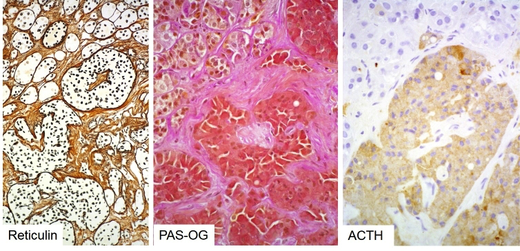
Figure 3b-11: Histology of corticotroph hyperplasia causing Cushing’s disease. Note distended but still intact reticulin network of hyperplastic pituitary acini (left) populated by a homogeneous population of deeply basophilic cells (center) expressing ACTH (right).

  • Agreement on Use
  • Supporters
  • Donations
  • Privacy Policy
  • Instructions for Authors (5/03/23)

Copyright © 2000-2025, MDText.com, Inc. All rights reserved.