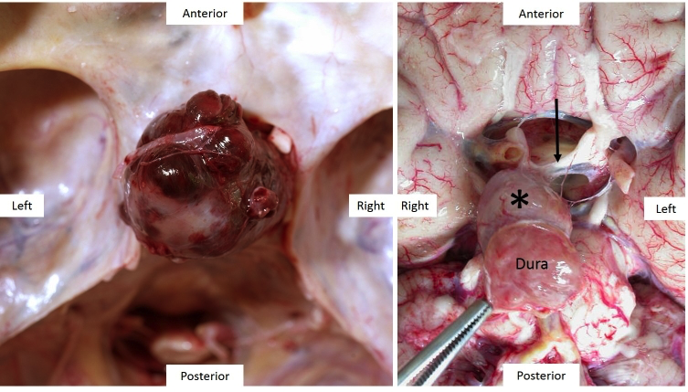
Figure 3b-15.1+15.2: MRI and macroscopic pathology of gonadotroph pituitary macroadenomas. Gonadotroph adenomas are usually clinically silent and thus tend to present as space occupying lesions compressing the optic chiasm, pituitary stalk or hypothalamus. 3b-15.1: Sagittal MRI (left) and post-mortem view of the same tumour. Note the suprasellar extension and compression of the hypothalamus. 3b-15.2: Two further gonadotroph macroadenomas seen in situ in the skull base (left) and the base of the brain (right) compressing the optic chiasm. Note in the left image the anatomical relationship to the sphenoid wings, right optic nerve and basal vessels of the brain (one of which is stuck to the rostral surface of the macroadenoma). The adenoma (asterisk) in the right image has grown through the diaphragma sellae and therefore is attached to dura mater – which is used to pull the adenoma away from the chiasmatic cistern to reveal the chiasm (arrow) and optic nerves.