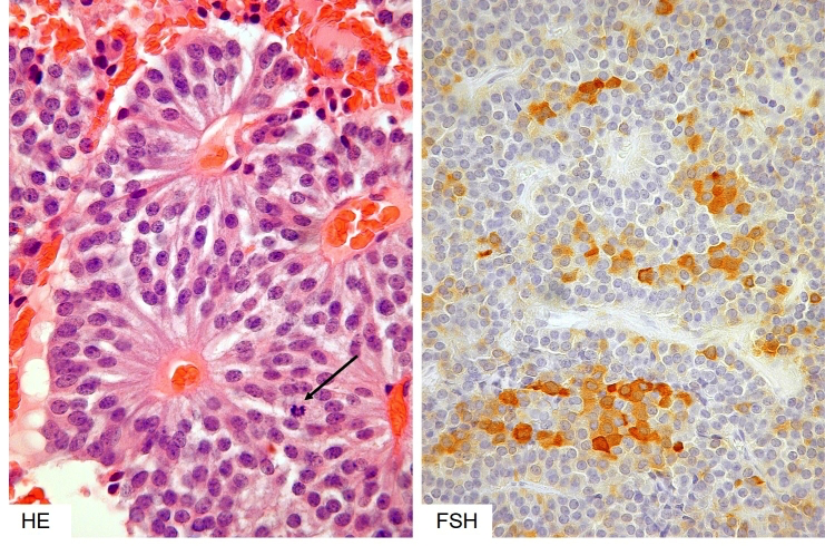
Figure 3b-16: Histology of a gonadotroph (non-functioning) adenoma. In their typical form these adenomas have a distinct architecture comprising perivascular rosettes of neuroendocrine cells with a distinct polarity of their processes towards the vascular lumen (left) and always patchy and focal, rather than diffuse expression of FSH and LH (right). Note a rare mitosis in the HE image (left, arrow).