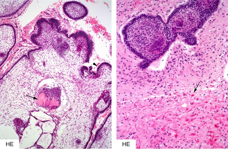
Figure 3b-18: Histology of adamantinomatous craniopharyngioma. This highly distinctive neoplasm consists of loosely arranged ‘stellate’ reticular stroma, palisading peripheral epithelium and nodules of ‘ghost’ cells (degenerated keratinocytes, arrow in the left image). Finger-like protrusions of neoplastic epithelium commonly invade the hypothalamic brain parenchyma resulting in a dense, hypereosinophilic Rosenthal fibre gliosis (arrow and bottom half of right image).