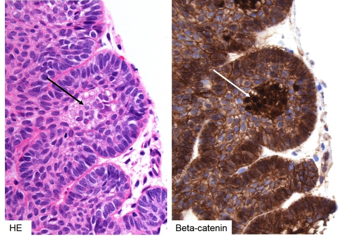
Figure 3b-19: Histology of adamantinomatous craniopharyngioma – beta-catenin. Nodules or whorls of tinctorially distinct epithelium is often seen in the invading edge of the adamantinomatous subtype (left, black arrow). The cells in these nests demonstrate nuclear translocation of beta-catenin (right, white arrow), indicating activated WNT signalling.