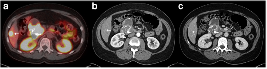
Figure 4. Overview of (A) 68Ga-PET/CT, multiphase (B) atrial and (C) portal vein CT scan images from patient with partially cystic PNEN. The arrow indicates a liver metastasis which is only visible on the 68Ga-PET/CT scan. This figure has been adapted from Current treatment options in oncology, Morse B., Al-Toubah T. and Montilla-Soler J., Anatomic and functional imaging of neuroendocrine tumors, 21 (9): 75 © 2020 (67).