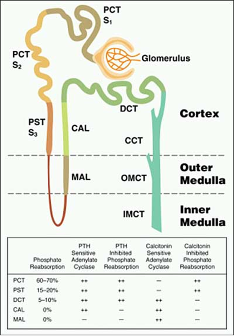
Figure 2. Distribution of Pi reabsorption and hormone-dependent adenylate cyclase activity throughout the renal tubule. The renal tubules consist of a proximal convoluted tubule (PCT), composed of an S1, S2 and S3 segment, a proximal straight tubule (PST), also known as the S3 segment, the loop of Henle, the medullary ascending limb (MAL), the cortical ascending limb (CAL), the distal convoluted tubule (DCT) and three segments of the collecting tubule: the cortical collecting tubule (CCT); the outer medullary collecting tubule (OMCT); and the inner medullary collecting tubule (IMCT). Pi reabsorption occurs primarily in the PCT but is present is the PST and DCT, sites at which parathyroid hormone (PTH) dependent adenylate cyclase is localized. In contrast, calcitonin alters Pi transport at sites devoid of calcitonin dependent adenylate cyclase, suggesting that Pi reabsorption in response to this stimulus occurs by a distinctly different mechanism.