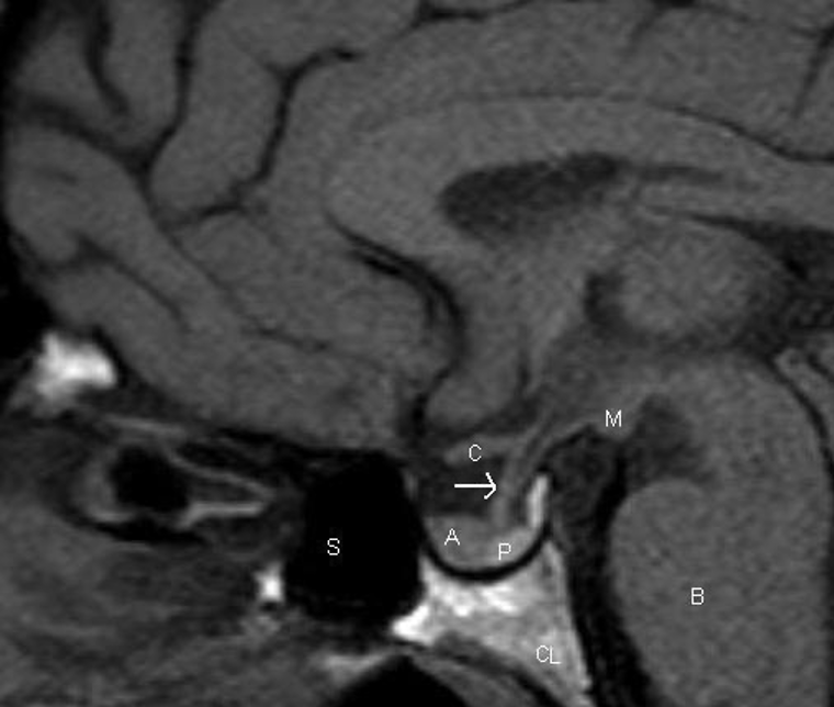
Figure 1. Sagittal T1 weighted unenhanced image of the pituitary fossa demonstrate normal anatomy. The anterior pituitary tissue, A, is visible within the sella and the posterior pituitary bright spot, P, is evident behind it. The stalk (arrow) is well seen with a small cleft of CSF visible within it superiorly –the infundibular recess of the third ventricle. The optic chiasm, C, and mamillary bodies, M, are seen in the suprasellar region. B – brainstem, S – sphenoid air sinus, CL – clivus.