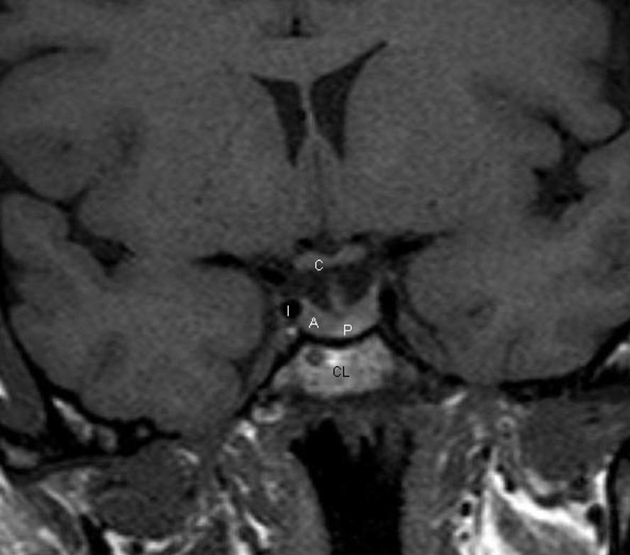
Figure 2. Coronal T1 weighted unenhanced image of the pituitary fossa. The anterior pituitary gland, A, is within the fossa. The posterior pituitary bright spot is visible centrally, P. The stalk is seen extending up into the suprasellar region. The optic chiasm, C, is visible. The cavernous segments of the carotid arteries, I, are seen within the cavernous sinuses, which form the lateral boundaries of the pituitary fossa.