The best Clinical Endocrinology source in the world, and FREE.

Endotext requires free registration
Login or Register

Menu

Skip to content
  • Home / Sections
  • Clinical Guidelines
  • Webinar Case Studies
  • Editorial Team
  • Country Associates
  • About
  • Instructions for Authors

etx-pit-ch3-fig16

Published July 18, 2020 at 934 × 1170 in Radiology of the Pituitary.
← Previous Next →

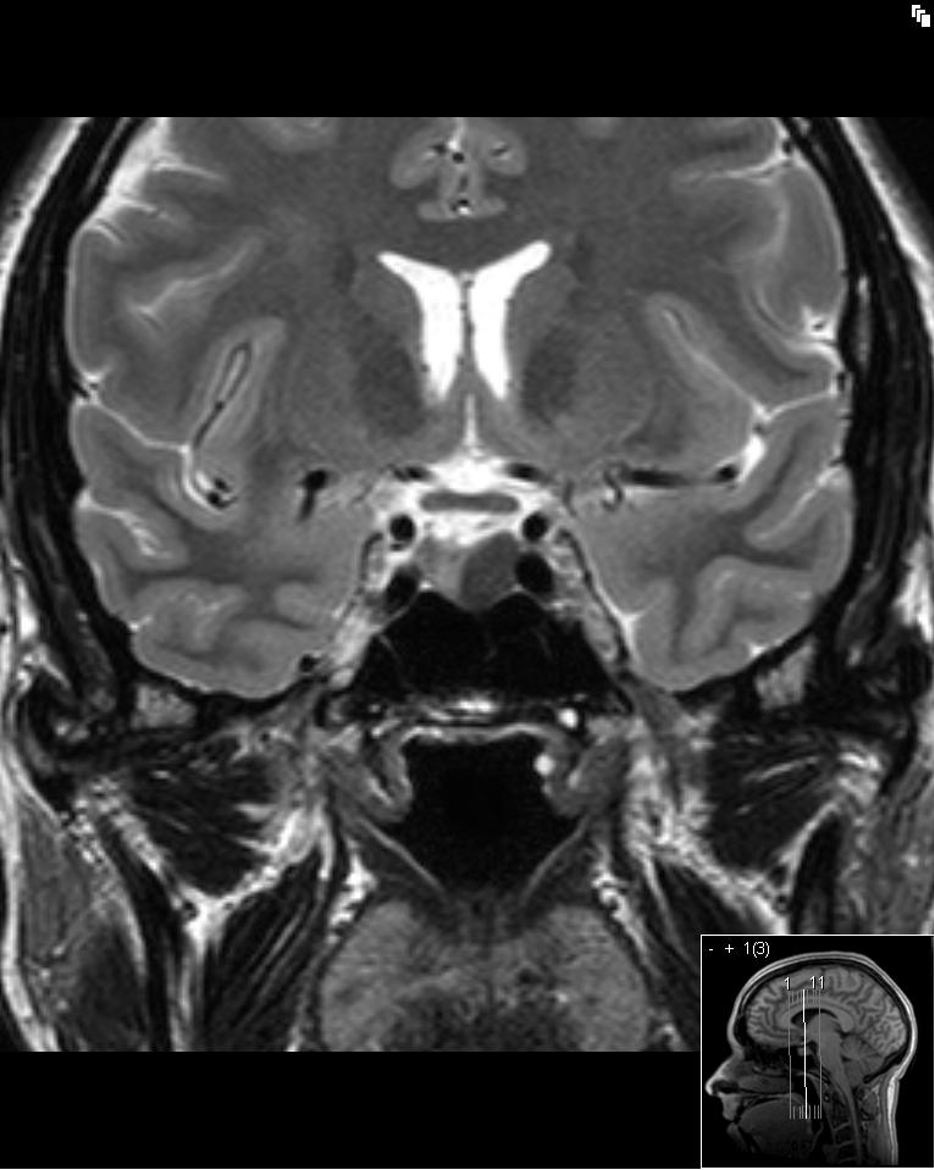
Figure 16. A coronal T2 weighted image shows a low T2 signal adenoma in the left side of the gland (arrow) which represented a densely granulated GH secreting adenoma.

  • Agreement on Use
  • Supporters
  • Donations
  • Privacy Policy
  • Instructions for Authors (5/03/23)

Copyright © 2000-2025, MDText.com, Inc. All rights reserved.