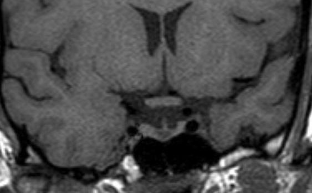
Figure 17. A coronal unenhanced T1 weighted image demonstrating a small Rathke’s cyst. This is seen to lie just below the insertion of the pituitary stalk and centrally within the gland. Although it is possible that a small cystic adenoma could have these appearances, this is a very typical location for a Rathke’s cyst.