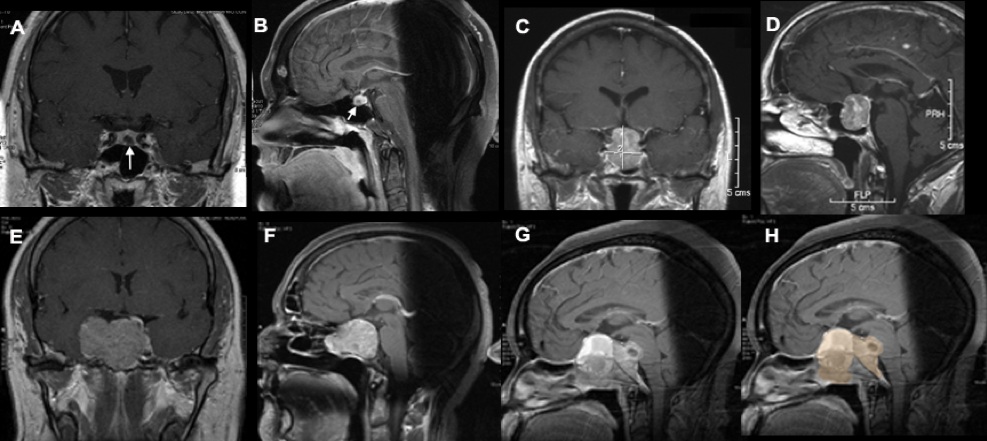
Figure 1. Tumor Classification based on size. Microadenoma: Coronal and sagittal T1 weighted MRIs with contrast with arrow indicating the location of the tumor (A and B). Macroadenoma: Coronal and sagittal T1 weighted MRIs of a typical macroadenoma (C and D). Giant invasive macroadenoma: Coronal and sagittal T1 MRIs with contrast in a patient in whom the tumor compresses the right temporal lobe and invades the sphenoid sinus (E and F). In another patient, the sagittal MRI reveals a tumor that has not only invaded the sphenoid sinus but compresses the brainstem; the tumor is highlighted (G and H).