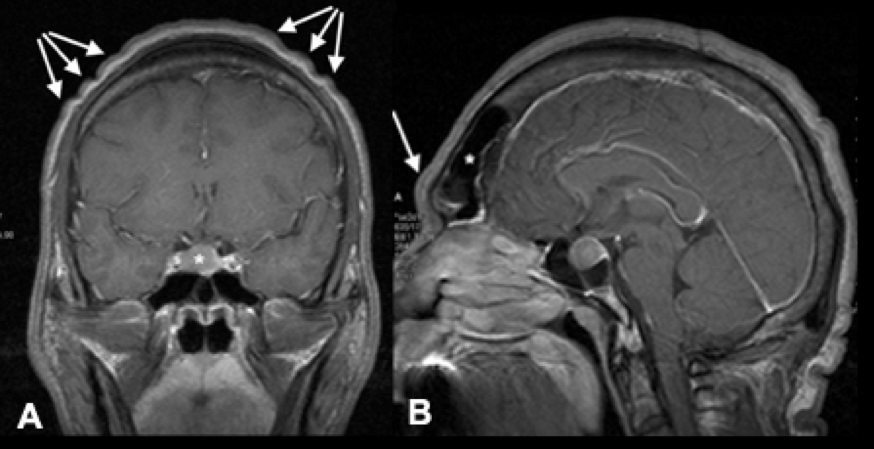
Figure 2. Acromegaly. A. Coronal T1 weighted MRI with contrast in a patient with an intrasellar GH secreting adenoma. Arrows indicate the common finding of “cutis gyrata”. B. Sagittal T1 weighted MRI in the same patient with arrows indicating the frontal bossing and the enlarged frontal sinus, and * the tumor.