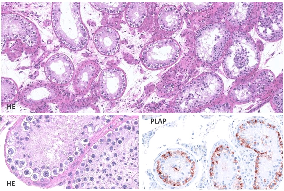
Figure 1. Histology of carcinoma in situ testis (CIS), also known as intratubular germ cell neoplasia (ITGCN) or testicular intraepithelial neoplasia (TIN)
The upper panel (hematoxyllin-eosin, HE staining shows a low magnification view of CIS in a typical pattern with only CIS cells and Sertoli cells present inside tubules. CIS tubules have a smaller diameter than normal seminiferous tubules. On the right side of this image a few tubules with decreased spermatogenesis – which are typically seen in a biopsy with CIS – are visible. The lower left image shows a fragment of a CIS tubule side-by-side with a tubule with preserved spermatogenesis. The lower right image displays CIS cells visualised by immunohistochemical staining for placental-like alkaline phosphatase (PLAP).