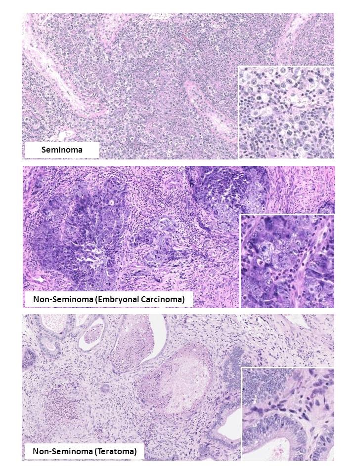
Figure 2. Histology of main types of testicular germ cell tumors.
The large images show a general histology pattern of a seminoma (upper panel) and two most often seen types of non-seminoma: embryonal carcinoma (middle panel) and teratoma (bottom panel). Small square pictures on the right show cellular characteristics in a greater magnification. All sections are stained with hematoxyllin-eosin (HE).