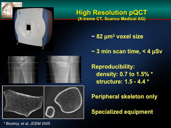
Figure 1. This is a high resolution QCT image of the distal radius of an individual patient. One can image the trabecular bone of the peripheral skeleton and define measures of bone “quality” by specific measurements. It is still not clear whether these measurements provide a better insight into fracture risk than DXA.