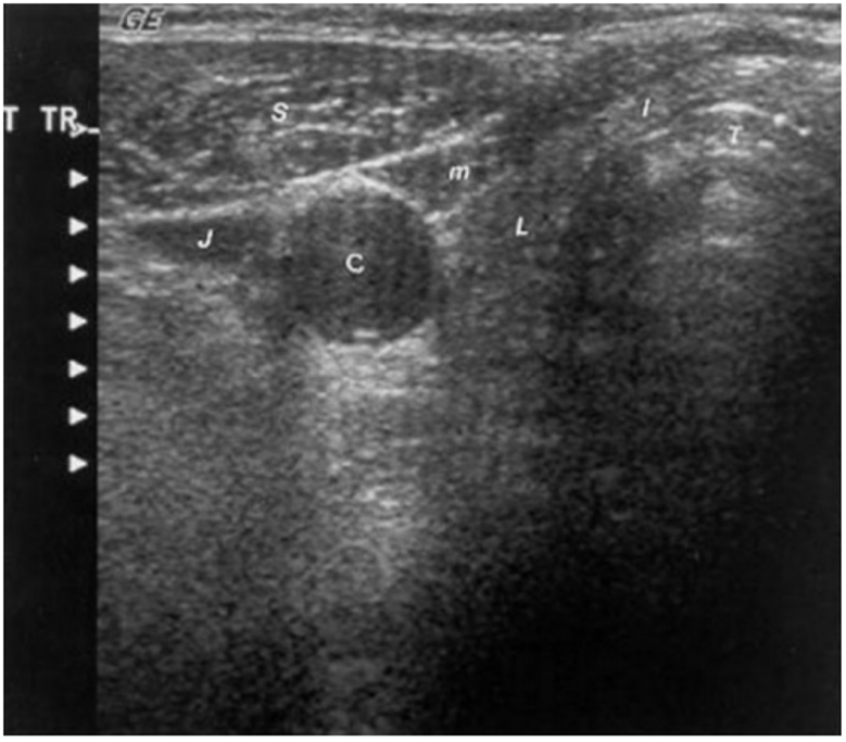
Figure 1. Sonogram of the neck in the transverse plane showing a normal right thyroid lobe and isthmus. L = small thyroid lobe in a patient who is taking suppressive amounts of L-thyroxine, I = isthmus, T = tracheal ring (the dense white arc represents calcification, distal to it reflects artifact), C = carotid artery (note the enhanced echoes deep to the fluid-filled blood vessel), J = jugular vein, S = sternocleidomastoid muscle, m = strap muscle.