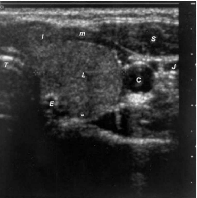
Figure 2. Sonogram of the left lobe of the thyroid gland in the transverse plane showing a rounded lobe of a goiter. L = enlarged lobe, I = widened isthmus, T = trachea, C = carotid artery (note the enhanced echoes deep to the fluid-filled blood vessel), J = jugular vein, S = sternocleidomastoid muscle, m = strap muscles, E = esophagus.