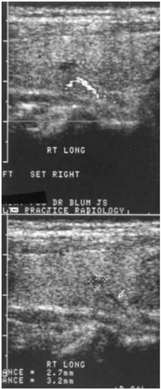
Figure 6. Sonograms of the right thyroid lobe in the longitudinal plane showing a 2.7 x 3.2 mm hypoechoic nodule that is delineated in the lower panel by the xx and ++ symbols. Note the linear hypoechoic structure below that (arrow). In the upper panel the bright structure is a Doppler signal and indicates a blood vessel below the nodule. The nodule is not vascular.