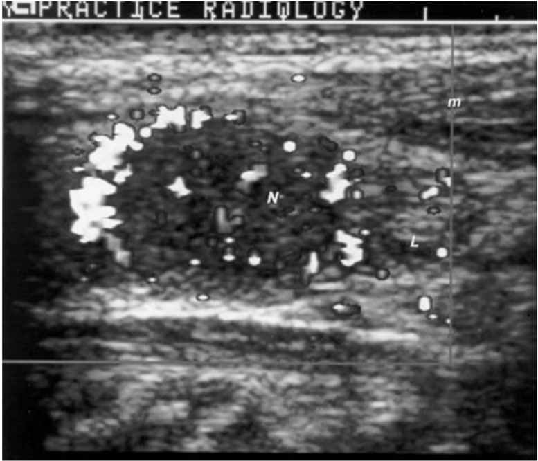
Figure 5. Sonogram of the neck in the longitudinal plane showing a hypoechogenic nodule that was surrounded by an echo free rim, called a halo. Doppler examination demonstrates great vascularity in the halo, identified as bright spots. Small blood vessels are also seen elsewhere. N = nodule, L = heterogeneous thyroid lobe, m = muscle.