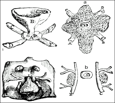
Fig. 2.Plates from the seventh book of the first edition (1543) of theFabricaby Andreas Vesalius, showing what is believed to be the oldest anatomical images in Western literature of the hypothalamic-pituitary unit. (Courtesy of the Library of the Department of Human Anatomy of the University of Bologna, Italy, with permission.) 1) Enlarged view of the pituitary gland (A), hypothalamic infundibulum (B) and ducts comprising theforamen lacerumand superior orbital fissure (C, D, E, F) believed to drain the brain mucus or phlegm (in Latinpituita) from the pituitary gland to the nasopharynx; 2) anatomical relationships between theinfundibulum(D), the duraldiaphragma sellae(F), the internal carotid arteries (C, D) and occulomotor nerves (G); 3) composite image including a) an enlarged view of therete mirabilisformed as a reticular plexus by the carotid arteries entering (A, B) and emerging (C, D) around the pituitary gland (E); b) detailed view of the reticular plexus arising from the carotids (B, C) on each side of the pituitary (A). (From Toni R., Ancient views on the hypothalamic-pituitary-thyroid axis: an historical and epistemological perspective, Pituitary 3: 83-95, 2000).