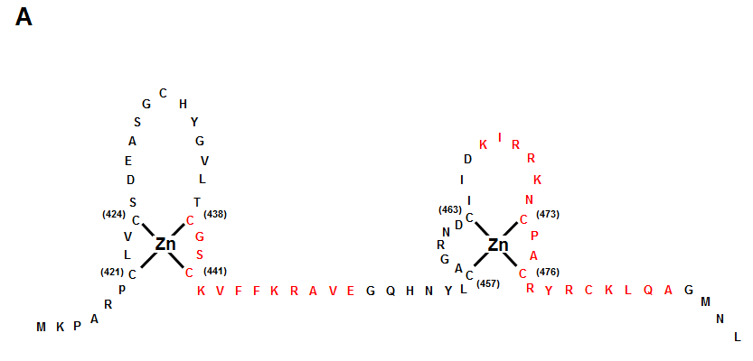
Figure 3: Structure of GR DBD and its interaction with GRE DNA
A: Zinc finger structures in the DNA-binding domain of hGR. Numbered eight cystein residues chelate Zn2+ to form two separate finger structures. Red-colored amino acid residues form -helical structures. Box with bold line indicates lever arm, while that with dashed line shows D-box. Modified from (361).