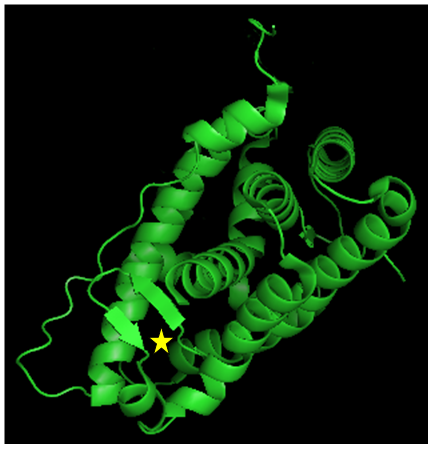
Figure 4:Structure of the GRα LBD.The GRα consists of 12 α-helices and 4 small β-strands that fold into a three-layer helical domain. Helices 1 and 3 form one side of a helical sandwich, while helices 7 and 10 form the other side. The middle layer of helices 4, 5, 8, and 9 are present in the top but not in bottom half of the protein, thus creating a ligand-binding pocket (shown as yellow star) in the bottom half of the LBD, which is surrounded by helices 3, 4, 11 and 12. The image was created with the MacPyMOL software using 3K22 of the RCSB Protein Data Bank (http://www.rcsb.org/pdb/home/home.do).