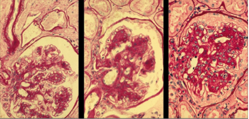
Figure 3. Light microscopy photographs of glomeruli in sequential kidney biopsies performed at baseline and after 5 and 10 years of follow-up in a long-standing normoalbuminuric type 1 diabetic patient with progressive mesangial expansion and renal function deterioration. A. Note the diffuse and nodular mesangial expansion and arteriolar hyalinosis in this glomerulus from a patient who was normotensive and normoalbuminuric at the time of this baseline biopsy, 21 years after diabetes onset [Periodic Acid Schiff (PAS) X 400]. B. 5-year follow-up biopsy showing worsening of the diffuse and nodular mesangial expansion and arteriolar hyalinosis in this now microalbuminuric patient with declining GFR (PAS X 400). C. 10-year follow-up biopsy showing more advanced diabetic glomerulopathy in this now proteinuric patient with further reduced GFR. Note also the multiple small glomerular probably efferent arterioles in the hilar region of this glomerulus (PAS X 400), and in the glomerulus in Fig. 3A above. Source: Reprinted with permission from National Kidney Foundation. Pathogenesis and Pathophysiology of Diabetic Nephropathy. Caramori ML, Mauer M. Primer on Kidney Diseases, 5th Edition, Greenberg A, et al., Copyright 2009 (253).