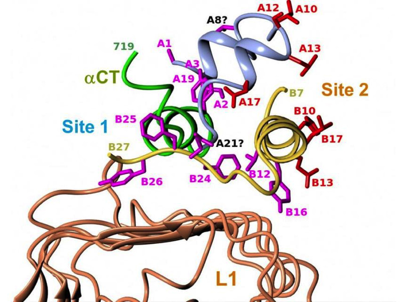
Figure 6. Structure of the site 1 insulin-receptor complex. Detailed view of insulin’s site 1 and site 2 residues in the ternary complex between insulin, L1 and αCT that includes insulin’s B-chain C-terminus. The figure shows the structural relationship of insulin’s site 1 (in magenta) and site 2 (in red) residues with the receptor’s αCT and L1 surfaces. See table 1 of reference 40 for more detailed description of the contacts. Drawn by Marek Brzozowski using the CCP4MG programme. PDB file: 4OGA. From reference 40, used with permission.
Figure 6. Structure of the site 1 insulin-receptor complex. Detailed view of insulin's site 1 and site 2 residues in the ternary complex between insulin, L1 and αCT that includes insulin's B-chain C-terminus. The figure shows the structural relationship of insulin's site 1 (in magenta) and site 2 (in red) residues with the receptor's αCT and L1 surfaces. See table 1 of reference 40 for more detailed description of the contacts. Drawn by Marek Brzozowski using the CCP4MG programme. PDB file: 4OGA. From reference 40, used with permission.