Archives

Androgen Physiology, Pharmacology Use and Misuse

ABSTRACT


Testosterone, together with its bioactive metabolites dihydrotestosterone and estradiol, determines the development and maintenance of male sexual differentiation and the characteristic mature masculine features. Defects in androgen action at various epochs of life produce characteristic clinical features. From an outline of the biochemistry and physiology of androgen action, the pathophysiology of defects in androgen action are derived and defined. The pharmacology of testosterone and its applications to replacement therapy for pathological hypogonadism as well as for pharmacological androgen therapy based on using either testosterone or synthetic androgens is described.


INTRODUCTION


An androgen, or male sex hormone, is defined as a substance capable of developing and maintaining masculine characteristics in reproductive tissues (notably the genital tract, secondary sexual characteristics, and fertility) and contributing to the anabolic status of somatic tissues. Testosterone together with its potent metabolite, dihydrotestosterone (DHT), are the principal androgens in the circulation of mature male mammals. Testosterone has a characteristic four ring C18 steroid structure and is synthesized mainly by Leydig cells, located in the interstitium of the testis between the seminiferous tubules. Leydig cell secretion creates a very high local concentration of testosterone in the testis as well as a steep downhill concentration gradient into the bloodstream maintaining circulating testosterone levels which exert characteristic androgenic effects on distant androgen sensitive target tissues. The classical biological effects of androgens are primarily mediated by binding to the androgen receptor, a member of the steroid nuclear receptor superfamily encoded by a single gene located on the X chromosome, which then leads to a characteristic patterns of gene expression by regulating the transcription of an array of androgen responsive target genes. This physiological definition of an androgen in the whole animal is now complemented by a biochemical and pharmacological definition of an androgen as a chemical that effectively competes with testosterone binding to the androgen receptor (1) to stimulate post-receptor functions in isolated cells or cell-free systems. In addition, non-genomic mechanisms of androgen action involving rapid, membrane-mediated nontranscriptional processes in the cytoplasm have been described but not yet fully characterized (2-4).


Testosterone is used clinically at physiologic doses for androgen replacement therapy and, at typically higher doses, testosterone or synthetic androgens based on its structure are also used for pharmacologic androgen therapy. The principal goal of androgen replacement therapy is to restore a physiologic pattern of androgen exposure to all tissues. Such treatment is usually restricted to the major natural androgen, testosterone, and aims to replicate physiological circulating testosterone levels and the full spectrum (including pre-receptor androgen activation) of endogenous androgen effects on tissues and recapitulating the natural history of efficacy and safety. Pharmacologic androgen therapy exploits the anabolic or other effects of androgens on muscle, bone, and other tissues as hormonal drugs that aim to modify the natural history of the underlying disorder and are judged on their efficacy, safety, and relative cost effectiveness like other therapeutic agents. Insight into the physiology of testosterone is a prequisite for understanding and making most effective use of androgen pharmacology (5, 6).


TESTOSTERONE PHYSIOLOGY


Biosynthesis


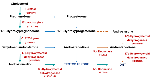
Testosterone is synthesized by an enzymatic sequence of steps from cholesterol (7, 8) (Figure 1) within the 500 million Leydig cells located in the interstitial compartment of the testis between the seminiferous tubules, which constitutes approximately 5% of mature testis volume (see Endotext, Endocrinology of Male Reproduction, Chapter entitled Endocrinology of the Male Reproductive System and Spermatogenesis, for details) (9). The cholesterol is predominantly formed by de novo synthesis from acetate, although preformed cholesterol either from intracellular cholesterol ester stores or extracellular supply from circulating low-­density lipoproteins also contributes (8). Testosterone biosynthesis involves two multifunctional cytochrome P-450 complexes involving hydroxylations and side-chain scissions (cholesterol side-chain cleavage [CYP11A1 or P450scc which produces C20 and C22 hydroxylation and C20,22 lyase activity] and 17-hydroxylase/17,20 lyase [CYP17A1 or P450c17 which hydroxylates the C17 and then excises two carbons (20 & 21) coverting a 21 to a 19 carbon structure]) together with 3 and 17b-hydroxysteroid dehydrogenases and ∆4,5 isomerase (8). The highly tissue-selective regulation of the 17,20 lyase activity (active in gonads but inactive in adrenals) independently of 17-hydroxylase activity (active in all steroidogenic tissues) is a key branch-point in steroidogenic pathways. Both activities reside in a single, multifunctional protein with the directionality of pathway flux determined by enzyme co-factors, notably electron supply from NADPH via the P450 oxidoreductase (POR), a membrane-bound flavoprotein serving diverse roles as a reductase, and cytochrome b5 (10, 11). In addition, some extragonadal biosynthesis of testosterone and dihydrotestosterone from circulating weak adrenal androgen precursor DHEA within specific tissues has been described (12); however, the net contribution of adrenal androgens to circulating testosterone in men is minor (13, 14) though it makes a much larger proportional contribution to circulating testosterone in women (15, 16).

FIGURE 1. Pathways of testosterone biosynthesis and action. In men, testosterone biosynthesis occurs almost exclusively in mature Leydig cells by the enzymatic sequences illustrated. Cholesterol originates predominantly by the de novo synthesis pathway from acetyl CoA with luteinizing hormone regulating the rate limiting step, the conversion of cholesterol to pregnenolone within mitochondria, while remaining enzymatic steps occur in smooth endoplasmic reticulum. The 5 and 4 steroidal pathways are on the left and right, respectively. Testosterone and its androgenic metabolite, dihydrotestosterone, exert biological effects directly through binding to the androgen receptor and indirectly through aromatization of testosterone to estradiol, which allows action via binding to the estrogen receptor (ER). The androgen and ERs are members of the steroid nuclear receptor superfamily with highly homologous structure differing mostly in the C-terminal ligand binding domain. The LH receptor has the structure of a G-protein linked receptor with its characteristic seven transmembrane spanning helical regions and a large extracellular domain which binds the LH molecule. LH is a dimeric glycoprotein hormone consisting of an  subunit common to other pituitary glycoprotein hormones and a  subunit specific to LH. Most sex steroids bind to sex hormone binding globulin (SHBG) which binds tightly and carries the majority of testosterone in the bloodstream.

Testicular testosterone secretion is principally governed by luteinizing hormone (LH) through its regulation of the rate-limiting conversion of cholesterol to pregnenolone within Leydig cell mitochondria by the cytochrome P-450 cholesterol side-chain cleavage enzyme complex located on the inner mitochondrial membrane. Cholesterol supply to mitochondrial steroidogenic enzymes is governed by proteins including sterol carrier protein 2 (17). This facilitates cytoplasmic transfer of cholesterol to mitochondria together with steroidogenic acute regulatory protein (18) and translocator protein (19), which govern cholesterol transport across the mitochondrial membrane. All subsequent enzymatic steps are located in the Leydig cell endoplasmic reticulum. The high testicular production rate of testosterone creates both high local concentrations (up to 1 μg/g tissue, ~100 times higher than blood concentrations) and rapid turnover (200 times per day) of intratesticular testosterone (20); however, the precise physical state in which such high concentrations of intratesticular testosterone and related steroids exist in the testis remains to be clarified.


Secretion


Testosterone is secreted at adult levels during three epochs of male life: transiently during the first trimester of intrauterine life (coinciding with masculine genital tract differentiation), during early neonatal life as the perinatal androgen surge (with still undefined physiologic significance), and continually after puberty to maintain virilization. The dramatic somatic changes of male puberty are triggered by the striking increases in testicular secretion of testosterone, rising ~30-fold over levels which prevail in pre-pubertal children and in women or castrate men originating from extra-testicular sources. After middle age, there are gradual decreases in circulating testosterone as well as increases in gonadotrophin and sex hormone–binding globulin (SHBG) levels (21, 22) with these trends being absent till late old age among men who remain in excellent health (23, 24) but exaggerated by the coexistence of chronic illness (22, 25-27). In addition there are temporal trends including increasing prevalence of obesity (28-30)and artefactual method-specific changes in testosterone immunoassays that deviate from reference mass spectrometry-based measurements (31, 32). These age-related changes from the accumulation of chronic disease states are functionally attributable to impaired hypothalamic regulation of testicular function (33-36), as well as Leydig cell attrition (9) and dysfunction (37-39) and atherosclerosis of testicular vessels (40). As a result, the ageing hypothalamic-pituitary-testicular axis progressively increasingly operates with multi-level functional defects that, in concert, lead to reduced circulating testosterone levels during male ageing (41, 42).


Testosterone, like other lipophilic steroids secreted from steroidogenic tissues, leaves the testis by diffusing down a concentration gradient across cell membranes into the bloodstream, with smaller amounts appearing in the lymphatics and tubule fluid. After male puberty, over 95% of circulating testosterone is derived from testicular secretion with the remainder arising from extragonadal conversion of precursors with negligible intrinsic androgenic potency such as dehydroepiandrosterone and androstenedione. These weak androgens, predominantly originating from the adrenal cortex, constitute a large circulating reservoir of precursors for conversion to bioactive sex steroids in extragonadal tis­sues including the liver, kidney, muscle, and adipose tissue. Unlike in women where adrenal androgens are the major source of biologically active androgen precursors, endogenous adrenal androgens contribute negligibly to direct virilization of men (13) and residual circulating and tissue androgens after medical or surgical castration have minimal biologic effect on androgen-sensitive prostate cancer (43). Conversely, however, adrenal androgens make a proportionately larger contribution to the much lower circulating testosterone concentrations in children and women (~5% of men) in whom blood testosterone is derived approximately equally from direct gonadal secretion and indirectly from peripheral interconversion of adrenal androgen precursors (15, 16). Exogenous dehydroepiandrosterone at physiologic replacement doses of 50 mg/day orally (15) is incapable of providing adequate blood testosterone for androgen replacement in men but produces dose-dependent increases in circulating estradiol in men (44, 45) and hyperandrogenism in women (14).


Hormone production rates can be calculated from either estimating metabolic clearance rate (from bolus injection or steady-state isotope infusion using high specific-activity tracers) and mean circulating testosterone levels (46, 47) or by estimation of testicular arteriovenous differences and testicular blood flow rate (48). These methods give consistent estimates of a testosterone production rate of 3 to 10 mg/day using tritiated (49, 50) or nonradioactive deuterated (51)tracers with interconversion rates of approximately 4% to dihydrotestosterone (DHT) (50, 52) and 0.2% to estradiol (53) under the assumption of steady-state conditions (hours to days). These steady-state methods are a simplification that neglects diurnal rhythm (54, 55), episodic fluctuation in circulating testosterone levels over shorter periods (minutes to hours) entrained by pulsatile LH secretion (56) and postural influence on hepatic blood flow (49). The major known determinants of testosterone metabolic clearance rate are circulating SHBG concentration (57), diurnal rhythmn (51) and postural effects on hepatic blood flow (49, 51). Major genetic influences on circulating testosterone levels mediated via changes in SHBG (58-61) and other mechanisms (50) have been described as well as environmental (28, 29, 51) factors.

Transport


Testosterone circulates in blood at concentrations greater than its aqueous solubility by binding to circulating plasma proteins. The most important is SHBG, a high affinity but low capacity binding protein (62), and other low affinity binding proteins include albumin, corticosteroid binding globulin (63) and a1 acid glycoprotein (64). Testosterone binds avidly to circulating SHBG, a homodimer of two glycoprotein subunits each comprising 373 amino acids with 3 glycosylation sites, 2 N-linked and 1 O-linked and containing a single high-affinity steroid binding site (65). The two binding sites in the homodimer display dynamic, co-operative binding affinities upon sequential binding of an androgen (62). The affinity of SHBG for binding testosterone is subject to genetic polymorphisms (66) but is not altered by acquired liver disease (67). It remains unknown as to whether it is influenced by other chronic diseases or pregnancy (when circulating levels increase). SHBG is secreted into the circulation by human, but not rodent, liver as well as into the seminiferous tubules of the testis by rodent, but not human, Sertoli cells where it is known as testicular androgen-binding protein (68), and by the placenta where it may contribute to the rise in blood SHBG during pregnancy (69). As a product of hepatic secretion, circulating SHBG levels are particularly influenced by first-pass effects on the liver of oral drugs including sex steroids. Circulating SHBG (and thereby total testosterone) concentrations are characteri­stically decreased (androgens, glucocorticoids) or increased (estrogens, thyroxine) by supraphysiologic hormone concentrations at the liver such as produced by oral administration (first pass effects) or by high-dose parenteral injections of androgens. In contrast, endogenous sex steroids and parenteral (non-oral) administration, which maintain predominantly physiologic circulating hormone concentrations (transdermal, depot implants), have minimal effects on blood SHBG levels (70, 71) (72). Other modifiers of circulating SHBG levels include up-regulation by acute or chronic liver disease and androgen deficiency and down-regulation by obesity, protein-losing states (65), non-alcoholic fatty liver disease (73, 74) and, rarely, genetic SHBG deficiency(75-77). Under physiologic conditions, 60% to 70% of circulating testosterone is SHBG bound with the remainder bound to lower affinity, high-capacity binding sites (albumin, a1 acid glycoprotein, corticosteroid binding protein) and 1% to 2% remaining non-protein bound.


Transfer of hydrophobic steroids into tissues is presumed to occur passively according to physicochemical partitioning between the hydrophobic protein binding sites on circulating binding proteins, the hydrophilic aqueous extracellular fluid and the lipophilic cellular plasma membranes. According to the free hormone hypothesis (78-80), recently restated and updated (62), the free (non-protein bound) fraction of testosterone is the most biologically active with the loosely protein-bound testosterone constituting a less accessible but mobilizable fraction, with the largest moiety tightly bound to SHBG constituting only an inactive reservoir. The free hormone hypothesis derived from now outdated 1970’s pharmacological theory on the mechanism of drug-drug interactions as due to mutual protein binding displacement; however, this theory is long superseded in molecular pharmacology by well-established physiological mechanisms such as cytochrome P450 enzyme induction, drug transporter activity and cognate mechanisms unrelated to binding to circulating proteins (81). As the free and/or bioavailable fractions would also have enhanced access to sites of testosterone inactivation by degradative metabolism that terminates androgen action, the free fractions may equally be considered the most evanescent and least active so that the net biological significance of the free or bioavailable fractions remains unclear and undermines a theoretical basis for the free hormone hypothesis. Furthermore empirical evidence indicates that, rather than being biologically inert, SHBG participates actively in cellular testosterone uptake via specific SHBG membrane receptors, uptake mechanisms and signaling via G protein and cyclic AMP (82-86). These mechanisms include the megalin receptor, a multi-valent low-density lipoprotein endocytic receptor located on cell surface membranes that can mediate receptor-mediated cellular uptake of SHBG loaded with testosterone by endocytosis (87, 88) and might influence tissue androgen action (89, 90). Consequently, lacking a physiological basis for the free hormone hypothesis (91) and with empirical evidence in its favor scarce and speculative,  it is refuted by intensive, prospective clinical evaluation (92). Hence, the biological significance of partitioning circulating testosterone into these derived fractions remains to be firmly established and its clinical application is unknown or possibly misleading. Furthermore, direct measurement of free testosterone requires laborious, manual methods only available in research or specialist pathology laboratories. Where available, they are costly and lack any external quality control programs or validated reference ranges. As a result, calculations purporting to replicate dialysis-based measurements are often substituted for direct measurements. These formulae come in two different formats – equilibrium binding equations requiring assumptions on testosterone binding stoichiometry and arbitrary plug-in binding affinity estimates (Sodergard (93), Vermeulen (94), Zakharov (95)) or assumption-free empirical methods (Ly(96, 97), Nanjee-Wheeler (98)) calibrated directly to dialysis-based laboratory measurements. Direct comparison has proven that empirical equations are more accurate compared with laboratory dialysis-based measurements (95, 96, 99, 100). Furthermore, calculations of free testosterone using any formula do not contribute significant to mortality or morbidity prognosis for older men’s health beyond accurate measurement of serum testosterone by liquid chromatography-mass spectrometry (92).


Measurement


Measuring blood testosterone concentration is an important part of the clinical evaluation of androgen status and for confirming a clinical and pathological diagnosis of androgen deficiency. The circulating testosterone concentration is a surrogate measure for whole body testosterone production rate and the inferred impact of androgens on tissues. However, the reliance on a spot measurement of blood testosterone concentration neglects changes in the whole body metabolic clearance rate as well as other factors influencing net androgen effects at tissue levels. These include the efficiency of blood testosterone transfer into adjacent tissues during capillary transit as well as pre-receptor, receptor and post-receptor factors influencing the testosterone activation, inactivation and action in that tissue. Circulating testosterone levels are also dynamic and feature distinct circhoral and diurnal rhythms. Circhoral LH pulsatility entrains some pulsatility in blood testosterone levels (36) although the buffering effects of the circulating steroid-binding proteins dampens the pulsatility of blood testosterone concentrations. This is illustrated by comparison with the strikingly pulsatile patterns of circulating testosterone in rodents which lack hepatic SHBG gene expression thereby having no circulating SHBG to buffer testosterone fluctuations (101, 102). Diurnal patterns of morning peak testosterone levels and nadir levels in the mid-afternoon are evident in younger and healthy older men (54) but lost in some ageing men (55). Consequently, it is conventional practice to standardize testosterone measurements to morning blood samples on at least 2 different days.


The advent of steroid radioimmunoassay in the 1970’s made it feasible to measure blood testosterone concentrations affordably with speed and sensitivity. However, cross-reacting steroids and non-specific matrix effects are limitations on modern direct (non-extraction) testosterone immunoassays relative to the high specificity of mass spectrometry-based methods, the reference method (103). In the next decades, the steep rise in demand for testosterone measurements in clinical practice and research led to method simplications to integrate steroid immunoassays into automated immunoassay platforms. These changes, notably eliminating preparative solvent extraction and chromatography as well as introducing bulky non-authentic tracers, undermine the specificity of unextracted testosterone immunoassays (104), particularly at the low circulating testosterone levels such as in women and children (105). Even at the higher testosterone concentrations in men, commercial testosterone immunoassays demonstrate wide discrepancies due to method-specific bias (32). New generation, bench-top mass spectrometers with higher sensitivity and throughput now overcome these limitations of testosterone immunoassays.


Assays to measure blood “free” testosterone levels directly in serum samples have been developed using tracer reference methods of equilibrium dialysis (106, 107) or ultrafiltration (108, 109) or calculated various formulae based on immunoassay measurement of total testosterone and SHBG (93, 110). Similarly, another derived testosterone measure, bioavailable testosterone, is defined as the non-SHBG bound testosterone (in effect the combination of albumin-bound plus unbound testosterone) and can also be measured directly or calculated by a formula from total testosterone and SHBG and albumin measurements. Some estimates of free testosterone, notably the direct analog assay (111, 112) and the free testosterone index (113) are invalid for use in men. As measurement of “free” or “bioavailable” testosterone is laborious, calculational formulae with limited validation (93, 110, 114) have been widely used; however, these estimates for “free” (115-117) or “bioavailable”(118, 119) testosterone are not accurate in large scale evaluation. Overall, the clinical utility of various derived (“free”, “bioavailable”) measures of testosterone arising from the unproven free hormone hypothesis remain to be established; consequently, they have minimal involvement in consensus clinical guidelines for diagnosis and management of androgen deficiency.

Metabolism


After testicular secretion, a small proportion of testosterone undergoes activation to two bioactive metabolites, estradiol and DHT, whereas the bulk of secreted testosterone undergoes inactivation by hepatic phase I and II metabolism to inactive oxidized and conjugated metabolites for urinary and/or biliary excretion (figure 2) (120).

FIGURE 2. Pathways of Testosterone Action. In men, most (>95%) testosterone is produced under LH stimulation through its specific receptor, a heptahelical G-protein coupled receptor located on the surface membrane of the steroidogenic Leydig cells. The daily production of testosterone (5-7 mg) is disposed along one of four major pathways. The direct pathway of testosterone action is characteristic of skeletal muscle in which testosterone itself binds to and activates the androgen receptor. In such tissues there is little metabolism of testosterone to biologically active metabolites. The amplification pathway is characteristic of the prostate and hair follicle in which testosterone is converted by the type 2 5 reductase enzyme into the more potent androgen, dihydrotestosterone. This pathway produces local tissue-based enhancement of androgen action in specific tissues according to where this pathway is operative. The local amplification mechanism was the basis for the development of prostate-selective inhibitors of androgen action via 5 reductase inhibition, the forerunner being finasteride. The diversification pathway of testosterone action allows testosterone to modulate its biological effects via estrogenic effects that often differ from androgen receptor mediated effects. The diversification pathway, characteristic of bone and brain, involves the conversion of testosterone to estradiol by the enzyme aromatase which then interacts with the ERs  and/or . Finally, the inactivation pathway occurs mainly in the liver with oxidation and conjugation to biologically inactive metabolites that are excreted by the liver into the bile and by the kidney into the urine.

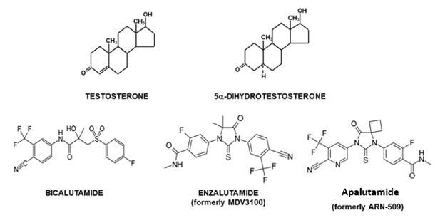
The amplification pathway converts ~4% of circulating testosterone to the more potent, pure androgen, DHT (50, 52). DHT has higher binding affinity to (121) and 3-10 time greater molar potency in transactivation (122-124) of the androgen receptor relative to testosterone. Testosterone is converted to the most potent natural androgen DHT by the 5a-reductase enzyme that ­originates from two distinct genes (I and II) (125). Type 1 5a-­reductase is expressed in the liver, kidney, skin, and brain, whereas type 2 5a-reductase is characteristically expressed strongly in the prostate but also at lower levels in the skin (hair follicles) and liver (125). Congenital 5a-reductase deficiency due to mutation of the type 2 enzyme protein (126) leads to a distinctive form of genital ambiguity causing undermasculinization of genetic males, who may be raised as females, but in whom puberty leads to marked virilization including phallic growth, normal testis development and spermatogenesis (127) and bone density (128) as well as, occasionally, masculine gender reorientation (129). Prostate development remains rudimentary (130) and sparse body hair without balding is characteristic (131). This remarkable natural history reflects the dependence of urogenital sinus derivative tissues on strong expression of 5a-reductase as a local androgen amplification mechanism for their full development. This amplification mechanism for androgen action was exploited in developing azasteroid 5a-reductase inhibitors (132). As the type 2 5a-reductase enzyme results in over 95% of testosterone entering the prostate being converted to the more potent androgen DHT (133), blockade of that isoenzyme (the expression of which is largely restricted to the prostate) confines the inhibition of testosterone action to the prostate (and other urogenital sinus tissue derivatives) without blocking extra-prostatic androgen action. DHT circulates at ~10% of blood testosterone concentrations, due to spillover from the prostate (134, 135) and nonprostatic sources (136). Whereas genetic mutations disrupting type 2 5a-reductase produce disorders of urogenital sinus derived tissues in men and mice (137), genetic inactivation of type 1 5a-reductase has no male phenotype in mice and no mutations of the human type 1 enzyme have been reported. Whether this reflects the type I enzyme having an unexpected phenotype or an evolutionarily conserved vital function, remains unclear. A third 5a-reductase enzyme (type 3, SRD5A3) has been described (138) but is widely expressed in human tissues, lacks steroidal 5a-reductase activity and has other roles in fatty acid metabolism (139).


An important issue is whether eliminating intraprostatic androgen amplification by inhibition of 5a-reductase can prevent prostate disease. Two major randomized, placebo-controlled studies of men at high risk of (but without diagnosed) prostate cancer have both shown that oral 5a reductase inhibitors (finasteride, dutasteride) reduced the incidence of low-grade prostate cancer as well as prevalence of lower urinary tract symptoms from benign prostate hyperplasia(140, 141). The Prostate Cancer Prevention Trial (PCPT) was a major 10-year chemoprevention study randomizing 18,882 men over 55 years of age without known prostate disease to daily treatment with 5 mg finasteride (inhibitor of type 2 5a reductase) or placebo observed a cumulative 25% reduction after 7 years of treatment in early stage, organ-confined, low-grade prostate cancer. Another study randomized over 8231 men aged 50-75 years with serum PSA <10 ng/mL and negative prostate biopsy to either daily treatment with 0.5 mg dutasteride (inhibitor of both type 1 and 2 5a reductases) or placebo for 4 years observed a 23% reduction in incidence of biopsy-proven prostate cancer. Although neither study was designed to determine mortality benefit, both showed no reduction in higher grade, but still organ-confined, cancers. Although this stage selectivity may be explained by diagnostic biases due to drug effects on prostate size and histology (142, 143), registration for chemoprevention of prostate cancer was refused by FDA (144). Overall, the evidence indicates that 5α-reductase inhibition reduces the incidence of early stage, screen-detected and organ-confined prostate cancers but there is insufficient evidence that such treatment reduces mortality from advanced (metastatic) prostate cancer. Whether or not preventive use of prostatic 5a-reductase inhibition in men with high prostate cancer risk proves warranted, novel synthetic androgens refractory to 5a-reductive amplification may have advantages for clinical development.


The diversification pathway of androgen action involves testosterone being converted by the enzyme aromatase to estradiol (145) to activate estrogen receptors (ERs) (also see Endotext, Endocrinology of Male Reproduction, Chapter entitled Estrogens and Male Reproduction). Although this involves only a small proportion (~0.2%) of testosterone output, the higher molar potency of estradiol (~100-fold higher vs testosterone) makes aromatization a potentially important mecha­nism to diversify androgen action via ER-mediated effects in tissues where aromatase is expressed. The diversification pathway is governed by the cytochrome P-450 enzyme (CYP19) aromatase (145, 146). In eugonadal men, most (~80%) circulating estradiol is derived from extratesticular aromatization (53). The biological importance of aromatization in male physiology was first recognized in the early 1970’s (147) when the local conversion of testosterone to estradiol within the neural tissues was identified and subseqeuntly shown to have an important role in mediating testosterone action, including negative feedback as well as activational and organisational effects, on the brain (148). More recently the importance of local aromatization in testosterone action has been reinforced by the striking developmental defects in bone and other tissues of men and mice with genetic inactivation of aromatase, leading to complete estrogen deficiency due to genetic inactivation of the aromatase (149). This phenotype is also strikingly similar to that of a man (150) and mice (150) with genetic mutations inactivating ERa. Furthermore, men with aromatase deficiency treated with exogenous estradioll or other estrogens also demonstrated significant bone maturation. By contrast, genetic inactivation of ERb has no effect on male mice (151) and no human mutations have been reported. Aromatase expression in tissue such as bone (152) and brain (148) may influence development and function by variation in aromatization that modulates local tissue-specific androgen action. By contrast other tissues, like mature liver and muscle, express little or no aromatase. Nevertheless, despite the importance of aromatization for male bone physiology, other observations indicate that androgens acting via androgen receptors have important additional direct effects on bone. These include the greater mass of bone in men despite very low circulating estradiol concentrations compared with young women (153), the failure of androgen insensitive rats lacking functional androgen receptors but normal estradiol and ERs to maintain bone mass of normal males (154)and the ability of nonaromatizable androgens to increase bone mass in estrogen-deficient women (155, 156). Testosterone action on bone and in the brain cannot be accounted for solely as a prohormone for local estradiol production (and action via estrogen receptors a and/or b) and androgen receptor mediated effects are required to manifest the full spectrum of testosterone effects on bone (157, 158) and in the brain (159). Conflicting evidence is available about the need for aromatization to mediate the effects of testosterone on male sexual function. One study using aromatase inhibitor-induced estrogen deficiency showed partial dependence (160) whereas another using DHT-induced estrogen deficiency showing no requirement of male sexual function for aromatization (161). Further studies are needed to fully understand the significance of aromatization in maintaining androgen action in mature male animals (162).


Testosterone is metabolized to inactive metabolites in the liver, kidney, gut, muscle, and adipose tissue. Inactivation is predominantly by hepatic oxidases (phase I metabolism), notably cytochrome P-450 3A family (163) leading ultimately to oxidation of most oxygen moieties followed by hepatic conjugation to glucuronides (phase II metabolism), which are rendered sufficiently hydrophilic for renal excretion. Uridine diphospho (UDP) glucuronosyl transferase (UGT) enzymes UGT2B7, UGT2B15 and UGT2B17 catalyze most phase II metabolism (glucuronidation) of testosterone with 2B17 being quantitatively the most important (164). A functional polymorphism of UGT 2B17, a deletion mutation several times more frequent in Asian than European populations (165), explains the concordant population difference in testosterone to epitestosterone (T/E) ratio (165), a World AntiDoping Agency-approved urine screening test for testosterone doping in sport, which constitutes an ethnic differential, false negative in surveillance for exogenous testosterone doping (166).


The metabolic clearance rate of testosterone is reduced by increases in circulating SHBG levels (57) or decreases in hepatic blood flow (e.g. posture) (49) or liver function. Theoretically, drugs that influence hepatic oxidase activity could alter metabolic inactivation of testosterone, but empirical examples of sufficient magnitude to influence clinical practice are rare. Rapid hepatic metabolic inactivation of testosterone leads to both low oral bioavailability (167, 168) and short duration of action when injected parenterally (169). To achieve sustained androgen replacement, these limitations dictate the need to deliver testosterone via parenteral depot products (e.g., injectable testosterone esters, testosterone implants, transdermal testosterone) or oral delivery systems that either bypass hepatic portal absorption (buccal (170, 171), ­sublingual (170, 172), gut lymphatic (173)) or use synthetic androgens with substituents rendering them resistant to first pass hepatic inactivation (174).


Regulation


During sexual differentiation in early intrauterine life, the testosterone required for masculine sexual differentiation is secreted by fetal Leydig cells. The regulation of this fetal Leydig cell testosterone secretion appears to differ between species. Higher primate and equine placenta secrete a chorionic gonadotropin during early fetal life (175) that may drive fetal human Leydig cell steroidogenesis (176) at the relevant time. By contrast, in subprimate mammals male sexual differentiation occurs without expression of any placental gonadotropin and prior to the time when pituitary gonadotropin secretion starts so that fetal Leydig cell testosterone secretion may be autonomous of gonadotropin stimulation during fetal development of most mammalian species (177).


Puberty is a complex series of maturational events originating during fetal life but completing only during adolescence. Recent genetic studies have enlightened the understanding of the physiology of puberty and its timing without yet identifying the ultimate trigger that initiates puberty. All components of the hypothalamo-pituitary testicular (HPT) axis are established during fetal life and its first activation is during the neonatal period manifested as a transient surge lasting several months in circulating testosterone (“mini-puberty”) reaching adult male levels and creating androgen imprinting in non-reproductive tissues. Following the neonatal surge, the HPT axis activity becomes quiescent during a decade of childhood under the restraint of mechanisms revealed by recent genetic studies to involve activities of at least four genes. These studies have identified premature (precocious) onset of puberty arising from inactivating mutations of makorin ring finger protein 3 (MKRN3) (178) and delta-like homolog 1 (DLK1) (179) as well as activating mutations of kisspeptin (KISS1) and its receptor (KISSR1) (180) indicating that these genes are involved in the mechanism of the childhood restraint of puberty. Alleviation of this restraint mechanism at the end of childhood unleashes the hormonal cascade of puberty. Although the ultimate trigger remains elusive, its activation initiates the timely onset of mature patterns of pulsatile GnRH secretion. This involves a complex and still poorly understood integration of a cascade of over 50 genes/proteins (180, 181). It is known that monogenic mutations in GnRH receptor, kisspeptin (KISS1 and its receptor), neurokinin B (TAC3 and its receptor), FGF8 or FGF17 and their FGFR1 receptor, prokineticin-2 (PROK) and its receptor (PROKR2) as well as compound heterozygotes and oligogenic inheritance of combinations of known mutated alleles disrupt the normal timing of puberty. Variants include reversible forms of gonadotropin deficiency, and this growing spectrum of underlying genetic susceptibilities probably overlaps and accounts for the variability in timing of puberty onset in the community.


Pulsatile hypothalamic GnRH secretion is the final common pathway driving pituitary gonadotrophin secretion that leads to testicular growth and maturation resulting in completion of spermatogenesis and steroidogenesis to produce adult male circulating testosterone concentrations. This results in testicular growth, the earliest and most salient external manifestation of male puberty, being initiated between 9 and 14 years of age with earlier onset defined as precocious and later onset as delayed puberty. The timing of puberty is under strong but complex genetic control with strong patterns detected in familial, twin and community studies (182-187). Disorders in this complex mechanism are relatively common with delayed puberty evident in 2% of adolescents although precocious puberty is rare with a frequency between 1:5000-10,000 involving only 10-20% in males (181, 188).


The still mysterious suprahypothalamic process initiating puberty involves a developmental clock and multiple permissive processes (189, 190) that lift the central neuroendocrine restraint on the final common pathway that drives reproductive function in the mature male – the episodic secretion of gonadotropin-releasing hormone (GnRH) from hypothalamic neurons (56). Various explanatory theories including the gonadostat, somatometer (191), neurally-driven changes in GABAergic inhibition and glutaminergic stimulation (192), triggering by kisspeptin-1 secretion and activating its receptor GPR54 (193, 194) and epigenetic factors (195) are proposed to explain the restraint and resurgence of the hypothalamic GnRH pulse generator without a comprehensive picture having yet emerged (190). Hypothalamic GnRH neurons are functional at birth but, after the perinatal androgen surge, remain tonically suppressed during infantile life. Puberty is initiated by a maturation process that awakens the dormant hypothalamic GnRH neurons to unleash mature cirhoral patterns of pulsatile GnRH secretion which in turn entrains pulsatile LH secretion from pituitary gonadotropes. Initially this resurgence of pulsatile GnRH and LH secretion occurs mainly during sleep (196) but eventually extends throughout the day with a persisting association with an underlying diurnal rhythmn. The timing and tempo of male puberty is under tight genetic control, encompassing nutrition influences on body weight and composition (185), with a correspondingly growing number of genetic causes of delayed puberty identified (197). Environmental factors that optimize growth (eg high socio-economic status with better nutrition and health care) may explain secular trends to earlier puberty with increased statural growth (198, 199) whereas claims that exposure to hormonally active chemical pollution contributes to earlier puberty (200) remain speculative (201) and not supported by available evidence in boys (202, 203). Very large UK population-based studies, using voice breaking as a self-reported marker of male puberty, show that both early and late male puberty are associated with a wide-range of adverse health outcomes (204, 205).


After birth, testicular testosterone output is primarily regulated by the pulsatile pattern of pituitary LH secretion. This is driven by the episodic secretion of GnRH from hypothalamic neurons into the pitu­itary portal bloodstream providing a direct short circuit route to pituitary gonadotropes. Under this regular by intermittent GnRH stimulation, pituitary gonadotrophs secrete LH in high amplitude pulses at ~60-90 min intervals with minimal intervening LH secretion between pulses with the net effect that circulating LH levels are distinctly pulsatile. This pulsatile pattern of trophic hormone exposure maintains Leydig cell sensitivity to LH to maintain mature male patterns of testicular testosterone secretion (206).


LH stimulates Leydig cell steroidogenesis via increasing substrate (cholesterol) availability and activating rate-limiting steroidogenic enzyme and cholesterol transport proteins (8, 18). LH is a dimeric glycoprotein consisting of an asubunit common to the other glycoprotein hormones (human chorionic gonadotropin (hCG), follicle-stimulating hormone, and thyrotropin-stimulating hormone) and a b subunit providing distinctive biologic specificity for each dimeric glycoprotein hormone by dictating its specific binding to the LH/hCG rather than the folliclle-stimulating hormone or thyroid stimulating hormone receptors (207, 208). These cell surface ­receptors are highly homologous members of the heptahelical, G protein–linked family of membrane receptors. LH receptors are located on Leydig cell surface membranes and use signal transduction mechanisms involving primarily cyclic adenosine monophosphate as well as calcium as second messengers to cause protein kinase–­dependent protein phosphorylation and DNA transcription, ultimately resulting in testosterone secretion (209). Functionally, hCG is a natural, long-acting analogue of LH because they both bind to the same LH/CG receptor and their b subunits are nearly identical except that hCG has a C-terminal extension of 31 amino acids containing four O-linked, sialic acid–capped carbohydrate side chains. These glycosylation differences confer greater resistance to degradation, which prolongs circulating residence time and biologic activity compared with LH (210, 211), a feature that has been exploited to engineer longer acting analog of other circulating hormones such as FSH (212), TSH (213) and erythropoietin (214).


Additional fine tuning of Leydig cell testosterone secretion is provided by paracrine factors originating within the testis (215). These include cytokines, inhibin, activin, follistatin, prostaglandins E2 and F2a, insulin-like and other growth factors as well as still uncharacterized factors secreted by Sertoli cells. LH also influences testicular vascular physiology by stimulating Leydig cell secretion of vasoactive and vascular growth factors (216).


Testosterone is a key element in the negative testicular feedback cycle through its inhibition of hypothalamic GnRH and, consequently, pituitary gonadotropin secretion. Such negative feedback involves both testosterone effects via androgen receptors as well as aromatization to estradiol within the hypothalamus (217, 218). These culminate in reduction of GnRH pulse frequency in the hypothalamus together with reductions in amplitude of LH pulses due to both reduced GnRH quantal secretion as well as gonadotropin response to GnRH stimulation (206). By contrast, the small proportion of estradiol in the bloodstream that is directly secreted from the testes (~20%) means that cirulating estradiol is under minimal physiological regulation and unlikely to be a major influence on negative feedback regulation of physiological gonadotropin secretion in men.

Action


Androgen action involves pre-receptor, receptor and post-receptor mechanisms that are centered on the binding of testosterone (or an analog) to the androgen receptor. Testosterone undergoes pre-receptor activation by conversion to potent bioactive metabolites, DHT and estradiol. The steroidogenic enzyme 5a-reductase has two isozymes, types 1 and 2, which form a local androgen amplification mechanism converting testosterone to the most potent natural androgen, DHT (219). The two isozymes have different chromosomal location and distinct biochemical features but are homologous genes (125). They are structurally and functionally unrelated to a third 5a-reductase (SRD5A3) which may have physiological role in fatty acid rather than steroidal biochemistry (138, 139). This local androgen amplification mechanism is exemplified in urogenital sinus derived tissues, notably external and internal genitalia and the prostate, which characteristically express high levels of 5a-reductase type 2 (125). Other tissues such as nongenital skin and liver express 5a-reductase type 1.


The other form of pre-receptor androgen activation is conversion of testosterone to estradiol by the enzyme aromatase (220) which diversifies androgen action by facilitating effects mediated via ERs (221). Consequently, while DHT may be considered a pure androgen because its bioactivity is solely mediated via AR, testosterone has a wider spectrum of action which includes diversification by aromatization and ER mediated effects. These pre-receptor mechanisms provide testosterone with a versatile and subtle range of regulatory mechanisms prior to receptor mediated effects, depending on the balance between direct AR mediated vs indirect actiational and/or ER mediated mechanisms. In addition, tissues vary in their androgenic thresholds and dose-response characteristics to testosterone and its bioactive metabolites.


The role of aromatization in androgen action was originally identified by the 1970s in the brain (222) whereby local expression of the aromatase enzyme within brain regions leads to local production of estradiol to mediate testosterone effects selectively in that region via ER and not AR mechanisms. Subsequently, the importance of aromatization to androgen action on bone was identified through investigations of inactivating mutations in ERα in men (223) and mice (150, 224). The role of aromatization in the estrogen-mediated effects of testosterone action is clearly shown by studies of Finkelstein et al who use the paradigm of complete suppression of endogenous testosterone production by administration of a depot GnRH analog with a range of doses of add-back testosterone without and with an aromatase inhibitor (anastrozole), the latter to investigate the effects of selective estrogen deficiency (225). These studies showed that aromatization was important in mediating testosterone effects in reducing fat mass and sexual function but not on muscle mass or strength. However,in another study using a different design, a high dosage of the non-aromatizable androgen dihydrotestosterone (vs. placebo) to induce selective complete estrogen deficiency in healthy men, demonstrated complete preservation of sexual function (161). Testosterone effects on bone involve dual mediation via indirect mechanisms, via aromatization to estradiol and ER-mediated effects, as well as via direct AR-mediated effects (226). In male mice, aromatization of testosterone must occur locally within bone as circulating estradiol levels are too low to activate ERs ; however, the role of local vs circulating estradiol effects on male bone remain to be clarified. A much wider role of estrogen action in male health is now identified (227). In that light the off-label use of aromatase inhibitors carries the risk of adverse effects on brain (manifest as sexual dysfunction), fat, and bone.


ANDROGEN RECEPTOR

The androgen receptor is required for masculine sexual differentiation and sexual maturation that ultimately leads to development of a mature testis capable of supporting spermatogenesis and testosterone production that form the basis for male fertility. The human androgen receptor is specified by a single X chromosome encoded gene located at Xq11-12 that specifies a protein of 919 amino acids (1), a classical member of the large nuclear receptor superfamily (228) which includes receptors for the 5 mammalian steroid classes (androgen, estrogen, progesterone, glucocorticoid, mineralocorticoid) as well as for thyroid hormones, retinoic acid and vitamin D and numerous orphan receptors where the ligand was originally not identified (229). Androgen receptor expression is not confined to reproductive tissues and it is ubiquitously expressed, athough levels of expression and androgen sensitivity of non-reproductive tissues vary.


The androgen receptor gene has 8 exons specifying a protein of 919 amino acids with the characteristic structure of mammalian steroid receptors. It has an N-terminal domain (NTD) that specifies a long transactivating functional domain (exon 1), a middle region specifying a DNA-binding domain (DBD) consisting of two zinc fingers (exons 2 and 3) separated by a hinge region from the C-terminal ligand binding domain (LBD) which specifies the steroid binding pocket (exons 4 to 8).


The NTD (exon 1) is relatively long comprising over half (535/919) the overall length of the AR. It has the least conserved sequence compared with other steroid receptors with a flexible and mobile tertiary structure harbouring a transactivation domain (AF-1) that interacts with AR co-regulator proteins and target genes (230). Its loose, naturally disordered structure (231) also contains three homopolymeric repeat sequences (glutamine, glycine, proline) with the most important being the CAG triplet (glutamine) repeat polymorphism (232). The less variable glycine (usually 24 residues) and proline (9 residues) repeat polymorphisms have little apparent independent pathophysiological significance although linkage dysequilibrium between the glutamine and glycine repeat polymorphisms requires haplotype analysis for interpretation (232). Among healthy people, where the glutamine repeat polymorphism has alleles of lengths between 5 and 35 (population mean ~21), the length of the glutamine repeat is inversely proportional to AR transcriptional efficiency so that this polymorphism dictates genetic differences between individuals in the androgen sensitivity of their target tissues (233). This genetic variation in in vivo tissue androgen sensitivity is proven experimentally (234) but, although modest in magnitude, influences physiological responses to endogenous testosterone in prostate size (235) and erythropoeisis (236) in carefully controlled studies. Wider epidemiological implications of population variation in the genetic androgen sensitivity as specified by the polyglutamine repeat have been studied in a variety of potentially androgen sensitive disorders (reviewed in (232)) including reproductive health disorders and hormone dependent cancers in men and women as well as non-gonadal disorders where there are significant gender disparities in prevalence. In men these include prostate (237) and other male preponderant cancers (liver, gastrointestinal, head & neck), prostate hypertrophy, cryptorchidism and hypospadias, male infertility (238)whereas in women they include reproductive health disorders (polycystic ovary syndrome, premature ovarian failure, endometriosis, uterine leiomyoma, preeclampsia) and hormone dependent cancers (breast, ovary, uterus). In addition, studies have also examined risks of obesity and cardiovascular disease, mental and behavioural disorders including dementia, psychosis, migraine, and personality disorders (232). However, as in many large-scale genetic association studies (239), the findings remain mostly inconsistent reflecting methdological limitations notably in recruitment, participation and publication bias as well as multiple hypothesis testing all of which tend to inflate spurious associations.


Remarkably, the pathological elongation of the polygutamine (CAG triplet) repeat to lengths of over 37 cause a neurodegenerative disease, Spinal Bulbar Muscular Atrophy (SBMA, also known as Kennedy’s syndrome). This is a form of late-onset, slow progressing but ultimately fatal motor neuron disease (240), one of several late-onset neurodegenerative polyglutamine repeat disorders (241). Although the extreme length of the polyglutamine repeat does determine mild androgen resistance, these men usually have normal reproductive function including fertility and virilization prior to diagnosis in mid-life (242). Furthermore, since complete androgen receptor inactivation in humans and other mammals does not cause motor neuron disease and female carriers are protected from symptomatic neurodegeneration, SBMA represents a toxic gain-of-function involving pathological protein aggregates of the mutant AR (243) like other genetic polyglutamine repeat neurodegenerative diseases do with other proteins (244). Surprisingly, transgenic mouse models of SBMA suggest that testosterone deprivation by medical castration using a GnRH agonist may slow progression of neuropathy (243) and that genetic (245) or pharmacological (246)administration of IGF-I may slow disease progression. However the first major clinical trial of leuprolide, a GnRH analog, failed to demonstrate neuromuscular benefit in swallowing (247) and further studies of selected subgroups and therapeutic targets are warranted (248, 249).


The DBD (exons 2 and 3) consists of ~70 amino acids with a high proportion of basic amino acids including eight cysteines distributed as two sets of 4 cysteineseach forming a zinc cooordination center for a single zinc atom, thereby creating two zinc fingers. The DBD is highly conserved between steroid receptors reflecting its tightly defined function of forming the two zinc fingers that bind to DNA by intercalating between its grooves. The first zinc finger (exon 2) is directly involved with the major DNA groove of the androgen response element through a proximal (P-box) region whereas the second zinc finger (exon 3) is also responsible for enhancing receptor dimerization through its distal (D-box) region.


The hinge region (first half of exon 4) of ~40 amino acids between the DBD and LBD is considered a flexible linker region but may have additional functions involving interactions with DNA (nuclear localization, androgen response element) and protein (AR dimerization, co-regulators) which influence AR transcriptional activity.


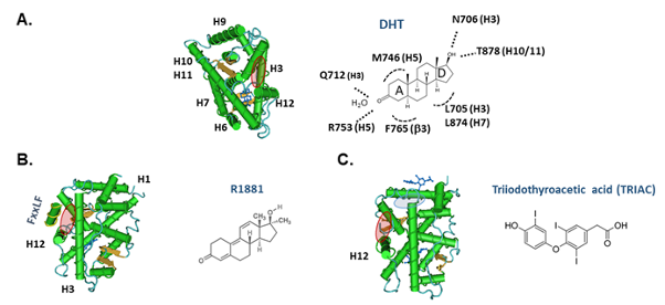
The LBD (mid-exon 4 to 8) of AR comprises ~250 amino acids which specify a steroid binding pocket which creates the characteristic high affinity, stable and selective binding of testosterone, DHT and synthetic androgens. While the LBD’s overall architecture is broadly conserved among nuclear receptors, the AR sequence diverges significantly to ensure the specificity of binding from other steroid classes and their different cognate ligands. Structural studies of the AR’s LBD shows it has similar tertiary conformation as other steroid receptors (most closely resembling PR) with 12 stretches of a helix interspersed with short b pleated sheets. The most C terminal helix 12 seals the binding pocket and influences whether a bound ligand acts as an agonist or antagonist as well as forming a hydrophobic surface for binding of co-regulator proteins that modify transcriptional activity of the androgen target genes. The LBD also participates in receptor dimerization, nuclear localization and transactivation via its activation function (AF-2) domain.


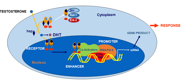
The AR has a predominantly nuclear location in androgen target cells regardless of whether bound to its ligand or not, unlike other steroid receptors which are more often evenly distributed between cytoplasm and nucleus when not bound to their cognate ligands. Androgen binding to the C-terminal LBD causes a conformational change in the androgen receptor protein and dimerization to facilitate binding of the ligand-loaded receptor to segments of DNA featuring a characteristic palindromic motif known as an androgen-response element, located in the promoter regions of androgen target genes. Ligand binding leads to shedding of heat shock proteins 70 and 90 that act as a molecular chaperone for the unliganded androgen receptor (250). Specific binding of the dimerized, ligand-bound androgen receptor complex to tandem androgen-response elements initiates gene transcription so that the androgen receptor acts as a ligand-activated transcription factor. Androgen receptor transcriptional activation is governed by a large number of coregulators (251, 252) whose tissue distribution and modulation of androgen action remain incompletely understood.


Androgen Insensitivity


Mutations in the androgen receptor are relatively common with over 1000 different mutations recorded by 2012 (253)in the McGill database (http://androgendb.mcgill.ca/) making androgen insensitivity the most frequent form of genetic hormone resistance. As the androgen receptor is an X chromosomal gene, functionally significant AR mutations are effectively expressed in all affected males because they are hemizygous. By contrast, women bearing these mutations (including the obligate heterzygote mothers of affected males) are silent carriers without any overt phenotype because they have a balancing allele as well as their circulating testosterone levels never rise to post-pubertal male levels sufficient to activate AR mediated effects.


Germline AR mutations produce a very wide spectrum of effects from functionally silent polymorphisms to androgen insensitivity syndromes that display phenotypes proportionate to the impairment of AR function and, thereby, the degree of deficit in androgen action (1). These clinical manifestations extend from a complete androgen insensitivity syndrome (CAIS, formerly known as testicular feminization) which produces a well developed female external phenotype in a spectrum spanning across all grades of undervirilized male phenotype to, at the other extreme, a virtually normal male phenotype. The severity of androgen insensitivity can be categorized most simply as complete, partial and mild although a more detailed 7 stage Quigley classification based on degree of hypospadias, phallic development, labioscrotal fusion and public/axillary hair is also described (1, 232). The degree of urogenital sinus derivative development together with testis descent provide clinical clues to the degree of androgen sensitivity. In addition, somatic androgen receptor mutations, notably generated during androgen deprivation treatment of prostate cancer (254), result in generation and expression of mutations and splice variants of the androgen receptor in a form of accelerated molecular evolution which may result in resistance to androgen effects and/or efficacy of androgen deprivation treatment (255).


CAIS due to completely inactivating AR mutations results in a 46XY individual with a hormonally active testis that secretes abundant testosterone but which cannot activate AR-mediated action so no male internal or external genitalia or somatic features develop. However, testosterone aromatization to estradiol is unimpeded, leading to the development of normal female somatic features including breast and external genital development after puberty. The population prevalence of CAIS is estimated to be at least 1:20,000 male births or 1-2% among female infants with inguinal hernia (1). The typical presentation of CAIS is a relatively tall, normally developed girl with delayed puberty and/or primary amenorrhea. The clinical features usually include well developed breasts, hips and female fat pattern deposition, acne-free facial complexion with minimal axillary and pubic hair with testes located within an inguinal hernia or in the abdominal cavity. The uterus and fallopian tubes are absent and the vagina is short and blind ending reflecting unimpeded effects of testicular AMH secretion causing regression of Mullerian structures including the upper third of the vagina. Earlier diagnosis is increasingly possible where a prenatal 46XY karyotype is discrepant from a female phenotype on ultrasound or at birth or among female infants presenting with inguinal hernia (256). The family history may be informative with infertile maternal (but not paternal) aunts consistent with an X-linked inheritance. Laboratory investigations of post-pubertal individuals show elevated blood LH, SHBG (at adult female levels) and testosterone (at adult male levels) prior to gonadectomy. The androgen sensitivity index, the product of LH and testosterone concentrations, is elevated (257). These features reflect high amplitude and frequency LH pulses due to the absence of effective negative androgenic feedback on the hypothalamus as well as the increased LH drive to maintain high-normal male levels of testicular testosterone secretion. In untreated individuals, failure to suppress blood SHBG with short-term, high dose androgen administration may be useful confirmation of androgen resistance (258, 259). After gonadectomy, blood LH and FSH increase to castrate levels but are partially suppressed by estradiol replacement therapy.


Long-term management includes (a) reinforcing female gender identity with counseling to cope with eventual infertility and acceptance of the genetic diagnosis, (b) post-pubertal gonadectomy to prevent the risk of gonadoblastoma (especially if the gonad is impalpable) but allowing the completion of puberty balanced against the low risks of tumour at that age and of unwanted virilization due to any residual AR function or mosaicism (260), and (c) post-gonadectomy estrogen replacement therapy to maintain bone density, breast development and quality of life. Long-term bone density is often subnormal for age due not only to the deficit in androgen action but also inadequate post-gonadectomy estrogen replacement, often resulting from suboptimal adherence to medication (128, 261-263). Although the long-term outcomes for AR mutations based on large prospective studies of a consistent management approach remain very limited, the clinical outcomes for individuals with CAIS reared as females are reported as successful (264, 265) although some gender role and psychosexual functional outcomes remain suboptimal (266-268).


Partial androgen insensitivity syndrome (PAIS) is characterized by a full range of external genital virilization and breast development from female to male phenotype, reflecting the functional severity of the AR mutation. A simple clinical guide to the severity of the deficit in AR function is provided by the level of testis descent and phallic development. PAIS was originally recognised under a variety of eponymously named syndromes (Reifenstein, Gilbert-Dreyfus, Lubs, Rosewater) and only more recently clearly distinguished from other developmental disorders of 46XY individuals with incomplete virilization especially those due to steroidogenic enzyme defects. Severe forms of PAIS with minimal AR function produce a predominantly female phenotype with clitoromegaly whereas PAIS with mutations displaying more functional AR are characterized by a male phenotype with various grades of labioscrotal formation (varying from minimal posterior partial labial fusion to labioscrotal fusion and bifid, ruggose scrotum) and hypospadias (urinary orofice ranging from perineal aperture to hypospadia with meatus at locations along penile shaft to the corona), micropenis and gynecomastia, each in inverse proportion to the AR function. These features have been combined into a External Masculinization Score (EMS) ranging from 0 (female) to 12 (male) based on degree of scrotal fusion, phallic development, location of urethral meatus and testis descent each scored 0-3 (269). The biochemical finding in PAIS are similar to those of CAIS but with a wide spectrum of severity from mildly virilized, predominantly female to an undervirilized male phenotype. The increase in blood LH and testosterone are less severe and consistent but the androgen sensitivity index (257) may help confirm the diagnosis of androgen resistance. Unlike CAIS, which usually presents during adolescence with failure of puberty, PAIS usually presents at birth with ambiguous genitalia requiring a crucial and decisive clinical judgement on sex of rearing to be made rapidly. The expert pediatric endocrinologist must balance the need for early genital surgery and vicarious decision-making against the risk of possible subsequent regret by the affected individual as an adult. This makes for inevitably complex, difficult and contentious choices as the available systematic prospective evidence from long-term follow-up of sex or rearing is still limited. Most intersex individuals due to PAIS, especially those with an EMS of 4 or more (270), are raised as males (269). Genital surgery for hypospadias is often required and usually uncertainty remains about the adequacy of the potential for post-pubertal virilization due to either endogenous or exogenous testosterone. If pubertal progression is inadequate, exogenous testosterone may be useful but higher than usual dosage may be required to get satisfactory effects. Long-term follow-up of PAIS raised as males has shown apparently adequate psychosexual function despite phallic underdevelopment, limited somatic virilization and dissatisfaction with outcomes by some patients as adults (268, 271). For those to be reared as females, the management is similar to that for CAIS and involves early genital surgery and pre-pubertal gonadectomy to prevent unwanted virilization.


Mild androgen insensitivity (MAIS) is the most minor form of androgen insensitivity dsplaying near-normal male phenotype with only subtle changes in hair patterns relative to family norms (less body and facial hair, absence of temporal recession or balding) and/or minor defects restricted to spermatogenesis alone. The blood LH and testosterone concentrations are usually but not always elevated although the androgen sensitivity index, the product of serum LH and testosterone concentrations, is more consistently raised. In common with mutations in many other genes, making a clear distinction between the most minor grades of clinical pathology and a silent, functionally insignificant polymorphism is challenging and depends on reproducing experimentally the functional consequences of the mutation in an authentic biological system. Ideally such verification is performed in vivo (eg in genetically modified mouse models) but, as this is laborious and expensive, it is rarely undertaken. The functional verification of putative mutations is usually undertaken by either in silico prediction of functional effects of structural protein changes from sequence data or in vitro studies of cultured cells or cell-free systems aiming to characterize protein functions. Nevertheless, although informative, the biological fidelity of these surrogate endpoints relative to the in vivo effects on androgen action may remain questionable.


All types of mutations have been reported in the AR gene including disruption of the reading frame by deletions, insertions, splice site interruption and frame-shift which usually produce major interference with function as well as the more common single base substitutions with effects ranging from nil to complete functional inactivation. In addition, mutation can produce less common mechanisms of interrupting AR function such as inefficient translation, unstable protein, or aberrant translational start sites all leading to reduced expression of functional AR protein. Mutations occur throughout the AR gene, probably at random; however, those reported are distributed unevenly because the most important functional regions of the gene are sensitive to even minor changes in sequence whereas the more variable regions may tolerate sequence changes without functional consequences. Over 90% of known mutations are single base substitutions which have pathophysiological consequences when they change the amino acid sequence in the functionally critical DBD or LBD regions whereas sequence changes in other regions may not alter AR function thereby constituting silent polymorphisms. For example, despite forming more than half the AR sequence, few functionally important mutations are reported in the NTD (exon1). Those described in exon 1 mostly represent major disruptions of the AR protein due to creation of a premature stop codon, a major deletion or frame shift mutation causing mistranslation onward from exon 1 whereas point mutations are more likely to constitute functionally insignificant (silent) polymorphisms. Mutations in the LBD, comprising ~25% of AR sequence, constitute the majority (~60%) of reported mutations whereas mutations in the DBD, representing ~7% of AR sequence, constitute ~14% of cases (272). The functional effects of these two types of mutations generally differ in that LBD mutations demonstrate various degrees of reduced affinity and/or loosened specificity of ligand binding characteristics whereas DBD mutations demonstrate normal ligand binding but reduced or absent receptor binding to DNA. The profusion of AR mutations has created numerous experiments of Nature with multiple different mutations involving the same amino acid with the physiological consequences depending generally on how conservative is the amino acid substitution. Nevertheless, there are exceptions to such categorization with mutations in regions other than the DBD or LBD sometimes unexpectedly affecting DNA or ligand binding properties presumably through physical interaction effects in the tertiary structure of the AR in its 3 dimensional topography.


The familial occurrence of androgen insensitivity due to X-linked inheritance of mutated AR makes carrier detection and prenatal genetic diagnosis feasible. A carrier female has a 50% chance of having a child bearing the mutant AR allele so they would be either a carrier female or an affected male and 50% of her fertile daughters will also be carriers. A specific mutation detection test needs to be established usually involving PCR-based genotyping for point mutations athough other mutational mechanisms may require more complex genotyping methods. For prenatal genetic diagnosis now usually applied to chorionic villus samples, the genetic diagnosis must be rapid, reliable and efficient. However, accurate genetic counselling relies on the a consistent and predictable phenotype for any specific genotype. This is usually, but not invariably, true for AR mutations as the clinical manifestations for the same mutation are usually consistent in CAIS with rare exceptions (273) whereas for PAIS the phenotype may vary even within a single family with significant implications for sex of rearing and/or need for genital surgery so that skilled genetic counselling is essential (274). Discrepancies in the fidelity of phenotype within families, or between unrelated individual bearing the identical mutation, is relatively common in PAIS and may be attributable to somatic mosaicism (275) or the effect of modifier genes that influence androgen action such as 5a reductase (276). An exotic, complex DNA breakage repair slippage mechanism has also been described to produce mutiple mutations within a single family (277). Wider population genetic screening for AR mutations is not currently cost-effective because, despite diminishing costs for increasingly facile genetic testing, the large number of different mutations featuring diverse mechanisms and variable phenotype which still mostly predict a normal life expectancy but a diminished quality of life that is difficult to cost or cure (278).


Acquired androgen insensitivity during life can arise either through postnatal somatic or germline AR mutations or by non-genetic, non-receptor mechanisms that hinder androgen action. Among overt cases of androgen insensitivity, ~30% are absent in the mother’s germline so must arise as a de novo mutation in the postnatal maternal germline (275) or in the fetal germline soon after fertilization (279). Somatic AR mutations, arising de novo postnatally in the stem cell pool of repopulating cells, are theoretically possible but have not been reported. Somatic AR mutations are relatively common in prostate cancer usually arising in late stage disease palliatively treated by androgen deprivation when AR mutations and functional splice variants are reported (255). The switch of highly androgen dependent prostate cancer cells to an androgen deplete milieu may encourage clonal selection of androgen insensitive sublines to proliferate in the terminal stage of the disease. Genetic instability of prostate cancer cells may also contribute to this process although somatic AR mutations are rare in other cancers such as liver (280) or breast (281) cancer in the absence of androgen deprivation. Somatic AR mutation in prostate cancer cells are responsible for the paradoxical anti-androgen withdrawal syndromes observed with non-steroidal (flutamide, bicalutamide, nilutamide) or steroidal (cyproterone, megestrol) (282, 283) treatment. In this state, anti-androgen withdrawal or switch-over (283) produces remission of worsening disease attributable to the occurrence of a de novo AR mutation in prostate cancer cells which alters ligand specificity turning the non-steroidal antiandrogens into AR agonists (255, 284). The LNCaP prostate cell line widely used in cancer cell biology research harbors a mutated AR (T877A) which occurs relatively frequently in prostate cancer metastases and can cause the flutamide withdrawal syndrome (285). Since the Nobel prize-winning discovery in the 1940’s of androgen deprivation as palliative treament of advanced prostate cancer (286), targeting of AR in the treatment of prostate cancer has focused on surgical or medical castration to eliminate AR’s cognate endogenous ligand, testosterone. After transient remission following castration, however, prostate cancers resume growth in the apparently androgen independent terminal, treatment resistant stage of the disease. Although castration eliminates the major (>95%) contribution to overall androgen synthesis, ongoing production of androgens from other tissues expressing steroidogenic enzymes, such as the adrenal (287) and prostate tumors (288), has been proposed to explain the late development of apparent androgen independence. Extensive clinical trials of maximum androgen blockade which aims to more thoroughly ablate androgen action by adding anti-androgens to castration, however, have produced only minimal improvement in survival (289), possibly due to antiandrogens countering the deleterious initial “flare” effect of superactive GnRH analogs used for medical castration. A more effective approach has been the development of abiraterone, a rationally designed, mechanism-based inhibitor of CYP17A1 (17-hydroxylase/17,20 lyase) incorporating a 16-17 double bond to inhibit 17-hydroxylation. Abiraterone has proven effective and well-tolerated in treatment of late stage, apparently androgen independent prostate cancer (290) although the blockade of glucocorticoid and mineralocorticoid synthesis requires adrenal replacement therapy. In addition, newer androgen receptor blockers also provided promising new therapeutic approaches especially for castration-resistent advanced prostate cancer (291).


Acquired androgen insensitivity may occur without AR mutations by mechanisms such as drugs including non-steroidal (flutamide, bicalutamide, nilutamide) and steroidal (cyproterone acetate), drugs that block part of testosterone activation such as 5a reductase inhibitors (finasteride, dutasteride) or estrogen antagonists or aromatase inhibitors. In addition, drugs may have physiological effects or pharmacological actions that oppose various steps in androgen action such as LH and FSH suppression by estrogens or progestins or that cause an increase in circulating SHBG which may influence testosterone transfer from blood into tissues to produce a functional phenocopy of androgen insensitivity.


Acquired androgen insensitivity in various disease states is reported with hormonal findings reflecting impeded androgen action which may be reversible with alleviation of the underlying disease. The disease-related mechanisms that impede androgen action vary but the most frequent is increase in hepatic SHBG secretion due to the underlying disease and/or its drug treatments that impede androgen action by reducing testosterone transport from blood to tissues as part of its overall reduction in metabolic clearance rate of testosterone. For example, in hyperthyroidism, increased blood LH and testosterone concentrations with clinical features of androgen deficiency (292) are mediated by increased circulating SHBG due to thyroid hormone-induced hepatic SHBG secretion (293) whereas in hypothyroidism the reduced blood testosterone and SHBG are rapidly corrected by thyroid hormone repacement therapy (292). In epilepsy, anticonvulsant-induced increase in hepatic SHBG secretion appears to be a common denominator in the near ubiquitous reproductive endocrine abnormalities in men with epilepsy (294). The relative contributions of impaired tissue transfer of testosterone, reduced testosterone metabolic clearance rate (295) or direct anti-androgenic effects of valproate (296) remain to be clarified. A similar mechanism of disease- and/or drug-induced increases in hepatic SHBG secretion may explain apparent acquired androgen insensitivity, often reversible with alleviation of the underlying disease, in various other conditions such as gluten enteropathy (297, 298), Wilson’s disease (299), relapsed acute intermittent porphyria (300), acute alcoholism (301), chronic liver disease and transplantation (67, 302).

PHARMACOLOGY OF ANDROGENS

Indications for Androgen Therapy


Androgen therapy can be classified as physiologic replacement or pharmacologic therapy according to the dose, type of androgen, and objectives of treatment. Androgen replacement therapy aims to restore tissue androgen exposure in androgen-deficient men due to pathological hypogonadism (disorders of the reproductive system) to levels comparable with those of eugonadal men. Using the natural androgen testosterone and a dose limited to one that maintains blood testosterone levels within the eugonadal range, androgen replacement therapy aims to restore the full spectrum of androgen effects while replicating the efficacy and safety experience of eugonadal men of similar age. Androgen replacement therapy is unlikely to prolong life because androgen deficiency, whether due to castration (303-307) or biological disorder (308) has minimal effect in shortening life expectancy (309). As an alternative, pharmacologic androgen therapy uses androgens without restriction on androgen type or dose but aims to produce androgen effects on muscle, bone, brain, or other tissues. In such pharmacological treatment, regardless of androgen status, an androgen is used therapeutically to exploit the anabolic or other effects of androgens on muscle, bone, and other tissues as hormonal drugs in various non-reproductive disorders. Such pharmacological androgen therapy is neither constrained to using the natural androgen, testosterone, nor it is limited to physiological replacement doses or their equivalent. Rather, it is judged on its efficacy, safety, and relative cost-effectiveness for that specific indication just as any other hormonal or xenobiotic non-hormonal therapeutic drug. Many older uses of pharmacologic androgen therapy are now considered second-line therapies as more specific treatments are developed (310). For example, erythropoietin has largely supplanted androgen therapy for anemia due to marrow or renal failure and improved first-line drug treatments for endometriosis, osteoporosis and advanced breast cancer have similarly relegated androgen therapy to a last resort while newer mechanism-based agents in development for hereditary angioedema may displace 17a-alkylated androgens (311, 312). Nevertheless in many clinical situations, pharmacological androgen therapy remains a cost-effective option with a long-established efficacy and safety profile.


Androgen Replacement Therapy


The sole unequivocal clinical indication for testosterone treatment is in replacement therapy for androgen deficient men suffering from pathological disorders of their reproductive system (hypothalamus, pituitary, testis) that prevent the testes from producing sufficient testosterone supply to meet the body’s usual needs. Establishing a pathological basis for androgen replacement therapy requires identifying well-defined disorders of the hypothalamus, pituitary or testis which have a known and clearly defined pathological basis. These disorders can, and often do, lead to persistent testosterone deficiency either due to disorders of the testis, where damaged Leydig cells cannot produce sufficient testosterone, or disorders of the hypothalamus and/or pituitary, where impaired pituitary luteinizing hormone (LH) secretion reduces the sole driving force to testosterone production by Leydig cells.


The principal goal of androgen (testosterone) replacement therapy is to restore a physiologic pattern of net tissue androgen exposure in androgen deficient men whose damaged reproductive systems are unable to secrete adequate testosterone to levels comparable with those of eugonadal men. This treatment uses only the natural androgen, testosterone, aimed at restoring a physiologic pattern of androgen exposure using a dose limited to that which maintains blood testosterone levels within the eugonadal range. Such treatment aims to restore the full spectrum of androgen effects when endogenous testosterone production fails due to pathological disorders of the reproductive system (testicular-hypothalamic-pituitary axis). This requires restricting replacement therapy to the major natural androgen, testosterone, which aims to not only replicate physiological circulating testosterone levels but also to provide testosterone’s two bioactive metabolites, DHT and estradiol, so that all 3 bioactive sex steroids are available to androgen target tissues. Synthetic androgens are unsuitable because they are incapable of metabolism to the more potent 5α reduced metabolites or being aromatized to estrogens. The overall goal of such replacement therapy is to replicate the efficacy and safety experience of eugonadal men of similar age by recreating the full spectrum of endogenous natural androgen effects on tissues so as to recapitulate the natural history of efficacy and safety of endogenous testosterone.


The prevalence of male hypogonadism requiring androgen repacement therapy in the general community can be estimated from the known prevalence of Klinefelter’s syndrome (15.6 per 1000 male births in 33 prospective birth survey studies (313)) because Klinefelter syndrome accounts for 25-35% of men requiring androgen replacement therapy. The estimated prevalence of ~5 per 1000 men in the general community makes androgen deficiency the most common hormonal deficiency disorder among men. Although life expectancy is not reduced by castration as an adult (303-307) or only minimally (~2 years) shortened (308) by life-long androgen deficiency, the hormonal deficit causes preventable morbidity and a suboptimal quality of life (313). Due to its variable and often subtle clinical features, androgen deficiency remains significantly underdiagnosed, thus denying sufferers simple and effective medical treatment with often striking benefits. Only ~20% of men with Klinefelter ­syndrome characterized by the highly distinctive tiny (<4 mL) testes, are diagnosed during their lifetime (314) indicating that most men go through life without a single pelvic examination by any medical professional in stark contrast to the usual expectation of reproductive health care for women.


The testis has two physiological functions, spermatogenesis and steroidogenesis, either of which can be impaired independently, resulting in infertility or androgen deficiency, respectively, so the term hypogonadism is inherently ambiguous. However, hypogonadism of any cause may require androgen replacement therapy if the deficit in endogenous testosterone production is sufficient to cause clinical and biochemical manifestations of androgen deficiency. Androgen deficiency is a clinical diagnosis with a characteristic presentation and underlying pathological basis in hypothalamus, pituitary or testis disorder, and confirmed by blood hormone assays (see other Endotext chapters for details). The clinical features of androgen deficiency vary according to the severity, chronicity, and epoch of life at presentation. These include ambiguous genitalia, microphallus, delayed puberty, sexual dysfunction, infertility, osteoporosis, anemia, flushing, muscular ache, lethargy, lack of stamina or endurance, easy fatigue, or incidental biochemical diagnosis. For each androgen deficient man, his leading clinical symptoms of androgen deficiency are distinctive, reproducible and corresponds to a specific blood testosterone threshold for any individual but both the symptom(s) and threshold vary between men (315). Because the underlying disorders are mostly irreversible, lifelong treatment is usually required. Androgen replacement therapy can rectify most clinical features of androgen deficiency apart from defective spermatogenesis (316). When fertility is required in gonadotropin-deficient men, spermatogenesis can be initiated by treatment with pulsatile GnRH (317) (if pituitary gonadotroph function is intact (318)) or gonadotropins (319) to substitute for pituitary gonadotropin secretion (320) (see also Endotext, Endocrinology of Male Reproduction, Hypogonadotropic Hypogonadism (HH) and Gonadtropin Therapy). The short half-life of LH would require multi-daily injections rendering it unsuitable for gonadotrophin therapy (321). Instead practical gonadotropin therapy uses hCG, a placental heterodimeric glycoprotein which has a much longer duration of action allowing it to be administered every two or three days. The chorionic gonadotropin hCG consists of an identical a subunit as LH (also the same as in FSH and TSH) combined with a distinct b subunit that is highly homologous to the LH b subunit except for a C terminal extension of 22 amino acids which includes four O-linked sialic acid-capped, carbohydrate side chains. This C terminal extension markedly prolongs the circulating half-life of hCG relative to LH thereby making it a naturally occuring long-acting LH analog. Both endogenous LH and hCG act on the Leydig cell LH/hCG receptor to stimulate endogenous testosterone production. Pharmaceutical hCG, originally purified from pregnancy urine and more recently its recombinant form, can be administered 2-3 times weekly for several months. Where spermatogenesis remains persistently suboptimal, recombinant FSH may subsequently be added (319). Once fertility is no longer required and any pregnancy has passed the 1st trimester, androgen replacement therapy usually reverts to the simpler and cheaper use of testosterone while preserving the ability subsequently to reinitiate spermatogenesis by gonadotropin replacement (319). The potential value of hCG therapy in gonadotropin-deficient adolescents to produce timely testis growth replicating physiologic puberty (322), rather than reliance on exogenous testosterone which leaves a dormant testis but remains standard management, has yet to be fully evaluated (323, 324).


The extension of testosterone replacement therapy to men with partial, subclinical or compensated androgen deficiency states remain of unproven value (figure 3). Biochemical features of Leydig cell dysfunction, notably persistently elevated LH with low to normal levels of testosterone constituting a high LH/testosterone ratio are observed in aging men (325-327), in men with testicular dysfunction associated with male infertility (328), or after chemotherapy-induced testicular damage (329-332). Although such features may signify mild androgen deficiency, substantial clinical benefits from testosterone replacement therapy remain to be demonstrated (333, 334). Furthermore, testosterone administration may have deleterious effects on spermatogenesis so that its potential adverse effect on men’s fertility must be considered with regard to their marital and fertility status.


Hormonal male contraception can be considered a form of androgen replacement therapy because all currently envisaged regimens aiming to suppress spermatogenesis (and thereby endogenous testosterone production) by inhibiting gonadotropin secretion, use testosterone either alone or with a progestin or a GnRH antagonist (335) (see also Endotext, Endocrinology of Male Reproduction, Male Contraception). As a consequence, exogenous testosterone is required to replace endogenous testosterone secretion.

FIGURE 3. The Hypothalamo-Pituitary Testicular Axis in Health and Intrinsic and Extrinsic Diseases. Schematic representation of the hypothalamo-pituitary-testicular (HPT) system in health (left panel) and in disease (right panel). Note in the right panel the distinction between organic disorders of the reproductive system causing pathologcal hypogonadism leading to male infertility and/or androgen deficiency and, alternatively, non-reproductive disorders which lead to an adaptive hypothalamic response to systemic non-reproductive disorders. While these non-reproductive disorders may lead to a reduced serum testosterone, that is neither a testosterone deficiency state nor warrants testosterone treatment without convincing evidence of safety and efficacy.

Pharmacologic Androgen Therapy


Pharmacologic androgen therapy uses androgens to maxi­mal therapeutic efficacy within adequate safety limits but without regard to androgen type, dose, duration of treatment, or gender. In this, the goal is to improve mortality and/or morbidity of an underlying non-gonadal disease through eliciting androgen effects on muscle, bone, brain, or other target tissues. To obtain morbidity benefits requires that androgens must modify the natural history of an underlying disease, a goal not yet achieved in any nongonadal disorder. Morbidity benefits are more achievable in aiming to improve quality of life by enhancing muscle, bone, brain, or other androgen-sensitive function (including mood elevation) as an adjuvant therapy in non-reproductive diseases. Such treatment is judged by the efficacy, safety, and cost-effectiveness standards of other drugs but very few studies fulfill the requirements of adequate study design (prospective design, randomization, placebo control, objective and validated end points, adequate power, and appropriate duration) (310, 336). Accordingly, the role of pharmacological androgen therapy is mostly relegated to an affordable but second line, supportive or adjunctive therapy (336).


The range of pharmacologic uses of androgens include: treatment of anemia due to marrow or renal failure; osteoporosis especially where estrogen therapy is contraindicated; advanced ER-positive breast cancer; hereditary angioedema (C1 esterase inhibitor deficiency); and for immunologic, pulmonary, and muscular diseases (reviewed in detail (336)). In anemia due to renal or marrow failure, androgens have proven beneficial effects on morbidity by improving hemoglobin levels, reducing transfusion requirements and improving quality of life. However, characteristically androgens do not improve mortality as they do not change the natural history of the underlying disease. In renal anemia, androgens are equally effective with erythropoeitin in maintaining hemoglobin levels and reducing transfusion requirement (337-339). However, their virilizing effects in women are limiting so that the affordability and augmentation of erythropoeitin effects by androgens provides an ongoing adjuvant role in older men or where erythropoeitin is unavailable (337-339). Similarly, in anemia due to marrow failure androgens reduce transfusion dependence but do not improve survival from the underlying marrow disorders. They remain secondary line, supportive therapy for men in whom marrow transplantation is not feasible or fails.


Although these traditional indications for androgen therapy are often superseded by more specific, effective but costly treatments, androgens usually persist as second-line, empirical therapies for which the lower cost and/or equivalent or synergistic efficacy may still favor androgen therapy in some settings. For historical reasons, pharmacologic androgen therapy has often involved synthetic, orally active 17a-alkylated androgens despite their hepatotoxicity including cholestasis, hepatitis, adenoma and peliosis (340, 341). Other than in treating angioedema, in which direct hepatic effects of 17a-alkyl androgens (rather than androgen action per se) may be crucial to increasing circulating C1 esterase inhibitor levels to prevent attacks (342-344), safer (nonhepatotoxic) testosterone preparations should generally be favored for long-term clinical use, although the risk-benefit balance may vary according to prognosis. For hereditary angioedema, newer mechanism-based, more specific and costly therapies such as purified or recombinant C1 inhibitor and bradykinin or kallekrein antagonists may overtake the traditional role of 17a-alkylated androgens such as danazol for long-term prophylaxis of hereditary angioedema (311, 312, 345) or endometriosis. In most clinical applications, pharmacological androgen therapy remains a cost-effective option relative to newer, more costly therapies.


An important watershed was the proof via a well-designed, placebo-controlled randomized clinical trial that pharmacologic testosterone doses increase muscular size and strength even in eugonadal men (346), overturning prior belief to the contrary (347). Testosterone has clear dose-dependent effects, extending from below to well above the physiological concentrations without evidence of a plateau, on muscle size and strength (but not performance function or fatigue) in young (348) and older (349) men with similar magnitude of ultimate effect (350). Nevertheless, ageing reduced the responsiveness of older muscle to testosterone as the same doses produced higher blood testosterone concentrations in older men. The higher blood testosterone concentrations are the result of decreased testosterone metabolic clearance rate due to age-related higher blood SHBG concentrations (351). Similarly, erythropoeitic effects of testosterone are greater in older men who developed a higher rate of polycythemia (352). Diverse androgen-sensitive effects including changes in metabolic function, cognition, mood and sexual function were minimal at physiological testosterone doses (353, 354). The wide dose-response to testosterone through and beyond the physiologic range suggests that androgens may have beneficial effects in reversing the frailty observed in many medical settings. Whether such effects can be applied effectively and safely (355) to improve frailty and quality of life in chronic disease or in male ageing remains an important challenge to be determined.


Pharmacological androgen therapy for human immunodeficiency virus (HIV) infection in the absence of classical hypogonadism has been investigated for its effects on disease-associated morbidity, notably AIDS wasting. However pharmacologic androgen therapy does not alter the natural history of underlying disease and the objective functional benefits remain modest being confined to reversing some aspects of AIDS wasting. The rationale for pharmacologic androgen therapy in AIDS wasting is that body weight loss is an important determinant of survival in AIDS and other terminal diseases with death estimated to occur when lean body mass reaches 66% of ideal (356). This leads to the hypothesis that androgens may delay death by increasing appetite and/or body weight. Meta-analysis of randomized, placebo-controlled studies of pharmacologic androgen therapy in HIV-positive men with AIDS wasting indicate modest increase in lean and decreased fat mass with additive effects from resistance training but inconsistent improvement in quality of life (357, 358). Among HIV-positive men without wasting there is less improvement in body composition and none in quality of life although, in affluent countries, there is a popular subculture of androgen abuse (359). The oral progestin, megestrol acetate, used alone as an appetite stimulant induces profound gonadotropin and testosterone suppression to castrate levels and predominanty increases fat mass rather than reversing the loss of muscle (360, 361).


It is now well recognized that chronic use of opiates has multiple effects on the human endocrine system (362), including prominent mu-opioid receptor mediated effects on the hypothalamus resulting in suppression of pituitary LH secretion and thereby testicular testosterone production (363, 364). However, despite an open-label study suggesting quality of life benefits for testosterone replacement therapy (365), placebo-controlled studies show no clinically significant benefit (366, 367) possibly due to failure to rectify non-androgenic effects of opiates.


A special application of pharmacologic androgen treatment is its use in women with estrogen-resistant menopausal symptoms such as loss of energy or libido. The similarity of blood testosterone in women, children, and orchidectomized men indicates that the term female androgen deficiency is not meaningful in women (368) with normal adrenal function (369). In women with adrenal failure due to hypothalamo-pituitary or adrenal disease, DHEA replacement therapy (14) has significant but modest clinical benefits in some (369, 370) but not all (371, 372) studies with relatively frequent, mild virilizing side-effects. Similar effects are observed using testosterone instead of DHEA (373). Well controlled studies of testosterone administration for menopausal symptoms or sexual hypofunction in women with normal adrenal function show strong placebo effects (374, 375) but minimal or no consistent symptomatic benefits (376) despite supraphysiological blood testosterone levels (374). High-dose testosterone used at male androgen replacement therapy doses (377, 378) produce markedly supraphysiologic blood testosterone levels and virilization including voice changes and androgenic alopecia (379-381). Lower but still supraphysiologic testosterone doses and blood levels increase bone density in menopausal women (382) but produce virilizing adverse effects (hirsutism, acne) in short-term studies. Overall the long-term efficacy and safety risks for cardiovascular disease and hormone dependent cancers (breast, uterus, ovary) for testosterone therapy in women remain unclear (383-385). Studies of testosterone administration as a form of adjuvant pharmacologic androgen therapy in women with chronic medical disorders such as anorexia nervosa (386), HIV (387) and systemic lupus erythematosus (388) have little consistent effect on disease activity or quality of life including sexual function.


Many important questions and opportunities remain for pharmacologic androgen therapy in nongonadal disease, but careful clinical trials are essential for proper evaluation (336). Recent well designed placebo-controlled clinical studies of pharmacologic androgen therapy in chronic disease have been reported. In men with severe chronic obstructive pulmonary disease it produces modest increases in muscle mass and strength with improved quality of life but no effect on underlying lung function (389-391) whereas oral megestrol administration had similar effects despite marked suppression of blood testosterone levels (392). Similarly, although in an observational study chronic heart failure is associated with lower blood testosterone that is proportional to the decrease in cardiac function and which predicts survival (393), a placebo-controlled prospective study of testosterone administration showed improvement in effort-dependent exercise capacity but not in left ventricular function or survival (394). This discrepancy suggests that the lowered blood testosterone is the consequence of a non-specific adaptive reaction of the reproductive hormonal axis to chronic disease (ontogenic regression (395)) rather than a detrimental effect susceptible to being overcome by androgen supplementation. Both testosterone and its non-aromatizable derivative nandrolone, produce increased bone density in men with glucocorticoid-induced osteoporosis with minimal short-term side-effects (396, 397). The best opportunities for future evaluation of adjuvant use of androgen therapy in men with nongonadal disease include steroid-induced osteoporosis; wasting due to AIDS or cancer cachexia; and chronic respiratory, rheumatologic, and some neuromuscular diseases. In addition, the role of pharmacologic androgen therapy in recovery and/or rehabilitation after severe catabolic illness such as burns, critical illness, or major surgery are promising (398) but requires thorough evaluation because detrimental effects may occur (399). Future studies of adjuvant androgen therapy require high-quality clinical data involving randomization and placebo controls as well as finding the optimal dose and authentic clinical, rather than surrogate, end points.

FIGURE 4. Serum Testosterone Across the Lifespan in Men and Women. Serum testosterone, SHBG & calculated "free" testosterone in males and females over the lifetime as determined in >100,000 consecutive blood samples from a single laboratory. After Handelsman et al. Ann Clin Biochem 2016.

Testosterone Treatment for Male Ageing

The prospect of ameliorating male ageing by androgen therapy has long been of interest and recently has been the subject of many observational and short-term interventional controlled ­clinical trials. The consensus from population-based cross—sectional (325, 326) and longitudinal studies (27, 400, 401) is that circulating testosterone concentrations fall by up to ~1% per annum from mid-life onward, an age-related decline that is accelerated by the presence of concomitant chronic disease (401) and associated with decreases in tissue androgen levels (402, 403) as well as numerous co-morbidities of male ageing (327, 404) (figure 4). Numerous cross-sectional and longitudinal observational studies show that low blood testosterone is associated with greater all-cause and/or cardiovascular mortality summarized in multiple meta-analyses (405-412). An observational study of older war veterans reported testosterone treatment was associated with better survival (413); however, bias in the non-randomized design allowing for preferential treatment of healthier men with testosterone may explain those findings (414). However, as observational studies cannot ascribe causality it remains likely that such reductions in blood testosterone may be a consequence rather than a cause of the increased mortality.


Interventional studies have remained too small and short-term to resolve this dilemma. Definitive evidence as to whether androgen treatment ameliorates age-related changes in bodily function and improves quality of life requires high quality, randomized placebo-controlled clinical trials using testosterone (415), DHT (416, 417) or hCG (418) or synthetic androgens (419); however, so far the only consistent changes observed in well controlled studies of at least 3 months duration have been small increases in lean (muscle) and decreases in fat mass.


The best available summary evidence from meta-analyses indicates no or only inconsistent benefits in bone (420, 421), muscle (422), sexual function (423, 424) and detrimental effects on cardiovascular disease/risk factors (405-412) and polycythemia (425). As a result, the 2004 Institute of Medicine report (426) recommended a priority to acquire more convincing, target-defining feasibility evidence to justify a large-scale clinical trial to weight potential benefits against risks of accelerating cardiovascular and prostate disease.


Arising from that recommendation, the NIH-funded series of inter-related ‘Testosterone Trials’ reported their main result in which 790 older men (>65 years), mostly obese, hypertensive, ex-smokers (ie men with “andropause/lowT” but not pathological hypogonadism), treated daily with testosterone for one year showed a modest improvement in sexual function compared with placebo (427). The improvement in sexual function, about 1/3 increase over baseline sexual activity, waned during the year’s treatment and there was no concomitant improvement in either vitality or physical activity compared with placebo. The benefit in sexual function was less robust than the effects of PDE5 inhibitors (427) and of uncertain clinical significance, insufficient to warrant initiating testosterone treatment of older men (428). These findings do not materially change the unfavorable balance of evidence for testosterone treatment for functional causes of a low serum testosterone in the absence of pathological hypogonadism. Although these results fail to meet the 2004 mandate of the Institute of Medicine (now National Academy of Medicine) for sufficient short-term efficacy to warrant public funding of a large scale efficacy trial, the FDA has mandated an industry-funded safety study (TRAVERSE) to investigate major adverse cardiovascular events involving 6000 patients with a low serum testosteronbe but no pathological hypogonadism randomized to daily testosterone vs placebo gel treated for up to 5 years.


The major hypothetical population risk from androgen therapy for male ageing remains increased cardiovascular disease (309) as was proven unexpectedly by the WHI study for the risks of estrogen replacement for menopause (429). Cardiovascular disease has earlier onset and greater severity in men resulting in a 2-3-fold higher age-specific risks of cardiovascular death compared with women (430). The male disadvantage in cardiovascular disease has a complex pathogenesis with androgens having apparently beneficial effects including in regulating cardiac ion channel fluxes that dictate QT interval length, cardiac ventricular repolarization and lesser risk of arrthymia (431-439) as well as angiogenesis (440, 441) which must be integrated with other apparently deleterious effects (309, 442). Prospective observational data remains conflicting, with low blood testosterone predicting subsequent cardiovascular death in some (443, 444) but not other (445-447) studies. Testosterone therapy for older, frail men may increase adverse cardiovascular events (355), side-effects that may be under reported (448) in previous studies not reporting such hazards (449). Observational data linking cardiovascular disease with low blood testosterone levels may however be the consequence of non-specific effects of chronic cardiovascular disease and/or confounding effects by major cardiovascular risk factors, like diabetes and obesity. The latter interpretations are supported by Mendelian randomization studies which report only non-causal relationships (450, 451) albeit with important methodological caveats (452).


Similarly, for the more feared but quantitatively less significant late-life prostate diseases, although their androgen dependence is well established, it is also known that life-long androgen deficiency (Klinefelter’s syndrome) reduces risk of fatal prostate cancer (453) and prevailing endogenous testosterone levels in healthy men do not predict risk of subsequent prostate cancer (454, 455).


These epidemiological observations are consistent with either circulating testosterone levels being a biomarker, a non-specific barometer of ill-health, or else that restoring circulating testosterone to eugonadal levels could reduce age-related cardiovascular and prostate disease (the “andropause” hypothesis, also known as “LowT” or”late-onset hypogonadism”). Independent critical analyses have concluded that it is not valid to extrapolate the features of pathological hypogonadism in younger men to older men with possibly age-related hypogonadism (456). Nor did a comprehensive meta-analysis identify any valid basis for testosterone treatment of such older men (457). Decisive testing of these alternatives requires an adequately powered, placebo-controlled, prospective, randomized clinical trial (426). As the decisive safety and efficacy evidence on testosterone supplementation for male ageing remains distant, interim clinical guidelines have been developed by academic and professional societies (458-461) ostensibly aiming to restrain the unproven testosterone prescribing which nevertheless escalated over recent decades in Australia (462), Europe (463, 464) and most dramatically in North America (465-468). Consequently, more recent clinical guidelines have curbed the tacit promotion of testosterone precsribing for men without pathological hypogonadism (469, 470).


At present, testosterone treatment ­cannot be recommended as routine treatment for male ageing (see also Endotext, Endocrinology of Male Reproduction, Age-Related Changes in the Male Reproductive Axis). Nevertheless, androgen replacement therapy may be used cautiously even in older men with pre-existing pathological pituitary-gonadal disorders causing androgen deficiency if contraindications such as prostate cancer are excluded.


Androgen Misuse and Abuse


Misuse of androgens involves medical prescription without a valid clinical indication and outside an approved clinical trial, and androgen abuse is the use of androgens for nonmedical purposes. Medical misuse includes prescribing androgens for male infertility (471) or sexual dysfunction in men without androgen deficiency (423) where there are no likely benefits or as a tonic for non-specific symptoms in older men (“male menopause”, ‘andropause”, “late-onset hypogonadism”) (426) or women (368) where safe and effective use is unproven. Although there is no exact boundary defining overuse, mass marketing and promotion to fend off ageing in the absence of reliable evidence are hallmarks of systemic misuse of androgens. Androgens have a mystique of youthful virility making them ideal for manipulative marketing to the wealthy, worried well as they grow older.


ANDROGEN MISUSE: PATTERNS OF TESTOSTERONE PRESCRIPTION

Despite the absence of any new approved indications beyond the treatment of pathological classical hypogonadism, testosterone prescribing has displayed major and progressive increases especially since 2000 (472). The market for testosterone prescribing has increased 100-fold from $18 million in the late 1980’s (465) to $1.8 billion in this decade (468). In Australia, for example, where a national health scheme provides accurate prescription data, striking differences between states, increasing use of costlier newer products and partially effective regulatory curbs on unproven testosterone prescribing were reported (462, 473). Similar increases are also described in the USA (465, 467), Switzerland (464) and UK (463). A 2013 study of international trends in testosterone prescribing based on per capita sales of testosterone usage and pooling all products into standardized testosterone usage estimates (per person per month), showed testosterone usage increased in every world region and for 37 of 41 countries surveyed over the 11 years (2000-11) (468). The increases were most striking in North America where they rose 40-fold in Canada and 10-fold in the USA over only a decade. Other estimates from the US confirm an increase in testosterone prescribing although with a lower increase when based on selective sources such as private insurance databases (467, 474, 475) or the Veterans Administration (VA) system (476). These lower increases underestimate the national usage, indicating the efficacy of formulary or other restrictions constraining unjustified testosterone prescribing but implies much greater increases in testosterone usage outside those populations served by private medical insurance and the VA system.


The increased testosterone prescribing appears to be primarily for older men and driven by clinical guidelines that endorse testosterone prescribing for age-related low circulating testosterone concentration (459, 477, 478), commonly referred to as “LowT” or “late-onset hypogonadism”. The major factors driving these increases include direct-to-consumer advertising as part of a broad spectrum of pharmaceutical promotional activities as well as permissive clinical prescribing guidelines from professional and single-issue societies. The latter have, in concert, tacitly encouraged, facilitated, and promoted increased off-label testosterone prescribing, bypassing the requirement for high quality clinical evidence of safety and efficacy. Prescribing guidelines that systematically eliminated the fundamental distinction between pathological hypogonadism and functional causes of a low circulating testosterone have significantly contributed to legitimizing epidemic-like increases in testosterone prescription overuse based upon highly inflated incidence data for “hypogonadism” (468).


The known prevalence of pathological androgen deficiency (~0.5% of men (479)) equates to a figure of ~15 defined monthly doses per year per 1000 population. Population linkage registry data from the UK (453) and Denmark (314)prove severe under-diagnosis of Klinefelter’s syndrome, the most frequent cause of pathological AD. Nevertheless, it is highly unlikely that recent steep increases in testosterone prescribing and use can be attributed to rectifying the under-diagnosis of KS, which is generally diagnosed in young male adults. Not only does total testosterone prescribing in some countries exceed the maximal amount that could be attributed to pathological AD, there is no evidence the diagnosis of KS has increased in recent years (473, 480).


By contrast, the estimated population prevalence of “andropause” among older men is up to 40% (481) or more usually claimed in the range 10-25% (326, 482, 483) with even the lowest estimates of 2-3% (484) representing major (5-100 fold) increases in potential market size over pathological hypogonadism.


ANDROGEN ABUSE


(see also Endotext, Endocrinology of Male Reproduction Section, Performance Enhancing Hormone Doping in Sport)

Androgen abuse originated in the 1950s as a product of the Cold War (485) whereby communist Eastern European countries could develop national programs to achieve short-term propaganda victories over the West in Olympic and international sports (486). This form of cheating was readily taken up by individual athletes seeking personal rewards of fame and fortune in elite competitive sports. Over decades, androgen abuse has become endemic in developed countries with sufficient affluence to support drug abuse subcultures. Androgen abuse is cultivated by underground folklore among athletes and trainers, particularly in power sports and body building, promoting the use of so-called “anabolic steroids” to enhance personal image and sports performance. A lucrative illicit industry is fostered through wildly speculative underground publications promoting the use of prodigious androgen doses in combination (“stacking”) and/or cycling regimens. The myotrophic benefits of supraphysiologic androgen doses in eugonadal men were long doubted (347) in the belief that alleged performance gains were attributable to placebo responses involving effects of motivation, training and diet, This belief was overturned by a randomized, placebo-controlled clinical study showing decisively that supraphysiologic testosterone doses (600 mg testosterone enanthate weekly) for 10 weeks increases muscular size and strength (346). In well-controlled studies of eugonadal young (487) and older (350) men, testosterone shows strong linear relationships of dose with muscular size and strength throughout and beyond the physiologic range. The additional dose-dependent increases in erythropoesis (352) and mood (488) may also enhance the direct myotrophic benefits of supraphysiologic androgen dose. While these studies prove the unequivocal efficacy of supraphysiological androgen dosage to increase muscle size and strength even in eugonadal men, the specific benefits for skilled athletic performance depend on the sport involved with greatest advantages evident in power sports. The overall safety of the sustained supraphysiological androgen exposure in these settings remains undefined, notably for cardiovascular and prostate disease as well as psychiatric sequelae (489).


Progressively, the epidemic of androgen abuse has spread from elite power athletes so that the majority of abusers are no longer athletes but recreational and cosmetic users wishing to augment body building or occupational users working in security-related professions (490). A remarkable meta-analysis of 271 papers reporting prevalence of androgen abuse within various populations comprising 2.8 million people (490), deduced a lifetime (“ever use”) prevalence of 6.4% for males and 1.6% for females with higher rates for recreational sports (18.4%), athletes (13.4%), prisoners (12.4%), drug users (8.0%), high school students (2.3%) compared with non-athletes (1.0%). As an illicit activity, the extent of androgen abuse in the general community is difficult to estimate, although point estimates of prevalence are more feasible in captive populations such as high schools. The prevalence of self-reported lifetime (“ever”) use is estimated to be 66 in the United States (491), 58 in Sweden (492), 32 in Australia (493), and 28 in South Africa (494) per 1000 boys in high school, with a much lower prevalence among girls. Predictors of androgen abuse in high schools are consistent across many cultures include truancy, availability of disposable income, minority ethnic or migrant status and there is significant overlap with typical features of adolescent abuse of other drugs. Voluntary self-report of androgen abuse understates prevalence of drug use among weight lifters (495) and prisoners (496, 497).


Abusers consume androgens from many sources including veterinary, inert, or counterfeit preparations, obtained mostly through illicit sales by underground networks with a small proportion obtained from compliant doctors. Highly sensitive urinary drug screening methods for detection of natural and synthetic androgens, standardized by the Word AntiDoping Agency (WADA) for international and national sporting bodies as a deterrent, has contributed to the progressive elimination of known androgens from elite sporting events. The persistent demand for androgens as the most potent known ergogenic drugs has led to the production in unlicensed laboratories of illicit designer androgens such as norbolethone (498), tetrahydrogestrinone (499, 500) and dimethyltestosterone (501) custom-developed for elite professional athletes to evade doping detection. The rapid identification of these designer androgens has meant that they have been seldom, if ever, used (502). Corresponding legislation has also been introduced by some governments to regulate clinical use of androgens and to reduce illicit supply of marketed androgens. While overall, the community epidemic of androgen abuse driven by user demands shows little signs of abating (503, 504), rigorous detection is reducing demand in elite sports and similar trends have been reported in the long running Monitoring the Future Project (http://www.monitoringthefuture.org/) whereby self-reported androgen abuse peaked in US high schools around 2000 and is now abating.


Androgen abuse is associated with reversible depression of spermatogenesis and fertility (505-509), gynecomastia (510), hepatotoxicity due to 17a-alkylated androgens (511), HIV and hepatitis from needle sharing (512-516) although the infectious risks are lower than among other iv drug users due to less needle and syringe sharing (517), local injury and sepsis from injections (518, 519), overtraining injuries (520), rhabdomyolysis (521), popliteal artery entrapment (522), cerebral (523) or deep vein thrombosis and pulmonary embolism (524), cerebral hemorrhage (525), convulsions (526) as well as mood and/or behavioral disturbances (527, 528). The medical consequences of androgen abuse for the cardiovascular system have been reviewed (529-533), but only few anecdotal reports are available relating to prostate diseases (534-536). However, for both, long-term consequences of androgen abuse based on anecdotal reporting are likely to be significantly underestimated due to underreporting of past androgen use and non-systematic follow-up. Few well controlled prospective clinical studies of the cardiovascular (537, 538) or prostatic (350, 487, 539) effects of high dose androgens have been reported. Most available clinical studies consist of non-randomized, observational comparisons of androgen users compared with non or discontinued users (540-552). However, such retrospective observational studies suffer from ascertainment, participation and other bias so that important unrecognized determinants of outcomes may not be measured. Given the low community prevalence of androgen abuse, well designed, sufficiently powerful retrospective case-control studies are required to define the long-term risks of cardiovascular and prostate disease (553). The best available evidence ­suggests elite athletes have longer life expectancy due to reduced cardiovascular disease (554, 555). This benefit, however, is least evident among power athletes, the group with highest likelihood of past androgen abuse, a finding confirmed by a small study finding a greater than 4-fold increase in premature deaths (from suicide, cardiovascular disease, liver failure and lymphoma) among 62 former power athletes compared with population norms (556). More definitive studies are required, but, at present, largely anecdotal information suggests that serious short-term medical danger is limited considering the extent of androgen abuse, that androgens are not physically addictive (557, 558) and that most androgen abusers eventually discontinue drug use. After cessation of prolonged use of high-dose androgens, recovery of the hypothalamic-pituitary-testicular axis may be delayed for months and up to 2 years (559), creating a transient gonadotropin and androgen deficiency withdrawal state (560-563). This may lead to temporary androgen deficiency symptoms and/or oligozoospemia and infertility that eventually abate without requiring additional hormonal treatments. Although hCG can induce spermatogenesis (507, 564), like exogenous testosterone, it further delays recovery of the reproductive axis and perpetuates the drug abuse cycle (565). There remains anecdotal evidence from experienced observers that prolonged hypothalamic-pituitary suppression by high dose exogenous androgens may not always be fully reversible after even a year off exogenous androgens, resembling the incomplete reversibility of GnRH analog suppression of circulating testosterone in older men after cessation of prolonged medical castration for prostate cancer (566, 567). An educational program intervention had modest success in deterring androgen abuse among secondary school footballers (568) and more effective interventions to prevent and/or halt androgen abuse capable of overcoming strong contrary social incentives of fame and fortune are yet to be defined.

Practical Goals of Androgen Replacement Therapy


The goal of androgen replacement therapy is to replicate the physiologic actions of endogenous testosterone, usually for the remainder of life as the pathological basis of hypogonadism is usually irreversible disorders of the hypothalamus, pituitary or testis. This requires rectifying the deficit and maintaining androgenic/anabolic effects on bone (153, 569), muscle (349), blood-forming marrow (352, 570), sexual function (71, 571), and other androgen-responsive tissues. The ideal product for long-term androgen replacement therapy should be a safe, effective, convenient, and inexpensive form of testosterone with long-acting depot properties providing steady-state blood testosterone levels due to reproducible, zero-order release kinetics. Androgen replacement therapy usually employs testosterone rather than synthetic androgens for reasons of safety and ease of monitoring. The aim is to maintain physiologic testosterone levels and resulting tissue androgen effects. Synthetic steroidal and non-steroidal androgens are likely to lack the full spectrum of testosterone tissue effects due to local amplification by 5a reductase to DHT and/or diversification to act on ERa by aromatization to estradiol. The practical goal of androgen replacement therapy is therefore to maintain stable, physiologic testosterone levels for prolonged periods using convenient depot testos­terone formulations that facilitate compliance and avoid either supranormal or excessive fluctuation of androgen levels. The adequacy of testosterone replacement therapy is important for optimal outcomes (572) as suboptimal testosterone regimens, whether due to inadequate dosage or poor compliance, produce suboptimal bone density (573-575) compared with maintenance of age-specific norms achieved with adequate testosterone regimens (572, 576). Differences in testosterone-induced bone density according to type of hypogonadism (577) may be attributable to delay in onset and/or suboptimal testosterone dose in early onset androgen deficiency (578, 579) leading to reduced peak bone mass achieved in early manhood. Similarly, the severity of the androgen deficiency also predicts the magnitude of the restorative effect of testosterone replacement with greatest effects early in treatment of severe androgen deficiency (569, 572) whereas only minimal effects are evident for testosterone treatment of mild androgen deficiency (333, 334). The potential for individual tailoring of testosterone replacement dose according to an individual’s pharmacogenetic background of androgen sensitivity has been proposed by a study showing that the magnitude of the prostate growth response to exogenous testosterone in androgen deficient men is inversely related to the CAG triplet (polyglutamine) repeat length in exon 1 of the androgen receptor (235). However, this polyglutamine repeat is inversely related to ambient blood testosterone levels (580) consistent with the reciprocal relationship between repeat lengths and AR transactivational activity. Hence this polymorphism is only a weak modulator of tissue androgen sensitivity. Whether the magnitude of this pharmacogenetic effect is sufficiently large and significantly influences other androgen-sensitive end points will determine whether this approach is useful in practice.

Pharmacologic Features of Androgens


The major features of the clinical pharmacology of testosterone are its short circulating half-life and low oral bioavailability, both largely attributable to rapid hepatic conversion to biologically inactivate oxidized and glucuronidated excretory metabolites. The pharmaceutical ­development of practical testosterone products has been geared to overcoming these limitations. This has led to the development of parenteral depot formulations (injectable, implantable, transdermal), or products to bypass the hepatic portal system (sublingual, buccal, gut lymphatic absorption, transdermal) as well as orally active synthetic androgens that resist hepatic degradation (120, 581).


Androgens are defined pharmacologically by their binding and activation of the androgen receptor (1). Testosterone is the model androgen featuring a 19-carbon, four-ring steroid structure with two oxygens (3-keto, 17b-hydroxy) including a ∆4 nonaromatic A ring. Testosterone derivatives have been developed to enhance intrinsic androgenic potency, prolong duration of action, and/or improve oral bioavailability of synthetic androgens. Major ring structural modifications of testosterone include 17b-esterification, 19-nor-methyl, 17a-alkyl, 1-methyl, 7a-methyl, and D-homoandrogens. Most synthetic androgens are 17a-alkylated analogs of testosterone developed to exploit the fact that introducing a one (methyl) or two (ethyl, ethinyl) carbon group at the 17a position of the D ring allowed for oral bioactivity by reducing hepatic oxidative degradative metabolism. In 1998 the first nonsteroidal androgens, modified from nonsteroidal aryl propionamide antiandrogen structures, were reported (582) followed by quinoline, tetrahydroquinoline and hydantoin derivatives (583).


The identification of a single gene and protein for the androgen receptor in 1988 (584-586) explains the physiologic observation that, at equivalent doses, all androgens have essentially similar effects (587). The term “anabolic steroid” was invented during the post-WWII golden age of steroid pharmacology to define an idealized androgen lacking virilizing features but maintaining myotrophic properties so that it could be used safely in chidren and women. Although this quest proved illusory and was abandoned after all industry efforts failed to identify such a hypothetical synthetic androgen, the obsolete term “anabolic steroid” persists mainly as a lurid descriptor in popular media despite continuing to make a false distinction where there is no difference. Better understanding of the metabolic activation of androgens via 5a-reduction and aromatization in target tissues and the tissue-specific partial agonist/antagonist properties of some synthetic androgens may lead to more physiological concepts of tissue-specific androgen action (“specific androgen receptor modulator”) governed by the physiological processes of pre-receptor androgen activation as well as post-receptor interaction with co-regulator proteins analogous to the development of synthetic estrogen partial agonists with tissue specificity (“specific estrogen receptor modulator”) (588). The potential for new clinical therapeutic indications of novel tissue-selective androgens in clinical development remain to be fully evaluated (589).


Formulation, Route, and Dose


UNMODIFIED TESTOSTERONE

Testosterone Implants


Implants of fused crystalline testosterone provide stable, physiologic testosterone levels for as long as 6 months after a single implantation procedure (590). Typically, four 200-mg pellets are inserted under the skin of the lateral abdominal wall or hip using in-office minor surgery and a local anesthetic. No suture or antibiotic is required, and the pellets are fully biodegradable and thus do not require removal. This old testosterone formulation (591) has excellent depot properties, with testosterone being absorbed by simple dissolution from a solid reservoir into extracellular fluid at a rate governed by the solubility of testosterone in the extracellular fluid resulting in a standard 800 mg testosterone dose releasing ~5 mg per day (592) replicating the testosterone production rate in healthy eugonadal men (49-51, 593). The long duration of action makes it popular among younger androgen-deficient men as reflected by a high continuation rate (594). The major limitations of this form of testosterone administration are the cumbersome implantation procedure and extrusion of a single pellet after ~5% of proce­dures. Extrusions are more frequent among thin men undertaking vigorous physical activities (594) but surface washing (595), antibiotic impregnation (596) or varying the site of implantation or track geometry (597) do not reduce extrusion rate. Other side effects such as bleeding or infection are rare (<1%) (594). Recent studies using a smaller (75 mg) implant reproduce these features although requiring administration of a larger number of pellets (598-600). Despite its clinical advantages and popularity, this simple, non-patented ­technology has limited commercial marketing appeal and, consequently, is not widely available apart from compounding chemists and niche manufacturers (598).

Transdermal Testosterone


Delivery of testosterone across the skin has long been of interest (174). More recently products delivering testosterone via adhesive dermal patches and gels have been developed to maintain physiologic testosterone levels by daily application. The first transdermal patch was developed for scrotal application where the thin, highly vascular skin facilitates steroid absorption (601, 602) and scrotal patches showed long-term efficacy (603) including minimal skin irritation (604, 605). However, their large size, need for shaving and ­disproportionately high increase blood DHT levels due to 5a-reduction of testosterone during transdermal passage led to the development of a smaller non-scrotal patch (606) effective for long-term use (607). For non-scrotal patches, the smaller size and application to less permeable non-scrotal (trunk, proximal limb) skin limit testosterone absorption. Although this can be enhanced by heating (608), in practice this required inclusion of absorption enhancers that cause skin irritation (604, 605) of varying severity (609). Although skin irritation may be reduced by topical corticosteroid cream (610), the majority of users experience some skin reaction with ~25% having to discontinue due to dermal intolerance (394).


Dermal testosterone (611) or DHT (612, 613) gels developed in Europe are now more widely available as topical gels (571, 614-619), solution (620) or a cream (72). They must be applied daily on the trunk or axilla, and the volatile hydroalcoholic gel base evaporates rapidly with a short-lived stinging sensation but is relatively nonirritating to the skin so there are few discontinuations for adverse skin reactions (571, 621). Transdermal testosterone delivery depends on a small fraction (typically <5%) of testosterone applied to the skin in the dermal gel or solution transfering into the skin where it forms a secondary reservoir in the stratum corneum. From this depot, testosterone is gradually released into the circulation by diffusion down a concentration gradient into the blood stream. A novel and well accepted form of transdermal testosterone delivery is application of a testosterone cream to the sccrotum taking advantage of the thin, highly vascular scrotal skin which demonstrates an 8-fold greater permeability to testosterone than truncal skin (622). As a large amount of testosterone remains on the skin after topical application, transfer of testosterone by direct skin contact is a risk for an intimate partner (623-625) or children (626-631). Serum testosterone concentrations are increased in non-dosed female partners making direct skin contact with men using transdermal testosterone products (632). Creating a physical barrier such as using a testosterone transdermal patch (633) or covering the application site with clothing (632, 634) reduces this risk. Washing off excess gel from the application site after a short time (<30 min) may reduce the risk of transfer (632, 634, 635) but also reduces effective testosterone absorption in some (636) but not all (637) studies. Unlike transdermal patches, topical gel or solutions have considerable misuse and abuse potential.

Testosterone Microspheres


Suspensions of biodegradable microspheres, consisting of polyglycolide-lactide matrix similar to absorbable suture material and laden with testosterone, can deliver stable, physiologic levels of testosterone for 2 to 3 months after intramuscular injection (638, 639). Subsequent findings (640) suggest that the practical limitations of microsphere technology such as loading capacity, large injection volumes, and batch variability may be overcome.

Oral Testosterone


Finely milled testosterone (167, 641) or testosterone suspended in an oil vehicle (642, 643) have low oral bioavailability requiring high daily doses (200–400 mg) to maintain physiologic testosterone levels. Such a heavy androgen load causes prominent hepatic enzyme induction (644) without hepatotoxicity (645). Although effective in small studies (646), oral testosterone is not commercially available and little used. Sufficiently high oral testosterone doses (400-900 mg daily) also reduce serum SHBG (647) which may explain the concomitant acceleration of testosterone metabolism (167, 646, 648).


Buccal and Sublingual Testosterone


Buccal or sublingual delivery of testosterone is an old technology (170) designed to bypass the avid first-pass hepatic metabolism of testosterone that is inevitable with the portal route of absorption. Once absorbed into the general circulation, however, testosterone is rapidly inactivated in accord with its short circulating half-time. Revivals of this technology include testosterone in a sublingual cyclodextrin formulation (649) and in a buccal lozenge (171, 650). The multiple daily dosing required to maintain physiologic testosterone levels are drawbacks for long-term androgen replacement using such products, and their effectiveness and acceptability remain to be established. Like all transepithelial (nonparenteral) testosterone delivery ­systems, disproportionate amounts of testosterone undergo 5a-reduction during local absorption, resulting in higher blood DHT levels than those in eugonadal men (651). Because intraprostatic DHT is produced locally within the prostate and unlikely to be affected by changes in circulating DHT levels as well as the fact that prostate ­diseases remain rare among androgen ­deficient men receiving androgen replacement therapy, the higher blood DHT levels appear to pose no real risk of accelerating prostate disease (652).


TESTOSTERONE ESTERS

FIGURE 5. Schematic overview of the pharmacology of testosterone esters. Testosterone is esterified through the 17 β hydroxyl group with fatty acid esters of different aliphatic or other chain length which is a biologically inactive pro-drug. The esterified testosterone in an oil vehicle is injected deeply into a muscle forming a local drug depot from which the testosterone ester is released at a slow rate determined by its physico-chemical partitioning according to the testosterone ester’s hydrophobicity. Once the testosterone ester exits the depot and enters the extracellular fluids, it is rapidly hydrolyzed by ubiquitous non-specific esterases thereby releasing the testosterone into the general circulation.

Injectable Testosterone

The most widely used testosterone formulation for many decades has been intramuscular injection of testosterone esters (figure 5), formed by 17b-esterification of testosterone with fatty acids of various aliphatic and/or aromatic chain lengths, injected in a vegetable oil vehicle (653). This depot product relies on retarded release of the testosterone ester from the oil vehicle injection depot because esters undergo rapid hydrolysis by ubiquitous esterases to liberate free testosterone into the circulation. The pharmacokinetics and pharmacodynamics of androgen esters is therefore primarily determined by ester side-chain length, volume of oil vehicle, and site of injection via hydrophobic physicochemical partitioning of the androgen ester between the hydrophobic oil vehicle and the aqueous extracellular fluid (654).


The short 3-carbon aliphatic ester side-chain of testosterone propionate gives the product a brief duration of action requiring injections of 25 to 50 mg at 1-2 day intervals for effective testosterone replacement therapy. In contrast, the 7-carbon side-chain of testosterone enanthate has a longer duration of action so that it is used at doses of 200 to 250 mg per 10 to 14 days for androgen replacement therapy in hypogonadal men (655-657) and has been for decades the most widely used form of testosterone used in replacement therapy. Other testosterone esters (cypionate, cyclohexane carboxylate) have simillar pharmacokinetics making them pharmacologically equivalent to testosterone enanthate (658). Similarly, mixtures of short- and longer acting testosterone esters also have essentially the same pharmacokinetics of the longest ester.


Longer acting testosterone esters, testosterone buciclate and undecanoate, intended to provide depot release over months rather than weeks, have been developed. Testosterone buciclate (trans-4-n-butyl cyclohexane carboxylate) is an insoluble testosterone ester in an aqueous suspension that produces prolonged testosterone release due to steric hindrance of ester side-chain hydrolysis slowing the liberation of unesterified testosterone. Although the buciclate ester produces blood testosterone levels in the low-normal physiologic range for up to 4 months after injection in nonhuman primates (659) as well as hypogonadal (660) and eugonadal (661) men, product development has not progressed. Injectable testosterone undecanoate, an ester of an 11 carbon aliphatic fatty acid, in an oil vehicle provides a longer (~12 weeks) duration of action (662-664) now widely marketed as a long-acting injectable depot testosterone product. Due to its limited solubility in the castor oil vehicle, testosterone undecanoate is administered as a 1000 mg dose in a large (4 mL) injection volume at 12 week intervals after the first and one 6 week loading dose or multiple loading doses (665) or in the USA, 750 mg in 3ml volume administered at the start of treatment and then 4 and subsequently at 10 weekly intervals. Once available, the rapid uptake of long-acting injectables show that they are very popular among younger hypogonadal men whereas transdermal products are more suited for older men in case of need to rapidly discontinue testosterone treatment such as after diagnosis of prostate cancer. The relatively long duration of action is also well suited to male hormonal contraception either alone in Chinese men (666) or as part of an androgen-progestin combination (667-669). For treatment of androgen deficiency, although its longer duration of action entails fewer injections with advantages for convenience and compliance, the efficacy and safety does not differ from that of the shorter acting testosterone enanthae (670).


Although testosterone esters in oil vehicle are approved for im injection, they can be effectively used by subcutaneous injection (671) in their marketed formulation or via a pre-filled autoinjector (672). There is increasing use of subcutaneous injections (1 ml) of medium acting testosterone esters (cypionate or enanthate) in oil vehicle especially for masculinizing female to male transgender (673-675). However, the sc use of larger volume (4 ml) for injecting testosterone undecanoate is feasible but less popular than im injection (676).

Oral Testosterone Undecanoate


Oral testosterone undecanoate, a suspension of the ester in 40-mg oil-filled capsules, is administered as 160 to 240 mg in two or more doses per day (677). The hydrophobic, long aliphatic chain ester in a castor oil/propylene glycol laurate vehicle favors preferential absorption into chylomicrons entering the gastrointestinal lymphatics and largely bypassing hepatic first-pass metabolism (173). Oral testosterone undecanoate is not absorbed under fasting conditions but is taken up when ingested with food (678) containing a moderate amount (at least 19 gm) of fat (679). Although oral testosterone undecanoate produces a disproportionate increase in serum DHT which is unaffected by concomitant administration of an oral 5 α reductase inhibitor (680); such modest increases in circulating DHT would have no impact on prostate size (681) or apparent risk of prostate cancer (454, 682) presumably because DHT of extra-prostatic origin fails to increase intra-prostatic DHT concentrations (683). Its low oral bioavailability (684) and short duration of action requiring high and multiple daily doses of testosterone lead to only modest clinical efficacy compared with injectable testosterone esters (657, 685). Widely marketed, it may cause gastrointestinal intolerance but has otherwise well established safety (682). A new formulation of oral testosterone undecanoate was approved for US marketing in 2019 (686) over four decades after its introduction in Europe (687) to close the gap in the market for a safe non-hepatotoxic oral androgen. Its limitations in efficacy, notably its capricious bioavailability, make it a second choice (657), unless parenteral therapy is best avoided (e.g., bleeding disorders, anticoagulation) or a low dose, as for induction of male puberty, must be provided (688, 689) as a better option than the hepatotoxic alkylated androgen, oxandrolone (690).


SYNTHETIC ANDROGENS

Synthetic androgens include both steroidal and non-steroidal androgens. Synthetic steroidal androgens, most developed by 1970, comprise categories of 17a-alkylated androgens, 1-methyl androgens and nandrolone and its derivatives (figure 6).
 

FIGURE 6. Testosterone and its pharmacological derivatives. Listed are the most common synthetic androgens displaying their structural relationship with testosterone.

Steriodal Androgens


Most oral androgens are hepatotoxic 17a-alkylated andro­gens (methyltestosterone, fluoxymesterone, oxymetholone, oxandrolone, ethylestrenol, stanozolol, danazol, methandrostenolone, norethandrolone) making them unacceptable for ­long-term androgen replacement therapy. The 1-methyl androgen mesterolone is an orally active DHT analog that undergoes neither amplification by 5a reduction nor aromatization but it is free of hepatotoxicity. Mesterolone is not used for long-term androgen replacement due to the need for multiple daily dosing, its poorly defined pharmacology (691) and suboptimal efficacy at standard dose (570, 577). For historical reasons, the other marketed 1-methyl androgen methenolone is used almost exclusively in anemia due to marrow failure (692, 693) although it has no specific pharmacological advantage over testosterone or other androgens.


Nandrolone (19-nor testosterone) is a widely used injectable androgen in the form of aliphatic fatty acid esters in an oil vehicle mainly for treatment of postmenopusal osteoporosis where it is effective at increasing bone density and reducing fracture rate (694, 695). It is also the most popular androgen abused in sports doping and in body building. Nandrolone is a naturally occuring steroid but is not normally secreted in the human bloodstream although it occurs as an intermediate in the aromatization of testosterone to estradiol by the aromatase enzyme (696). This enzyme complex undertakes two successive hydroxylations on the angular C19 methyl group of testosterone followed by a cleavage of the C10-C19 bond to releases formic acid and aromatize the A ring (697). Nandrolone represents a penultimate step of the molecule undergoing aromatization bound to the enzyme complex with the C19 methyl group excised but a still non-aromatized A ring. Paradoxically, despite being an intermediate in the aromatization reaction, nandrolone is virtually not aromatized after parenteral administration in men (698, 699), presumably because it is a very poor substrate for the human aromatase enzyme (700). It is susceptible to amplification by 5a reductase with its 5a reduced metabolites being moderately activated in androgenic potency (701). The minimal aromatizability of nandrolone makes it suitable for treatment of osteoporosis in women in whom estrogen therapy is contraindicated due to hormone sensitive cancers (breast, uterus) or for older women, although virilization limits its acceptability (702).


Synthetic nandrolone derivatives 7a-methyl 19-nortestosterone (MENT) (703) and 7a, 11b-dimethyl 19-nortestosterone (dimethandrolone) (704) are potent, non-hepatotoxic androgens. MENT is being developed as a depot androgen (705) for androgen replacement (706) and male contraception in an androgen-progestin combination regimen (707) while dimethandrolone has potential for male contraception as a single steroid with dual androgen and progestin activity (708). As nandrolone derivatives, these synthetic androgens are less susceptible to amplification by 5a-reduction (700, 709) whereby their 5a-reduced metabolites have reduced AR binding affinity (710). Disparities in reported susceptibility to aromatization vary from minimal using a recombinant human aromatase assay (700)whereas greater aromatization is reported using purified human or equine placental aromatase (709, 711, 712). The inability of MENT to maintain bone density in androgen deficient men (575) may be due to underdosing rather than an intrinsic feature of this synthetic androgen but illustrates the need for thorough dose titration in different tissues for synthetic androgens that may not possess the full characteristic spectrum of testosterone effects.


Nonsteroidal Androgens


The first nonsteroidal androgen was reported in 1998 (582) and the first placebo-controlled randomized clinical trial in 2013 (713). None are yet approved for clinical use but registration studies are underway for enobosarm (714). Based on structural modifications of the nonsteroidal class of the aryl-propionamide antiandrogens (bicalutamide, flutamide), these compounds offer the possibility of orally active, potent androgens. Subsequently, additional classes of non-steroidal androgen based on structures including quinolines, hydantoins, tetracyclic indoles and oxachrysensones have been reported. Lacking the classical steroid ring structure such androgens are likely to be not subject to androgen activation either by 5a reductase or aromatization but, if taken orally, subject to first-pass hepatic metabolism. Such hepatic metabolism can eliminate in vivo bioactivity of analogs with potent in vitro androgenic effects (715) whereas metabolically resistant analogs can produce potent and disproportionate androgenic effects on the liver in transit. Many of the novel non-steroidal androgens demonstrate potent androgenic effects experimentally on muscle, bone and sexual function while minimizing prostate effects in experimental animals but none have yet undergone full clinical evaluation. These selective effects may be attributable to the tissue-selective distribution of 5a-reductase as a local tissue, pre-receptor androgen amplification mechanism (716) or more complex mechanisms involving ligand-induced receptor conformation changes and/or post-receptor co-regulator interaction mechanisms such as define the tissue selectivity and agonist/antagonist specificity of non-steroidal estrogen partial agonists (717). These features suggest that non-steroidal androgens have potential for development into pharmacologic androgen therapy regimens as tissue-selective mixed or partial androgen agonists (“selective androgen receptor modulators”, SARM) (419, 718). Conversely, they are not ideal for androgen replacement therapy where the full spectrum of testosterone effects including aromatization is idealy required, especially for tissues such as the brain (148, 159) and bone (153) where aromatization is a prominent feature of testosterone action. The clinical efficacy and safety of non-steroidal androgens have yet to be reported and none are yet marketed. Whether the hepatotoxicity of antiandrogens (719, 720) will also be a feature of non-steroidal androgens remains to be determined.


Choice of Preparation


The choice of testosterone product for androgen replacement therapy depends on physician experience and patient preference, involving factors such as convenience, availability, familiarity, cost, and tolerance of frequent injections. Preparations of testosterone or its esters are favored over synthetic androgens for all androgen replacement therapy applications by virtue of their long record of safety and efficacy, ease of dose titration and of monitoring of blood levels as well as the possibility that synthetic androgens lack the full spectrum of testosterone effects through pre-receptor tissue activational mechanisms (5a reduction, aromatization). The hepatotoxicity of synthetic 17a-alkylated androgens (340, 341) makes them unsuitable for long-term androgen replacement therapy.


Cross-over studies indicate that patients prefer testosterone formulations that maintain stable blood levels and smoother clinical effects. This is best achieved by testosterone products that form effective depots for sustained release such as long-acting testosterone implants (6 monthly) (657) and injectable testosterone undecanoate (3 monthly) (721, 722) or shorter-acting daily transdermal gels (71). These are an improvement over the previous standard of intramuscular injections of older testosterone esters (enanthate, mixed esters) in an oil vehicle every 2-3 weeks (655, 657, 658) which produce characteristically wide fluctuations in testosterone levels and corresponding roller-coaster symptomatic effects.


There are few well-established formulation or route-dependent differences between various testosterone products once adequate doses are administered. As with estrogen replacement (723, 724), testosterone effects on SHBG are effectively manifestations of hepatic overdose (725) so that oral ingestion of either 17a-alkylated androgens (726) or oral testosterone undecanoate (657) cause prominent lowering of SHBG levels due to prominent first-pass hepatic effects. By contrast long or short acting sustained-action testosterone depot products cause at most minor transient decreases, mirroring blood testosterone levels, or no effects on blood SHBG (590, 639, 657, 660, 721). The more convenient and well tolerated depot testosterone products which maintain steady-state delivery patterns (71, 590, 657, 721, 722) are supplanting the older, short-term (2-3 week) injectable testosterone esters (enanthate, mixed esters) as the mainstays of androgen replacement therapy.


Side Effects of Androgen Therapy


See also Endotext, Endocrinology of Male Reproduction, Androgens and Cardiovascular Disease in Men


Serious adverse effects from androgen replacement therapy using physiological testosterone doses for appropriate indications are rare. This corresponds to the observation that testosterone is the only hormone without a well defined, spontaneously occurring clinical syndrome of hormone excess in men. However, supraphysiological doses of synthetic androgens in pharmacological androgen therapy or the massive doses of androgen abusers as well as unphysiological use of androgens in chidren or women may produce unwanted androgenic side effects. Oral 17aalkylated androgens also risk a wide range of hepatic adverse effects. Virtually all androgenic side effects are rapidly reversible on cessation of treatment apart from inappropriate virilization in children or women in which voice deepening, ­terminal body hair, or stunting of final height may be irreversible.


STEROIDAL EFFECTS


Androgen replacement therapy activates physical and mental activity to enhance mood, behavior, and libido, thereby reversing their impairment during androgen deficiency (727). In otherwise healthy men, however, additional testosterone at doses equivalent to testosterone replacement doses (eg for male contraception or “andropause”) mood or behaviour changes are not evident (354, 728-734) or minimal (669). Even among healthy young men having very high androgen doses there are few mood or behaviour changes (488, 735-738) except for a small minority (~5%) of paid clinical trial vounteers who display a hypomanic reaction, reversible on androgen discontinuation (488). However, such adverse behavioural reactions were not observed in larger studies of testosterone administration to unpaid healthy men (666, 669, 739, 740). The higher prevalence of adverse behavioral effects reported among androgen abusers may be related not only to the massive androgen doses but also to high levels of background psychological disturbance (527), drug habituation (557), and anticipation (741) which predispose to behavioral disturbances reported during this form of drug abuse (727, 742).


Excessive or undesirable androgenic effects may be experienced during androgen therapy due to intrinsic androgenic effects in inappropriate settings (e.g., virilization in women or children). In a few untreated hypogonadal men, mainly in newly diagnosed older men, initiation of androgen treatment with standard doses occasionally produces an unfamiliar and even intolerable increase in libido and erection frequency. If this occurs, more gradual acclimatization to full testosterone dose with counseling of men and their partners may occasionally be helpful but usually adequate advice before starting treatment is sufficient.


Seborrhea and acne are commonly associated with high androgen levels in either the steep rise in endogenous testosterone during puberty or among androgen abusers. In contrast to the predominantly facial distribution of adolescent acne, androgen-induced acne with onset well after puberty is characteristically truncal in distribution and provides a useful clinical clue to androgen abuse (743). Acne is unusual during testosterone replacement therapy being mainly restricted to a few susceptible individuals during establishment of treatment with shorter-acting ­intramuscular testosterone esters, probably related to their generation of transient supraphysiologic testosterone concentrations in the days after injection (570, 655). Acne is rare with depot testosterone products that maintain steady-state physiologic blood testosterone levels. Androgen-induced acne is usually adequately managed with topical measures and/or broad-spectrum antibiotics, if required, with either dose reduction or a switch to steady-state delivery (gel, long-acting injectable) that avoids supraphysiologic peak blood testosterone concentrations. Increased body hair and temporal hair loss or balding may also be seen even with physiologic testosterone replacement in susceptible men.


Modest weight gain (up to 5kg) reflecting anabolic effects on muscle mass is also common. Gynecomastia is a feature of androgen deficiency but may appear during androgen replacement therapy, especially during use of aromatizable androgens such as testosterone that increase circulating estradiol levels at times when androgenic effects are inadequate (e.g., a too low or infrequent dose or unreliable compliance with treatment).


Obstructive sleep apnea causes a mild lowering of blood testosterone concentrations that is rectified by effective continuous positive airway pressure treatment (744). Although testosterone treatment has precipitated obstructive sleep apnea (745) and has potential adverse effects on sleep in older men (746), the prevalence of obstructive sleep apnea precipitated by testosterone treatment remains unclear. The risk is rare in younger androgen deficient men, but is higher among older men with the steeply rising background prevalence of obstructive sleep apnea with age. Hence, screening for obstructive sleep apnea by asking about daytime sleepiness and partner reports of loud and irregular snoring, especially among overweight men with large collar size, is wise for older men starting testosterone treatment but not routinely required for young men with classic androgen deficiency.


THROMBOEMBOLISM


Testosterone treatment of men without pathological hypogonadism were first reported to be associated with an increased risk of venous thromboembolism in a large UK general practice database (747) especially in the first 3-6 months after starting treatment (747-749). These findings were confirmed in another study (750) whereas other studies (751-754) considering only overall or cumulative thromboembolism risk without distinguishing early from later time periods following start of treatment, did not report an increased risk. The mechanism for early coagulopathies might be underlying thrombophilia-hypofibrinolysis due to Factor V Leiden, high lipoprotein (a) or lupus anticoagulant who are at risk of early thromboembolism (748) with recurrence despite adequate anticoagulation (755); however, whether testosterone treatment increases risk remains unclear. Endogenous testosterone is reportedly a risk factor for thromboembolism in a two sample Mendelian randomization study (756) but not confirmed in a 10-year follow-up of 1350 Norwegian men in a population-based study (757). 


HEPATOTOXICITY


Hepatotoxicity is a well-recognized but uncommon side effect of 17a-alkylated (340) whereas the occurrence of liver disorders in patients using non-17a alkylated androgens such as testosterone, nandrolone, and 1-methyl androgens (methenolone, mesterolone) are no more than by chance (341). This is consistent with the evidence of direct toxic effects on liver cells of alkylated but not non-alkylated androgens (758). The risk of 17a alkylated androgen-induced hepatotoxicity is unrelated to the indication for use, although association with certain underlying conditions may be related to intensity of diagnostic surveillance (341). It is possible, but unproven, that the risks are dose-dependent although relatively few cases are reported among women using low dose methyl-testosterone (759, 760) while clinical management of children using the alkylated androgen oxandolone often omits liver function tests. However, even if the risks are dose-dependent, the therapeutic margin is narrow. By contrast, the rates of hepatotoxicity among androgen abusers who typically use supraphysiological, often massive, doses remain difficult to quantify due to underreporting of the extent of illicit usage and dosage but abnormal liver function tests are common in androgen abusers when checked incidentally as part of other health evaluation.


Biochemical hepatotoxicity may involve either a cholestatic or hepatitic pattern and usually abates with cessation of steroid ingestion. Elevation of blood transaminases without gamma-glutamyl transferase may be attributable to rhabdomyolysis rather than to hepatotoxicity if confirmed by increased creatinine kinase (761). Major hepatic abnormalities are related to use of 17-alkylated androgens include peliosis hepatis (blood-filled cysts) (762) and hepatic rupture, adenoma, angiosarcoma (763, 764) and carcinoma; however, these risks do not apply to testosterone or other nonalkylated androgens such as nandrolone or 1-methyl androgens. Prolonged use of 17a-alkylated androgens, if unavoidable, requires regular clinical examination together with biochemical monitoring of hepatic function, the latter not required for non-alkylated androgens. If biochemical abnormalities are detected, treatment with 17a-alkylated androgens should cease and safer androgens may be substituted without concern. Where structural lesions are suspected, radionuclide scan, ultrasonography, or abdominal computed tomography scan should precede hepatic biopsy during which severe bleeding may be provoked in peliosis hepatis. Because equally effective and safer alternatives exist, the hepatotoxic 17a-alkylated androgens should not be used for long-term androgen replacement therapy. By contrast, pharmacological androgen therapy often uses 17a alkylated androgens for historical reasons rather than the non-hepatotoxic alternatives. In these situations, the risk-benefit analysis needs to be judged according to the clinical circumstances.


Complications related to testosterone products may be related to dosage, mode of administration or idiosyncratic reactions to constituents. Intramuscular injections of oil vehicle may cause local pain, bleeding, or bruising and, rarely, coughing fits or fainting due to pulmonary oil microembolization (POME) (765) as a minor variant of accidental self-injection oil embolism (766, 767). In a study of over 3000 consecutive injections by experienced nurses, POME occurred at a rate of ~2% (768) but is often unrecognised or under-reported (769) due to the transient symptoms. There was also no bruising or bleeding reported even among men using anticoagulants and/or antiplatelet drugs (upper confidence limit of risk ~1%) (768). Inadvertent subcutaneous administration of the oil vehicle is highly irritating and may cause pain, inflammation, or even dermal necrosis. Allergy to the vegetable oil vehicle (sesame, castor, arachis) used in testosterone ester injections is very rare, and even patients allergic to peanuts may tolerate arachis (peanut) oil. Self-injection by body-builders of large volumes of sesame or other oils may cause exuberant local injection site reactions (770) or even oil embolism (766). Long-term fibrosis at intramuscular injection sites might be expected but has not been reported. Oral testosterone undecanoate may causes gastrointestinal intolerance due to the castor oil/propylene glycol laurate suspension vehicle. Testosterone implants may be associated with extrusion of implants or bleeding, infection, or scarring at implant sites (594). Parenteral injection of testosterone undecanoate (721) or biodegradable microspheres (640) involves a large injection volume that may cause discomfort. Transdermal patches applied to the trunk cause skin irritation in most men, some with quite severe burn-like lesions (609, 771) with a significant minority (~20%) are unable to continue use . Skin irritation may be reduced in prevalence or ameliorated by concurrent use of topical corticosteroid cream at the application site (610) while transdermal testosterone gels (571) or solution (621) are rarely irritating. Topical testosterone gels can cause virilization via transfer of ­androgens through topical skin-to-skin contact with children (626-631) or sexual partners (623, 624). These problems can be avoided by covering the application site with clothing or washing off excess gel after a short time (772).


Monitoring of Androgen Replacement Therapy


Monitoring of androgen replacement therapy involves, pri­marily, clinical observations to optimize androgen effects including ensuring the continuation of treatment and surveillance for side effects. Once testosterone dosage is well established, androgen replacement therapy requires only very limited, judicious use of biochemical ­testing or hormone assays to verify adequacy of dosage when in doubt or following changes of product or dosage. Testosterone and its esters at conventional doses for replacement therapy are sufficiently safe not to require routine biochemical monitoring of liver, kidney or electrolytes.


Clinical monitoring depends on serial observation of improvement in the key presenting features of androgen deficiency. Androgen-deficient men as a group may report subjective improvement in one or more of a variety of symptoms (some only recognized in retrospect) including energy, well-being, psychosocial drive, initiative, and assertiveness as well as sexual activity (especially libido and ejaculation frequency), increased truncal and facial hair growth and muscular strength and endurance. Individual men will become familiar with their own leading androgen deficiency symptom(s), and these appear in predictable sequence and at consistent blood testosterone thresholds towards the end of any treatment cyce (315, 773). Subjective symptoms of genuine androgen deficiency are alleviated quickly, typically within 3 weeks and reach plateau within 2-3 months (774) whereas persistent symptoms after 3 months may represent placebo responses reflecting the non-specificity of androgen deficiency symptoms and the unusually prominent expectations in the community for testosterone treatment. Objective and sensitive measures of androgen action are highly desirable but not available for most androgen-responsive tissues (775). The main biochemical measures available for monitoring of androgenic effects include hemoglobin and trough reproductive hormone (testosterone, LH, FSH) levels. In androgen deficient men, hemoglobin typically increases by ~10% (or up to 20 g/L) with standard testosterone doses (352, 570, 776). Excessive hemoglobin responses (hematocrit ≥0.54, or ≥0.50 with higher risk of cardio- or ceberebrovascular ischemia) occur as a rare (~1%) idiosyncratic reaction which is more frequent at older age (352) explaining the higher prevalence of polycythemia in older testosterone-treated men (777). Testosterone-induced polycythemia is dose-dependent (352, 778) being related to the supraphysiological peak blood testosterone levels observed with shorter-acting testosterone ester injections (570) or trough blood testosterone during treatment with injectable testosterone (778) although it can occur at high enough androgen doses in older men even with transdermal products (779). Such androgen-induced secondary polycythemia is characteristically negative for JAK2 mutations distinguishing it from primary polycythemia rubra vera (780) and usually resolves with reducing testosterone dose and/or switching to more steady-state testosterone delivery systems (implants, injectable testosterone undecanoate or transdermal gel) (781) and only very rarely is venesection and/or anticoagulation required. Circulating testosterone and gonadotropin levels must be considered in relation to time since last testosterone dose. Trough levels (immediately before next scheduled dose) may be helpful in establishing adequacy of depot testosterone regimens. In the presence of normal testosterone, negative feedback on hypothalamic GnRH and pituitary LH secretion (in men with hyper­gonadotropic hypogonadism), plasma LH levels are elevated in rough proportion to the degree of androgen deficiency. In severe androgen deficiency, virtually castrate LH levels may be present, and, conversely, circulating LH levels provide a sensitive and specific index of tissue testosterone effects (590, 655) especially with more steady-state testosterone delivery by depot-type products. Suppression of LH into the eugonadal range indicates adequate androgen replacement therapy, whereas persistent nonsuppression after the first few months of treatment is an indication of inadequate dose or pattern of testosterone levels. In hypogonadotropic hypogonadism, however, impaired hypothalamic-pituitary function diminishes circulating LH levels regardless of androgen effects, so blood LH levels do not reflect tissue androgenic effects.


Blood testosterone measurements are valuable before treatment for diagnosis and after start of treatment to check adequacy of dosage if in doubt and, during long-term treatment, only to evaluate changes in treatment dosage or product. During depot testosterone treatment which achieves quasi steady-state blood testosterone levels, trough blood testosterone levels taken prior to the next dose may detect patients whose treatment is suboptimal and whose dose and/or treatment interval need modification. Blood testosterone levels are not helpful for monitoring of oral testosterone undecanoate while pharmacological androgen therapy using any synthetic androgens would lower endogenous blood testosterone levels. Serial evaluation of bone density (especially vertebral trabecular bone) by dual photon absorptiometry at 1- to 2-year intervals may be helpful as a time-integrated measure to verify the adequacy of tissue androgen effects (420, 572).


Although chronic androgen deficiency protects against prostate disease (130, 782, 783), prostate size of androgen-deficient men receiving androgen replacement therapy is restored to, but does not exceed, age-appropriate norms (784, 785). Even prolonged (2 years) high doses of exogenous DHT did not significantly increase age-related prostate growth in middle-aged men without known prostate disease (681). Between-subject variability in response to testosterone replacement is partly explained by genetic sensitivity to testosterone, which is inversely related to length of the CAG triplet (polyglutamine) repeat polymorphism in exon 1 of the androgen receptor (235). Furthermore, because neither endogenous blood testosterone nor circulating levels of other androgen predicts subse­quent development of prostate cancer (454), maintaining physiologic testosterone concentrations should ensure no higher rates of prostate disease than eugonadal men of ­similar age (786).


The potential long-term risks for cardiovascular disease of androgen replacement and pharmacologic androgen therapy remain uncertain. Although men have two to three times the prevalence (430) as well as earlier onset and more severe atherosclerotic cardiovascular disease than women, the precise role of blood testosterone and of androgen treatment in this marked gender disparity is still poorly understood (309). Although low blood testosterone concentration is a risk factor for cardiovascular disease and testosterone effects include vasodilation and amelioration of coronary ischemia as well as potentially deleterious effects, it is not possible to predict the net clinical risk-benefit of androgen replacement therapy on cardiovascular disease. Hence, during androgen replacement therapy, it is prudent to aim at maintaining physiologic testosterone concentrations and surveillance of cardiovascular and prostate disease should be comparable with, and no more intensive than, that for eugonadal men of equivalent age (786). The effects of pharmacologic androgen therapy, in which the androgen dose is not necessarily restricted to eugonadal limits, on cardiovascular and prostate disease are still more difficult to predict, and surveillance then depends on the nature, severity, and life expectancy of the underlying ­disease.


Contraindications and Precautions for Androgen Replacement Therapy


Contraindications to androgen replacement therapy are prostate or breast cancer, because these tumors may be androgen responsive, and pregnancy, in which transplacental passage of androgens may disturb fetal sexual differentiation, notably risking virilization of a female fetus.


The Nobel Prize-winning recognition in the 1940’s that prostate cancer was androgen dependent led to castration being ever since the main treatment for advanced prostate cancer for which it prolongs life but is not curative. This approach led to long-held concern about testosterone treatment of men with advanced prostate cancer (286) for fear of relapse, based however, largely on anecdotal observations (787, 788). Recent studies have challenged this belief as intermittent rather than sustained androgen blockade (789), rapid androgen cycling (790), androgen priming (791, 792) or even testosterone administration (793, 794) have all shown promising, albeit counter-intuitive, results. Meta-analyses suggest that neither ambient circulating testosterone concentrations nor testosterone treatment predict future prostate cancer (454, 455, 795). Furthermore, the increasing diagnosis of organ-confined prostate cancer detected by PSA screening among younger men requires different considerations including the continuation of testosterone replacement therapy following curative treatment of the prostate cancer with careful monitoring (796-798). This is consistent with the fact that endogenous circulating androgens (testosterone, dihydrotestosterone) do not predict subsequent prostate cancer (454) and even prolonged (2 year) administration of high doses of exogenous DHT does not accelerate mid-life prostate growth rate in middle-aged men without prostate disease (681) presumably because exogenous DHT does not increase intra-prostatic androgen concentration (683). Hence local, organ-confined prostate cancer following treatment with curative intent may be an exception to the otherwise absolute contraindication to testosterone for men with a diagnosis of prostate cancer.


Precautions and/or careful monitoring of androgen use are required in (1) initiating treatment in older men with newly diagnosed androgen deficiency who may experience unfamiliar and intolerable initial changes in libido; (2) men subject to occupational monitoring by drug testing (including elite athletes) who may be sanctioned or disqualified for drug use; (3) androgen deficient men with residual spermatogenesis who are planning fertility in the near future who may wish to delay or bank sperm prior to starting treatment; (4) women of reproductive age, especially those who use their voice professionally, who may become irreversibly virilized; (5) prepubertal children in whom inappropriate androgen treatment risks precocious ­sexual development, virilization and premature epiphyseal closure with compromised final adult height; (6) patients with bleeding disorders or those undergoing anticoagulation or antiplatelet treatment when parenteral admin­istration may cause severe bruising or bleeding; (7) sex steroid–sensitive epilepsy or migraine; and (8) older and especially obese men with subclinical obstructive sleep apnea.


Some traditional warnings about risks of androgen treatment which appear on older product information appear to be rarely or never observed in modern clinical practice. An example of this is hypercalcemia, originally described during pharmacological androgen therapy for advanced breast cancer with metastases (799) although direct causation was not well established (800), but this not been reported with androgen use for other indications. Similarly, fluid overload from sodium and fluid retention due to cardiac or renal failure or severe hypertension is rare and probably confined to high dose pharmacological androgen therapy (799) whereas controlled clinical trials suggest androgens may improve cardiac function and quality of life (394), rather than having detrimental effects, in men with chronic heart failure.


REFERENCES


1.       Quigley CA, DeBellis A, Marschke KB, El-Awady MK, Wilson EM, French FF (1995) Androgen receptor defects: historical, clinical and molecular perspectives. Endocrine Reviews 16:271-321

2.       Foradori CD, Weiser MJ, Handa RJ (2008) Non-genomic actions of androgens. Frontiers in Neuroendocrinology 29:169-181

3.       Michels G, Hoppe UC (2008) Rapid actions of androgens. Frontiers in Neuroendocrinology 29:182-198

4.       Gonzalez-Montelongo MC, Marin R, Gomez T, Diaz M (2010) Androgens are powerful non-genomic inducers of calcium sensitization in visceral smooth muscle. Steroids 75:533-538

5.       Kochakian CD ed. 1976 Anabolic-Androgenic Steroids. Berlin: Springer-Verlag

6.       Nieschlag E, Behre HM eds. 2012 Testosterone: Action.Deficiency.Substitution. 4th ed. Cambridge: Cambridge University Press

7.       Hall PF 1988 Testicular steroid synthesis: organization and regulation. In: Knobil E, Neill J eds. The Physiology of Reproduction. New York: Raven Press; 975-998

8.       Miller WL, Auchus RJ (2011) The molecular biology, biochemistry, and physiology of human steroidogenesis and its disorders. Endocrine Reviews 32:81-151

9.       Neaves WB, Johnson L, Porter JC, Parker CR, Petty CS (1984) Leydig cell numbers, daily sperm production, and serum gonadotropin levels in aging men. Journal of Clinical Endocrinology and Metabolism 55:756-763

10.     Miller WL, Tee MK (2015) The post-translational regulation of 17,20 lyase activity. Molecular and Cellular Endocrinology 408:99-106

11.     Peng HM, Im SC, Pearl NM, Turcu AF, Rege J, Waskell L, Auchus RJ (2016) Cytochrome b5 Activates the 17,20-Lyase Activity of Human Cytochrome P450 17A1 by Increasing the Coupling of NADPH Consumption to Androgen Production. Biochemistry 55:4356-4365

12.     Labrie F (2004) Adrenal androgens and intracrinology. Seminars in Reproductive Medicine 22:299-309

13.     Oesterling JE, Epstein JI, Walsh PC (1986) The inability of adrenal androgens to stimulate the adult human prostate: an autopsy evaluation of men with hypogonadotropic hypogonadism and panhypopituitarism. Journal of Urology 136:1030-1034

14.     Young J, Couzinet B, Nahoul K, Brailly S, Chanson P, Baulieu EE, Schaison G (1997) Panhypopituitarism as a model to study the metabolism of dehydroepiandrosterone (DHEA) in humans. Journal of Clinical Endocrinology and Metabolism 82:2578-2585

15.     Arlt W, Justl HG, Callies F, Reincke M, Hubler D, Oettel M, Ernst M, Schulte HM, Allolio B (1998) Oral dehydroepiandrosterone for adrenal androgen replacement: pharmacokinetics and peripheral conversion to androgens and estrogens in young healthy females after dexamethasone suppression. Journal of Clinical Endocrinology and Metabolism 83:1928-1934

16.     Davison SL, Bell R, Donath S, Montalto JG, Davis SR (2005) Androgen levels in adult females: changes with age, menopause, and oophorectomy. Journal of Clinical Endocrinology and Metabolism 90:3847-3853

17.     Gallegos AM, Atshaves BP, Storey SM, Starodub O, Petrescu AD, Huang H, McIntosh AL, Martin GG, Chao H, Kier AB, Schroeder F (2001) Gene structure, intracellular localization, and functional roles of sterol carrier protein-2. Progress in Lipid Research 40:498-563

18.     Miller WL (2016) Disorders in the initial steps of steroid hormone synthesis. Journal of Steroid Biochemistry and Molecular Biology

19.     Midzak A, Zirkin B, Papadopoulos V (2015) Translocator protein: pharmacology and steroidogenesis. Biochemical Society Transactions 43:572-578

20.     Jarow JP, Zirkin BR (2005) The androgen microenvironment of the human testis and hormonal control of spermatogenesis. Annals of the New York Academy of Sciences 1061:208-220

21.     Gray A, Berlin JA, McKinlay JB, Longcope C (1991) An examination of research design effects on the association of testosterone and male aging: results of a meta-analysis. Journal of Clinical Epidemiology 44:671-684

22.     Kaufman JM, Vermeulen A (2005) The decline of androgen levels in elderly men and its clinical and therapeutic implications. Endocrine Reviews 26:833-876

23.     Wu FC, Tajar A, Pye SR, Silman AJ, Finn JD, O'Neill TW, Bartfai G, Casanueva F, Forti G, Giwercman A, Huhtaniemi IT, Kula K, Punab M, Boonen S, Vanderschueren D (2008) Hypothalamic-pituitary-testicular axis disruptions in older men are differentially linked to age and modifiable risk factors: the European Male Aging Study. Journal of Clinical Endocrinology and Metabolism 93:2737-2745

24.     Sartorius G, Spasevska S, Idan A, Turner L, Forbes E, Zamojska A, Allan C, Ly L, Conway A, McLachlan R, Handelsman D (2012) Serum Testosterone, Dihydrotestosterone and Estradiol Concentrations in Older Men Self-Reporting Very Good Health:The Healthy Man Study. Clinical Endocrinology 77:755-763

25.     Handelsman DJ, Staraj S (1985) Testicular size: the effects of aging, malnutrition, and illness. Journal of Andrology 6:144-151

26.     Gray A, Feldman HA, McKinlay JB, Longcope C (1991) Age, disease, and changing sex hormone levels in middle-aged men: results of the Massachussetts Male Aging Study. Journal of Clinical Endocrinology and Metabolism 73:1016-1025

27.     Feldman HA, Longcope C, Derby CA, Johannes CB, Araujo AB, Coviello AD, Bremner WJ, McKinlay JB (2002) Age trends in the level of serum testosterone and other hormones in middle-aged men: longitudinal results from the Massachusetts male aging study. Journal of Clinical Endocrinology and Metabolism 87:589-598

28.     Andersson AM, Jensen TK, Juul A, Petersen JH, Jorgensen T, Skakkebaek NE (2007) Secular decline in male testosterone and sex hormone binding globulin serum levels in Danish population surveys. Journal of Clinical Endocrinology and Metabolism 92:4696-4705

29.     Travison TG, Araujo AB, Hall SA, McKinlay JB (2009) Temporal trends in testosterone levels and treatment in older men. Curr Opin Endocrinol Diabetes Obes 16:211-217

30.     Perheentupa A, Makinen J, Laatikainen T, Vierula M, Skakkebaek NE, Andersson AM, Toppari J (2013) A cohort effect on serum testosterone levels in Finnish men. European Journal of Endocrinology 168:227-233

31.     Taieb J, Mathian B, Millot F, Patricot MC, Mathieu E, Queyrel N, Lacroix I, Somma-Delpero C, Boudou P (2003) Testosterone measured by 10 immunoassays and by isotope-dilution gas chromatography-mass spectrometry in sera from 116 men, women, and children. Clinical Chemistry 49:1381-1395

32.     Sikaris K, McLachlan RI, Kazlauskas R, de Kretser D, Holden CA, Handelsman DJ (2005) Reproductive hormone reference intervals for healthy fertile young men: evaluation of automated platform assays. Journal of Clinical Endocrinology and Metabolism 90:5928-5936.

33.     Vermeulen A, Desylpere JP, Kaufman JM (1989) Influence of antiopioids on luteinizing hormone pulsatility in aging men. Journal of Clinical Endocrinology and Metabolism 68:68-72

34.     Deslypere JP, Kaufman JM, Vermeulen T, Vogelaers D, Vandalem JL, Vermeulen A (1987) Influence of age on pulsatile luteinizing hormone release and responsiveness of the gonadotrophs to sex hormone feedback in men. Journal of Clinical Endocrinology and Metabolism 64:68-73

35.     Veldhuis JD, Urban RJ, Lizarralde G, Johnson ML, Iranmanesh A (1992) Attenuation of luteinizing hormone secretory burst amplitude as a proximate basis for the hypoandrogenism of healthy aging men. Journal of Clinical Endocrinology and Metabolism 75:707-713

36.     Liu PY, Pincus SM, Takahashi PY, Roebuck PD, Iranmanesh A, Keenan DM, Veldhuis JD (2006) Aging attenuates both the regularity and joint synchrony of LH and testosterone secretion in normal men: analyses via a model of graded GnRH receptor blockade. Am J Physiol Endocrinol Metab 290:E34-E41

37.     Mulligan T, Iranmanesh A, Gheorghiu S, Godschalk M, Veldhuis JD (1995) Amplified nocturnal luteinizing hormone (LH) secretory burst frequency with selective attenuation of pulsatile (but not basal) testosterone secretion in healthy aged men: possible Leydig cell desensitization to endogenous LH signaling--a clinical research center study. Journal of Clinical Endocrinology and Metabolism 80:3025-3031

38.     Mulligan T, Iranmanesh A, Kerzner R, Demers LW, Veldhuis JD (1999) Two-week pulsatile gonadotropin releasing hormone infusion unmasks dual (hypothalamic and Leydig cell) defects in the healthy aging male gonadotropic axis. European Journal of Endocrinology 141:257-266

39.     Liu PY, Takahashi PY, Roebuck PD, Iranmanesh A, Veldhuis JD (2005) Aging in healthy men impairs recombinant human luteinizing hormone (LH)-stimulated testosterone secretion monitored under a two-day intravenous pulsatile LH clamp. Journal of Clinical Endocrinology and Metabolism 90:5544-5550

40.     Regadera J, Nistal M, Paniagua R (1985) Testis, epididymis, and spermatic cord in elderly men: correlation of angiographic and histologic studies with systemic arteriosclerosis. Archives of Pathology and Laboratory Medicine 109:663-667

41.     Veldhuis JD, Keenan DM, Iranmanesh A (2005) Mechanisms of ensemble failure of the male gonadal axis in aging. Journal of Endocrinological Investigation 28:8-13

42.     Veldhuis JD, Keenan DM, Liu PY, Iranmanesh A, Takahashi PY, Nehra AX (2009) The aging male hypothalamic-pituitary-gonadal axis: pulsatility and feedback. Molecular and Cellular Endocrinology 299:14-22

43.     Prostate Cancer Trialists' Collaborative Group (2000) Maximum androgen blockade in advanced prostate cancer: an overview of the randomised trials. Lancet 355:1491-1498

44.     Arlt W, Callies F, Koehler I, van Vlijmen JC, Fassnacht M, Strasburger CJ, Seibel MJ, Huebler D, Ernst M, Oettel M, Reincke M, Schulte HM, Allolio B (2001) Dehydroepiandrosterone supplementation in healthy men with an age-related decline of dehydroepiandrosterone secretion. Journal of Clinical Endocrinology and Metabolism 86:4686-4692

45.     Nair KS, Rizza RA, O'Brien P, Dhatariya K, Short KR, Nehra A, Vittone JL, Klee GG, Basu A, Basu R, Cobelli C, Toffolo G, Dalla Man C, Tindall DJ, Melton LJ, 3rd, Smith GE, Khosla S, Jensen MD (2006) DHEA in elderly women and DHEA or testosterone in elderly men. New England Journal of Medicine 355:1647-1659

46.     Baird DT, Horton R, Longcope C, Tait JF (1969) Steroid dynamics under steady-state conditions. Recent Progress in Hormone Research 25:611-664

47.     Gurpide E 1975 Tracer Methods in Hormone Research. New York: Springer

48.     Setchell BP 1978 The Mammalian Testis. London: Paul Elek

49.     Southren AL, Gordon GG, Tochimoto S (1968) Further studies of factors affecting metabolic clearance rate of testosterone in man. Journal of Clinical Endocrinology and Metabolism 28:1105-1112

50.     Santner S, Albertson B, Zhang GY, Zhang GH, Santulli M, Wang C, Demers LM, Shackleton C, Santen RJ (1998) Comparative rates of androgen production and metabolism in Caucasian and Chinese subjects. Journal of Clinical Endocrinology and Metabolism 83:2104-2109

51.     Wang C, Catlin DH, Starcevic B, Leung A, DiStefano E, Lucas G, Hull L, Swerdloff RS (2004) Testosterone metabolic clearance and production rates determined by stable isotope dilution/tandem mass spectrometry in normal men: influence of ethnicity and age. Journal of Clinical Endocrinology and Metabolism 89:2936-2941

52.     Ishimaru T, Edmiston WA, Pages L, Horton R (1978) Splanchnic extraction and conversion of testosterone and dihydrotestosterone in man. Journal of Clinical Endocrinology and Metabolism 46:528-533

53.     Longcope C, Sato K, McKay C, Horton R (1984) Aromatization by splanchnic tissue in men. Journal of Clinical Endocrinology and Metabolism 58:1089-1093

54.     Diver MJ, Imtiaz KE, Ahmad AM, Vora JP, Fraser WD (2003) Diurnal rhythms of serum total, free and bioavailable testosterone and of SHBG in middle-aged men compared with those in young men. Clinical Endocrinology 58:710-717

55.     Bremner WJ, Vitiello MV, Prinz PN (1983) Loss of circadian rhythmicity in blood testosterone levels with aging in normal men. Journal of Clinical Endocrinology and Metabolism 56:1278-1281

56.     Keenan DM, Takahashi PY, Liu PY, Roebuck PD, Nehra AX, Iranmanesh A, Veldhuis JD (2006) An ensemble model of the male gonadal axis: illustrative application in aging men. Endocrinology 147:2817-2828

57.     Petra P, Stanczyk FZ, Namkung PC, Fritz MA, Novy ML (1985) Direct effect of sex-steroid binding protein (SBP) of plasma on the metabolic clearance rate of testosterone in the rhesus macaque. Journal of Steroid Biochemistry and Molecular Biology 22:739-746

58.     Vanbillemont G, Bogaert V, De Bacquer D, Lapauw B, Goemaere S, Toye K, Van Steen K, Taes Y, Kaufman JM (2009) Polymorphisms of the SHBG gene contribute to the interindividual variation of sex steroid hormone blood levels in young, middle-aged and elderly men. Clinical Endocrinology 70:303-310

59.     Ohlsson C, Wallaschofski H, Lunetta KL, Stolk L, Perry JR, Koster A, Petersen AK, Eriksson J, Lehtimaki T, Huhtaniemi IT, Hammond GL, Maggio M, Coviello AD, Ferrucci L, Heier M, Hofman A, Holliday KL, Jansson JO, Kahonen M, Karasik D, Karlsson MK, Kiel DP, Liu Y, Ljunggren O, Lorentzon M, Lyytikainen LP, Meitinger T, Mellstrom D, Melzer D, Miljkovic I, Nauck M, Nilsson M, Penninx B, Pye SR, Vasan RS, Reincke M, Rivadeneira F, Tajar A, Teumer A, Uitterlinden AG, Ulloor J, Viikari J, Volker U, Volzke H, Wichmann HE, Wu TS, Zhuang WV, Ziv E, Wu FC, Raitakari O, Eriksson A, Bidlingmaier M, Harris TB, Murray A, de Jong FH, Murabito JM, Bhasin S, Vandenput L, Haring R (2011) Genetic determinants of serum testosterone concentrations in men. PLoS Genet 7:e1002313

60.     Jin G, Sun J, Kim ST, Feng J, Wang Z, Tao S, Chen Z, Purcell L, Smith S, Isaacs WB, Rittmaster RS, Zheng SL, Condreay LD, Xu J (2012) Genome-wide association study identifies a new locus JMJD1C at 10q21 that may influence serum androgen levels in men. Human Molecular Genetics 21:5222-5228

61.     Xita N, Tsatsoulis A (2010) Genetic variants of sex hormone-binding globulin and their biological consequences. Molecular and Cellular Endocrinology 316:60-65

62.     Goldman AL, Bhasin S, Wu FCW, Krishna M, Matsumoto AM, Jasuja R (2017) A Reappraisal of Testosterone's Binding in Circulation: Physiological and Clinical Implications. Endocr Rev 38:302-324

63.     Dunn JF, Nisula BC, Rodbard D (1981) Transport of steroid hormones: binding of 21 endogenous steroids to both testosterone-binding globulin and corticosteroid-binding globulin in human plasma. Journal of Clinical Endocrinology and Metabolism 53:58-68

64.     Fournier T, Medjoubi NN, Porquet D (2000) Alpha-1-acid glycoprotein. Biochimica et Biophysica Acta 1482:157-171

65.     Hammond GL, Wu TS, Simard M (2012) Evolving utility of sex hormone-binding globulin measurements in clinical medicine. Curr Opin Endocrinol Diabetes Obes 19:183-189

66.     Wu TS, Hammond GL (2014) Naturally occurring mutants inform SHBG structure and function. Molecular Endocrinology 28:1026-1038

67.     Luppa PB, Thaler M, Schulte-Frohlinde E, Schreiegg A, Huber U, Metzger J, Fahrner CL, Hackney AC (2006) Unchanged androgen-binding properties of sex hormone-binding globulin in male patients with liver cirrhosis. Clinical Chemistry and Laboratory Medicine 44:967-973

68.     Selva DM, Hammond GL (2006) Human sex hormone-binding globulin is expressed in testicular germ cells and not in sertoli cells. Hormone and Metabolic Research 38:230-235

69.     Diaz L, Queipo G, Carino C, Nisembaum A, Larrea F (1997) Biologically active steroid and thyroid hormones stimulate secretion of sex hormone-binding globulin by human term placenta in culture. Archives of Medical Research 28:29-36

70.     Handelsman DJ, Conway AJ, Boylan LM (1990) Pharmacokinetics and pharmacodynamics of testosterone pellets in man. Journal of Clinical Endocrinology and Metabolism 71:216-222

71.     Wang C, Cunningham G, Dobs A, Iranmanesh A, Matsumoto AM, Snyder PJ, Weber T, Berman N, Hull L, Swerdloff RS (2004) Long-term testosterone gel (AndroGel) treatment maintains beneficial effects on sexual function and mood, lean and fat mass, and bone mineral density in hypogonadal men. Journal of Clinical Endocrinology and Metabolism 89:2085-2098

72.     Wittert GA, Harrison RW, Buckley MJ, Wlodarczyk J (2016) An open-label, phase 2, single centre, randomized, crossover design bioequivalence study of AndroForte 5 testosterone cream and Testogel 1% testosterone gel in hypogonadal men: study LP101. Andrology 4:41-45

73.     Jaruvongvanich V, Sanguankeo A, Riangwiwat T, Upala S (2017) Testosterone, Sex Hormone-Binding Globulin and Nonalcoholic Fatty Liver Disease: a Systematic Review and Meta-Analysis. Ann Hepatol 16:382-394

74.     Sarkar M, VanWagner LB, Terry JG, Carr JJ, Rinella M, Schreiner PJ, Lewis CE, Terrault N (2019) Sex Hormone-Binding Globulin Levels in Young Men Are Associated With Nonalcoholic Fatty Liver Disease in Midlife. Am J Gastroenterol 114:758-763

75.     Ahrentsen OD, Jensen HK, Johnsen SG (1982) Sex-hormone-binding globulin deficiency. Lancet 2:377

76.     Hogeveen KN, Cousin P, Pugeat M, Dewailly D, Soudan B, Hammond GL (2002) Human sex hormone-binding globulin variants associated with hyperandrogenism and ovarian dysfunction. Journal of Clinical Investigation 109:973-981

77.     Vos MJ, Mijnhout GS, Rondeel JM, Baron W, Groeneveld PH (2014) Sex hormone binding globulin deficiency due to a homozygous missense mutation. Journal of Clinical Endocrinology and Metabolism 99:E1798-1802

78.     Pardridge WM (1987) Plasma protein-mediated transport of steroid and thyroid hormones. American Journal of Physiology 252:E157-164

79.     Mendel CM (1989) The free hormone hypothesis: a physiologically based mathematical model. Endocrine Reviews 10:232-274

80.     Ekins R (1990) Measurement of free hormones in blood. Endocrine Reviews 11:5-46

81.     Rowland M, Tozer TN 2011 Chapter 17: Drug Interactions. In: Clinical Pharmacokinetics and Pharmacodynamics: Concepts and Applications. 4th ed. Baltimore: Wolters Kluwer/Lippincott Willaims & Wilkins; 483-525

82.     Queipo G, Deas M, Arranz C, Carino C, Gonzalez R, Larrea F (1998) Sex hormone-binding globulin stimulates chorionic gonadotrophin secretion from human cytotrophoblasts in culture. Human Reproduction 13:1368-1373

83.     Rosner W, Hryb DJ, Khan MS, Nakhla AM, Romas NA (1999) Sex hormone-binding globulin mediates steroid hormone signal transduction at the plasma membrane. Journal of Steroid Biochemistry and Molecular Biology 69:481-485

84.     Rosner W, Hryb DJ, Khan MS, Nakhla AM, Romas NA (1999) Androgen and estrogen signaling at the cell membrane via G-proteins and cyclic adenosine monophosphate. Steroids 64:100-106

85.     Kahn SM, Li YH, Hryb DJ, Nakhla AM, Romas NA, Cheong J, Rosner W (2008) Sex hormone-binding globulin influences gene expression of LNCaP and MCF-7 cells in response to androgen and estrogen treatment. Advances in Experimental Medicine and Biology 617:557-564

86.     Hamada A, Sissung T, Price DK, Danesi R, Chau CH, Sharifi N, Venzon D, Maeda K, Nagao K, Sparreboom A, Mitsuya H, Dahut WL, Figg WD (2008) Effect of SLCO1B3 haplotype on testosterone transport and clinical outcome in caucasian patients with androgen-independent prostatic cancer. Clinical Cancer Research 14:3312-3318

87.     Hammes A, Andreassen TK, Spoelgen R, Raila J, Hubner N, Schulz H, Metzger J, Schweigert FJ, Luppa PB, Nykjaer A, Willnow TE (2005) Role of endocytosis in cellular uptake of sex steroids. Cell 122:751-762

88.     Adams JS (2005) "Bound" to work: the free hormone hypothesis revisited. Cell 122:647-649

89.     Poole CN, Roberts MD, Dalbo VJ, Sunderland KL, Kerksick CM (2011) Megalin and androgen receptor gene expression in young and old human skeletal muscle before and after three sequential exercise bouts. J Strength Cond Res 25:309-317

90.     Holt SK, Karyadi DM, Kwon EM, Stanford JL, Nelson PS, Ostrander EA (2008) Association of megalin genetic polymorphisms with prostate cancer risk and prognosis. Clinical Cancer Research 14:3823-3831

91.     Handelsman DJ (2017) Free Testosterone: Pumping up the Tires or Ending the Free Ride? Endocr Rev 38:297-301

92.     Hsu B, Cumming RG, Blyth FM, Naganathan V, Waite LM, Le Couteur DG, Seibel MJ, Handelsman DJ (2018) Evaluating Calculated Free Testosterone as a Predictor of Morbidity and Mortality Independent of Testosterone for Cross-sectional and 5-Year Longitudinal Health Outcomes in Older Men: The Concord Health and Ageing in Men Project. J Gerontol A Biol Sci Med Sci 73:729-736

93.     Sodergard R, Backstrom T, Shanbhag V, Carstensen H (1982) Calculation of free and bound fractions of testosterone and estradiol-17 beta to human plasma proteins at body temperature. Journal of Steroid Biochemistry 16:801-810

94.     Vermeulen A, Verdonck L, Kaufman JM (1999) A critical evaluation of simple methods for the estimation of free testosterone in serum. J Clin Endocrinol Metab 84:3666-3672

95.     Zakharov MN, Bhasin S, Travison TG, Xue R, Ulloor J, Vasan RS, Carter E, Wu F, Jasuja R (2015) A multi-step, dynamic allosteric model of testosterone's binding to sex hormone binding globulin. Mol Cell Endocrinol 399:190-200

96.     Ly LP, Sartorius G, Hull L, Leung A, Swerdloff RS, Wang C, Handelsman DJ (2010) Accuracy of calculated free testosterone formulae in men. Clin Endocrinol (Oxf) 73:382-388

97.     Sartorius G, Ly LP, Sikaris K, McLachlan R, Handelsman DJ (2009) Predictive accuracy and sources of variability in calculated free testosterone estimates. Ann Clin Biochem 46:137-143

98.     Nanjee MN, Wheeler MJ (1985) Plasma free testosterone--is an index sufficient? Ann Clin Biochem 22 ( Pt 4):387-390

99.     Hackbarth JS, Hoyne JB, Grebe SK, Singh RJ (2011) Accuracy of calculated free testosterone differs between equations and depends on gender and SHBG concentration. Steroids 76:48-55

100.   Salameh WA, Redor-Goldman MM, Clarke NJ, Reitz RE, Caulfield MP (2010) Validation of a total testosterone assay using high-turbulence liquid chromatography tandem mass spectrometry: total and free testosterone reference ranges. Steroids 75:169-175

101.   Ellis GB, Desjardins C (1982) Male rats secrete luteinizing hormone and testosterone episodically. Endocrinology 110:1618-1627

102.   Coquelin A, Desjardins C (1982) Luteinizing hormone and testosterone secretion in young and old male mice. American Journal of Physiology 243:E257-263

103.   Shackleton C (2010) Clinical steroid mass spectrometry: a 45-year history culminating in HPLC-MS/MS becoming an essential tool for patient diagnosis. Journal of Steroid Biochemistry and Molecular Biology 121:481-490

104.   Rosner W, Auchus RJ, Azziz R, Sluss PM, Raff H (2007) Position statement: Utility, limitations, and pitfalls in measuring testosterone: an Endocrine Society position statement. Journal of Clinical Endocrinology and Metabolism 92:405-413

105.   Herold DA, Fitzgerald RL (2003) Immunoassays for testosterone in women: better than a guess? Clinical Chemistry 49:1250-1251

106.   Umstot ES, Baxter JE, Andersen RN (1985) A theoretically sound and practicable equilibrium dialysis method for measuring percentage of free testosterone. Journal of Steroid Biochemistry 22:639-648

107.   Swinkels LM, Ross HA, Benraad TJ (1987) A symmetric dialysis method for the determination of free testosterone in human plasma. Clinica Chimica Acta 165:341-349

108.   Hammond GL, Nisker JA, Jones LA, Siiteri PK (1980) Estimation of the percentage of free steroid in undiluted serum by centrifugal ultrafiltration-dialysis. Journal of Biological Chemistry 255:5023-5026

109.   Vlahos I, MacMahon W, Sgoutas D, Bowers W, Thompson J, Trawick W (1982) An improved ultrafiltration method for determining free testosterone in serum. Clinical Chemistry 28:2286-2291

110.   Vermeulen A, Verdonck L, Kaufman JM (1999) A critical evaluation of simple methods for the estimation of free testosterone in serum. Journal of Clinical Endocrinology and Metabolism 84:3666-3672

111.   Rosner W (2001) An extraordinarily inaccurate assay for free testosterone is still with us. Journal of Clinical Endocrinology and Metabolism 86:2903.

112.   Fritz KS, McKean AJ, Nelson JC, Wilcox RB (2008) Analog-based free testosterone test results linked to total testosterone concentrations, not free testosterone concentrations. Clinical Chemistry 54:512-516

113.   Kapoor P, Luttrell BM, Williams D (1993) The free androgen index is not valid for adult males. Journal of Steroid Biochemistry and Molecular Biology 45:325-326

114.   Mazer NA (2009) A novel spreadsheet method for calculating the free serum concentrations of testosterone, dihydrotestosterone, estradiol, estrone and cortisol: with illustrative examples from male and female populations. Steroids 74:512-519

115.   Ly LP, Handelsman DJ (2005) Empirical estimation of free testosterone from testosterone and sex hormone-binding globulin immunoassays. European Journal of Endocrinology 152:471-478

116.   Ly LP, Sartorius G, Hull L, Leung A, Swerdloff RS, Wang C, Handelsman DJ (2010) Accuracy of calculated free testosterone formulae in men. Clinical Endocrinology 73:382-388

117.   Sartorius G, Ly LP, Sikaris K, McLachlan R, Handelsman DJ (2009) Predictive accuracy and sources of variability in calculated free testosterone estimates. Annals of Clinical Biochemistry 46:137-143

118.   Egleston BL, Chandler DW, Dorgan JF (2010) Validity of estimating non-sex hormone-binding globulin bound testosterone and oestradiol from total hormone measurements in boys and girls. Annals of Clinical Biochemistry 47:233-241

119.   Giton F, Fiet J, Guechot J, Ibrahim F, Bronsard F, Chopin D, Raynaud JP (2006) Serum bioavailable testosterone: assayed or calculated? Clinical Chemistry 52:474-481

120.   Van Eenoo P, Delbeke FT (2006) Metabolism and excretion of anabolic steroids in doping control--new steroids and new insights. Journal of Steroid Biochemistry and Molecular Biology 101:161-178

121.   Kumar N, Crozat A, Li F, Catterall JF, Bardin CW, Sundaram K (1999) 7alpha-methyl-19-nortestosterone, a synthetic androgen with high potency: structure-activity comparisons with other androgens. Journal of Steroid Biochemistry and Molecular Biology 71:213-222

122.   Deslypere JP, Young M, Wilson JD, McPhaul MJ (1992) Testosterone and 5 alpha-dihydrotestosterone interact differently with the androgen receptor to enhance transcription of the MMTV-CAT reporter gene. Molecular and Cellular Endocrinology 88:15-22

123.   Zhou ZX, Lane MV, Kemppainen JA, French FS, Wilson EM (1995) Specificity of ligand-dependent androgen receptor stabilization: receptor domain interactions influence ligand dissociation and receptor stability. Molecular Endocrinology 9:208-218

124.   McRobb L, Handelsman DJ, Kazlauskas R, Wilkinson S, McLeod MD, Heather AK (2008) Structure-activity relationships of synthetic progestins in a yeast-based in vitro androgen bioassay. Journal of Steroid Biochemistry and Molecular Biology 110:39-47

125.   Russell DW, Wilson JD (1994) Steroid 5 alpha-reductase: two genes/two enzymes. Annual Review of Biochemistry 63:25-61

126.   Thigpen AE, Davis DL, Milatovich A, Mendonca B, Imperato-McGinley J, Griffin JE, Francke U, Wilson JD, Russell DW (1992) Molecular genetics of steroid 5a-reductase 2 deficiency. Journal of Clinical Investigations 90:799-809

127.   Cai LQ, Fratiani CM, Gautier T, Imperato-McGinley J (1994) Dihydrotestosterone regulation of semen in males pseudohermaphrodites with 5-a reductase deficiency. Journal of Clinical Endocrinology and Metabolism 79:409-414

128.   Sobel V, Schwartz B, Zhu YS, Cordero JJ, Imperato-McGinley J (2006) Bone mineral density in the complete androgen insensitivity and 5alpha-reductase-2 deficiency syndromes. Journal of Clinical Endocrinology and Metabolism 91:3017-3023

129.   Imperato-McGinley J, Peterson RE, Gautier T, Sturla E (1979) Androgens and the evolution of male gender identity among male pseudohemaphrodites with 5-a reductase deficiency. New England Journal of Medicine 300:1233-1237

130.   Imperato-McGinley J, Gautier T, Zirinsky K, Hom T, Palomo O, Stein E, Vaughan ED, Markisz JA, deArellano ER, Kazam E (1992) Prostate visualization studies in males homozygous and heterozygous for 5-a reductase deficiency. Journal of Clinical Endocrinology and Metabolism 75:1022-1026

131.   Imperato-McGinley J, Zhu YS (2002) Androgens and male physiology the syndrome of 5alpha-reductase-2 deficiency. Molecular and Cellular Endocrinology 198:51-59

132.   Steers WD (2001) 5alpha-reductase activity in the prostate. Urology 58:17-24; discussion 24.

133.   Frick J, Aulitzky W (1991) Physiology of the prostate. Infection 19 Suppl 3:S115-118

134.   Gisleskog PO, Hermann D, Hammarlund-Udenaes M, Karlsson MO (1998) A model for the turnover of dihydrotestosterone in the presence of the irreversible 5 alpha-reductase inhibitors GI198745 and finasteride. Clinical Pharmacology and Therapeutics 64:636-647

135.   Miller LR, Partin AW, Chan DW, Bruzek DJ, Dobs AS, Epstein JI, Walsh PC (1998) Influence of radical prostatectomy on serum hormone levels. Journal of Urology 160:449-453

136.   Toorians AW, Kelleher S, Gooren LJ, Jimenez M, Handelsman DJ (2003) Estimating the contribution of the prostate to blood dihydrotestosterone. Journal of Clinical Endocrinology and Metabolism 88:5207-5211

137.   Zhu YS, Katz MD, Imperato-McGinley J (1998) Natural potent androgens: lessons from human genetic models. Baillieres Clinical Endocrinology and Metabolism 12:83-113

138.   Uemura M, Tamura K, Chung S, Honma S, Okuyama A, Nakamura Y, Nakagawa H (2008) Novel 5 alpha-steroid reductase (SRD5A3, type-3) is overexpressed in hormone-refractory prostate cancer. Cancer Sci 99:81-86

139.   Cantagrel V, Lefeber DJ, Ng BG, Guan Z, Silhavy JL, Bielas SL, Lehle L, Hombauer H, Adamowicz M, Swiezewska E, De Brouwer AP, Blumel P, Sykut-Cegielska J, Houliston S, Swistun D, Ali BR, Dobyns WB, Babovic-Vuksanovic D, van Bokhoven H, Wevers RA, Raetz CR, Freeze HH, Morava E, Al-Gazali L, Gleeson JG (2010) SRD5A3 is required for converting polyprenol to dolichol and is mutated in a congenital glycosylation disorder. Cell 142:203-217

140.   Thompson IM, Goodman PJ, Tangen CM, Lucia MS, Miller GJ, Ford LG, Lieber MM, Cespedes RD, Atkins JN, Lippman SM, Carlin SM, Ryan A, Szczepanek CM, Crowley JJ, Coltman CA, Jr. (2003) The influence of finasteride on the development of prostate cancer. New England Journal of Medicine 349:215-224

141.   Andriole GL, Bostwick DG, Brawley OW, Gomella LG, Marberger M, Montorsi F, Pettaway CA, Tammela TL, Teloken C, Tindall DJ, Somerville MC, Wilson TH, Fowler IL, Rittmaster RS (2010) Effect of dutasteride on the risk of prostate cancer. New England Journal of Medicine 362:1192-1202

142.   Lucia MS, Epstein JI, Goodman PJ, Darke AK, Reuter VE, Civantos F, Tangen CM, Parnes HL, Lippman SM, La Rosa FG, Kattan MW, Crawford ED, Ford LG, Coltman CA, Jr., Thompson IM (2007) Finasteride and high-grade prostate cancer in the Prostate Cancer Prevention Trial. Journal of the National Cancer Institute 99:1375-1383

143.   Redman MW, Tangen CM, Goodman PJ, Lucia MS, Coltman CA, Jr., Thompson IM (2008) Finasteride does not increase the risk of high-grade prostate cancer: a bias-adjusted modeling approach. Cancer Prev Res (Phila) 1:174-181

144.   Kim J, Amos CI, Logothetis C (2011) 5alpha-Reductase inhibitors for prostate-cancer prevention. New England Journal of Medicine 365:2340

145.   Simpson ER, Zhao Y, Agarwal VR, Michael MD, Bulun SE, Hinshelwood MM, Graham-Lorence S, Sun T, Fisher CR, Qin K, Mendelson CR (1997) Aromatase expression in health and disease. Recent Progress in Hormone Research 52:185-213; discussion 213-184

146.   Bulun SE, Takayama K, Suzuki T, Sasano H, Yilmaz B, Sebastian S (2004) Organization of the human aromatase p450 (CYP19) gene. Seminars in Reproductive Medicine 22:5-9

147.   Naftolin F (1994) Brain aromatization of androgens. Journal of Reproductive Medicine 39:257-261

148.   Roselli CF (2007) Brain aromatase: roles in reproduction and neuroprotection. Journal of Steroid Biochemistry and Molecular Biology 106:143-150

149.   Jones ME, Boon WC, Proietto J, Simpson ER (2006) Of mice and men: the evolving phenotype of aromatase deficiency. Trends Endocrinol Metab 17:55-64

150.   Lubahn DB, Moyer JS, Golding TS, Couse JF, Korach KS, Smithies O (1993) Alteration of reproductive function but not prenatal sexual development after insertional disruption of the mouse estrogen receptor gene. Proceedings of the National Academy of Sciences USA 90:11162-11166

151.   Couse JE, Mahato D, Eddy EM, Korach KS (2001) Molecular mechanism of estrogen action in the male: insights from the estrogen receptor null mice. Reproduction, Fertility, and Development 13:211-219

152.   Gennari L, Nuti R, Bilezikian JP (2004) Aromatase activity and bone homeostasis in men. Journal of Clinical Endocrinology and Metabolism 89:5898-5907

153.   Vanderschueren D, Vandenput L, Boonen S, Lindberg MK, Bouillon R, Ohlsson C (2004) Androgens and bone. Endocrine Reviews 25:389-425

154.   Vandenput L, Swinnen JV, Boonen S, Van Herck E, Erben RG, Bouillon R, Vanderschueren D (2004) Role of the androgen receptor in skeletal homeostasis: the androgen-resistant testicular feminized male mouse model. Journal of Bone and Mineral Research 19:1462-1470

155.   Chesnut CH, 3rd, Ivey JL, Gruber HE, Matthews M, Nelp WB, Sisom K, Baylink DJ (1983) Stanozolol in postmenopausal osteoporosis: therapeutic efficacy and possible mechanisms of action. Metabolism: Clinical and Experimental 32:571-580

156.   Need AG, Nordin BEC, Chatterton BE (1993) Double-blind placebo-controlled trial of treatment of osteoporosis with the anabolic steroid nandrolone decanoate. Osteoporosis International 3 Suppl 1:S218-222

157.   Venken K, De Gendt K, Boonen S, Ophoff J, Bouillon R, Swinnen JV, Verhoeven G, Vanderschueren D (2006) Relative impact of androgen and estrogen receptor activation in the effects of androgens on trabecular and cortical bone in growing male mice: a study in the androgen receptor knockout mouse model. Journal of Bone and Mineral Research 21:576-585

158.   Callewaert F, Venken K, Ophoff J, De Gendt K, Torcasio A, van Lenthe GH, Van Oosterwyck H, Boonen S, Bouillon R, Verhoeven G, Vanderschueren D (2009) Differential regulation of bone and body composition in male mice with combined inactivation of androgen and estrogen receptor-alpha. FASEB Journal 23:232-240

159.   Zuloaga DG, Puts DA, Jordan CL, Breedlove SM (2008) The role of androgen receptors in the masculinization of brain and behavior: what we've learned from the testicular feminization mutation. Hormones and Behavior 53:613-626

160.   Finkelstein JS, Lee H, Burnett-Bowie SA, Pallais JC, Yu EW, Borges LF, Jones BF, Barry CV, Wulczyn KE, Thomas BJ, Leder BZ (2013) Gonadal steroids and body composition, strength, and sexual function in men. New England Journal of Medicine 369:1011-1022

161.   Sartorius GA, Ly LP, Handelsman DJ (2014) Male sexual function can be maintained without aromatization: randomized placebo-controlled trial of dihydrotestosterone (DHT) in healthy, older men for 24 months. J Sex Med 11:2562-2570

162.   Vanderschueren D, Gaytant J, Boonen S, Venken K (2008) Androgens and bone. Curr Opin Endocrinol Diabetes Obes 15:250-254

163.   Zhou SF (2008) Drugs behave as substrates, inhibitors and inducers of human cytochrome P450 3A4. Current Drug Metabolism 9:310-322

164.   Chouinard S, Yueh MF, Tukey RH, Giton F, Fiet J, Pelletier G, Barbier O, Belanger A (2008) Inactivation by UDP-glucuronosyltransferase enzymes: the end of androgen signaling. Journal of Steroid Biochemistry and Molecular Biology 109:247-253

165.   Jakobsson J, Ekstrom L, Inotsume N, Garle M, Lorentzon M, Ohlsson C, Roh HK, Carlstrom K, Rane A (2006) Large differences in testosterone excretion in Korean and Swedish men are strongly associated with a UDP-glucuronosyl transferase 2B17 polymorphism. Journal of Clinical Endocrinology and Metabolism 91:687-693

166.   Schulze JJ, Lundmark J, Garle M, Skilving I, Ekstrom L, Rane A (2008) Doping test results dependent on genotype of uridine diphospho-glucuronosyl transferase 2B17, the major enzyme for testosterone glucuronidation. Journal of Clinical Endocrinology and Metabolism 93:2500-2506

167.   Johnsen SG, Bennet EP, Jensen VG (1974) Therapeutic effectiveness of oral testosterone. Lancet ii:1473-1475

168.   Frey H, Aakvag A, Saanum D, Falch J (1979) Bioavailability of testosterone in males. European Journal of  Clinical Pharmacology 16:345-349

169.   Parkes AS (1938) Effective absorption of hormones. British Medical Journal:371-373

170.   Lisser H, Escamilla RF, Curtis LE (1942) Testosterone therapy of male eunuchoids. III Sublingual administration of testosterone compounds. Journal of Clinical Endocrinology 2:351-360

171.   Korbonits M, Slawik M, Cullen D, Ross RJ, Stalla G, Schneider H, Reincke M, Bouloux PM, Grossman AB (2004) A comparison of a novel testosterone bioadhesive buccal system, striant, with a testosterone adhesive patch in hypogonadal males. Journal of Clinical Endocrinology and Metabolism 89:2039-2043

172.   Wang C, Eyre DR, Clark R, Kleinberg D, Newman C, Iranmanesh A, Veldhuis J, Dudley RE, Berman N, Davidson T, Barstow TJ, Sinow R, Alexander G, Swerdloff RS (1996) Sublingual testosterone replacement improves muscle mass and strength, decreases bone resorption, and increases bone formation markers in hypogonadal men--a clinical research center study. Journal of Clinical Endocrinology and Metabolism 81:3654-3662

173.   Shackleford DM, Faassen WA, Houwing N, Lass H, Edwards GA, Porter CJ, Charman WN (2003) Contribution of lymphatically transported testosterone undecanoate to the systemic exposure of testosterone after oral administration of two andriol formulations in conscious lymph duct-cannulated dogs. Journal of Pharmacology and Experimental Therapeutics 306:925-933

174.   Foss GL (1939) Clinical administration of androgens. Lancet i:502-504

175.   Bousfield GR, Butnev VY, Gotschall RR, Baker VL, Moore WT (1996) Structural features of mammalian gonadotropins. Molecular and Cellular Endocrinology 125:3-19

176.   Gromoll J, Eiholzer U, Nieschlag E, Simoni M (2000) Male hypogonadism caused by homozygous deletion of exon 10 of the luteinizing hormone (LH) receptor: differential action of human chorionic gonadotropin and LH. Journal of Clinical Endocrinology and Metabolism 85:2281-2286

177.   O'Shaughnessy PJ, Baker P, Sohnius U, Haavisto AM, Charlton HM, Huhtaniemi I (1998) Fetal development of Leydig cell activity in the mouse is independent of pituitary gonadotroph function. Endocrinology 139:1141-1146

178.   Abreu AP, Dauber A, Macedo DB, Noel SD, Brito VN, Gill JC, Cukier P, Thompson IR, Navarro VM, Gagliardi PC, Rodrigues T, Kochi C, Longui CA, Beckers D, de Zegher F, Montenegro LR, Mendonca BB, Carroll RS, Hirschhorn JN, Latronico AC, Kaiser UB (2013) Central precocious puberty caused by mutations in the imprinted gene MKRN3. N Engl J Med 368:2467-2475

179.   Dauber A, Cunha-Silva M, Macedo DB, Brito VN, Abreu AP, Roberts SA, Montenegro LR, Andrew M, Kirby A, Weirauch MT, Labilloy G, Bessa DS, Carroll RS, Jacobs DC, Chappell PE, Mendonca BB, Haig D, Kaiser UB, Latronico AC (2017) Paternally Inherited DLK1 Deletion Associated With Familial Central Precocious Puberty. J Clin Endocrinol Metab 102:1557-1567

180.   Roberts S, Kaiser UB (2020) GENETICS IN ENDOCRINOLOGY: Genetic etiologies of central precocious puberty and the role of imprinted genes. Eur J Endocrinol

181.   Howard SR (2019) The Genetic Basis of Delayed Puberty. Front Endocrinol (Lausanne) 10:423

182.   Meikle AW, Bishop DT, Stringham JD, West DW (1986) Quantitating genetic and nongenetic factors that determine plasma sex steroid variation in normal male twins. Metabolism 35:1090-1095

183.   Eaves L, Silberg J, Foley D, Bulik C, Maes H, Erkanli A, Angold A, Costello EJ, Worthman C (2004) Genetic and environmental influences on the relative timing of pubertal change. Twin Res 7:471-481

184.   Grotzinger AD, Mann FD, Patterson MW, Herzhoff K, Tackett JL, Tucker-Drob EM, Paige Harden K (2018) Twin models of environmental and genetic influences on pubertal development, salivary testosterone, and estradiol in adolescence. Clin Endocrinol (Oxf) 88:243-250

185.   Silventoinen K, Haukka J, Dunkel L, Tynelius P, Rasmussen F (2008) Genetics of pubertal timing and its associations with relative weight in childhood and adult height: the Swedish Young Male Twins Study. Pediatrics 121:e885-891

186.   Day FR, Perry JR, Ong KK (2015) Genetic Regulation of Puberty Timing in Humans. Neuroendocrinology 102:247-255

187.   Busch AS, Hollis B, Day FR, Sørensen K, Aksglaede L, Perry JRB, Ong KK, Juul A, Hagen CP (2019) Voice break in boys-temporal relations with other pubertal milestones and likely causal effects of BMI. Hum Reprod 34:1514-1522

188.   Sultan C, Gaspari L, Maimoun L, Kalfa N, Paris F (2018) Disorders of puberty. Best Pract Res Clin Obstet Gynaecol 48:62-89

189.   Sisk CL, Foster DL (2004) The neural basis of puberty and adolescence. Nature Neuroscience 7:1040-1047

190.   Abreu AP, Kaiser UB (2016) Pubertal development and regulation. Lancet Diabetes Endocrinol 4:254-264

191.   Veldhuis JD (2003) Neuroendocrine facets of human puberty. Neurobiology of Aging 24 Suppl 1:S93-S119; discussion S121-112

192.   Terasawa E, Fernandez DL (2001) Neurobiological mechanisms of the onset of puberty in primates. Endocrine Reviews 22:111-151

193.   de Roux N, Genin E, Carel JC, Matsuda F, Chaussain JL, Milgrom E (2003) Hypogonadotropic hypogonadism due to loss of function of the KiSS1-derived peptide receptor GPR54. Proceedings of the National Academy of Sciences of the United States of America 100:10972-10976

194.   Seminara SB, Messager S, Chatzidaki EE, Thresher RR, Acierno JS, Jr., Shagoury JK, Bo-Abbas Y, Kuohung W, Schwinof KM, Hendrick AG, Zahn D, Dixon J, Kaiser UB, Slaugenhaupt SA, Gusella JF, O'Rahilly S, Carlton MB, Crowley WF, Jr., Aparicio SA, Colledge WH (2003) The GPR54 gene as a regulator of puberty. New England Journal of Medicine 349:1614-1627

195.   Lomniczi A, Ojeda SR (2016) The Emerging Role of Epigenetics in the Regulation of Female Puberty. Endocrine Development 29:1-16

196.   Wu FC, Borrow SM, Nicol K, Elton R, Hunter WM (1989) Ontogeny of pulsatile gonadotrophin secretion and pituitary responsiveness in male puberty in man: a mixed longitudinal and cross-sectional study. Journal of Endocrinology 123:347-359

197.   Pedersen-White JR, Chorich LP, Bick DP, Sherins RJ, Layman LC (2008) The prevalence of intragenic deletions in patients with idiopathic hypogonadotropic hypogonadism and Kallmann syndrome. Molecular Human Reproduction 14:367-370

198.   Delemarre-van de Waal HA (2005) Secular trend of timing of puberty. Endocrine Development 8:1-14

199.   Ong KK, Ahmed ML, Dunger DB (2006) Lessons from large population studies on timing and tempo of puberty (secular trends and relation to body size): the European trend. Molecular and Cellular Endocrinology 254-255:8-12

200.   Parent AS, Teilmann G, Juul A, Skakkebaek NE, Toppari J, Bourguignon JP (2003) The timing of normal puberty and the age limits of sexual precocity: variations around the world, secular trends, and changes after migration. Endocrine Reviews 24:668-693

201.   Gluckman PD, Hanson MA (2006) Changing times: the evolution of puberty. Molecular and Cellular Endocrinology 254-255:26-31

202.   Slyper AH (2006) The pubertal timing controversy in the USA, and a review of possible causative factors for the advance in timing of onset of puberty. Clinical Endocrinology 65:1-8

203.   Juul A, Teilmann G, Scheike T, Hertel NT, Holm K, Laursen EM, Main KM, Skakkebaek NE (2006) Pubertal development in Danish children: comparison of recent European and US data. International Journal of Andrology 29:247-255; discussion 286-290

204.   Day FR, Elks CE, Murray A, Ong KK, Perry JR (2015) Puberty timing associated with diabetes, cardiovascular disease and also diverse health outcomes in men and women: the UK Biobank study. Sci Rep 5:11208

205.   Day FR, Bulik-Sullivan B, Hinds DA, Finucane HK, Murabito JM, Tung JY, Ong KK, Perry JR (2015) Shared genetic aetiology of puberty timing between sexes and with health-related outcomes. Nat Commun 6:8842

206.   Veldhuis JD, Keenan DM, Pincus SM (2010) Regulation of complex pulsatile and rhythmic neuroendocrine systems: the male gonadal axis as a prototype. Progress in Brain Research 181:79-110

207.   Chin WW, Boime I eds. 1990 Glycoprotein Hormones: Structure, Synthesis and Biologic Function. Norwell: Serono Symposia, USA

208.   Boime I, Ben-Menahem D (1999) Glycoprotein hormone structure-function and analog design. Recent Progress in Hormone Research 54:271-288; discussion 288-279

209.   Ascoli M, Fanelli F, Segaloff DL (2002) The lutropin/choriogonadotropin receptor, a 2002 perspective. Endocrine Reviews 23:141-174

210.   Rosa C, Amr S, Birken S, Wehmann R, Nisula B (1984) Effect of desialylation of human chorionic gonadotropin on its metabolic clearance rate in humans. Journal of Clinical Endocrinology and Metabolism 59:1215-1219

211.   Muyan M, Furuhashi M, Sugahara T, Boime I (1996) The carboxy-terminal region of the beta-subunits of luteinizing hormone and chorionic gonadotropin differentially influence secretion and assembly of the heterodimers. Molecular Endocrinology 10:1678-1687

212.   Bouloux PM, Handelsman DJ, Jockenhovel F, Nieschlag E, Rabinovici J, Frasa WL, de Bie JJ, Voortman G, Itskovitz-Eldor J (2001) First human exposure to FSH-CTP in hypogonadotrophic hypogonadal males. Human Reproduction 16:1592-1597.

213.   Joshi L, Murata Y, Wondisford FE, Szkudlinski MW, Desai R, Weintraub BD (1995) Recombinant thyrotropin containing a beta-subunit chimera with the human chorionic gonadotropin-beta carboxy-terminus is biologically active, with a prolonged plasma half-life: role of carbohydrate in bioactivity and metabolic clearance. Endocrinology 136:3839-3848

214.   Fares F, Ganem S, Hajouj T, Agai E (2007) Development of a long-acting erythropoietin by fusing the carboxyl-terminal peptide of human chorionic gonadotropin beta-subunit to the coding sequence of human erythropoietin. Endocrinology 148:5081-5087

215.   Saunders PT (2003) Germ cell-somatic cell interactions during spermatogenesis. Reproduction Supplement 61:91-101

216.   Rudolfsson SH, Wikstrom P, Jonsson A, Collin O, Bergh A (2004) Hormonal regulation and functional role of vascular endothelial growth factor a in the rat testis. Biology of Reproduction 70:340-347

217.   Schnorr JA, Bray MJ, Veldhuis JD (2001) Aromatization mediates testosterone's short-term feedback restraint of 24-hour endogenously driven and acute exogenous gonadotropin-releasing hormone-stimulated luteinizing hormone and follicle-stimulating hormone secretion in young men. Journal of Clinical Endocrinology and Metabolism 86:2600-2606

218.   Pitteloud N, Dwyer AA, DeCruz S, Lee H, Boepple PA, Crowley WF, Jr., Hayes FJ (2008) Inhibition of luteinizing hormone secretion by testosterone in men requires aromatization for its pituitary but not its hypothalamic effects: evidence from the tandem study of normal and gonadotropin-releasing hormone-deficient men. Journal of Clinical Endocrinology and Metabolism 93:784-791

219.   Wilson JD (2001) The role of 5alpha-reduction in steroid hormone physiology. Reproduction, Fertility, and Development 13:673-678

220.   Simpson ER (2004) Aromatase: biologic relevance of tissue-specific expression. Seminars in Reproductive Medicine 22:11-23

221.   Cooke PS, Nanjappa MK, Ko C, Prins GS, Hess RA (2017) Estrogens in Male Physiology. Physiol Rev 97:995-1043

222.   Naftolin F, Ryan KJ, Petro Z (1971) Aromatization of androstenedione by the diencephalon. J Clin Endocrinol Metab 33:368-370

223.   Smith EP, Boyd J, Frank GR, Takahashi H, Cohen RM, Specker B, Williams TC, Lubahn DB, Korach KS (1994) Estrogen resistance caused by a mutation in the estrogen-receptor gene in a man. N Engl J Med 331:1056-1061

224.   Krege JH, Hodgin JB, Couse JF, Enmark E, Warner M, Mahler JF, Sar M, Korach KS, Gustafsson JA, Smithies O (1998) Generation and reproductive phenotypes of mice lacking estrogen receptor beta. Proc Natl Acad Sci U S A 95:15677-15682

225.   Finkelstein JS, Lee H, Burnett-Bowie SA, Pallais JC, Yu EW, Borges LF, Jones BF, Barry CV, Wulczyn KE, Thomas BJ, Leder BZ (2013) Gonadal steroids and body composition, strength, and sexual function in men. N Engl J Med 369:1011-1022

226.   Vanderschueren D, Laurent MR, Claessens F, Gielen E, Lagerquist MK, Vandenput L, Borjesson AE, Ohlsson C (2014) Sex steroid actions in male bone. Endocr Rev 35:906-960

227.   Russell N, Grossmann M (2019) MECHANISMS IN ENDOCRINOLOGY: Estradiol as a male hormone. Eur J Endocrinol 181:R23-R43

228.   Gronemeyer H, Gustafsson JA, Laudet V (2004) Principles for modulation of the nuclear receptor superfamily. Nature Reviews Drug Discovery 3:950-964

229.   Shi Y (2007) Orphan nuclear receptors in drug discovery. Drug Discov Today 12:440-445

230.   Adachi M, Takayanagi R, Tomura A, Imasaki K, Kato S, Goto K, Yanase T, Ikuyama S, Nawata H (2000) Androgen-insensitivity syndrome as a possible coactivator disease. New England Journal of Medicine 343:856-862

231.   McEwan IJ, Lavery D, Fischer K, Watt K (2007) Natural disordered sequences in the amino terminal domain of nuclear receptors: lessons from the androgen and glucocorticoid receptors. Nucl Recept Signal 5:e001

232.   Rajender S, Singh L, Thangaraj K (2007) Phenotypic heterogeneity of mutations in androgen receptor gene. Asian Journal of Andrology 9:147-179

233.   Zitzmann M, Nieschlag E (2003) The CAG repeat polymorphism within the androgen receptor gene and maleness. International Journal of Andrology 26:76-83.

234.   Simanainen U, Brogley M, Gao YR, Jimenez M, Harwood DT, Handelsman DJ, Robins DM (2011) Length of the human androgen receptor glutamine tract determines androgen sensitivity in vivo. Molecular and Cellular Endocrinology 342:81-86

235.   Zitzmann M, Depenbusch M, Gromoll J, Nieschlag E (2003) Prostate volume and growth in testosterone-substituted hypogonadal men are dependent on the CAG repeat polymorphism of the androgen receptor gene: a longitudinal pharmacogenetic study. Journal of Clinical Endocrinology and Metabolism 88:2049-2054

236.   Zitzmann M, Nieschlag E (2007) Androgen receptor gene CAG repeat length and body mass index modulate the safety of long-term intramuscular testosterone undecanoate therapy in hypogonadal men. Journal of Clinical Endocrinology and Metabolism 92:3844-3853

237.   Zeegers MP, Kiemeney LA, Nieder AM, Ostrer H (2004) How strong is the association between CAG and GGN repeat length polymorphisms in the androgen receptor gene and prostate cancer risk? Cancer Epidemiology, Biomarkers and Prevention 13:1765-1771

238.   Davis-Dao CA, Tuazon ED, Sokol RZ, Cortessis VK (2007) Male infertility and variation in CAG repeat length in the androgen receptor gene: a meta-analysis. Journal of Clinical Endocrinology and Metabolism 92:4319-4326

239.   Ioannidis JP, Ntzani EE, Trikalinos TA, Contopoulos-Ioannidis DG (2001) Replication validity of genetic association studies. Nature Genetics 29:306-309

240.   Palazzolo I, Gliozzi A, Rusmini P, Sau D, Crippa V, Simonini F, Onesto E, Bolzoni E, Poletti A (2008) The role of the polyglutamine tract in androgen receptor. Journal of Steroid Biochemistry and Molecular Biology 108:245-253

241.   Thomas PS, Jr., Fraley GS, Damian V, Woodke LB, Zapata F, Sopher BL, Plymate SR, La Spada AR (2006) Loss of endogenous androgen receptor protein accelerates motor neuron degeneration and accentuates androgen insensitivity in a mouse model of X-linked spinal and bulbar muscular atrophy. Human Molecular Genetics 15:2225-2238

242.   Atsuta N, Watanabe H, Ito M, Banno H, Suzuki K, Katsuno M, Tanaka F, Tamakoshi A, Sobue G (2006) Natural history of spinal and bulbar muscular atrophy (SBMA): a study of 223 Japanese patients. Brain 129:1446-1455

243.   Adachi H, Waza M, Katsuno M, Tanaka F, Doyu M, Sobue G (2007) Pathogenesis and molecular targeted therapy of spinal and bulbar muscular atrophy. Neuropathology and Applied Neurobiology 33:135-151

244.   Ross CA, Poirier MA (2005) Opinion: What is the role of protein aggregation in neurodegeneration? Nature Reviews Molecular Cell Biology 6:891-898

245.   Palazzolo I, Stack C, Kong L, Musaro A, Adachi H, Katsuno M, Sobue G, Taylor JP, Sumner CJ, Fischbeck KH, Pennuto M (2009) Overexpression of IGF-1 in muscle attenuates disease in a mouse model of spinal and bulbar muscular atrophy. Neuron 63:316-328

246.   Rinaldi C, Bott LC, Chen KL, Harmison GG, Katsuno M, Sobue G, Pennuto M, Fischbeck KH (2012) IGF-1 administration ameliorates disease manifestations in a mouse model of spinal and bulbar muscular atrophy. Molecular Medicine

247.   Katsuno M, Banno H, Suzuki K, Takeuchi Y, Kawashima M, Yabe I, Sasaki H, Aoki M, Morita M, Nakano I, Kanai K, Ito S, Ishikawa K, Mizusawa H, Yamamoto T, Tsuji S, Hasegawa K, Shimohata T, Nishizawa M, Miyajima H, Kanda F, Watanabe Y, Nakashima K, Tsujino A, Yamashita T, Uchino M, Fujimoto Y, Tanaka F, Sobue G (2010) Efficacy and safety of leuprorelin in patients with spinal and bulbar muscular atrophy (JASMITT study): a multicentre, randomised, double-blind, placebo-controlled trial. Lancet Neurol 9:875-884

248.   Banno H, Katsuno M, Suzuki K, Tanaka F, Sobue G (2012) Pathogenesis and molecular targeted therapy of spinal and bulbar muscular atrophy (SBMA). Cell and Tissue Research 349:313-320

249.   Grunseich C, Fischbeck KH (2015) Spinal and Bulbar Muscular Atrophy. Neurologic Clinics 33:847-854

250.   Prescott J, Coetzee GA (2006) Molecular chaperones throughout the life cycle of the androgen receptor. Cancer Letters 231:12-19

251.   Heinlein CA, Chang C (2002) Androgen receptor (AR) coregulators: an overview. Endocrine Reviews 23:175-200

252.   Smith CL, O'Malley BW (2004) Coregulator function: a key to understanding tissue specificity of selective receptor modulators. Endocrine Reviews 25:45-71

253.   Gottlieb B, Beitel LK, Nadarajah A, Paliouras M, Trifiro M (2012) The androgen receptor gene mutations database: 2012 update. Human Mutation 33:887-894

254.   Antonarakis ES, Lu C, Wang H, Luber B, Nakazawa M, Roeser JC, Chen Y, Mohammad TA, Chen Y, Fedor HL, Lotan TL, Zheng Q, De Marzo AM, Isaacs JT, Isaacs WB, Nadal R, Paller CJ, Denmeade SR, Carducci MA, Eisenberger MA, Luo J (2014) AR-V7 and resistance to enzalutamide and abiraterone in prostate cancer. New England Journal of Medicine 371:1028-1038

255.   McCrea E, Sissung TM, Price DK, Chau CH, Figg WD (2016) Androgen receptor variation affects prostate cancer progression and drug resistance. Pharmacological Research 114:152-162

256.   Sarpel U, Palmer SK, Dolgin SE (2005) The incidence of complete androgen insensitivity in girls with inguinal hernias and assessment of screening by vaginal length measurement. Journal of Pediatric Surgery 40:133-136; discussion 136-137

257.   Hiort O, Holterhus PM, Horter T, Schulze W, Kremke B, Bals-Pratsch M, Sinnecker GH, Kruse K (2000) Significance of mutations in the androgen receptor gene in males with idiopathic infertility. Journal of Clinical Endocrinology and Metabolism 85:2810-2815

258.   Belgorosky A, Rivarola MA (1985) Sex hormone binding globulin response to testosterone. An androgen sensitivity test. Acta Endocrinologica 109:130-138

259.   Sinnecker GH, Hiort O, Nitsche EM, Holterhus PM, Kruse K (1997) Functional assessment and clinical classification of androgen sensitivity in patients with mutations of the androgen receptor gene. German Collaborative Intersex Study Group. European Journal of Pediatrics 156:7-14

260.   Cheikhelard A, Morel Y, Thibaud E, Lortat-Jacob S, Jaubert F, Polak M, Nihoul-Fekete C (2008) Long-term followup and comparison between genotype and phenotype in 29 cases of complete androgen insensitivity syndrome. Journal of Urology 180:1496-1501

261.   Marcus R, Leary D, Schneider DL, Shane E, Favus M, Quigley CA (2000) The contribution of testosterone to skeletal development and maintenance: lessons from the androgen insensitivity syndrome. Journal of Clinical Endocrinology and Metabolism 85:1032-1037

262.   Danilovic DL, Correa PH, Costa EM, Melo KF, Mendonca BB, Arnhold IJ (2007) Height and bone mineral density in androgen insensitivity syndrome with mutations in the androgen receptor gene. Osteoporosis International 18:369-374

263.   Han TS, Goswami D, Trikudanathan S, Creighton SM, Conway GS (2008) Comparison of bone mineral density and body proportions between women with complete androgen insensitivity syndrome and women with gonadal dysgenesis. European Journal of Endocrinology 159:179-185

264.   Wisniewski AB, Migeon CJ, Meyer-Bahlburg HF, Gearhart JP, Berkovitz GD, Brown TR, Money J (2000) Complete androgen insensitivity syndrome: long-term medical, surgical, and psychosexual outcome. Journal of Clinical Endocrinology and Metabolism 85:2664-2669

265.   Hines M, Ahmed SF, Hughes IA (2003) Psychological outcomes and gender-related development in complete androgen insensitivity syndrome. Archives of Sexual Behavior 32:93-101

266.   Minto CL, Liao KL, Conway GS, Creighton SM (2003) Sexual function in women with complete androgen insensitivity syndrome. Fertility and Sterility 80:157-164

267.   Brinkmann L, Schuetzmann K, Richter-Appelt H (2007) Gender assignment and medical history of individuals with different forms of intersexuality: evaluation of medical records and the patients' perspective. J Sex Med 4:964-980

268.   Hughes IA, Werner R, Bunch T, Hiort O (2012) Androgen insensitivity syndrome. Seminars in Reproductive Medicine 30:432-442

269.   Ahmed SF, Cheng A, Dovey L, Hawkins JR, Martin H, Rowland J, Shimura N, Tait AD, Hughes IA (2000) Phenotypic features, androgen receptor binding, and mutational analysis in 278 clinical cases reported as androgen insensitivity syndrome. Journal of Clinical Endocrinology and Metabolism 85:658-665

270.   Deeb A, Mason C, Lee YS, Hughes IA (2005) Correlation between genotype, phenotype and sex of rearing in 111 patients with partial androgen insensitivity syndrome. Clinical Endocrinology 63:56-62

271.   Migeon CJ, Wisniewski AB, Gearhart JP, Meyer-Bahlburg HF, Rock JA, Brown TR, Casella SJ, Maret A, Ngai KM, Money J, Berkovitz GD (2002) Ambiguous genitalia with perineoscrotal hypospadias in 46,XY individuals: long-term medical, surgical, and psychosexual outcome. Pediatrics 110:e31

272.   Gottlieb B, Beitel LK, Wu JH, Trifiro M (2004) The androgen receptor gene mutations database (ARDB): 2004 update. Human Mutation 23:527-533

273.   Rodien P, Mebarki F, Mowszowicz I, Chaussain JL, Young J, Morel Y, Schaison G (1996) Different phenotypes in a family with androgen insensitivity caused by the same M780I point mutation in the androgen receptor gene. Journal of Clinical Endocrinology and Metabolism 81:2994-2998

274.   Boehmer AL, Brinkmann O, Bruggenwirth H, van Assendelft C, Otten BJ, Verleun-Mooijman MC, Niermeijer MF, Brunner HG, Rouwe CW, Waelkens JJ, Oostdijk W, Kleijer WJ, van der Kwast TH, de Vroede MA, Drop SL (2001) Genotype versus phenotype in families with androgen insensitivity syndrome. Journal of Clinical Endocrinology and Metabolism 86:4151-4160

275.   Kohler B, Lumbroso S, Leger J, Audran F, Grau ES, Kurtz F, Pinto G, Salerno M, Semitcheva T, Czernichow P, Sultan C (2005) Androgen insensitivity syndrome: somatic mosaicism of the androgen receptor in seven families and consequences for sex assignment and genetic counseling. Journal of Clinical Endocrinology and Metabolism 90:106-111

276.   Boehmer AL, Brinkmann AO, Nijman RM, Verleun-Mooijman MC, de Ruiter P, Niermeijer MF, Drop SL (2001) Phenotypic variation in a family with partial androgen insensitivity syndrome explained by differences in 5alpha dihydrotestosterone availability. Journal of Clinical Endocrinology and Metabolism 86:1240-1246

277.   MacLean HE, Favaloro JM, Warne GL, Zajac JD (2006) Double-strand DNA break repair with replication slippage on two strands: a novel mechanism of deletion formation. Human Mutation 27:483-489

278.   Rogowski W (2006) Genetic screening by DNA technology: a systematic review of health economic evidence. International Journal of Technology Assessment in Health Care 22:327-337

279.   Hiort O, Sinnecker GH, Holterhus PM, Nitsche EM, Kruse K (1998) Inherited and de novo androgen receptor gene mutations: investigation of single-case families. Journal of Pediatrics 132:939-943

280.   Yeh SH, Chiu CM, Chen CL, Lu SF, Hsu HC, Chen DS, Chen PJ (2007) Somatic mutations at the trinucleotide repeats of androgen receptor gene in male hepatocellular carcinoma. International Journal of Cancer 120:1610-1617

281.   Hiort O, Naber SP, Lehners A, Muletta-Feurer S, Sinnecker GH, Zollner A, Komminoth P (1996) The role of androgen receptor gene mutations in male breast carcinoma. Journal of Clinical Endocrinology and Metabolism 81:3404-3407

282.   Paul R, Breul J (2000) Antiandrogen withdrawal syndrome associated with prostate cancer therapies: incidence and clinical significance. Drug Safety 23:381-390

283.   Suzuki H, Okihara K, Miyake H, Fujisawa M, Miyoshi S, Matsumoto T, Fujii M, Takihana Y, Usui T, Matsuda T, Ozono S, Kumon H, Ichikawa T, Miki T (2008) Alternative nonsteroidal antiandrogen therapy for advanced prostate cancer that relapsed after initial maximum androgen blockade. Journal of Urology 180:921-927

284.   Hara T, Miyazaki J, Araki H, Yamaoka M, Kanzaki N, Kusaka M, Miyamoto M (2003) Novel mutations of androgen receptor: a possible mechanism of bicalutamide withdrawal syndrome. Cancer Research 63:149-153

285.   Miyamoto H, Rahman MM, Chang C (2004) Molecular basis for the antiandrogen withdrawal syndrome. Journal of Cellular Biochemistry 91:3-12

286.   Huggins C, Hodges CV (1941) Studies on prostatic cancer. I. The effect of castration, of estrogen and of androgen injection on serum phosphatases in metastatic carcinoma of the prostate. Cancer Research 1:293-297

287.   Labrie F, Cusan L, Gomez JL, Martel C, Berube R, Belanger P, Belanger A, Vandenput L, Mellstrom D, Ohlsson C (2008) Comparable amounts of sex steroids are made outside the gonads in men and women: Strong lesson for hormone therapy of prostate and breast cancer. Journal of Steroid Biochemistry and Molecular Biology

288.   Attard G, Reid AH, Yap TA, Raynaud F, Dowsett M, Settatree S, Barrett M, Parker C, Martins V, Folkerd E, Clark J, Cooper CS, Kaye SB, Dearnaley D, Lee G, de Bono JS (2008) Phase I clinical trial of a selective inhibitor of CYP17, abiraterone acetate, confirms that castration-resistant prostate cancer commonly remains hormone driven. Journal of Clinical Oncology 26:4563-4571

289.   Anonymous (2000) Maximum androgen blockade in advanced prostate cancer: an overview of the randomised trials. Prostate Cancer Trialists' Collaborative Group. Lancet 355:1491-1498

290.   Pezaro CJ, Mukherji D, De Bono JS (2012) Abiraterone acetate: redefining hormone treatment for advanced prostate cancer. Drug Discov Today 17:221-226

291.   Scher HI, Fizazi K, Saad F, Taplin ME, Sternberg CN, Miller K, de Wit R, Mulders P, Chi KN, Shore ND, Armstrong AJ, Flaig TW, Flechon A, Mainwaring P, Fleming M, Hainsworth JD, Hirmand M, Selby B, Seely L, de Bono JS, Investigators A (2012) Increased survival with enzalutamide in prostate cancer after chemotherapy. New England Journal of Medicine 367:1187-1197

292.   Meikle AW (2004) The interrelationships between thyroid dysfunction and hypogonadism in men and boys. Thyroid 14 Suppl 1:S17-25

293.   Payne KL, Loidl NM, Lim CF, Topliss DJ, Stockigt JR, Barlow JW (1997) Modulation of T3-induced sex hormone-binding globulin secretion by human hepatoblastoma cells. European Journal of Endocrinology 137:415-420

294.   Isojarvi J (2008) Disorders of reproduction in patients with epilepsy: antiepileptic drug related mechanisms. Seizure 17:111-119

295.   Wheeler MJ, Toone BK, ADannatt, Fenwick PB, Brown S (1991) Metabolic clearance rate of testosterone in male epileptic patients on anti-convulsant therapy. Journal of Endocrinology 129:465-468

296.   Death AK, McGrath KC, Handelsman DJ (2005) Valproate is an anti-androgen and anti-progestin. Steroids 70:946-953

297.   Green JRB, Goble HL, Edwards CRW, Dawson AM (1977) Reversible insensitivity to androgens in men with untreated gluten enteropathy. Lancet i:280-282

298.   Farthing MJR, Rees LH, Edwards CRW, Dawson AM (1983) Male gonadal dysfunction in coeliac disease: 2. Sex hormones. Gut 24:127-135

299.   Frydman M, Kauschansky A, Bonne-Tamir B, Nassar F, Homburg R (1991) Assessment of the hypothalamic-pituitary-testicular function in male patients with Wilson's disease. Journal of Andrology 12:180-184

300.   Herrick AL, McColl KE, Wallace AM, Moore MR, Goldberg A (1990) Elevation of hormone-binding globulins in acute intermittent porphyria. Clinica Chimica Acta 187:141-148

301.   Iturriaga H, Lioi X, Valladares L (1999) Sex hormone-binding globulin in non-cirrhotic alcoholic patients during early withdrawal and after longer abstinence. Alcohol and Alcoholism 34:903-909

302.   Handelsman DJ, Strasser S, McDonald JA, Conway AJ, McCaughan GW (1995) Hypothalamic-pituitary testicular function in end-stage non-alcoholic liver disease before and after liver transplantation. Clinical Endocrinology 43:331-337

303.   Hamilton JB, Mestler GE (1969) Mortality and survival: comparison of eunuchs with intact men and women in a mentally retarded population. Journal of Gerontology 24:395-411

304.   Nieschlag E, Nieschlag S, Behre HM (1993) Lifespan and testosterone. Nature 366:215

305.   Jenkins JS (1998) The voice of the castrato. Lancet 351:1877-1880

306.   Eyben FE, Graugaard C, Vaeth M (2005) All-cause mortality and mortality of myocardial infarction for 989 legally castrated men. European Journal of Epidemiology 20:863-869

307.   Min KJ, Lee CK, Park HN (2012) The lifespan of Korean eunuchs. Current Biology 22:R792-793

308.   Bojesen A, Juul S, Birkebaek N, Gravholt CH (2004) Increased mortality in Klinefelter syndrome. Journal of Clinical Endocrinology and Metabolism 89:3830-3834

309.   Liu PY, Death AK, Handelsman DJ (2003) Androgens and cardiovascular disease. Endocrine Reviews 24:313-340

310.   Liu PY, Handelsman DJ 2004 Androgen therapy in non-gonadal disease. In: Nieschlag E, Behre HM eds. Testosterone: Action, Deficiency and Substitution. 3rd ed. Berlin: Springer-Verlag; 445-495

311.   Zuraw BL (2008) Clinical practice. Hereditary angioedema. New England Journal of Medicine 359:1027-1036

312.   Longhurst H, Cicardi M (2012) Hereditary angio-oedema. Lancet 379:474-481

313.   Handelsman DJ (2007) Update in andrology. Journal of Clinical Endocrinology and Metabolism 92:4505-4511

314.   Bojesen A, Juul S, Gravholt CH (2003) Prenatal and postnatal prevalence of Klinefelter syndrome: a national registry study. Journal of Clinical Endocrinology and Metabolism 88:622-626

315.   Kelleher S, Conway AJ, Handelsman DJ (2004) Blood testosterone threshold for androgen deficiency symptoms. Journal of Clinical Endocrinology and Metabolism 89:3813-3817

316.   Schaison G, Young J, Pholsena M, Nahoul K, Couzinet B (1993) Failure of combined follicle-stimulating hormone-testosterone administration to initiate and/or maintain spermatogenesis in men with hypogonadotropic hypogonadism. Journal of Clinical Endocrinology and Metabolism 77:1545-1549

317.   Pitteloud N, Hayes FJ, Dwyer A, Boepple PA, Lee H, Crowley WF, Jr. (2002) Predictors of outcome of long-term GnRH therapy in men with idiopathic hypogonadotropic hypogonadism. Journal of Clinical Endocrinology and Metabolism 87:4128-4136

318.   Wang C, Tso SC, Todd D (1989) Hypogonadotropic hypogonadism in severe beta-thalassemia: effect of chelation and pulsatile gonadotropin-releasing hormone therapy. Journal of Clinical Endocrinology and Metabolism 68:511-516

319.   Liu PY, Baker HW, Jayadev V, Zacharin M, Conway AJ, Handelsman DJ (2009) Induction of spermatogenesis and fertility during gonadotropin treatment of gonadotropin-deficient infertile men: predictors of fertility outcome. Journal of Clinical Endocrinology and Metabolism 94:801-808

320.   Dwyer AA, Raivio T, Pitteloud N (2015) Gonadotrophin replacement for induction of fertility in hypogonadal men. Best Practice and Research Clinical Endocrinology and Metabolism 29:91-103

321.   Handelsman DJ, Goebel C, Idan A, Jimenez M, Trout G, Kazlauskas R (2009) Effects of recombinant human LH and hCG on serum and urine LH and androgens in men. Clinical Endocrinology 71:417-428

322.   Barrio R, de Luis D, Alonso M, Lamas A, Moreno JC (1999) Induction of puberty with human chorionic gonadotropin and follicle-stimulating hormone in adolescent males with hypogonadotropic hypogonadism. Fertility and Sterility 71:244-248

323.   Bouvattier C, Maione L, Bouligand J, Dode C, Guiochon-Mantel A, Young J (2011) Neonatal gonadotropin therapy in male congenital hypogonadotropic hypogonadism. Nat Rev Endocrinol 8:172-182

324.   Howard S, Dunkel L (2016) Sex Steroid and Gonadotropin Treatment in Male Delayed Puberty. Endocrine Development 29:185-197

325.   Belanger A, Candas B, Dupont A, Cusan L, Diamond P, Gomez JL, Labrie F (1994) Changes in serum concentrations of conjugated and unconjugated steroids in 40- to 80-year-old men. Journal of Clinical Endocrinology and Metabolism 79:1086-1090

326.   Orwoll E, Lambert LC, Marshall LM, Phipps K, Blank J, Barrett-Connor E, Cauley J, Ensrud K, Cummings S (2006) Testosterone and estradiol among older men. Journal of Clinical Endocrinology and Metabolism 91:1336-1344

327.   Travison TG, Araujo AB, Kupelian V, O'Donnell AB, McKinlay JB (2007) The relative contributions of aging, health, and lifestyle factors to serum testosterone decline in men. Journal of Clinical Endocrinology and Metabolism 92:549-555

328.   Andersson AM, Jorgensen N, Frydelund-Larsen L, Rajpert-De Meyts E, Skakkebaek NE (2004) Impaired Leydig cell function in infertile men: a study of 357 idiopathic infertile men and 318 proven fertile controls. Journal of Clinical Endocrinology and Metabolism 89:3161-3167

329.   Howell SJ, Radford JA, Adams JE, Shalet SM (2000) The impact of mild Leydig cell dysfunction following cytotoxic chemotherapy on bone mineral density (BMD) and body composition. Clinical Endocrinology 52:609-616

330.   Howell SJ, Radford JA, Smets EM, Shalet SM (2000) Fatigue, sexual function and mood following treatment for haematological malignancy: the impact of mild Leydig cell dysfunction. British Journal of Cancer 82:789-793

331.   Gerl A, Muhlbayer D, Hansmann G, Mraz W, Hiddemann W (2001) The impact of chemotherapy on Leydig cell function in long term survivors of germ cell tumors. Cancer 91:1297-1303

332.   Somali M, Mpatakoias V, Avramides A, Sakellari I, Kaloyannidis P, Smias C, Anagnostopoulos A, Kourtis A, Rousso D, Panidis D, Vagenakis A (2005) Function of the hypothalamic-pituitary-gonadal axis in long-term survivors of hematopoietic stem cell transplantation for hematological diseases. Gynecological Endocrinology 21:18-26

333.   Howell SJ, Radford JA, Adams JE, Smets EM, Warburton R, Shalet SM (2001) Randomized placebo-controlled trial of testosterone replacement in men with mild Leydig cell insufficiency following cytotoxic chemotherapy. Clinical Endocrinology 55:315-324

334.   Merza Z, Blumsohn A, Mah PM, Meads DM, McKenna SP, Wylie K, Eastell R, Wu F, Ross RJ (2006) Double-blind placebo-controlled study of testosterone patch therapy on bone turnover in men with borderline hypogonadism. International Journal of Andrology 29:381-391

335.   Nieschlag E (2010) Male hormonal contraception. Handb Exp Pharmacol:197-223

336.   Handelsman DJ 2011 Androgen therapy in non-gonadal disease. In: Nieschlag E, Behre HM eds. Testosterone: Action, Deficiency and Substitution. 4th ed. Cambridge: Cambridge University Press; 372-407

337.   Teruel JL, Aguilera A, Marcen R, Antolin JN, Otero GG, Ortuno J (1996) Androgen therapy for anaemia of chronic renal failure. Scandanavian Journal of Urology and Nephrology 30:403-408

338.   Gascon A, Belvis JJ, Berisa F, Iglesias E, Estopinan V, Teruel JL (1999) Nandrolone decanoate is a good alternative for the treatment of anemia in elderly male patients on hemodialysis. Geriatric Nephrology and Urology 9:67-72

339.   Navarro JF, Mora C, Macia M, Garcia J (2002) Randomized prospective comparison between erythropoietin and androgens in CAPD patients. Kidney International 61:1537-1544.

340.   Ishak KG, Zimmerman HJ (1987) Hepatotoxic effects of the anabolic-androgenic steroids. Seminars in Liver Disease 7:230-236

341.   Velazquez I, Alter BP (2004) Androgens and liver tumors: Fanconi's anemia and non-Fanconi's conditions. American Journal of Hematology 77:257-267

342.   Sloane DE, Lee CW, Sheffer AL (2007) Hereditary angioedema: Safety of long-term stanozolol therapy. Journal of Allergy and Clinical Immunology 120:654-658

343.   Banerji A, Sloane DE, Sheffer AL (2008) Hereditary angioedema: a current state-of-the-art review, V: attenuated androgens for the treatment of hereditary angioedema. Annals of Allergy, Asthma, and Immunology 100:S19-22

344.   Bork K, Bygum A, Hardt J (2008) Benefits and risks of danazol in hereditary angioedema: a long-term survey of 118 patients. Annals of Allergy, Asthma, and Immunology 100:153-161

345.   Sabharwal G, Craig T (2015) Recombinant human C1 esterase inhibitor for the treatment of hereditary angioedema due to C1 inhibitor deficiency (C1-INH-HAE). Expert Rev Clin Immunol 11:319-327

346.   Bhasin S, Storer TW, Berman N, Callegari C, Clevenger B, Phillips J, Bunnell TJ, Tricker R, Shirazi A, Casaburi R (1996) The effects of supraphysiologic doses of testosterone on muscle size and strength in normal men. New England Journal of Medicine 335:1-7

347.   Elashoff JD, Jacknow AD, Shain SG, Braunstein GD (1991) Effects of anabolic-androgenic steroids on muscular strength. Annals of Internal Medicine 115:387-393

348.   Storer TW, Magliano L, Woodhouse L, Lee ML, Dzekov C, Dzekov J, Casaburi R, Bhasin S (2003) Testosterone dose-dependently increases maximal voluntary strength and leg power, but does not affect fatigability or specific tension. Journal of Clinical Endocrinology and Metabolism 88:1478-1485

349.   Storer TW, Woodhouse L, Magliano L, Singh AB, Dzekov C, Dzekov J, Bhasin S (2008) Changes in muscle mass, muscle strength, and power but not physical function are related to testosterone dose in healthy older men. Journal of the American Geriatrics Society

350.   Bhasin S, Woodhouse L, Casaburi R, Singh AB, Mac RP, Lee M, Yarasheski KE, Sinha-Hikim I, Dzekov C, Dzekov J, Magliano L, Storer TW (2005) Older men are as responsive as young men to the anabolic effects of graded doses of testosterone on the skeletal muscle. Journal of Clinical Endocrinology and Metabolism 90:678-688

351.   Coviello AD, Lakshman K, Mazer NA, Bhasin S (2006) Differences in the apparent metabolic clearance rate of testosterone in young and older men with gonadotropin suppression receiving graded doses of testosterone. Journal of Clinical Endocrinology and Metabolism 91:4669-4675

352.   Coviello AD, Kaplan B, Lakshman KM, Chen T, Singh AB, Bhasin S (2008) Effects of graded doses of testosterone on erythropoiesis in healthy young and older men. Journal of Clinical Endocrinology and Metabolism 93:914-919

353.   Singh AB, Hsia S, Alaupovic P, Sinha-Hikim I, Woodhouse L, Buchanan TA, Shen R, Bross R, Berman N, Bhasin S (2002) The effects of varying doses of T on insulin sensitivity, plasma lipids, apolipoproteins, and C-reactive protein in healthy young men. Journal of Clinical Endocrinology and Metabolism 87:136-143

354.   Gray PB, Singh AB, Woodhouse LJ, Storer TW, Casaburi R, Dzekov J, Dzekov C, Sinha-Hikim I, Bhasin S (2005) Dose-dependent effects of testosterone on sexual function, mood and visuospatial cognition in older men. Journal of Clinical Endocrinology and Metabolism 90:3838-3846

355.   Basaria S, Coviello AD, Travison TG, Storer TW, Farwell WR, Jette AM, Eder R, Tennstedt S, Ulloor J, Zhang A, Choong K, Lakshman KM, Mazer NA, Miciek R, Krasnoff J, Elmi A, Knapp PE, Brooks B, Appleman E, Aggarwal S, Bhasin G, Hede-Brierley L, Bhatia A, Collins L, LeBrasseur N, Fiore LD, Bhasin S (2010) Adverse events associated with testosterone administration. The New England Journal of Medicine 363:109-122

356.   Kotler DP, Tierney AR, Wang J, Pierson RN (1989) Magnitude of body-cell-mass depletion and the timing of death from wasting in AIDS. American Journal of Clinical Nutrition 50:444-447

357.   Moyle GJ, Schoelles K, Fahrbach K, Frame D, James K, Scheye R, Cure-Bolt N (2004) Efficacy of selected treatments of HIV wasting: a systematic review and meta-analysis. Journal of Acquired Immune Deficiency Syndromes 37 Suppl 5:S262-276

358.   Johns K, Beddall MJ, Corrin RC (2005) Anabolic steroids for the treatment of weight loss in HIV-infected individuals. Cochrane Database Syst Rev:CD005483

359.   Bolding G, Sherr L, Maguire M, Elford J (1999) HIV risk behaviours among gay men who use anabolic steroids. Addiction 94:1829-1835

360.   Lambert CP, Sullivan DH, Freeling SA, Lindquist DM, Evans WJ (2002) Effects of testosterone replacement and/or resistance exercise on the composition of megestrol acetate stimulated weight gain in elderly men: a randomized controlled trial. Journal of Clinical Endocrinology and Metabolism 87:2100-2106

361.   Mulligan K, Zackin R, Von Roenn JH, Chesney MA, Egorin MJ, Sattler FR, Benson CA, Liu T, Umbleja T, Shriver S, Auchus RJ, Schambelan M (2007) Testosterone supplementation of megestrol therapy does not enhance lean tissue accrual in men with human immunodeficiency virus-associated weight loss: a randomized, double-blind, placebo-controlled, multicenter trial. Journal of Clinical Endocrinology and Metabolism 92:563-570

362.   Vuong C, Van Uum SH, O'Dell LE, Lutfy K, Friedman TC (2010) The effects of opioids and opioid analogs on animal and human endocrine systems. Endocrine Reviews 31:98-132

363.   Bawor M, Bami H, Dennis BB, Plater C, Worster A, Varenbut M, Daiter J, Marsh DC, Steiner M, Anglin R, Coote M, Pare G, Thabane L, Samaan Z (2015) Testosterone suppression in opioid users: a systematic review and meta-analysis. Drug and Alcohol Dependence 149:1-9

364.   O'Rourke TK, Jr., Wosnitzer MS (2016) Opioid-Induced Androgen Deficiency (OPIAD): Diagnosis, Management, and Literature Review. Current Urology Reports 17:76

365.   Daniell HW, Lentz R, Mazer NA (2006) Open-label pilot study of testosterone patch therapy in men with opioid-induced androgen deficiency. J Pain 7:200-210

366.   Basaria S, Travison TG, Alford D, Knapp PE, Teeter K, Cahalan C, Eder R, Lakshman K, Bachman E, Mensing G, Martel MO, Le D, Stroh H, Bhasin S, Wasan AD, Edwards RR (2015) Effects of testosterone replacement in men with opioid-induced androgen deficiency: a randomized controlled trial. Pain 156:280-288

367.   Huang G, Travison TG, Edwards RR, Basaria S (2016) Effects of Testosterone Replacement on Pain Catastrophizing and Sleep Quality in Men with Opioid-Induced Androgen Deficiency. Pain Med

368.   Wierman ME, Basson R, Davis SR, Khosla S, Miller KK, Rosner W, Santoro N (2006) Androgen therapy in women: an Endocrine Society Clinical Practice guideline. Journal of Clinical Endocrinology and Metabolism 91:3697-3710

369.   Arlt W, Callies F, van Vlijmen JC, Koehler I, Reincke M, Bidlingmaier M, Huebler D, Oettel M, Ernst M, Schulte HM, Allolio B (1999) Dehydroepiandrosterone replacement in women with adrenal insufficiency New England Journal of Medicine 341:1013-1020

370.   Gurnell EM, Hunt PJ, Curran SE, Conway CL, Pullenayegum EM, Huppert FA, Compston JE, Herbert J, Chatterjee VK (2008) Long-term DHEA replacement in primary adrenal insufficiency: a randomized, controlled trial. Journal of Clinical Endocrinology and Metabolism 93:400-409

371.   Johannsson G, Burman P, Wiren L, Engstrom BE, Nilsson AG, Ottosson M, Jonsson B, Bengtsson BA, Karlsson FA (2002) Low dose dehydroepiandrosterone affects behavior in hypopituitary androgen-deficient women: a placebo-controlled trial. Journal of Clinical Endocrinology and Metabolism 87:2046-2052

372.   Lovas K, Gebre-Medhin G, Trovik TS, Fougner KJ, Uhlving S, Nedrebo BG, Myking OL, Kampe O, Husebye ES (2003) Replacement of dehydroepiandrosterone in adrenal failure: no benefit for subjective health status and sexuality in a 9-month, randomized, parallel group clinical trial. Journal of Clinical Endocrinology and Metabolism 88:1112-1118

373.   Miller KK, Biller BM, Beauregard C, Lipman JG, Jones J, Schoenfeld D, Sherman JC, Swearingen B, Loeffler J, Klibanski A (2006) Effects of testosterone replacement in androgen-deficient women with hypopituitarism: a randomized, double-blind, placebo-controlled study. Journal of Clinical Endocrinology and Metabolism 91:1683-1690

374.   Shifren JL, Braunstein GD, Simon JA, Casson PR, Buster JE, Redmond GP, Burki RE, Ginsburg ES, Rosen RC, Leiblum SR, Caramelli KE, Mazer NA (2000) Transdermal testosterone treatment in women with impaired sexual function after oophorectomy. New England Journal of Medicine 343:682-688

375.   Davis S, Papalia MA, Norman RJ, O'Neill S, Redelman M, Williamson M, Stuckey BG, Wlodarczyk J, Gard'ner K, Humberstone A (2008) Safety and efficacy of a testosterone metered-dose transdermal spray for treating decreased sexual satisfaction in premenopausal women: a randomized trial. Annals of Internal Medicine 148:569-577

376.   Somboonporn W, Davis S, Seif MW, Bell R (2005) Testosterone for peri- and postmenopausal women. Cochrane Database Syst Rev:CD004509

377.   Greenblatt RB, Barfield WE, Garner JF, Calk GL, Harrod JP (1950) Evaluation of an estrogen, androgen, estrogen-androgen combination, and a placebo in the treatment of the menopause. Journal of Clinical Endocrinology and Metabolism 10:1547-1558

378.   Sherwin BB, Gelfand MM (1987) The role of androgens in the maintenance of sexual functioning in oophorectomized women. Psychosomatic Medicine 49:397-409

379.   Urman B, Pride SM, Yuen BH (1991) Elevated serum testosterone, hirsutism, and virilism associated with combined androgen-estrogen hormone replacement therapy. Obstetrics and Gynecology 77:1124-1131

380.   Gerritsma EJ, Brocaar MP, Hakkesteegt MM, Birkenhager JC (1994) Virilization of the voice in post-menopausal women due to the anabolic steroid nandrolone decanoate (Decadurabolin). The effects of medication for one year. Clin Otolaryngol Allied Sci 19:79-84

381.   Baker J (1999) A report on alterations to the speaking and singing voices of four women following hormonal therapy with virilizing agents. Journal of Voice 13:496-507

382.   Davis SR, McCloud P, Strauss BJG, Burger H (1995) Testosterone enhances estradiol's effects on postmenopausal bone density and sexuality. Maturitas 21:227-236

383.   Elraiyah T, Sonbol MB, Wang Z, Khairalseed T, Asi N, Undavalli C, Nabhan M, Firwana B, Altayar O, Prokop L, Montori VM, Murad MH (2014) Clinical review: The benefits and harms of systemic testosterone therapy in postmenopausal women with normal adrenal function: a systematic review and meta-analysis. Journal of Clinical Endocrinology and Metabolism 99:3543-3550

384.   Davis SR, Wahlin-Jacobsen S (2015) Testosterone in women--the clinical significance. Lancet Diabetes Endocrinol 3:980-992

385.   Reed BG, Bou Nemer L, Carr BR (2016) Has testosterone passed the test in premenopausal women with low libido? A systematic review. Int J Womens Health 8:599-607

386.   Miller KK, Grieco KA, Klibanski A (2005) Testosterone administration in women with anorexia nervosa. Journal of Clinical Endocrinology and Metabolism 90:1428-1433

387.   Choi HH, Gray PB, Storer TW, Calof OM, Woodhouse L, Singh AB, Padero C, Mac RP, Sinha-Hikim I, Shen R, Dzekov J, Dzekov C, Kushnir MM, Rockwood AL, Meikle AW, Lee ML, Hays RD, Bhasin S (2005) Effects of testosterone replacement in human immunodeficiency virus-infected women with weight loss. Journal of Clinical Endocrinology and Metabolism 90:1531-1541

388.   Gordon C, Wallace DJ, Shinada S, Kalunian KC, Forbess L, Braunstein GD, Weisman MH (2008) Testosterone patches in the management of patients with mild/moderate systemic lupus erythematosus. Rheumatology 47:334-338

389.   Ferreira IM, Verreschi IT, Nery LE, Goldstein RS, Zamel N, Brooks D, Jardim JR (1998) The influence of 6 months of oral anabolic steroids on body mass and respiratory muscles in undernourished COPD patients. Chest 114:19-28.

390.   Casaburi R, Bhasin S, Cosentino L, Porszasz J, Somfay A, Lewis MI, Fournier M, Storer TW (2004) Effects of testosterone and resistance training in men with chronic obstructive pulmonary disease. American Journal of Respiratory and Critical Care Medicine 170:870-878

391.   Svartberg J, Aasebo U, Hjalmarsen A, Sundsfjord J, Jorde R (2004) Testosterone treatment improves body composition and sexual function in men with COPD, in a 6-month randomized controlled trial. Respiratory Medicine 98:906-913

392.   Weisberg J, Wanger J, Olson J, Streit B, Fogarty C, Martin T, Casaburi R (2002) Megestrol acetate stimulates weight gain and ventilation in underweight COPD patients. Chest 121:1070-1078

393.   Jankowska EA, Biel B, Majda J, Szklarska A, Lopuszanska M, Medras M, Anker SD, Banasiak W, Poole-Wilson PA, Ponikowski P (2006) Anabolic deficiency in men with chronic heart failure: prevalence and detrimental impact on survival. Circulation 114:1829-1837

394.   Malkin CJ, Pugh PJ, West JN, van Beek EJ, Jones TH, Channer KS (2006) Testosterone therapy in men with moderate severity heart failure: a double-blind randomized placebo controlled trial. European Heart Journal 27:57-64

395.   Handelsman DJ, Dong Q 1992 Ontogenic regression: a model of stress and reproduction. In: Sheppard K, Boublik JH, Funder JW eds. Stress and Reproduction. New York: Raven Press; 333-345

396.   Reid IR, Wattie DJ, Evans MC, Stapleton JP (1996) Testosterone therapy in glucocorticoid-treated men. Archives of Internal Medicine 156:1173-1177

397.   Crawford BA, Liu PY, Kean M, Bleasel J, Handelsman DJ (2003) Randomised, placebo-controlled trial of androgen effects on bone and muscle in men requiring long-term systemic glucocorticoid therapy. Journal of Clinical Endocrinology and Metabolism 88:3167-3176

398.   Jeschke MG, Finnerty CC, Suman OE, Kulp G, Mlcak RP, Herndon DN (2007) The effect of oxandrolone on the endocrinologic, inflammatory, and hypermetabolic responses during the acute phase postburn. Annals of Surgery 246:351-360; discussion 360-352

399.   Bulger EM, Jurkovich GJ, Farver CL, Klotz P, Maier RV (2004) Oxandrolone does not improve outcome of ventilator dependent surgical patients. Annals of Surgery 240:472-478; discussion 478-480

400.   Harman SM, Metter EJ, Tobin JD, Pearson J, Blackman MR (2001) Longitudinal effects of aging on serum total and free testosterone levels in healthy men. Baltimore Longitudinal Study of Aging. Journal of Clinical Endocrinology and Metabolism 86:724-731.

401.   Travison TG, Shackelton R, Araujo AB, Hall SA, Williams RE, Clark RV, O'Donnell AB, McKinlay JB (2008) The natural history of symptomatic androgen deficiency in men: onset, progression, and spontaneous remission. Journal of the American Geriatrics Society 56:831-839

402.   Deslypere JP, Vermeulen A (1981) Aging and tissue androgens. Journal of Clinical Endocrinology and Metabolism 53:430-434

403.   Deslypere JP, Vermeulen A (1985) Influence of age on steroid concentration in skin and striated muscle in women and in cardiac muscle and lung tissue in men. Journal of Clinical Endocrinology and Metabolism 60:648-653

404.   Mohr BA, Guay AT, O'Donnell AB, McKinlay JB (2005) Normal, bound and nonbound testosterone levels in normally ageing men: results from the Massachusetts Male Ageing Study. Clinical Endocrinology 62:64-73

405.   Corona G, Monami M, Rastrelli G, Aversa A, Tishova Y, Saad F, Lenzi A, Forti G, Mannucci E, Maggi M (2011) Testosterone and metabolic syndrome: a meta-analysis study. J Sex Med 8:272-283

406.   Brand JS, van der Tweel I, Grobbee DE, Emmelot-Vonk MH, van der Schouw YT (2011) Testosterone, sex hormone-binding globulin and the metabolic syndrome: a systematic review and meta-analysis of observational studies. International Journal of Epidemiology 40:189-207

407.   Ruige JB, Mahmoud AM, De Bacquer D, Kaufman JM (2011) Endogenous testosterone and cardiovascular disease in healthy men: a meta-analysis. Heart 97:870-875

408.   Araujo AB, Dixon JM, Suarez EA, Murad MH, Guey LT, Wittert GA (2011) Clinical review: Endogenous testosterone and mortality in men: a systematic review and meta-analysis. Journal of Clinical Endocrinology and Metabolism 96:3007-3019

409.   Toma M, McAlister FA, Coglianese EE, Vidi V, Vasaiwala S, Bakal JA, Armstrong PW, Ezekowitz JA (2012) Testosterone supplementation in heart failure: a meta-analysis. Circ Heart Fail 5:315-321

410.   Borst SE, Shuster JJ, Zou B, Ye F, Jia H, Wokhlu A, Yarrow JF (2014) Cardiovascular risks and elevation of serum DHT vary by route of testosterone administration: a systematic review and meta-analysis. BMC Med 12:211

411.   Onasanya O, Iyer G, Lucas E, Lin D, Singh S, Alexander GC (2016) Association between exogenous testosterone and cardiovascular events: an overview of systematic reviews. Lancet Diabetes Endocrinol

412.   Alexander GC, Iyer G, Lucas E, Lin D, Singh S (2016) Cardiovascular Risks of Exogenous Testosterone Use Among Men: A Systematic Review and Meta-Analysis. American Journal of Medicine

413.   Shores MM, Smith NL, Forsberg CW, Anawalt BD, Matsumoto AM (2012) Testosterone treatment and mortality in men with low testosterone levels. Journal of Clinical Endocrinology and Metabolism 97:2050-2058

414.   Wu FC (2012) Caveat emptor: does testosterone treatment reduce mortality in men? Journal of Clinical Endocrinology and Metabolism 97:1884-1886

415.   Gruenewald DA, Matsumoto AM (2003) Testosterone supplementation therapy for older men: potential benefits and risks. Journal of the American Geriatrics Society 51:101-115

416.   Ly LP, Jimenez M, Zhuang TN, Celermajer DS, Conway AJ, Handelsman DJ (2001) A double-blind, placebo-controlled, randomized clinical trial of transdermal dihydrotestosterone gel on muscular strength, mobility, and quality of life in older men with partial androgen deficiency. Journal of Clinical Endocrinology and Metabolism 86:4078-4088.

417.   Kunelius P, Lukkarinen O, Hannuksela ML, Itkonen O, Tapanainen JS (2002) The effects of transdermal dihydrotestosterone in the aging male: a prospective, randomized, double blind study. Journal of Clinical Endocrinology and Metabolism 87:1467-1472

418.   Liu PY, Wishart SM, Handelsman DJ (2002) A double-blind, placebo-controlled, randomized clinical trial of recombinant human chorionic gonadotropin on muscle strength and physical function and activity in older men with partial age-related androgen deficiency. Journal of Clinical Endocrinology and Metabolism 87:3125-3135

419.   Bhasin S, Calof OM, Storer TW, Lee ML, Mazer NA, Jasuja R, Montori VM, Gao W, Dalton JT (2006) Drug insight: Testosterone and selective androgen receptor modulators as anabolic therapies for chronic illness and aging. Nat Clin Pract Endocrinol Metab 2:146-159

420.   Isidori AM, Giannetta E, Greco EA, Gianfrilli D, Bonifacio V, Isidori A, Lenzi A, Fabbri A (2005) Effects of testosterone on body composition, bone metabolism and serum lipid profile in middle-aged men: a meta-analysis. Clinical Endocrinology 63:280-293

421.   Tracz MJ, Sideras K, Bolona ER, Haddad RM, Kennedy CC, Uraga MV, Caples SM, Erwin PJ, Montori VM (2006) Testosterone use in men and its effects on bone health. A systematic review and meta-analysis of randomized placebo-controlled trials. Journal of Clinical Endocrinology and Metabolism 91:2011-2016

422.   Ottenbacher KJ, Ottenbacher ME, Ottenbacher AJ, Acha AA, Ostir GV (2006) Androgen treatment and muscle strength in elderly men: A meta-analysis. Journal of the American Geriatrics Society 54:1666-1673

423.   Isidori AM, Giannetta E, Gianfrilli D, Greco EA, Bonifacio V, Aversa A, Isidori A, Fabbri A, Lenzi A (2005) Effects of testosterone on sexual function in men: results of a meta-analysis. Clinical Endocrinology 63:381-394

424.   Bolona ER, Uraga MV, Haddad RM, Tracz MJ, Sideras K, Kennedy CC, Caples SM, Erwin PJ, Montori VM (2007) Testosterone use in men with sexual dysfunction: a systematic review and meta-analysis of randomized placebo-controlled trials. Mayo Clinic Proceedings 82:20-28

425.   Calof OM, Singh AB, Lee ML, Kenny AM, Urban RJ, Tenover JL, Bhasin S (2005) Adverse events associated with testosterone replacement in middle-aged and older men: a meta-analysis of randomized, placebo-controlled trials. Journals of Gerontology Series A, Biological Sciences and Medical Sciences 60:1451-1457

426.   Liverman CT, Blazer DG eds. 2004 Testosterone and Aging: Clinical Research Directions. Washington, DC: Institute of Medicine: The National Academies Press

427.   Snyder PJ, Bhasin S, Cunningham GR, Matsumoto AM, Stephens-Shields AJ, Cauley JA, Gill TM, Barrett-Connor E, Swerdloff RS, Wang C, Ensrud KE, Lewis CE, Farrar JT, Cella D, Rosen RC, Pahor M, Crandall JP, Molitch ME, Cifelli D, Dougar D, Fluharty L, Resnick SM, Storer TW, Anton S, Basaria S, Diem SJ, Hou X, Mohler ER, 3rd, Parsons JK, Wenger NK, Zeldow B, Landis JR, Ellenberg SS, Testosterone Trials I (2016) Effects of Testosterone Treatment in Older Men. New England Journal of Medicine 374:611-624

428.   Orwoll ES (2016) Establishing a Framework--Does Testosterone Supplementation Help Older Men? New England Journal of Medicine 374:682-683

429.   Rossouw JE, Anderson GL, Prentice RL, LaCroix AZ, Kooperberg C, Stefanick ML, Jackson RD, Beresford SA, Howard BV, Johnson KC, Kotchen JM, Ockene J (2002) Risks and benefits of estrogen plus progestin in healthy postmenopausal women: principal results From the Women's Health Initiative randomized controlled trial. Jama 288:321-333

430.   Kalin MF, Zumoff B (1990) Sex hormones and coronary disease: a review of the clinical studies. Steroids 55:330-352

431.   Zhang Y, Ouyang P, Post WS, Dalal D, Vaidya D, Blasco-Colmenares E, Soliman EZ, Tomaselli GF, Guallar E (2011) Sex-steroid hormones and electrocardiographic QT-interval duration: findings from the third National Health and Nutrition Examination Survey and the Multi-Ethnic Study of Atherosclerosis. American Journal of Epidemiology 174:403-411

432.   Carnes CA, Dech SJ (2002) Effects of dihydrotestosterone on cardiac inward rectifier K(+) current. International Journal of Andrology 25:210-214

433.   Liu XK, Katchman A, Whitfield BH, Wan G, Janowski EM, Woosley RL, Ebert SN (2003) In vivo androgen treatment shortens the QT interval and increases the densities of inward and delayed rectifier potassium currents in orchiectomized male rabbits. Cardiovascular Research 57:28-36

434.   Fulop L, Banyasz T, Szabo G, Toth IB, Biro T, Lorincz I, Balogh A, Peto K, Miko I, Nanasi PP (2006) Effects of sex hormones on ECG parameters and expression of cardiac ion channels in dogs. Acta Physiol (Oxf) 188:163-171

435.   Ridley JM, Shuba YM, James AF, Hancox JC (2008) Modulation by testosterone of an endogenous hERG potassium channel current. Journal of Physiology and Pharmacology 59:395-407

436.   Wu ZY, Chen K, Haendler B, McDonald TV, Bian JS (2008) Stimulation of N-terminal truncated isoform of androgen receptor stabilizes human ether-a-go-go-related gene-encoded potassium channel protein via activation of extracellular signal regulated kinase 1/2. Endocrinology 149:5061-5069

437.   Charbit B, Christin-Maitre S, Demolis JL, Soustre E, Young J, Funck-Brentano C (2009) Effects of testosterone on ventricular repolarization in hypogonadic men. American Journal of Cardiology 103:887-890

438.   Pecori Giraldi F, Toja PM, Filippini B, Michailidis J, Scacchi M, Stramba Badiale M, Cavagnini F (2010) Increased prevalence of prolonged QT interval in males with primary or secondary hypogonadism: a pilot study. International Journal of Andrology 33:e132-138

439.   van Noord C, Rodenburg EM, Stricker BH (2011) Invited commentary: sex-steroid hormones and QT-interval duration. American Journal of Epidemiology 174:412-415

440.   Sieveking DP, Lim P, Chow RW, Dunn LL, Bao S, McGrath KC, Heather AK, Handelsman DJ, Celermajer DS, Ng MK (2010) A sex-specific role for androgens in angiogenesis. Journal of Experimental Medicine 207:345-352

441.   Lecce L, Lam YT, Lindsay LA, Yuen SC, Simpson PJ, Handelsman DJ, Ng MK (2014) Aging impairs VEGF-mediated, androgen-dependent regulation of angiogenesis. Molecular Endocrinology 28:1487-1501

442.   Wu FC, von Eckardstein A (2003) Androgens and coronary artery disease. Endocrine Reviews 24:183-217

443.   Khaw KT, Dowsett M, Folkerd E, Bingham S, Wareham N, Luben R, Welch A, Day N (2007) Endogenous testosterone and mortality due to all causes, cardiovascular disease, and cancer in men: European prospective investigation into cancer in Norfolk (EPIC-Norfolk) Prospective Population Study. Circulation 116:2694-2701

444.   Laughlin GA, Barrett-Connor E, Bergstrom J (2008) Low serum testosterone and mortality in older men. Journal of Clinical Endocrinology and Metabolism 93:68-75

445.   Smith GD, Ben-Shlomo Y, Beswick A, Yarnell J, Lightman S, Elwood P (2005) Cortisol, testosterone, and coronary heart disease: prospective evidence from the Caerphilly study. Circulation 112:332-340

446.   Araujo AB, Kupelian V, Page ST, Handelsman DJ, Bremner WJ, McKinlay JB (2007) Sex steroids and all-cause and cause-specific mortality in men. Archives of Internal Medicine 167:1252-1260

447.   Maggio M, Lauretani F, Ceda GP, Bandinelli S, Ling SM, Metter EJ, Artoni A, Carassale L, Cazzato A, Ceresini G, Guralnik JM, Basaria S, Valenti G, Ferrucci L (2007) Relationship between low levels of anabolic hormones and 6-year mortality in older men: the aging in the Chianti Area (InCHIANTI) study. Archives of Internal Medicine 167:2249-2254

448.   Xu L, Freeman G, Cowling BJ, Schooling CM (2013) Testosterone therapy and cardiovascular events among men: a systematic review and meta-analysis of placebo-controlled randomized trials. BMC Med 11:108

449.   Handelsman DJ (2011) An old emperor finds new clothing: rejuvenation in our time. Asian Journal of Andrology 13:125-129

450.   Haring R, Teumer A, Volker U, Dorr M, Nauck M, Biffar R, Volzke H, Baumeister SE, Wallaschofski H (2013) Mendelian randomization suggests non-causal associations of testosterone with cardiometabolic risk factors and mortality. Andrology 1:17-23

451.   Zhao J, Jiang C, Lam TH, Liu B, Cheng KK, Xu L, Au Yeung SL, Zhang W, Leung GM, Schooling CM (2014) Genetically predicted testosterone and cardiovascular risk factors in men: a Mendelian randomization analysis in the Guangzhou Biobank Cohort Study. International Journal of Epidemiology 43:140-148

452.   Gong J, Zubair N (2015) Commentary: Mendelian randomization, testosterone, and cardiovascular disease. International Journal of Epidemiology 44:621-622

453.   Swerdlow AJ, Higgins CD, Schoemaker MJ, Wright AF, Jacobs PA (2005) Mortality in patients with Klinefelter syndrome in Britain: a cohort study. Journal of Clinical Endocrinology and Metabolism 90:6516-6522

454.   Roddam AW, Allen NE, Appleby P, Key TJ (2008) Endogenous sex hormones and prostate cancer: a collaborative analysis of 18 prospective studies. Journal of the National Cancer Institute 100:170-183

455.   Boyle P, Koechlin A, Bota M, d'Onofrio A, Zaridze DG, Perrin P, Fitzpatrick J, Burnett AL, Boniol M (2016) Endogenous and exogenous testosterone and the risk of prostate cancer and increased prostate specific antigen (PSA): a meta-analysis. BJU International

456.   Millar AC, Lau AN, Tomlinson G, Kraguljac A, Simel DL, Detsky AS, Lipscombe LL (2016) Predicting low testosterone in aging men: a systematic review. Cmaj 188:E321-330

457.   Huo S, Scialli AR, McGarvey S, Hill E, Tugertimur B, Hogenmiller A, Hirsch AI, Fugh-Berman A (2016) Treatment of Men for "Low Testosterone": A Systematic Review. PLoS One 11:e0162480

458.   Conway AJ, Handelsman DJ, Lording DW, Stuckey B, Zajac JD (2000) Use, misuse and abuse of androgens: The Endocrine Society of Australia consensus guidelines for androgen prescribing. Medical Journal of Australia 172:220-224

459.   Petak SM, Nankin HR, Spark RF, Swerdloff RS, Rodriguez-Rigau LJ, American Association of Clinical E (2002) American Association of Clinical Endocrinologists Medical Guidelines for clinical practice for the evaluation and treatment of hypogonadism in adult male patients--2002 update. Endocr Pract 8:440-456

460.   Nieschlag E, Swerdloff R, Behre HM, Gooren LJ, Kaufman JM, Legros JJ, Lunenfeld B, Morley JE, Schulman C, Wang C, Weidner W, Wu FC (2005) Investigation, treatment and monitoring of late-onset hypogonadism in males: ISA, ISSAM, and EAU recommendations. International Journal of Andrology 28:125-127

461.   Bhasin S, Cunningham GR, Hayes FJ, Matsumoto AM, Snyder PJ, Swerdloff RS, Montori VM (2006) Testosterone therapy in adult men with androgen deficiency syndromes: an endocrine society clinical practice guideline. Journal of Clinical Endocrinology and Metabolism 91:1995-2010

462.   Handelsman DJ (2012) Pharmacoepidemiology of testosterone prescribing in Australia, 1992-2010. Medical Journal of Australia 196:642-645

463.   Gan EH, Pattman S, S HSP, Quinton R (2013) A UK epidemic of testosterone prescribing, 2001-2010. Clinical Endocrinology 79:564-570

464.   Nigro N, Christ-Crain M (2012) Testosterone treatment in the aging male: myth or reality? Swiss Medical Weekly 142:w13539

465.   Bhasin S, Singh AB, Mac RP, Carter B, Lee MI, Cunningham GR (2003) Managing the risks of prostate disease during testosterone replacement therapy in older men: recommendations for a standardized monitoring plan. Journal of Andrology 24:299-311

466.   Tan RS, Salazar JA (2004) Risks of testosterone replacement therapy in ageing men. Expert Opin Drug Saf 3:599-606

467.   Baillargeon J, Urban RJ, Ottenbacher KJ, Pierson KS, Goodwin JS (2013) Trends in androgen prescribing in the United States, 2001 to 2011. JAMA Intern Med 173:1465-1466

468.   Handelsman DJ (2013) Global trends in testosterone prescribing, 2000-2011: expanding the spectrum of prescription drug misuse. Medical Journal of Australia 199:548-551

469.   Yeap BB, Grossmann M, McLachlan RI, Handelsman DJ, Wittert GA, Conway AJ, Stuckey BG, Lording DW, Allan CA, Zajac JD, Burger HG (2016) Endocrine Society of Australia position statement on male hypogonadism (part 1): assessment and indications for testosterone therapy. Medical Journal of Australia 205:173-178

470.   Yeap BB, Grossmann M, McLachlan RI, Handelsman DJ, Wittert GA, Conway AJ, Stuckey BG, Lording DW, Allan CA, Zajac JD, Burger HG (2016) Endocrine Society of Australia position statement on male hypogonadism (part 2): treatment and therapeutic considerations. Medical Journal of Australia 205:228-231

471.   Vandekerckhove P, Lilford R, Vail A, Hughes E (2000) Androgens versus placebo or no treatment for idiopathic oligo/asthenospermia. Cochrane Database Syst Rev:CD000150

472.   Gabrielsen JS, Najari BB, Alukal JP, Eisenberg ML (2016) Trends in Testosterone Prescription and Public Health Concerns. Urologic Clinics of North America 43:261-271

473.   Handelsman DJ (2004) Trends and regional differences in testosterone prescribing in Australia, 1991-2001. Medical Journal of Australia 181:419-422

474.   Layton JB, Li D, Meier CR, Sharpless J, Sturmer T, Jick SS, Brookhart MA (2014) Testosterone Lab Testing and Initiation in the United Kingdom and the United States, 2000-2011. Journal of Clinical Endocrinology and Metabolism:jc20133570

475.   Rao PK, Boulet SL, Mehta A, Hotaling J, Eisenberg ML, Honig SC, Warner L, Kissin DM, Nangia AK, Ross LS (2016) Trends in Testosterone Replacement Therapy Use Among Reproductive-Age US Men, 2003-2013. Journal of Urology

476.   Jasuja GK, Bhasin S, Reisman JI, Berlowitz DR, Rose AJ (2015) Ascertainment of Testosterone Prescribing Practices in the VA. Medical Care 53:746-752

477.   Bhasin S, Cunningham GR, Hayes FJ, Matsumoto AM, Snyder PJ, Swerdloff RS, Montori VM (2010) Testosterone therapy in men with androgen deficiency syndromes: an Endocrine Society clinical practice guideline. Journal of Clinical Endocrinology and Metabolism 95:2536-2559

478.   Wang C, Nieschlag E, Swerdloff R, Behre HM, Hellstrom WJ, Gooren LJ, Kaufman JM, Legros JJ, Lunenfeld B, Morales A, Morley JE, Schulman C, Thompson IM, Weidner W, Wu FC (2009) Investigation, treatment, and monitoring of late-onset hypogonadism in males: ISA, ISSAM, EAU, EAA, and ASA recommendations. Journal of Andrology 30:1-9

479.   Handelsman DJ 2010 Androgen Physiology, Pharmacology and Abuse. In: DeGroot LJ, Jameson JL eds. Endocrinology. 6th ed. Philadelphia: Elsevier Saunders; 2469-2498

480.   Herlihy AS, Halliday JL, Cock ML, McLachlan RI (2011) The prevalence and diagnosis rates of Klinefelter syndrome: an Australian comparison. Medical Journal of Australia 194:24-28

481.   Mulligan T, Frick MF, Zuraw QC, Stemhagen A, McWhirter C (2006) Prevalence of hypogonadism in males aged at least 45 years: the HIM study. International Journal of Clinical Practice 60:762-769

482.   Araujo AB, O'Donnell AB, Brambilla DJ, Simpson WB, Longcope C, Matsumoto AM, McKinlay JB (2004) Prevalence and incidence of androgen deficiency in middle-aged and older men: estimates from the Massachusetts Male Aging Study. Journal of Clinical Endocrinology and Metabolism 89:5920-5926

483.   Haring R, Ittermann T, Volzke H, Krebs A, Zygmunt M, Felix SB, Grabe HJ, Nauck M, Wallaschofski H (2010) Prevalence, incidence and risk factors of testosterone deficiency in a population-based cohort of men: results from the study of health in Pomerania. Aging Male 13:247-257

484.   Wu FC, Tajar A, Beynon JM, Pye SR, Silman AJ, Finn JD, O'Neill TW, Bartfai G, Casanueva FF, Forti G, Giwercman A, Han TS, Kula K, Lean ME, Pendleton N, Punab M, Boonen S, Vanderschueren D, Labrie F, Huhtaniemi IT (2010) Identification of late-onset hypogonadism in middle-aged and elderly men. New England Journal of Medicine 363:123-135

485.   Hoberman JM, Yesalis CE (1995) The history of synthetic testosterone. Scientific American 272:76-81

486.   Franke WW, Berendonk B (1997) Hormonal doping and androgenization of athletes: a secret program of the German Democratic Republic government. Clinical Chemistry 43:1262-1279

487.   Bhasin S, Woodhouse L, Casaburi R, Singh AB, Bhasin D, Berman N, Chen X, Yarasheski KE, Magliano L, Dzekov C, Dzekov J, Bross R, Phillips J, Sinha-Hikim I, Shen R, Storer TW (2001) Testosterone dose-response relationships in healthy young men. Am J Physiol Endocrinol Metab 281:E1172-1181

488.   Pope HG, Jr., Kouri EM, Hudson JI (2000) Effects of supraphysiologic doses of testosterone on mood and aggression in normal men: a randomized controlled trial. Archives of General Psychiatry 57:133-140; discussion 155-136

489.   Kanayama G, Hudson JI, Pope HG, Jr. (2008) Long-term psychiatric and medical consequences of anabolic-androgenic steroid abuse: A looming public health concern? Drug and Alcohol Dependence 98:1-12

490.   Sagoe D, Molde H, Andreassen CS, Torsheim T, Pallesen S (2014) The global epidemiology of anabolic-androgenic steroid use: a meta-analysis and meta-regression analysis. Annals of Epidemiology 24:383-398

491.   Buckley WE, Yesalis CE, Freidl KE, Anderson WA, Streit AL, Wright JE (1988) Estimated prevalence of anabolic steroid use among male high school students. Journal of the American Medical Association 260:3441-3445

492.   Nilsson S (1995) Androgenic anabolic steroid use among male adolescents in Falkenberg. Europen Journal of Clinical Pharmacology 48:9-11

493.   Handelsman DJ, Gupta L (1997) Prevalence and risk factors for anabolic-androgenic steroid abuse in Australian secondary school students. International Journal of Andrology 20:159-164

494.   Lambert MI, Titlestad SD, Schwellnus MP (1998) Prevalence of androgenic-anabolic steroid use in adolescents in two regions of South Africa. South African Medical Journal 88:876-880

495.   Ferenchick GS (1996) Validity of self-report in identifying anabolic steroid use among weightlifters. Journal of General Internal Medicine 11:554-556

496.   Pope HG, Kouri EM, Powell KF, Campbell C, Katz DL (1996) Anabolic-androgenic steroid use among 133 prisoners. Comprehensive Psychiatry 37:322-327

497.   Isacsson G, Garle M, Ljung EB, Asgard U, Bergmen U (1998) Anabolic steroids and violent crime - an epidemiological study at a jail in Stockholm, Sweden. Comprehensive Psychiatry 39:203-205

498.   Catlin DH, Ahrens BD, Kucherova Y (2002) Detection of norbolethone, an anabolic steroid never marketed, in athletes' urine. Rapid Communication in Mass Spectrometry 16:1273-1275

499.   Death AK, McGrath KC, Kazlauskas R, Handelsman DJ (2004) Tetrahydrogestrinone is a potent androgen and progestin. Journal of Clinical Endocrinology and Metabolism 89:2498-2500

500.   Catlin DH, Sekera MH, Ahrens BD, Starcevic B, Chang YC, Hatton CK (2004) Tetrahydrogestrinone: discovery, synthesis, and detection. Rapid Communication in Mass Spectrometry 18:1245-1249

501.   Sekera MH, Ahrens BD, Chang YC, Starcevic B, Georgakopoulos C, Catlin DH (2005) Another designer steroid: discovery, synthesis, and detection of 'madol' in urine. Rapid Communication in Mass Spectrometry 19:781-784

502.   Handelsman DJ, Heather A (2008) Androgen abuse in sports. Asian Journal of Andrology 10:403-415

503.   vandenBerg P, Neumark-Sztainer D, Cafri G, Wall M (2007) Steroid use among adolescents: longitudinal findings from Project EAT. Pediatrics 119:476-486

504.   McCabe SE, Brower KJ, West BT, Nelson TF, Wechsler H (2007) Trends in non-medical use of anabolic steroids by U.S. college students: results from four national surveys. Drug and Alcohol Dependence 90:243-251

505.   Holma PK (1977) Effects of an anabolic steroid (metandienone) on spermatogenesis. Contraception 15:151-162

506.   Knuth UA, Maniera H, Nieschlag E (1989) Anabolic steroids and semen parameters in bodybuilders. Fertility and Sterility 52:1041-1047

507.   Turek PJ, Williams RH, Gilbaugh JH, Lipshultz LI (1995) The reversibility of anabolic steroid-induced azoospermia. Journal of Urology 153:1628-1630

508.   Sorensen M, Ingerslev HJ (1995) Azoospermia in two bodybuilders taking anabolic steroids. Ugeskrift for Laeger 157:1044-1045

509.   Gazvani MR, Buckett W, Luckas MJ, Aird IA, Hipkin LJ, Lewis-Jones DI (1997) Conservative management of azoospermia following steroid abuse. Human Reproduction 12:1706-1708

510.   Reyes RJ, Zicchi S, Hamed H, Chaudary MA, Fentiman IS (1995) Surgical correction of gynaecomastia in bodybuilders. British Journal of Clinical Practice 49:177-179

511.   Friedl KE (1990) Reappraisal of health risks associated with use of high doses of oral and injectable androgenic steroids. NIDA Research Monograph 102:142-177

512.   Sklarek HM, Mantovani RP, Erens E, Heisler D, Niederman MS, Fein AM (1984) AIDS in a bodybuilder using anabolic steroids [letter]. New England Journal of Medicine 311:1701

513.   Nemechek PM (1991) Anabolic steroid users--another potential risk group for HIV infection [letter]. New England Journal of Medicine 325:357

514.   Henrion R, Mandelbrot L, Delfieu D (1992) HIV contamination after injections of anabolic steroids (letter). Presse Medicale 21:218

515.   Rich JD, Dickinson BP, Merriman NA, Flanigan TP (1998) Hepatitis C virus infection related to anabolic-androgenic steroid injection in a recreational weight lifter [letter]. American Journal of Gastroenterology 93:1598

516.   Aitken C, Delalande C, Stanton K (2002) Pumping iron, risking infection? Exposure to hepatitis C, hepatitis B and HIV among anabolic-androgenic steroid injectors in Victoria, Australia. Drug and Alcohol Dependence 65:303-308

517.   Rich JD, Dickinson BP, Feller A, Pugatch D, Mylonakis E (1999) The infectious complications of anabolic-androgenic steroid injection. International Journal of Sports Medicine 20:563-566

518.   Khankhanian NK, Hammers YA (1992) Exuberant local tissue reaction to intramuscular injection of nandrolone decanoate (Deca-Durabolin) - a steroid compound in sesame seed oil base - mimicking soft tissue malignant tumors: a case report and review of the literature. Military Medicine 157:670-674

519.   Evans NA (1997) Local complications of self administered anabolic steroid injections. British Journal of Sports Medicine 31:349-350

520.   Freeman BJ, Rooker GD (1995) Spontaneous rupture of the anterior cruciate ligament after anabolic steroids. British Journal of Sports Medicine 29:274-275

521.   Daniels JM, van Westerloo DJ, de Hon OM, Frissen PH (2006) [Rhabdomyolysis in a bodybuilder using steroids]. Nederlands Tijdschrift Voor Geneeskunde 150:1077-1080

522.   Lepori M, Perren A, Gallino A (2002) The popliteal-artery entrapment syndrome in a patient using anabolic steroids. New England Journal of Medicine 346:1254-1255

523.   Sahraian MA, Mottamedi M, Azimi AR, Moghimi B (2004) Androgen-induced cerebral venous sinus thrombosis in a young body builder: case report. BMC Neurol 4:22

524.   Liljeqvist S, Hellden A, Bergman U, Soderberg M (2008) Pulmonary embolism associated with the use of anabolic steroids. Eur J Intern Med 19:214-215

525.   Alaraj AM, Chamoun RB, Dahdaleh NS, Haddad GF, Comair YG (2005) Spontaneous subdural haematoma in anabolic steroids dependent weight lifters: reports of two cases and review of literature. Acta Neurochirurgica 147:85-87; discussion 87-88

526.   Petersson A, Garle M, Granath F, Thiblin I (2007) Convulsions in users of anabolic androgenic steroids: possible explanations. Journal of Clinical Psychopharmacology 27:723-725

527.   Pope HG, Katz DL (1988) Affective and psychotic symptoms associated with anabolic steroid use. American Journal of Psychiatry 145:487-490

528.   Bahrke MS, Yesalis CE, Wright JE (1996) Psychological and behavioural effects of endogenous testosterone levels and anabolic-androgenic steroids among male. An update. Sports Medicine 22:367-390

529.   Rockhold RW (1993) Cardiovascular toxicity of anabolic steroids. Annual Review of Pharmacology and Toxicology 33:497-520

530.   Melchert RB, Welder AA (1995) Cardiovascular effects of androgenic-anabolic steroids. Medicine and Science in Sports and Exercise 27:1252-1262

531.   Sullivan ML, Martinez CM, Gennis P, Gallagher EJ (1998) The cardiac toxicity of anabolic steroids. Progress in Cardiovascular Diseases 41:1-15

532.   Dhar R, Stout CW, Link MS, Homoud MK, Weinstock J, Estes NA, 3rd (2005) Cardiovascular toxicities of performance-enhancing substances in sports. Mayo Clinic Proceedings 80:1307-1315

533.   Furlanello F, Serdoz LV, Cappato R, De Ambroggi L (2007) Illicit drugs and cardiac arrhythmias in athletes. Eur J Cardiovasc Prev Rehabil 14:487-494

534.   Larkin GL (1991) Carcinoma of the prostate. New England Journal of Medicine 324:1892

535.   Roberts JT, Essenhigh DM (1986) Adenocarcinoma of prostate in 40 year old body-builder. Lancet 2:742

536.   Nakata S, Hasumi M, Sato J, Ogawa A, Yamanaka H (1997) Prostate cancer associated with long-term intake of patent medicine containing methyltestosterone: a case report. Hinyokika Kiyo - Acta Urologica Japonica 43:791-793

537.   Hartgens F, Cheriex EC, Kuipers H (2003) Prospective echocardiographic assessment of androgenic-anabolic steroids effects on cardiac structure and function in strength athletes. International Journal of Sports Medicine 24:344-351

538.   Chung T, Kelleher S, Liu PY, Conway AJ, Kritharides L, Handelsman DJ (2007) Effects of testosterone and nandrolone on cardiac function: a randomized, placebo-controlled study. Clinical Endocrinology 66:235-245

539.   Jin B, Turner L, Walters WAW, Handelsman DJ (1996) Androgen or estrogen effects on the human prostate. Journal of Clinical Endocrinology and Metabolism 81:4290-4295

540.   Palatini P, Giada F, Garavelli G, Sinisi F, Mario L, Michieletto M, Baldo-Enzi G (1996) Cardiovascular effects of anabolic steroids in weight-trained subjects. Journal of Clinical Pharmacology and New Drugs 36:1132-1140

541.   Krieg A, Scharhag J, Albers T, Kindermann W, Urhausen A (2007) Cardiac tissue Doppler in steroid users. International Journal of Sports Medicine 28:638-643

542.   D'Andrea A, Caso P, Salerno G, Scarafile R, De Corato G, Mita C, Di Salvo G, Severino S, Cuomo S, Liccardo B, Esposito N, Calabro R (2007) Left ventricular early myocardial dysfunction after chronic misuse of anabolic androgenic steroids: a Doppler myocardial and strain imaging analysis. British Journal of Sports Medicine 41:149-155

543.   Lane HA, Grace F, Smith JC, Morris K, Cockcroft J, Scanlon MF, Davies JS (2006) Impaired vasoreactivity in bodybuilders using androgenic anabolic steroids. European Journal of Clinical Investigation 36:483-488

544.   Nottin S, Nguyen LD, Terbah M, Obert P (2006) Cardiovascular effects of androgenic anabolic steroids in male bodybuilders determined by tissue Doppler imaging. American Journal of Cardiology 97:912-915

545.   Urhausen A, Albers T, Kindermann W (2004) Are the cardiac effects of anabolic steroid abuse in strength athletes reversible? Heart 90:496-501

546.   Climstein M, O'Shea P, Adams KJ, DeBeliso M (2003) The effects of anabolic-androgenic steroids upon resting and peak exercise left ventricular heart wall motion kinetics in male strength and power athletes. Journal of Science and Medicine in Sport 6:387-397

547.   Grace F, Sculthorpe N, Baker J, Davies B (2003) Blood pressure and rate pressure product response in males using high-dose anabolic androgenic steroids (AAS). Journal of Science and Medicine in Sport 6:307-312

548.   Karila TA, Karjalainen JE, Mantysaari MJ, Viitasalo MT, Seppala TA (2003) Anabolic androgenic steroids produce dose-dependant increase in left ventricular mass in power atheletes, and this effect is potentiated by concomitant use of growth hormone. International Journal of Sports Medicine 24:337-343

549.   Sader MA, Griffiths KA, McCredie RJ, Handelsman DJ, Celermajer DS (2001) Androgenic anabolic steroids and arterial structure and function in male bodybuilders. Journal of the American College of Cardiology 37:224-230

550.   Di Bello V, Giorgi D, Bianchi M, Bertini A, Caputo MT, Valenti G, Furioso O, Alessandri L, Paterni M, Giusti C (1999) Effects of anabolic-androgenic steroids on weight-lifters' myocardium: an ultrasonic videodensitometric study. Medicine and Science in Sports and Exercise 31:514-521

551.   Dickerman RD, Schaller F, Zachariah NY, McConathy WJ (1997) Left ventricular size and function in elite bodybuilders using anabolic steroids. Clinical Journal of Sports Medicine 7:90-93

552.   Di Bello V, Pedrinelli R, Giorgi D, Bertini A, Talarico L, Caputo MT, Massimiliano B, Dell'Omo G, Paterni M, Giusti C (1997) Ultrasonic videodensitometric analysis of two different models of left ventricular hypertrophy. Athlete's heart and hypertension. Hypertension 29:937-944

553.   Thiblin I, Petersson A (2005) Pharmacoepidemiology of anabolic androgenic steroids: a review. Fundamental and Clinical Pharmacology 19:27-44

554.   Sarna S, Sahi T, Koskenvuo M, Kaprio J (1993) Increased life expectancy of world class male athletes. Medicine and Science in Sports and Exercise 25:237-244

555.   Sarna S, Kaprio J, Kujala UM, Koskenvuo M (1997) Health status of former elite athletes. The Finnish experience. Aging 9:35-41

556.   Parssinen M, Kujala U, Vartiainen E, Sarna S, Seppala T (2000) Increased premature mortality of competitive powerlifters suspected to have used anabolic agents. International Journal of Sports Medicine 21:225-227

557.   Kashkin KB, Kleber HD (1989) Hooked on hormones? An anabolic steroid addiction hypothesis. Journal of the American Medical Association 262:3166-3170

558.   Fingerhood MI, Sullivan JT, Testa M, Jasinski DR (1997) Abuse liability of testosterone. Journal of Psychopharmacology 11:59-63

559.   Gazvani MR, Buckett W, Luckas MJ, Aird IA, Hipkin LJ, Lewis-Jones DI (1997) Conservative management of azoospermia following steroid abuse. Hum Reprod 12:1706-1708

560.   Gill GV (1998) Anabolic steroid induced hypogonadism treated with human chorionic gonadotropin. Postgrad Med J 74:45-46

561.   Boyadjiev NP, Georgieva KN, Massaldjieva RI, Gueorguiev SI (2000) Reversible hypogonadism and azoospermia as a result of anabolic-androgenic steroid use in a bodybuilder with personality disorder. A case report. J Sports Med Phys Fitness 40:271-274

562.   Torres-Calleja J, Gonzalez-Unzaga M, DeCelis-Carrillo R, Calzada-Sanchez L, Pedron N (2001) Effect of androgenic anabolic steroids on sperm quality and serum hormone levels in adult male bodybuilders. Life Sci 68:1769-1774

563.   Shankara-Narayana N, Yu C, Savkovic S, Desai R, Fennell C, Turner L, Jayadev V, Conway AJ, Kockx M, Ridley L, Kritharides L, Handelsman DJ (2020) Rate and Extent of Recovery from Reproductive and Cardiac Dysfunction Due to Androgen Abuse in Men. J Clin Endocrinol Metab 105

564.   Menon DK (2003) Successful treatment of anabolic steroid-induced azoospermia with human chorionic gonadotropin and human menopausal gonadotropin. Fertility and Sterility 79 Suppl 3:1659-1661

565.   Drakeley A, Gazvani R, Lewis-Jones I (2004) Duration of azoospermia following anabolic steroids. Fertility and Sterility 81:226

566.   Nejat RJ, Rashid HH, Bagiella E, Katz AE, Benson MC (2000) A prospective analysis of time to normalization of serum testosterone after withdrawal of androgen deprivation therapy. Journal of Urology 164:1891-1894

567.   Kaku H, Saika T, Tsushima T, Ebara S, Senoh T, Yamato T, Nasu Y, Kumon H (2006) Time course of serum testosterone and luteinizing hormone levels after cessation of long-term luteinizing hormone-releasing hormone agonist treatment in patients with prostate cancer. Prostate 66:439-444

568.   Goldberg L, MacKinnon DP, Elliot DL, Moe EL, Clarke G, Cheong J (2000) The adolescents training and learning to avoid steroids program: preventing drug use and promoting health behaviors. Archives of Pediatrics and Adolescent Medicine 154:332-338

569.   Behre HM, Kliesch S, Leifke E, Link TM, Nieschlag E (1997) Long-term effect of testosterone therapy on bone mineral density in hypogonadal men. Journal of Clinical Endocrinology and Metabolism

 82:2386-2390

570.   Jockenhovel F, Vogel E, Reinhardt W, Reinwein D (1997) Effects of various modes of androgen substitution therapy on erythropoiesis. European Journal of Medical Research 2:293-298

571.   Wang C, Swedloff RS, Iranmanesh A, Dobs A, Snyder PJ, Cunningham G, Matsumoto AM, Weber T, Berman N (2000) Transdermal testosterone gel improves sexual function, mood, muscle strength, and body composition parameters in hypogonadal men. Testosterone Gel Study Group. Journal of Clinical Endocrinology and Metabolism 85:2839-2853

572.   Aminorroaya A, Kelleher S, Conway AJ, Ly LP, Handelsman DJ (2005) Adequacy of androgen replacement influences bone density response to testosterone in androgen-deficient men. European Journal of Endocrinology 152:881-886

573.   Wong FH, Pun KK, Wang C (1993) Loss of bone mass in patients with Klinefelter's syndrome despite sufficient testosterone replacement. Osteoporosis International 3:3-7

574.   Ishizaka K, Suzuki M, Kageyama Y, Kihara K, Yoshida K (2002) Bone mineral density in hypogonadal men remains low after long-term testosterone replacement. Asian Journal of Andrology 4:117-121

575.   Anderson RA, Wallace AM, Sattar N, Kumar N, Sundaram K (2003) Evidence for tissue selectivity of the synthetic androgen 7 alpha-methyl-19-nortestosterone in hypogonadal men. Journal of Clinical Endocrinology and Metabolism 88:2784-2793

576.   Zacharin MR, Pua J, Kanumakala S (2003) Bone mineral density outcomes following long-term treatment with subcutaneous testosterone pellet implants in male hypogonadism. Clinical Endocrinology 58:691-695

577.   Schubert M, Bullmann C, Minnemann T, Reiners C, Krone W, Jockenhovel F (2003) Osteoporosis in male hypogonadism: responses to androgen substitution differ among men with primary and secondary hypogonadism. Hormone Research 60:21-28

578.   Finkelstein JS, Klibanski A, Neer RM (1996) A longitudinal evaluation of bone mineral density in adult men with histories of delayed puberty. Journal of Clinical Endocrinology and Metabolism 81:1152-1155

579.   Finkelstein JS, Neer RM, Biller BM, Crawford JD, Klibanski A (1992) Osteopenia in men with a history of delayed puberty. New England Journal of Medicine 326:600-604

580.   Huhtaniemi IT, Pye SR, Limer KL, Thomson W, O'Neill TW, Platt H, Payne D, John SL, Jiang M, Boonen S, Borghs H, Vanderschueren D, Adams JE, Ward KA, Bartfai G, Casanueva F, Finn JD, Forti G, Giwercman A, Han TS, Kula K, Lean ME, Pendleton N, Punab M, Silman AJ, Wu FC (2009) Increased estrogen rather than decreased androgen action is associated with longer androgen receptor CAG repeats. Journal of Clinical Endocrinology and Metabolism 94:277-284

581.   Kicman AT (2008) Pharmacology of anabolic steroids. British Journal of Pharmacology 154:502-521

582.   Dalton JT, Mukherjee A, Zhu Z, Kirkovsky L, Miller DD (1998) Discovery of nonsteroidal androgens. Biochemical and Biophysical Research Communications 244:1-4

583.   Thevis M, Schanzer W (2008) Mass spectrometry of selective androgen receptor modulators. Journal of Mass Spectrometry 43:865-876

584.   Lubahn D, Joseph DR, Sullivan PM, Williard HF, French FS, Wilson EM (1988) Cloning of the human androgen receptor complementary DNA and localisation to the X-chromosome. Science 240:327-330

585.   Chang CS, Kokontis J, Liao ST (1988) Molecular cloning of human and rat complementary DNA encoding androgen receptors. Science 240:324-326

586.   Trapman J, Klaassen P, Kuiper GG, van der Korput JA, Faber PW, van Rooij HC, Geurts van Kessel A, Voorhorst MM, Mulder E, Brinkmann AO (1988) Cloning, structure and expression of a cDNA encoding the human androgen receptor. Biochemical and Biophysical Research Communications 153:241-248

587.   Wilson JD (1980) The use and misuse of androgens. Metabolism: Clinical and Experimental 29:1278-1295

588.   Negro-Vilar A (1999) Selective androgen receptor modulators (SARMs): a novel approach to androgen therapy for the new millennium. Journal of Clinical Endocrinology and Metabolism 84:3459-3462

589.   Bhasin S, Jasuja R (2009) Selective androgen receptor modulators as function promoting therapies. Current Opinion in Clinical Nutrition and Metabolic Care 12:232-240

590.   Handelsman DJ, Conway AJ, Boylan LM (1990) Pharmacokinetics and pharmacodynamics of testosterone pellets in man. Journal of Clinical Endocrinology and Metabolism 71:216-222

591.   Deansley R, Parkes AS (1938) Further experiments on the administration of hormones by the subcutaneous implantation of tablets. Lancet ii:606-608

592.   Kelleher S, Howe C, Conway AJ, Handelsman DJ (2004) Testosterone release rate and duration of action of testosterone pellet implants. Clinical Endocrinology 60:420-428

593.   Vierhapper H, Nowotny P, Waldhausl W (2003) Reduced production rates of testosterone and dihydrotestosterone in healthy men treated with rosiglitazone. Metabolism: Clinical and Experimental 52:230-232

594.   Handelsman DJ, Mackey MA, Howe C, Turner L, Conway AJ (1997) Analysis of testosterone implants for androgen replacement therapy. Clinical Endocrinology 47:311-316

595.   Kelleher S, Turner L, Howe C, Conway AJ, Handelsman DJ (1999) Extrusion of testosterone pellets: a randomized controlled clinical study. Clinical Endocrinology 51:469-471

596.   Kelleher S, Conway AJ, Handelsman DJ (2002) A randomised controlled clinical trial of antibiotic impregnation of testosterone pellet implants to reduce extrusion rate. European Journal of Endocrinology 146:513-518

597.   Kelleher S, Conway AJ, Handelsman DJ (2001) Influence of implantation site and track geometry on the extrusion rate and pharmacology of testosterone implants. Clinical Endocrinology 55:531-536

598.   Cavender RK, Fairall M (2009) Subcutaneous testosterone pellet implant (Testopel) therapy for men with testosterone deficiency syndrome: a single-site retrospective safety analysis. J Sex Med 6:3177-3192

599.   Pastuszak AW, Mittakanti H, Liu JS, Gomez L, Lipshultz LI, Khera M (2012) Pharmacokinetic evaluation and dosing of subcutaneous testosterone pellets. Journal of Andrology 33:927-937

600.   McCullough AR, Khera M, Goldstein I, Hellstrom WJ, Morgentaler A, Levine LA (2012) A multi-institutional observational study of testosterone levels after testosterone pellet (Testopel((R))) insertion. J Sex Med 9:594-601

601.   Bals-Pratsch M, Knuth UA, Yoon YD, Nieschlag E (1986) Transdermal testosterone substitution therapy for male hypogonadism. Lancet 2:943-946

602.   Findlay JC, Place VA, Snyder PJ (1987) Transdermal delivery of testosterone. Journal of Clinical Endocrinology and Metabolism 64:266-268

603.   Bals-Pratsch M, Langer K, Place VA, Nieschlag E (1988) Substitution therapy of hypogonadal men with transdermal testosterone over one year. Acta Endocrinologica 118:7-13

604.   Jordan WP, Jr., Atkinson LE, Lai C (1998) Comparison of the skin irritation potential of two testosterone transdermal systems: an investigational system and a marketed product. Clinical Therapeutics 20:80-87

605.   Jordan WP, Jr. (1997) Allergy and topical irritation associated with transdermal testosterone administration: a comparison of scrotal and nonscrotal transdermal systems. American Journal of Contact Dermatitis 8:108-113

606.   Meikle AW, Mazer NA, Moellmer JF, Stringham JD, Tolman KG, Sanders SW, Odell WD (1992) Enhanced transdermal delivery of testosterone across nonscrotal skin produces physiological concentrations of testosterone and its metabolites in hypogonadal men. Journal of Clinical Endocrinology and Metabolism 74:623-628

607.   Arver S, Dobs AS, Meikle AW, Caramelli KE, Rajaram L, Sanders SW, Mazer NA (1997) Long-term efficacy and safety of a permeation-enhanced testosterone transdermal system in hypogonadal men. Clinical Endocrinology 47:727-737

608.   Shomaker TS, Zhang J, Ashburn MA (2001) A pilot study assessing the impact of heat on the transdermal delivery of testosterone. Journal of Clinical Pharmacology 41:677-682

609.   Bennett NJ (1998) A burn-like lesion caused by a testosterone transdermal system. Burns 24:478-480

610.   Wilson DE, Kaidbey K, Boike SC, Jorkasky DK (1998) Use of topical corticosteroid pretreatment to reduce the incidence and severity of skin reactions associated with testosterone transdermal therapy. Clinical Therapeutics 20:299-306

611.   Guerin JF, Rollet J (1988) Inhibition of spermatogenesis in men using various combinations of oral progestagens and percutaneous or oral androgens. International Journal of Andrology 11:187-199

612.   Fiet J, Morville R, Chemana D, Villette JM, Gourmel B, Brerault JL, Dreux C (1982) Percutaneous absorption of 5a-dihydrotestosterone in man. I Plasma androgen and gonadotrophin levels in normal adult men after percutaneous administration of 5a-dihydrotestosterone. International Journal of Andrology 5:586-594

613.   Chemana D, Morville R, Fiet J, Villette JM, Tabuteau F, Brerault JL, Passa P (1982) Percutaneous absorption of 5a-dihydrotestosterone in man. II Percutaneous administration of 5a-dihydrotestosterone in hypogonadal men with idiopathic haemochromatosis; clinical, metabolic and hormonal effectiveness. International Journal of Andrology 5:595-606

614.   Wang C, Iranmanesh A, Berman N, McDonald V, Steiner B, Ziel F, Faulkner SM, Dudley RE, Veldhuis JD, Swerdloff RS (1998) Comparative pharmacokinetics of three doses of percutaneous dihydrotestosterone gel in healthy elderly men--a clinical research center study. Journal of Clinical Endocrinology and Metabolism 83:2749-2757

615.   Swerdloff RS, Wang C, Cunningham G, Dobs A, Iranmanesh A, Matsumoto AM, Snyder PJ, Weber T, Longstreth J, Berman N (2000) Long-term pharmacokinetics of transdermal testosterone gel in hypogonadal men. Journal of Clinical Endocrinology and Metabolism 85:4500-4510

616.   Rolf C, Kemper S, Lemmnitz G, Eickenberg U, Nieschlag E (2002) Pharmacokinetics of a new transdermal testosterone gel in gonadotrophin-suppressed normal men. European Journal of Endocrinology 146:673-679

617.   Steidle C, Schwartz S, Jacoby K, Sebree T, Smith T, Bachand R (2003) AA2500 testosterone gel normalizes androgen levels in aging males with improvements in body composition and sexual function. Journal of Clinical Endocrinology and Metabolism 88:2673-2681

618.   McNicholas TA, Dean JD, Mulder H, Carnegie C, Jones NA (2003) A novel testosterone gel formulation normalizes androgen levels in hypogonadal men, with improvements in body composition and sexual function. BJU International 91:69-74

619.   Kuhnert B, Byrne M, Simoni M, Kopcke W, Gerss J, Lemmnitz G, Nieschlag E (2005) Testosterone substitution with a new transdermal, hydroalcoholic gel applied to scrotal or non-scrotal skin: a multicentre trial. European Journal of Endocrinology 153:317-326

620.   Wang C, Ilani N, Arver S, McLachlan RI, Soulis T, Watkinson A (2011) Efficacy and safety of the 2% formulation of testosterone topical solution applied to the axillae in androgen-deficient men. Clinical Endocrinology 75:836-843

621.   Muram D, Melby T, Alles Kingshill E (2012) Skin reactions in a phase 3 study of a testosterone topical solution applied to the axilla in hypogonadal men. Current Medical Research and Opinion 28:761-766

622.   Iyer R, Mok SF, Savkovic S, Turner L, Fraser G, Desai R, Jayadev V, Conway AJ, Handelsman DJ (2017) Pharmacokinetics of testosterone cream applied to scrotal skin. Andrology 5:725-731

623.   Delanoe D, Fougeyrollas B, Meyer L, Thonneau P (1984) Androgenisation of female partners of men on medroxyprogesterone acetate/percutaneous testosterone contraception. Lancet i:276

624.   Moore N, Paux G, Noblet C, Andrejak M (1988) Spouse-related drug side-effects. Lancet 1:468

625.   de Ronde W (2009) Hyperandrogenism after transfer of topical testosterone gel: case report and review of published and unpublished studies. Human Reproduction 24:425-428

626.   Yu YM, Punyasavatsu N, Elder D, D'Ercole AJ (1999) Sexual development in a two-year-old boy induced by topical exposure to testosterone. Pediatrics 104:e23

627.   Bhowmick SK, Ricke T, Rettig KR (2007) Sexual precocity in a 16-month-old boy induced by indirect topical exposure to testosterone. Clinical Pediatrics 46:540-543

628.   Brachet C, Vermeulen J, Heinrichs C (2005) Children's virilization and the use of a testosterone gel by their fathers. European Journal of Pediatrics 164:646-647

629.   Svoren BM, Wolfsdorf JI (2005) Sexual development in a 21 month-old boy caused by testosterone contamination of a topical hydrocortisone cream. Journal of Pediatric Endocrinology and Metabolism 18:507-510

630.   Kunz GJ, Klein KO, Clemons RD, Gottschalk ME, Jones KL (2004) Virilization of young children after topical androgen use by their parents. Pediatrics 114:282-284

631.   Franklin SL, Geffner ME (2003) Precocious puberty secondary to topical testosterone exposure. Journal of Pediatric Endocrinology and Metabolism 16:107-110

632.   Stahlman J, Britto M, Fitzpatrick S, McWhirter C, Testino SA, Brennan JJ, Zumbrunnen TL (2012) Serum testosterone levels in non-dosed females after secondary exposure to 1.62% testosterone gel: effects of clothing barrier on testosterone absorption. Current Medical Research and Opinion 28:291-301

633.   Mazer N, Fisher D, Fischer J, Cosgrove M, Bell D, Eilers B (2005) Transfer of transdermally applied testosterone to clothing: a comparison of a testosterone patch versus a testosterone gel. J Sex Med 2:227-234

634.   Stahlman J, Britto M, Fitzpatrick S, McWhirter C, Testino SA, Brennan JJ, Zumbrunnen TL (2012) Effect of application site, clothing barrier, and application site washing on testosterone transfer with a 1.62% testosterone gel. Current Medical Research and Opinion 28:281-290

635.   Rolf C, Kemper S, Lemmnitz G, Eickenberg U, Nieschlag E (2002) Pharmacokinetics of a new transdermal testosterone gel in gonadotrophin-suppressed normal men. Eur J Endocrinol 146:673-679

636.   de Ronde W, Vogel S, Bui HN, Heijboer AC (2011) Reduction in 24-hour plasma testosterone levels in subjects who showered 15 or 30 minutes after application of testosterone gel. Pharmacotherapy 31:248-252

637.   Rolf C, Knie U, Lemmnitz G, Nieschlag E (2002) Interpersonal testosterone transfer after topical application of a newly developed testosterone gel preparation. Clin Endocrinol (Oxf) 56:637-641

638.   Burris AS, Ewing LL, Sherins RJ (1988) Initial trial of slow-release testosterone microspheres in hypogonadal men. Fertility and Sterility 50:493-497

639.   Bhasin S, Swerdloff RS, Steiner B, Peterson MA, Meridores T, Galmirini M, Pandian MR, Goldberg R, Berman N (1992) A biodegradable testosterone microcapsule formulation provides uniform eugonadal levels of testosterone for 10-11 weeks in hypogonadal men. Journal of Clinical Endocrinology and Metabolism 74:75-83

640.   Amory JK, Anawalt BD, Blaskovich PD, Gilchriest J, Nuwayser ES, Matsumoto AM (2002) Testosterone release from a subcutaneous, biodegradable microcapsule formulation (Viatrel) in hypogonadal men. Journal of Andrology 23:84-91

641.   Page ST, Bremner WJ, Clark RV, Bush MA, Zhi H, Caricofe RB, Smith PM, Amory JK (2008) Nanomilled oral testosterone plus dutasteride effectively normalizes serum testosterone in normal men with induced hypogonadism. Journal of Andrology 29:222-227

642.   Amory JK, Bremner WJ (2005) Oral testosterone in oil plus dutasteride in men: a pharmacokinetic study. Journal of Clinical Endocrinology and Metabolism 90:2610-2617

643.   Amory JK, Page ST, Bremner WJ (2006) Oral testosterone in oil: pharmacokinetic effects of 5alpha reduction by finasteride or dutasteride and food intake in men. Journal of Andrology 27:72-78

644.   Johnsen SG, Kampmann JP, Bennet EP, Jorgensen F (1976) Enzyme induction by oral testosterone. Clinical Pharmacology and Therapeutics 20:233-237

645.   Johnsen S (1978) Long-term oral testosterone and liver function. Lancet 1:50

646.   Daggett PR, Wheeler MJ, Nabarro JD (1978) Oral testosterone, a reappraisal. Hormone Research 9:121-129

647.   Lee A, Rubinow K, Clark RV, Caricofe RB, Bush MA, Zhi H, Roth MY, Page ST, Bremner WJ, Amory JK (2012) Pharmacokinetics of modified slow-release oral testosterone over 9 days in normal men with experimental hypogonadism. Journal of Andrology 33:420-426

648.   Amory JK, Bush MA, Zhi H, Caricofe RB, Matsumoto AM, Swerdloff RS, Wang C, Clark RV (2011) Oral testosterone with and without concomitant inhibition of 5alpha-reductase by dutasteride in hypogonadal men for 28 days. Journal of Urology 185:626-632

649.   Salehian B, Wang C, Alexander G, Davidson T, McDonald V, Berman N, Dudley RE, Ziel F, Swerdloff RS (1995) Pharmacokinetics, bioefficacy, and safety of sublingual testosterone cyclodextrin in hypogonadal men: comparison to testosterone enanthate--a clinical research center study. Journal of Clinical Endocrinology and Metabolism 80:3567-3575

650.   Dobs AS, Hoover DR, Chen MC, Allen R (1998) Pharmacokinetic characteristics, efficacy, and safety of buccal testosterone in hypogonadal males: a pilot study. Journal of Clinical Endocrinology and Metabolism 83:33-39

651.   Mazer N, Bell D, Wu J, Fischer J, Cosgrove M, Eilers B (2005) Comparison of the steady-state pharmacokinetics, metabolism, and variability of a transdermal testosterone patch versus a transdermal testosterone gel in hypogonadal men. J Sex Med 2:213-226

652.   Swerdloff RS, Wang C (1998) Dihydrotestosterone: a rationale for its use as a non-aromatizable androgen replacement therapeutic agent. Baillieres Clinical Endocrinology and Metabolism 12:501-506

653.   Junkman K (1957) Long-acting steroids in reproduction. Recent Progress in Hormone Research 13:380-419

654.   Minto C, Howe C, Wishart S, Conway AJ, Handelsman DJ (1997) Pharmacokinetics and pharmacodynamics of nandrolone esters in oil vehicle: effects of ester, injection site and volume. Journal of Pharmacology and Experimental Therapeutics 281:93-102

655.   Snyder PJ, Lawrence DA (1980) Treatment of male hypogonadism with testosterone enanthate. Journal of Clinical Endocrinology and Metabolism 51:1335-1339

656.   Cantrill JA, Dewis P, Large DM, Newman M, Anderson DC (1984) Which testosterone replacement therapy? Clinical Endocrinology 24:97-107

657.   Conway AJ, Boylan LM, Howe C, Ross G, Handelsman DJ (1988) A randomised clinical trial of testosterone replacement therapy in hypogonadal men. International Journal of Andrology 11:247-264

658.   Behre HM, Wang C, Handelsman DJ, Nieschlag E 2004 Pharmacology of testosterone preparations. In: Nieschlag E, Behre HM eds. Testosterone: Action Deficiency Substitution. 3rd ed. Cambridge: Cambridge University Press; 405-444

659.   Weinbauer GF, Marshall GR, Nieschlag E (1986) New injectable testosterone ester maintains serum testosterone of castrated monkeys in the normal range for four months. Acta Endocrinol 113:128-132

660.   Behre HM, Nieschlag E (1992) Testosterone buciclate (20 Aet-1) in hypogonadal men: pharmacokinetics and pharmacodynamics of the new long-acting androgen ester. Journal of Clinical Endocrinology and Metabolism 75:1204-1210

661.   Behre HM, Baus S, Kliesch S, Keck C, Simoni M, Nieschlag E (1995) Potential of  testosterone buciclate for male contraception: endocrine differences between responders and nonresponders. Journal of Clinical Endocrinology and Metabolism 80:2394-2403

662.   Zhang GY, Gu YQ, Wang XH, Cui YG, Bremner WJ (1998) A pharmacokinetic study of injectable testosterone undecanoate in hypogonadal men. Journal of Andrology 19:761-768

663.   Nieschlag E, Buchter D, Von Eckardstein S, Abshagen K, Simoni M, Behre HM (1999) Repeated intramuscular injections of testosterone undecanoate for substitution therapy in hypogonadal men. Clinical Endocrinology 51:757-763.

664.   von Eckardstein S, Nieschlag E (2002) Treatment of male hypogonadism with testosterone undecanoate injected at extended intervals of 12 weeks: a phase II study. Journal of Andrology 23:419-425

665.   Schubert M, Minnemann T, Hubler D, Rouskova D, Christoph A, Oettel M, Ernst M, Mellinger U, Krone W, Jockenhovel F (2004) Intramuscular testosterone undecanoate: pharmacokinetic aspects of a novel testosterone formulation during long-term treatment of men with hypogonadism. Journal of Clinical Endocrinology and Metabolism 89:5429-5434

666.   Gu YQ, Wang XH, Xu D, Peng L, Cheng LF, Huang MK, Huang ZJ, Zhang GY (2003) A multicenter contraceptive efficacy study of injectable testosterone undecanoate in healthy Chinese men. Journal of Clinical Endocrinology and Metabolism 88:562-568

667.   Gu YQ, Tong JS, Ma DZ, Wang XH, Yuan D, Tang WH, Bremner WJ (2004) Male hormonal contraception: effects of injections of testosterone undecanoate and depot medroxyprogesterone acetate at eight-week intervals in chinese men. Journal of Clinical Endocrinology and Metabolism 89:2254-2262

668.   Qoubaitary A, Meriggiola C, Ng CM, Lumbreras L, Cerpolini S, Pelusi G, Christensen PD, Hull L, Swerdloff RS, Wang C (2006) Pharmacokinetics of testosterone undecanoate injected alone or in combination with norethisterone enanthate in healthy men. Journal of Andrology 27:853-867

669.   Mommers E, Kersemaekers WM, Elliesen J, Kepers M, Apter D, Behre HM, Beynon J, Bouloux PM, Costantino A, Gerbershagen HP, Gronlund L, Heger-Mahn D, Huhtaniemi I, Koldewijn EL, Lange C, Lindenberg S, Meriggiola MC, Meuleman E, Mulders PF, Nieschlag E, Perheentupa A, Solomon A, Vaisala L, Wu FC, Zitzmann M (2008) Male hormonal contraception: a double-blind, placebo-controlled study. Journal of Clinical Endocrinology and Metabolism 93:2572-2580

670.   Jockenhovel F, Minnemann T, Schubert M, Freude S, Hubler D, Schumann C, Christoph A, Ernst M (2009) Comparison of long-acting testosterone undecanoate formulation versus testosterone enanthate on sexual function and mood in hypogonadal men. European Journal of Endocrinology 160:815-819

671.   Singh GK, Turner L, Desai R, Jimenez M, Handelsman DJ (2014) Pharmacokinetic-pharmacodynamic study of subcutaneous injection of depot nandrolone decanoate using dried blood spots sampling coupled with ultrapressure liquid chromatography tandem mass spectrometry assays. J Clin Endocrinol Metab 99:2592-2598

672.   Kaminetsky JC, McCullough A, Hwang K, Jaffe JS, Wang C, Swerdloff RS (2019) A 52-Week Study of Dose Adjusted Subcutaneous Testosterone Enanthate in Oil Self-Administered via Disposable Auto-Injector. J Urol 201:587-594

673.   Olson J, Schrager SM, Clark LF, Dunlap SL, Belzer M (2014) Subcutaneous Testosterone: An Effective Delivery Mechanism for Masculinizing Young Transgender Men. LGBT Health 1:165-167

674.   Spratt DI, Stewart, II, Savage C, Craig W, Spack NP, Chandler DW, Spratt LV, Eimicke T, Olshan JS (2017) Subcutaneous Injection of Testosterone Is an Effective and Preferred Alternative to Intramuscular Injection: Demonstration in Female-to-Male Transgender Patients. J Clin Endocrinol Metab 102:2349-2355

675.   Wilson DM, Kiang TKL, Ensom MHH (2018) Pharmacokinetics, safety, and patient acceptability of subcutaneous versus intramuscular testosterone injection for gender-affirming therapy: A pilot study. Am J Health Syst Pharm 75:351-358

676.   Turner L, Ly LP, Desai R, Singh GKS, Handelsman TD, Savkovic S, Fennell C, Jayadev V, Conway A, Handelsman DJ (2019) Pharmacokinetics and Acceptability of Subcutaneous Injection of Testosterone Undecanoate. J Endocr Soc 3:1531-1540

677.   Kohn FM, Schill WB (2003) A new oral testosterone undecanoate formulation. World Journal of Urology 21:311-315

678.   Bagchus WM, Hust R, Maris F, Schnabel PG, Houwing NS (2003) Important effect of food on the bioavailability of oral testosterone undecanoate. Pharmacotherapy 23:319-325

679.   Schnabel PG, Bagchus W, Lass H, Thomsen T, Geurts TB (2007) The effect of food composition on serum testosterone levels after oral administration of Andriol Testocaps. Clinical Endocrinology 66:579-585

680.   Roth MY, Dudley RE, Hull L, Leung A, Christenson P, Wang C, Swerdloff R, Amory JK (2011) Steady-state pharmacokinetics of oral testosterone undecanoate with concomitant inhibition of 5alpha-reductase by finasteride. International Journal of Andrology 34:541-547

681.   Idan A, Griffiths KA, Harwood DT, Seibel MJ, Turner L, Conway AJ, Handelsman DJ (2010) Long-term effects of dihydrotestosterone treatment on prostate growth in healthy, middle-aged men without prostate disease: a randomized, placebo-controlled trial. Annals of Internal Medicine 153:621-632

682.   Gooren LJ (1994) A ten-year safety study of the oral androgen testosterone undecanoate. Journal of Andrology 15:212-215.

683.   Page ST, Lin DW, Mostaghel EA, Marck BT, Wright JL, Wu J, Amory JK, Nelson PS, Matsumoto AM (2011) Dihydrotestosterone administration does not increase intraprostatic androgen concentrations or alter prostate androgen action in healthy men: a randomized-controlled trial. Journal of Clinical Endocrinology and Metabolism 96:430-437

684.   Tauber U, Schroder K, Dusterberg B, Matthes H (1986) Absolute bioavailability of testosterone after oral administration of testosterone-undecanoate and testosterone. European Journal of Drug Metabolism and Pharmacokinetics 11:145-149

685.   Ahmed SF, Tucker P, Mayo A, Wallace AM, Hughes IA (2004) Randomized, crossover comparison study of the short-term effect of oral testosterone undecanoate and intramuscular testosterone depot on linear growth and serum bone alkaline phosphatase. Journal of Pediatric Endocrinology and Metabolism 17:941-950

686.   Swerdloff RS, Wang C, White WB, Kaminetsky J, Gittelman MC, Longstreth JA, Dudley RE, Danoff TM (2020) A New Oral Testosterone Undecanoate Formulation Restores Testosterone to Normal Concentrations in Hypogonadal Men. J Clin Endocrinol Metab 105

687.   Nieschlag E, Mauss J, Coert A, Kicovic P (1975) Plasma androgen levels in men after oral administration of testosterone or testosterone undecanoate. Acta Endocrinol (Copenh) 79:366-374

688.   Butler GE, Sellar RE, Walker RF, Hendry M, Kelnar CJH, Wu FCW (1992) Oral testosterone undecanoate in the management of delayed puberty in boys: pharmacokinetics and effects on sexual maturation and growth. Journal of Clinical Endocrinology and Metabolism 75:37-44

689.   Brown DC, Butler GE, Kelnar CJ, Wu FC (1995) A double blind, placebo controlled study of the effects of low dose testosterone undecanoate on the growth of small for age, prepubertal boys. Archives of Disease in Childhood 73:131-135

690.   Albanese A, Kewley GD, Long A, Pearl KN, Robins DG, Stanhope R (1994) Oral treatment for constitutional delay of growth and puberty in boys: a randomised trial of an anabolic steroid or testosterone undecanoate. Archives of Disease in Childhood 71:315-317

691.   Luisi M, Franchi E (1980) Double-blind group comparative study of testosterone undecanoate and mesterolone in hypogonadal male patients. Journal of Endocrinological Investigations 3:305-308

692.   (1979) Androgen therapy of aplastic anaemia--a prospective study of 352 cases. Scandinavian Journal of Haematology 22:343-356

693.   Shimoda K, Shide K, Kamezaki K, Okamura T, Harada N, Kinukawa N, Ohyashiki K, Niho Y, Mizoguchi H, Omine M, Ozawa K, Haradaa M (2007) The effect of anabolic steroids on anemia in myelofibrosis with myeloid metaplasia: retrospective analysis of 39 patients in Japan. International Journal of Hematology 85:338-343

694.   Gennari C, AgnusDei D, Gonnelli S, Nardi P (1989) Effects of nandrolone decanoate therapy on bone mass and calcium metabolism in women with established post-menopausal osteoporosis: a double-blind placebo-controlled study. Maturitas 11:187-197

695.   Frisoli A, Jr., Chaves PH, Pinheiro MM, Szejnfeld VL (2005) The effect of nandrolone decanoate on bone mineral density, muscle mass, and hemoglobin levels in elderly women with osteoporosis: a double-blind, randomized, placebo-controlled clinical trial. Journals of Gerontology Series A, Biological Sciences and Medical Sciences 60:648-653

696.   Simpson ER, Mahendroo MS, Means GD, Kilgore MW, Hinshelwood MM, Graham-Lorence S, Amarneh B, Ito Y, Fisher CR, Michael MD, et al. (1994) Aromatase cytochrome P450, the enzyme responsible for estrogen biosynthesis. Endocrine Reviews 15:342-355

697.   Hong Y, Yu B, Sherman M, Yuan YC, Zhou D, Chen S (2007) Molecular basis for the aromatization reaction and exemestane-mediated irreversible inhibition of human aromatase. Molecular Endocrinology 21:401-414

698.   Hobbs CJ, Jones RE, Plymate SR (1996) Nandrolone, a 19-nortestosterone, enhances insulin-independent glucose uptake in normal men. Journal of Clinical Endocrinology and Metabolism 81:1582-1585

699.   Behre HM, Kliesch S, Lemcke B, von Eckardstein S, Nieschlag E (2001) Suppression of spermatogenesis to azoospermia by combined administration of GnRH antagonist and 19-nortestosterone cannot be maintained by this non-aromatizable androgen alone. Human Reproduction 16:2570-2577

700.   Attardi BJ, Pham TC, Radler LC, Burgenson J, Hild SA, Reel JR (2008) Dimethandrolone (7alpha,11beta-dimethyl-19-nortestosterone) and 11beta-methyl-19-nortestosterone are not converted to aromatic A-ring products in the presence of recombinant human aromatase. Journal of Steroid Biochemistry and Molecular Biology 110:214-222

701.   Lemus AE, Enriquez J, Garcia GA, Grillasca I, Perez-Palacios G (1997) 5alpha-reduction of norethisterone enhances its binding affinity for androgen receptors but diminishes its androgenic potency. Journal of Steroid Biochemistry and Molecular Biology 60:121-129

702.   Geusens P (1995) Nandrolone decanoate: pharmacological properties and therapeutic use in osteoporosis. Clinical Rheumatology 14:32-39.

703.   Sundaram K, Kumar N (2000) 7alpha-methyl-19-nortestosterone (MENT): the optimal androgen for male contraception and replacement therapy. International Journal of Andrology 23 Suppl 2:13-15

704.   Cook CE, Kepler JA (2005) 7alpha,11beta-Dimethyl-19-nortestosterone: a potent and selective androgen response modulator with prostate-sparing properties. Bioorganic and Medicinal Chemistry Letters 15:1213-1216

705.   Suvisaari J, Sundaram K, Noe G, Kumar N, Aguillaume C, Tsong YY, Lahteenmaki P, Bardin CW (1997) Pharmacokinetics and pharmacodynamics of 7a-methyl-19-nortestosterone after intramuscular administration in healthy men. Human Reproduction 12:967-973

706.   Sundaram K, Kumar N, Bardin CW (1994) 7 alpha-Methyl-19-nortestosterone: an ideal androgen for replacement therapy. Recent Progress in Hormone Research 49:373-376

707.   Sundaram K, Kumar N, Bardin CW (1993) 7a-Methyl-nortestosterone (MENT): the optimal androgen for male contraception. Annals of Medicine 25:199-205

708.   Attardi BJ, Hild SA, Reel JR (2006) Dimethandrolone undecanoate: a new potent orally active androgen with progestational activity. Endocrinology 147:3016-3026

709.   Sundaram K, Kumar N, Monder C, Bardin CW (1995) Different patterns of metabolism determine the relative anabolic activity of 19-norandrogens. Journal of Steroid Biochemistry and Molecular Biology 53:253-257

710.   Toth M, Zakar T (1982) Relative binding affinities of testosterone, 19-nortestosterone and their 5 alpha-reduced derivatives to the androgen receptor and to other androgen-binding proteins: a suggested role of 5 alpha-reductive steroid metabolism in the dissociation of "myotropic" and "androgenic" activities of 19-nortestosterone. Journal of Steroid Biochemistry 17:653-660

711.   LaMorte A, Kumar N, Bardin CW, Sundaram K (1994) Aromatization of 7 alpha-methyl-19-nortestosterone by human placental microsomes in vitro. Journal of Steroid Biochemistry and Molecular Biology 48:297-304

712.   Moslemi S, Dintinger T, Dehennin L, Silberzahn P, Gaillard JL (1993) Different in vitro metabolism of 7 alpha-methyl-19-nortestosterone by human and equine aromatases. European Journal of Biochemistry 214:569-576

713.   Dobs AS, Boccia RV, Croot CC, Gabrail NY, Dalton JT, Hancock ML, Johnston MA, Steiner MS (2013) Effects of enobosarm on muscle wasting and physical function in patients with cancer: a double-blind, randomised controlled phase 2 trial. Lancet Oncology 14:335-345

714.   Crawford J, Prado CM, Johnston MA, Gralla RJ, Taylor RP, Hancock ML, Dalton JT (2016) Study Design and Rationale for the Phase 3 Clinical Development Program of Enobosarm, a Selective Androgen Receptor Modulator, for the Prevention and Treatment of Muscle Wasting in Cancer Patients (POWER Trials). Current Oncology Reports 18:37

715.   Yin D, Xu H, He Y, Kirkovsky LI, Miller DD, Dalton JT (2002) Pharmacology, pharmacokinetics, and metabolism of  acetothiolutamide, a novel nonsteroidal agonist for the  androgen receptor. Journal of Pharmacology and Experimental Therapeutics 304:1323-1333.

716.   Gao W, Dalton JT (2007) Ockham's razor and selective androgen receptor modulators (SARMs): are we overlooking the role of 5alpha-reductase? Mol Interv 7:10-13

717.   Sengupta S, Jordan VC (2008) Selective estrogen modulators as an anticancer tool: mechanisms of efficiency and resistance. Advances in Experimental Medicine and Biology 630:206-219

718.   Gao W, Dalton JT (2007) Expanding the therapeutic use of androgens via selective androgen receptor modulators (SARMs). Drug Discov Today 12:241-248

719.   Thole Z, Manso G, Salgueiro E, Revuelta P, Hidalgo A (2004) Hepatotoxicity induced by antiandrogens: a review of the literature. Urologia Internationalis 73:289-295

720.   Manso G, Thole Z, Salgueiro E, Revuelta P, Hidalgo A (2006) Spontaneous reporting of hepatotoxicity associated with antiandrogens: data from the Spanish pharmacovigilance system. Pharmacoepidemiol Drug Saf 15:253-259

721.   Minnemann T, Schubert M, Hubler D, Gouni-Berthold I, Freude S, Schumann C, Oettel M, Ernst M, Mellinger U, Sommer F, Krone W, Jockenhovel F (2007) A four-year efficacy and safety study of the long-acting parenteral testosterone undecanoate. Aging Male 10:155-158

722.   Saad F, Kamischke A, Yassin A, Zitzmann M, Schubert M, Jockenhel F, Behre HM, Gooren L, Nieschlag E (2007) More than eight years' hands-on experience with the novel long-acting parenteral testosterone undecanoate. Asian Journal of Andrology 9:291-297

723.   Stege R, Frohlander N, Carlstrom K, Pousette A, von Schoultz B (1987) Steroid-sensitive proteins, growth hormone and somatomedin C in prostatic cancer: effects of parenteral and oral estrogen therapy. Prostate 10:333-338

724.   Serin IS, Ozcelik B, Basbug M, Aygen E, Kula M, Erez R (2001) Long-term effects of continuous oral and transdermal estrogen replacement therapy on sex hormone binding globulin and free testosterone levels. European Journal of Obstetrics, Gynecology, and Reproductive Biology 99:222-225

725.   von Schoultz B, Carlstrom K (1989) On the regulation of sex-hormone-binding globulin. A challenge of an old dogma and outlines of an alternative mechanism. Journal of Steroid Biochemistry and Molecular Biology 32:327-334

726.   Small M, Beastall GH, Semple CG, Cowan RA, Forbes CD (1984) Alterations of hormone levels in normal males given the anabolic steroid stanozolol. Clinical Endocrinology 21:49-55

727.   Christiansen K 2004 Behavioural correlates of testosterone. In: Nieschlag E, Behre HM eds. Testosterone: Action Deficiency Substitution. 3rd ed. Cambridge: Cambridge University Press; 125-172

728.   Anderson RA, Bancroft J, Wu FCW (1992) The effects of exogenous testosterone on sexuality and mood of normal men. Journal of Clinical Endocrinology and Metabolism 75:1503-1507

729.   Buena F, Peterson MA, Swerdloff RS, Pandian MR, Steiner BS, Galmarini M, Lutchmansingh P, Bhasin S (1993) Sexual function does not change when serum testosterone levels are pharmacologically varied within the normal male range. Fertility and Sterility 59:1118-1123

730.   Bagatell CJ, Heiman JR, Matsumoto AM, Rivier JE, Bremner WJ (1994) Metabolic and behavioral effects of high-dose, exogenous testosterone in healthy men. Journal of Clinical Endocrinology and Metabolism 79:561-567

731.   Tricker R, Casaburi R, Storer TW, Clevenger B, Berman N, Shirazi A, Bhasin S (1996) The effects of supraphysiological doses of testosterone on angry behavior in healthy eugonadal men--a clinical research center study. Journal of Clinical Endocrinology and Metabolism 81:3754-3758

732.   O'Connor DB, Archer J, Hair WM, Wu FC (2002) Exogenous testosterone, aggression, and mood in eugonadal and hypogonadal men. Physiology and Behavior 75:557-566

733.   O'Connor DB, Archer J, Wu FC (2004) Effects of testosterone on mood, aggression, and sexual behavior in young men: a double-blind, placebo-controlled, cross-over study. Journal of Clinical Endocrinology and Metabolism 89:2837-2845

734.   Meriggiola MC, Cerpolini S, Bremner WJ, Mbizvo MT, Vogelsong KM, Martorana G, Pelusi G (2006) Acceptability of an injectable male contraceptive regimen of norethisterone enanthate and testosterone undecanoate for men. Human Reproduction 21:2033-2040

735.   Su TP, Pagliaro M, Schmidt PJ, Pickar D, Wolkowitz O, Rubinow DR (1993) Neuropsychiatric effects of anabolic steroids in male normal volunteers. Journal of the American Medical Association 269:2760-2764

736.   Yates WR, Perry PJ, MacIndoe J, Holman T, Ellingrod V (1999) Psychosexual effects of three doses of testosterone cycling in normal men. Biological Psychiatry 45:254-260

737.   Daly RC, Su TP, Schmidt PJ, Pagliaro M, Pickar D, Rubinow DR (2003) Neuroendocrine and behavioral effects of high-dose anabolic steroid administration in male normal volunteers. Psychoneuroendocrinology 28:317-331

738.   Schmidt PJ, Berlin KL, Danaceau MA, Neeren A, Haq NA, Roca CA, Rubinow DR (2004) The effects of pharmacologically induced hypogonadism on mood in healthy men. Archives of General Psychiatry 61:997-1004

739.   WHO Task Force on Methods for the Regulation of Male Fertility (1996) Contraceptive efficacy of testosterone-induced azoospermia and oligozoospermia in normal men. Fertility and Sterility 65:821-829

740.   WHO Task Force on Methods for the Regulation of Male Fertility (1990) Contraceptive efficacy of testosterone-induced azoospermia in normal men. Lancet 336:955-959

741.   Bjorkvist K, Nygren T, Bjorklund AC, Bjorkvist SE (1994) Testosterone intake and aggressiveness: real effect or anticipation? Aggressive Behavior 20:17-26

742.   Archer J (1991) The influence of testosterone on human aggression. British Journal of Psychiatry 82:1-28

743.   Melnik B, Jansen T, Grabbe S (2007) Abuse of anabolic-androgenic steroids and bodybuilding acne: an underestimated health problem. J Dtsch Dermatol Ges 5:110-117

744.   Grunstein RR, Handelsman DJ, Lawrence SJ, Blackwell C, Caterson ID, Sullivan CE (1989) Hypothalamic dysfunction in sleep apnea: reversal by nasal continuous positive airways pressure. Journal of Clinical Endocrinology and Metabolism 68:352-358

745.   Sandblom RE, Matsumoto AM, Scoene RB, Lee KA, Giblin EC, Bremner WJ, Pierson DJ (1983) Obstructive sleep apnea induced by testosterone administration. New England Journal of Medicine 308:508-510

746.   Liu PY, Yee BJ, Wishart SM, Jimenez M, Jung DG, Grunstein RR, Handelsman DJ (2003) The short-term effects of high dose testosterone on sleep, breathing and function in older men. Journal of Clinical Endocrinology and Metabolism 88:3605-3613

747.   Martinez C, Suissa S, Rietbrock S, Katholing A, Freedman B, Cohen AT, Handelsman DJ (2016) Testosterone treatment and risk of venous thromboembolism: population based case-control study. BMJ 355:i5968

748.   Glueck CJ, Goldenberg N, Wang P (2018) Thromboembolism peaking 3 months after starting testosterone therapy: testosterone-thrombophilia interactions. J Investig Med 66:733-738

749.   Glueck CJ, Goldenberg N, Budhani S, Lotner D, Abuchaibe C, Gowda M, Nayar T, Khan N, Wang P (2011) Thrombotic events after starting exogenous testosterone in men with previously undiagnosed familial thrombophilia. Transl Res 158:225-234

750.   Walker RF, Zakai NA, MacLehose RF, Cowan LT, Adam TJ, Alonso A, Lutsey PL (2019) Association of Testosterone Therapy With Risk of Venous Thromboembolism Among Men With and Without Hypogonadism. JAMA Intern Med 180:190-197

751.   Baillargeon J, Urban RJ, Morgentaler A, Glueck CJ, Baillargeon G, Sharma G, Kuo YF (2015) Risk of Venous Thromboembolism in Men Receiving Testosterone Therapy. Mayo Clin Proc 90:1038-1045

752.   Li H, Benoit K, Wang W, Motsko S (2016) Association between Use of Exogenous Testosterone Therapy and Risk of Venous Thrombotic Events among Exogenous Testosterone Treated and Untreated Men with Hypogonadism. J Urol 195:1065-1072

753.   Cole AP, Hanske J, Jiang W, Kwon NK, Lipsitz SR, Kathrins M, Learn PA, Sun M, Haider AH, Basaria S, Trinh QD (2018) Impact of testosterone replacement therapy on thromboembolism, heart disease and obstructive sleep apnoea in men. BJU Int 121:811-818

754.   Houghton DE, Alsawas M, Barrioneuvo P, Tello M, Farah W, Beuschel B, Prokop LJ, Layton JB, Murad MH, Moll S (2018) Testosterone therapy and venous thromboembolism: A systematic review and meta-analysis. Thromb Res 172:94-103

755.   Glueck CJ, Goldenberg N, Wang P (2018) Testosterone Therapy, Thrombophilia, Venous Thromboembolism, and Thrombotic Events. J Clin Med 8

756.   Luo S, Au Yeung SL, Zhao JV, Burgess S, Schooling CM (2019) Association of genetically predicted testosterone with thromboembolism, heart failure, and myocardial infarction: mendelian randomisation study in UK Biobank. Bmj 364:l476

757.   Svartberg J, Braekkan SK, Laughlin GA, Hansen JB (2009) Endogenous sex hormone levels in men are not associated with risk of venous thromboembolism: the Tromso study. Eur J Endocrinol 160:833-838

758.   Welder AA, Robertson JW, Melchert RB (1995) Toxic effects of anabolic-androgenic steroids in primary rat hepatic cell cultures. Journal of Pharmacological and Toxicological Methods 33:187-195

759.   Gitlin N, Korner P, Yang HM (1999) Liver function in postmenopausal women on estrogen-androgen hormone replacement therapy: a meta-analysis of eight clinical trials. Menopause 6:216-224

760.   Gelfand MM, Wiita B (1997) Androgen and estrogen-androgen hormone replacement therapy: a review of the safety literature, 1941 to 1996. Clinical Therapeutics 19:383-404; discussion 367-388

761.   Pertusi R, Dickerman RD, McConathy WJ (2001) Evaluation of aminotransferase elevations in a bodybuilder using anabolic steroids: hepatitis or rhabdomyolysis? Journal of the American Osteopathic Association 101:391-394

762.   Tsokos M, Erbersdobler A (2005) Pathology of peliosis. Forensic Science International 149:25-33

763.   Falk H, Thomas LB, Popper H, Ishak KG (1979) Hepatic angiosarcoma associated with androgenic-anabolic steroids. Lancet 2:1120-1123

764.   Daneshmend TK, Bradfield JW (1979) Hepatic angiosarcoma associated with androgenic-anabolic steroids. Lancet 2:1249

765.   Mackey MA, Conway AJ, Handelsman DJ (1995) Tolerability of intramuscular injections of testosterone ester in an oil vehicle. Human Reproduction 10:862-865

766.   Bhagat R, Holmes IH, Kulaga A, Murphy F, Cockcroft DW (1995) Self-injection with olive oil. A cause of lipoid pneumonia. Chest 107:875-876

767.   Hjort M, Hoegberg LC, Almind M, Jansen T (2015) Subacute fat-embolism-like syndrome following high-volume intramuscular and accidental intravascular injection of mineral oil. Clin Toxicol (Phila) 53:230-232

768.   Middleton T, Turner L, Fennell C, Savkovic S, Jayadev V, Conway AJ, Handelsman DJ (2015) Complications of injectable testosterone undecanoate in routine clinical practice. European Journal of Endocrinology 172:511-517

769.   Meyer RJ, Mann M (2015) Pulmonary oil micro-embolism (POME) syndrome: a review and summary of a large case series. Current Medical Research and Opinion 31:837-841

770.   Darsow U, Bruckbauer H, Worret WI, Hofmann H, Ring J (2000) Subcutaneous oleomas induced by self-injection of sesame seed oil for muscle augmentation. Journal of the American Academy of Dermatology 42:292-294

771.   Lawrentschuk N, Fleshner N (2009) Severe irritant contact dermatitis causing skin ulceration secondary to a testosterone patch. TheScientificWorldJournal 9:333-338

772.   Rolf C, Knie U, Lemmnitz G, Nieschlag E (2002) Interpersonal testosterone transfer after topical application of a newly developed testosterone gel preparation. Clinical Endocrinology 56:637-641

773.   Zitzmann M, Faber S, Nieschlag E (2006) Association of specific symptoms and metabolic risks with serum testosterone in older men. Journal of Clinical Endocrinology and Metabolism 91:4335-4343

774.   Jockenhovel F, Minnemann T, Schubert M, Freude S, Hubler D, Schumann C, Christoph A, Gooren L, Ernst M (2009) Timetable of effects of testosterone administration to hypogonadal men on variables of sex and mood. Aging Male 12:113-118

775.   Mooradian AD, Morley JE, Korenman SG (1987) Biological actions of androgens. Endocrine Reviews 8:1-28

776.   Palacios A, Campfield LA, McClure RD, Steiner B, Swerdloff RS (1983) Effect of testosterone enanthate on hematopoesis in normal men. Fertility and Sterility 40:100-104

777.   Drinka PJ, Jochen AL, Cuisinier M, Bloom R, Rudman I, Rudman D (1995) Polycythemia as a complication of testosterone replacement therapy in nursing home men with low testosterone levels. Journal of the American Geriatric Society 43:899-901

778.   Ip FF, di Pierro I, Brown R, Cunningham I, Handelsman DJ, Liu PY (2010) Trough serum testosterone predicts the development of polycythemia in hypogonadal men treated for up to 21 years with subcutaneous testosterone pellets. European Journal of Endocrinology 162:385-390

779.   Viallard JF, Marit G, Mercie P, Leng B, Reiffers J, Pellegrin JL (2000) Polycythaemia as a complication of transdermal testosterone therapy. British Journal of Haematology 110:237-238

780.   Tefferi A (2012) Polycythemia vera and essential thrombocythemia: 2012 update on diagnosis, risk stratification, and management. American Journal of Hematology 87:285-293

781.   Siddique H, Smith JC, Corrall RJ (2004) Reversal of polycythaemia induced by intramuscular androgen replacement using transdermal testosterone therapy. Clinical Endocrinology 60:143-145

782.   Wu JP, Gu FL (1987) The prostate 41-65 years post castration. Chinese Medical Journal 100:271-272

783.   Swerdlow AJ, Schoemaker MJ, Higgins CD, Wright AF, Jacobs PA (2005) Cancer incidence and mortality in men with Klinefelter syndrome: a cohort study. Journal of the National Cancer Institute 97:1204-1210

784.   Behre HM, Bohmeyer J, Nieschlag E (1994) Prostate volume in testosterone-treated and untreated hypogonadal men in comparison to age-matched normal controls. Clinical Endocrinology 40:341-349

785.   Jin B, Conway AJ, Handelsman DJ (2001) Effects of androgen deficiency and replacement on prostate zonal volumes. Clinical Endocrinology 54:437-445.

786.   Handelsman DJ 1998 The safety of androgens: prostate and cardiovascular disease. In: Wang C ed. Male Reproductive Function. Boston: Kluwer Academic Publishers; 173-190

787.   Fowler JE, Jr., Whitmore WF, Jr. (1981) The response of metastatic adenocarcinoma of the prostate to exogenous testosterone. Journal of Urology 126:372-375

788.   Fowler JE, Jr., Whitmore WF, Jr. (1982) Considerations for the use of testosterone with systemic chemotherapy in prostatic cancer. Cancer 49:1373-1377

789.   Wright JL, Higano CS, Lin DW (2006) Intermittent androgen deprivation: clinical experience and practical applications. Urologic Clinics of North America 33:167-179, vi

790.   Feltquate D, Nordquist L, Eicher C, Morris M, Smaletz O, Slovin S, Curley T, Wilton A, Fleisher M, Heller G, Scher HI (2006) Rapid androgen cycling as treatment for patients with prostate cancer. Clinical Cancer Research 12:7414-7421

791.   Manni A, Bartholomew M, Caplan R, Boucher A, Santen R, Lipton A, Harvey H, Simmonds M, White-Hershey D, Gordon R, et al. (1988) Androgen priming and chemotherapy in advanced prostate cancer: evaluation of determinants of clinical outcome. Journal of Clinical Oncology 6:1456-1466

792.   Santen RJ, Manni A, English HF, Heitjan D (1990) Androgen-primed chemotherapy-experimental confirmation of efficacy. Journal of Steroid Biochemistry and Molecular Biology 37:1115-1120

793.   Szmulewitz R, Mohile S, Posadas E, Kunnavakkam R, Karrison T, Manchen E, Stadler WM (2009) A randomized phase 1 study of testosterone replacement for patients with low-risk castration-resistant prostate cancer. European Urology 56:97-103

794.   Morris MJ, Huang D, Kelly WK, Slovin SF, Stephenson RD, Eicher C, Delacruz A, Curley T, Schwartz LH, Scher HI (2009) Phase 1 trial of high-dose exogenous testosterone in patients with castration-resistant metastatic prostate cancer. European Urology 56:237-244

795.   Cui Y, Zong H, Yan H, Zhang Y (2014) The effect of testosterone replacement therapy on prostate cancer: a systematic review and meta-analysis. Prostate Cancer Prostatic Dis 17:132-143

796.   Kaufman JM, Graydon RJ (2004) Androgen replacement after curative radical prostatectomy for prostate cancer in hypogonadal men. Journal of Urology 172:920-922

797.   Rhoden EL, Averbeck MA, Teloken PE (2008) Androgen replacement in men undergoing treatment for prostate cancer. J Sex Med 5:2202-2208

798.   Morgentaler A (2009) Testosterone therapy in men with prostate cancer: scientific and ethical considerations. Journal of Urology 181:972-979

799.   (1971) Hormones in advanced cancer. British Medical Journal 2:760-763

800.   Muggia FM (1990) Overview of cancer-related hypercalcemia: epidemiology and etiology. Seminars in Oncology 17:3-9


Paraneoplastic Syndromes Related to Neuroendocrine Tumors

ABSTRACT


Neuroendocrine neoplasms (NENs) are rare tumors that display marked heterogeneity with varying natural history, biological behavior, response to therapy and prognosis. Their management is complex, particularly as some may be associated with a secretory syndrome, and is undertaken in the context of a multidisciplinary team including a variety of surgical and medical options. The term paraneoplastic syndrome (PNS) is used to define a spectrum of symptoms attributed to the production of biologically active substances secreted from tumors not related to their specific organ or tissue of origin and/or production of autoantibodies against tumor cells. The majority of these syndromes is associated with hormonal and neurological symptoms. Currently, no specific underlying pathogenic mechanism has been identified although a number of plausible hypotheses have been put forward. PNSs can precede, occur concomitantly or present at a later stage of tumor development and may complicate the patient’s clinical course, response to treatment, and impact overall prognosis. Their detection can facilitate the diagnosis of the underlying neoplasia, monitor response to treatment, detect early recurrences, and correlate with prognosis. Clinical awareness and the incorporation into clinical practice of 68Ga-labelled somatostatin analogue positron emission tomography, and other evolving biomarkers have substantially contributed to the identification of patients harboring such syndromes. When associated with tumors of low malignant potential PNSs usually do not affect long-term outcome. Conversely, in cases of highly malignant tumors, endocrine PNSs are usually associated with poorer survival outcomes. The development of well-designed prospective multicenter trials remains a priority in the field in order to fully characterize these syndromes and provide evidence-based diagnostic and therapeutic protocols.


INTRODUCTION


Neuroendocrine neoplasms (NENs) are rare tumors with an estimated annual incidence of approximately 3-5 cases /100,000 inhabitants. Due to increased utilization of modern more sensitive diagnostic tools their incidence has risen over time (1,2). NENs are predominately located in the gastrointestinal and bronchopulmonary systems but may rarely arise in other organs such as the ovaries or the urinary bladder (3). They display marked heterogeneity with varying natural history, biological behavior, response to treatment and prognosis. In general, NENs exhibit a relatively indolent course but can develop metastases and a subset can display an aggressive behavior. They are derived from cells that have the ability to synthesize and secrete a variety of metabolically active substances that are related to distinct clinical syndromes. These secretory products are characteristic of the tissue of origin and secretory tumors are denoted “functioning”. This distinguishes them from tumors originating from cells which do not produce any substances associated with recognized clinical syndromes or produce biologically inactive substances that do not have any clinical consequences. The latter tumors are called “non-functioning” and can cause symptoms, along with functioning tumors, due to mass effects and compression of surrounding vital structures (4). The non-specific immunohistochemical markers chromogranin A (CgA) and synaptophysin have been used to establish the neuroendocrine nature of these tumors and in this context tumors expressing these markers are classified as NENs (Table 1) (1,5,6). According to the proliferative index (PI) Ki-67, defined by immunohistochemical staining for nuclear Ki-67 protein expression, gastro-entero-pancreatic NENs (GEP-NENs) are classified into grade 1 (G1) or 2 (G2) if Ki-67 PI is ≤2 or between 3 and 20% respectively and grade 3 (G3) if Ki-67 PI is > 20% (7,8). Recently, the degree of tumor differentiation has been taken into consideration, and the proposed World Health Organization (WHO) classification of 2019 divided all the GEP-NENs into well-differentiated G3 neuroendocrine tumors (G3 NETs) and poorly-differentiated neuroendocrine carcinomas (G3 NECs). This distinction is of clinical significance as it correlates with the clinical behavior, the response to treatment and the overall prognosis (9,10) (Table 1). Lung NENs classification by the WHO on the contrary, is not based on Ki-67 but on mitotic counts and assessment of necrosis (11). Thus, they are classified into four histological variants, namely typical carcinoid (TC), atypical carcinoid (AC), large cell neuroendocrine carcinoma (LCNEC) and small cell lung carcinoma (SCLC).


Table 1. Classification of Gastro-Entero-Pancreatic Neuroendocrine Neoplasms (WHO 2019)


Ki67 index (%)

Mitotic Index

Well Differentiated NENs


Neuroendocrine Tumor (NET) G1

< 3

< 2/10 HPF

Neuroendocrine Tumor (NET) G2

3-20

2-20/10 HPF

Neuroendocrine Tumor (NET) G3

> 20

> 20/10 HPF

Poorly Differentiated NENs


Neuroendocrine Carcinoma (NEC) G3

> 20

> 20/10 HPF

HPF= high-power field


More than 100 years ago, it was recognized that patients with malignant tumors may develop symptoms that cannot be attributed to direct tumor invasion/compression or to a clinical syndrome associated with a secretory product derived from the specific cell of origin (12). The term paraneoplastic syndrome (PNS) was first described in 1940s and is used to define a spectrum of symptoms attributed to the production of hormones, growth factors, cytokines and/or other substances by the tumor cells not designated to release these specific compounds or as of consequence of immune cross-reactivity between tumor and normal host tissues (13). In some instances, these syndromes are caused by the secretory products, mainly peptide hormones, of neuroendocrine cells that are widely dispersed throughout the lung, gastrointestinal (GI) tract, pancreas, thyroid gland, adrenal medulla, skin, prostate and breast (1,14). The clinical manifestations of these ectopic hormonal secretion syndromes may be clinically indistinguishable to those encountered when the neoplastic lesion is found in the expected site of origin (eutopic hormonal secretion), thus causing diagnostic dilemmas (12).


It has been estimated that PNSs affect approximately 8-15% of patients suffering from malignant neoplasms, mostly involving the lung, breast, and gastrointestinal system (13-17). NENs are the type of tumors that are expected to exhibit the highest prevalence of PNSs due to their inherent synthetic and secretory capacity. However, to date the prevalence of NEN-related PNSs is still obscure due the limited availability of data (16,18-21). Following the continuing rise in the prevalence of NENs and the significant application of the available diagnostic modalities, it is expected that the prevalence of PNSs related to NENs will also rise (1,22).


A PNS can develop during different phases of the evolution of the neoplastic process. It can present before the diagnosis of the underlying malignancy and help making the diagnosis of a previously unsuspected neoplasm at an early disease stage (5,23). Furthermore, the presence of a PNS and the related etiological factor may be useful in following the clinical course of the disease, in monitoring the response to treatment, and/or detecting early recurrence of the neoplasm (16,17). Effective and prompt diagnosis and treatment of the PNS may substantially improve overall clinical outcomes. However, the development of a PNS does not always correlate with the stage of the disease, the malignant potential of the tumor and the overall prognosis. Furthermore, in the presence of highly aggressive tumors or extensive disease burden, management of these syndromes may be difficult (5,24).


It is therefore critical to recognize the presence of a PNS and to record common and uncommon cases related to NENs in order to provide further information regarding the clinical manifestations, the natural history and the overall prognosis and improve the clinical outcome of the patients.


CLASSIFICATION


According to the clinical manifestations, the NENs-related PNSs may be classified as (5):

  • Humoral PNSs
  • Neurological PNSs
  • Other less common manifestations


PATHOGENESIS


Although several hypotheses have been proposed regarding the pathogenesis of PNSs, the precise mechanism that leads to the development of PNS remains largely unknown. All human cells carry the same genetic information of which only part is expressed through their life span. Neoplastic transformation is linked to alterations of oncogenes, tumor suppressor genes, and apoptotic mechanisms that control cell growth (17,25). In addition, under certain conditions specific alterations of gene functions may activate genes that regulate hormonal synthesis, particularly in the context of an underlying neoplastic process, leading to the development of a PNS. Inappropriate gene expression heralds the unscheduled appearance of a gene product in a non-designated tissue or organ leading to a PNS, as encountered in many different animal species (23). Similar underlying mechanisms may  operate to initiate a PNS, i.e. by activating hormone production, changing the activity of genes that regulate the expression of genes involved in hormonal synthesis, or by antibody formation (16,17). However, the exact mechanism that initiates ectopic hormonal synthesis and release at a specific time point during the neoplastic transformation still remains to be elucidated.


Ectopically produced substances are mostly peptides or glycoproteins while rarely biogenic amines, steroids and thyroid hormones are associated with the development of a PNS (5,24). The clinical manifestations are produced as a result of the direct secretion of these substances from the tumor, arising from tissue other than the endocrine gland or tissue that normally produces them, to the circulation. On top of this, the secreted products may also exert paracrine and autocrine effects. The term ‘ectopic hormonal production’ leading to a PNS is used whenever these compounds are secreted in such proportion that may be related to clinical manifestations and are found in large quantities in the serum (14,26). In some instances, the synthesis and/or processing of these substances in malignant tissues may be different from that of the eutopically-secreted hormone (27). In addition, it has recently been observed that a minority of patients with pancreatic NENs multiple hormone secretion was detected at diagnosis and alteration of the hormonal secretion may be observed during the disease course (28).


PNSs may also develop secondary to antibody formation induced by the expression of antigens, usually expressed in neuronal tissue, by some neoplasms. The recognition of antigens in neuronal tissues by these antibodies leads to the development of neurological symptoms (5).


DIAGNOSIS


The wide application of modern diagnostic modalities has contributed immensely to the identification and characterization of PNSs derived from NENs. It is important for physicians to be familiar with the clinical presentation of a PNS as well as with the imaging modalities and the laboratory tests that would allow the prompt and effective diagnosis and treatment.


In order to qualify a spectrum of symptoms as a PNS several clinical, biochemical and histopathological criteria have to be fulfilled (Table 2). In the context of a specific clinical syndrome, the demonstration of increased levels of a humoral compound in the circulation along with in situ hybridization to detect the specific substance’s mRNA and immunohistochemistry to demonstrate its protein presence in the tumor tissue, provide the evidence for diagnosing a PNS (12,17). It has to be noted that in some cases, molecular forms different form a eutopically produced compound may be secreted in the circulation while endocrine dynamic function tests may also be required to prove the ectopic secretion of a substance (12,17). Furthermore, a number of autoantibodies have been shown to be of diagnostic significance in neurological PNSs (5).


Table 2. Criteria for Defining a PNS

·       Clinical

Presence of a distinct clinical syndrome attributed to a secretory product

Remission or improvement of the syndrome following treatment and/or reappearance following recurrence

·       Biochemical

Abnormally regulated elevated secretory product and/or significant gradient between the venous effluent of the tumor and the arterial level of the same product

·       Histopathological

Presence of bio/immuno-reactive and relevant mRNA of the secretory product in tumor tissue

Synthetic and secretory ability of the product by tumor cells in vitro


General circulating biomarkers associated with NENs include CgA and neuron specific enolase (NSE) while there are multiple studies investigating the role of several biomarkers as well as of genetic and epigenetic alterations, including circulating tumor cells (CTCs), circulating tumor DNA (ctDNA), histone modifications, mRNA transcripts (NeTest), and miRNAs, as prognostic factors and predictors of response to treatment (29-31)


Conventional imaging modalities such as computerized tomography (CT), magnetic resonance imaging (MRI), colonoscopy, gastroscopy, and endoscopic ultrasound (EUS) are used for detection of the primary tumor and metastatic lesions (8,32). As the majority of NENs express high levels of somatostatin receptors (SSTRs), these neoplasms can also be detected with somatostatin receptor imaging by 111In-labelled scintigraphy (SRS; Octreoscan or Tektrotyd) or by 68Ga-labelled positron emission tomography (PET; 68Ga-DOTATOC and 68Ga-DOTATATE PET/CT) that allow whole body scanning. 68Ga-laballed somatostatin analogue PET/CT has been proved to be the most sensitive method for the diagnosis and staging of NENs (Figure 1) (32). Furthermore, 18F-fluorodeoxyglucose (FDG) PET/CT is a whole body imaging procedure that assesses glycolytic metabolism and has higher sensitivity than SRS in G3 tumors (8,32).

Figure 1. Increased uptake of a pNEN in 68Ga-PET/CT; pNEN: pancreatic neuroendocrine neoplasm; 68Ga-PET/CT: 68Ga DOTATOC Positron emission tomography/computed tomography

HUMORAL PARANEOPLASTIC SYNDROMES IN NENs


Hypercalcemia


Humoral hypercalcemia is one of the commonest PNSs that occurs in up to 10% of patients with advanced malignancies and is associated with a poor prognosis as the 30-day mortality can be up to 50% (33,34). In the absence of osseous metastases and parathyroid gland disease, hypercalcemia in cancer patients may be caused from ectopic secretion of parathyroid hormone (PTH), 1,25 dihydroxy-vitamin D3, or PTH-related protein (PTHrP) (5,24).


The vast majority (>80%) of NEN-related hypercalcemia is secondary to the ectopic secretion of PTHrP (24,35). PTHrP was first isolated in 1987 from cancer cell lines and a tumor associated with hypercalcemia. It is considered to be the most common cause of humoral hypercalcemia of malignancy (36,37). It binds to PTH receptor as well as to other receptors and exerts effects other that PTH on tissues such as the skin, the breast, and the anterior pituitary (34,35). Hypomethylation of the PTHrP promoter has been implicated as a mechanism of its aberrant gene expression (37).


The first case of a PTHrP-producing malignant NEN was observed in a patient presenting with a pancreatic NEN (pNEN) and severe hypercalcemia during pregnancy (38). Since then, several reports have been published that describe patients with metastatic NENs presenting with biochemical and/or immunohistochemical PTHrP-related hypercalcemia (21,39). A recent retrospective case series reported that hypersecretion of PTHrP by metastatic GEP-NENs is a rare event that seems to be exclusively associated with metastatic pNENs (20). Interestingly, a case of a brown tumor in a patient with long-standing PTHrP related hypercalcemia has been described confirming the relevant biological homology of this peptide to the native hormone (40). Despite the fact other PTHrP-secreting neoplasms display a poor prognosis, patients with NEN-related PTHrP production have a much better outcome (39). In addition, there are cases of benign pheochromocytoma that have been associated with PTHrP-related hypercalcemia (41).


Very few cases of ectopic PTH secretion from NENs have been documented mainly from SCLCs, pheochromocytoma, and MTC (42-44) . A case of PTH-related hypercalcemia in a patient with metastatic poorly differentiated small-cell pNEC has also been described (45). There is a recent report of hypercalcemia observed in a patient with glucagon cell hyperplasia and neoplasia (Mahvash Syndrome) but the exact pathophysiology of hypercalcemia in this case remains unclear since PTH, PTHrP, and 1,25 dihydroxy-vitamin D3 were low. Activation of the calcium sensing receptor by the hyperaminoacidemia or the concurrently found increased levels of glucagon-like 1 peptide (GLP-1) could contribute to the hypercalcemia through an undefined mechanism (46).


Clinical manifestations of hypercalcemia include nausea, vomiting, polyuria, constipation cognitive dysfunction and coma (47). Symptom severity depends not only on the degree of hypercalcemia but also on the rapidity of onset and the patient’s baseline renal function. In patients with PTHrP-related hypercalcemia, typical laboratory findings include increased calcium levels, low phosphate levels, low or inappropriately normal PTH, and increased PTHrP and nephrogenous cAMP levels (5,24). The optimal management of paraneoplastic hypercalcemia is treatment of the underlying tumor. It has been observed that the most successful treatment options for PTHrP-producing GEP-NENs are long acting somatostatin analogues (SSAs) and peptide receptor radionuclide therapy (PRRT) using radiolabeled SSAs whereas multiple anti-tumors modalities may be required to control cases of refractory hypercalcemia in inoperable patients (20). However, in severe cases, medical treatment of hypercalcemia according to recent guidelines may also be required (47,48). Intravenous administration of zoledronic acid is superior to pamidronate for patients with malignancy associated hypercalcemia, including humoral causes(49). Denosumab can be considered in bisphosphonate-refractory disease(50). On top of this, the calcimimetic cinacalcet and the tyrosine kinase inhibitor (TKI) sunitinib that has been observed to cause hypocalcemia may be effective in treating NEN-related hypercalcemia (51) (52).


Ectopic Vasopressin & Atrial Natriuretic Peptide Secretion


Vasopressin (ADH) is produced within the hypothalamus and stored in nerve terminals of the posterior pituitary as well as in a subset of neuroendocrine cells in the lung (53). Additional processing can also occur in SCLC cells and other neoplasms that can also synthesize and secrete oxytocin (5). The syndrome of inappropriate anti-diuretic hormone secretion (SIADH) was first described in the early 1950s. It is characterized by hypo-osmotic, euvolemic hyponatremia in the absence of plasma hypotonicity and occurs in 1-2% of all patients with malignant tumors (54,55). Atrial natriuretic peptide (ANP) is synthesized from the cardiac atria and can initiate natriuresis and hypotension when ectopically produced by NENs (56,57). However, severe cases of hyponatremia are mostly associated with SIADH (5).


SIADH is most commonly found in SCLCs, while cases of large cell lung carcinomas (LCLCs) have also been reported (54,58,59). Although vasopressin levels are increased in up to 50% of patients with SCLCs, only 15% of patients develop the syndrome (60). In addition, SIADH has been observed in rare cases of sinonasal NEC, pNEC, small cell rectum NEC and NEC of the uterine cervix (61-64). Recently, a patient with a grade 1 insulinoma has been reported that developed SIADH during the disease course and after disease progression (65). Immunohistochemical examination of the tumor tissue at autopsy was diffusely positive for vasopressin while the initial tissue biopsy was negative for vasopressin. In addition, there are some rare reports of patients with SCLC secreting both ACTH and ADH. They tend to have more extensive disease and are more likely to have a poor prognosis, with a survival time of 2-4 months after the diagnosis, because the disease is refractory to treatment. Interestingly, ectopic adrenocorticotropic hormone (ACTH) secretion may mask SIADH due to the antagonistic action of cortisol and ADH on renal sodium excretion (66,67).


In contrast to the majority of chronic causes of hyponatremia that may develop gradually and be relatively asymptomatic, hyponatremia secondary to ectopic hormonal production can develop abruptly and be associated with severe symptoms (68). The diagnosis of SIADH secretion is made by demonstrating a urinary osmolality that exceeds 100mOsm/kg of water in the presence of low effective plasma osmolality in a euvolemic individual (68). In SCLC, SIADH has been associated with a higher propensity for central nervous system metastases, poor response to chemotherapy and advanced stage of cancer (59). The grade of hyponatremia at short-term follow-up was also predictive for long-term survival (69). There appears to be no clinical and/or biochemical features distinguishing the origin of the tumor although most severe symptoms are encountered in patients with highly aggressive tumors (70).


Treating the underlying neoplasm is the best means of correcting the hyponatremia (71). In the absence of symptoms, gradual correction of the hyponatremia is appropriate and involves adequate solute intake and fluid restriction (71,72). In the presence of symptoms increasing serum sodium by 0.5-1 mmol/L/hour for a total of 8 mmol/L during the first day is required to render the patient asymptomatic; this can be enhanced by promoting free-water excretion with furosemide (71). Alternatively, the management of SIADH may be enhanced by the recent introduction of the vasopressin antagonists “vaptans”, that can raise Na+ levels up to 5 mEq/L/day (72,73). Tolvaptan is hepatotoxic and should not be used in patients with liver disease. Intravenous conivaptan is very effective in correcting hyponatremia and baseline mental status in hospitalized patients (74,75). It has recently been shown that prompt endocrine input improved time for correction of hyponatremia and shortened length of hospitalization, and the widespread provision of endocrine input should be considered (75-77).


Cushing's Syndrome (CS)


The ectopic Cushing’s syndrome (ECS) that develops secondary to the secretion of ACTH and less often of corticotrophin-releasing hormone (CRH) by non-pituitary tumors comprises 10%-20% of ACTH-dependent CS and 5-10 of all types of CS(78-81).


In a recent study looking at a large cohort of patients with NENs, the reported prevalence of ECS was 1,9% (21). NENs associated with ectopic ACTH secretion are mainly derived from the lung (bronchial carcinoids, SCLC and rarely LCLC), thymus, pancreas, thyroid (MTC), chromaffin cell tumors (pheochromocytomas, paragangliomas, neuroblastomas), and rarely from the ovary or prostate(79,82-86). Bronchial carcinoids (3–55%), are the most frequent causes of ectopic ACTH secretion in more recent series, whereas SCLC represented the most common tumor associated with ECS in early series (3–50%)(18,25,79,87). Ferreira et al, have recently reported a case of an aggressive MTC that produced both ACTH and serotonin (88). Unknown primary tumors account for 12–37% of all causes of ectopic ACTH production (89). In the majority of cases, these occult tumors are located in the lung and ACTH-secreting lung carcinoids or carcinoid tumourlets as small as 2-3 mm have been documented (79,90). However, other rare sites such as the appendix have also been described (91,92).


Rarely ECS may result from CRH production from SCLCs, MTCs, carcinoids, pNENs, and pheochromocytomas accounting for approximately 5% of all cases of ECS (81,84,93,94). Such patients have high CRH levels in plasma and tumor tissue whereas plasma ACTH levels are also increased. CS due to CRH production does not have a distinctive presentation and endocrine testing may resemble an interplay between ectopic and eutopic production. In addition, there are some rare reports of ECS associated with NENs secreting both CRH and ACTH(95,96).


The ectopic ACTH syndrome is caused by abnormal expression of the POMC gene product in response to ectopic activation of the pituitary-specific promoter of this gene (97). Large amounts of biologically active ACTH are found in tumor tissue, although immunoreactive ACTH may also be found at high concentrations in tumor extracts from patients without clinical manifestations of CS (5). In addition, up to 30% of SCLCs hypersecrete ACTH that may be bio-inactive following incomplete processing and thus not capable of inducing a clinical syndrome (89,98). Using bisulphite sequencing and hypomethylation in five thymic carcinoid tumors resected from patients with ectopic ACTH syndrome, its presence correlated with POMC over-expression and the ectopic ACTH syndrome (97). Methylation near the response element for the tissue-specific POMC activator PTX1 diminishes POMC expression, implying that the methylation and expression patterns are likely to be set early or prior to neoplastic transformation and that targeted de novo methylation might be a potential therapeutic strategy (27).


The clinical manifestations of ECS display significant heterogeneity according to the severity of hypercortisolism and the malignant potential of the underlying tumor. Lung carcinoids, thymic carcinoids and pheochromocytomas cause the ‘indolent’ type of ECS exhibiting gradual onset of typical symptoms and signs of CS resembling Cushing’s disease (99). SCLC, pNENs, malignant pheochromocytomas, thymic carcinomas, and MTCs are associated with an ‘aggressive’ type of ECS caused by very high ACTH and cortisol levels and present with rapid onset of clinical signs and symptoms including weight loss, hyperpigmentation, hypertension, hypokalemia, diabetes mellitus, and psychiatric alterations (99). The time interval between the appearance of the first symptoms of ECS and the diagnosis of the tumor is approximately 3–4 months for SCLCs, 6–8 months for pNENs and 6-24 months for lung carcinoids (99). However, there is a considerable overlap between these two types of ECS and could be viewed as a continuum rather than two different types. In rare instances, cyclic ACTH secretion may render the diagnosis extremely difficult. Cyclic ECS characterized by episodes of hypercortisolism interspersed with phases of normal cortisol production or adrenal insufficiency, is usually associated with indolent tumors and its long-term course may be variable (100-103). There are some reports of spontaneous remission of ECS after treatment with steroidogenesis inhibitors but it is unknown whether medical treatment played any role in the resolution of hypercortisolism (100,101).


The discrimination between ECS and Cushing’s disease may be quite challenging as both pituitary tumors and lung carcinoids are often small in size and difficult to detect. It has been suggested that no single endocrine test and/or imaging procedure are accurate enough to diagnose and localize ectopic ACTH/CRH-producing tumors, particularly as false positive inferior petrosal sinus sampling (IPSS) results may occasionally be obtained, albeit very rarely (5,104,105). Frete et al, have recently proposed a non-invasive diagnostic strategy in ACTH-dependent CS in order to decrease the requirement of IPSS using the combination of CRH and desmopressin tests along with pituitary MRI and thin-slice whole-body CT scan, a protocol that was associated with 100% positive predictive value for Cushing’s disease (106). However, small ectopic sources may still contain many of the intrinsic regulatory mechanisms of corticotroph tumors and respond to endocrine testing making the differential diagnosis really challenging. Hence, the IPSS remains the gold standard test to identify a pituitary versus ectopic source of ACTH as it is associated with sensitivity and specificity > 95% (78,107). In case of confounding results, 68Ga-labelled somatostatin analogue PET/CT has been proved to be a sensitive functional imaging study that identifies occult tumors after conventional imaging and impacts clinical care in the majority of patients (108).


The management of ECS relies on successful control of the underlying malignancy and treatment of comorbidities. The ideal treatment is complete excision of the ACTH-secreting tumor that can be performed rapidly or after preoperative preparation using cortisol-lowering drugs (109). Ketoconazole and metyrapone are used as first line treatment due to their efficacy and safety, while the glucocorticoid receptor antagonist mifepristone, dopamine agonists, and SSAs have also been shown to be effective in small series (78,110-112). When rapid correction of the hypercortisolism is required intravenous etomidate can be used (113). Occasionally hypercortisolism may be extremely severe and difficult to control with adrenolytic medication, necessitating bilateral adrenalectomy (109).


In a study of 29 patients with ECS related to thoracic or GEP-NENs the median overall survival (OS) was 41 months. However, only the first 5-year survival of patients with ECS was shorter compared to patients with no ECS (18). Daskalakis et al, showed that patients with ECS of extra-thoracic origin demonstrated shorter OS compared to patients with ECS of lung or thymic origin while patients with lung carcinoids displayed comparable 5-year and 10-year OS rates irrespectively of the presence of ECS (21). Multiple factors affect the prognosis of patients with ECS. A recent retrospective analysis of 110 patients suffering from NENs and ECS found that OS was significantly higher in lung carcinoids compared with pNENs and occult tumors and in G1 NENs compared with G2 and G3 (90). Negative predictive factors for survival were the severity of hypercortisolism and the presence of hypokalemia, diabetes mellitus, and distant metastases. Improved survival was observed in patients who underwent surgical removal of the NEN, while adrenalectomy improved short-term survival. Furthermore, a retrospective study of 886 patients with NENs found that in patients with ECS multiple hormone secretion was associated with shorter OS (114).


Acromegaly


Acromegaly secondary to non-pituitary tumors is rare and accounts for less than 1% of cases of acromegaly. Ectopic acromegaly is mostly related to growth hormone-releasing hormone (GHRH)-hypersecretion and rarely to growth hormone (GH) itself (63,115,116). NENs most commonly associated with GHRH hypersecretion are bronchial and thymic carcinoids, pNENs, SCLCs, and pheochromocytomas. A few patients with multiple endocrine neoplasia type 1 (MEN-1) syndrome and GHRH-producing pNENs have also been described (116-122). Ectopic GH secretion from NENs has been rarely reported, whereas. a case of acromegaly and CS caused by a NEN arising within a sacrococcygeal teratoma has recently been described (123,124).


Clinical presentation is not different to that of pituitary origin while biochemical findings are also similar in both pituitary-related and ectopic acromegaly, characterized by elevated insulin-like growth factor 1 (IGF1) and GH levels, with the latter failing to suppress following an oral glucose tolerance test (OGTT). Serum GHRH has been proposed as a useful diagnostic tool which could be used as a marker for disease activity or tumor recurrence (125). Pituitary imaging is not always helpful in differentiation between pituitary-related and ectopic acromegaly. Normal pituitary or uniform pituitary enlargement are the expected findings in cases of ectopic acromegaly. However, in a recent review of 63 pituitary MRIs in patients suffering from ectopic acromegaly, 13 cases were reported as pituitary adenoma, highlighting the importance of MRI evaluation by an experienced radiologist (125).

Treatment of ectopic acromegaly is mainly surgical and involves resection of the responsible tumor either with a curative intent or as debulking surgery. When surgical treatment is not feasible or in case of metastatic disease, SSAs can also be useful for the treatment of the tumor and the biochemical control of acromegaly (4,126).


Hypoglycemia


Tumor-associated or paraneoplastic hypoglycemia occurs rarely and is caused by insulin-producing non islet-cell tumors and tumors secreting substances that can induce hypoglycemia by non-insulin mediated mechanism, a condition called non-islet cell tumor hypoglycemia (NICTH) (127,128). This condition is mainly secondary to the hypersecretion of insulin growth factor 2 (IGF2) precursor that is not cleaved producing increased amounts of “big-IGF2″ (127,129). This molecule has a molecular mass of 10-17 kDa, that is substantially bigger than the 7.5 kDa mature IGF2. This structure has substantially reduced affinity to its cognate binding protein, leading to increased free levels that exerts its effect to insulin and IGF receptors. As a result, serum insulin is low and serum GH levels are suppressed contributing further to hypoglycemia; IGF1 levels are usually also low (130,131). The confirmation of the diagnosis is not often given by a high level of IGF-2 but by a high IGF-2: IGF-1 ratio. A ratio greater than 10:1 is highly suggestive of IGF-2 precursor secretion (131). Although “big-IGF2” is mostly secreted by tumors of mesenchymal and epithelial origin, rare cases of NENs and pheochromocytomas have also been described (5,131,132). The diagnosis should always be suspected in patients presenting with hypoglycemic symptoms, particularly in the presence of a malignant tumor. Acromegalic skin changes have also been described in patients with NICTH(133).


The possibility of hypoglycemia due to insulin secretion from non-islet-cell tumors is controversial and a few cases have been described. Furrer et al, have described a primary NEN of the liver that manifested initially as extrapituitary acromegaly and a typical carcinoid syndrome, and later on as a hyperinsulinemic hypoglycemic syndrome (134). Li et al, reported a case of ectopic insulinoma in the pelvis secondary to rectum neuroendocrine tumor (135). In addition, a few cases of insulin-secreting NENs of the cervix, ovaries and kidney and paragangliomas have also been described (136-139).  A rare case of a LCLC with recurrent hypoglycemia, low insulin and big IGF2 levels and increased IGF1 levels has also been described, while there are reports of somatostatinomas or GLP-1 secreting tumors that caused hypoglycemia (140-142).


Treatment relates to that of the underlying neoplasm, stage and grade of the disease. Patients with NICTH may undergo complete remission following surgical removal of the tumor; even partial removal often may reduce or abolish the hypoglycemia (143). Both human GH and glucocorticoids can induce a substantial effect while SSAs can also be used with caution as they can inhibit the secretion of counter-regulatory to hypoglycemia hormones (4,144,145). Although mTOR pathway blockade may represent a possible target regarding the management of malignant insulinoma-induced NICTH, an interesting case of an adrenocortical carcinoma secreting IGF-2 not responding to everolimus was recently reported (146). It appears that either IGF-2 does not cause hypoglycemia by activation of the insulin receptor, which is improbable, or that the mode of action of everolimus in this situation was not downstream of the insulin receptor. It is possible that the IGF1-R and insulin receptor A or B may form receptor hybrids when co-expressed on the same cell (146).


Ectopic Secretion of Other Peptidic (Including Pituitary) Hormones


Although extremely rare, a few cases of ectopic luteinizing hormone (LH) production from pNENs have been described (147,148). No definite case of ectopic TSH has clearly been described, whereas ectopic prolactin production has been reported in association with SCLCs (4,16,94,149). Tumor-associated β-human chorionic gonadotrophin (β-hCG) production has been demonstrated in SCLCs and pNENs clinically associated with gynecomastia in men, menstrual irregularity and virilization in women and precocious puberty in children (94). A case report of a boy with severe arterial hypertension and hyperandrogenism due to ectopic secretion of β-hCG by a pheochromocytoma has been recently published (150). Human Placental lactogen (hPL) is normally produced in the latter part of gestation and stimulates the mammary gland, but has been shown to be secreted by SCLCs and pheochromocytomas; its secretion may be associated with gynecomastia (151). Ectopic renin secretion is extremely rare and has been described in a SCLC, paraganglioma, and a carcinoid tumor accompanied by hypertension and hypokalemia. An increased ratio of pro-renin to renin is found due to inefficient processing of renin by the tumors (152,153). A few cases of ectopic production of vasoactive intestinal polypeptide (VIP) causing watery diarrhea arising from a SCLC, MTC, and a pheochromocytoma have been described (154,155). Several cases of pheochromocytomas presenting with flushing, hypotension or normal blood pressure in the context of excessive catecholamine secretion and elevated calcitonin gene-related peptide (CGRP) and/or VIP levels have been documented (156-158). CGRP-producing NENs secrete larger forms of calcitonin than MTC. A few patients with documented ghrelin overproduction from a pNEN and a carcinoid of the stomach but without any obvious clinical symptoms and/or acromegalic features have also been described (159,160). PNSs secondary to the ectopic production of other gut peptides, although relatively rare, are increasingly being described. Gastrin-releasing peptide (GRP) is present in highest concentration in SCLCs and, besides gastrin hypersecretion, may act as an autocrine growth factor (161). A case of a GLP-1 and somatostatin secreting NEN presenting with reactive hypoglycemia and hyperglycemia has been reported (142). Several cases of pNENs and carcinoid tumors with elevated calcitonin levels associated with no clinical symptoms but causing diagnostic confusion with MTC have been described; such cases usually do not exhibit a calcitonin rise in response to pentagastrin or calcium stimulation (162,163). In addition, increased secretion of calcitonin has been detected in a case with a metastatic esophageal NEN (164).


Tumor-induced osteomalacia (TIO) is a rare PNS manifesting with bone and muscular pains, bone fractures, and sometimes loss of height and weight(165). The first evidence of a circulating factor that could cause phosphate wasting in humans was described when a tumor transplanted into nude mice caused hypophosphatemia (166). Fibroblast growth factor FGF-23 is secreted by the bones and was first identified as the phosphaturic agent when mutations in FGF-23 gene were linked to autosomal dominant hypophosphatemic rickets (ADHR) (167). In cases of TIO, FGF-23 secretion is elevated leading to dysregulation of the FGF-23 degradation pathway (168). Tumors usually bearing the ability to over-secrete FGF-23 are generally of mesenchymal origin, but there are cases of an adenocarcinoma of the colon and prostate (169-171). Although to date there is no direct association of this PNS with NENs, its presence has for the most part not been actively sought.


Cytokines


There is increasing evidence indicating that several cytokines, particularly interleukin-6 (IL-6), can be secreted directly by NENs (172). IL-6 plays an important role in the development of inflammatory reactions by stimulating the production of acute phase proteins while inhibiting albumin synthesis. A PNS presenting with fever and increased acute phase proteins has been shown to be associated with elevated IL-6 levels (172-174). In this context, several patients with pheochromocytoma, pyrexia, marked inflammatory signs and elevated IL-6 levels have been described. In all of these patients symptoms subsided by removal of the tumor while immunohistochemical IL-6 expression was demonstrated in the tumors (175,176).


Immune-mediated PNS may develop in less than 1 in 10.000 patients with cancer (177). The frequency of neurological PNSs in patients with NENs is unclear but may range from 0.01% to 8% of patients (178).


Table 3: Neurological and Dermatological Paraneoplastic Syndromes and Responsible Autoantibodies Related to NENs

Neurological PNSs


Lambert-Eaton myasthenic syndrome (LEMS)

Cerebellar degeneration

Limbic encephalitis

Visceral plexopathy

Cancer-associated retinopathy

Autonomic dysfunction

Responsible Auto-Ab

Anti-voltage-gated calcium channels (P/Q type)

-

Anti-Hu, anti-Ma2

Type 1 antineuronal nuclear antibodies

Anti-23 kd CAR antigen

-

NEN

SCLC, carcinoid

SCLC

SCLC, carcinoid

SCLC

SCLC

SCLC, carcinoid

Dermatologic PNSs

Scleroderma-like

Palmar fasciitis

Flushing-Rosacea

Dermatomyositis

TEN-like syndrome

Pellagra

NEN

SCLC, carcinoid

SCLC

SCLC, carcinoid

SCLC

SCLC

SCLC, carcinoid

PNSs: Paraneoplastic Syndromes, NEN: Neuroendocrine Neoplasms, SCLC: Small cell lung carcinoma, TEN-like: Toxic Epidermal Necrolysis-like syndrome

A number of patients have been described presenting with subacute or chronic proximal muscle weakness, mainly of the pelvic and shoulder girdle muscles, and more rarely involvement of the cranial nerves, that may improve with movement. These patients have been shown to suffer from the Lambert-Eaton myasthenic syndrome (LEMS), an uncommon presynaptic neuromuscular junction disorder. In this disease, antibodies produced by the tumor cells target voltage-gated calcium channels, which function in the release of acetylcholine from presynaptic sites, particularly the P/Q-type (57). More than 50% of well-documented cases of Eaton-Lambert syndrome have been reported in association with SCLC (57). A  few cases of LEMS have been described in association with atypical carcinoid tumors and these remitted following successful treatment (179). Patients may also present with an ataxic gate, loss of coordination, dysarthria, and nystagmus, all symptoms suggestive that are suffering from the paraneoplastic cerebellar degeneration (PCD) syndrome. This PNS has mainly been linked to SCLCs and its pathogenesis relates to autoantibody-induced destruction of Purkinje cells (180,181). A few cases of other non-SCLC NEN related paraneoplastic cerebellar degeneration cases have been published (182). Limbic encephalitis is a multifocal inflammatory disorder characterized by personality changes, irritability, memory loss, seizures and, in some cases, dementia (183). Recently, two cases of limbic encephalitis associated with a thymic carcinoid and an anorectal small cell NEC have been reported (183,184).


Tannoury et al, published a case series of 15 patients with gastrointestinal NENs who presented with neurological symptoms and displayed no evidence of a direct link between the tumors and their symptoms (177). Most of them (85%) presented with well recognized syndromes including encephalopathy and peripheral neuropathy. Of the 6 patients whose serum antineuronal antibodies were assayed, five had high titers while the clinical syndrome improved after debulking surgery and treatment with corticosteroids and/or immunosuppressive drugs. These findings suggest that the neurological symptoms may have been related, in part at least, to immune-mediated PNS.


Other Less Common Manifestations


The association of photoreceptor degeneration and SCLC, termed cancer-associated retinopathy (CAR), has been attributed to autoantibodies produced by malignant cells that react with a 23-kDa retinal antigen termed 23-kDa CAR antigen and manifests clinically as ring scotomatous visual field loss, and attenuated arteriole caliber (185). Cases of orthostatic hypotension secondary to autonomic dysfunction and nephrotic syndrome have also been reported in patients with SCLCs and carcinoid tumors (57,186). In addition, a recently published case report described a patient with a well-differentiated duodenal NEN and nephrotic syndrome due to minimal change glomerulonephritis (187).


SUMMARY


PNSs are commonly encountered in patients with NENs reflecting their multipotent potential and ability to synthesize and secrete biologically active substances and/or autoantibodies that can cause distinct clinical syndromes. These syndromes may precede the diagnosis of the tumor and their presence along with measurement of the responsible compound can be used as means to monitor response to treatment and disease recurrence. The majority of these syndromes are related to the production of peptidic hormones that cause symptoms mimicking the clinical syndromes produced by the eutopic secretion of these substances. Since it is expected that the incidence of NENs will increase as a result of a real increase in cases or as more cases being readily diagnosed due to physician awareness and better diagnostic tools it is likely that the incidence of PNSs related to these tumors will also increase. It is therefore important to identify and register such cases to develop evidence-based diagnostic and therapeutic guidelines.


REFERENCES


  1. Modlin IM, Oberg K, Chung DC, Jensen RT, de Herder WW, Thakker RV, Caplin M, Delle Fave G, Kaltsas GA, Krenning EP, Moss SF, Nilsson O, Rindi G, Salazar R, Ruszniewski P, Sundin A. Gastroenteropancreatic neuroendocrine tumours. The Lancet Oncology. 2008;9(1):61-72.
  2. Huguet I, Grossman AB, O'Toole D. Changes in the Epidemiology of Neuroendocrine Tumours. Neuroendocrinology. 2017;104(2):105-111.
  3. Yao JC, Hassan M, Phan A, Dagohoy C, Leary C, Mares JE, Abdalla EK, Fleming JB, Vauthey JN, Rashid A, Evans DB. One hundred years after "carcinoid": epidemiology of and prognostic factors for neuroendocrine tumors in 35,825 cases in the United States. Journal of clinical oncology : official journal of the American Society of Clinical Oncology. 2008;26(18):3063-3072.
  4. Kaltsas GA, Besser GM, Grossman AB. The diagnosis and medical management of advanced neuroendocrine tumors. Endocrine reviews. 2004;25(3):458-511.
  5. Kaltsas G, Androulakis, II, de Herder WW, Grossman AB. Paraneoplastic syndromes secondary to neuroendocrine tumours. Endocrine-related cancer. 2010;17(3):R173-193.
  6. Rindi G, Capella C, Solcia E. Introduction to a revised clinicopathological classification of neuroendocrine tumors of the gastroenteropancreatic tract. The quarterly journal of nuclear medicine : official publication of the Italian Association of Nuclear Medicine. 2000;44(1):13-21.
  7. Kloppel G, Rindi G, Perren A, Komminoth P, Klimstra DS. The ENETS and AJCC/UICC TNM classifications of the neuroendocrine tumors of the gastrointestinal tract and the pancreas: a statement. Virchows Archiv : an international journal of pathology. 2010;456(6):595-597.
  8. Tsoli M, Chatzellis E, Koumarianou A, Kolomodi D, Kaltsas G. Current best practice in the management of neuroendocrine tumors. Therapeutic advances in endocrinology and metabolism. 2019;10:2042018818804698.
  9. Basturk O, Yang Z, Tang LH, Hruban RH, Adsay V, McCall CM, Krasinskas AM, Jang KT, Frankel WL, Balci S, Sigel C, Klimstra DS. The high-grade (WHO G3) pancreatic neuroendocrine tumor category is morphologically and biologically heterogenous and includes both well differentiated and poorly differentiated neoplasms. The American journal of surgical pathology. 2015;39(5):683-690.
  10. Nagtegaal ID, Odze RD, Klimstra D, Paradis V, Rugge M, Schirmacher P, Washington KM, Carneiro F, Cree IA, Board WHOCoTE. The 2019 WHO classification of tumours of the digestive system. Histopathology. 2020;76(2):182-188.
  11. Travis WD, Brambilla E, Nicholson AG, Yatabe Y, Austin JHM, Beasley MB, Chirieac LR, Dacic S, Duhig E, Flieder DB, Geisinger K, Hirsch FR, Ishikawa Y, Kerr KM, Noguchi M, Pelosi G, Powell CA, Tsao MS, Wistuba I, Panel WHO. The 2015 World Health Organization Classification of Lung Tumors: Impact of Genetic, Clinical and Radiologic Advances Since the 2004 Classification. Journal of thoracic oncology : official publication of the International Association for the Study of Lung Cancer. 2015;10(9):1243-1260.
  12. Keffer JH. Endocrinopathy and ectopic hormones in malignancy. Hematology/oncology clinics of North America. 1996;10(4):811-823.
  13. Pelosof LC, Gerber DE. Paraneoplastic syndromes: an approach to diagnosis and treatment. Mayo Clinic proceedings. 2010;85(9):838-854.
  14. Agarwala SS. Paraneoplastic syndromes. The Medical clinics of North America. 1996;80(1):173-184.
  15. Henry K. Paraneoplastic syndromes: Definitions, classification, pathophysiology and principles of treatment. Seminars in diagnostic pathology. 2019;36(4):204-210.
  16. Bollanti L, Riondino G, Strollo F. Endocrine paraneoplastic syndromes with special reference to the elderly. Endocrine. 2001;14(2):151-157.
  17. Pierce ST. Paraendocrine syndromes. Current opinion in oncology. 1993;5(4):639-645.
  18. Kamp K, Alwani RA, Korpershoek E, Franssen GJ, de Herder WW, Feelders RA. Prevalence and clinical features of the ectopic ACTH syndrome in patients with gastroenteropancreatic and thoracic neuroendocrine tumors. European journal of endocrinology. 2016;174(3):271-280.
  19. Schneider R, Waldmann J, Swaid Z, Ramaswamy A, Fendrich V, Bartsch DK, Schlosser K. Calcitonin-secreting pancreatic endocrine tumors: systematic analysis of a rare tumor entity. Pancreas. 2011;40(2):213-221.
  20. Kamp K, Feelders RA, van Adrichem RC, de Rijke YB, van Nederveen FH, Kwekkeboom DJ, de Herder WW. Parathyroid hormone-related peptide (PTHrP) secretion by gastroenteropancreatic neuroendocrine tumors (GEP-NETs): clinical features, diagnosis, management, and follow-up. The Journal of clinical endocrinology and metabolism. 2014;99(9):3060-3069.
  21. Daskalakis K, Chatzelis E, Tsoli M, Papadopoulou-Marketou N, Dimitriadis GK, Tsolakis AV, Kaltsas G. Endocrine paraneoplastic syndromes in patients with neuroendocrine neoplasms. Endocrine. 2019;64(2):384-392.
  22. Oberg K, Krenning E, Sundin A, Bodei L, Kidd M, Tesselaar M, Ambrosini V, Baum RP, Kulke M, Pavel M, Cwikla J, Drozdov I, Falconi M, Fazio N, Frilling A, Jensen R, Koopmans K, Korse T, Kwekkeboom D, Maecke H, Paganelli G, Salazar R, Severi S, Strosberg J, Prasad V, Scarpa A, Grossman A, Walenkamp A, Cives M, Virgolini I, Kjaer A, Modlin IM. A Delphic consensus assessment: imaging and biomarkers in gastroenteropancreatic neuroendocrine tumor disease management. Endocrine connections. 2016;5(5):174-187.
  23. Tarin D. Update on clinical and mechanistic aspects of paraneoplastic syndromes. Cancer metastasis reviews. 2013;32(3-4):707-721.
  24. Dimitriadis GK, Angelousi A, Weickert MO, Randeva HS, Kaltsas G, Grossman A. Paraneoplastic endocrine syndromes. Endocrine-related cancer. 2017;24(6):R173-R190.
  25. Isidori AM, Kaltsas GA, Pozza C, Frajese V, Newell-Price J, Reznek RH, Jenkins PJ, Monson JP, Grossman AB, Besser GM. The ectopic adrenocorticotropin syndrome: clinical features, diagnosis, management, and long-term follow-up. The Journal of clinical endocrinology and metabolism. 2006;91(2):371-377.
  26. Odell WD. Endocrine/metabolic syndromes of cancer. Seminars in oncology. 1997;24(3):299-317.
  27. Newell-Price J. Proopiomelanocortin gene expression and DNA methylation: implications for Cushing's syndrome and beyond. The Journal of endocrinology. 2003;177(3):365-372.
  28. Crona J, Norlen O, Antonodimitrakis P, Welin S, Stalberg P, Eriksson B. Multiple and Secondary Hormone Secretion in Patients With Metastatic Pancreatic Neuroendocrine Tumours. The Journal of clinical endocrinology and metabolism. 2016;101(2):445-452.
  29. Oberg K, Couvelard A, Delle Fave G, Gross D, Grossman A, Jensen RT, Pape UF, Perren A, Rindi G, Ruszniewski P, Scoazec JY, Welin S, Wiedenmann B, Ferone D, Antibes Consensus Conference p. ENETS Consensus Guidelines for Standard of Care in Neuroendocrine Tumours: Biochemical Markers. Neuroendocrinology. 2017;105(3):201-211.
  30. Malczewska A, Kos-Kudla B, Kidd M, Drozdov I, Bodei L, Matar S, Oberg K, Modlin IM. The clinical applications of a multigene liquid biopsy (NETest) in neuroendocrine tumors. Advances in medical sciences. 2020;65(1):18-29.
  31. Gaudenzi G, Dicitore A, Carra S, Saronni D, Pozza C, Giannetta E, Persani L, Vitale G. MANAGEMENT OF ENDOCRINE DISEASE: Precision medicine in neuroendocrine neoplasms: an update on current management and future perspectives. European journal of endocrinology. 2019;181(1):R1-R10.
  32. Tan EH, Tan CH. Imaging of gastroenteropancreatic neuroendocrine tumors. World journal of clinical oncology. 2011;2(1):28-43.
  33. Ralston SH, Gallacher SJ, Patel U, Campbell J, Boyle IT. Cancer-associated hypercalcemia: morbidity and mortality. Clinical experience in 126 treated patients. Annals of internal medicine. 1990;112(7):499-504.
  34. Lumachi F, Brunello A, Roma A, Basso U. Medical treatment of malignancy-associated hypercalcemia. Current medicinal chemistry. 2008;15(4):415-421.
  35. Van den Eynden GG, Neyret A, Fumey G, Rizk-Rabin M, Vermeulen PB, Bouizar Z, Body JJ, Dirix LY. PTHrP, calcitonin and calcitriol in a case of severe, protracted and refractory hypercalcemia due to a pancreatic neuroendocrine tumor. Bone. 2007;40(4):1166-1171.
  36. Suva LJ, Winslow GA, Wettenhall RE, Hammonds RG, Moseley JM, Diefenbach-Jagger H, Rodda CP, Kemp BE, Rodriguez H, Chen EY, et al. A parathyroid hormone-related protein implicated in malignant hypercalcemia: cloning and expression. Science. 1987;237(4817):893-896.
  37. Strewler GJ. The physiology of parathyroid hormone-related protein. The New England journal of medicine. 2000;342(3):177-185.
  38. Abraham P, Ralston SH, Hewison M, Fraser WD, Bevan JS. Presentation of a PTHrP-secreting pancreatic neuroendocrine tumour, with hypercalcaemic crisis, pre-eclampsia, and renal failure. Postgraduate medical journal. 2002;78(926):752-753.
  39. Srirajaskanthan R, McStay M, Toumpanakis C, Meyer T, Caplin ME. Parathyroid hormone-related peptide-secreting pancreatic neuroendocrine tumours: case series and literature review. Neuroendocrinology. 2009;89(1):48-55.
  40. Kanakis G, Kaltsas G, Granberg D, Grimelius L, Papaioannou D, Tsolakis AV, Oberg K. Unusual complication of a pancreatic neuroendocrine tumor presenting with malignant hypercalcemia. The Journal of clinical endocrinology and metabolism. 2012;97(4):E627-631.
  41. Kimura S, Nishimura Y, Yamaguchi K, Nagasaki K, Shimada K, Uchida H. A case of pheochromocytoma producing parathyroid hormone-related protein and presenting with hypercalcemia. The Journal of clinical endocrinology and metabolism. 1990;70(6):1559-1563.
  42. Demura M, Yoneda T, Wang F, Zen Y, Karashima S, Zhu A, Cheng Y, Yamagishi M, Takeda Y. Ectopic production of parathyroid hormone in a patient with sporadic medullary thyroid cancer. Endocrine journal. 2010;57(2):161-170.
  43. Weiss ES, Doty J, Brock MV, Halvorson L, Yang SC. A case of ectopic parathyroid hormone production by a pulmonary neoplasm. The Journal of thoracic and cardiovascular surgery. 2006;131(4):923-924.
  44. Kukreja SC, Hargis GK, Rosenthal IM, Williams GA. Pheochromocytoma causing excessive parathyroid hormone production and hypercalcemia. Annals of internal medicine. 1973;79(6):838-840.
  45. Doyle MA, Malcolm JC. An unusual case of malignancy-related hypercalcemia. International journal of general medicine. 2013;7:21-27.
  46. Gild ML, Tsang V, Samra J, Clifton-Bligh RJ, Tacon L, Gill AJ. Hypercalcemia in Glucagon Cell Hyperplasia and Neoplasia (Mahvash Syndrome): A New Association. The Journal of clinical endocrinology and metabolism. 2018;103(9):3119-3123.
  47. Walsh J, Gittoes N, Selby P, Society for Endocrinology Clinical C. SOCIETY FOR ENDOCRINOLOGY ENDOCRINE EMERGENCY GUIDANCE: Emergency management of acute hypercalcaemia in adult patients. Endocrine connections. 2016;5(5):G9-G11.
  48. Makras P, Papapoulos SE. Medical treatment of hypercalcaemia. Hormones. 2009;8(2):83-95.
  49. Major P, Lortholary A, Hon J, Abdi E, Mills G, Menssen HD, Yunus F, Bell R, Body J, Quebe-Fehling E, Seaman J. Zoledronic acid is superior to pamidronate in the treatment of hypercalcemia of malignancy: a pooled analysis of two randomized, controlled clinical trials. Journal of clinical oncology : official journal of the American Society of Clinical Oncology. 2001;19(2):558-567.
  50. Hu MI, Glezerman I, Leboulleux S, Insogna K, Gucalp R, Misiorowski W, Yu B, Ying W, Jain RK. Denosumab for patients with persistent or relapsed hypercalcemia of malignancy despite recent bisphosphonate treatment. Journal of the National Cancer Institute. 2013;105(18):1417-1420.
  51. Valdes-Socin H, Almanza MR, Fernandez-Ladreda MT, Daele DV, Polus M, Chavez M, Beckers A. Use of cinacalcet and sunitinib to treat hypercalcaemia due to a pancreatic neuroendocrine tumor. Archives of endocrinology and metabolism. 2017;61(5):506-509.
  52. Alexandraki KI, Daskalakis K, Tsoli M, Grossman AB, Kaltsas GA. Endocrinological Toxicity Secondary to Treatment of Gastroenteropancreatic Neuroendocrine Neoplasms (GEP-NENs). Trends in endocrinology and metabolism: TEM. 2020;31(3):239-255.
  53. Gainer H, Wray S. Oxytocin and vasopressin. From genes to peptides. Annals of the New York Academy of Sciences. 1992;652:14-28.
  54. Castillo JJ, Vincent M, Justice E. Diagnosis and management of hyponatremia in cancer patients. The oncologist. 2012;17(6):756-765.
  55. Schwartz WB, Bennett W, Curelop S, Bartter FC. A syndrome of renal sodium loss and hyponatremia probably resulting from inappropriate secretion of antidiuretic hormone. The American journal of medicine. 1957;23(4):529-542.
  56. Kangawa K, Fukuda A, Kubota I, Hayashi Y, Minamitake Y, Matsuo H. Human atrial natriuretic polypeptides (hANP): purification, structure synthesis and biological activity. Journal of hypertension Supplement : official journal of the International Society of Hypertension. 1984;2(3):S321-323.
  57. Marchioli CC, Graziano SL. Paraneoplastic syndromes associated with small cell lung cancer. Chest surgery clinics of North America. 1997;7(1):65-80.
  58. Raftopoulos H. Diagnosis and management of hyponatremia in cancer patients. Supportive care in cancer : official journal of the Multinational Association of Supportive Care in Cancer. 2007;15(12):1341-1347.
  59. Thajudeen B, Salahudeen AK. Role of tolvaptan in the management of hyponatremia in patients with lung and other cancers: current data and future perspectives. Cancer management and research. 2016;8:105-114.
  60. Moses AM, Scheinman SJ. Ectopic secretion of neurohypophyseal peptides in patients with malignancy. Endocrinology and metabolism clinics of North America. 1991;20(3):489-506.
  61. Howard JD, Jr., Deveaux PG. Syndrome of inappropriate antidiuretic hormone associated with rectal small cell neuroendocrine carcinoma: a case report. Journal of surgical case reports. 2017;2017(7):rjx136.
  62. Pescatori LC, Festa S, Panzuto F, Pilozzi E, Prosperi D, Rinzivillo M, Pizzichini P, Iaquinto G, Iannicelli E, Mene P, Delle Fave G, Capurso G. A Case of Pancreatic Small Cell Neuroendocrine Carcinoma Associated With SIADH. Pancreas. 2016;45(5):e20-22.
  63. Kim DY, Yun HJ, Lee YS, Lee HN, Kim CJ. Small cell neuroendocrine carcinoma of the uterine cervix presenting with syndrome of inappropriate antidiuretic hormone secretion. Obstetrics & gynecology science. 2013;56(6):420-425.
  64. Vasan NR, Medina JE, Canfield VA, Gillies EM. Sinonasal neuroendocrine carcinoma in association with SIADH. Head & neck. 2004;26(1):89-93.
  65. Alshaikh OM, Yoon JY, Chan BA, Krzyzanowska MK, Butany J, Asa SL, Ezzat S. Pancreatic Neuroendocrine Tumor Producing Insulin and Vasopressin. Endocrine pathology. 2018;29(1):15-20.
  66. Agarwal KA, Soe MH. Beyond the Dual Paraneoplastic Syndromes of Small-Cell Lung Cancer with ADH and ACTH Secretion: A Case Report with Literature Review and Future Implications. Case reports in oncological medicine. 2018;2018:4038397.
  67. Kosuda A, Shirahata T, Kudo N, Uehara Y, Miyawaki M, Hagiwara A, Murakami R, Shimizu K. Long-term Survival of a Patient with Small Cell Lung Cancer Secreting ADH and ACTH Simultaneously, Following the Prolonged Use of Amrubicin. Internal medicine. 2020;59(1):107-112.
  68. Adrogue HJ, Madias NE. Hyponatremia. The New England journal of medicine. 2000;342(21):1581-1589.
  69. Shepshelovich D, Leibovitch C, Klein A, Zoldan S, Milo G, Shochat T, Rozen-zvi B, Gafter-Gvili A, Lahav M. The syndrome of inappropriate antidiuretic hormone secretion: Distribution and characterization according to etiologies. European journal of internal medicine. 2015;26(10):819-824.
  70. Selmer C, Madsen JC, Torp-Pedersen C, Gislason GH, Faber J. Hyponatremia, all-cause mortality, and risk of cancer diagnoses in the primary care setting: A large population study. European journal of internal medicine. 2016;36:36-43.
  71. Ellison DH, Berl T. Clinical practice. The syndrome of inappropriate antidiuresis. The New England journal of medicine. 2007;356(20):2064-2072.
  72. Ghali JK, Koren MJ, Taylor JR, Brooks-Asplund E, Fan K, Long WA, Smith N. Efficacy and safety of oral conivaptan: a V1A/V2 vasopressin receptor antagonist, assessed in a randomized, placebo-controlled trial in patients with euvolemic or hypervolemic hyponatremia. The Journal of clinical endocrinology and metabolism. 2006;91(6):2145-2152.
  73. Schrier RW, Gross P, Gheorghiade M, Berl T, Verbalis JG, Czerwiec FS, Orlandi C, Investigators S. Tolvaptan, a selective oral vasopressin V2-receptor antagonist, for hyponatremia. The New England journal of medicine. 2006;355(20):2099-2112.
  74. Yasir M, Mechanic OJ. Syndrome of Inappropriate Antidiuretic Hormone Secretion (SIADH). StatPearls. Treasure Island (FL)2020.
  75. Tzoulis P, Waung JA, Bagkeris E, Carr H, Khoo B, Cohen M, Bouloux PM. Real-life experience of tolvaptan use in the treatment of severe hyponatraemia due to syndrome of inappropriate antidiuretic hormone secretion. Clinical endocrinology. 2016;84(4):620-626.
  76. Grant P, Ayuk J, Bouloux PM, Cohen M, Cranston I, Murray RD, Rees A, Thatcher N, Grossman A. The diagnosis and management of inpatient hyponatraemia and SIADH. European journal of clinical investigation. 2015;45(8):888-894.
  77. Verbalis JG, Grossman A, Hoybye C, Runkle I. Review and analysis of differing regulatory indications and expert panel guidelines for the treatment of hyponatremia. Current medical research and opinion. 2014;30(7):1201-1207.
  78. Hayes AR, Grossman AB. The Ectopic Adrenocorticotropic Hormone Syndrome: Rarely Easy, Always Challenging. Endocrinology and metabolism clinics of North America. 2018;47(2):409-425.
  79. Ilias I, Torpy DJ, Pacak K, Mullen N, Wesley RA, Nieman LK. Cushing's syndrome due to ectopic corticotropin secretion: twenty years' experience at the National Institutes of Health. The Journal of clinical endocrinology and metabolism. 2005;90(8):4955-4962.
  80. Sharma ST, Nieman LK, Feelders RA. Cushing's syndrome: epidemiology and developments in disease management. Clinical epidemiology. 2015;7:281-293.
  81. Nakhjavani M, Amirbaigloo A, Rabizadeh S, Rotondo F, Kovacs K, Ghazi AA. Ectopic cushing's syndrome due to corticotropin releasing hormone. Pituitary. 2019;22(5):561-568.
  82. Wijewardene AA, Glastras SJ, Learoyd DL, Robinson BG, Tsang VHM. ACTH-secreting medullary thyroid cancer: a case series. Endocrinology, diabetes & metabolism case reports. 2017;2017.
  83. Verma R, Lambert A, Katz HH, Benson SJ. Ectopic ACTH-producing large cell neuroendocrine Pancoast tumour presenting as Horner syndrome. BMJ case reports. 2017;2017.
  84. Elliott PF, Berhane T, Ragnarsson O, Falhammar H. Ectopic ACTH- and/or CRH-producing pheochromocytomas. The Journal of clinical endocrinology and metabolism. 2020.
  85. Lawrence L, Zhang P, Choi H, Ahmad U, Arrossi V, Purysko A, Makin V. A unique case of ectopic Cushing's syndrome from a thymic neuroendocrine carcinoma. Endocrinology, diabetes & metabolism case reports. 2019;2019.
  86. Soundarrajan M, Zelada H, Fischer JV, Kopp P. Ectopic Adrenocorticotropic Hormone Syndrome Due to Metastatic Prostate Cancer with Neuroendocrine Differentiation. AACE clinical case reports. 2019;5(3):e192-e196.
  87. Shepherd FA, Laskey J, Evans WK, Goss PE, Johansen E, Khamsi F. Cushing's syndrome associated with ectopic corticotropin production and small-cell lung cancer. Journal of clinical oncology : official journal of the American Society of Clinical Oncology. 1992;10(1):21-27.
  88. Ferreira M, Leal CTS, Ferreira LV, Ezequiel DGA, Costa MB. Atypical Presentation of a Medullary Thyroid Carcinoma Producing Acth and Serotonin. Case reports in oncology. 2019;12(3):742-748.
  89. Newell-Price J, Bertagna X, Grossman AB, Nieman LK. Cushing's syndrome. Lancet. 2006;367(9522):1605-1617.
  90. Davi MV, Cosaro E, Piacentini S, Reimondo G, Albiger N, Arnaldi G, Faggiano A, Mantovani G, Fazio N, Piovesan A, Arvat E, Grimaldi F, Canu L, Mannelli M, Ambrogio AG, Pecori Giraldi F, Martini C, Lania A, Albertelli M, Ferone D, Zatelli MC, Campana D, Colao A, Scaroni C, Terzolo M, De Marinis L, Cingarlini S, Micciolo R, Francia G. Prognostic factors in ectopic Cushing's syndrome due to neuroendocrine tumors: a multicenter study. European journal of endocrinology. 2017;176(4):453-461.
  91. Grossman AB, Kelly P, Rockall A, Bhattacharya S, McNicol A, Barwick T. Cushing's syndrome caused by an occult source: difficulties in diagnosis and management. Nature clinical practice Endocrinology & metabolism. 2006;2(11):642-647.
  92. Diwaker C, Shah RK, Patil V, Jadhav S, Lila A, Bandgar T, Shah N. 68Ga-DOTATATE PET/CT of Ectopic Cushing Syndrome Due to Appendicular Carcinoid. Clinical nuclear medicine. 2019;44(11):881-882.
  93. Ballav C, Naziat A, Mihai R, Karavitaki N, Ansorge O, Grossman AB. Mini-review: pheochromocytomas causing the ectopic ACTH syndrome. Endocrine. 2012;42(1):69-73.
  94. DeLellis RA, Xia L. Paraneoplastic endocrine syndromes: a review. Endocrine pathology. 2003;14(4):303-317.
  95. Streuli R, Krull I, Brandle M, Kolb W, Stalla G, Theodoropoulou M, Enzler-Tschudy A, Bilz S. A rare case of an ACTH/CRH co-secreting midgut neuroendocrine tumor mimicking Cushing's disease. Endocrinology, diabetes & metabolism case reports. 2017;2017.
  96. Fountas A, Giotaki Z, Ligkros N, Tsakiridou ED, Tigas S, Saeger W, Tsatsoulis A. Cushing's Syndrome Due to CRH and ACTH Co-secreting Pancreatic Tumor--Presentation of a New Case Focusing on Diagnostic Pitfalls. Endocrine pathology. 2015;26(3):239-242.
  97. Ye L, Li X, Kong X, Wang W, Bi Y, Hu L, Cui B, Li X, Ning G. Hypomethylation in the promoter region of POMC gene correlates with ectopic overexpression in thymic carcinoids. The Journal of endocrinology. 2005;185(2):337-343.
  98. Limper AH, Carpenter PC, Scheithauer B, Staats BA. The Cushing syndrome induced by bronchial carcinoid tumors. Annals of internal medicine. 1992;117(3):209-214.
  99. Araujo Castro M, Marazuela Azpiroz M. Two types of ectopic Cushing syndrome or a continuum? Review. Pituitary. 2018;21(5):535-544.
  100. Loh KC, Gupta R, Shlossberg AH. Spontaneous remission of ectopic Cushing's syndrome due to pheochromocytoma: a case report. European journal of endocrinology. 1996;135(4):440-443.
  101. Sharma ST, Nieman LK. Prolonged remission after long-term treatment with steroidogenesis inhibitors in Cushing's syndrome caused by ectopic ACTH secretion. European journal of endocrinology. 2012;166(3):531-536.
  102. Liebowitz G, White A, Hadani M, Gross DJ. Fluctuating hyper-hypocortisolaemia: a variant of Cushing's syndrome. Clinical endocrinology. 1997;46(6):759-763.
  103. Wang K, Liu F, Wu C, Liu Y, Qi L, Yang X, Zheng H, Ma A, Wu J, Yan F, Hou X, Chen L, Dong M, Hou W. Cyclic Cushing's syndrome caused by neuroendocrine tumor: a case report. Endocrine journal. 2019;66(2):175-180.
  104. Loli P, Vignati F, Grossrubatscher E, Dalino P, Possa M, Zurleni F, Lomuscio G, Rossetti O, Ravini M, Vanzulli A, Bacchetta C, Galli C, Valente D. Management of occult adrenocorticotropin-secreting bronchial carcinoids: limits of endocrine testing and imaging techniques. The Journal of clinical endocrinology and metabolism. 2003;88(3):1029-1035.
  105. Young J, Deneux C, Grino M, Oliver C, Chanson P, Schaison G. Pitfall of petrosal sinus sampling in a Cushing's syndrome secondary to ectopic adrenocorticotropin-corticotropin releasing hormone (ACTH-CRH) secretion. The Journal of clinical endocrinology and metabolism. 1998;83(2):305-308.
  106. Frete C, Corcuff JB, Kuhn E, Salenave S, Gaye D, Young J, Chanson P, Tabarin A. Non-invasive diagnostic strategy in ACTH-dependent Cushing's syndrome. The Journal of clinical endocrinology and metabolism. 2020.
  107. Pereira CA, Ferreira L, Amaral C, Alves V, Xavier J, Ribeiro I, Cardoso H. Diagnostic accuracy of Bilateral Inferior Petrosal Sinus Sampling: The Experience of a Tertiary Centre. Experimental and clinical endocrinology & diabetes : official journal, German Society of Endocrinology [and] German Diabetes Association. 2019.
  108. Wannachalee T, Turcu AF, Bancos I, Habra MA, Avram AM, Chuang HH, Waguespack SG, Auchus RJ. The Clinical Impact of [(68) Ga]-DOTATATE PET/CT for the Diagnosis and Management of Ectopic Adrenocorticotropic Hormone - Secreting Tumours. Clinical endocrinology. 2019;91(2):288-294.
  109. Young J, Haissaguerre M, Viera-Pinto O, Chabre O, Baudin E, Tabarin A. MANAGEMENT OF ENDOCRINE DISEASE: Cushing's syndrome due to ectopic ACTH secretion: an expert operational opinion. European journal of endocrinology. 2020;182(4):R29-R58.
  110. Cassier PA, Abou-Amara-Olivieri S, Artru P, Lapalus MG, Riou JP, Lombard-Bohas C. Mifepristone for ectopic ACTH secretion in metastatic endocrine carcinomas: report of two cases. European journal of endocrinology. 2008;158(6):935-938.
  111. de Bruin C, Pereira AM, Feelders RA, Romijn JA, Roelfsema F, Sprij-Mooij DM, van Aken MO, van der Lelij AJ, de Herder WW, Lamberts SW, Hofland LJ. Coexpression of dopamine and somatostatin receptor subtypes in corticotroph adenomas. The Journal of clinical endocrinology and metabolism. 2009;94(4):1118-1124.
  112. Pivonello R, Ferone D, de Herder WW, Faggiano A, Bodei L, de Krijger RR, Lombardi G, Colao A, Lamberts SW, Hofland LJ. Dopamine receptor expression and function in corticotroph ectopic tumors. The Journal of clinical endocrinology and metabolism. 2007;92(1):65-69.
  113. Lebek-Szatanska A, Nowak KM, Zgliczynski W, Baum E, Zylka A, Papierska L. Low-dose etomidate for the management of severe hypercortisolaemia in different clinical scenarios: a case series and review of the literature. Therapeutic advances in endocrinology and metabolism. 2019;10:2042018819825541.
  114. Lase I, Strele I, Gronberg M, Kozlovacki G, Welin S, Janson ET. Multiple hormone secretion may indicate worse prognosis in patients with ectopic Cushing's syndrome. Hormones. 2020;19(3):351-360.
  115. Melmed S. Medical progress: Acromegaly. The New England journal of medicine. 2006;355(24):2558-2573.
  116. Biermasz NR, Smit JW, Pereira AM, Frolich M, Romijn JA, Roelfsema F. Acromegaly caused by growth hormone-releasing hormone-producing tumors: long-term observational studies in three patients. Pituitary. 2007;10(3):237-249.
  117. Ramsay JA, Kovacs K, Asa SL, Pike MJ, Thorner MO. Reversible sellar enlargement due to growth hormone-releasing hormone production by pancreatic endocrine tumors in a acromegalic patient with multiple endocrine neoplasia type I syndrome. Cancer. 1988;62(2):445-450.
  118. Thorner MO, Frohman LA, Leong DA, Thominet J, Downs T, Hellmann P, Chitwood J, Vaughan JM, Vale W. Extrahypothalamic growth-hormone-releasing factor (GRF) secretion is a rare cause of acromegaly: plasma GRF levels in 177 acromegalic patients. The Journal of clinical endocrinology and metabolism. 1984;59(5):846-849.
  119. Faglia G, Arosio M, Bazzoni N. Ectopic acromegaly. Endocrinology and metabolism clinics of North America. 1992;21(3):575-595.
  120. Filosso PL, Ruffini E, Solidoro P, Roffinella M, Lausi PO, Lyberis P, Oliaro A, Guerrera F. Neuroendocrine tumors of the thymus. Journal of thoracic disease. 2017;9(Suppl 15):S1484-S1490.
  121. Kyriakakis N, Trouillas J, Dang MN, Lynch J, Belchetz P, Korbonits M, Murray RD. Diagnostic challenges and management of a patient with acromegaly due to ectopic growth hormone-releasing hormone secretion from a bronchial carcinoid tumour. Endocrinology, diabetes & metabolism case reports. 2017;2017.
  122. Weiss DE, Vogel H, Lopes MB, Chang SD, Katznelson L. Ectopic acromegaly due to a pancreatic neuroendocrine tumor producing growth hormone-releasing hormone. Endocrine practice : official journal of the American College of Endocrinology and the American Association of Clinical Endocrinologists. 2011;17(1):79-84.
  123. Melmed S, Ezrin C, Kovacs K, Goodman RS, Frohman LA. Acromegaly due to secretion of growth hormone by an ectopic pancreatic islet-cell tumor. The New England journal of medicine. 1985;312(1):9-17.
  124. Babiker T, Kyrodimou E, Berney DM, Gurnell M, Drake WM, Brooke A. Acromegaly and Cushing's syndrome caused by a neuroendocrine tumor arising within a sacrococcygeal teratoma. Clinical case reports. 2017;5(11):1768-1771.
  125. Borson-Chazot F, Garby L, Raverot G, Claustrat F, Raverot V, Sassolas G, group GTE. Acromegaly induced by ectopic secretion of GHRH: a review 30 years after GHRH discovery. Annales d'endocrinologie. 2012;73(6):497-502.
  126. Tsoli M, Alexandraki K, Xanthopoulos C, Kassi E, Kaltsas G. Medical Treatment of Gastrointestinal Neuroendocrine Neoplasms. Hormone and metabolic research = Hormon- und Stoffwechselforschung = Hormones et metabolisme. 2020.
  127. Douillard C, Jannin A, Vantyghem MC. Rare causes of hypoglycemia in adults. Annales d'endocrinologie. 2020;81(2-3):110-117.
  128. Iglesias P, Diez JJ. Management of endocrine disease: a clinical update on tumor-induced hypoglycemia. European journal of endocrinology. 2014;170(4):R147-157.
  129. Daughaday WH, Trivedi B, Baxter RC. Serum "big insulin-like growth factor II" from patients with tumor hypoglycemia lacks normal E-domain O-linked glycosylation, a possible determinant of normal propeptide processing. Proceedings of the National Academy of Sciences of the United States of America. 1993;90(12):5823-5827.
  130. de Groot JW, Rikhof B, van Doorn J, Bilo HJ, Alleman MA, Honkoop AH, van der Graaf WT. Non-islet cell tumour-induced hypoglycaemia: a review of the literature including two new cases. Endocrine-related cancer. 2007;14(4):979-993.
  131. Jannin A, Espiard S, Benomar K, Do Cao C, Mycinski B, Porte H, D'Herbomez M, Penel N, Vantyghem MC. Non-islet-cell tumour hypoglycaemia (NICTH): About a series of 6 cases. Annales d'endocrinologie. 2019;80(1):21-25.
  132. Macfarlane DP, Leese GP. Hypoglycaemia, phaeochromocytoma and features of acromegaly: a unifying diagnosis? QJM : monthly journal of the Association of Physicians. 2011;104(11):983-986.
  133. Marks V, Teale JD. Tumours producing hypoglycaemia. Diabetes/metabolism reviews. 1991;7(2):79-91.
  134. Furrer J, Hattenschwiler A, Komminoth P, Pfammatter T, Wiesli P. Carcinoid syndrome, acromegaly, and hypoglycemia due to an insulin-secreting neuroendocrine tumor of the liver. The Journal of clinical endocrinology and metabolism. 2001;86(5):2227-2230.
  135. Li TN, Liu Z, Zhang Y, Wang F. Ectopic insulinomas in the pelvis secondary to rectum neuroendocrine tumour. BMJ case reports. 2018;2018.
  136. Seckl MJ, Mulholland PJ, Bishop AE, Teale JD, Hales CN, Glaser M, Watkins S, Seckl JR. Hypoglycemia due to an insulin-secreting small-cell carcinoma of the cervix. The New England journal of medicine. 1999;341(10):733-736.
  137. Uysal M, Temiz S, Gul N, Yarman S, Tanakol R, Kapran Y. Hypoglycemia due to ectopic release of insulin from a paraganglioma. Hormone research. 2007;67(6):292-295.
  138. Battocchio M, Zatelli MC, Chiarelli S, Trento M, Ambrosio MR, Pasquali C, De Carlo E, Dassie F, Mioni R, Rebellato A, Fallo F, Degli Uberti E, Martini C, Vettor R, Maffei P. Ovarian tumors secreting insulin. Endocrine. 2015;49(3):611-619.
  139. Ramkumar S, Dhingra A, Jyotsna V, Ganie MA, Das CJ, Seth A, Sharma MC, Bal CS. Ectopic insulin secreting neuroendocrine tumor of kidney with recurrent hypoglycemia: a diagnostic dilemma. BMC endocrine disorders. 2014;14:36.
  140. Nauck MA, Reinecke M, Perren A, Frystyk J, Berishvili G, Zwimpfer C, Figge AM, Flyvbjerg A, Lankisch PG, Blum WF, Kloppel G, Schmiegel W, Zapf J. Hypoglycemia due to paraneoplastic secretion of insulin-like growth factor-I in a patient with metastasizing large-cell carcinoma of the lung. The Journal of clinical endocrinology and metabolism. 2007;92(5):1600-1605.
  141. Sugiyama T, Nakanishi M, Hoshimoto K, Uebanso T, Inoue K, Endo H, Minoura S, Yasuda K, Noda M. Severely fluctuating blood glucose levels associated with a somatostatin-producing ovarian neuroendocrine tumor. The Journal of clinical endocrinology and metabolism. 2012;97(11):3845-3850.
  142. Todd JF, Stanley SA, Roufosse CA, Bishop AE, Khoo B, Bloom SR, Meeran K. A tumour that secretes glucagon-like peptide-1 and somatostatin in a patient with reactive hypoglycaemia and diabetes. Lancet. 2003;361(9353):228-230.
  143. Teale JD, Marks V. Glucocorticoid therapy suppresses abnormal secretion of big IGF-II by non-islet cell tumours inducing hypoglycaemia (NICTH). Clinical endocrinology. 1998;49(4):491-498.
  144. Teale JD, Blum WF, Marks V. Alleviation of non-islet cell tumour hypoglycaemia by growth hormone therapy is associated with changes in IGF binding protein-3. Annals of clinical biochemistry. 1992;29 ( Pt 3):314-323.
  145. Perros P, Simpson J, Innes JA, Teale JD, McKnight JA. Non-islet cell tumour-associated hypoglycaemia: 111In-octreotide imaging and efficacy of octreotide, growth hormone and glucocorticosteroids. Clinical endocrinology. 1996;44(6):727-731.
  146. Korevaar TI, Ragazzoni F, Weaver A, Karavitaki N, Grossman AB. IGF2-induced hypoglycemia unresponsive to everolimus. QJM : monthly journal of the Association of Physicians. 2014;107(4):297-300.
  147. Brignardello E, Manti R, Papotti M, Allia E, Campra D, Isolato G, Cassinis MC, Fronda G, Boccuzzi G. Ectopic secretion of LH by an endocrine pancreatic tumor. Journal of endocrinological investigation. 2004;27(4):361-365.
  148. Piaditis G, Angellou A, Kontogeorgos G, Mazarakis N, Kounadi T, Kaltsas G, Vamvakidis K, Lloyd RV, Horvath E, Kovacs K. Ectopic bioactive luteinizing hormone secretion by a pancreatic endocrine tumor, manifested as luteinized granulosa-thecal cell tumor of the ovaries. The Journal of clinical endocrinology and metabolism. 2005;90(4):2097-2103.
  149. Pasquini E, Faustini-Fustini M, Sciarretta V, Saggese D, Roncaroli F, Serra D, Frank G. Ectopic TSH-secreting pituitary adenoma of the vomerosphenoidal junction. European journal of endocrinology. 2003;148(2):253-257.
  150. Chiale F, Abrigo E, Lonati L, Di Rosa G, Morra I, Peruzzi L, Einaudi S. Severe arterial hypertension and hyperandrogenism in a boy: a rare case of catecholamine- and beta-HCG-secreting pheochromocytoma. Journal of pediatric endocrinology & metabolism : JPEM. 2019;32(10):1193-1197.
  151. Weintraub BD, Kadesky YM. Fractionation of antibodies to human chorionic somatomammotropin: application to radioimmunoassay. The Journal of clinical endocrinology and metabolism. 1971;33(3):432-435.
  152. Dayal Y, Lin HD, Tallberg K, Reichlin S, DeLellis RA, Wolfe HJ. Immunocytochemical demonstration of growth hormone-releasing factor in gastrointestinal and pancreatic endocrine tumors. American journal of clinical pathology. 1986;85(1):13-20.
  153. Leckie BJ, Birnie G, Carachi R. Renin in Wilms' tumor: prorenin as an indicator. The Journal of clinical endocrinology and metabolism. 1994;79(6):1742-1746.
  154. Said SI. Evidence for secretion of vasoactive intestinal peptide by tumours of pancreas, adrenal medulla, thyroid and lung: support for the unifying APUD concept. Clinical endocrinology. 1976;5 Suppl:201S-204S.
  155. Hu X, Cao W, Zhao M. Octreotide reverses shock due to vasoactive intestinal peptide-secreting adrenal pheochromocytoma: A case report and review of literature. World journal of clinical cases. 2018;6(14):862-868.
  156. Fisher BM, MacPhee GJ, Davies DL, McPherson SG, Brown IL, Goldberg A. A case of watery diarrhoea syndrome due to an adrenal phaeochromocytoma secreting vasoactive intestinal polypeptide with coincidental autoimmune thyroid disease. Acta endocrinologica. 1987;114(3):340-344.
  157. Herrera MF, Stone E, Deitel M, Asa SL. Pheochromocytoma producing multiple vasoactive peptides. Archives of surgery. 1992;127(1):105-108.
  158. Takami H, Shikata J, Kakudo K, Ito K. Calcitonin gene-related peptide in patients with endocrine tumors. Journal of surgical oncology. 1990;43(1):28-32.
  159. Corbetta S, Peracchi M, Cappiello V, Lania A, Lauri E, Vago L, Beck-Peccoz P, Spada A. Circulating ghrelin levels in patients with pancreatic and gastrointestinal neuroendocrine tumors: identification of one pancreatic ghrelinoma. The Journal of clinical endocrinology and metabolism. 2003;88(7):3117-3120.
  160. Tsolakis AV, Portela-Gomes GM, Stridsberg M, Grimelius L, Sundin A, Eriksson BK, Oberg KE, Janson ET. Malignant gastric ghrelinoma with hyperghrelinemia. The Journal of clinical endocrinology and metabolism. 2004;89(8):3739-3744.
  161. Cuttitta F, Carney DN, Mulshine J, Moody TW, Fedorko J, Fischler A, Minna JD. Bombesin-like peptides can function as autocrine growth factors in human small-cell lung cancer. Nature. 1985;316(6031):823-826.
  162. Schneider R, Heverhagen AE, Moll R, Bartsch DK, Schlosser K. Differentiation between thyroidal and ectopic calcitonin secretion in patients with coincidental thyroid nodules and pancreatic tumors - a report of two cases. Experimental and clinical endocrinology & diabetes : official journal, German Society of Endocrinology [and] German Diabetes Association. 2010;118(8):520-523.
  163. Uccella S, Blank A, Maragliano R, Sessa F, Perren A, La Rosa S. Calcitonin-Producing Neuroendocrine Neoplasms of the Pancreas: Clinicopathological Study of 25 Cases and Review of the Literature. Endocrine pathology. 2017;28(4):351-361.
  164. Fertig RM, Alperstein A, Diaz C, Klingbeil KD, Vangara SS, Misawa R, Reed J, Gaudi S. Metastatic neuroendocrine tumor of the esophagus with features of medullary thyroid carcinoma. Intractable & rare diseases research. 2017;6(3):224-229.
  165. Dadoniene J, Miglinas M, Miltiniene D, Vajauskas D, Seinin D, Butenas P, Kacergius T. Tumour-induced osteomalacia: a literature review and a case report. World journal of surgical oncology. 2016;14(1):4.
  166. Miyauchi A, Fukase M, Tsutsumi M, Fujita T. Hemangiopericytoma-induced osteomalacia: tumor transplantation in nude mice causes hypophosphatemia and tumor extracts inhibit renal 25-hydroxyvitamin D 1-hydroxylase activity. The Journal of clinical endocrinology and metabolism. 1988;67(1):46-53.
  167. White KE, Jonsson KB, Carn G, Hampson G, Spector TD, Mannstadt M, Lorenz-Depiereux B, Miyauchi A, Yang IM, Ljunggren O, Meitinger T, Strom TM, Juppner H, Econs MJ. The autosomal dominant hypophosphatemic rickets (ADHR) gene is a secreted polypeptide overexpressed by tumors that cause phosphate wasting. The Journal of clinical endocrinology and metabolism. 2001;86(2):497-500.
  168. Hannan FM, Athanasou NA, Teh J, Gibbons CL, Shine B, Thakker RV. Oncogenic hypophosphataemic osteomalacia: biomarker roles of fibroblast growth factor 23, 1,25-dihydroxyvitamin D3 and lymphatic vessel endothelial hyaluronan receptor 1. European journal of endocrinology. 2008;158(2):265-271.
  169. Ramon I, Kleynen P, Valsamis J, Body JJ, Karmali R. Hypophosphatemia related to paraneoplastic Cushing syndrome in prostate cancer: cure after bilateral adrenalectomy. Calcified tissue international. 2011;89(6):442-445.
  170. Leaf DE, Pereira RC, Bazari H, Juppner H. Oncogenic osteomalacia due to FGF23-expressing colon adenocarcinoma. The Journal of clinical endocrinology and metabolism. 2013;98(3):887-891.
  171. Mak MP, da Costa e Silva VT, Martin RM, Lerario AM, Yu L, Hoff PM, de Castro G, Jr. Advanced prostate cancer as a cause of oncogenic osteomalacia: an underdiagnosed condition. Supportive care in cancer : official journal of the Multinational Association of Supportive Care in Cancer. 2012;20(9):2195-2197.
  172. Fukumoto S, Matsumoto T, Harada S, Fujisaki J, Kawano M, Ogata E. Pheochromocytoma with pyrexia and marked inflammatory signs: a paraneoplastic syndrome with possible relation to interleukin-6 production. The Journal of clinical endocrinology and metabolism. 1991;73(4):877-881.
  173. Dawson J, Harding LK. Phaeochromocytoma presenting as pyrexia of undetermined origin: diagnosis using gallium-67. British medical journal. 1982;284(6323):1164.
  174. Yoshizaki K, Matsuda T, Nishimoto N, Kuritani T, Taeho L, Aozasa K, Nakahata T, Kawai H, Tagoh H, Komori T, et al. Pathogenic significance of interleukin-6 (IL-6/BSF-2) in Castleman's disease. Blood. 1989;74(4):1360-1367.
  175. Carvalho Cunha N, Gomes L, Saraiva J, Paiva I. Interleukin-6 Producing Pheochromocytoma: A Rare Cause of Systemic Inflammatory Response Syndrome. Case reports in endocrinology. 2019;2019:7906272.
  176. Suzuki K, Miyashita A, Inoue Y, Iki S, Enomoto H, Takahashi Y, Takemura T. Interleukin-6-producing pheochromocytoma. Acta haematologica. 1991;85(4):217-219.
  177. Tannoury J, de Mestier L, Hentic O, Ruszniewski P, Creange A, Sobhani I. Contribution of Immune-Mediated Paraneoplastic Syndromes to Neurological Manifestations of Neuroendocrine Tumours: Retrospective Study. Neuroendocrinology. 2020.
  178. Brizzi MP, Sonetto C, Tampellini M, Di Maio M, Volante M, Scagliotti GV. Unusual paraneoplastic neurological syndrome secondary to a well differentiated pancreatic neuroendocrine tumor: a case report and review of the literature. BMC cancer. 2015;15:914.
  179. Burns TM, Juel VC, Sanders DB, Phillips LH, 2nd. Neuroendocrine lung tumors and disorders of the neuromuscular junction. Neurology. 1999;52(7):1490-1491.
  180. Brain L, Wilkinson M. Subacute cerebellar degeneration associated with neoplasms. Brain : a journal of neurology. 1965;88(3):465-478.
  181. Posner JB. Paraneoplastic cerebellar degeneration. The Canadian journal of neurological sciences Le journal canadien des sciences neurologiques. 1993;20 Suppl 3:S117-122.
  182. Balducci G, Frontoni M, Bocchetti T, Angelini D, Di Giacomo G, Ziparo V. Malignant gastric carcinoid and paraneoplastic cerebellar degeneration. The European journal of surgery = Acta chirurgica. 1999;165(12):1193-1196.
  183. Davis M, Ravenel JG. Paraneoplastic limbic encephalitis due to thymic carcinoid. Journal of thoracic oncology : official publication of the International Association for the Study of Lung Cancer. 2008;3(12):1484-1486.
  184. Longo R, Wagner M, Savenkoff B, de Castaing MC, Desiro G, Tubail Z, Hennequin L, Mahmoud SB, Marcon N, Quetin P, Campitiello M, Plastino F. A paraneoplastic limbic encephalitis from an anorectal small cell neuroendocrine carcinoma: a case report. BMC neurology. 2019;19(1):304.
  185. Thirkill CE, Roth AM, Keltner JL. Paraneoplastic retinopathy syndrome. Ophthalmology. 1993;100(2):147.
  186. Becker BN, Goldin G, Santos R, Glick A, Johnson DH, Breyer JA, Schulman GS. Carcinoid tumor and the nephrotic syndrome: a novel association between neoplasia and glomerular disease. Southern medical journal. 1996;89(2):240-242.
  187. Montague E, Hockenhull K, Lamarca A, Al-Sayed T, Hubner RA. Duodenal neuroendocrine tumour associated with minimal change glomerulonephritis. BMJ case reports. 2019;12(8).

Diabetes in the Elderly

ABSTRACT


The number of older adults with diabetes is increasing in the United States and worldwide due to increased lifespan and the increased prevalence of diabetes in the geriatric population. One-third of the U.S. population over 65 years old has diabetes with a projection of two-fold increased prevalence for those 65-74, and four-fold increased prevalence for those >75 years of age from 2005 to 2025. Diabetes is a major cause of morbidity and mortality in this population, with the latter largely attributable to macrovascular complications. Older diabetics also carry a disproportionate burden of microvascular complications, presumably related to longer duration of diabetes. This chapter reviews goals of diabetes care and how to achieve these goals in the geriatric population.


PREVALENCE


The number of older adults with diabetes is increasing in the United States and worldwide due to increased lifespan and the increased prevalence of diabetes in the geriatric population. One third of the U.S. population over 65 years old has diabetes, and one half of older adults have prediabetes (1). Diabetes is a major cause of morbidity and mortality in this population, with the latter largely attributable to macrovascular complications. Older diabetics also carry a disproportionate burden of microvascular complications, presumably related to longer duration of diabetes (2). This chapter reviews the goals of diabetes care and how to achieve these goals in the geriatric population. 


Age and weight are both risk factors for the development of diabetes. It has been noted that in normal aging there is a 2 mg/dL/decade rise in fasting plasma glucose, placing elderly patients at increased risk for the development of diabetes. Weight gain and decreased muscle mass are often seen with increasing age, resulting in worsened insulin resistance at the level of muscle and fat. Hence, beta cell function is taxed not only by impaired function with age per se, but also through worsening insulin resistance. Additionally, in the elderly there are often concomitant diseases, decreased activity, and medications which can worsen insulin resistance.


CLASSIFICATION OF DIABETES


The types of diabetes in the elderly population span the spectrum, including Type 1, Type 2, latent autoimmune diabetes of adulthood, and other types. The last classification group includes diabetes due to underlying defined genetic syndromes; drugs, toxins, or endocrinopathy induced diabetes; and a variety of other relatively uncommon etiologies (see the American Diabetes Association [ADA] diabetes classification for further details) (3).


Type 1 diabetes mellitus results from autoimmune destruction of the beta-cells of the pancreas, ultimately leading to insulin deficiency. It occurs in genetically susceptible people and is influenced by environmental factors. Latent autoimmune diabetes of adulthood is a subset of type 1 diabetes with onset in adulthood. These patients have a slower loss of beta cell function than do traditional type 1 patients. Hence, they may initially be able to achieve glycemic control on oral agents for a period of time before needing to be transitioned to insulin. These patients are more often thin and may lack a family history of diabetes. They should be closely monitored for beta cell failure with need for transition to insulin to prevent development of ketosis (3).


Type 2 diabetes mellitus results from increased insulin resistance which is superimposed on an inability of the pancreas to keep up with the insulin needs of the person (3). Type 2 diabetes can generally be treated with lifestyle changes and oral agents early in its course. However, beta cell function progressively declines, often with ultimate beta cell failure, thereby requiring insulin treatment. Over 90% of diabetics are type 2; they tend to be overweight or obese and have a strong family history of diabetes (4).


DIAGNOSIS


The diagnostic criteria for diabetes remain constant across all ages. Diabetes is diagnosed with fasting glucose greater than or equal to 126 mg/dl; symptoms of hyperglycemia and a random glucose equal to or greater than 200 mg/dl; a 75-gram oral glucose tolerance test with a two- hour value equal to or greater than 200 mg/dl; or A1C≥ 6.5%. For diagnosis of diabetes, two abnormal test results on the same test sample are needed, or confirmation of the abnormal test must be done on another day, unless unequivocal symptoms of hyperglycemia are present (5).  


In an elderly population, screening for diabetes should be considered in light of its increased prevalence. The ADA recommends that all adults over age 45 are screened for diabetes and prediabetes, and if the results are normal, it can be repeated in three years. If the patient is found to have prediabetes (impaired fasting glucose with FPG 100-125 mg/dl, impaired glucose tolerance with 2-hour glucose 140-199 mg/dl on 75-gram oral glucose tolerance test, or A1C 5.7-6.4%,), screening is recommended yearly (5).  


There is a distinction between diabetes diagnosed at an earlier age as opposed to diagnosis while elderly. Patients who have had diabetes for a longer period of time have an increased rate of microvascular complications compared with those with a diagnosis of diabetes at a later age.  The incidence of macrovascular complications appears to be similar in older patients with diabetes regardless of duration of the disease (6).


MANAGEMENT


This section will address some common diabetes management issues in an elderly population.  Please see the chapters of Endotext on modalities of treatment of diabetes for further details.


The American Geriatrics Society (AGS) guidelines for the management of diabetes in the elderly identify syndromes which elderly patients with diabetes are at increased risk of having in comparison to age matched non-diabetic patients (Table 1) (5, 6). Care of the elderly diabetic patient should include heightened screening and treatment of these syndromes. In addition to the areas targeted by the AGS, other targeted areas of therapy of elderly patients with diabetes include: hypoglycemia, hyperglycemia, medication errors, and vision problems. 


Table 1.  Associated Syndromes in Elderly Diabetic Patients

Polypharmacy

Depression

Cognitive Impairment

Urinary Incontinence

Injurious Falls

Vision Impairment

Pain


Polypharmacy


The AGS guidelines (6) indicate that elderly diabetics are often on multiple prescription medications for their diabetes as well as other comorbidities. This can lead to increased side effects, drug-drug interactions, and confusion about how and when to take medications. Each assessment of an elderly patient with diabetes should address and document what medications a patient is taking and how they are being taken. Documentation of potential adverse effects as well as benefits and risks of a medication should occur with each new medication prescribed (6).


Depression


When compared with age-matched non-diabetic patients, elderly patients with diabetes are at increased risk of depression.  Additionally, older adults with diabetes and depression have higher risk for functional disability (7). The AGS guidelines identify that there is under-detection and undertreatment of depression in the elderly diabetic population. It is therefore recommended that one screens for depression in an older adult (≥65-year-old) with diabetes mellitus during the initial evaluation period (first 3 months) (5, 6). In addition, when an elderly patient with diabetes presents with new symptoms, consideration should be given to depression as an etiology of these symptoms (6).


Cognitive Impairment


There is an increased risk of cognitive impairment in elderly patients with diabetes (8, 9). Diabetic retinopathy (10) and hypoglycemia (11, 12) have been linked to memory loss and increased risk of dementia. This impairment may hinder their ability to comply with treatment recommendations and medications (6), and may contribute to increased mortality (13). The AGS recommends an assessment of cognitive status with the initial visit of a patient with diabetes and with any change in clinical condition (6).


Urinary Incontinence


It is well known that elderly female patients with diabetes have an increased risk of urinary incontinence. However, it has been recently reported that there is also an increased risk of incontinence in older men with diabetes (14); this should be kept in mind in the evaluation and management of these patients. Urinary incontinence may be associated with social isolation, as well as increased risk of falls and fractures. An initial assessment and examination to evaluate the etiology of urinary incontinence should be performed. The AGS guidelines note that factors which may exacerbate urinary incontinence in female patients with diabetes include:  polyuria due to hyperglycemia, neurogenic bladder, fecal impaction, bladder prolapse, atrophic vaginitis, vaginal candidiasis, and urinary tract infections (6).


Injurious Falls


The increased risk of falls in elderly patients with diabetes is associated with significant morbidity and mortality. It has been reported that 30.6% of older individuals with diabetes have recurrent falls compared with 19.4% of individuals without diabetes (15). Potential factors related to this increased risk include polypharmacy, visual impairment, peripheral neuropathy, and hypoglycemia. The increased fall risk is particularly true in elderly patients using insulin (16). Hence, it is recommended that one screen for fall risk as well as provide education on fall prevention (6, 17). 


Vision Impairment


Older adults with diabetes have a higher prevalence of vision impairment (18), and visual impairment has been linked to increased risk of falls, isolation, and depression.


Pain


Elderly patients with diabetes are at risk for neuropathic pain. This pain is often undertreated.  The AGS recommends screening for evidence of persistent pain during the initial evaluation and treatment of this pain (6).


Hypoglycemia


The UKPDS showed that hypoglycemia was one of the limiting factors in achieving optimal glycemic control (19). Older age is an important risk factor for hypoglycemia (20). Several factors contribute to the greater frequency of hypoglycemia, including declining renal function and drug interactions. Moreover, elderly adults may be more susceptible to severe hypoglycemia due to reduced recognition of hypoglycemic symptoms. Hypoglycemia is also associated with increased morbidity and mortality in the geriatric population.


To minimize the risk of hypoglycemia, the hemoglobin A1c goal should be less restrictive for elderly who are frail, have significant comorbidities, or have life expectancy of less than 5 years (5). Moreover, it is important to simplify the treatment regimen with the aim to reduce polypharmacy. Also, if insulin treatment is initiated, it is imperative to avoid use of solely insulin sliding scale, as this increases risk of both hypoglycemia and hyperglycemia (21).   

Continuous glucose monitoring (CGM) can also be considered in selected elderly patients. A recent randomized clinical trial showed a significant improvement in hypoglycemia in older adults with type 1 diabetes (22).  


TREATMENT


Treatment goals in older patients with diabetes should reflect the significant heterogeneity of this population in terms of comorbidities, life expectancy, self-care capabilities, psychological elements, and social support. Hence, they need to be individualized to be consistent with these factors as well as based upon patient and/ or family goals and willingness/ capability to comply with medication and lifestyle recommendations (6, 11, 23, 24). Additionally, patients with dementia represent a unique challenge that may necessitate modification of treatment goals.  For all elderly patients, treatment goals should reflect a high level of concern over the risks associated with hypoglycemia (25); further, we must recognize the risks of excessive hyperglycemia, including dehydration, electrolyte abnormalities, urinary incontinence, dizziness, falls, and hyperglycemic crisis. Many studies of tight glycemic control excluded the elderly, and it is only more recently that we have guidelines of specific recommendation for elderly patients with diabetes. The AGS, Endocrine Society, and ADA recommend an A1C target of 7.5-8% in most older adults, whereas higher A1C is reasonable in frail adults with multiple comorbidities and with a life expectancy less than 5 years (6-7, 24). Lower A1C (7.5%) may be appropriate in an older adult with few comorbidities and good functional status (6-7, 24) (see Table 2).  


Table 2. Treatment Targets for Older Patients with Diabetes

Patient characteristics and overall health

A1C goal

Fasting/ pre-prandial and HS BGs

BP goal

Lipid goal

Generally healthy (0-2 coexisting chronic illnesses, intact cognitive and functional status.) Longer life expectancy.

<7.5%

90-130 mg/dl premeal, 90-150 mg/dl QHS

<140/90

On statin therapy.

Intermediate health (3 or more comorbidities and mild cognitive or functional impairment). Intermediate life expectancy

<8.0%

90-150 mg/dl

100-180 mg/dl QHS

<140/90

On statin therapy

Very poor health (End stage medical condition, residence in LTC facility, severe cognitive impairment), Limited life expectancy, tight control of uncertain benefit

<8.5%

100-180 mg/dl

150-180 mg/dl QHS

<150/90

Consider statin therapy


Diet and exercise remain the cornerstones of therapy for diabetes and should be emphasized at each patient visit (5). Medication choices are presented as described in the treatment algorithm published by the American Diabetes Association (ADA) and European Association for the Study of Diabetes (EASD) (26-28) and Endocrine Society Guidelines (24); we have incorporated limitations related to treatment of older persons (Table 3).


Of note, based upon the 2020 ADA guidelines, providers should consider GLP-receptor agonist and SGLT2 inhibitor therapy after metformin independent of A1c in patients at high risk or with established atherosclerotic cardiovascular disease (ASCVD), heart failure, or kidney disease. In patients with ASCVD, GLP-1 receptor agonist therapy is preferable, whereas SGLT2 inhibitors are preferred in patients with heart failure or kidney disease with adequate renal function (3).


Table 3. Glucose Lowering Medications

Medication

A1C % reduction

Hypoglycemia

risk

Limitations in older adults

Initial therapy (monotherapy)

Biguanide (metformin) 

1-2

negligible

- Caution with decreased renal function (use submax dose for eGFR<45 ml/min, stop if <30 ml/min)

- GI side effects

- Possible weight loss

-Monitor yearly for B12 deficiency

Addition of a second drug (Two-drug Therapy)

GLP1 receptor agonist

0.5-1.5

Negligible (as monotherapy)

-GI side effects (pancreatitis contraindication)

- Injectable (requires training) except for oral semaglutide

- Weight loss

- Cost 

SGLT-2 Inhibitors

0.4-1.16

Negligible

-Hypovolemia

- Acute Kidney Injury

- Urinary tract infection

- Genital candidiasis

Sulfonylurea

1-1.5

Moderate/

high

-Fasting hypoglycemia

-Caution with decreased renal function (not recommended if eGFR<30 ml/min)

-Avoid glyburide

Meglitinide

0.5-1.5

Moderate

- Complexity: frequent dosing, carbohydrate counting

- Hypoglycemia if not correctly used

Thiazolidinedione

0.5-1.4

Negligible

- Edema: contraindicated in NYHA Class III or IV heart failure

- Increase risk of long bone fracture

-Increased risk bladder cancer?

-Weight gain

DPP-4 Inhibitor

0.5-0.8

Negligible

- Contraindicated w/ history of pancreatitis

- Potential increased risk of CHF (saxagliptin, alogliptin)

- Dose adjustment for renal impairment except linagliptin

- Cost

Basal Insulin

variable

High

- Injectable (requires training)

- Weight gain


Biguanides (Metformin)


The main action of metformin is reducing hepatic glucose production.   Significant benefits of metformin include absence of hypoglycemia when used as monotherapy as well as absence of weight gain (28). The most common side-effects associated with metformin include bloating, flatulence, and diarrhea. These generally improve with low dose initiation and slow titration. 


The most worrisome, although very rare, side-effect of metformin is lactic acidosis. It is seen in patients with impaired renal function, active liver disease, sepsis, heart failure, or advanced pulmonary disease. Since metformin is exclusively excreted by the kidneys, submaximal doses should be used when creatinine clearance is below 45 ml/min; its use is absolutely contraindicated when the creatinine clearance is ≤30 ml/min (29).  Additionally, metformin should not be newly initiated when the eGFR is <45 ml/min, and it should be temporarily suspended in situations in which renal function may rapidly decline such as during hospitalizations and at the time of iodine related contrast exams.     


Long term treatment with metformin is associated with vitamin B12 deficiency, and the B12 level should be checked in patients on long term therapy, with repletion as indicated (30).


Metformin is optimal first line therapy for diabetes management in elderly patients in whom it is important to avoid hypoglycemia.  


GLP-1 Receptor Agonists


Exenatide (Byetta®, Bydureon®), Liraglutide (Victoza®), Dulaglutide (Trulicity®), Lixisenatide (Adlyxin®) and Semaglutide (Ozempic®, Rybelsus®) act as analogs of the incretin glucagon-like peptide-1.  They thereby enhance glucose stimulated insulin secretion, inhibit secretion of glucagon in a glucose dependent manner, slow gastric emptying, and act centrally to promote satiety. These agents result in significant weight loss in most, but not all patients. They are indicated as monotherapy as well as for use in combination with sulfonylureas and/or metformin, long acting insulin (31), or in combination with prandial insulin (32, 33).  Exenatide and lixisenatide generally reduce A1c by 0.5-1%, whereas extended release exenatide, liraglutide, dulaglutide and semaglutide have been noted to be more potent in A1C lowering, achieving reductions of up to 1.5%.  Further, studies comparing addition of prandial insulin or GLP-1 receptor agonists to basal insulin therapy have revealed similar A1C efficacy with less hypoglycemia and weight gain (33, 34).  Up to 40% of patients have gastrointestinal side effects including nausea, vomiting and abdominal discomfort. These tend to decrease over time, but sometimes require the drug to be stopped (35). The association between these agents and acute pancreatitis is controversial; a recent meta-analysis of four large cardiovascular outcome studies did not demonstrate an increased risk of pancreatitis or pancreatic cancer with GLP-1 receptor agonist treatment (36).


Longer acting agents have also been associated with an increased risk of thyroid C-cell tumors in rodents; they should not be used in patients with a personal or family history of MEN-2 or medullary thyroid cancer. Additionally, these agents are associated with hypoglycemia when used in combination with sulfonylureas and/ or insulin, and one may consider decreasing the dose of sulfonylurea when an incretin mimetic is initiated or titrated. The cardiovascular outcome trials for liraglutide, semaglutide, and dulaglutide revealed a decreased risk in fatal and nonfatal myocardial infarction and stroke as well as death over a 2-5.4-year period (37, 38, 39). Exenatide is dosed twice daily by subcutaneous injection (35), liraglutide and lixisenatide are dosed once daily and exenatide extended release, dulaglutide and semaglutide are dosed weekly. An oral formulation of semaglutide is now available.  


A GLP-1 receptor agonist should be considered in patients with diabetes and known cardiovascular disease or high cardiovascular risk (age >55 with vascular stenosis, left ventricular hypertrophy, eGFR<60 ml/min, or albuminuria).


Sodium-Glucose Cotransporter 2 (SGLT2) Inhibitors


Canagliflozin (Invokana®), empagliflozin (Jardiance ®), ertugliflozin (Steglatro ®) and dapagliflozin (Farxiga ®) are FDA approved drugs that inhibit renal absorption of glucose resulting in increased urinary glucose excretion. They reduce A1c by 0.37%-1.16%, have low risk for hypoglycemia and result in a modest decrease in blood pressure as well as weight loss. Empagliflozin and canagliflozin have shown decreased risk of fatal and nonfatal myocardial infarction and stroke in diabetic patients with cardiovascular disease (40,41), and all agents in this class decrease hospitalization for heart failure (hHF) and progression of chronic kidney disease (40-43). There is an increased risk of genital candidiasis as well as urinary infection (44) associated with SGLT2 inhibitor use; lightheadedness is uncommon but a concern for older patients. Notably, for individuals 75 years of age or older, canagliflozin has been shown to have a higher incidence of adverse events secondary to osmotic diuresis and volume depletion (45).  All agents are most effective for glucose lowering with eGFR of >60 ml/min, but they have been shown greater benefit for preventing cardiovascular events with lower eGFR (46). An SLGT2 inhibitor should be considered in patients with diabetes and heart failure, especially those with reduced ejection fraction, to decrease hospitalization for HF, MACE, and cardiovascular death.  This class should be considered for patients with known cardiovascular disease (after the GLP-1 receptor agonists), or eGFR<60 ml/mins. Additionally, these agents slow the progression of renal disease.


Sulfonylureas


Glyburide (Diabeta®, Glynase®, Micronase®), glipizide (Glucotrol®), and glimepiride (Amaryl®) bind to the sulfonylurea receptors on pancreatic beta cells and stimulate insulin release in a non-glucose mediated manner. They have a long track record of safety with a very extensive history of use (47). 


Sulfonylureas are all hepatically metabolized and should be avoided in active liver disease.  Glyburide and its active metabolites are renally cleared and thus should be avoided in those with renal disease as this can lead to profound and prolonged hypoglycemia. Glipizide has inactive metabolites, and glimepiride is cleared through biliary circulation and thus may be safer in patients with renal impairment (28). 


Side effects of sulfonylureas include hypoglycemia and weight gain. Glyburide is of most concern in this arena, making glipizide or glimepiride preferred in those >65 years of age. In patients with erratic dietary intake, or if hypoglycemia occurs, sulfonylureas can be changed to a short acting insulin secretagogue (meglitinide) or DPP-4 inhibitor.


Meglitinides


Nateglinide (Starlix®) and repaglinide (Prandin®) also stimulate insulin release by binding to the sulfonylurea receptor, stimulating non-glucose mediated insulin release. In contrast to the older sulfonylurea agents, the meglitinides have a rapid onset and offset of action. Hence, they need to be taken shortly before each carbohydrate containing meal and are more effective in controlling postprandial hyperglycemia. These medications may pose a compliance problem in the elderly population who may have difficulty remembering such frequent dosing. They are, however, ideally suited to patients with inconsistent meal times or variable appetites. 


Repaglinide is more efficacious in lowering A1C than nateglinide (47).  Side effects include hypoglycemia and weight gain. These medications are metabolized in the liver and should not be used with active liver disease, but they are quite useful in older patients with renal insufficiency.


Thiazolidinediones 


Pioglitazone (Actos®) and rosiglitazone (Avandia®)  activate PPAR-gamma, which leads to improved insulin sensitivity, mainly at the level of fat and muscle.  As a result, they may preserve beta cell function to some degree and increase the duration until additional therapy is required (46). Thiazolidinediones generally have a very slow onset of action, and hence months may elapse before their full impact on glycemic control is evident.  


Thiazolidinediones have several remarkable side effects.  Weight gain has been noted due to increased fat deposition in the subcutaneous depot. Both medications in this class cause fluid retention that can result in increased incidence of peripheral edema as well as heart failure; this has resulted in a black box warning by the FDA. Additionally, both agents appear to cause increased appendicular bone loss and fractures (48) which is potentially problematic in our older patients with osteoporosis. Lastly, these medications undergo hepatic metabolism and should not be used in patients with hepatic dysfunction. A meta-analysis concluded that rosiglitazone may cause increased risk of myocardial infarction as well (49). This resulted in rosiglitazone’s withdrawal from the European market in September of 2010, and a severe restriction on its use being placed by the FDA; the FDA removed the restriction on rosiglitazone use in 2013 based on additional studies, indicating that there is no increased cardiovascular risk. In contrast, several studies have indicated that pioglitazone reduces cardiovascular risk (50) and appears beneficial for patient with nonalcoholic fatty liver disease and non-alcoholic steatohepatitis (51).  In light of the side effects seen with this class of medications, their use should be considered third line.


Dipeptidylpeptidase IV Inhibitors (DPP4 Inhibitors)


Sitagliptin (Januvia®), saxagliptin (Onglyza®), alogliptin (Nesina®) and linagliptin (Tradjenta®) act to inhibit the breakdown of intrinsically made GLP-1 and GIP, thereby enhancing glucose stimulated insulin secretion and suppressing glucagon secretion in a glucose-dependent manner. They can be used as monotherapy or in combination with metformin or thiazolidinedione and insulin. They do not appear to cause hypoglycemia when used as monotherapy or in combination with metformin or thiazolidinediones (52). Treatment with DPP-4 inhibitors provides similar glycemic control as seen with sulfonylureas with less hypoglycemia and weight gain in elderly patients (53). It has been reported that these agents can cause severe joint pain, and this resolves with stopping the medication (54). There is a possible increased risk of pancreatitis and pre-cancerous changes of the pancreas (55,56). The cardiovascular outcome trial with saxagliptin (57) revealed a slightly increased risk of hospitalization for heart failure with its use.  This was not seen in the cardiovascular outcome trial with alogliptin (58), but the FDA concluded that it “may increase the risk of heart failure” (2/11/14 FDA Drug Safety Communication), and both drugs have warnings in their labelling. In contrast, there was no observed increased risk with sitagliptin or linagliptin (59-60).

There was no noted increase in pancreatic cancer or pancreatitis in these large, longer trials but meta-analysis of these trials did suggest an increased risk of pancreatitis.


Insulin 


Exogenous insulin replaces or augments the total insulin present to achieve glycemic control. Insulin can be added to oral therapy in the elderly diabetic population as a basal injection of intermediate or long acting insulin (61). However, if this does not achieve glycemic control, transition can be made to an insulin regimen with basal and prandial components; in this case, most oral diabetes medications can be discontinued, thus helping to eliminate polypharmacy. In elderly patients with a variable appetite, one can dose the prandial insulin post meal based upon grams of carbohydrate consumed to reduce the risk of hypoglycemia (62, 63). Because of the high risk of hypoglycemia in the elderly population, simplified regimens using long acting morning basal insulin may be preferred to prevent nocturnal hypoglycemia; further, there should be greater caution when titrating the insulin dose (64). Insulin therapy can be especially burdensome for an elderly patient because of the complexity of the treatment. Visual impairment can be addressed with the use of a pen device to dispense insulin or the attachment of a magnifying glass to the syringe. Because insulin is degraded by the kidneys, care must be taken to reduce the dose in the setting of renal impairment to avoid hypoglycemia.


Elderly patients with diabetes who should be considered for insulin therapy at the onset include those with type 1 diabetes, diabetes secondary to pancreatic insufficiency, or those with a history of ketonuria, weight loss, or severe symptoms (26). It is notable that the American Diabetes Association has incorporated in their guidelines an algorithm published by Munshi and colleagues (65) which encourages simplification of regimens consisting of multiple daily injections of insulin for patients with type 2 diabetes and intact C-peptide. This algorithm encourages substitution of prandial insulin with oral therapy(ies) which do not cause hypoglycemia, and use of morning insulin glargine, a long acting insulin analog, as a means of decreasing hypoglycemia overall. Recent studies (66) document that we have not significantly decreased the use of insulin or decreased rates of hypoglycemia in our older patients, and it is key that we make this a focus of our care. Combination of insulin-GLP-1 receptor agonist such as glargine insulin/lixisenatide (iGlarLixi) (Soliqua) and degludec insulin/liraglutide (iDegLira) (Xultophy) are available for use and they can simplify therapy for some patients.


α-Glucosidase Inhibitors 


Acarbose (Precose®) and miglitol (Glyset®) reduce absorption of glucose at the level of the small intestine by inhibiting alpha-glucosidase at the brush border. This results in a reduction of postprandial hyperglycemia, with a decrease in A1c by 0.5-1% (67). These medications have the benefit of not causing hypoglycemia when used as monotherapy. However, when used in conjunction with other agents, hypoglycemia can occur and needs to be treated with glucose specifically, as the absorption of other carbohydrates is delayed by inhibition of the intestinal breakdown.


The main side-effects which limit patients’ compliance are abdominal bloating, flatulence, and diarrhea.  These can be improved by limiting carbohydrate intake in a meal and by slowly titrating the medication.


Acarbose is contraindicated in patients with active hepatic disease.  Miglitol is absorbed and excreted by the kidneys and is contraindicated with significant renal disease (67).


Amylin Analogues


The only amylin analog on the market is pramlintide (Symlin®).  This agent acts by inhibiting postprandial glucagon release, thereby reducing hepatic glucose output, delaying gastric emptying, and enhancing satiety. These actions lead to improvement in postprandial hyperglycemia, and there may be some associated weight loss. A1C is decreased by 0.3-0.5% (68). Pramlintide is indicated as adjunctive therapy for patients with type 1 or 2 diabetes who inject insulin at mealtimes and have failed to achieve adequate glycemic control. Hypoglycemia associated with its use can be severe, especially in type 1 diabetics, and reduction of mealtime insulin doses is recommended when therapy with pramlintide is initiated. Additional drawbacks of pramlintide therapy include its high cost as well as the need to take additional subcutaneous injections prior to each meal, thereby increasing complexity of treatment for elderly diabetic patients.


GOALS OF TREATMENT


The United Kingdom Prospective Diabetes Study (UKPDS) in patients with type 2 diabetes and the Diabetes Control and Complications Trial (DCCT) in patients with type 1 diabetes revealed decreased onset and slowed progression of microvascular complications with tight glycemic control (20, 69). This came at the expense of increased frequency of hypoglycemia. Hence, in an elderly diabetic population which may be prone to frailty, one needs to carefully balance the expected benefits with risk. Therapeutic goals should address the wishes of the patient and family and should take into consideration patient co-morbidities as well as life expectancy.  Hence, therapeutic goals need to be tailored for each individual patient (5, 26).


In addition to a focus on glycemic control, care should also be taken in the elderly population to focus on additional goals of therapy. The elderly population with diabetes has a very high rate of macrovascular and microvascular complications, and hyperglycemia is only one of the contributors to these complications (16, 69).  Hence, other risk factors for complications, including hypertension, hyperlipidemia, and smoking, need to be addressed in order to optimize outcomes. 


The AGS, Endocrine Society, and ADA guidelines provide guidance for additional aspects of care for elderly patients with diabetes (5, 6, 24). These include the therapies listed below which target the macrovascular complications of diabetes:


  • For older adults with diabetes target A1c should be <7.5% if generally healthy, <8% if multiple coexisting chronic illness, high risk for hypoglycemia and fall, and < 8.5% if limited life expectancy (5, 24).      
  • Use of daily aspirin for primary prevention of cardiovascular disease is no longer recommended because the increased risk of bleeding outweighs the reduction in cardiovascular events.  
  • For older adults with diabetes, target blood pressure should be <140/90 mm Hg if tolerated and <150/90 if short life expectancy, end stage chronic disease, or living in a long-term care facility (6). There is a potential harm in lowering the systolic BP <120 mm Hg. The previous systolic BP target <130 mm Hg did not show a better cardiovascular outcome for individuals with diabetes than BP 130-140 mm Hg (70). 
  • Serum lipids should be treated as well.  This includes measurement of an annual fasting lipid panel.  Lifestyle modification should be initiated with a focus on heart-healthy diet emphasizing intake of vegetables, fruits, whole grains, legumes, healthy protein sources and oils, as well as increased physical activity. It is recommended to treat all diabetics age 40 and older with statin therapy. Dosing can be moderate-intensity in case of no additional risk factors and high-intensity for patients with additional cardiovascular risk factors. In patients with known atherosclerotic cardiovascular disease (ASCVD) and LDL>70 mg/dl, it is reasonable to add ezetimibe to maximally tolerated statin therapy; if on this combination therapy very high-risk patients still have LDL>70 mg/dl, addition of a PCSK9 inhibitor is reasonable (5, 71). In older adults, lipid lowering therapy should be individualized considering the life expectancy and tolerability (5).
  • Tobacco cessation is recommended, and physicians should offer counseling and pharmacological intervention to assist with smoking cessation.   


The AGS as well as ADA treatment guidelines also address screening for microvascular complications:


  • Retinal exam is recommended at diagnosis and every year in high risk patients (3, 5, 6). This latter group includes elderly diabetic patients with symptomatic eye changes, retinopathy, glaucoma, cataracts, A1c > 8%, type 1 diabetes, and blood pressure above goal (5). 
  • Foot examination is recommended at least annually (3, 5, 6).
  • Screening for microalbuminuria is recommended at diagnosis and annually (3, 5, 6), although there is little evidence supporting annual microalbuminuria screening in the older adult with limited life expectancy (6).


Finally, the AGS and ADA guidelines recommend education of the patients regarding their diabetes.  Education should include home capillary blood glucose monitoring, symptoms, and treatment of hypoglycemia and hyperglycemia, nutrition counseling, exercise, as well as foot care (3, 5, 6).


CONCLUSIONS


The treatment of diabetes in the elderly population depends on clinical recognition and diagnosis of the disease. Individualized treatment goals can be achieved with individualized therapeutic regimens. Lifestyle modification, including diet and exercise, should be the cornerstones of therapy. Care should be taken to avoid complications of therapy, especially hypoglycemia. Finally, prevention of microvascular and macrovascular complications should be undertaken, targeting the multiple contributors noted above, as the elderly diabetic population is especially at risk for these complications.


ACKNOWLEDGEMENT  


We thank Dr. Samira Kirmiz for her contribution to a prior version of the manuscript.


REFERENCES


  1. Menke A, Casagrade S,,Geiss, L, Cowie CC. Prevalence and Trends in diabetes among adults in the United States, 1988-2012. JAMA. 2015; 314:1021-29.
  2. Gregg EW, Engelgau MM, Narayan V. Complications of diabetes in elderly people. BMJ 325:916-917, 2002.
  3. American Diabetes Association. Standards of Medical Care in Diabetes- 2020. Diabetes Care 43(Supp1): S14-31, 2020.
  4. Laiterpong N, Karter AJ, Liu JY, Mofer HH, Sudore R, Schillinger D, John PM, Huang ES. Correlates of quality of life in older adults with diabetes: the diabetes & aging study. Diabetes Care. 2011; 34: 21749-53..
  5. American Diabetes Association. Standards of Medical Care in Diabetes- 2020. Diabetes Care. 43 (Supp 1):S15-22 (classification and diagnosis), S152-62 (older adults)..  
  6. Guidelines Abstracted from the American Geriatrics Society Guidelines for Improving the Care of Older Adults with Diabetes Mellitus: 2013 Update. JAGS 61:2020-2026, 2013.
  7. Egede, LE. Diabetes, major depression, and functional disability among U.S. adults. Diabetes Care 27: 421-8, 2004.
  8. Gregg EW, Yaffe K, Cauley JA, et al. Is Diabetes Associated with Cognitive Impairment and Cognitive Decline Among Older Women?  Study of Osteoporotic Fractures Research Group.  Arch Intern Med 160:174-180, 2000.
  9. Chau PH, Woo J, Lee CH, et al. Older people with diabetes have higher risk of depression, cognitive and functional impairment: implication for diabetes services. J Nutr Health Aging 15:751-5, 2011.
  10. Ding J, Strachan MW, Reynolds RM, et al. Diabetic retinopathy and cognitive decline in older people with type 2 diabetes: the Edinburgh type 2 Diabetes Study. Diabetes 59:2883-9, 2010.
  11. Feil DG, Rajan M, Soroka O, et al. Risk of hypoglycemia in older veterans with dementia and cognitive impairment: implication for practice and policy. J Am Geriatr Soc 59:2263-72, 2011. 
  12. Yaffe K, Falvey CM, Hamilton N, et al. Association between hypoglycemia and dementia in a biracial cohort of older adult with diabetes mellitus. JAMA, 173:1300-6, 2013.
  13. Feil D, Unutzer J. Cognitive impairment, chronic medical illness, and risk of mortality in an elderly cohort. Am J Geriatr Psych 11:551-560, 2003.
  14. Lu FP, Chan DC, Kuo HK, Wu SC. Sex difference in the impact of diabetes on the risk of geriatric conditions. Geriatr Gerontol Int. 13:116-22, 2013.
  15. Pijers E, Ferreira I, De Jongh RT, et al. Older individuals with diabetes have an increased risk of recurrent falls: analysis of potential mediating factor: The Longitudinal Ageing Study Amsterdam. Age Ageing. 41:358-65, 2012.
  16. Corriere M, Rooparinesingh N, Kalyani RR. Epidemiology of diabetes and diabetes complications in elderly: an emerging public health burden. Curr Diab Rep, 13: 805-13, 2013.
  17. Tinetti ME, Williams TF, Mayewski R. Fall Risk Index for Elderly Patients Based on Number of Chronic Disabilities. Am J Med 80:429-434, 1986.
  18. Kalyani RR, Saudek CD, Brancati FL, Selvin E. Association of diabetes, comorbidity and A1c with functional disability in older adults: results from the National Health and Nutrition Examination Survey (NHANES), 1999-20006. Diabetes Care, 33:1055-60, 2010.
  19. United Kingdom Prospective Diabetes Study Group. Intensive blood glucose control with sulphonylureas or insulin compared with conventional treatment and risk of complications in patients with type 2 diabetes. Lancet 352:837-853, 1998.
  20. Ligthelm RJ, Kaiser M, Vora J, Yale JF. Insulin use in elderly adults: risk of hypoglycemia and strategy for care. J Am Geriatr Soc. 60: 1532-15412, 2012.

21.    Munshi N, Florez H, Huang ES. Management of Diabetes in Long-term Care and Skilled Nursing Facilities: A Position Statement of the American Diabetes Association. Diabetes Care 39:308-318, 2016.

22.    Pratley RE, Kanapka LG, Rickels MR, et al. Effect of Continuous Glucose Monitoring on Hypoglycemia in Older Adults With Type 1 Diabetes: A Randomized Clinical Trial. JAMA, 323: 2397-2406, 2020.

23.    Selvin E et al. The Burden and Treatment of Diabetes in Elderly Individuals in the U.S., Diabetes Care 21(11):2415-9, 2006.

  1. LeRoith D, Biessels GJ, Braithwaite SS, Casaneuva FF, Draznin B, et al. Treatment of Diabetes in Older Adults: An Endocrine Society Clinical Practice Guideline. J Clin Endocrinol Metab 2019; 104:1-55.
  2. Olson D, Norris S. Diabetes in older adults: Overview of the AGS guidelines for the treatment of diabetes mellitus in geriatric populations.  Geriatrics 59:18-24, 2004.
  3. Inzucchi SE, Bergenstal RM, Buse JB, et al. Management of hyperglycemia in type 2 diabetes: a patient centered approach. Position statement of the ADA and EASD). Diabetes Care. 35(6):1364-1379, 2012.
  4. SE Inzucchi, RM Bergenstal, JB Buse et al. Management of Hyperglycemia in Type 2 Diabetes, 2015: A Patient Centered Approach. Update to a Position Statement of the American Diabetes Association and the European Association for the Study of Diabetes. Diabetes Care, 38:140-149, 2015.
  5. Bailey CJ, Turner RC. Metformin. N Engl J Med 334:574-9, 1996.
  6. U.S. Food and Drug Administration.FDA Drug Safety Communication: FDA revises warnings regarding use of the diabetes medicine metformin in certain patients with reduced kidney function. 8 April 2016. Accessed at www.fda.gov/Drugs/DrugSafety/ucm493244.htm on 16 June 2016.
  7. Kancherla V, Elliott JL Jr., Patel BB, et al.Long-term metformin therapy and monitoring for vitamin B12 deficiency among older veterans. J Am Geritar Soc. doi: 10.111.jgs.14761, 2017.
  8. Victoza [package insert]. Princeton, NJ: Novo Nordisk Inc.; Jan 2010.

32.    Amblee A. Mode of administration of dulaglutide: implications for treatment adherence. Patient preference adherence. 2016 2:10:975-82.

  1. M Diamant, MA Nauck, R Shaginian et al. Glucagon-like peptide receptor agonist or bolus insulin with optimized basal insulin in type 2 diabetes. Diabetes Care. 37:2763-73, 2014.
  2. C Eng, CK Kramer, B Zinman and R Retnakaran. Glucagon-like peptide-1 receptor agonist and basal insulin combination treatment for the management of type 2 diabetes: a systematic review and meta-analysis. Lancet. 384:2228-34, 2014.
  3. Butler PC, Elashoff M, Elashoff R, Gale EA. A critical analysis of the clinical use of incretin-based therapies: Are the GLP-1 therapies safe?  Diabetes Care. 38(7):2118-25, 2013. 
  4. Bethel MA, Patel RA, Merrill P, Lokhnygina Y, et al. Cardiovascular outcomes with glucagon-like peptide-1 receptor agonists in patients with type 2 diabetes: a meta-analysis. Lancet Diabetes Endocrinol 2018; 6:105-113
  5. SP Marso, GH Daniels, K Brown-Frandsen et al. Liraglutide and cardiovascular outcomes in type 2 diabetes. New Engl J Med. 375:311-22, 2016.
  6. Marso SP, Bain SC, Consoli A, Eliaschewitz FG, Jodar E, Leiter LA et al. Semaglutide and Cardiovascular Outcomes in Patients with Type 2 Diabetes. New Engl J Med. 2016; 375:1834-44.
  7. Gerstein HC, Colhoun HM, Dagenais GR, Diaz R, Lakshmanan M. Pais P et al. Dulaglutide and Cardiovascular Outcomes in Type 2 Diabetes (REWIND): a double-blind, randomized placebo-controlled trial. The Lancet 2019; 394:121-130.
  8. Zinman B, Wanner C, Lachin J. Empagliflozin, Cardiovascular Outcomes, and Mortality in Type 2 Diabetes. N Engl J Med. 373:2117-2128, 2015.
  9. Neal B, Perkovic V, Mahaffey KW, de Zeeuw D, Fulcher G, Erondu N, Shaw W, Law G Desai M, Matthews DR, for the CANVAS Program Collaborative Group. New Engl J Med. 2017; 377:644-57.
  10. McMurray JJV, Solomon SD, Inzuchi SE, Kover L, Kosibood MN, Martinez FS, et al for the DAPA-HF Trial Committees and Investigators. Dapagliflozin in Patients with Heart Failure and Reduced Ejection Fraction. New Engl J Med. 2019; 381:1995-2008.
  11. Perkovic V, Jardine JM, Neal B, Bompoint S, et al for the CREDENCE Trial Investigators. Canagliflozin and Renal Outcomes in Type 2 Diabetes and Nephropathy. New Engl J Med. 2019; 380:2295-306.
  12. Dietrich E, Powell J, Taylor JR. Canagliflozin: a novel treatment option for type 2 diabetes. Drug Des Devel Ther. 7:1399-1408, 2013.
  13. Sinclair A., Bode B, Harris S, et al. Efficacy and Safety of Canagliflozin in Individuals Aged 75 and Older with Type 2 Diabetes Mellitus: A Pooled Analysis. JAGS. 64:543-552, 2016.
  14. Zelniker TA, Wiviott SD, Raz I, et al. SGLT2 inhibitors for primary and secondary prevention of cardiovascular and renal outcomes in type 2 diabetes: a systematic review and meta-analysis of cardiovascular outcome trials. Lancet 2019; 393:31-39)
  15. Nateglinide. The Medical Letter. 43(1101): 29-30, 2001.
  16. Kahn S, et al. for the ADOPT Study Group. Glycemic Durability of Rosiglitazone, Metformin, or Glyburide Monotherapy.  N Engl J Med 355:2427-2443, 2006.
  17. Nissen S, Wolski K. Effect of Rosiglitazone on the Risk of Myocardial Infarction and Death from Cardiovascular Causes. NEJM 356(24):2457-2471, 2007.
  18. DeFronzo RA, Inzucchi S, Abdul-Ghani M, Nissen SE. Pioglitazone: The forgotten, cost-effective cardioprotective drug for type 2 diabetes. Diab Vasc Dis Res. 2019 Mar;16(2):133-143. PMID: 30706731.
  19. Cusi K, Orsak B, Bril F, et al. Long-Term Pioglitazone Treatment for Patients With Nonalcoholic Steatohepatitis and Prediabetes or Type 2 Diabetes Mellitus: A Randomized Trial. Ann Int Med. 165(5):305-15, 2016.
  20. Budd J, Cusi K. What Does the Primary Care Physician Need to Know? Am J Med. 2020 May;133(5):536-543. PMID: 32017891. 51. Drucker D, Easley C, Kirkpatrick P.  Sitagliptin.  Nat Rev Drug Dis  6:109-110, 2007.
  21. Shankar R, Engel SS, Xu L, et al. Sitagliptin provides similar glycemic improvement with less hypoglycemia in the elderly with type 2 diabetes mellitus compared to sulfonylurea. Diabetes. 61:A278, 2012.
  22. Chaicha-Brom T, Yasmeen T. DPP-IV Inhibitor-associated arthralgias. Endocr Pract 2013: 19:377.
  23. Singh S, Chang HY, Richards TM, Weiner JP, Clark JM, Segal JB. Glucagonlike peptide 1-based therapies and risk of hospitalization for acute pancreatitis in type 2 diabetes mellitus: a population-based matched case-control study. JAMA Intern Med. 173(7):534-9, 2013.
  24. 56. Elashoff M, Matveyenko AV, Gier B, Elashoff R, Butler PC. Pancreatitis, pancreatic, and thyroid cancer with glucagon-like peptide-1-based therapies. Gastroenterology. 141(1):150-6, 2011.
  25. BM Scirica, DL Bhatt, E Braunwald et al. Saxagliptin and Cardiovacscular Outcomes in Patients with Type 2 Diabetes Mellitus. New Engl J Med. 369:1317-26, 2013.
  26. WB White, CP Cannon, SR Heller et al. Alogliptin after acute coronary syndrome in patients with type 2 diabetes. New Engl J Med. 369:1327-35, 2013.
  27. JB Green, A Bethel, PW Armstrong et al. Effect of Sitagliptin on Cardiovascular Outcomes in Type 2 Diabetes. New Engl J Med. 373:232-242, 2015.
  28. Rosenstock J, Perkovic V, Johansen OE. Effect of Linagliptin vs Placebo on Major Cardiovascular Events in Adults With Type 2 Diabetes and High Cardiovascular and Renal Risk: The CARMELINA Randomized Clinical Trial. JAMA. 2019 Jan 1;321(1):69-79.
  29. Riddle M et al. Treat to Target. Diabetes Care. 26:3080-3086, 2003.
  30. Brunner GA, Hirschberger S, Sendlhofer G, Wutte A, Ellmerer M, Balent B, Schaupp L, Krejs GJ, Pieber TR. Post-prandial administration of the insulin analogue insulin aspart in patients with Type 1 diabetes mellitus. Diabetic Medicine. 17(5):371-375, 2000.
  31. Garg SK, Rosenstock J, Ways K. Optimized Basal-bolus insulin regimens in type 1 diabetes: insulin glulisine versus regular human insulin in combination with Basal insulin glargine. Endocrine Practice. 11(1):11-17, 2000.
  32. Sakharova O, Inzucchi S. Treatment of diabetes in the elderly: Addressing its complexities in this high-risk group.  Postgraduate medicine 119:19-29, 2005.
  33. Mushi M, Slyne C, Segal A. Simplification of insulin regime in older adults and risk of hypoglycemia. JAMA Intern Med. 174:1116-1124, 2014.65. Lipska KJ, Yao X, Herin J, McCoy RG, Ross JS, Steinman MA, Inzucchi SE, Gill TM, Krumholz HM, and Shah ND. Trends in Drug Utilization, Glycemic Control, and Rates of Severe Hypoglycemia, 2006013. Diabetes Care 2017; 40:468-75.
  34. McCoy RG, Lipska KJ, Van Houten HK, Shah ND. Paradox of Glycemic Management: Multimorbidity, glycemic control, and high risk medication use among adults with diabetes. BMJ Open Diab Res Care.2020; 8:e001007, doi:10.1136/bmjdrc-2019-001007.
  35. Derosa G,Maffioli P. α-Glucosidase inhibitors and their use in clinical practice. Arch Med Sci. 8(5):899-906, 2012. 
  36. Riddle MC and Drucker DJ. Emerging Therapies Mimicking the Effects of Amylin and Glucagon-Like Peptide 1.  Diabetes Care. 29:535-49, 2006.
  37. The Diabetes Control and Complications Trial Research Group. The effect of intensive treatment of diabetes on the development and progression of long-term complications in insulin-dependent diabetes mellitus. N Engl J Med. 329: 977-986, 1993.
  38. Cooper-DeHoff RM, Gong Y, Handberg EM, et al. Tight blood pressure control and cardiovascular outcomes among hypertensive patients with diabetes and coronary artery disease. JAMA., 304:61-68, 2010.
  39. Grundy SM, Stone NJ, Bailey AL, Beam C, et al.  2018 Guideline on the Management of Blood Cholesterol. J Am Coll Cardiol. 2018; DOI: 10.1016/j.jacc.2018.11.003.


Medical Management of the Postoperative Bariatric Surgery Patient

ABSTRACT


Bariatric surgery can result in substantial weight loss and significant metabolic improvements.  Therefore, clinicians should be prepared to taper treatments for weight-related chronic metabolic diseases. For patients with type 2 diabetes, early and dramatic improvements in glucose homeostasis require anticipatory management. This includes insulin dose reductions, discontinuation of certain oral agents, and close monitoring. Antihypertensive medications should be adjusted to avoid hypotension. Even after postoperative improvements in dyslipidemia, some patients will continue to meet criteria for statin therapy. While many obesity-related diseases will improve, clinicians should also be prepared to manage postoperative medical and nutritional complications. Micronutrient deficiencies are common, and professional guidelines provide recommendations for preoperative screening, universal postoperative supplementation, micronutrient monitoring, and repletion strategies. Changes in gastrointestinal physiology may result in dumping syndrome, and patients may report early gastrointestinal and vasomotor symptoms after eating. In contrast, post-gastric bypass hypoglycemia is a rare complication of malabsorptive procedures, resulting in insulin-mediated hypoglycemia after carbohydrate-containing meals. Rapid weight loss may increase risk of cholelithiasis, which can be mitigated by ursodiol. After malabsorptive procedures, enteric hyperoxaluria and other factors may result in nephrolithiasis, which can be addressed with hydration, dietary interventions, and calcium. All bariatric surgeries induce a high bone turnover state, with declining bone mineral density (BMD) and increased fracture risk. Appropriate strategies include adequate calcium and vitamin D supplementation and age-appropriate BMD screening. Long-term strategies to prevent weight regain include adherence to healthy lifestyle practices, identification and avoidance of medications that promote weight gain, and prescribing weight-loss medications. In summary, given dramatic physiologic changes with bariatric surgery, clinicians should be prepared to taper treatments for chronic metabolic diseases, to manage postoperative medical and nutritional complications, and to identify and manage risk for weight regain. 


INTRODUCTION


Bariatric surgery is a highly effective treatment for obesity, inducing substantial and durable weight loss and improvement in obesity-related comorbidities (1). Moreover, it reduces mortality (2-4). The surgical treatment of obesity is discussed in another Endotext chapter, with sections devoted to the modern bariatric surgical procedures including the biliopancreatic diversion with duodenal switch (BPD/DS), Roux-en-Y gastric bypass (RYGB), sleeve gastrectomy (SG), and laparoscopic adjustable gastric band (LAGB) (5). This chapter also addresses the benefits of bariatric surgery on obesity-related conditions including type 2 diabetes (5). 


As the postoperative bariatric surgery patient population increases with time, it is crucial that endocrinologists and primary care providers have the training and tools required to meet the population’s medical needs. In this chapter, we first review the postoperative approach to chronic co-morbid medical conditions, focusing on type 2 diabetes, hypertension, and dyslipidemia. We then discuss potential long-term complications of bariatric surgery (Table 1), including the pathophysiology, screening, and treatment of those potential complications.


Table 1. Potential Medical and Nutritional Complications of Bariatric Surgery om

Micronutrient deficiencies

Dumping syndrome

Post-gastric bypass hypoglycemia

Cholelithiasis

Nephrolithiasis

Bone loss and fracture


POSTOPERATIVE APPROACH TO CHRONIC METABOLIC CONDITIONS


In the perioperative and early postoperative periods (usually the first 30 to 90 days after surgery), a patient’s surgeon will monitor closely for surgical complications such as anastomotic leak, deep vein thrombosis, and infection. An experienced dietitian generally assists with meal initiation and progression. Later, regular follow-up with the surgeon—including, eventually, annual follow-up for life—is important for the assessment of weight loss success and the reinforcement of necessary lifestyle modifications. Typically, the primary care provider or endocrinologist assumes responsibility for the early and later postoperative management of chronic medical conditions, including diabetes, hypertension, and dyslipidemia. This section summarizes the effects of bariatric surgery on those conditions and recommended approach to management.


Postoperative Diabetes Management


Bariatric surgery results in dramatic improvements in glucose homeostasis and type 2 diabetes (T2D). After RYGB in particular, these improvements are both weight loss-dependent and weight loss-independent, with weight loss-independent effects likely mediated by alterations in gut hormones, gastrointestinal tract nutrient sensing, bile acid metabolism, and the gut microbiome (6,7). Due to these complex factors and the effects of postoperative calorie restriction, improvement in glucose homeostasis is evident within days to weeks following RYGB (8,9). In an early systematic review and meta-analysis, diabetes remission was observed in 99% of those with T2D who underwent BPD/DS, 84% of those who underwent RYGB, and 48% of those who underwent LAGB (1). Of participants in the Longitudinal Assessment of Bariatric Surgery-2 (LABS-2) study with T2D, 59% of RYGB participants and 25% of LAGB participants were in diabetes remission 7 years after surgery (10). Even after controlling for differences in amount of weight lost, the diabetes remission rate after RYGB was almost double that after LAGB (11). The newer SG procedure appears to be positioned between RYGB and LAGB in T2D effectiveness (12-14).


The endocrinologist or primary care provider caring for a bariatric surgery patient with T2D must anticipate a quick and potentially dramatic improvement in glycemic status. Typically, oral insulin secretagogues (sulfonylureas and meglitinides) are discontinued at the time of surgery in order to decrease hypoglycemia risk. Insulin doses should be decreased in the hospital and upon discharge home, with strict instructions provided to the patient for the self-monitoring of blood glucose levels and adjustments of insulin doses to avoid hypoglycemia. Metformin is often continued postoperatively, with appropriate caution exercised in patients with reduced kidney function, until blood glucose levels and hemoglobin A1c in the subsequent months suggest that it can be discontinued. While incretin-based therapies (GLP-1 receptor agonists and DPP-4 inhibitors) theoretically could be continued safely, they are often discontinued postoperatively because of the clear effects of bariatric surgery on incretin physiology. Thiazolidinediones and SGLT2 inhibitors could also be theoretically continued but are often discontinued in part due to expected postoperative changes in insulin sensitivity and volume status. Alpha glucosidase inhibitors should be discontinued due to their gastrointestinal effects. 


Regardless of the initial postoperative T2D medication regimen, close glucose monitoring is critical. For patients using insulin or an insulin secretagogue, this must include patient self-monitoring of blood glucose levels with a clear plan for adjustments. For others, self-monitoring may be reassuring and should be individualized. Hemoglobin A1c monitoring should be routinely continued long-term (years). While glucose control improves to the point of full remission in most patients in the year after bariatric surgery (70% or more (10)  depending on the procedure), certain patients are at higher risk for not achieving remission or for having diabetes recur over time, including older patients, those with a longer-duration of diabetes, and those who were using insulin or required more than one non-insulin medication (11,15). Such patients are characterized by a greater impairment in insulin secretory capacity. Recently published long-term data elucidate the proportions of T2D patients who achieve and maintain full remission: In a cohort of RYGB patients, of those with T2D preoperatively, 75% had remitted 2 years postoperatively, 62% at 6 years, and 51% at 12 years (15). In the LABS-2 study, 7 years after surgery, 60% of RYGB participants and 20% of LAGB participants were in diabetes remission (10).


In patients not reaching glycemic targets or experiencing relapse, diabetes therapies can be resumed or added. A reasonable approach is first to add metformin, and then if needed to add one or more other weight-neutral or weight loss-promoting agents such as a GLP-1 receptor agonist, a DPP-4 inhibitor, or an SGLT2 inhibitor.   


Postoperative Hypertension Management


Reductions in systolic and diastolic blood pressure have been demonstrated at just one week after RYGB (16), suggesting weight loss-dependent and weight loss-independent mechanisms (17). An early systematic review and meta-analysis of bariatric surgery outcomes demonstrated that, of patients with preoperative diagnosis of hypertension, hypertension resolved completely after surgery in 62% and resolved or improved in 79% (1). Frank remission was observed in 83% of those who underwent BPD/DS, 68% of those who underwent RYGB, and 43% of those who underwent LAGB. Subsequent studies have yielded less impressive but still very favorable results (17,18). For example, of participants in the LABS-2 study with hypertension, 38% of RYGB participants and 17% of LAGB participants had complete remission of hypertension 3 years after surgery (19), and 33% of RYGB participants and 17% of LAGB participants had complete remission after 7 years (10). The newer SG procedure also has a substantial effect on hypertension, with resolution or improvement in the majority of cases (20), although a recent meta-analysis concluded that the odds of resolution of hypertension was greater after RYGB than SG (21).


Because the effect of bariatric surgery on blood pressure is thought to be variable and potentially less durable than on glucose metabolism, the Clinical Practice Guidelines of the American Association of Clinical Endocrinologists (AACE), The Obesity Society (TOS), and American Society for Metabolic and Bariatric Surgery (ASMBS) recommend against the preemptive discontinuation of antihypertensive medications (22). Rather, endocrinologists and primary care providers should pay close attention to blood pressure at every postoperative clinic visit and adjust medications when indicated.


Postoperative Dyslipidemia Management


Bariatric surgery may improve dyslipidemia by altering diet, various endocrine and inflammatory factors, bile acid metabolism, and potentially even the intestinal microbiome (23). An early systematic review and meta-analysis of bariatric surgery outcomes demonstrated that among patients undergoing LAGB, RYGB, gastroplasty, or BPD/DS, hyperlipidemia improved in 79%, hypercholesterolemia improved in 71%, and hypertriglyceridemia improved in 82% (1). Of participants in the Longitudinal Assessment of Bariatric Surgery-2 (LABS-2) study, 62% of RYGB participants and 27% of LAGB participants had remission of dyslipidemia 3 years after surgery (19), and percentages were generally similar 7 years after surgery (10). Regarding SG, a systematic review confirmed its effectiveness for the treatment of dyslipidemia (24). In STAMPEDE, a randomized controlled trial (RCT) of RYGB, SG, or intensive medical therapy alone among overweight and obese patients with T2D, both RYGB and SG increased HDL and decreased TG levels compared to placebo (13). Changes in LDL levels were not different between groups, although the number of medications needed to treat hyperlipidemia was lower in the surgical groups than the medical therapy group.


Unlike insulin and antihypertensive medications, which must be decreased or discontinued when no longer needed in order to avoid the acute dangers of overtreatment, lipid-lowering medications may be continued during the metabolically dynamic early postoperative period.  Moreover, even after postoperative improvement in dyslipidemia, many bariatric surgery patients will continue to meet criteria for statin use based on the current American College of Cardiology/American Heart Association guideline (25) and National Lipid Association recommendations (26), especially those at very high risk for cardiovascular events including secondary prevention. With this in mind, for many patients, endocrinologists and primary care providers should be cautious about creating expectations that statin therapy will be discontinued postoperatively. Instead, a patient’s cardiovascular risk should be periodically evaluated and the potential of role of statins discussed in an individualized manner. 


Medication Adjustments


Nonsteroidal anti-inflammatory drugs (NSAIDs) should be avoided after bariatric surgery because of risk of gastric and marginal ulcer development (27). In many bariatric centers, proton pump inhibitor therapy is prescribed postoperatively, as evidence from cohort studies suggests that it may decrease ulcer risk (28). Endocrinologists and primary care providers should be prepared to make adjustments to the dose of any medication that is dosed based on weight (e.g., levothyroxine), and to consider potential effects of malabsorption on a patient’s usual oral medications.


PREVENTION AND TREATMENT OF POSTOPERATIVE MEDICAL AND NUTRITIONAL COMPLICATIONS


While a patient’s surgeon monitors closely for postoperative surgical complications, the primary care provider or endocrinologist often identifies and manages postoperative medical and nutritional complications. This section reviews these potential complications (Table 1), with attention to pathophysiology, screening, and therapeutic approach.


Micronutrient Deficiencies


Given the dietary changes, rerouting of nutrient flow, and gut anatomy/physiology alterations that occur after bariatric surgery, patients who undergo these procedures are at risk for micronutrient deficiencies. Some of these deficiencies can result in severe consequences, such as neuropathy, heart failure, and encephalopathy. Therefore, it is essential that patients comprehend the importance of compliance and the need for lifelong supplementation. Patients who have malabsorptive procedures, such as RYGB or BPD/DS, are at highest risk for micronutrient deficiencies and require a more extensive preoperative nutritional evaluation and postoperative monitoring and supplementation. But even with restrictive procedures, decreased oral intake and poor tolerance to certain food groups may also increase the risk for micronutrient deficiencies.


Tables 2-5 represent recommendations that have been adapted and modified from the American Society for Metabolic and Bariatric Surgery (ASMBS) Integrated Health Nutrition Guidelines (29), Clinical Practice Guidelines from the combined American Association of Clinical Endocrinologists (AACE), The Obesity Society (TOS), and ASMBS (22), and The Endocrine Society Clinical Practice Guidelines (30). These recommendations for adults reflect general guidelines, and patients with specific diseases may require further evaluation and closer monitoring. For example, nutritional anemias resulting from malabsorptive bariatric surgical procedures in the setting of appropriate iron repletion might also involve other micronutrient deficiencies in vitamin B12, folate, protein, copper, selenium and zinc, and these should be evaluated. 


Preoperative micronutrient screening recommendations are listed in Table 2.  Ideally, preexisting micronutrient deficiencies would be corrected prior to surgery in order to avoid clinically symptomatic or severe disease.  Suboptimal levels of 25-hydroxyvitamin D are particularly common and may require supplementation prior to surgery.


Table 2. Preoperative Micronutrient Screening Recommendations

Micronutrient

Surgical population

Screening laboratory test (optional tests)

Thiamine

All

Thiamine

Vitamin B12 (cobalamin)

All

Vitamin B12 (optional: MMA)

Folate

(folic acid)

All

Folate (optional: RBC folate, homocysteine, MMA)

Iron

All

Iron, TIBC, ferritin

Vitamin D

All

25-hydroxyvitamin D

Calcium

All

Calcium (optional: intact PTH, 24-hour urinary calcium)

Vitamin A

RYGB, BPD/DS*

Vitamin A

Zinc

RYGB, BPD/DS

Zinc

Copper

RYGB, BPD/DS

Copper and ceruloplasmin

Table modified from the ASMBS Integrated Health Nutritional Guidelines (29)

*Recommendation from the Endocrine Society Clinical Practice Guideline (30)


Universal postoperative supplementation (Table 3) is an important component of postoperative care. For example, vitamin B12 deficiency is common after RYGB without adequate supplementation, and oral doses of 350 mcg/day have been shown to maintain normal plasma B12 levels. Other suggested micronutrient doses are either based on expert opinion or are similar to the recommended dietary allowance (RDA).


Table 3. Recommended Postoperative Supplementation of Vitamins and Minerals

Micronutrient

Supplementation

Within a multivitamin with minerals product

Thiamine

12 mg/day

Vitamin B12 (cobalamin)

Oral or sublingual: 350-500 mcg/day

Intranasal: 1000 mcg/week*

Intramuscular: 1000 mcg/month

Folate (folic acid)

400-800 mcg/day

Women of childbearing age: 800-1000 mcg/day

Iron

18 mg/day elemental iron

RYGB, SG, BPD/DS or menstruating women: 45-60 mg/day

Take separately from calcium supplements

Vitamin D

D3 3000 IU/day

Vitamin A

LAGB: vitamin A 5000 IU/day

RYGB or SG: vitamin A 5,000-10,000 IU/day

BPD/DS: vitamin A 10,000 IU/day

Vitamin E

15 mg/day

Vitamin K

LAGB, SG or RYGB: 90-120 mcg/day

BPD/DS: 300 mcg/day

Zinc

SG or LAGB: 8-11 mg/day

RYGB: 8-22 mg/day

BPD/DS: 16-22 mg/day

Copper

SG or LAGB: 1 mg/day

RYGB or BPD/DS: 2 mg/day

As separate supplementation

Calcium

LAGB, SG, RYGB: calcium 1200-1500 mg/day (diet + supplements)

BPD/DS: calcium 1800-2400 mg/day (diet + supplements)

(as calcium citrate, in divided doses)

Table modified from the ASMBS Integrated Health Nutritional Guidelines (29)

*Recommendation from the Endocrine Society Clinical Practice Guideline (30)


Most micronutrients are provided in multivitamins, and chewable multivitamins are recommended postoperatively. Multivitamins for the general population can be used, provided that attention is paid to the product’s micronutrient contents. The ASMBS recommends one general multivitamin tablet daily for patients who have had LAGB, or 2 general multivitamin tablets daily for those undergoing SG, RYGB or BPD/DS. As an alternative to general multivitamins, bariatric surgery-specific, high-potency multivitamins are available and often contain the recommended doses of micronutrients in one tablet daily.


Multivitamins do not contain the recommended doses of calcium, as calcium can impede the absorption of other micronutrients. Therefore, separate calcium supplementation is usually required. Calcium citrate is the preferred form of supplemental calcium, as it is better absorbed than calcium carbonate in the state of impaired gastric acid production. A patient’s dietary calcium intake should be considered when determining the dose of a calcium supplement, as the recommended intakes are generally total daily intakes (diet plus supplements). Iron absorption may be enhanced by co-administration of vitamin C (500-1000 mg) to create an acidic environment or when taken with meat.  If inadequate absorption or intolerance occurs, parenteral iron replacement may be necessary.


A suggested schedule for postoperative biochemical monitoring is listed in Table 4.  Patients who develop micronutrient deficiencies may need more frequent monitoring.


Table 4. Schedule for Postoperative Micronutrient Monitoring


6 months

12 months

18 months

24 months

Annually

Vitamin B12

X

X

X

X

X

Folate

X

X

X

X

X

Iron, ferritin

X

X

X

X

X

25-hydroxyvitamin D

X

X

X

X

X

Calcium

X

X

X

X

X

Intact PTH

X

X

X

X

X

24-hour urinary calcium

X

X


X

X

Thiamine

Optional

Optional

Optional

Optional

Optional

Vitamin A


Optional

Optional

Zinc

Optional

Optional


Optional

Optional

Copper


Optional


Optional

Table modified from the Endocrine Society Clinical Practice Guideline (30)

Examinations should be performed after RYGB or BPD/DS.  All of these could be suggested for patients submitted to restrictive surgery where frank deficiencies are less common. Some surgeons perform additional early biochemical evaluation 3 months postoperatively, and the AACE/TOS/ASMBS Clinical Practice Guidelines suggest evaluation earlier than 6 months for some micronutrients (22).

Recommendation from the AACE/TOS/ASMBS Clinical Practice Guidelines (22)


Oral repletion is often sufficient for correcting micronutrient deficiencies, although parenteral therapy may be required in severe disease. After a repletion course, biochemical testing should be performed and a maintenance dose should be established.  Micronutrient deficiencies may co-exist; for example, malabsorptive procedures may result in deficiencies of the fat-soluble vitamins A, E and K.


Table 5. Repletion Recommendations for Micronutrient Deficiencies

Micronutrient

Repletion recommendation

Thiamine

Oral: 100 mg 2-3 times daily

IM: 250 mg daily for 3-5 days or 100-250 mg monthly

IV: 200 mg 2-3 times daily to 500 mg 1-2 times daily for 3-5 days, followed by 250 mg/day for 3-5 days

Severe disease: administer thiamine prior to dextrose-containing solutions

Vitamin B12 (cobalamin)

Oral: 1000 mcg/day

IM: 1000 mcg/month to 1000-3000 mcg/6-12 months

Folate (folic acid)

1000 mcg/day orally

Iron

150-200 mg elemental iron/day, up to 300 mg 2-3 times daily

Calcium may impair iron absorption

Consider co-administration of vitamin C to enhance absorption

Consider IV iron infusions for severe/refractory iron deficiency

Vitamin D

D3 6000 IU/day or D2 50,000 IU 1-3 times per week, or more if needed to achieve and maintain 25-hydroxyvitamin D >30 ng/mL

Calcium

Increase dose and titrate to normalize PTH ± 24-hr urinary calcium level*

Vitamin A

10,000-25,000 IU/day orally until clinical improvement (1-2 weeks)

With corneal changes: 50,000-100,000 IU IM x 3 days, then 50,000 IU/day IM for 2 weeks

Vitamin E

Optimal therapeutic dose not clearly defined, consider 100-400 IU/day

Vitamin K

Acute malabsorption: 10 mg parentally

Chronic malabsorption: 1-2 mg/day orally or 1-2 mg/week parentally

Zinc

There is insufficient evidence to make a dose-related recommendation

Copper

Mild-moderate deficiency: oral copper gluconate or sulfate 3-8 mg/day

Severe deficiency: 2-4 mg/day of intravenous copper x 6 days

Table modified from the ASMBS Integrated Health Nutritional Guidelines (29)

IM, intramuscular; IV, intravenous

Recommendation from the AACE/TOS/ASMBS Clinical Practice Guidelines (22)

*In chronic kidney disease, PTH goal should be appropriate for renal function (31,32)


Dumping Syndrome and Post-Gastric Bypass Hypoglycemia


Early and late dumping syndromes are a result of altered gastrointestinal anatomy and hormone secretion after bariatric surgery. The two syndromes have distinct symptomatology and pathophysiology though there is considerable overlap in dietary triggers and treatment approaches. Late dumping syndrome is hallmarked by hypoglycemia and will henceforth be referred to as post-gastric bypass hypoglycemia (PGBH).


Early Dumping Syndrome


Early dumping syndrome (DS) typically occurs within 1 hour of eating and is characterized by both gastrointestinal (nausea, abdominal fullness, diarrhea) and vasomotor symptoms (fainting, sleepiness, weakness, diaphoresis, palpitations, and desire to lie down) (33). Dumping syndrome symptoms can appear as early as 6 weeks after surgery and has been reported to affect up to 20% according to large survey studies and up to 40% in smaller prospective studies of individuals who have undergone both restrictive and malabsorptive procedures (34-37). The pathophysiology of DS is not completely understood but is thought to be due to both a rapid delivery of nutrients to the small intestine causing an osmotic shift of intravascular fluid to the intestinal lumen as well as an increased release of gastrointestinal hormones that disrupt motility and hemodynamic status (38-40). There is debate in the literature on whether DS is an adaptive consequence of bariatric surgery that helps restrict food intake and aids weight loss versus an adverse consequence that reduces quality of life and does not contribute to weight loss (34,41,42).


The diagnosis of DS should be made after the exclusion of more serious entities such as intestinal fistulas, adhesions, ischemia, herniation, obstipation, and gallstone disease which may have shared clinical features (39). There are validated questionnaires as well as provocation tests that have been used to confirm DS in research settings. Oral glucose challenge with an increase in heart rate and hematocrit (indicating hemoconcentration) is one such approach (33,43,44).


The first line treatment for DS is to modify the diet so as to avoid foods that worsen symptoms (oftentimes calorie-dense foods with high fat/refined sugar content and low in fiber), eating small volume meals, not eating and drinking at the same time, eating slowly, chewing well, and avoiding alcohol. Indeed, patients often implement these changes on their own and, over time, symptom severity improves or resolves in many (if not most) patients. In addition, lying down for 30 minutes after eating to slow gastric emptying and mitigate symptoms of hypovolemia may be helpful if symptoms occur (45). There are several small interventional studies and case reports that support the use of dietary supplements (e.g., pectin, guar gum) that increase food viscosity and reduced symptoms of DS, however low palatability and potential choking hazard and bowel obstruction are downsides to their use (39). Somatostatin analogs have also been tested in small studies, although this class of drugs are expensive, involve subcutaneous or intramuscular injections, and have gastrointestinal side effects (39). Enteral tube feedings or bariatric surgery reversal have been reported to improve symptoms when all else fails (39). 


Post Gastric Bypass Hypoglycemia


Post-gastric bypass hypoglycemia (PGBH) is a rare complication of bariatric surgery that occurs several months to years after procedures that rapidly pass nutrients through the stomach (or stomach remnant) directly to the small intestine and has not been reported with restrictive procedures. It is defined by the presence of postprandial hypoglycemia (plasma glucose concentration < 55 mg/dL) manifesting with neuroglycopenic symptoms such as confusion or loss of consciousness which resolve when glucose levels are normalized (Whipple's Triad) (46).  PGBH is insulin mediated, stimulated by a carbohydrate containing meal, and is distinct from dumping syndrome in that it occurs 1-3 hours after eating without vasomotor symptoms (39).


The reported prevalence of PGBH varies widely in the literature depending on the methodology of measurement. In a retrospective nationwide cohort study performed in Sweden, involving >5000 individuals who had undergone bariatric surgery, the rate of hypoglycemia (and related symptoms such as dizziness, visual disturbances, syncope and seizures) as ascertained by diagnosis codes was low but significantly higher in patients without diabetes who had undergone RYGB (0.2%) compared to the general reference population (0.04%) (47).  A large cross-sectional database analysis of 145,582 US subjects who underwent RYGB and 29,930 who underwent SG showed that only 0.1% and 0.02% had self-reported hypoglycemia as a postoperative complication (48). Another US study involving mailed questionnaires to subjects who had undergone bariatric surgery reported that 11% had experienced severe or medically confirmed hypoglycemia though, interestingly, the only significant correlate of these severe postoperative hypoglycemic episodes was a history of pre-operative hypoglycemic symptoms (49).


The exact pathophysiology of PGBH is not entirely understood. In one case series, six individuals with biochemical confirmation of PGBH underwent selective arterial calcium stimulation testing followed by partial pancreatectomy (50).  Pathological analysis of pancreatic samples confirmed an insulinoma in one, while five had evidence for beta cell hyperplasia and hypertrophy compared to obese controls who had undergone pancreatectomy for pancreatic cancer. The authors of a subsequent study using the same pathology samples taken from the affected post-RYGB patients but compared to otherwise healthy lean and obese controls found no evidence for post-RYGB islet hypertrophy or “nesidioblastosis” and postulated that hyperinsulinemia may instead be due to hyper functioning of existing beta cells (51). A commonly proposed mechanism for such beta cell “hyperfunction” is the large increase in GLP-1 response to meals that occurs after gastric bypass (52-54). In two separate studies, individuals with PGBH had higher levels of GLP-1 that were generated in response to a mixed meal challenge compared to bariatric patients without symptoms (52,53). However, similar symptoms and effects have not been reported with long-term use of GLP-1 agonists used for the management of type 2 diabetes and obesity. Interestingly, despite large increases in GLP-1 secretion, post-prandial glucagon levels are not suppressed in both non-symptomatic patients after RYGB and PGBH patients, nor does glucagon treatment readily reverse this condition. 


Alternatively, a reasonable explanation for the state of post-prandial hyperinsulinemic-hypoglycemia after RYGB in some patients may come down to a mismatch between the clearance of glucose and insulin after the meal. Gastric emptying is accelerated after RYGB leading to earlier and higher peaks of both glucose and insulin compared to non-surgical controls. Without a pyloric valve regulating nutrient entry to the gut, however, glucose levels also fall quickly. Since insulin clearance occurs at a fixed rate, insulin levels may not be able to fall commensurate with the drop in glucose levels, and without a pyloric valve to provide a more piecemeal entry, a mismatch may ensue.


If suspected, a careful history of symptoms consistent with PGBH should be ascertained and other etiologies of hypoglycemia should be ruled out (e.g., medication-induced hypoglycemia and rarely an insulinoma can be unmasked when insulin resistance improves after surgically induced weight loss). Although there is no standardized test to confirm PGBH, a mixed-meal tolerance test with confirmatory serum glucose levels both before and at 30-minute intervals after the meal is commonly used (55). Alternatively, 3-day continuous glucose monitoring performed in the context of an individual's normal eating pattern has been demonstrated to be sensitive in detecting PGBH (56). Oral glucose tolerance testing is less useful as individuals who have undergone RYGB commonly experience low glucose levels following an oral glucose load without symptoms of hypoglycemia (57,58).


Suggested treatments for PGBH ranging from dietary modification to more extreme measures such as gastric bypass reversal have been reported. Recommended dietary modifications consist of small frequent meals that do not result in large, rapid carbohydrate delivery to the small intestine. These meals should be high in fiber and protein and very low in simple carbohydrates (59). Successful use of medications such as acarbose, nifedipine, somatostatin, and diazoxide has been described in case reports and small series (60-63). As a last resort, symptoms have been shown to resolve with re-introduction of nutrient flow through the stomach and duodenum either by gastric-tube feedings or reversal of the gastric bypass. Due to future risk of diabetes and frequent symptom recurrence, PGBH treatment involving distal pancreatectomy is no longer recommended (55).


Cholelithiasis


Rapid weight loss after bariatric surgery promotes gallstone formation by increasing the lithogenicity of bile, with hypersaturation of the bile with cholesterol and with increased mucin production (64,65). Gallbladder hypomotility contributes to this process (66). Further, additional risk factors for cholelithiasis, including obesity, female sex, and premenopausal status, are already prevalent in the bariatric surgery patient population. Indeed, after RYGB, reported incidence of cholelithiasis ranges from 7% to 53%, with most figures around 30%, substantially higher than in the general population (67). A recent study of patients undergoing SG documented a similarly elevated incidence of radiographic cholelithiasis (68).   


Ursodeoxycholic acid, commonly known as ursodiol, can successfully reduce risk of postoperative cholelithiasis. In a multicenter randomized controlled trial of RYGB patients, ursodiol at any of 3 doses decreased risk compared to placebo, with 43% of patients in the placebo group forming gallstones on ultrasound by the 6-month postoperative time point, vs. 8% of patients in a 300 mg twice daily group. The efficacy of prophylactic ursodiol after bariatric surgery was subsequently confirmed in a meta-analysis of this and 4 other RCTs (69), and a recent randomized controlled trial demonstrated that ursodiol decreased cholelithiasis incidence 6 months after SG (68). As a result of these data, a common practice is to treat bariatric surgery patients with ursodiol 300 mg twice daily for the 6 months following surgery.


Cholecystectomy is sometimes performed at the time of bariatric surgery, but in whom it should be performed is controversial and variable between surgeons (67). Some surgeons perform prophylactic cholecystectomy at the time of surgery; some perform cholecystectomy if preoperative ultrasound reveals gallstones, even if asymptomatic; and some perform concomitant cholecystectomy only if both pathology and symptoms exist.


Nephrolithiasis


Bariatric surgery increases risk for new-onset nephrolithiasis. This increased risk is procedure-specific and is proportionate to the degree of procedure-induced malabsorption: greatest after BPD/DS, moderate following RYGB, and risk similar to the nonsurgical population following SG and LAGB (70-72). For example, in one recent retrospective cohort study, the comorbidity-adjusted relative hazard of nephrolithiasis was 4.15 (2.16-8.00) after the most malabsorptive procedures and 2.13 (1.30-3.49) after RYGB; the risk after SG and LAGB was similar to that of obese controls (71).


The pathophysiologic mechanisms of kidney stone formation after RYGB and BPD/DS include low urinary volume and low urinary citrate, but the driving mechanism relates to high urinary oxalate in the setting of malabsorption (enteric hyperoxaluria) (70,73,74). Normally, dietary calcium binds dietary oxalate, precipitates out as calcium oxalate, and is excreted in the feces.  In the setting of malabsorption, non-absorbed fatty acids preferentially bind calcium in the intestine, leaving high concentrations of unbound oxalate that can passively diffuse into the blood, where it is filtered and excreted by the kidneys. Under predisposing conditions—such as low urinary volume—urinary oxalate may precipitate with urinary calcium to form kidney stones.  Further, colonic permeability to oxalate may increase with exposure to unconjugated bile salts and long chain fatty acids, both of which increase after bariatric surgery. Finally, it is speculated that postoperative alterations in gut microbiota, and particularly in the oxalate-degrading Oxalobacter formigenes, might also contribute to hyperoxaluria (70,73,74).


Therapeutic strategies to mitigate nephrolithiasis risk after bariatric surgery (Table 7) are similar to those for the general population (75).  Fluid intake to achieve a urine volume of at least 2.5 L/day can be a challenge when a small stomach pouch restricts overall intake and a patient should be counseled to drink fluids between rather than with meals. This highlights the need for the sipping of water throughout the day. A registered dietitian can help a patient achieve a diet low in oxalate-rich foods that also meets the patient’s other dietary needs. Some patients may assume that consumption of calcium will increase kidney stone risk and thus may benefit from teaching that adequate calcium consumption (from diet and calcium citrate supplements) is necessary to limit oxalate absorption and avoid enteric hyperoxaluria.


Table 7. Therapeutic Strategies to Decrease Risk of Kidney Stones

Strategy

Rationale

Hydration to achieve urine volume of ≥ 2.5 L/day

Dilute urine

Li   Limitation of oxalate-rich foods (e.g., spinach, nuts, vitamin C)

Limit oxalate absorption

Low fat diet

Limit oxalate absorption

Ad Adequate calcium consumption (diet ± calcium citrate supplements)

Limit oxalate absorption

Low salt and low non-dairy animal protein diet

Increase urinary citrate

Potassium citrate therapy if urinary citrate low

Increase urinary citrate


Bone Loss and Fracture Risk


Bariatric surgery has a significant impact on bone metabolism. All bariatric procedures induce a high postoperative bone turnover state. For example, after RYGB biochemical markers of bone resorption have been shown to double in the first postoperative year (76-79). Bone mineral density (BMD) assessed by dual-energy X-ray absorptiometry (DXA) decreases (76-79), and while there has been concern about potential unreliability of DXA assessment in the setting of marked weight loss and changing soft tissue composition (80,81), declines in BMD have now been demonstrated clearly using quantitative computed tomography (QCT) at the axial skeleton and high-resolution peripheral QCT at the appendicular skeleton (82-86). Decline in BMD has been most consistently reported after RYGB (77,78,84), but also after BPD/DS (87,88) and SG (86,89-91). After LAGB, DXA-assessed BMD decreases modestly at the proximal hip but not at the spine (77,78), with reductions in hip density smaller than those after RYGB (92). While some loss of bone mass may be an appropriate physiological response to weight loss, BMD has been shown to decline progressively after RYGB, even after weight stabilization (83,90,93) and mild weight regain (93).


Ultimately, the important question is whether fracture risk increases after bariatric surgery. Recent studies have now indicated that fracture risk is indeed higher after bariatric surgery in comparison to obese (94-96), non-obese (95), and general population (97) nonsurgical controls.  There may be bias introduced when studies identify obese nonsurgical controls based on the assignment of diagnostic codes for morbid obesity, as those nonsurgical patients may be sicker.  However, recent studies with BMI-matching also demonstrate an increase in fracture risk (98,99). Fracture risk after bariatric surgery appears to vary by bariatric procedure, with the risk most clearly defined for RYGB (98). Fracture risk is higher after RYGB than LAGB (100,101).  Risk for fracture might be lower after SG (96,102), although longer-term data are needed for SG, the newer procedure, before conclusions should be drawn.


Negative skeletal effects resulting from bariatric surgery appear to be multifactorial (79,103-105).  Potential mechanisms include the decreased skeletal loading with weight loss; loss of muscle mass; changes in levels of fat-secreted hormones (adipokines), sex steroids, and gut-derived hormones; changes in bone marrow adipose tissue (106); and, importantly, nutritional factors including vitamin D deficiency, inadequate calcium intake, and calcium malabsorption.  Intestinal calcium absorption has been shown to decrease after RYGB even in the setting of optimized vitamin D status (84),  presumably because the bypassed duodenum and proximal jejunum are the usually predominant sites of active, transcellular, 1,25-dihydroxyvitamin D-mediated calcium uptake, and the distal intestine is unable to compensate. In response to calcium malabsorption after RYGB, parathyroid hormone (PTH) secretion increases, and the effects of PTH include an increase in bone resorption in order to maintain serum calcium concentration. Meanwhile, bone resorption also increases due to non-PTH-mediated processes like mechanical unloading and changes in the hormonal milieu. This mobilization of calcium from the skeleton may actually dampen the need for greater PTH secretion (Figure 1).

Figure 1. Effects of RYGB on calcium homeostasis.  Reprinted from J Steroid Biochem Mol Biol, Schafer AL, Vitamin D and intestinal calcium transport after bariatric surgery, 173:202-210, 2017 (107), with permission from Elsevier.


Strategies that aim to decrease the risk of postoperative skeletal complications have been included in the AACE/TOS/ASMBS Clinical Practice Guidelines (22) and Endocrine Society Clinical Practice Guidelines (30), as well as in an additional position statement from the ASMBS (108).  A reasonable approach is described in Table 8. 


Preoperatively, testing of 25-hydroxyvitamin D level with treatment of vitamin D deficiency is recommended for patients preparing to undergo any bariatric surgical procedure. DXA scanning should be performed based on age-appropriate recommendations of the National Osteoporosis Foundation (109) or the United States Preventive Services Task Force (110); other patients with risk factors for osteoporosis or fracture could also undergo baseline BMD assessment, although there is no evidence to support that approach. 


Postoperatively, universal supplementation with calcium and vitamin D are necessary after any bariatric surgical procedure; even after procedures without a malabsorptive component since restricted food intake and variety poses a risk for micronutrient deficiencies. After RYGB, SG, and LAGB, a total calcium intake of 1200-1500 mg/day from diet and supplements (as needed) is recommended. After BPD/DS, a higher calcium intake may be necessary. Supplemental calcium should be provided as chewable calcium citrate in divided doses. An initial postoperative vitamin D supplement of 3000 IU/day is reasonable for most patients regardless of procedure. Postoperative laboratory monitoring should include 25-hydroxyvitamin D, calcium, albumin, phosphorus, and PTH levels. The vitamin D supplement dose can be titrated to achieve and maintain a 25-hydroxyvitamin D level of at least 30 ng/mL. If secondary hyperparathyroidism is present despite an optimized 25-hydroxyvitamin D level, the most likely cause is inadequate calcium intake or absorption; a low 24-hour urinary calcium level would support this. Increased calcium intake would be appropriate, with follow-up laboratory testing to confirm normalization of PTH level. (PTH level should, of course, be interpreted and targeted based on renal function.)  Professional organizations have differed in their recommendations about postoperative DXA, in light of the absence of evidence about the utility of such screening.


Table 8. Pre- and Postoperative Skeletal Health Strategies

Preoperative strategies


Check 25-hydroxyvitamin D and replete low levels


DXA based on age-appropriate screening


Consider DXA in select patients

Postoperative strategies

Supplementation

Calcium, as calcium citrate, to achieve total daily calcium intakes:

     LAGB, SG, RYGB: Calcium 1200-1500 mg/day from diet + supplements

     BPD/DS: Calcium 1800-2400 mg/day from diet + supplements


Vitamin D 3000 IU, titrate to ≥30 ng/mL

Lab monitoring

Calcium, albumin, phosphorus, PTH, 25-hydroxyvitamin D after 3 months, then every 6-12 months


24-hour urinary calcium if additional data is needed (e.g., elevated PTH)

BMD monitoring

DXA based on age-appropriate screening; consider in others after 2 years


Other strategies which may benefit the skeletal health of the bariatric surgery patient include exercise—particularly weight-bearing and muscle-loading exercise—and higher protein intake, as these mitigate loss of bone mass during non-surgical weight loss in older adults. A randomized controlled trial of a multipronged intervention of exercise, calcium, vitamin D, and protein supplementation was shown to attenuate—although not entirely prevent—postoperative increases in bone turnover markers and declines in BMD after RYGB and sleeve gastrectomy (91).


For those who have had bariatric surgery and are found to be osteoporotic, there are very few data to guide management. Antiresorptive osteoporosis medications such as bisphosphonates and denosumab should only be considered after appropriate therapy for calcium and vitamin D insufficiency and confirmation that adequate calcium and vitamin D status are maintained. Otherwise, there is a meaningful risk of medication-induced hypocalcemia (111). If pharmacotherapy is prescribed, a parenterally administered agent is recommended due to concerns about adequate gastrointestinal absorption and potential anastomotic ulceration with orally administered bisphosphonates.  Research is needed to guide osteoporosis management in postoperative bariatric surgery population.


WEIGHT REGAIN AFTER BARIATRIC SURGERY


Given that obesity is a chronic disease and sustained weight loss requires ongoing management, understanding the durability of weight loss after bariatric surgery is of critical importance. Unfortunately, published studies reporting weight loss after bariatric surgery thus far tend to be short-term (many with < 5 years follow-up), and longer studies often lack high retention rates and/or adequate control groups (112,113). Additionally, the literature on long-term weight loss mostly addresses LAGB and RYGB, and the literature on SG is just emerging.  Furthermore, methods of quantifying weight change vary across studies, including percentage excess weight loss (%EWL) and percentage weight loss (%WL) (Table 9), making comparisons between studies challenging. Percentage weight loss (%WL) may be the best method for measuring weight change after bariatric surgery (114), as it is least confounded by preoperative BMI and allows surgical studies to be compared to non-surgical interventions.  However, this method is not widely used in the surgical literature. 


Table 9. Hypothetical Comparison of Anthropometrics, Including Total Weight Loss, Excess Weight Loss, and Percentage Weight Loss, Following Bariatric Surgery.

Example patient

Baseline

Scenario 1:

Post-op BMI 30 kg/m2

Scenario 2:

Post-op BMI 35 kg/m2

Weight

120 kg (264 lbs)

79 kg (175 lb)

93 kg (204 lb)

Height

163 cm (64 in)

---

---

BMI

45 kg/m2

30 kg/m2

35 kg/m2

Ideal Weight (if BMI 25 kg/m2)

66 kg (145 lbs)

---

---

Excess Weight

(Weight above ideal weight)

54 kg (119 lbs)

14 kg (30 lb)

27 kg (59 lb)

Excess BMI

(BMI above 25 kg/m2)

20 kg/m2

5 kg/m2

10 kg/m2

Total Weight Loss

(baseline weight - post op weight)

---

40 kg (89 lbs)

27 kg (60 lbs)

% Weight Loss

(Total weight loss/baseline weight x 100)

---

44%

33%

% Excess Weight Loss  

(Total weight loss/excess weight x 100)


75%

50%

% Excess BMI Loss

(Excess BMI - total BMI loss)


75%

50%


Using RYGB as an example, several studies with long-term follow-up and high retention rates highlight expected weight trajectories (Figure 2 and Table 10). It is important for patients to understand that the amount of weight loss can be highly variable between people, and that soon after achieving a postoperative weight loss nadir, it is not unusual to have a slight weight regain before achieving a new weight stabilization. These findings were highlighted recently in an analysis from the Longitudinal Assessment of Bariatric Surgery (LABS) Study, a 10-center observational cohort study in the U.S. that followed 2,348 participants after RYGB (n=1,738) or LAGB (n=610). Serial weight measurements were obtained in person for 82.9% of participants up to 7 years after surgery (10). The weight nadir was typically achieved between 6 months and 2 years after this procedure, with a mean weight loss 7 years after RYGB of 28.4% (95%CI, 27.6-29.2) with 3.9% weight regain having been observed between years 3-7. Grouping individuals by similarly modeled weight-loss trajectories identified six distinct patterns (Figure 2).  Roughly 75% of individuals achieved a 7-year weight loss of 25% or more from baseline (Groups 3 to 6). Less than 5% of patients lost less than 10% of their initial weight while 13.3% lost 45% or more. These patterns of weight loss closely mirrored achieved weight loss by 6 months and all but one group experienced some weight rebound between postoperative years one to six (10). 

Figure 2. Weight change trajectory groups following RYGB.  Lines indicate modeled group trajectories; data markers and median values; bars, interquartile range (IQR) of observed data. Negative value indicates weight loss from baseline (10).


Table 10. Observational Studies of Long-Term Weight Loss Following RYGB

Author (Year)

Study Type

Study size (% follow up)

Weight loss at follow up

Courcoulas (2018)

Prospective

N=1130 (86%)

28.4% WL at 7 years

Adams (2012)

Prospective

N=417 (92.6%)

27.7% WL at 6 years

Maciejewski (2016)

Retrospective

N=688 (81.9%)

28% WL at 10 years

Christou (2006)

Retrospective

N=288 (83%)

68.1% EWL at 12 years

Carbajo (2017)

Retrospective

N=1200 (87% at 6 years, 74% at 8 years, 72% at 10 years, 70% at 12 years)

77% EWL at 6 years

73% EWL at 8 years

70% EWL at 10 years

70% EWL at 12 years

Pories (1995)

Retrospective

N=574 (96%)

55% EWL at 10 years

In the LABS study, 7-year weight loss after LAGB averaged only 15% with 25% losing ≤ 5% of baseline weight and another 5% regaining all their lost weight and more (10).  Data on weight loss following SG is still emerging, but typically runs roughly 2% to 5% less than RYGB by either %EWL or %TWL criteria, with greater variability between patients (13,120-123).


Adams et al. prospectively followed a cohort of 417 subjects undergoing RYGB at a Utah-based surgical group (115) for 6 years, 92.6% of whom had follow-up weights, mostly obtained via in person measurement or medical chart review.  Weight change was compared to 2 control groups: those who sought but did not undergo surgery (72.9% follow-up) and matched controls from a local healthcare database (96.9% follow-up). The RYGB group had the greatest mean adjusted weight loss from baseline to postoperative year 2 at 34.9%, decreasing to 27.7% in postoperative year 6. The authors report the absolute difference between these two figures as “percent weight regain” of 7.2%.  Additionally, among RYGB patients, 94% had lost >20% of baseline weight at year 2, though 76% had maintained >20% weight loss at year 6. The control groups experienced negligible weight change.


A Veterans Administration (VA) retrospective cohort study evaluated 10-year weight loss outcomes among 1787 individuals who underwent RYGB, comparing these to 5,305 non-surgical matches derived from the VA electronic health record (116).  Among eligible patients, 81.9% of RYGB patients and 67.4% of non-surgical matches had follow-up data at 10 years.  Percentage weight loss in the RYGB group was 31% (n= 1,755) at year 1 and 28% (n=564) at year 10. The control group had lost only modest amounts of weight in follow-up, and the difference in weight loss between RYGB and controls was calculated at 30% and 21% in postoperative years 1 and 10, respectively.


Christou et al. of McGill University retrospectively studied 272 patients who had undergone RYGB, 83% of whom were available for in-person or phone follow-up (117). Among all patients, the greatest %EWL was 89% at the 2.5 years postoperative time-point, and this reduced to 68.1% at the 12 years postoperative time-point. Thus, approximately 18% of excess weight loss in the second year was regained by year 12. At 10 years, among patients with a starting BMI <50 kg/m2, “excellent” surgical response (postoperative BMI <30 kg/m2) and “good” surgical response (postoperative BMI 30-35 kg/m2) were achieved in 51% and 29%, respectively.  Among those with a baseline BMI >50 kg/m2, the results were less positive: 13% achieved an excellent response, and 29% achieved a good response. Rates of follow-up were similar between the two groups.


Carbajo et al. of Spain performed a database analysis of 1,200 patients who underwent one-anastomosis gastric bypass (a modification of RYGB) and had at least 6 years follow-up (118).  Mean preoperative BMI was 46 kg/m2 (range, 33-86 kg/m2).  Among the 1,200, follow-up rates at 6, 8, 10 and 12 years were 87% (n=233), 74% (n=607), 72% (n=759) and 70% (n=839), with roughly half followed up in person and half via electronic correspondence.  %EWL was 77% for 6-year follow-up, 73% for 8-year follow-up, and 70% for 10- and 12-year follow-up.  Percentage weight loss in the first 5 years of operation was not reported.


Pories et al. of East Carolina University School of Medicine retrospectively evaluated %EWL in patients who underwent RYGB from 1980-1994 (119).  Among the 608 operated on, 574 were alive at the time analysis, and 553 of those remained in contact (i.e., 96% follow-up).  Among the 553, 49% were examined in person, and the remainder were interviewed by telephone.  Mean %EWL values at years 1, 2, 10, and 14 were 69% (n=506), 58% (n=407), 55% (n=158), and 49% (n=10), respectively.  Thus, the average excess weight loss at 14 years is 20% less than at year 1.


Overall, among RYGB studies with high retention rates, the greatest average weight loss (nadir) is typically reported in the first two years with %EWL ranging from 69-89% and %WL of 31-35%.  In general, about 10-20% of the maximum weight lost after surgery is regained when patients are followed for six years or longer. However, %EWL remains between 49-70% and %WL nearly 30%, which far exceeds any non-surgical weight loss interventions.  


Medical Management of Postoperative Weight Regain


In observational studies, clinical predictors of insufficient weight loss or weight regain after bariatric surgery have identified specific diet and exercise practices, female sex, older age, higher initial BMI, presence of T2D, psychological factors, and non-white race, although the influence of any individual factor is relatively small (10,124,125). While the actual physiology that explains the long-term weight rebound following both RYGB and SG or why some individuals achieve 50% weight loss (or more) and others regain all their lost weight remains unknown at present, it is possible traditional influencers of body weight are playing a role, including genetics (126,127) and the postoperative use of medications that promote weight gain (128).


Interventions to stabilize or restore weight loss that have compared lifestyle or psychological support to usual care after surgery have shown (with some exceptions) to be minimally effective, but the studies conducted thus far have been relatively small (124). Several observational studies suggest that medical (drug) weight loss therapy may be a promising modality to aid in weight loss after bariatric surgery. The largest published study to date on the use of pharmacologic agents to reverse weight regain or weight loss plateau came from 319 patients who underwent RYGB (n=258) or SG (n=61) at 2 academic centers and had been prescribed one or more weight loss medications (129) with at least 1 year of follow-up (130).  The medications included FDA-approved weight-loss rugs (e.g., phentermine, liraglutide, lorcaserin, orlistat) and off-label use of medications with potential weight-lowering effects (e.g., topiramate monotherapy, metformin, pramlinitide, and canagliflozin). The medications were more often started for weight regain (78.5%) than weight loss plateau (21.5%), and the mean start time of a medication was earlier after SG (mean of 23.2 months) than RYGB (mean of 59.3 months). Overall, 54% of patients lost at least 5% of weight, 30% lost at least 10%, and 15% lost more than 15%. Topiramate use was associated with highest success, with a 1.9 odds ratio of achieving at least 10% weight loss. Other small observational studies have shown efficacy of topiramate, liraglutide, phentermine, and phentermine/topiramate combination for post bariatric surgery weight loss (131-135). A recent randomized, controlled trial of liraglutide 1.8 mg given to patients with persistent or recurrent T2DM after RYGB or SG for six months showed an additional 4 kg weight loss compared to placebo (136).


While historically weight loss variability or regain after weight-loss surgery has been attributed to “poor habits” or “failure” on the patient’s part to adhere to recommended food intake, it is now recognized that such variability is similar to other chronic diseases where some individuals respond well to certain therapies while others do not or in which progression of the underlying disease state necessitates combination therapies (such as in T2DM as the islet cell impairment progresses over time). It is therefore important to continue to support the patient who experiences postoperative weight regain by emphasizing continued healthy lifestyle practices, identifying medical conditions or medications that might be contributing to their weight gain and either stopping them or switching to weight neutral medications, and considering adding in weight loss medications.


CONCLUSIONS


The postoperative management of the bariatric surgery patient requires an interdisciplinary team, including the surgeon, dietitian, and endocrinologist and/or primary care provider. It is critical that endocrinologists and primary care providers have the training and tools required to meet the population’s medical needs, which include the management of chronic metabolic conditions and the prevention and treatment of postoperative medical and nutritional complications during lifelong follow-up. The teamwork of informed and experienced clinicians can optimize the long-term benefits of bariatric surgery. 


ACKNOWLEDGMENTS


Dr. Schafer’s research is supported by the National Institute of Diabetes, Digestive, and Kidney Diseases (NIDDK), National Institutes of Health (NIH) (R01 DK107629 and R21 DK112126).


REFERENCES


  1. Buchwald H, Avidor Y, Braunwald E, Jensen MD, Pories W, Fahrbach K, Schoelles K. Bariatric surgery: a systematic review and meta-analysis. JAMA. 2004;292(14):1724-1737.
  2. Adams TD, Gress RE, Smith SC, Halverson RC, Simper SC, Rosamond WD, Lamonte MJ, Stroup AM, Hunt SC. Long-term mortality after gastric bypass surgery. N Engl J Med. 2007;357(8):753-761.
  3. Sjostrom L, Narbro K, Sjostrom CD, Karason K, Larsson B, Wedel H, Lystig T, Sullivan M, Bouchard C, Carlsson B, Bengtsson C, Dahlgren S, Gummesson A, Jacobson P, Karlsson J, Lindroos AK, Lonroth H, Naslund I, Olbers T, Stenlof K, Torgerson J, Agren G, Carlsson LM, Swedish Obese Subjects S. Effects of bariatric surgery on mortality in Swedish obese subjects. N Engl J Med. 2007;357(8):741-752.
  4. Arterburn DE, Olsen MK, Smith VA, Livingston EH, Van Scoyoc L, Yancy WS, Jr., Eid G, Weidenbacher H, Maciejewski ML. Association between bariatric surgery and long-term survival. JAMA. 2015;313(1):62-70.
  5. O'Brien P. Surgical Treatment of Obesity. In: Feingold KR, Anawalt B, Boyce A, Chrousos G, Dungan K, Grossman A, Hershman JM, Kaltsas G, Koch C, Kopp P, Korbonits M, McLachlan R, Morley JE, New M, Perreault L, Purnell J, Rebar R, Singer F, Trence DL, Vinik A, Wilson DP, eds. Endotext. South Dartmouth (MA)2000.
  6. Laferrere B, Teixeira J, McGinty J, Tran H, Egger JR, Colarusso A, Kovack B, Bawa B, Koshy N, Lee H, Yapp K, Olivan B. Effect of weight loss by gastric bypass surgery versus hypocaloric diet on glucose and incretin levels in patients with type 2 diabetes. J Clin Endocrinol Metab. 2008;93(7):2479-2485.
  7. Pucci A, Batterham RL. Endocrinology of the Gut and the Regulation of Body Weight and Metabolism. In: Feingold KR, Anawalt B, Boyce A, Chrousos G, Dungan K, Grossman A, Hershman JM, Kaltsas G, Koch C, Kopp P, Korbonits M, McLachlan R, Morley JE, New M, Perreault L, Purnell J, Rebar R, Singer F, Trence DL, Vinik A, Wilson DP, eds. Endotext. South Dartmouth (MA)2000.
  8. Isbell JM, Tamboli RA, Hansen EN, Saliba J, Dunn JP, Phillips SE, Marks-Shulman PA, Abumrad NN. The importance of caloric restriction in the early improvements in insulin sensitivity after Roux-en-Y gastric bypass surgery. Diabetes Care. 2010;33(7):1438-1442.
  9. Martinussen C, Bojsen-Moller KN, Dirksen C, Jacobsen SH, Jorgensen NB, Kristiansen VB, Holst JJ, Madsbad S. Immediate enhancement of first-phase insulin secretion and unchanged glucose effectiveness in patients with type 2 diabetes after Roux-en-Y gastric bypass. American journal of physiology Endocrinology and metabolism. 2015;308(6):E535-544.
  10. Courcoulas AP, King WC, Belle SH, Berk P, Flum DR, Garcia L, Gourash W, Horlick M, Mitchell JE, Pomp A, Pories WJ, Purnell JQ, Singh A, Spaniolas K, Thirlby R, Wolfe BM, Yanovski SZ. Seven-Year Weight Trajectories and Health Outcomes in the Longitudinal Assessment of Bariatric Surgery (LABS) Study. JAMA Surg. 2018;153(5):427-434.
  11. Purnell JQ, Selzer F, Wahed AS, Pender J, Pories W, Pomp A, Dakin G, Mitchell J, Garcia L, Staten MA, McCloskey C, Cummings DE, Flum DR, Courcoulas A, Wolfe BM. Type 2 Diabetes Remission Rates After Laparoscopic Gastric Bypass and Gastric Banding: Results of the Longitudinal Assessment of Bariatric Surgery Study. Diabetes Care. 2016;39(7):1101-1107.
  12. Cho JM, Kim HJ, Lo Menzo E, Park S, Szomstein S, Rosenthal RJ. Effect of sleeve gastrectomy on type 2 diabetes as an alternative treatment modality to Roux-en-Y gastric bypass: systemic review and meta-analysis. Surgery for obesity and related diseases : official journal of the American Society for Bariatric Surgery. 2015;11(6):1273-1280.
  13. Schauer PR, Bhatt DL, Kirwan JP, Wolski K, Aminian A, Brethauer SA, Navaneethan SD, Singh RP, Pothier CE, Nissen SE, Kashyap SR, Investigators S. Bariatric Surgery versus Intensive Medical Therapy for Diabetes - 5-Year Outcomes. N Engl J Med. 2017;376(7):641-651.
  14. Hutter MM, Schirmer BD, Jones DB, Ko CY, Cohen ME, Merkow RP, Nguyen NT. First report from the American College of Surgeons Bariatric Surgery Center Network: laparoscopic sleeve gastrectomy has morbidity and effectiveness positioned between the band and the bypass. Annals of surgery. 2011;254(3):410-420; discussion 420-412.
  15. Adams TD, Davidson LE, Litwin SE, Kim J, Kolotkin RL, Nanjee MN, Gutierrez JM, Frogley SJ, Ibele AR, Brinton EA, Hopkins PN, McKinlay R, Simper SC, Hunt SC. Weight and Metabolic Outcomes 12 Years after Gastric Bypass. N Engl J Med. 2017;377(12):1143-1155.
  16. Ahmed AR, Rickards G, Coniglio D, Xia Y, Johnson J, Boss T, O'Malley W. Laparoscopic Roux-en-Y gastric bypass and its early effect on blood pressure. Obesity surgery. 2009;19(7):845-849.
  17. Owen JG, Yazdi F, Reisin E. Bariatric Surgery and Hypertension. American journal of hypertension. 2017.
  18. Schiavon CA, Drager LF, Bortolotto LA, Amodeo C, Ikeoka D, Berwanger O, Cohen RV. The Role of Metabolic Surgery on Blood Pressure Control. Current atherosclerosis reports. 2016;18(8):50.
  19. Courcoulas AP, Christian NJ, Belle SH, Berk PD, Flum DR, Garcia L, Horlick M, Kalarchian MA, King WC, Mitchell JE, Patterson EJ, Pender JR, Pomp A, Pories WJ, Thirlby RC, Yanovski SZ, Wolfe BM, Longitudinal Assessment of Bariatric Surgery C. Weight change and health outcomes at 3 years after bariatric surgery among individuals with severe obesity. JAMA. 2013;310(22):2416-2425.
  20. Sarkhosh K, Birch DW, Shi X, Gill RS, Karmali S. The impact of sleeve gastrectomy on hypertension: a systematic review. Obesity surgery. 2012;22(5):832-837.
  21. Li J, Lai D, Wu D. Laparoscopic Roux-en-Y Gastric Bypass Versus Laparoscopic Sleeve Gastrectomy to Treat Morbid Obesity-Related Comorbidities: a Systematic Review and Meta-analysis. Obesity surgery. 2016;26(2):429-442.
  22. Mechanick JI, Youdim A, Jones DB, Garvey WT, Hurley DL, McMahon MM, Heinberg LJ, Kushner R, Adams TD, Shikora S, Dixon JB, Brethauer S. Clinical practice guidelines for the perioperative nutritional, metabolic, and nonsurgical support of the bariatric surgery patient--2013 update: cosponsored by American Association of Clinical Endocrinologists, the Obesity Society, and American Society for Metabolic & Bariatric Surgery. Endocrine practice : official journal of the American College of Endocrinology and the American Association of Clinical Endocrinologists. 2013;19(2):337-372.
  23. Bays HE, Jones PH, Jacobson TA, Cohen DE, Orringer CE, Kothari S, Azagury DE, Morton J, Nguyen NT, Westman EC, Horn DB, Scinta W, Primack C. Lipids and bariatric procedures part 1 of 2: Scientific statement from the National Lipid Association, American Society for Metabolic and Bariatric Surgery, and Obesity Medicine Association: EXECUTIVE SUMMARY. Journal of clinical lipidology. 2016;10(1):15-32.
  24. Al Khalifa K, Al Ansari A, Alsayed AR, Violato C. The impact of sleeve gastrectomy on hyperlipidemia: a systematic review. Journal of obesity. 2013;2013:643530.
  25. Stone NJ, Robinson JG, Lichtenstein AH, Bairey Merz CN, Blum CB, Eckel RH, Goldberg AC, Gordon D, Levy D, Lloyd-Jones DM, McBride P, Schwartz JS, Shero ST, Smith SC, Jr., Watson K, Wilson PW, Eddleman KM, Jarrett NM, LaBresh K, Nevo L, Wnek J, Anderson JL, Halperin JL, Albert NM, Bozkurt B, Brindis RG, Curtis LH, DeMets D, Hochman JS, Kovacs RJ, Ohman EM, Pressler SJ, Sellke FW, Shen WK, Smith SC, Jr., Tomaselli GF. 2013 ACC/AHA guideline on the treatment of blood cholesterol to reduce atherosclerotic cardiovascular risk in adults: a report of the American College of Cardiology/American Heart Association Task Force on Practice Guidelines. Circulation. 2014;129(25 Suppl 2):S1-45.
  26. Jacobson TA, Ito MK, Maki KC, Orringer CE, Bays HE, Jones PH, McKenney JM, Grundy SM, Gill EA, Wild RA, Wilson DP, Brown WV. National Lipid Association recommendations for patient-centered management of dyslipidemia: part 1 - executive summary. Journal of clinical lipidology. 2014;8(5):473-488.
  27. Coblijn UK, Lagarde SM, de Castro SM, Kuiken SD, van Wagensveld BA. Symptomatic marginal ulcer disease after Roux-en-Y gastric bypass: incidence, risk factors and management. Obesity surgery. 2015;25(5):805-811.
  28. Ying VW, Kim SH, Khan KJ, Farrokhyar F, D'Souza J, Gmora S, Anvari M, Hong D. Prophylactic PPI help reduce marginal ulcers after gastric bypass surgery: a systematic review and meta-analysis of cohort studies. Surgical endoscopy. 2015;29(5):1018-1023.
  29. Parrott J, Frank L, Rabena R, Craggs-Dino L, Isom KA, Greiman L. American Society for Metabolic and Bariatric Surgery Integrated Health Nutritional Guidelines for the Surgical Weight Loss Patient 2016 Update: Micronutrients. Surgery for obesity and related diseases : official journal of the American Society for Bariatric Surgery. 2017;13(5):727-741.
  30. Heber D, Greenway FL, Kaplan LM, Livingston E, Salvador J, Still C, Endocrine S. Endocrine and nutritional management of the post-bariatric surgery patient: an Endocrine Society Clinical Practice Guideline. J Clin Endocrinol Metab. 2010;95(11):4823-4843.
  31. Ketteler M, Block GA, Evenepoel P, Fukagawa M, Herzog CA, McCann L, Moe SM, Shroff R, Tonelli MA, Toussaint ND, Vervloet MG, Leonard MB. Executive summary of the 2017 KDIGO Chronic Kidney Disease-Mineral and Bone Disorder (CKD-MBD) Guideline Update: what's changed and why it matters. Kidney international. 2017;92(1):26-36.
  32. Isakova T, Nickolas TL, Denburg M, Yarlagadda S, Weiner DE, Gutierrez OM, Bansal V, Rosas SE, Nigwekar S, Yee J, Kramer H. KDOQI US Commentary on the 2017 KDIGO Clinical Practice Guideline Update for the Diagnosis, Evaluation, Prevention, and Treatment of Chronic Kidney Disease-Mineral and Bone Disorder (CKD-MBD). American journal of kidney diseases : the official journal of the National Kidney Foundation. 2017.
  33. Laurenius A, Olbers T, Naslund I, Karlsson J. Dumping syndrome following gastric bypass: validation of the dumping symptom rating scale. Obesity surgery. 2013;23(6):740-755.
  34. Banerjee A, Ding Y, Mikami DJ, Needleman BJ. The role of dumping syndrome in weight loss after gastric bypass surgery. Surg Endosc. 2013;27(5):1573-1578.
  35. Nielsen JB, Pedersen AM, Gribsholt SB, Svensson E, Richelsen B. Prevalence, severity, and predictors of symptoms of dumping and hypoglycemia after Roux-en-Y gastric bypass. Surg Obes Relat Dis. 2016;12(8):1562-1568.
  36. Papamargaritis D, Koukoulis G, Sioka E, Zachari E, Bargiota A, Zacharoulis D, Tzovaras G. Dumping symptoms and incidence of hypoglycaemia after provocation test at 6 and 12 months after laparoscopic sleeve gastrectomy. Obesity surgery. 2012;22(10):1600-1606.
  37. Tzovaras G, Papamargaritis D, Sioka E, Zachari E, Baloyiannis I, Zacharoulis D, Koukoulis G. Symptoms suggestive of dumping syndrome after provocation in patients after laparoscopic sleeve gastrectomy. Obesity surgery. 2012;22(1):23-28.
  38. MacGregor I, Parent J, Meyer JH. Gastric emptying of liquid meals and pancreatic and biliary secretion after subtotal gastrectomy or truncal vagotomy and pyloroplasty in man. Gastroenterology. 1977;72(2):195-205.
  39. van Beek AP, Emous M, Laville M, Tack J. Dumping syndrome after esophageal, gastric or bariatric surgery: pathophysiology, diagnosis, and management. Obesity reviews : an official journal of the International Association for the Study of Obesity. 2017;18(1):68-85.
  40. Lawaetz O, Blackburn AM, Bloom SR, Aritas Y, Ralphs DN. Gut hormone profile and gastric emptying in the dumping syndrome. A hypothesis concerning the pathogenesis. Scandinavian journal of gastroenterology. 1983;18(1):73-80.
  41. Miras AD, le Roux CW. Mechanisms underlying weight loss after bariatric surgery. Nature reviews Gastroenterology & hepatology. 2013;10(10):575-584.
  42. Emous M, Wolffenbuttel BHR, Totte E, van Beek AP. The short- to mid-term symptom prevalence of dumping syndrome after primary gastric-bypass surgery and its impact on health-related quality of life. Surgery for obesity and related diseases : official journal of the American Society for Bariatric Surgery. 2017.
  43. Sigstad H. A clinical diagnostic index in the diagnosis of the dumping syndrome. Changes in plasma volume and blood sugar after a test meal. Acta medica Scandinavica. 1970;188(6):479-486.
  44. Linehan IP, Weiman J, Hobsley M. The 15-minute dumping provocation test. Br J Surg. 1986;73(10):810-812.
  45. Ukleja A. Dumping syndrome: pathophysiology and treatment. Nutr Clin Pract. 2005;20(5):517-525.
  46. Rariy CM, Rometo D, Korytkowski M. Post-Gastric Bypass Hypoglycemia. Current diabetes reports. 2016;16(2):19.
  47. Marsk R, Jonas E, Rasmussen F, Naslund E. Nationwide cohort study of post-gastric bypass hypoglycaemia including 5,040 patients undergoing surgery for obesity in 1986-2006 in Sweden. Diabetologia. 2010;53(11):2307-2311.
  48. Sarwar H, Chapman WH, 3rd, Pender JR, Ivanescu A, Drake AJ, 3rd, Pories WJ, Dar MS. Hypoglycemia after Roux-en-Y gastric bypass: the BOLD experience. Obesity surgery. 2014;24(7):1120-1124.
  49. Lee CJ, Clark JM, Schweitzer M, Magnuson T, Steele K, Koerner O, Brown TT. Prevalence of and risk factors for hypoglycemic symptoms after gastric bypass and sleeve gastrectomy. Obesity (Silver Spring). 2015;23(5):1079-1084.
  50. Service GJ, Thompson GB, Service FJ, Andrews JC, Collazo-Clavell ML, Lloyd RV. Hyperinsulinemic hypoglycemia with nesidioblastosis after gastric-bypass surgery. N Engl J Med. 2005;353(3):249-254.
  51. Meier JJ, Butler AE, Galasso R, Butler PC. Hyperinsulinemic hypoglycemia after gastric bypass surgery is not accompanied by islet hyperplasia or increased beta-cell turnover. Diabetes Care. 2006;29(7):1554-1559.
  52. Goldfine AB, Mun EC, Devine E, Bernier R, Baz-Hecht M, Jones DB, Schneider BE, Holst JJ, Patti ME. Patients with neuroglycopenia after gastric bypass surgery have exaggerated incretin and insulin secretory responses to a mixed meal. J Clin Endocrinol Metab. 2007;92(12):4678-4685.
  53. Salehi M, Prigeon RL, D'Alessio DA. Gastric bypass surgery enhances glucagon-like peptide 1-stimulated postprandial insulin secretion in humans. Diabetes. 2011;60(9):2308-2314.
  54. Korner J, Bessler M, Inabnet W, Taveras C, Holst JJ. Exaggerated glucagon-like peptide-1 and blunted glucose-dependent insulinotropic peptide secretion are associated with Roux-en-Y gastric bypass but not adjustable gastric banding. Surg Obes Relat Dis. 2007;3(6):597-601.
  55. Eisenberg D, Azagury DE, Ghiassi S, Grover BT, Kim JJ. ASMBS Position Statement on Postprandial Hyperinsulinemic Hypoglycemia after Bariatric Surgery. Surgery for obesity and related diseases : official journal of the American Society for Bariatric Surgery. 2017;13(3):371-378.
  56. Kefurt R, Langer FB, Schindler K, Shakeri-Leidenmuhler S, Ludvik B, Prager G. Hypoglycemia after Roux-En-Y gastric bypass: detection rates of continuous glucose monitoring (CGM) versus mixed meal test. Surgery for obesity and related diseases : official journal of the American Society for Bariatric Surgery. 2015;11(3):564-569.
  57. Roslin M, Damani T, Oren J, Andrews R, Yatco E, Shah P. Abnormal glucose tolerance testing following gastric bypass demonstrates reactive hypoglycemia. Surg Endosc. 2011;25(6):1926-1932.
  58. Kim SH, Liu TC, Abbasi F, Lamendola C, Morton JM, Reaven GM, McLaughlin TL. Plasma glucose and insulin regulation is abnormal following gastric bypass surgery with or without neuroglycopenia. Obesity surgery. 2009;19(11):1550-1556.
  59. Kellogg TA, Bantle JP, Leslie DB, Redmond JB, Slusarek B, Swan T, Buchwald H, Ikramuddin S. Postgastric bypass hyperinsulinemic hypoglycemia syndrome: characterization and response to a modified diet. Surg Obes Relat Dis. 2008;4(4):492-499.
  60. Guseva N, Phillips D, Mordes JP. Successful treatment of persistent hyperinsulinemic hypoglycemia with nifedipine in an adult patient. Endocrine practice : official journal of the American College of Endocrinology and the American Association of Clinical Endocrinologists. 2010;16(1):107-111.
  61. Spanakis E, Gragnoli C. Successful medical management of status post-Roux-en-Y-gastric-bypass hyperinsulinemic hypoglycemia. Obesity surgery. 2009;19(9):1333-1334.
  62. Ritz P, Vaurs C, Bertrand M, Anduze Y, Guillaume E, Hanaire H. Usefulness of acarbose and dietary modifications to limit glycemic variability following Roux-en-Y gastric bypass as assessed by continuous glucose monitoring. Diabetes Technol Ther. 2012;14(8):736-740.
  63. Mordes JP, Alonso LC. Evaluation, Medical Therapy, and Course of Adult Persistent Hyperinsulinemic Hypoglycemia After Roux-en-Y Gastric Bypass Surgery: A Case Series. Endocrine practice : official journal of the American College of Endocrinology and the American Association of Clinical Endocrinologists. 2015;21(3):237-246.
  64. Gustafsson U, Benthin L, Granstrom L, Groen AK, Sahlin S, Einarsson C. Changes in gallbladder bile composition and crystal detection time in morbidly obese subjects after bariatric surgery. Hepatology (Baltimore, Md). 2005;41(6):1322-1328.
  65. Shiffman ML, Sugerman HJ, Kellum JM, Moore EW. Changes in gallbladder bile composition following gallstone formation and weight reduction. Gastroenterology. 1992;103(1):214-221.
  66. Bastouly M, Arasaki CH, Ferreira JB, Zanoto A, Borges FG, Del Grande JC. Early changes in postprandial gallbladder emptying in morbidly obese patients undergoing Roux-en-Y gastric bypass: correlation with the occurrence of biliary sludge and gallstones. Obesity surgery. 2009;19(1):22-28.
  67. Grover BT, Kothari SN. Biliary issues in the bariatric population. The Surgical clinics of North America. 2014;94(2):413-425.
  68. Adams LB, Chang C, Pope J, Kim Y, Liu P, Yates A. Randomized, Prospective Comparison of Ursodeoxycholic Acid for the Prevention of Gallstones after Sleeve Gastrectomy. Obesity surgery. 2016;26(5):990-994.
  69. Uy MC, Talingdan-Te MC, Espinosa WZ, Daez ML, Ong JP. Ursodeoxycholic acid in the prevention of gallstone formation after bariatric surgery: a meta-analysis. Obesity surgery. 2008;18(12):1532-1538.
  70. Bhatti UH, Duffy AJ, Roberts KE, Shariff AH. Nephrolithiasis after bariatric surgery: A review of pathophysiologic mechanisms and procedural risk. International journal of surgery (London, England). 2016;36(Pt D):618-623.
  71. Lieske JC, Mehta RA, Milliner DS, Rule AD, Bergstralh EJ, Sarr MG. Kidney stones are common after bariatric surgery. Kidney international. 2015;87(4):839-845.
  72. Upala S, Jaruvongvanich V, Sanguankeo A. Risk of nephrolithiasis, hyperoxaluria, and calcium oxalate supersaturation increased after Roux-en-Y gastric bypass surgery: a systematic review and meta-analysis. Surgery for obesity and related diseases : official journal of the American Society for Bariatric Surgery. 2016;12(8):1513-1521.
  73. Canales BK, Hatch M. Kidney stone incidence and metabolic urinary changes after modern bariatric surgery: review of clinical studies, experimental models, and prevention strategies. Surgery for obesity and related diseases : official journal of the American Society for Bariatric Surgery. 2014;10(4):734-742.
  74. Sakhaee K, Poindexter J, Aguirre C. The effects of bariatric surgery on bone and nephrolithiasis. Bone. 2016;84:1-8.
  75. Pearle MS, Goldfarb DS, Assimos DG, Curhan G, Denu-Ciocca CJ, Matlaga BR, Monga M, Penniston KL, Preminger GM, Turk TM, White JR. Medical management of kidney stones: AUA guideline. The Journal of urology. 2014;192(2):316-324.
  76. Liu C, Wu D, Zhang JF, Xu D, Xu WF, Chen Y, Liu BY, Li P, Li L. Changes in Bone Metabolism in Morbidly Obese Patients After Bariatric Surgery: A Meta-Analysis. Obesity surgery. 2016;26(1):91-97.
  77. Yu EW. Bone metabolism after bariatric surgery. Journal of bone and mineral research : the official journal of the American Society for Bone and Mineral Research. 2014;29(7):1507-1518.
  78. Stein EM, Silverberg SJ. Bone loss after bariatric surgery: causes, consequences, and management. The lancet Diabetes & endocrinology. 2014;2(2):165-174.
  79. Scibora LM. Skeletal effects of bariatric surgery: examining bone loss, potential mechanisms and clinical relevance. Diabetes, obesity & metabolism. 2014;16(12):1204-1213.
  80. Tothill P, Hannan WJ, Cowen S, Freeman CP. Anomalies in the measurement of changes in total-body bone mineral by dual-energy X-ray absorptiometry during weight change. Journal of bone and mineral research : the official journal of the American Society for Bone and Mineral Research. 1997;12(11):1908-1921.
  81. Van Loan MD. Is dual-energy X-ray absorptiometry ready for prime time in the clinical evaluation of body composition? The American journal of clinical nutrition. 1998;68(6):1155-1156.
  82. Stein EM, Carrelli A, Young P, Bucovsky M, Zhang C, Schrope B, Bessler M, Zhou B, Wang J, Guo XE, McMahon DJ, Silverberg SJ. Bariatric surgery results in cortical bone loss. The Journal of clinical endocrinology and metabolism. 2013;98(2):541-549.
  83. Yu EW, Bouxsein ML, Putman MS, Monis EL, Roy AE, Pratt JS, Butsch WS, Finkelstein JS. Two-year changes in bone density after Roux-en-Y gastric bypass surgery. The Journal of clinical endocrinology and metabolism. 2015;100(4):1452-1459.
  84. Schafer AL, Weaver CM, Black DM, Wheeler AL, Chang H, Szefc GV, Stewart L, Rogers SJ, Carter JT, Posselt AM, Shoback DM, Sellmeyer DE. Intestinal Calcium Absorption Decreases Dramatically After Gastric Bypass Surgery Despite Optimization of Vitamin D Status. Journal of bone and mineral research : the official journal of the American Society for Bone and Mineral Research. 2015.
  85. Shanbhogue VV, Stoving RK, Frederiksen KH, Hanson S, Brixen K, Gram J, Jorgensen NR, Hansen S. Bone structural changes after gastric bypass surgery evaluated by HR-pQCT: a two-year longitudinal study. European journal of endocrinology / European Federation of Endocrine Societies. 2017;176(6):685-693.
  86. Bredella MA, Greenblatt LB, Eajazi A, Torriani M, Yu EW. Effects of Roux-en-Y gastric bypass and sleeve gastrectomy on bone mineral density and marrow adipose tissue. Bone. 2017;95:85-90.
  87. Tsiftsis DD, Mylonas P, Mead N, Kalfarentzos F, Alexandrides TK. Bone mass decreases in morbidly obese women after long limb-biliopancreatic diversion and marked weight loss without secondary hyperparathyroidism. A physiological adaptation to weight loss? Obesity surgery. 2009;19(11):1497-1503.
  88. Marceau P, Biron S, Lebel S, Marceau S, Hould FS, Simard S, Dumont M, Fitzpatrick LA. Does bone change after biliopancreatic diversion? Journal of gastrointestinal surgery : official journal of the Society for Surgery of the Alimentary Tract. 2002;6(5):690-698.
  89. Carrasco F, Basfi-Fer K, Rojas P, Valencia A, Csendes A, Codoceo J, Inostroza J, Ruz M. Changes in bone mineral density after sleeve gastrectomy or gastric bypass: relationships with variations in vitamin D, ghrelin, and adiponectin levels. Obesity surgery. 2014;24(6):877-884.
  90. Maghrabi AH, Wolski K, Abood B, Licata A, Pothier C, Bhatt DL, Nissen S, Brethauer SA, Kirwan JP, Schauer PR, Kashyap SR. Two-year outcomes on bone density and fracture incidence in patients with T2DM randomized to bariatric surgery versus intensive medical therapy. Obesity (Silver Spring, Md). 2015;23(12):2344-2348.
  91. Muschitz C, Kocijan R, Haschka J, Zendeli A, Pirker T, Geiger C, Muller A, Tschinder B, Kocijan A, Marterer C, Nia A, Muschitz GK, Resch H, Pietschmann P. The Impact of Vitamin D, Calcium, Protein Supplementation, and Physical Exercise on Bone Metabolism After Bariatric Surgery: The BABS Study. Journal of bone and mineral research : the official journal of the American Society for Bone and Mineral Research. 2016;31(3):672-682.
  92. Hsin MC, Huang CK, Tai CM, Yeh LR, Kuo HC, Garg A. A case-matched study of the differences in bone mineral density 1 year after 3 different bariatric procedures. Surgery for obesity and related diseases : official journal of the American Society for Bariatric Surgery. 2015;11(1):181-185.
  93. Vilarrasa N, San Jose P, Garcia I, Gomez-Vaquero C, Miras PM, de Gordejuela AG, Masdevall C, Pujol J, Soler J, Gomez JM. Evaluation of bone mineral density loss in morbidly obese women after gastric bypass: 3-year follow-up. Obesity surgery. 2011;21(4):465-472.
  94. Lu CW, Chang YK, Chang HH, Kuo CS, Huang CT, Hsu CC, Huang KC. Fracture Risk After Bariatric Surgery: A 12-Year Nationwide Cohort Study. Medicine. 2015;94(48):e2087.
  95. Rousseau C, Jean S, Gamache P, Lebel S, Mac-Way F, Biertho L, Michou L, Gagnon C. Change in fracture risk and fracture pattern after bariatric surgery: nested case-control study. BMJ (Clinical research ed). 2016.
  96. Paccou J, Martignène N, Lespessailles E, Babykina E, Pattou F, Cortet B, Ficheur G. Gastric Bypass But Not Sleeve Gastrectomy Increases Risk of Major Osteoporotic Fracture: French Population-Based Cohort Study. Journal of bone and mineral research : the official journal of the American Society for Bone and Mineral Research. 2020.
  97. Nakamura KM, Haglind EG, Clowes JA, Achenbach SJ, Atkinson EJ, Melton LJ, 3rd, Kennel KA. Fracture risk following bariatric surgery: a population-based study. Osteoporosis international : a journal established as result of cooperation between the European Foundation for Osteoporosis and the National Osteoporosis Foundation of the USA. 2014;25(1):151-158.
  98. Axelsson KF, Werling M, Eliasson B, Szabo E, Näslund I, Wedel H, Lundh D, Lorentzon M. Fracture Risk After Gastric Bypass Surgery: A Retrospective Cohort Study. Journal of bone and mineral research : the official journal of the American Society for Bone and Mineral Research. 2018;33(12):2122-2131.
  99. Fashandi AZ, Mehaffey JH, Hawkins RB, Schirmer B, Hallowell PT. Bariatric surgery increases risk of bone fracture. Surgical endoscopy. 2018;32(6):2650-2655.
  100. Yu EW, Lee MP, Landon JE, Lindeman KG, Kim SC. Fracture risk after bariatric surgery: Roux-en-Y gastric bypass versus adjustable gastric banding. Journal of bone and mineral research : the official journal of the American Society for Bone and Mineral Research. 2017.
  101. Yu EW, Kim SC, Sturgeon DJ, Lindeman KG, Weissman JS. Fracture Risk After Roux-en-Y Gastric Bypass vs Adjustable Gastric Banding Among Medicare Beneficiaries. JAMA surgery. 2019;154(8):746-753.
  102. Khalid SI, Omotosho PA, Spagnoli A, Torquati A. Association of Bariatric Surgery With Risk of Fracture in Patients With Severe Obesity. JAMA network open. 2020;3(6):e207419.
  103. Hage MP, El-Hajj Fuleihan G. Bone and mineral metabolism in patients undergoing Roux-en-Y gastric bypass. Osteoporosis international : a journal established as result of cooperation between the European Foundation for Osteoporosis and the National Osteoporosis Foundation of the USA. 2014;25(2):423-439.
  104. Brzozowska MM, Sainsbury A, Eisman JA, Baldock PA, Center JR. Bariatric surgery, bone loss, obesity and possible mechanisms. Obesity reviews : an official journal of the International Association for the Study of Obesity. 2013;14(1):52-67.
  105. Folli F, Sabowitz BN, Schwesinger W, Fanti P, Guardado-Mendoza R, Muscogiuri G. Bariatric surgery and bone disease: from clinical perspective to molecular insights. International journal of obesity (2005). 2012;36(11):1373-1379.
  106. Kim TY, Schwartz AV, Li X, Xu K, Black DM, Petrenko DM, Stewart L, Rogers SJ, Posselt AM, Carter JT, Shoback DM, Schafer AL. Bone Marrow Fat Changes After Gastric Bypass Surgery Are Associated With Loss of Bone Mass. Journal of bone and mineral research : the official journal of the American Society for Bone and Mineral Research. 2017.
  107. Schafer AL. Vitamin D and intestinal calcium transport after bariatric surgery. The Journal of steroid biochemistry and molecular biology. 2017;173:202-210.
  108. Kim J, Brethauer S, Committee ACI. Metabolic bone changes after bariatric surgery. Surgery for obesity and related diseases : official journal of the American Society for Bariatric Surgery. 2014.
  109. Cosman F, de Beur SJ, LeBoff MS, Lewiecki EM, Tanner B, Randall S, Lindsay R. Clinician's guide to prevention and treatment of osteoporosis. Osteoporos Int. 2014;25(10):2359-2381.
  110. Screening for osteoporosis: U.S. preventive services task force recommendation statement. Annals of internal medicine. 2011;154(5):356-364.
  111. Rosen CJ, Brown S. Severe hypocalcemia after intravenous bisphosphonate therapy in occult vitamin D deficiency. The New England journal of medicine. 2003;348(15):1503-1504.
  112. Puzziferri N, Roshek TB, 3rd, Mayo HG, Gallagher R, Belle SH, Livingston EH. Long-term follow-up after bariatric surgery: a systematic review. JAMA. 2014;312(9):934-942.
  113. Courcoulas AP, Yanovski SZ, Bonds D, Eggerman TL, Horlick M, Staten MA, Arterburn DE. Long-term outcomes of bariatric surgery: a National Institutes of Health symposium. JAMA surgery. 2014;149(12):1323-1329.
  114. Hatoum IJ, Kaplan LM. Advantages of percent weight loss as a method of reporting weight loss after Roux-en-Y gastric bypass. Obesity (Silver Spring). 2013;21(8):1519-1525.
  115. Adams TD, Davidson LE, Litwin SE, Kolotkin RL, LaMonte MJ, Pendleton RC, Strong MB, Vinik R, Wanner NA, Hopkins PN, Gress RE, Walker JM, Cloward TV, Nuttall RT, Hammoud A, Greenwood JL, Crosby RD, McKinlay R, Simper SC, Smith SC, Hunt SC. Health benefits of gastric bypass surgery after 6 years. JAMA. 2012;308(11):1122-1131.
  116. Maciejewski ML, Arterburn DE, Van Scoyoc L, Smith VA, Yancy WS, Jr., Weidenbacher HJ, Livingston EH, Olsen MK. Bariatric Surgery and Long-term Durability of Weight Loss. JAMA Surg. 2016;151(11):1046-1055.
  117. Christou NV, Look D, Maclean LD. Weight gain after short- and long-limb gastric bypass in patients followed for longer than 10 years. Ann Surg. 2006;244(5):734-740.
  118. Carbajo MA, Luque-de-Leon E, Jimenez JM, Ortiz-de-Solorzano J, Perez-Miranda M, Castro-Alija MJ. Laparoscopic One-Anastomosis Gastric Bypass: Technique, Results, and Long-Term Follow-Up in 1200 Patients. Obesity surgery. 2017;27(5):1153-1167.
  119. Pories WJ, Swanson MS, MacDonald KG, Long SB, Morris PG, Brown BM, Barakat HA, deRamon RA, Israel G, Dolezal JM, et al. Who would have thought it? An operation proves to be the most effective therapy for adult-onset diabetes mellitus. Ann Surg. 1995;222(3):339-350; discussion 350-332.
  120. Puzziferri N, Nakonezny PA, Livingston EH, Carmody TJ, Provost DA, Rush AJ. Variations of weight loss following gastric bypass and gastric band. Ann Surg. 2008;248(2):233-242.
  121. Salminen P, Helmio M, Ovaska J, Juuti A, Leivonen M, Peromaa-Haavisto P, Hurme S, Soinio M, Nuutila P, Victorzon M. Effect of Laparoscopic Sleeve Gastrectomy vs Laparoscopic Roux-en-Y Gastric Bypass on Weight Loss at 5 Years Among Patients With Morbid Obesity: The SLEEVEPASS Randomized Clinical Trial. JAMA. 2018;319(3):241-254.
  122. Peterli R, Wolnerhanssen BK, Peters T, Vetter D, Kroll D, Borbely Y, Schultes B, Beglinger C, Drewe J, Schiesser M, Nett P, Bueter M. Effect of Laparoscopic Sleeve Gastrectomy vs Laparoscopic Roux-en-Y Gastric Bypass on Weight Loss in Patients With Morbid Obesity: The SM-BOSS Randomized Clinical Trial. JAMA. 2018;319(3):255-265.
  123. Azagury D, Mokhtari TE, Garcia L, Rosas US, Garg T, Rivas H, Morton J. Heterogeneity of weight loss after gastric bypass, sleeve gastrectomy, and adjustable gastric banding. Surgery. 2019;165(3):565-570.
  124. Courcoulas AP, Christian NJ, O'Rourke RW, Dakin G, Patchen Dellinger E, Flum DR, Melissa Kalarchian PD, Mitchell JE, Patterson E, Pomp A, Pories WJ, Spaniolas K, Steffen K, Wolfe BM, Belle SH. Preoperative factors and 3-year weight change in the Longitudinal Assessment of Bariatric Surgery (LABS) consortium. Surg Obes Relat Dis. 2015;11(5):1109-1118.
  125. Shukla AP, He D, Saunders KH, Andrew C, Aronne LJ. Current concepts in management of weight regain following bariatric surgery. Expert Rev Endocrinol Metab. 2018;13(2):67-76.
  126. Noel OF, Still CD, Gerhard GS. Genetics of Bariatric Surgery Outcomes. Endocrinol Metab Clin North Am. 2016;45(3):623-632.
  127. Hatoum IJ, Greenawalt DM, Cotsapas C, Reitman ML, Daly MJ, Kaplan LM. Heritability of the weight loss response to gastric bypass surgery. J Clin Endocrinol Metab. 2011;96(10):E1630-1633.
  128. Verhaegen AA, Van Gaal LF. Drugs That Affect Body Weight, Body Fat Distribution, and Metabolism. In: Feingold KR, Anawalt B, Boyce A, Chrousos G, Dungan K, Grossman A, Hershman JM, Kaltsas G, Koch C, Kopp P, Korbonits M, McLachlan R, Morley JE, New M, Perreault L, Purnell J, Rebar R, Singer F, Trence DL, Vinik A, Wilson DP, eds. Endotext. South Dartmouth (MA)2000.
  129. Kumar RB, Aronne LJ. Pharmacologic Treatment of Obesity. In: De Groot LJ, Chrousos G, Dungan K, Feingold KR, Grossman A, Hershman JM, Koch C, Korbonits M, McLachlan R, New M, Purnell J, Rebar R, Singer F, Vinik A, eds. Endotext. South Dartmouth (MA)2000.
  130. Stanford FC, Alfaris N, Gomez G, Ricks ET, Shukla AP, Corey KE, Pratt JS, Pomp A, Rubino F, Aronne LJ. The utility of weight loss medications after bariatric surgery for weight regain or inadequate weight loss: A multi-center study. Surg Obes Relat Dis. 2017;13(3):491-500.
  131. Zilberstein B, Pajecki D, Garcia de Brito AC, Gallafrio ST, Eshkenazy R, Andrade CG. Topiramate after adjustable gastric banding in patients with binge eating and difficulty losing weight. Obes Surg. 2004;14(6):802-805.
  132. Pajecki D, Halpern A, Cercato C, Mancini M, de Cleva R, Santo MA. Short-term use of liraglutide in the management of patients with weight regain after bariatric surgery. Rev Col Bras Cir. 2013;40(3):191-195.
  133. Schwartz J, Chaudhry UI, Suzo A, Durkin N, Wehr AM, Foreman KS, Tychonievich K, Mikami DJ, Needleman BJ, Noria SF. Pharmacotherapy in Conjunction with a Diet and Exercise Program for the Treatment of Weight Recidivism or Weight Loss Plateau Post-bariatric Surgery: a Retrospective Review. Obesity surgery. 2016;26(2):452-458.
  134. Ard JD, Beavers DP, Hale E, Miller G, McNatt S, Fernandez A. Use of phentermine-topiramate extended release in combination with sleeve gastrectomy in patients with BMI 50 kg/m(2) or more. Surg Obes Relat Dis. 2019;15(7):1039-1043.
  135. Wharton S, Kuk JL, Luszczynski M, Kamran E, Christensen RAG. Liraglutide 3.0 mg for the management of insufficient weight loss or excessive weight regain post-bariatric surgery. Clin Obes. 2019;9(4):e12323.
  136. Miras AD, Perez-Pevida B, Aldhwayan M, Kamocka A, McGlone ER, Al-Najim W, Chahal H, Batterham RL, McGowan B, Khan O, Greener V, Ahmed AR, Petrie A, Scholtz S, Bloom SR, Tan TM. Adjunctive liraglutide treatment in patients with persistent or recurrent type 2 diabetes after metabolic surgery (GRAVITAS): a randomised, double-blind, placebo-controlled trial. The lancet Diabetes & endocrinology. 2019;7(7):549-559.


Hyperaldosteronism

ABSTRACT


Aldosterone regulation plays a crucial role in maintaining intravascular and effective circulating volume and potassium homeostasis; however, inappropriate regulation of aldosterone results in adverse cardiovascular and metabolic consequences. Hyperaldosteronism can be seen in a broad range of phenotypes. Approaching hyperaldosteronism by assessing plasma renin activity and hypertensive status is a simple method to narrow the potential etiologies. Breakthroughs in genetic and histopathological research have resulted in a major paradigm shift in understanding the causes of primary aldosteronism (PA). Germline and somatic mutations in membrane channels, such as potassium channels, that maintain the resting potential of zona glomerulosa cells have been implicated in a large subset of aldosterone producing adenomas. Approaching the diagnosis of PA with an initial screening test is recommended; an aldosterone/renin ratio (ARR)>20-30 ng/dl per ng/(ml·h) when the PRA is suppressed is highly suggestive of PA. Confirmation of autonomous aldosterone excess using recommended suppression tests should prompt imaging studies to localize the source of aldosterone excess. Adrenal venous sampling can be considered in most cases to confirm the location as unilateral or bilateral, and prevent erroneous diagnoses and treatment plans; however, some emerging data suggest that the use AVS may not influence outcomes as much as previously considered. In cases of unilateral PA, surgical treatment typically results in cure of hyperaldosteronism, and substantial improvements in blood pressure and potassium homeostasis. In cases of bilateral disease, and in unilateral disease where surgery is not preferred, medical management with mineralocorticoid receptor antagonists is usually effective. 


INTRODUCTION


Aldosterone is the principal mineralocorticoid in man. Its classical functions include regulation of extracellular volume and electrolyte homeostasis through its effects on the renal distal convoluted tubule. In this manner, aldosterone activates the mineralocorticoid receptor in principle cells of the distal nephron, resulting in increased expression of luminal epithelial sodium channels (ENaC) (1). Sodium is reabsorbed via ENaC resulting in a potent electronegative luminal potential that induces the efflux of cations from the principle cell, namely potassium and hydrogen ions. Thus, the net effect of this classical aldosterone action on the kidney is reabsorption of sodium (which ultimately will result in water reabsorption and intravascular volume expansion) and urinary excretion of potassium and hydrogen.


In addition to these classical actions of aldosterone in the kidney, the non-classical extra-renal actions of aldosterone, particularly on cardiovascular tissues such as the endothelium and myocardium, are now increasingly recognized in human disease (2,3).


ALDOSTERONE REGULATION AND ACTION


Physiologic Actions of Aldosterone


Aldosterone is synthesized in the zona glomerulosa of the adrenal gland. Its production is restricted to this layer of the adrenal cortex because of zonal-specific expression of aldosterone synthase (CYP11B2)(4), which is the key enzyme for aldosterone biosynthesis (5). Its expression is controlled by aldosterone secretagogues. Previous Immunohistochemistry studies of the adrenal gland reported that in early ages, cells express CYP11B2 in a continuous mode, whereas with increasing age, expression of CYP11B2 is less continuous, and thus in adults, CYP11B2-expressing cells are distributed in a diffuse manner in the subcapsular cortex among typical zona glomerulosa cells not expressing the enzyme; the CYP11B2-expressing area decreases with age (5,6). Aldosterone secretion is under the control of several factors: angiotensin II, potassium, and, to a lesser degree, adrenocorticotropic hormone (ACTH), endothelin 1 (ET-1), estrogens, and urotensin II (5,7). Its production can be upregulated acutely following increased expression and phosphorylation of the StAR protein or more chronically due to increased expression of CYP11B2 (5).


The renin-angiotensin system (RAS) is a principal regulator of aldosterone production. Renin, an enzyme produced in the juxtaglomerular apparatus of the kidney, catalyses the conversion of angiotensinogen (an inactive precursor peptide) to angiotensin I. Angiotensin I undergoes further enzymatic conversion by angiotensin-converting enzyme (ACE) to produce angiotensin II (AngII). AngII acts via the adrenal angiotensin receptor to stimulate the release of aldosterone by increasing the transcription of aldosterone synthase.


The physiologic role of the RAS is to regulate sodium homeostasis and thereby intravascular volume and arterial pressure. In normal physiology, renin secretion is stimulated by decreased delivery of chloride ion to the macula densa of the juxtaglomerular apparatus. This is typically the consequence of decreased systemic arterial pressure resulting in decreased renovascular pressure and glomerular filtration. Increased renin activity results in activation of the RAS and increased synthesis of AngII, an activator of Ca2+ influx and Ca2+/calmodulin-dependent protein kinases (CaMKs), stimulating transcription of CYP11B2 and aldosterone biosynthesis (5). AngII has many functions to counter the initial hypotensive and hypoperfusion insult:


  • AngII acts as a direct arterial vasopressor and can induce vasoconstriction to address the systemic hypotension
  • AngII stimulates vasopressin (antidiuretic hormone) release to induce distal nephron water reabsorption and expand intravascular volume
  • AngII acts at the proximal tubule of the nephron to maximize proximal sodium (and therefore water) reabsorption to expand intravascular volume
  • AngII maximizes renal sodium reabsorption by stimulating adrenal aldosterone synthesis; aldosterone then acts at the principle cell to increase sodium reabsorption as described earlier.


The net effect of these actions is a feedback loop whereby expansion of intravascular volume increases renal perfusion and glomerular filtration and decreases renin secretion (Figure 1). 

Figure 1. Renin-Dependent Aldosteronism. The physiologic relationship between the renin-angiotensin system and aldosterone regulation is referred to as “Renin-Dependent Aldosteronism,” also referred to as “Secondary Aldosteronism.” Decreased renal-vascular perfusion resulting in decreased glomerular filtration is sensed by juxtaglomerular cells. The consequent release of renin activates the renin-angiotensin system resulting in the synthesis of angiotensin II (AngII). AngII induces systemic vasoconstriction, increases proximal tubular sodium reabsorption, and stimulates aldosterone secretion. The net effect is increased renal sodium reabsorption and intravascular volume expansion which closes the feedback loop and corrects the initial stimulus to raise renin.


Aldosterone secretion can also be directly stimulated by high serum potassium, which increases transcription of aldosterone synthase in the zona glomerulosa. Potassium channels TASK-1, TASK-2, and TASK-3, coded by KCNK3, KCNK5, and KCNK9 genes, the TWIK-related potassium channel 1, and the G protein-activated inwardly rectifying potassium channel Kir3.4, which is coded by KCNJ5 and transports potassium out of the cell, keeping adrenocortical cells hyperpolarized under resting conditions (5,8).


ACTH is another aldosterone secretagogue, although its effect is modest and transient; ACTH is a 39-amino acid peptide, resulting from the cleavage of its proopiomelanocortin (POMC) precursor. It is produced by the anterior pituitary corticotropes, but, to a lesser degree, can be produced in the brain, adrenal medulla, skin, and placenta (9). It binds to melanocortin type 2 receptor (MC2R), stimulating both cortisol and aldosterone secretion (9). However, earlier and more recent data have suggested that the ACTH effect on aldosterone secretion may be more complex and underestimated. It has been reported that increasing StAR expression, as well as activation of the PKA pathway and calcium/calmodulin-dependent protein kinase, may lead to increased aldosterone secretion (10).  A recent study evaluated 61 normotensive and 113 hypertensive patients with normal aldosterone suppression in a combined fludrocortisone-dexamethasone suppression test (dexamethasone was administered to eliminate any stimulatory effect of ACTH on aldosterone secretion) and normal findings in computed tomography. All the patients underwent stimulation tests with 0.03 μg ACTH and among them, twenty-six individuals also had genetic studies. The study found that 27% of the hypertensive group exhibited increased aldosterone secretion following the test. Sequencing of the KCNJ5 gene revealed that 2 patients had two different heterozygous germline mutations. Interestingly, MR antagonist therapy was effective for blood pressure normalization (11). These findings led to the hypothesis that glomerulosa cells were primed by chronic stress-induced ACTH secretion, and, hence, became more sensitive to ACTH and/or REN/angiotensin II (11,12).


Pathophysiologic Actions of Aldosterone


Emerging evidence has implicated aldosterone, and specifically activation of the mineralocorticoid receptor, with cardiovascular and cardiometabolic diseases (13,14). The mineralocorticoid receptor is classically considered in the context of its expression in the distal nephron; however, it is now clear that this receptor is also expressed in the vasculature and heart and plays an important role in mediating cardiovascular pathophysiology. The non-classical effects of aldosterone have stemmed from dysregulated aldosterone physiology being linked with deleterious end-organ effects. Typically, this has been evidenced by inappropriately elevated levels of aldosterone in the setting of high dietary sodium intake (subclinical or clinical primary hyperaldosteronism). However, some evidence also suggests that inappropriately low levels of aldosterone on a restricted sodium diet, or in response to angiotensin II, are also associated with adverse cardiometabolic consequences (15–17).


Excess or inappropriate aldosterone activity has been associated with or shown to cause cardiac fibrosis, inflammation, and remodelling (18–20), pathologic insulin secretion and/or peripheral resistance, as well as the metabolic syndrome (17,21,22), kidney injury (23), and increased mortality (24). Intervention studies in animals and humans have supported these assertions by demonstrating the prevention of these deleterious effects with the use of mineralocorticoid antagonists (24,25). Taken together, this evolving body of evidence points towards subclinical aldosterone excess, particularly in the milieu of excessive dietary sodium intake, as a modifiable cardio-metabolic risk factor.


The mechanisms by which this can occur are many: 1) an adrenal tumor that autonomously secretes aldosterone; 2) unilateral or bilateral hyperplasia of the zona glomerulosa that oversecretes aldosterone; 3) or germline or somatic mutations that induce aldosterone hypersecretion that is decoupled from AngII signalling. Autonomous aldosterone excess results in continuous renal sodium reabsorption, intravascular volume expansion, hypertension, and renal-vascular hyperperfusion, and consequently suppression of the RAS. Yet despite this physiologic suppression of the RAS, aldosterone secretion continues unabated, resulting in a vicious cycle of hypertension and possibly also hypokalemia (Figure 2). Patients with PA, when compared with matched essential hypertensives, have increased left ventricular wall and carotid intima media thickness, as well as impaired diastolic and endothelial function (14,26,27). A higher incidence of atrial fibrillation, often hypokalemia-induced, coronary artery disease, and heart failure has been reported (28,29). PA is also associated with a higher incidence of negative cardiovascular outcomes (myocardial infarction and stroke) than essential hypertension with similar degree of blood pressure elevation (30–32).  Therefore, PA is considered to induce increased cardiovascular risk independent of blood pressure effects alone. The excess cardiovascular events associated with hyperaldosteronism were previously considered reversible if treatment with mineralocorticoid antagonists was administered  in time (33,34). However, newer data suggest that PA patients treated with MR antagonists had an approximately two-fold higher incidence of adverse cardiovascular events. Patients with PA also had a significantly higher death risk, as well as a higher incidence of atrial fibrillation and diabetes mellitus than people diagnosed with essential hypertension. The adjusted 10-year cumulative incidence difference for occurrence of cardiovascular morbidity for patients with PA and treatment with MR antagonists was reported to be 14.1 (95% CI 10.1-18.0) excess events per 100 individuals compared to those with essential hypertension (28).

Figure 2: Renin-Independent Aldosteronism or Primary Aldosteronism. The pathophysiologic relationship between the renin-angiotensin system and aldosterone regulation in Primary Aldosteronism is referred to as “Renin-Independent Aldosteronism”. See concept video at: https://www.youtube.com/watch?v=db9v9kNIiXU.


CAUSES OF MINERALOCORTICOID EXCESS SYNDROME


Mineralocorticoid excess states (Figure 3) comprise a group of disorders that can be separated into those mediated by the principal mineralocorticoid, aldosterone, and those caused by non-aldosterone etiologies (35).


Hyperaldosteronism can result from autonomous secretion of aldosterone from one or both adrenal glands, which is referred as PA. In this circumstance, the plasma renin activity (PRA) is suppressed (hyporeninemic hyperaldosteronism or renin-independent aldosteronism), and the plasma aldosterone to renin activity ratio is elevated. In secondary hyperaldosteronism, increased activation of the RAS is the initiating event, resulting in excess aldosterone production (hyperreninemic hyperaldosteronism or renin-dependent aldosteronism). Therefore, secondary hyperaldosteronism can be a normal physiologic phenomenon (such as in states of systemic hypovolemia or hypoperfusion) or can manifest as a pathologic entity when activation of the RAS is inappropriate relative to the state of the systemic vasculature. The distinction between primary and secondary causes of hyperaldosteronism is of importance, as the manifestations, as well as the subsequent testing and treatment, differ (35).

Figure 3. The Approach to Mineralocorticoid Excess Syndromes. See concept video  at https://www.youtube.com/watch?v=db9v9kNIiXU. Evaluation of renin as suppressed or unsuppressed is often the first algorithmic step to determine whether the underlying pathophysiology is renin or AngII-dependent versus renin or AngII-independent. Renin-independent states (low renin) can be further characterized as having a relatively high aldosterone (primary aldosteronism) or a suppressed aldosterone (pseudo primary aldosteronism). High renin states represent secondary aldosteronism and may present with hypertension or normotension, depending on the nature of disease.


CAUSES OF MINERALOCORTICOID EXCESS WITH LOW PLASMA RENIN ACTIVITY


Primary Aldosteronism


The five established morphological subtypes of PA include: aldosterone-producing adenoma (APA), bilateral adrenal hyperplasia (BAH), unilateral adrenal hyperplasia (UAH), glucocorticoid-remediable aldosteronism (GRA), and, rarely, adrenocortical carcinoma. A potential sixth subtype may involve a morphologically normal adrenal gland (without any tumor or hyperplasia) that harbors clusters of increased expression of aldosterone synthase: the aldosterone producing cell cluster (36,37). Recent advances in genetics and clinical research have dramatically enhanced our understanding of the pathogenesis of these subtypes and have raised the question of whether these entities are part of a larger spectrum of disorders that share genetic underpinnings (5,6).


APA/BAH/UAH


It is currently estimated that APA or UAH account for 30-40% of PA cases, whereas BAH accounts for the remaining 60% (38–40). Definitive diagnosis of the cause of PA can be a challenge in individual patients; however, making the correct diagnosis is of utmost importance, since the treatment for each underlying etiology may be different. APAs are often small tumors, usually less than 2 cm in diameter. Histopathology of APA reveals hybrid cells which have histological features of both zona glomerulosa and zona fasciculata cells. Unilateral adrenal hyperplasia (UAH), sometimes referred to as primary adrenal hyperplasia, shares many biochemical features with APA. This diagnosis is often made based on evidence of unilateral production of aldosterone in the absence of a discrete radiographic mass. Similar to APA, the hypertension and biochemical abnormalities with UAH may be cured or substantially ameliorated with unilateral adrenalectomy (40,41). BAH probably represents a spectrum of disorders (42,43). The extent of hyperaldosteronism is often milder in BAH compared to APA, and consequently the severity of hypertension, hypokalemia and suppression of PRA is often less. Adrenal carcinomas are a rare cause of primary aldosteronism. At the time of diagnosis, adrenal carcinomas are generally large (>4 cm) and may be producing one or multiple adrenal cortical hormones, including cortisol, aldosterone, and adrenal androgens.


EPIDEMIOLOGY OF ALDOSTERONE EXCESS


Epidemiology of Primary Aldosteronism


In 1954, Conn first reported the clinical syndrome of hypertension, hypokalemia, and metabolic alkalosis resulting from autonomous production of aldosterone due to an adrenal adenoma – a syndrome that continues to bear his name. Previous studies reported a prevalence of primary aldosteronism (PA) of 1-2 %, even in patients with adrenal incidentaloma and hypertension (44). Since that time, numerous studies have investigated the prevalence of primary aldosteronism (PA) and reported rates ranging up to 20%,  pending on the cut-offs of screening and diagnostic tests used (45–49). Disparity in these percentages is probably due to the use of different laboratory screening techniques, different definitions of a positive screening study indicative of PA, study design, and varying population ethnicity, and sampling source (21,42,43,50–52). Initial studies primarily diagnosed patients with PA if they had both hypertension and spontaneous (not diuretic-induced) hypokalemia. More recent reports, however, describe hypokalemic PA in only the minority of PA cases (<40%) (53), and describe an intermediate phenotype of normotensive PA with milder manifestations than the classic hypertensive PA . Many (up to 63%) of patients with PA may be normokalemic (30,44). A recent study suggested that PA was diagnosed in 12% of normotensive and normokalemic   people   with   adrenal   incidentalomas (12,56).


In patients with resistant hypertension, the addition of a mineralocorticoid antagonist has been associated with substantial efficacy in blood pressure lowering, suggesting that subclinical hyperaldosteronism may be more prevalent than recognized, within a range 17 and 23 % (44,57,58). In a study involving 1616 patients with resistant hypertension, 21% (338 pts) had an ARR of > 65 with concomitant plasma aldosterone concentrations of > 416 pmol/L (15 ng) (59). After salt suppression testing, only 11% (182 pts) of these patients had primary aldosteronism (59). Low renin hypertension is not always easy to differentiate from PA (60). Another study reported that 56% of 553 patients with primary aldosteronism had hypokalemia and 16% had cardio-and cerebrovascular comorbidities (30). A recent study investigated 327 people with hypertension and 90 control normotensive subjects with normal adrenal imaging. Serum aldosterone, active renin levels, aldosterone/active renin ratio were measured before and after a combined sodium chloride, fludrocortisone and dexamethasone suppression test (FDST). Post-FDST values were compared to cut-offs obtained from controls. Combined results of post-FDST aldosterone levels and ARR, revealed that 28·7% of the hypertensive patients had PA (61).


Screening for primary aldosteronism is generally recommended for patients with drug resistant hypertension, people with diuretic-induced or spontaneous hypokalemia, those with hypertension and a family history of early-onset hypertension or cerebrovascular accident at a young age, and those with hypertension and an adrenal incidentaloma (35,62,63).


Genetic Insights into the Causes of Primary Aldosteronism


Recent advances in the genetics of PA have provided novel insights into the pathogenesis of unilateral forms of PA. Familial types of the disease have been described.


FAMILIAL HYPERALDOSTERONISM TYPE I (FH-I) OR GLUCOCORTICOID-REMEDIABLE ALDOSTERONISM (GRA)


GRA (also known as familial hyperaldosteronism type I) is an autosomal dominant disorder characterized by a chimeric duplication, whereby the 5’-promotor region of the 11β-hydroxylase gene (regulated by ACTH) is fused to the coding sequences of the aldosterone synthase gene in a recombination event (gene defect in CYP11B1/CYPB2 -coding for 11beta-hydroxylase/aldosterone synthase). The result is that the aldosterone synthase gene (CYP11B2) is under the control of the promoter for the CYP11B1 gene, typically responsible for cortisol production under the regulation of ACTH. Aldosterone synthesis is therefore abnormally and solely regulated by ACTH (64,65). It leads to an ectopic expression of aldosterone synthase activity in the cortisol-producing zona fasciculata, making mineralocorticoid production regulated by corticotropin (49,66). The hybrid gene has been identified on chromosome 8. Under normal conditions, aldosterone secretion is mainly stimulated by hyperkalemia and angiotensin II. An increase of serum potassium of 0.1 mmol/L increases aldosterone by 35%. In familial hyperaldosteronism type 1 or glucocorticoid-remediable aldosteronism, urinary hybrid steroids 18-oxocortisol and 18-hydroxycortisol are approximately 20-fold higher than in sporadic aldosteronomas. Intracranial aneurysms and hemorrhagic stroke are clinical features frequently associated with familial hyperaldosteronism type 1 (67). The diagnosis is made by documenting dexamethasone suppression of serum aldosterone using the Liddle’s Test (dexamethasone 0.5 mg q 6h for 48h should reduce plasma aldosterone to nearly undetectable levels (below 4 ng/dl) or by genetic testing (Southern Blot or PCR) (68)(35).


FAMILIAL HYPERALDOSTERONISM TYPE II (FH-II)


It consists of a familial disease without unique phenotypic features or known genetic underpinnings), caused by mutations in the inwardly rectifying chloride channel CLCN2 (69) (70).


FH-III


FH-III (71) was associated with germline mutations in KCNJ5, a gene that encodes the inwardly-rectifying potassium channel GIRK4 (72) leading to an increase in aldosterone synthase expression and production of aldosterone (67). This type is characterized by severe childhood-onset hypertension, hypokalemia, remarkably high aldosterone-to-renin ratio, with marked adrenal enlargement and diffuse hyperplasia of the zona fasciculata.


This discovery set off international research efforts to investigate the role of potassium channel mutations in PA. Although the prevalence of KCNJ5 germline mutations is considered to be extremely low (73–75), investigators have now reported the presence of KCNJ5 somatic mutations in 30-50% of patients with APA’s that were previously classified as sporadic (73,75–82). Hence, the discovery of a rare familial form of PA has resulted in the understanding that somatic potassium channel mutations may be a highly prevalent cause of PA. In general, from the reports to date, somatic mutations in KCNJ5 appear to be associated with female gender, younger age, and higher aldosterone levels; however, these descriptions may reflect a significant sample selection bias.


Normally, adrenal zona glomerulosa cells maintain a hyperpolarized resting membrane potential that is largely regulated by potassium current. Depolarization of the cell (either by angiotensin II or hyperkalemia mediated inhibition of the potassium current) results in the opening of voltage-gated calcium channels, increased intracellular calcium signaling, and stimulation of aldosterone synthase. A gain-of-function mutation in GIRK4 results in sodium influx, cell depolarization, and increased aldosterone synthesis (83,84) (Figure 4-6). In this manner, mutations in channels that regulate the resting potential of zona glomerulosa cells have been implicated in the development of hyperaldosteronism. How these mutations may result in proliferation and adenoma production is not well understood. This understanding provoked further international collaborative research, especially among European research teams, to investigate the role of other cell membrane channels involved in maintaining zona glomerulosa cell resting potentials. This research has resulted in the discovery of somatic mutations in the sodium-potassium-ATPase, calcium-ATPase, and voltage-gated calcium channel all in the zona glomerulosa cell membrane in the pathogenesis of PA (70,85).

Figure 4. Adrenal zona glomerulosa cell membrane potential and KCNJ5 mutations. Normal resting equilibrium. The normal resting potential of zona glomerulsa cells is hyperpolarized thereby preventing calcium influx by inhibiting voltage-gated calcium channels.

Figure 5. Adrenal zona glomerulosa cell membrane potential and KCNJ5 mutations. Normal aldosterone stimulation. Activation of the angiotensin receptor (ATR1) by angiotensin II (ANG II) or extracellular hyperkalemia results in depolarization of the cell and resultant calcium influx via activated voltage-gated calcium channels. Calcium influx activates signaling to increase expression of aldosterone synthase and ultimately aldosterone production.

Figure 6. Adrenal zona glomerulosa cell membrane potential and KCNJ5 mutations. KCNJ5 mutations. Mutations in KCNJ5 result in permeability to Na+, resultant depolarization and calcium influx via voltage-gated calcium channels. Similarly, mutant Na+/K+ ATPase and Ca++ ATPase result in cell membrane depolarization and calcium influx.


FAMILIAL HYPERALDOSTEREONISM TYPE IV (FH IV)


Familial aldosteronism type IV results from germline mutations in the T-type calcium channel subunit gene CACNA1H (86). Germline mutations in CACNA1D (encoding a subunit of L-type voltage-gated calcium channel CaV1.3) are found in patients with primary aldosteronism sometimes associated with seizures, and other neurological abnormalities (87).


With continued collaborative research, it is expected the number of mutated gene products regulating the resting potential of zona glomerulosa cells implicated in the pathogenesis of PA will grow. Whether the identification of these mutations will translate to treatment modalities remains to be seen.


Insights into the Syndrome of Subclinical Primary Aldosteronism 


The histopathological discovery of aldosterone-producing cell clusters (APCCs), CYP11B2 expressing area and/or areas of abnormal foci of CYP11B2-expressing cells (6), sparked another leap in the understanding of PA pathogenesis (36,37). APCCs have now been identified in more than 50% of otherwise morphologically normal adrenal glands and are found with higher prevalence in older individuals (6). Recent studies reported decreased normal zona glomerulosa CYP11B2 expression and increased APCC expression with advancing age (6). Further, APCCs harbor somatic mutations known to increase autonomous aldosterone secretion in APAs (37). Although studies of APCCs to date lack biochemical or clinical correlates to confirm that this histopathological phenotype of aldosterone synthase overexpression induces renin-independent aldosteronism, they raised speculation that the APCC may represent a precursor for development of APA or BAH. For example, APCCs exist even in adrenal tissue adjacent to an aldosterone-producing adenoma (36), suggesting that APCCs have non-suppressible aldosterone synthase activity. Several clinical studies to date have shown mild or “subclinical” renin-independent aldosteronism in normotensives and early stage hypertensives, and that this phenotype increases the risk for cardiometabolic disease (17,47,88–90); however, none of these clinical studies had histopathological evidence to link APCC’s with the clinical phenotype. Therefore, future studies that integrate APCC histopathology with biochemical testing and incident clinical outcomes are needed to better characterize whether APCCs may represent the initial pathogenesis of PA (Figure 7).

Figure 7. Phenotype and clinical manifestations of Primary aldosteronism (PA) varies. Current clinical practice guidelines recommend screening for primary aldosteronism using in Grade III or resistant hypertension. The patients with overt primary aldosteronism have the highest risk for incident cardiovascular disease. In milder forms of autonomous aldosterone secretion with biochemically confirmed primary aldosteronism, where arterial pressure may be normal- high normal or with grade I-II hypertension, populations for whom primary aldosteronism screening is not routinely recommended, have unrecognized, yet biochemically overt primary aldosteronism. In cases of subclinical primary aldosteronism, renin-independent aldosterone secretion can be seen among healthy normotensive populations with no obvious clinical syndrome of MR overactivation is apparent, but subtle biochemical evidence of renin-independent aldosteronism (plasma renin activity suppressed and inappropriately “normal” or high aldosterone levels). These people may be at higher risk for developing hypertension. Recent hypothesis suggests that the newly described non-neoplastic foci of CYP11B2-expressing cells called aldosterone-producing cell clusters (APCC) may represent a precursor to APAs or bilateral adrenal hyperplasia (BAH) (Vaidya et al., 2018)


Congenital Adrenal Hyperplasia


Another mineralocorticoid-excess state with low plasma renin activity is congenital adrenal hyperplasia (CAH). The most common cause of CAH is 21-hydroxylase deficiency, which can result in variable insufficiencies of cortisol and aldosterone. However, much rarer forms of CAH, for example, 11β-hydroxylase deficiency and 17α-hydroxylase deficiency can result in monogenic hypertension due to hypermineralocorticoidism, caused by elevated deoxycortisol and deoxycorticosterone levels, and resultant excessive mineralocorticoid receptor activation (91,92) (Figure 3).


Apparent Mineralocorticoid Excess (AME) and Liddle’s syndrome


AME results from abnormal activation of the Type I mineralocorticoid receptor in the kidney by cortisol, secondary to an acquired (licorice ingestion or chewing tobacco) or congenital deficiency of the renal isoform of the type II isoenzyme of the corticosteroid 11-beta-dehydrogenase. This isoenzyme converts cortisol to the inactive cortisone in the renal distal convoluted tubule  (91,93). However, in case of this isoenzyme’s deficiency, the type I mineralocorticoid receptor is no longer ‘protected’ from activation by cortisol and responds to it as if it were aldosterone.


Mutations in 11β-hydroxysteroid dehydrogenase type 2 gene (HSD11B2) is a rare autosomal recessive disorder that is the main cause of AME, which is a form of low renin hypertension (94). The most common clinical manifestations are cardiovascular complications, severe hypertension, left ventricular hypertrophy, hypertensive retinopathy and nephrocalcinosis associated with hypokalemia. Death caused by cardiac arrest in adolescence has been reported (94).


In Liddle’s syndrome, constitutive activation of the renal epithelial sodium channel (ENaC) results from activating mutations in the ENaC gene. In both AME and Liddle’s syndromes, the intrinsic renal abnormalities described lead to unregulated and excessive sodium reabsorption, and therefore a biochemical phenotype of suppressed PRA, hypokalemia, and undetectable levels of plasma aldosterone (93).


CLINICAL FEATURES OF HYPERALDOSTERONISM


The clinical features of hyperaldosteronism are non-specific and variable, often resulting in or associated with hypertension. It is more important to distinguish whether the hyperaldosteronism is primary or secondary, as this pathophysiologic designation dictates the likely clinical syndrome (Table 1). Renal potassium wasting can result in hypokalemia. The phenotype depends largely on the underlying cause and the degree of the aldosterone excess, as well as the presence of other co-morbidities. The classic features of moderate-to-severe hypertension, hypokalemia, and metabolic alkalosis are highly suggestive of mineralocorticoid excess (usually primary aldosteronism). In the majority of cases, however, only subtle clues of hyperaldosteronism exist, such as the recent onset of refractory hypertension (defined as refractory to treatment with three classes of antihypertensives, including a diuretic) (43). Hypertension is common among patients with PA. Hypertension results from inappropriately high aldosterone secretion because of plasma volume expansion and increased peripheral vascular resistance. Hypertension may be severe or refractory to standard antihypertensive therapies. However, some patients are normotensive or have minimal blood pressure elevations and, as a result, severe hypertension is not a sine qua non for this diagnosis (17,47,55,88,95).


Spontaneous hypokalemia in any patient with or without concurrent hypertension warrants consideration of hyperaldosteronism as the etiology. Additionally, patients that develop severe hypokalemia after institution of a potassium-wasting diuretic (such as hydrochlorothiazide or furosemide) should be investigated. It should be noted that in the majority of cases of PA serum potassium levels are normal (43,54).


PA results in extracellular volume expansion secondary to excess sodium reabsorption. However, after the retention of several liters of isotonic saline, an escape from the renal sodium-retaining actions of aldosterone occurs in part due to the increased secretion of atrial natriuretic peptide. Therefore, peripheral edema is rarely a feature of PA if cardiac and renal functions are normal.


Metabolic alkalosis occurs secondary to renal distal tubule urinary hydrogen ion secretion. It is usually mild, causing no significant sequelae, and may go unnoticed. Hypomagnesemia and mild hypernatremia (likely secondary to resetting of the osmostat) can also be observed.

Rarely, patients experience neuromuscular symptoms, including paresthesias or weakness, due to the electrolyte disturbances caused by the hyperaldosteronism. Nephrogenic diabetes insipidus, caused by renal tubule antidiuretic hormone resistance due to the hypokalemia, can cause nocturia and mild polyuria and polydipsia. Atrial fibrillation and cardiac arrhythmias may occur and can be life threatening.


Table 1. CLINICAL MANIFESTATIONS OF PRIMARY ALDOSTERONISM

Classic Manifestations

·        Hypertension   18-25%

·        Resistant Hypertension   8%

·        Hypokalemia  (9 to 37%)

·        Hypervolemia

·        Metabolic alkalosis

Other Manifestations

Secondary to hypertension

·        Headaches    female (57-59%)  male (42-43%)

·        Retinopathy (rare)

·        Due to hypokalemia

·        Neuromuscular symptoms (cramps, paresthesias, weakness)

·        Nephrogenic diabetes insipidus

·        Cardiac arrhythmia (incl. atrial fibrillation)

·        Glucose intolerance / impaired insulin secretion

Secondary to direct actions of aldosterone on the cardiovascular system

·        Cardiac Hypertrophy/Fibrosis

·        Vascular smooth muscle hypertrophy

Secondary to a reset osmostat

Mild hypernatremia


DIAGNOSIS OF HYPERALDOSTERONISM


Secondary causes of hypertension (including hyperaldosteronism) should be considered initially in all hypertensive individuals. A thorough medical history and physical examination can greatly assist the clinician in deciding which patients should be further evaluated and what tests should be performed. Although the sensitivity of testing for hyperaldosteronism increases when limited to patients with moderate-to-severe hypertension, many patients with hyperaldosteronism have mild to moderate hypertension. The recent onset of refractory or accelerated hypertension, especially in a patient known to be previously normotensive, can be a valuable clinical clue. Therefore, the clinician must remain vigilant to the possibility of hyperaldosteronism, especially in the appropriate clinical setting.


Who to Screen for PA


The Endocrine Society has published clinical practice guidelines for the diagnosis and treatment of patients with PA (34). The task force recommends screening the following subtypes of patients deemed to be at high-risk for PA:


  1. Patients with sustained blood pressure >150/100 mmHg on three or more measurements on different days.
  2. Patients with hypertension resistant to three or more anti-hypertensive medications or patients requiring four or more anti-hypertensive medications to attain blood pressure control.
  3. Patients with hypertension and sleep apnea.
  4. Patients with hypertension associated with either spontaneous or diuretic-induced hypokalemia.
  5. Patients with hypertension and an incidentally discovered adrenal adenoma.
  6. Patients with hypertension with a family history of early-onset hypertension or cerebrovascular accident at age less than 40 years.
  7. All hypertensive first-degree relatives of patients with PA, although there is insufficient data from prospective studies to support this recommendation.


GRA should be considered in patients with early-onset hypertension (<20yr) in the setting of a suppressed PRA. A family history of PA or early cerebral hemorrhage (<40yr) should also raise suspicion for GRA. Screening of GRA kindreds has revealed that most affected individuals are not hypokalemic (43,96).


How to Screen for PA


Evaluation for PA begins with hormonal screening, specifically determination of plasma aldosterone concentration (PAC) and plasma renin activity (PRA) with validated, sensitive assays, for calculation of a plasma aldosterone to renin ratio (ARR). The use of automated direct renin concentration (DRC) rather than PRA is increasing as automated DRC assays are becoming more available. In most studies, given that serum aldosterone is expressed ng/dL and plasma renin activity (PRA) in ng/mL per hour, an ARR > 20 is considered suspicious for PA (95% sensitivity and 75% specificity). When aldosterone is measured in pmol/L, ARR greater than 900 is consistent with primary aldosteronism. An ARR >30, especially in the setting of a PAC > 15 ng/dL (555 pmol/L), has been shown to be 90% sensitive and 91% specific for the diagnosis of PA (29,43,97), whereas a ratio of >50 is virtually diagnostic of PA (97). The cut-off for ARR differs when using the DRC instead of PRA and differs further when employing SI units rather than conventional units (45). Interpretation of the ARR should be made after confirming that renin is suppressed in the setting of inappropriately high endogenous aldosterone production. The absence of renin suppression should raise suspicion for secondary aldosteronism and/or the use of medications that raise renin (mineralocorticoid receptor antagonists, renin inhibitors, renin-angiotensin-aldosterone system inhibitors, ENaC inhibitors, other diuretics that induce volume contraction).


To optimize the initial screening evaluation for PA, several aspects of the testing conditions must be considered (98). To begin with, the ARR is most sensitive when collected in the morning, after patients have been ambulatory for 2 hours, and have been seated for 5-15 minutes prior to blood drawing (43). Hypokalemia should also ideally be corrected prior to screening as it directly inhibits aldosterone secretion. Furthermore, drugs that alter aldosterone or renin secretion can result in false positive or false negative results. Beta-adrenergic blockers and central alpha agonists lower PRA secretion and often produce a false positive ARR in patients with essential hypertension. Diuretics, ACE-inhibitors (ACEI) and angiotensin receptor blockers (ARB) can increase PRA and result in false negative screening resultsHowever, if the ARR while on any medication is high, with frankly elevated PAC and suppressed PRA, the likelihood of primary aldosteronism remains remarkably high. The mineralocorticoid receptor antagonists spironolactone and eplerenone, as well as renin inhibitors, can cause false negative ARR by virtue of raising the PRA. If a PRA is suppressed while on a mineralocorticoid receptor antagonist, the ARR may still be interpretable; however, in the context of an unsuppressed PRA, mineralocorticoid receptor antagonists should be discontinued for weeks-to-months until the PRA is suppressed, before the ARR is informative.


Understanding the impact of various medications on the ARR helps in the interpretation of results. When possible, it is ideal to withdraw the antihypertensive agents described above that affect the ARR 2-4 weeks prior to screening for PA; spironolactone and eplerenone, because of longer effect duration, should be stopped at least 4-6 weeks prior to testing. However, withdrawal of anti-hypertensives may not be feasible in patients with moderate to severe hypertension. Medications with neutral effects on the ARR, such as non-dihydropyridine calcium channel blockers, hydralazine, or alpha-blockers, can be used instead to control arterial pressure during the screening evaluation.


In addition to the ARR, new studies have implicated other biomarkers that may have a high sensitivity for screening PA. Titers of angiotensin II type I receptor autoantibodies are elevated in PA, and have been shown to exhibit discriminatory capability in distinguishing patients with APA, BAH, essential hypertension, and normotension (99). Additionally, emerging evidence has implicated a complex cross-talk between adrenal hormones and parathyroid hormone regulation (100,101); parathyroid hormone levels may be able to distinguish those with PA from an APA (102).


Confirming the Diagnosis


In patients with a positive ARR, subsequent confirmation or exclusion of autonomous aldosterone secretion is necessary. Methods to demonstrate autonomy of aldosterone production focus on volume-expanding maneuvers. Options for volume expansion include oral sodium loading and intravenous saline infusion. Other confirmatory testing can be done by fludrocortisone suppression and captopril challenge (45). Combined fludrocortisone and dexamethasone suppression test and overnight diagnostic test using pharmaceutical RAAS (renin-angiotensin-aldosterone system) blockade with dexamethasone, captopril and valsartan (captopril was administered for inhibition of ACE activity, valsartan to counteract the remaining angiotensin activity and dexamethasone for suppression of the ACTH effect on aldosterone secretion) have also been suggested (103,104) (Table 2).


When prescribing the oral sodium loading test to confirm PA, patients should be instructed to consume a high sodium (200 mmol/day) diet for 4 days. This is best accomplished by adding 4 bouillon packets per day to a regular diet (each packet contains 1100 mg, or 48 mmol, of sodium). Sodium chloride tablets can also be used, though in our experience these may be poorly tolerated due to gastrointestinal upset. On the fourth day of high dietary sodium intake, a 24-hour urine collection for urinary aldosterone (or aldosterone excretion rate), creatinine, and sodium is collected. Oral salt loading should result in extra- and intra-vascular volume expansion and RAS suppression in normal individuals. Aldosterone excretion greater than 10-12 mcg/24h (ref. range <10 mcg/24h) in the presence of a urinary sodium excretion greater than 200 mmol/24 hours confirms the diagnosis of PA (45). The advantage of oral sodium loading is that it is easier for both the patient and clinician, as it can be performed on an outpatient basis without using hospital resources. However, this should not be performed on patients with severe uncontrolled blood pressure or moderate to severe, untreated hypokalemia. Blood pressure and potassium levels should be monitored during the testing, as hypertension and hypokalemia can be further precipitated or exacerbated with dietary sodium loading(43,105).


For the saline suppression test, 2 liters of isotonic saline are infused (500ml/h) over 4 hours. This test should not be performed in patients with compromised cardiac function due to the risk of pulmonary edema. Intravascular volume expansion should suppress the RAS. In normal subjects, PAC decreases below 5 ng/dL at the end of the saline infusion; levels greater than 10 ng/dL are considered diagnostic of autonomous aldosterone production. Values between 6 and 10 ng/dL are considered indeterminate (105,106).


Table 2. Tests to Confirm Primary Hyperaldosteronism

Confirmation Method

Protocol

Interpretation of Results

Oral Salt Suppression Test

·Increase sodium intake for 3-4 days via supplemental tablets or dietary sodium to >200 mmol/day

· Monitor blood pressure

· Provide potassium supplementation to ensure normal serum levels

· Measure 24h urinary aldosterone excretion and urinary sodium on 3rd or 4th day

· PA confirmed: if 24h urinary aldosterone excretion >12 mcg in setting of 24h sodium balance >200 mmol

· PA unlikely: if 24h urinary aldosterone excretion <10mcg

Intravenous Saline Infusion Test

· Infusion of 2L of normal saline after patient lies supine for 1 hour.

· Infuse 2L of normal saline over 4 hours (500 mL/h)

· Monitor blood pressure, heart rate, potassium

· Measure plasma renin and serum aldosterone at time=0h and time=4h

· PA confirmed: 4h aldosterone level > 10 ng/dL

· PA unlikely: 4h aldosterone level < 5 ng/dL


Captopril Challenge Test

· Administer 25-50mg of captopril in the seated position

· Measure renin and aldosterone at time=0h and again at time=2h

· Monitor blood pressure

· PA confirmed: serum aldosterone high and renin suppressed*

· PA unlikely: renin elevated, and aldosterone suppressed*


*varying interpretations without specific validated cut-offs

Fludrocortisone Suppression Test

· Administer 0.1 mg fludrocortisone q6h for 4 days

· Supplement 75-100 mmol of NaCl daily to ensure a urinary sodium excretion rate of 3 mmol/kg/body weight

· Monitor blood pressure

· Provide potassium supplementation to ensure normal serum levels

· Measure plasma renin and serum aldosterone in the morning of day 4 while

· PA confirmed: Seated serum aldosterone > 6 ng/dL on day 4 with PRA< 1ng/mL/h

· PA unlikely: suppressed aldosterone < 6 ng/dL

Fludrocortisone- dexamethasone suppression test 

Fludrocortisone- dexamethasone suppression test  (FDST) (61) Administration of sodium chloride (2 g  3 times daily with food) plus oral fludrocortisone (0.1 mg every 6 h for 4 days) along with potassium gluconate (4.68 g three times daily) to maintain  serum potassium within the normal  range (3.5–5.5 mEql/l). At midnight   on the 4th day 2 mg of dexamethasone are added (2 h after dinner)(12)

· PA confirmed: Upright plasma aldosterone >   82 pmol/l and ARR > 26 on day 5 at 0830 h (Simultaneous cortisol measurements (< 54 nmol/l) are required to confirm patients’ compliance)

Recumbent post-low dose dexamethasone suppression (LDDST)-saline infusion test

Dexamethasone administration 2 mg/day (0.5 mg/6 h) for 2 consecutive days. Maintain recumbent position early in the morning of the 3rd day (0830 h) and during  the i.v. infusion of 2 l 0.9% normal saline over 4 h. Sampling for renin, aldosterone, cortisol and potassium  drawn before initiation of infusion and    after 4 h with continuous monitoring  of BP and heart rate    (12)

· PA confirmed: Post-infusion plasma aldosterone <68 pmol/l and ARR < 10 pmol/mU

Captopril-valsartan -dexamethasone test (103)

Day 1 at midnight, at least 2h after the last meal: 2mg dexamethasone, 50mg captopril, and 320mg valsartan.

Day 2 morning: extra dose of 50mg captopril was given 1h before blood sampling, which was performed between 08:30 and 09:00 (cortisol, ALD, REN, ACTH, and potassium levels). All blood samples were drawn with the participants remaining seated in a non-stressful environment for at least 30 min.

· PA confirmed: Cutoff values of 0.3ng/dL/μU/mL (9pmol/IU) for ARR and 3.1ng/dL (85pmol/L) for aldosterone respectively


Identifying the Cause and Source of PA


Once the biochemical diagnosis of primary hyperaldosteronism has been confirmed, further testing is required to determine the etiology and identify the source of excessive aldosterone production. Distinguishing between APA, BAH, and less common forms of PA, such as GRA, is important. Unilateral adrenalectomy cures hypertension in 30-70% of patients with APA or UAH, and invariably reverses hypokalemia (38,107). In contrast, bilateral adrenalectomy in BAH cures hypertension in only <20% of patients (41,108). Hence, the treatment of choice is surgical in APA or UAH, and medical therapy is generally favored in BAH and GRA.


Biochemical characteristics can assist with the diagnosis of the various causes of PA. Age (<50 years old), severe hypokalemia (<3.0 mmol/L), high plasma aldosterone concentrations (> 25 ng/dl), and high urinary aldosterone excretion (>30 ug/24hr) favors the diagnosis of APA versus BAH. The presence of a classical unilateral Conn’s adenoma in addition to a serum potassium < 3.5 mmol/L or estimated glomerular filtration rate > 100 mL/min/1.73 m2 is nearly 100% specific for an APA. However, while sensitive or specific, these clinical tools lack validation in large cohorts, and therefore cannot be relied upon as a means to determine the underlying etiology in individual patients (40,43,97).


Patients with PA should undergo radiographic evaluation of the adrenal glands to localize the source and define the anatomy for potential surgical approaches. Computed tomography (CT) scanning with thin-slice (3mm) spiral technique is the best radiographic procedure to visualize the adrenal glands and serves primarily to exclude large masses that may represent adrenocortical carcinoma, which are usually more than 4 cm in size. Observation of a solitary hypodense adrenal nodule, usually < 2 cm in size, supports the diagnosis of APA. Adrenal adenomas typically are lipid-rich on CT scan (<10 HU) and have a greater than 50% washout of contrast after 10-15 minutes. However, even when biochemical features suggestive of APA are present, only one-third to one-half of patients have positive CT findings for a solitary adenoma (109,110). It is also not uncommon for both adrenal glands to be anatomically abnormal in patients with primary aldosteronism. Furthermore, it is emphasized that a radiographic abnormality does not correlate with a functional equivalent. Non-functioning adrenal ‘incidentalomas’ are not rare, especially in patients above the age of 40; these are radiographically indistinguishable from APA and can co-exist with an APA in the ipsilateral or contralateral adrenal gland. Recent studies suggest that Aldosterone-producing adenomas as well as non-functioning tumors are more likely to develop on the left side in patients with PA (111). However, data suggest that adrenal anatomy determined by CT scanning may wrongly predict etiology as well as lateralization of the aldosterone source in a significant proportion of patients (63,109).


Adrenal vein sampling (AVS) is a localization technique that is considered to be the ‘gold standard’ for distinguishing unilateral versus bilateral disease in PA. AVS involves sampling from the right and left adrenal veins, as well as from the inferior vena cava (IVC), for measurement of aldosterone and cortisol concentrations. Many favor performing AVS with adrenocorticotropin (ACTH) stimulation, which can be administered continuously or as a bolus, and may minimize stress-induced fluctuations in aldosterone secretion during the procedure as well as maximize aldosterone secretion from an APA (13,43,54,112). However, other studies indicate that ACTH does not significantly improve the diagnostic accuracy of the procedure, in part because it may increase secretion from the contralateral side more than from the APA itself and, therefore, blunts lateralization (13,113,114). It has been recently suggested that the use of cosyntropin stimulation can be justified only for centers with low experience to perform bilaterally simultaneous catheterization. In contrast, more experienced centers performing AVS should perform  catherization studies to avoid the confounding effect that cosyntropin may have on lateralization (114).


Multiple variables derived from AVS can be used to determine lateralization of aldosterone hypersecretion (115). Cortisol-corrected aldosterone ratios (A/C ratio) are determined by dividing the aldosterone concentrations from each location sampled by the cortisol concentration in the same location to correct for dilutional effects. Recent observational studies have also demonstrated that perhaps the most sensitive way to confirm contralateral suppression is when the ratio of the basal aldosterone concentration from the contralateral adrenal vein to the basal aldosterone concentration in the peripheral vein is less than 1.5 (13).


Using this approach, AVS has been reported to have a sensitivity of 95% and a specificity of 100% to detect unilateral disease (63). Adrenal vein sampling may not be necessary in patients with a high probability of APA by biochemical criteria, and a >1cm unilateral adrenal nodule with an anatomically normal contralateral gland if they are less than 40 years old (43,63). In all cases, if adrenal vein sampling is performed, it should be done by an experienced angiographer to increase the likelihood of a successful procedure (63).

There is a compelling argument against using adrenal venous sampling. Long considered the gold standard for localization and recommended by most experts and expert societies, adrenal venous sampling had never been tested in a randomized controlled trial until 2016. The “SPARTACUS” study was the first large randomized controlled trial to evaluate whether the use of adrenal venous sampling, when compared to decision making using the results of cross-sectional imaging, could influence clinical outcomes one year later (116). The study revealed no significant differences in antihypertensive medication needs or clinical manifestations for patients after 1 year of follow-up (116). Although medical therapy with an MR antagonist is the recommendation of choice for BAH, longitudinal and prospective studies dictating the optimal goals and targets to efficiently reduce cardiometabolic risk for these patients is lacking (45). Thus, this challenge to the long recommended liberal use of adrenal venous sampling suggests that empiric treatment with surgery or medication based on CT or MRI findings may yield an efficacious and cost-effective result (117,118). Several studies have debated the need for AVS or not and still no consensus has been obtained (119–122).


TREATMENT OF PRIMARY ALDOSTERONISM


Treatment for PA depends on the underlying etiology. The goals for optimal treatment are reduction of the adverse cardiovascular effects of chronic aldosterone excess, such as increased left ventricular mass increases/ stroke/ myocardial infarction/ heart failure and atrial fibrillation, normalization of the serum potassium and normalization of blood pressure, which often may persist after correction of the hyperaldosteronism.


Surgery is most often the treatment of choice for APA, and is often performed with laparoscopic techniques (anterior or posterior approaches) (123), which reduce patient recovery time and hospital cost. A newer treatment approach, and potential alternative to surgical resection, is radiofrequency ablation of a unilateral APA. Advances in imaging localization and radiofrequency techniques have demonstrated safe and effective ablations of APAs with long-term outcomes (with regard to blood pressure, potassium, and number of antihypertensives used) that are no different from surgical resection of APAs, but with arguably shorter hospital lengths of stay (124,125). However, several adverse effects have been reported, including hypertensive episodes, abdominal pain, hematuria, pancreatitis, pneumothorax, adrenal abscess formation, etc.(126–128). A clear advantage of radiofrequency ablation is the option to avoid surgery and instead pursue imaging guided needle placement and ablation; however, one clear disadvantage is the inability to obtain histopathology since the procedure destroys pathological tissue in situ. Resection or ablation of an APA may cure or ameliorate hypertension, and invariably reverses hypokalemia. Unilateral adrenalectomy cures hypertension in 30-70% of patients with APA or UAH (39,108). Data suggests that resolution of hypertension after adrenalectomy for PA is less likely if there is family history of hypertension and use of two or more antihypertensive agents pre-operatively (21,41,107). Caution should be exercised in the perioperative and postoperative management of APA patients. Pre-operatively, hypertension and hypokalemia should be well controlled, which may require the addition of a mineralocorticoid receptor antagonist (45). Post-operatively, suppression of aldosterone secretion in the contralateral adrenal gland is expected and may result in a transient hyporeninemic hypoaldosteronism state. As a result, some patients exhibit post-operative salt wasting, mild hyperkalemia, and are at increased risk of dehydration and orthostatic hypotension if sodium restricted. Potassium and mineralocorticoid receptor antagonists should be withdrawn after surgery. PAC can be measured post-operatively as an indication of surgical response, however, re-equilibration of PRA post-operatively can take several weeks to months. Blood pressure tends to show maximal improvement 1-6 months post-operatively. For patients who are not operative candidates, or choose not to undergo surgery, medical management of hyperaldosteronism should be pursued (47), as described below for BAH.


BAH is best treated medically with the use of a mineralocorticoid receptor (MR) antagonist. However, it should be noted that in situations of grossly asymmetric BAH (where AVS indicates that one adrenal gland is clearly producing the vast majority of aldosterone), unilateral adrenalectomy can be considered to ‘debulk’ the major contributor to aldosterone excess if it may improve the patient’s quality of life or overall well-being. Although medical therapy with an MR antagonist is the recommendation of choice for BAH, longitudinal and prospective studies dictating the optimal goals and targets to efficiently reduce cardiometabolic risk for these patients is lacking (45). When medical therapy is pursued in the vast majority of BAH cases, the available options are the mineralocorticoid receptor antagonists eplerenone or spironolactone, which prevent aldosterone from activating the MR, resulting sequentially in sodium loss, a decrease in plasma volume, and an elevation in PRA (129). Spironolactone doses required are usually between 50 mg and 400 mg per day, usually administered once daily. The dose can be up-titrated every two weeks, until serum potassium values of 4.5 mEq/L are achieved. Studies have reported reductions in mean systolic and diastolic blood pressure of 25% and 22%, respectively (130,131). However, while it is effective for controlling blood pressure and hypokalemia, the use of spironolactone is limited by side effects. Gynecomastia and erectile dysfunction often occur during long-term treatment in males due to the anti-androgenic actions of spironolactone (132). The incidence of gynecomastia in males after 6 months of use at a dose of > 150 mg/d was as high as 52% (133). In women, spironolactone may lead to menstrual dysfunction, primarily intermenstrual bleeding. Fatigue and gastrointestinal intolerance are other common side effects. Eplerenone, which has similar antagonistic actions at the type I renal MR, has no anti-androgen activity since it does not bind to androgen or progesterone receptors, and therefore has fewer side effects. It is felt to have 60% of the MR antagonist potency of spironolactone (43). Eplerenone has a short half-life and is more effective if given twice daily. Its starting dose is 25 mg, twice daily. In order to achieve a sufficient response in PA, doses higher than 100 mg/day are often needed (134,135). A targeted mid- to high-normal serum potassium concentration without the aid of potassium supplements may suggest sufficient mineralocorticoid receptor blockade. A monitoring of plasma renin activity with an optimal value higher than 1 ng/mL/hour, has also been suggested to significantly reduce risk of major cardiometabolic events and mortality (28). Several studies reported that PA patients treated with high doses of MR antagonists, whose renin activity was increased, had significantly less risk for major cardiovascular events (atrial fibrillation, incident diabetes, myocardial infarction, heart failure hospitalization, or stroke and incident mortality). Importantly, the excess risk for these cardiovascular events, as well as death and atrial fibrillation was reduced, compared with primary aldosteronism patients treated with lower doses of MR antagonists, whose renin activity remained suppressed/undetectable. In these patients. An approximately three-fold excess risk for cardiovascular events and atrial fibrillation and a 63% higher risk for death were reported, when compared with age-matched patients diagnosed with essential hypertension (28,29,136). However, optimization with high dosage of MR antagonists may  not be ideal in cases of glomerular filtration rate decline, when there is an increased risk of hyperkalemia with MR antagonist treatment (29).


When blood pressure is not controlled with spironolactone/eplerenone, or side-effects limit tolerability, the addition of other antihypertensive therapies may be required. Potassium-sparing diuretics, such as the ENaC inhibitors triamterene or amiloride, have been used, although they are usually not as effective as spironolactone (137). The dihydropyridine calcium channel antagonists have also been shown to effectively reduce blood pressure. Dietary sodium restriction (< 100 mmol/day), regular aerobic exercise, and maintenance of ideal body weight contribute to the success of pharmacologic treatment for hypertension in BAH (29). Novel treatment-agents, such as finerenone, a dihydropyridine‐based nonsteroidal MR antagonist, are under evaluation for the treatment of PA. This newer MR antagonist has shown in preclinical studies, as well as in a phase I clinical trial, a beneficial and antifibrotic effect on cardiac and/or vascular activity, along with minimal side-effects regarding renal function and renal sodium and potassium homeostasis (138,139).


Glucocorticoid-remediable aldosteronism (GRA) can be successfully treated with low doses of glucocorticoids such as dexamethasone (96). By inhibiting ACTH release, the abnormal production of aldosterone can be suppressed. The lowest dose of glucocorticoid that can normalize blood pressure and potassium levels should serve to minimize side effects. PRA and PAC can be measured to assess treatment effectiveness and prevent overtreatment. The MR antagonists eplerenone and spironolactone are alternative treatments of hypertension in GRA (140).


CAUSES OF MINERALOCORTICOID EXCESS WITH HIGH PLASMA RENIN ACTIVITY (SECONDARY ALDOSTERONISM)


Secondary aldosteronism is the result of the hypersecretion of aldosterone because of increased activation of the renin-angiotensin system (RAS). The subgroups are best understood by contrasting the etiologies that usually produce hypertension from those that do not (Figure 3).


Usually Normo- or Hypotensive 


The most common causes of secondary aldosteronism are medical illnesses that result from a reduction in perceived or effective circulating blood volume, such as congestive heart failure and nephrotic syndrome. Importantly, treatment and correction of the underlying medical illness and volume expansion results in reversal of the activated RAS. Secondary aldosteronism in a normotensive patient should also raise consideration for Gittleman’s and Barter’s syndrome (see Figure 3 and further discussion in Hypertension section).


Diuretic use can also cause secondary aldosteronism. The findings can mimic those seen in renovascular hypertension, especially in a hypertensive patient. With chronic diuretic use, moderate to severe extracellular and intravascular volume depletion results in renal hypoperfusion, increased release of renin, and subsequently excessive aldosterone production. In rare occasions, surreptitious use of diuretics can produce misleading biochemical findings. A high degree of suspicion should be present in the appropriate setting, such as unexplained hypokalemia in a medical or paramedical worker or an individual attempting to lose weight using pharmacologic methods.


Usually Hypertensive


It is important to distinguish renal vascular disease from renal vascular hypertension. While a large proportion of the adult population may have renal vascular disease (defined as a 50% or greater decrease in renal artery luminal diameter), only a small portion of these patients experience critical and clinically relevant renal hypoperfusion and ischemia (141). Therefore, documentation of both structural and functional abnormalities is required before therapeutic intervention in such patients.


Renovascular hypertension is defined as hypertension associated with either unilateral or bilateral ischemia of the renal parenchyma. There are numerous causes of this disorder. Atherosclerosis of the renal arteries is the most common, accounting for 90% of cases. Fibromuscular dysplasia accounts for less than 10% of cases (141). In these disorders, decreased renal perfusion causes tissue hypoxia and decreased perfusion pressure, thereby stimulating renin release from the juxtaglomerular cells, resulting in secondary aldosterone secretion. Coarctation of the aorta can produce a similar pathophysiology due to renal hypoperfusion.


Although renal vascular hypertension can affect patients of all ages, it is commonly seen in older adults (>50 years) due to the increased prevalence of atherosclerosis in this population. When found in patients younger than 50 years of age, renal vascular hypertension is more common in women, usually as a result of fibromuscular dysplasia of one of both of the renal arteries (141,142).


In very rare cases, juxtaglomerular cell tumors of the kidney that hypersecrete renin have been described (143). Such patients often have severe hypertension, accompanied by marked elevation of renin and aldosterone levels, hypokalemia, and a mass lesion in the kidney. Confirmation includes documentation of unilateral renin secretion in the absence of renal artery stenosis. While rare, such cases are important to diagnose, as surgical removal of the tumor can be curative.


CLINICAL MANIFESTATIONS OF SECONDARY ALDOSTERONISM


Secondary causes of hyperaldosteronism have broad phenotypic variation and cannot be stereotyped by classical manifestations.


DIAGNOSIS OF SECONDARY ALDOSTERONISM


When there is clinical suspicion for renovascular hypertension, and initial screening has revealed a normal or elevated PRA, further testing for renovascular hypertension should be considered. Clinical features that should raise suspicion for renovascular hypertension include abrupt-onset hypertension, unexplained acute or progressive renal dysfunction, renal dysfunction induced by renin-angiotensin-aldosterone system inhibitors, asymmetric renal dimensions, or suspicion of fibromuscular disease in a young patient. Importantly screening is only recommended if intervention will be pursued if a significant lesion is detected (142,144).


The diagnosis of renovascular hypertension requires two criteria: 1) the identification of a significant arterial obstruction (structural abnormality), and 2) evidence of excess renin secretion by the affected kidney (functional abnormality) (145). Structural abnormalities can be detected by a variety of imaging techniques. The gold standard is renal arteriography, but computed tomography (CT) scanning, duplex Doppler ultrasonography, and magnetic resonance angiography are reasonable non-invasive alternatives (142,144–146). Despite the multiple screening options, there is currently no single test that if negative completely excludes a stenotic lesion in the real arteries. Choosing among the various options is largely dependent on degree of clinical suspicion, availability of the technology, cost of the examination, and physician experience in performing and interpreting the results. The presence of renal insufficiency is an important consideration in determining the most appropriate diagnostic approach.


Evaluating the functional significance of a stenotic lesion in the renal arteries can be accomplished by captopril renography. For this procedure, 25-50 mg of captopril is administered one hour before a radioisotope is injected. Under normal conditions, administration of an ACE inhibitor reduces angiotensin II-mediated vasoconstriction and leads to relaxation of the efferent arteriole and an increase in glomerular filtration rate (GFR). This response is attenuated if the afferent blood flow is fixed by the presence of a stenotic lesion, and thus the difference between radioisotope excretion between the two kidneys is enhanced. Delayed excretion on the affected relative to the unaffected side provides functional evidence of renal artery narrowing (147). Although the captopril renogram is not recommended as a screening test for renal artery stenosis because of variable sensitivity and specificity depending on the populations studied (144), it is a tool for assessing the clinical significance of a stenotic lesion, and has high positive and negative predictive values for beneficial revascularization results (144).


TREATMENT OF SECONDARY ALDOSTERONISM


Renal artery stenosis is managed through medical therapy alone or combined with revascularization. The goal of treatment is blood pressure control, as well as prevention of decline in renal function and secondary cardiovascular disease (144,146). For renal artery fibromuscular dysplasia, primary angioplasty is the recommended endovascular procedure. In the case of atherosclerotic renovascular disease, angioplasty with stent placement is preferred over angioplasty alone, because data suggest improved outcomes in ostial renovascular stenosis. However, it must be noted that there is a paucity of level 1 data from randomized control trials demonstrating that revascularization has survival advantage in atherosclerotic renovascular disease (148). In all cases, an experienced interventional angiographer should perform angioplasty. Surgery for repair of renal vascular hypertension is reserved for patients with prior unsuccessful angioplasties.


Aggressive medical therapy should also be instituted and may be sufficient in many patients with atherosclerotic renovascular hypertension. Given the central role of the RAS in the pathophysiology of the disease, ACE inhibitors and ARB are the agents of choice for medical management and have anti-hypertensive as well as reno-protective effects. Caution must be taken, however, as initiation of either agent can rarely be associated with precipitation of acute renal failure, particularly in patients who have critical, bilateral renal artery stenosis. As a corollary, acute deterioration of renal function after initiation of these medications in patients with hypertension should prompt clinicians to consider the diagnosis of bilateral renal artery stenosis (141,144).


REFERENCES


  1. Soundararajan R, Pearce D, Ziera T. The role of the ENaC-regulatory complex in aldosterone-mediated sodium transport. Mol. Cell. Endocrinol. 2012;350(2):242–247.
  2. Funder JW, Pearce PT, Smith R, Smith AI. Mineralocorticoid action: Target tissue specificity is enzyme, not receptor, mediated. Science (80-. ). 1988;242(4878):583–585.
  3. Stowasser M. New perspectives on the role of aldosterone excess in cardiovascular disease. Clin. Exp. Pharmacol. Physiol. 2001;28(10):783–791.
  4. Nishimoto K, Koga M, Seki T, Oki K, Gomez-Sanchez EP, Gomez-Sanchez CE, Naruse M, Sakaguchi T, Morita S, Kosaka T, Oya M, Ogishima T, Yasuda M, Suematsu M, Kabe Y, Omura M, Nishikawa T, Mukai K. Immunohistochemistry of aldosterone synthase leads the way to the pathogenesis of primary aldosteronism. Mol. Cell. Endocrinol. 2017;441:124–133.
  5. Seccia TM, Caroccia B, Gomez-Sanchez EP, Gomez-Sanchez CE, Rossi GP. The Biology of Normal Zona Glomerulosa and Aldosterone-Producing Adenoma: Pathological Implications. Endocr. Rev. 2018;39(6):1029–1056.
  6. Nanba K, Vaidya A, Williams GH, Zheng I, Else T, Rainey WE. Age-related autonomous aldosteronism. Circulation 2017;136(4):347–355.
  7. Hattangady NG, Olala LO, Bollag WB, Rainey WE. Acute and chronic regulation of aldosterone production. Mol. Cell. Endocrinol. 2012;350(2):151–162.
  8. Oki K, Plonczynski MW, Lam ML, Gomez-Sanchez EP, Gomez-Sanchez CE. The potassium channel, Kir3.4 participates in angiotensin II-stimulated aldosterone production by a human adrenocortical cell line. Endocrinology 2012;153(9):4328–4335.
  9. El Ghorayeb N, Bourdeau I, Lacroix A. Role of ACTH and Other Hormones in the Regulation of Aldosterone Production in Primary Aldosteronism. Front. Endocrinol. (Lausanne). 2016;7. doi:10.3389/fendo.2016.00072.
  10. Gallo-Payet N. Adrenal and extra-adrenal functions of ACTH. J. Mol. Endocrinol. 2016;56(4):T135–T156.
  11. Markou A, Sertedaki A, Kaltsas G, Androulakis II, Marakaki C, Pappa T, Gouli A, Papanastasiou L, Fountoulakis S, Zacharoulis A, Karavidas A, Ragkou D, Charmandari E, Chrousos GP, Piaditis GP. Stress-induced aldosterone hyper-secretion in a substantial subset of patients with essential hypertension. J. Clin. Endocrinol. Metab. 2015;100(8):2857–2864.
  12. Piaditis G, Markou A, Papanastasiou L, Androulakis II, Kaltsas G. Progress in aldosteronism: A review of the prevalence of primary aldosteronism in pre-hypertension and hypertension. Eur. J. Endocrinol. 2015;172(5):R191–R203.
  13. El Ghorayeb N, Mazzuco TL, Bourdeau I, Mailhot JP, Zhu PS, Thérasse E, Lacroix A. Basal and post-ACTH aldosterone and its ratios are useful during adrenal vein sampling in primary aldosteronism. J. Clin. Endocrinol. Metab. 2016;101(4):1826–1835.
  14. Rossi G, Boscaro M, Ronconi V, Funder JW. Aldosterone as a cardiovascular risk factor. Trends Endocrinol. Metab. 2005;16(3):104–7.
  15. Redgrave J, Rabinowe S, Hollenberg NK, Williams GH. Correction of abnormal renal blood flow response to angiotensin II by converting enzyme inhibition in essential hypertensives. J. Clin. Invest. 1985;75(4):1285–1290.
  16. Shoback DM, Williams GH, Moore TJ, Dluhy RG, Podolsky S, Hollenberg NK. Defect in the sodium-modulated tissue responsiveness to angiotensin II in essential hypertension. J. Clin. Invest. 1983;72(6):2115–2124.
  17. Vaidya A, Underwood PC, Hopkins PN, Jeunemaitre X, Ferri C, Williams GH, Adler GK. Abnormal aldosterone physiology and cardiometabolic risk factors. Hypertension 2013;61(4):886–893.
  18. Weber KT, Brilla CG. Pathological hypertrophy and cardiac interstitium: Fibrosis and renin-angiotensin-aldosterone system. Circulation 1991;83(6):1849–1865.
  19. Weber KT. Aldosterone in congestive heart failure. N. Engl. J. Med. 2001;345(23):1689–1697.
  20. Rao AD, Shah R V., Garg R, Abbasi SA, Neilan TG, Perlstein TS, Di Carli MF, Jerosch-Herold M, Kwong RY, Adler GK. Aldosterone and myocardial extracellular matrix expansion in type 2 diabetes mellitus. In: American Journal of Cardiology.Vol 112.; 2013:73–78.
  21. Anderson GH, Blakeman N, Streeten DHP. The effect of age on prevalence of secondary forms of hypertension in 4429 consecutively referred patients. J. Hypertens. 1994;12(5):609–615.
  22. Musani SK, Vasan RS, Bidulescu A, Liu J, Xanthakis V, Sims M, Gawalapu RK, Samdarshi TE, Steffes M, Taylor HA, Fox ER. Aldosterone, c-reactive protein, and plasma b-type natriuretic peptide are associated with the development of metabolic syndrome and longitudinal changes in metabolic syndrome components : Findings from the jackson heart study. Diabetes Care 2013;36(10):3084–3092.
  23. Brown NJ. Contribution of aldosterone to cardiovascular and renal inflammation and fibrosis. Nat. Rev. Nephrol. 2013;9(8):459–469.
  24. Pitt B, Zannad F, Remme WJ, Cody R, Castaigne A, Perez A, Palensky J, Wittes J. The effect of spironolactone on morbidity and mortality in patients with severe heart failure. N. Engl. J. Med. 1999;341(10):709–717.
  25. Brilla CG, Matsubara LS, Weber KT. Antifibrotic effects of spironolactone in preventing myocardial fibrosis in systemic arterial hypertension. Am. J. Cardiol. 1993;71(3). doi:10.1016/0002-9149(93)90239-9.
  26. Bernini G, Galetta F, Franzoni F, Bardini M, Taurino C, Bernardini M, Ghiadoni L, Bernini M, Santoro G, Salvetti A. Arterial stiffness, intima-media thickness and carotid artery fibrosis in patients with primary aldosteronism. J. Hypertens. 2008;26(12):2399–2405.
  27. Tsuchiya K, Yoshimoto T, Hirata Y. Endothelial dysfunction is related to aldosterone excess and raised blood pressure. Endocr. J. 2009;56(4):553–9.
  28. Hundemer GL, Curhan GC, Yozamp N, Wang M, Vaidya A. Cardiometabolic outcomes and mortality in medically treated primary aldosteronism: a retrospective cohort study. Lancet Diabetes Endocrinol. 2018;6(1):51–59.
  29. Hundemer GL, Vaidya A. Primary Aldosteronism Diagnosis and Management: A Clinical Approach. Endocrinol. Metab. Clin. North Am. 2019;48(4):681–700.
  30. Born-Frontsberg E, Reincke M, Rump LC, Hahner S, Diederich S, Lorenz R, Allolio B, Seufert J, Schirpenbach C, Beuschlein F, Bidlingmaier M, Endres S, Quinkler M, Participants of the German Conn’s Registry. Cardiovascular and cerebrovascular comorbidities of hypokalemic and normokalemic primary aldosteronism: results of the German Conn’s Registry. J. Clin. Endocrinol. Metab. 2009;94(4):1125–30.
  31. Milliez P, Girerd X, Plouin PF, Blacher J, Safar ME, Mourad JJ. Evidence for an increased rate of cardiovascular events in patients with primary aldosteronism. J. Am. Coll. Cardiol. 2005;45(8):1243–1248.
  32. Savard S, Amar L, Plouin P-F, Steichen O. Cardiovascular complications associated with primary aldosteronism: a controlled cross-sectional study. Hypertens. (Dallas, Tex. 1979) 2013;62(2):331–6.
  33. Catena C, Colussi GL, Nadalini E, Chiuch A, Baroselli S, Lapenna R, Sechi LA. Cardiovascular outcomes in patients with primary aldosteronism after treatment. Arch. Intern. Med. 2008;168(1):80–85.
  34. Rossi GP, Cesari M, Cuspidi C, Maiolino G, Cicala MV, Bisogni V, Mantero F, Pessina AC. Long-term control of arterial hypertension and regression of left ventricular hypertrophy with treatment of primary aldosteronism. Hypertens. (Dallas, Tex. 1979) 2013;62(1):62–9.
  35. Koch CA, Papadopoulou-Marketou N, Chrousos GP. Overview of Endocrine Hypertension. 2000;(6):1–34.
  36. Nishimoto K, Nakagawa K, Li D, Kosaka T, Oya M, Mikami S, Shibata H, Itoh H, Mitani F, Yamazaki T, Ogishima T, Suematsu M, Mukai K. Adrenocortical zonation in humans under normal and pathological conditions. J. Clin. Endocrinol. Metab. 2010;95(5):2296–305.
  37. Nishimoto K, Tomlins SA, Kuick R, Cani AK, Giordano TJ, Hovelson DH, Liu C-J, Sanjanwala AR, Edwards MA, Gomez-Sanchez CE, Nanba K, Rainey WE. Aldosterone-stimulating somatic gene mutations are common in normal adrenal glands. Proc. Natl. Acad. Sci. U. S. A. 2015;112(33):E4591-9.
  38. Kaplan NM. The current epidemic of primary aldosteronism: causes and consequences. J. Hypertens. 2004;22(5):863–9.
  39. Stewart PM, Edwards CRW. The cortisol-cortisone shuttle and hypertension. J. Steroid Biochem. Mol. Biol. 1991;40(4–6):501–509.
  40. Stowasser M. Update in primary aldosteronism. J. Clin. Endocrinol. Metab. 2009;94(10):3623–3630.
  41. Meyer A, Brabant G, Behrend M. Long-term follow-up after adrenalectomy for primary aldosteronism. World J. Surg. 2005;29(2):155–9.
  42. Gordon RD, Klemm SA, Tunny TJ, Stowasser M. Primary aldosteronism: hypertension with a genetic basis. Lancet (London, England) 1992;340(8812):159–61.
  43. Funder JW, Carey RM, Fardella C, Gomez-Sanchez CE, Mantero F, Stowasser M, Young WF, Montori VM, Edwards H. Case detection, diagnosis, and treatment of patients with primary aldosteronism: An endocrine society clinical practice guideline. J. Clin. Endocrinol. Metab. 2008;93(9):3266–3281.
  44. Streeten DHP, Tomycz N, Anderson GH. Reliability of screening methods for the diagnosis of primary aldosteronism. Am. J. Med. 1979;67(3):403–413.
  45. Funder JW, Carey RM, Mantero F, Murad MH, Reincke M, Shibata H, Stowasser M, Young WF. The Management of Primary Aldosteronism: Case Detection, Diagnosis, and Treatment: An Endocrine Society Clinical Practice Guideline. J. Clin. Endocrinol. Metab. 2016;101(5):1889–916.
  46. Mosso L, Carvajal C, González A, Barraza A, Avila F, Montero J, Huete A, Gederlini A, Fardella CE. Primary aldosteronism and hypertensive disease. Hypertens. (Dallas, Tex. 1979) 2003;42(2):161–5.
  47. Baudrand R, Guarda FJ, Torrey J, Williams G, Vaidya A. Dietary Sodium Restriction Increases the Risk of Misinterpreting Mild Cases of Primary Aldosteronism. J. Clin. Endocrinol. Metab. 2016;101(11):3989–3996.
  48. Gouli A, Kaltsas G, Tzonou A, Markou A, Androulakis II, Ragkou D, Vamvakidis K, Zografos G, Kontogeorgos G, Chrousos GP, Piaditis G. High prevalence of autonomous aldosterone secretion among patients with essential hypertension. Eur. J. Clin. Invest. 2011;41(11):1227–1236.
  49. Fardella CE, Mosso L, Gómez-Sánchez C, Cortés P, Soto J, Gómez L, Pinto M, Huete A, Oestreicher E, Foradori A, Montero J. Primary Hyperaldosteronism in Essential Hypertensives: Prevalence, Biochemical Profile, and Molecular Biology 1 . J. Clin. Endocrinol. Metab. 2000;85(5):1863–1867.
  50. Lim PO, Rodgers P, Cardale K, Watson AD, MacDonald TM. Potentially high prevalence of primary aldosteronism in a primary-care population. Lancet 1999;353(9146):40.
  51. Rossi GP, Bernini G, Caliumi C, Desideri G, Fabris B, Ferri C, Ganzaroli C, Giacchetti G, Letizia C, Maccario M, Mallamaci F, Mannelli M, Mattarello M-J, Moretti A, Palumbo G, Parenti G, Porteri E, Semplicini A, Rizzoni D, Rossi E, Boscaro M, Pessina AC, Mantero F, PAPY Study Investigators. A prospective study of the prevalence of primary aldosteronism in 1,125 hypertensive patients. J. Am. Coll. Cardiol. 2006;48(11):2293–300.
  52. YOUNG WF, HOGAN MJ, KLEE GG, GRANT CS, van HEERDEN JA. Primary Aldosteronism: Diagnosis and Treatment. Mayo Clin. Proc. 1990;65(1):96–110.
  53. Koch CA, Chrousos GP. Endocrine hypertension : underlying mechanisms and therapy. Humana Press; 2013.
  54. Mulatero P, Stowasser M, Loh KC, Fardella CE, Gordon RD, Mosso L, Gomez-Sanchez CE, Veglio F, Young WF. Increased diagnosis of primary aldosteronism, including surgically correctable forms, in centers from five continents. J. Clin. Endocrinol. Metab. 2004;89(3):1045–1050.
  55. Markou A, Pappa T, Kaltsas G, Gouli A, Mitsakis K, Tsounas P, Prevoli A, Tsiavos V, Papanastasiou L, Zografos G, Chrousos GP, Piaditis GP. Evidence of primary aldosteronism in a predominantly female cohort of normotensive individuals: a very high odds ratio for progression into arterial hypertension. J. Clin. Endocrinol. Metab. 2013;98(4):1409–16.
  56. Pappa T, Papanastasiou L, Kaltsas G, Markou A, Tsounas P, Androulakis I, Tsiavos V, Zografos G, Vamvakidis K, Samara C, Piaditis G. Pattern of adrenal hormonal secretion in patients with adrenal adenomas: The relevance of aldosterone in arterial hypertension. J. Clin. Endocrinol. Metab. 2012;97(4). doi:10.1210/jc.2011-2874.
  57. Calhoun DA. Use of Aldosterone Antagonists in Resistant Hypertension. Prog. Cardiovasc. Dis. 2006;48(6):387–396.
  58. Calhoun DA, White WB. Effectiveness of the selective aldosterone blocker, eplerenone, in patients with resistant hypertension. J. Am. Soc. Hypertens. 2008;2(6):462–8.
  59. Douma S, Petidis K, Doumas M, Papaefthimiou P, Triantafyllou A, Kartali N, Papadopoulos N, Vogiatzis K, Zamboulis C. Prevalence of primary hyperaldosteronism in resistant hypertension: a retrospective observational study. Lancet (London, England) 2008;371(9628):1921–6.
  60. Ulick S, Blumenfeld JD, Atlas SA, Wang JZ, Vaughan ED. The unique steroidogenesis of the aldosteronoma in the differential diagnosis of primary aldosteronism. J. Clin. Endocrinol. Metab. 1993;76(4):873–8.
  61. Papanastasiou L, Markou A, Pappa T, Gouli A, Tsounas P, Fountoulakis S, Kounadi T, Tsiama V, Dasou A, Gryparis A, Samara C, Zografos G, Kaltsas G, Chrousos G, Piaditis G. Primary aldosteronism in hypertensive patients: Clinical implications and target therapy. Eur. J. Clin. Invest. 2014;44(8):697–706.
  62. Liu C, Kraja AT, Smith JA, Brody JA, Franceschini N, Bis JC, Rice K, Morrison AC, Lu Y, Weiss S, Guo X, Palmas W, Martin LW, Chen Y-DI, Surendran P, Drenos F, Cook JP, Auer PL, Chu AY, Giri A, Zhao W, Jakobsdottir J, Lin L-A, Stafford JM, Amin N, Mei H, Yao J, Voorman A, CHD Exome+ Consortium, ExomeBP Consortium, GoT2DGenes Consortium, T2D-GENES Consortium, Larson MG, Grove ML, Smith A V, Hwang S-J, Chen H, Huan T, Kosova G, Stitziel NO, Kathiresan S, Samani N, Schunkert H, Deloukas P, Myocardial Infarction Genetics and CARDIoGRAM Exome Consortia, Li M, Fuchsberger C, Pattaro C, Gorski M, CKDGen Consortium, Kooperberg C, Papanicolaou GJ, Rossouw JE, Faul JD, Kardia SLR, Bouchard C, Raffel LJ, Uitterlinden AG, Franco OH, Vasan RS, O’Donnell CJ, Taylor KD, Liu K, Bottinger EP, Gottesman O, Daw EW, Giulianini F, Ganesh S, Salfati E, Harris TB, Launer LJ, Dörr M, Felix SB, Rettig R, Völzke H, Kim E, Lee W-J, Lee I-T, Sheu WH-H, Tsosie KS, Edwards DRV, Liu Y, Correa A, Weir DR, Völker U, Ridker PM, Boerwinkle E, Gudnason V, Reiner AP, van Duijn CM, Borecki IB, Edwards TL, Chakravarti A, Rotter JI, Psaty BM, Loos RJF, Fornage M, Ehret GB, Newton-Cheh C, Levy D, Chasman DI. Meta-analysis identifies common and rare variants influencing blood pressure and overlapping with metabolic trait loci. Nat. Genet. 2016;48(10):1162–70.
  63. Young WF, Stanson AW, Thompson GB, Grant CS, Farley DR, Van Heerden JA. Role for adrenal venous sampling in primary aldosteronism. Surgery 2004;136(6):1227–1235.
  64. Lifton RP, Dluhy RG, Powers M, Rich GM, Cook S, Ulick S, Lalouel JM. A chimaeric 11 beta-hydroxylase/aldosterone synthase gene causes glucocorticoid-remediable aldosteronism and human hypertension. Nature 1992;355(6357):262–5.
  65. Lifton RP, Dluhy RG, Powers M, Ulick S, Lalouel JM. The molecular basis of glucocorticoid-remediable aldosteronism, a Mendelian cause of human hypertension. Trans. Assoc. Am. Physicians 1992;105:64–71.
  66. Charmandari E, Sertedaki A, Kino T, Merakou C, Hoffman DA, Hatch MM, Hurt DE, Lin L, Xekouki P, Stratakis CA, Chrousos GP. A novel point mutation in the KCNJ5 gene causing primary hyperaldosteronism and early-onset autosomal dominant hypertension. J. Clin. Endocrinol. Metab. 2012;97(8):E1532-9.
  67. Asbach E, Williams TA, Reincke M. Recent Developments in Primary Aldosteronism. Exp. Clin. Endocrinol. Diabetes 2016;124(6):335–41.
  68. Weinberger MH, Fineberg NS. The diagnosis of primary aldosteronism and separation of two major subtypes. Arch. Intern. Med. 1993;153(18):2125–9.
  69. Lafferty AR, Torpy DJ, Stowasser M, Taymans SE, Lin JP, Huggard P, Gordon RD, Stratakis CA. A novel genetic locus for low renin hypertension: familial hyperaldosteronism type II maps to chromosome 7 (7p22). J. Med. Genet. 2000;37(11):831–5.
  70. Vaidya A, Hamrahian AH, Auchus RJ. Genetics of primary aldosteronism. Endocr. Pract. 2015;21(4):400–5.
  71. Geller DS, Zhang J, Wisgerhof M V, Shackleton C, Kashgarian M, Lifton RP. A novel form of human mendelian hypertension featuring nonglucocorticoid-remediable aldosteronism. J. Clin. Endocrinol. Metab. 2008;93(8):3117–23.
  72. Choi M, Scholl UI, Yue P, Björklund P, Zhao B, Nelson-Williams C, Ji W, Cho Y, Patel A, Men CJ, Lolis E, Wisgerhof M V., Geller DS, Mane S, Hellman P, Westin G, Åkerström G, Wang W, Carling T, Lifton RP. K+ channel mutations in adrenal aldosterone-producing adenomas and hereditary hypertension. Science (80-. ). 2011;331(6018):768–772.
  73. Mulatero P, Tauber P, Zennaro M-C, Monticone S, Lang K, Beuschlein F, Fischer E, Tizzani D, Pallauf A, Viola A, Amar L, Williams TA, Strom TM, Graf E, Bandulik S, Penton D, Plouin P-F, Warth R, Allolio B, Jeunemaitre X, Veglio F, Reincke M. KCNJ5 mutations in European families with nonglucocorticoid remediable familial hyperaldosteronism. Hypertens. (Dallas, Tex. 1979) 2012;59(2):235–40.
  74. Scholl UI, Nelson-Williams C, Yue P, Grekin R, Wyatt RJ, Dillon MJ, Couch R, Hammer LK, Harley FL, Farhi A, Wang WH, Lifton RP. Hypertension with or without adrenal hyperplasia due to different inherited mutations in the potassium channel KCNJ5. Proc. Natl. Acad. Sci. U. S. A. 2012;109(7):2533–2538.
  75. Charmandari E, Sertedaki A, Kino T, Merakou C, Hoffman DA, Hatch MM, Hurt DE, Lin L, Xekouki P, Stratakis CA, Chrousos GP. A novel point mutation in the KCNJ5 gene causing primary hyperaldosteronism and early-onset autosomal dominant hypertension. J. Clin. Endocrinol. Metab. 2012;97(8):E1532-9.
  76. Mulatero P, Monticone S, Rainey WE, Veglio F, Williams TA. Role of KCNJ5 in familial and sporadic primary aldosteronism. Nat. Rev. Endocrinol. 2013;9(2):104–12.
  77. Boulkroun S, Beuschlein F, Rossi GP, Golib-Dzib JF, Fischer E, Amar L, Mulatero P, Samson-Couterie B, Hahner S, Quinkler M, Fallo F, Letizia C, Allolio B, Ceolotto G, Cicala MV, Lang K, Lefebvre H, Lenzini L, Maniero C, Monticone S, Perrocheau M, Pilon C, Plouin PF, Rayes N, Seccia TM, Veglio F, Williams TA, Zinnamosca L, Mantero F, Benecke A, Jeunemaitre X, Reincke M, Zennaro MC. Prevalence, clinical, and molecular correlates of KCNJ5 mutations in primary aldosteronism. Hypertension 2012;59(3):592–598.
  78. Azizan EAB, Murthy M, Stowasser M, Gordon R, Kowalski B, Xu S, Brown MJ, O’Shaughnessy KM. Somatic mutations affecting the selectivity filter of KCNJ5 are frequent in 2 large unselected collections of adrenal aldosteronomas. Hypertens. (Dallas, Tex. 1979) 2012;59(3):587–91.
  79. Åkerström T, Crona J, Delgado Verdugo A, Starker LF, Cupisti K, Willenberg HS, Knoefel WT, Saeger W, Feller A, Ip J, Soon P, Anlauf M, Alesina PF, Schmid KW, Decaussin M, Levillain P, Wängberg B, Peix J-L, Robinson B, Zedenius J, Bäckdahl M, Caramuta S, Iwen KA, Botling J, Stålberg P, Kraimps J-L, Dralle H, Hellman P, Sidhu S, Westin G, Lehnert H, Walz MK, Åkerström G, Carling T, Choi M, Lifton RP, Björklund P. Comprehensive re-sequencing of adrenal aldosterone producing lesions reveal three somatic mutations near the KCNJ5 potassium channel selectivity filter. PLoS One 2012;7(7):e41926.
  80. Monticone S, Hattangady NG, Nishimoto K, Mantero F, Rubin B, Cicala MV, Pezzani R, Auchus RJ, Ghayee HK, Shibata H, Kurihara I, Williams TA, Giri JG, Bollag RJ, Edwards MA, Isales CM, Rainey WE. Effect of KCNJ5 mutations on gene expression in aldosterone-producing adenomas and adrenocortical cells. J. Clin. Endocrinol. Metab. 2012;97(8):E1567-72.
  81. Taguchi R, Yamada M, Nakajima Y, Satoh T, Hashimoto K, Shibusawa N, Ozawa A, Okada S, Rokutanda N, Takata D, Koibuchi Y, Horiguchi J, Oyama T, Takeyoshi I, Mori M. Expression and mutations of KCNJ5 mRNA in Japanese patients with aldosterone-producing adenomas. J. Clin. Endocrinol. Metab. 2012;97(4):1311–1319.
  82. Nicolaides NC, Roberts ML, Kino T, Braatvedt G, Hurt DE, Katsantoni E, Sertedaki A, Chrousos GP, Charmandari E. A novel point mutation of the human glucocorticoid receptor gene causes primary generalized glucocorticoid resistance through impaired interaction with the LXXLL motif of the p160 coactivators: Dissociation of the transactivating and transreppressive activities. J. Clin. Endocrinol. Metab. 2014;99(5). doi:10.1210/jc.2013-3005.
  83. Stowasser M. Primary aldosteronism and potassium channel mutations. Curr. Opin. Endocrinol. Diabetes. Obes. 2013;20(3):170–9.
  84. Scholl UI, Lifton RP. New insights into aldosterone-producing adenomas and hereditary aldosteronism: mutations in the K+ channel KCNJ5. Curr. Opin. Nephrol. Hypertens. 2013;22(2):141–7.
  85. Beuschlein F. Regulation of aldosterone secretion: from physiology to disease. Eur. J. Endocrinol. 2013;168(6):R85-93.
  86. Wulczyn K, Perez-Reyes E, Nussbaum RL, Park M. Primary aldosteronism associated with a germline variant in CACNA1H. BMJ Case Rep. 2019;12(5). doi:10.1136/bcr-2018-229031.
  87. Seidel E, Schewe J, Scholl UI. Genetic causes of primary aldosteronism. Exp. Mol. Med. 2019;51(11). doi:10.1038/s12276-019-0337-9.
  88. Brown JM, Underwood PC, Ferri C, Hopkins PN, Williams GH, Adler GK, Vaidya A. Aldosterone dysregulation with aging predicts renal vascular function and cardiovascular risk. Hypertens. (Dallas, Tex. 1979) 2014;63(6):1205–11.
  89. Bentley-Lewis R, Adler GK, Perlstein T, Seely EW, Hopkins PN, Williams GH, Garg R. Body mass index predicts aldosterone production in normotensive adults on a high-salt diet. J. Clin. Endocrinol. Metab. 2007;92(11):4472–4475.
  90. Vasan RS, Evans JC, Larson MG, Wilson PWF, Meigs JB, Rifai N, Benjamin EJ, Levy D. Serum aldosterone and the incidence of hypertension in nonhypertensive persons. N. Engl. J. Med. 2004;351(1):33-41+111.
  91. New MI. Hypertension in congenital adrenal hyperplasia and apparent mineralocorticoid excess. In: Annals of the New York Academy of Sciences.Vol 970. New York Academy of Sciences; 2002:145–154.
  92. Raina R, Krishnappa V, Das A, Amin H, Radhakrishnan Y, Nair NR, Kusumi K. Overview of monogenic or Mendelian forms of hypertension. Front. Pediatr. 2019;7(JULY). doi:10.3389/fped.2019.00263.
  93. Quinkler M, Stewart PM. Hypertension and the cortisol-cortisone shuttle. J. Clin. Endocrinol. Metab. 2003;88(6):2384–92.
  94. Yau M, Haider S, Khattab A, Ling C, Mathew M, Zaidi S, Bloch M, Patel M, Ewert S, Abdullah W, Toygar A, Mudryi V, Al Badi M, Alzubdi M, Wilson RC, Al Azkawi HS, Nur Ozdemir H, Abu-Amer W, Hertecant J, Razzaghy-Azar M, Funder JW, Al Senani A, Sun L, Kim SM, Yuen T, Zaidi M, New MI. Clinical, genetic, and structural basis of apparent mineralocorticoid excess due to 11β-hydroxysteroid dehydrogenase type 2 deficiency. Proc. Natl. Acad. Sci. U. S. A. 2017;114(52):E11248–E11256.
  95. Kono T, Ikeda F, Oseko F, Imura H, Tanimura H. Normotensive primary aldosteronism: report of a case. J. Clin. Endocrinol. Metab. 1981;52(5):1009–13.
  96. Dluhy RG, Lifton RP. Glucocorticoid-Remediable Aldosteronism. J. Clin. Endocrinol. Metab. 1999;84(12):4341–4344.
  97. Young WF. Primary aldosteronism: Renaissance of a syndrome. Clin. Endocrinol. (Oxf). 2007;66(5):607–618.
  98. Tomaschitz A, Pilz S. Aldosterone to renin ratio--a reliable screening tool for primary aldosteronism? Horm. Metab. Res. 2010;42(6):382–91.
  99. Rossitto G, Regolisti G, Rossi E, Negro A, Nicoli D, Casali B, Toniato A, Caroccia B, Seccia TM, Walther T, Rossi GP. Elevation of angiotensin-II type-1-receptor autoantibodies titer in primary aldosteronism as a result of aldosterone-producing adenoma. Hypertens. (Dallas, Tex. 1979) 2013;61(2):526–33.
  100. Brown JM, Williams JS, Luther JM, Garg R, Garza AE, Pojoga LH, Ruan DT, Williams GH, Adler GK, Vaidya A. Human interventions to characterize novel relationships between the renin-angiotensin-aldosterone system and parathyroid hormone. Hypertens. (Dallas, Tex. 1979) 2014;63(2):273–80.
  101. Tomaschitz A, Ritz E, Pieske B, Rus-Machan J, Kienreich K, Verheyen N, Gaksch M, Grübler M, Fahrleitner-Pammer A, Mrak P, Toplak H, Kraigher-Krainer E, März W, Pilz S. Aldosterone and parathyroid hormone interactions as mediators of metabolic and cardiovascular disease. Metabolism. 2014;63(1):20–31.
  102. Rossi GP, Ragazzo F, Seccia TM, Maniero C, Barisa M, Calò LA, Frigo AC, Fassina A, Pessina AC. Hyperparathyroidism can be useful in the identification of primary aldosteronism due to aldosterone-producing adenoma. Hypertension 2012;60(2):431–436.
  103. Tsiavos V, Markou A, Papanastasiou L, Kounadi T, Androulakis II, Voulgaris N, Zachaki A, Kassi E, Kaltsas G, Chrousos GP, Piaditis GP. A new highly sensitive and specific overnight combined screening and diagnostic test for primary aldosteronism. Eur. J. Endocrinol. 2016;175(1):21–8.
  104. Markou A, Pappa T, Kaltsas G, Gouli A, Mitsakis K, Tsounas P, Prevoli A, Tsiavos V, Papanastasiou L, Zografos G, Chrousos GP, Piaditis GP. Evidence of primary aldosteronism in a predominantly female cohort of normotensive individuals: A very high odds ratio for progression into arterial hypertension. J. Clin. Endocrinol. Metab. 2013;98(4):1409–1416.
  105. Giacchetti G, Ronconi V, Lucarelli G, Boscaro M, Mantero F. Analysis of screening and confirmatory tests in the diagnosis of primary aldosteronism: Need for a standardized protocol. J. Hypertens. 2006;24(4):737–745.
  106. Rossi GP, Belfiore A, Bernini G, Desideri G, Fabris B, Ferri C, Giacchetti G, Letizia C, Maccario M, Mallamaci F, Mannelli M, Montemurro D, Palumbo G, Rizzoni D, Rossi E, Semplicini A, Agabiti-Rosei E, Pessina AC, Mantero F, PAPY Study Investigators. Prospective evaluation of the saline infusion test for excluding primary aldosteronism due to aldosterone-producing adenoma. J. Hypertens. 2007;25(7):1433–42.
  107. Celen O, O’Brien MJ, Melby JC, Beazley RM. Factors influencing outcome of surgery for primary aldosteronism. Arch. Surg. 1996;131(6):646–650.
  108. Blumenfeld JD, Sealey JE, Schlussel Y, Vaughan ED, Sos TA, Atlas SA, Muller FB, Acevedo R, Ulick S, Laragh JH. Diagnosis and treatment of primary hyperaldosteronism. Ann. Intern. Med. 1994;121(11):877–885.
  109. Kempers MJE, Lenders JWM, Van Outheusden L, Van Der Wilt GJ, Kool LJS, Hermus ARMM, Deinum J. Systematic review: Diagnostic procedures to differentiate unilateral from bilateral adrenal abnormality in primary aldosteronism. Ann. Intern. Med. 2009;151(5):329–337.
  110. Nwariaku FE, Miller BS, Auchus R, Holt S, Watumull L, Dolmatch B, Nesbitt S, Vongpatanasin W, Victor R, Wians F, Livingston E, Snyder WH. Primary hyperaldosteronism: effect of adrenal vein sampling on surgical outcome. Arch. Surg. 2006;141(5):497–502; discussion 502-3.
  111. Wada N, Shibayama Y, Yoneda T, Katabami T, Kurihara I, Tsuiki M, Ichijo T, Ogawa Y, Kawashima J, Sone M, Yoshimoto T, Matsuda Y, Fujita M, Kobayashi H, Tamura K, Kamemura K, Otsuki M, Okamura S, Naruse M. Lateralizing Asymmetry of Adrenal Imaging and Adrenal Vein Sampling in Patients With Primary Aldosteronism. J. Endocr. Soc. 2019;3(7):1393–1402.
  112. Sechi LA, Fabio A Di, Bazzocchi M, Uzzau A, Catena C. Intrarenal hemodynamics in primary aldosteronism before and after treatment. J. Clin. Endocrinol. Metab. 2009;94(4):1191–1197.
  113. Rossi GP, Ganzaroli C, Miotto D, De Toni R, Palumbo G, Feltrin G Pietro, Mantero F, Pessina AC. Dynamic testing with high-dose adrenocorticotrophic hormone does not improve lateralization of aldosterone oversecretion in primary aldosteronism patients. J. Hypertens. 2006;24(2):371–9.
  114. Deinum J, Groenewoud H, van der Wilt GJ, Lenzini L, Rossi GP. Adrenal venous sampling: cosyntropin stimulation or not? Eur. J. Endocrinol. 2019;181(3):D15–D26.
  115. Sacks BA, Brook OR, Brennan IM. Adrenal venous sampling: promises and pitfalls. Curr. Opin. Endocrinol. Diabetes. Obes. 2013;20(3):180–5.
  116. Dekkers T, Prejbisz A, Kool LJS, Groenewoud HJMM, Velema M, Spiering W, Kołodziejczyk-Kruk S, Arntz M, Kądziela J, Langenhuijsen JF, Kerstens MN, van den Meiracker AH, van den Born BJ, Sweep FCGJ, Hermus ARMM, Januszewicz A, Ligthart-Naber AF, Makai P, van der Wilt GJ, Lenders JWM, Deinum J. Adrenal vein sampling versus CT scan to determine treatment in primary aldosteronism: an outcome-based randomised diagnostic trial. Lancet Diabetes Endocrinol. 2016;4(9):739–746.
  117. Velema M, Dekkers T, Hermus A, Timmers H, Lenders J, Groenewoud H, Schultze Kool L, Langenhuijsen J, Prejbisz A, van der Wilt GJ, Deinum J. Quality of Life in Primary Aldosteronism: A Comparative Effectiveness Study of Adrenalectomy and Medical Treatment. J. Clin. Endocrinol. Metab. 2018;103(1):16–24.
  118. Gordon RD. Primary aldosteronism. J. Endocrinol. Invest. 1995;18(7):495–511.
  119. Monticone S, Tetti M, Burrello J, Buffolo F, De Giovanni R, Veglio F, Williams TA, Mulatero P. Familial hyperaldosteronism type III. J. Hum. Hypertens. 2017;31(12):776–781.
  120. Rossi GP. Update in adrenal venous sampling for primary aldosteronism. Curr. Opin. Endocrinol. Diabetes Obes. 2018;25(3):160–171.
  121. Zhu L, Zhang Y, Zhang H, Zhou W, Shen Z, Zheng F, Tang X, Tao B, Zhang J, Lu X, Xu J, Chu S, Zhu D, Gao P, Wang JG. Comparison between adrenal venous sampling and computed tomography in the diagnosis of primary aldosteronism and in the guidance of adrenalectomy. Med. (United States) 2016;95(39). doi:10.1097/MD.0000000000004986.
  122. Williams TA, Burrello J, Sechi LA, Fardella CE, Matrozova J, Adolf C, Baudrand R, Bernardi S, Beuschlein F, Catena C, Doumas M, Fallo F, Giacchetti G, Heinrich DA, Saint-Hilary G, Jansen PM, Januszewicz A, Kocjan T, Nishikawa T, Quinkler M, Satoh F, Umakoshi H, Widimský J, Hahner S, Douma S, Stowasser M, Mulatero P, Reincke M. Computed tomography and adrenal venous sampling in the diagnosis of unilateral primary aldosteronism. Hypertension 2018;72(3):641–649.
  123. Nehs MA, Ruan DT. Minimally invasive adrenal surgery: an update. Curr. Opin. Endocrinol. Diabetes. Obes. 2011;18(3):193–7.
  124. Sarwar A, Brook OR, Vaidya A, Sacks AC, Sacks BA, Goldberg SN, Ahmed M, Faintuch S. Clinical Outcomes following Percutaneous Radiofrequency Ablation of Unilateral Aldosterone-Producing Adenoma: Comparison with Adrenalectomy. J. Vasc. Interv. Radiol. 2016;27(7):961–967.
  125. Liu SYW, Chu CCM, Tsui TKC, Wong SKH, Kong APS, Chiu PWY, Chow FCC, Ng EKW. Aldosterone-producing adenoma in primary aldosteronism: CT-guided radiofrequency ablation-long-term results and recurrence rate. Radiology 2016;281(2):625–634.
  126. Yang MH, Tyan YS, Huang YH, Wang SC, Chen SL. Comparison of radiofrequency ablation versus laparoscopic adrenalectomy for benign aldosterone-producing adenoma. Radiol. Medica 2016;121(10):811–819.
  127. Chini EN, Brown MJ, Farrell MA, Charboneau JW. Hypertensive crisis in a patient undergoing percutaneous radiofrequency ablation of an adrenal mass under general anesthesia. Anesth. Analg. 2004;99(6):1867–9, table of contents.
  128. Keeling AN, Sabharwal T, Allen MJ, Hegarty NJ, Adam A. Hypertensive Crisis during Radiofrequency Ablation of the Adrenal Gland. J. Vasc. Interv. Radiol. 2009;20(7):990–991.
  129. Lim PO, Young WF, MacDonald TM. A review of the medical treatment of primary aldosteronism. J. Hypertens. 2001;19(3):353–361.
  130. Brown JJ, Davies DL, Ferriss JB, Fraser R, Haywood E, Lever AF, Robertson JIS, Haywood E, Brown JJ, Davies DL. Comparison of Surgery and Prolonged Spironolactone Therapy in Patients with Hypertension, Aldosterone Excess, and Low Plasma Renin. Br. Med. J. 1972;2(5816):729–734.
  131. Crane MG, Harris JJ. Effect of spironolactone in hypertensive patients. Am. J. Med. Sci. 1970;260(6):311–30.
  132. Karagiannis A, Tziomalos K, Papageorgiou A, Kakafika AI, Pagourelias ED, Anagnostis P, Athyros VG, Mikhailidis DP. Spironolactone versus eplerenone for the treatment of idiopathic hyperaldosteronism. Expert Opin. Pharmacother. 2008;9(4):509–515.
  133. Jeunemaitre X, Chatellier G, Kreft-Jais C, Charru A, Devries C, Plouin PF, Corvol P, Menard J. Efficacy and tolerance of spironolactone in essential hypertension. Am. J. Cardiol. 1987;60(10):820–825.
  134. Parthasarathy HK, Ménard J, White WB, Young WF, Williams GH, Williams B, Ruilope LM, McInnes GT, Connell JM, MacDonald TM. A double-blind, randomized study comparing the antihypertensive effect of eplerenone and spironolactone in patients with hypertension and evidence of primary aldosteronism. J. Hypertens. 2011;29(5):980–990.
  135. Tam TSC, Wu MH, Masson SC, Tsang MP, Stabler SN, Kinkade A, Tung A, Tejani AM. Eplerenone for hypertension. Cochrane Database Syst. Rev. 2017;2017(2). doi:10.1002/14651858.CD008996.pub2.
  136. Hundemer GL. Primary Aldosteronism: Cardiovascular Outcomes Pre- and Post-treatment. Curr. Cardiol. Rep. 2019;21(9). doi:10.1007/s11886-019-1185-x.
  137. Griffing GT, Cole AG, Aurecchia SA, Sindler BH, Komanicky P, Melby JC. Amiloride in primary hyperaldosteronism. Clin. Pharmacol. Ther. 1982;31(1):56–61.
  138. Bramlage P, Swift SL, Thoenes M, Minguet J, Ferrero C, Schmieder RE. Non-steroidal mineralocorticoid receptor antagonism for the treatment of cardiovascular and renal disease. Eur. J. Heart Fail. 2016;18(1):28–37.
  139. Grune J, Beyhoff N, Smeir E, Chudek R, Blumrich A, Ban Z, Brix S, Betz IR, Schupp M, Foryst-Ludwig A, Klopfleisch R, Stawowy P, Houtman R, Kolkhof P, Kintscher U. Selective mineralocorticoid receptor cofactor modulation as molecular basis for finerenone’s antifbrotic activity. Hypertension 2018;71(4):599–608.
  140. McMahon GT, Dluhy RG. Glucocorticoid-Remediable Aldosteronism. Cardiol. Rev. 2004;12(1):44–48.
  141. Safian RD, Textor SC. Renal-artery stenosis. N. Engl. J. Med. 2001;344(6):431–442.
  142. Safian RD. Atherosclerotic Renal Artery Stenosis. Curr. Treat. Options Cardiovasc. Med. 2003;5(2):91–101.
  143. Conn JW, Cohen EL, Lucas CP, McDonald WJ, Mayor GH, Blough WM, Eveland WC, Bookstein JJ, Lapides J. Primary reninism. Hypertension, hyperreninemia, and secondary aldosteronism due to renin-producing juxtaglomerular cell tumors. Arch. Intern. Med. 1972;130(5):682–96.
  144. Hirsch AT, Haskal ZJ, Hertzer NR, Bakal CW, Creager MA, Halperin JL, Hiratzka LF, Murphy WRC, Olin JW, Puschett JB, Rosenfield KA, Sacks D, Stanley JC, Taylor LM, White CJ, White J, White RA, Antman EM, Smith SC, Adams CD, Anderson JL, Faxon DP, Fuster V, Gibbons RJ, Hunt SA, Jacobs AK, Nishimura R, Ornato JP, Page RL, Riegel B. ACC/AHA 2005 practice guidelines for the management of patients with peripheral arterial disease (Lower extremity, renal, mesenteric, and abdominal aortic). Circulation 2006;113(11):e463–e654.
  145. Sfakianakis GN, Bourgoignie JJ, Georgiou M, Guerra JJ. Diagnosis of renovascular hypertension with ACE inhibition scintigraphy. Radiol. Clin. North Am. 1993;31(4):831–48.
  146. Nally J V, Olin JW, Lammert GK. Advances in noninvasive screening for renovascular disease. Cleve. Clin. J. Med. 1994;61(5):328–36.
  147. Setaro JF, Saddler MC, Chen CC, Hoffer PB, Roer DA, Markowitz DM, Meier GH, Gusberg RJ, Black HR. Simplified captopril renography in diagnosis and treatment of renal artery stenosis. Hypertens. (Dallas, Tex. 1979) 1991;18(3):289–98.
  148. Edwards MS, Corriere MA. Contemporary management of atherosclerotic renovascular disease. J. Vasc. Surg. 2009;50(5):1197–1210.


Osteogenesis Imperfecta

ABSTRACT


Osteogenesis imperfecta (OI, or Brittle Bone Disease) is a clinically and genetically heterogeneous group of heritable disorders of connective tissue. The incidence of forms recognizable at birth is 1:10-20,000. The hallmark feature of OI is bone fragility, with susceptibility to fracture from minimal trauma, as well as bone deformity and growth deficiency. OI has multiple secondary features, including macrocephaly, blue sclerae, dentinogenesis imperfecta, hearing loss, neurological defects (macrocephaly and basilar invagination), and cardiopulmonary complications (the major cause of mortality directly related to OI). The current paradigm of OI is that of a collagen-related disorder. The classical Sillence types of OI (types I-IV) with autosomal dominant inheritance comprise about 80-85% of cases and are caused by mutations in the genes that encode type I collagen, COL1A1 and COL1A2. These types encompass the full spectrum of OI severity, from perinatal lethal type II to progressively deforming type III to mild and diagnostically delayed type I. The rare forms of OI (types V-XVIII) delineated in the last decade have (except for type V and some XV) autosomal recessive inheritance and are caused by mutations in genes whose protein products interact with collagen for post-translational modification or folding. OI, regardless of etiology, requires clinical management and genetic analysis. Most individuals with OI have significant physical disabilities. The diagnostic work-up focuses on the skeletal system, including age-specific physical exam, a thorough family pedigree, radiographic examination, and DEXA. Differential diagnosis (child maltreatment, thanatophoric dysplasia, achondrogenesis type I, campomelic dysplasia, hypophosphatasia, osteoporosis) varies with patient age and OI severity. Genetic counseling, nonsurgical (e.g., rehabilitation, bracing, splinting), surgical, and pharmacological (bisphosphonates, anti-RANK ligand antibody, recombinant human parathyroid hormone analog, growth hormone) management are essential components of complete care for individuals who have OI. Fractures should be evaluated with standard x-rays and managed with reduction and realignment, as needed, to prevent loss of function and to interrupt a cycle of refracturing. Two pharmacologic treatment modalities target osteoclast bone resorption.  Bisphosphonates (synthetic analogs of pyrophosphate) induce osteoclast apoptosis. Maximum effects on bone histology and density occur within the first year following treatment.  Meta-analyses do not support significant reduction in long bone fractures in bisphosphonate-treated children. Anti-RANK ligand antibody improves bone mineral density in individuals with OI types I, III, IV and VI without accumulating in the bone matrix. Disturbance of calcium homeostasis is a clinically significant side effect. Anabolic therapy with growth hormone to ameliorate short stature in OI is successful for type I and about half of type IV OI children; responders also have improved bone histology, increased bone density and fewer fractures. Two antibody-based drugs with anabolic action on bone: anti-sclerostin, a negative regulator of bone formation in the Wnt pathway, and anti-TGF-β, a coordinator of bone remodeling produced by osteoblasts, have shown promising efficacy in early phase clinical trials and animal studies, respectively. Overall, a multidisciplinary approach to management of this set of disorders is most beneficial, with care centered on maximizing patient quality of life.


INTRODUCTION


Osteogenesis imperfecta (OI), also known as Brittle Bone Disease, is a clinically and genetically heterogeneous group of heritable disorders of connective tissue. The hallmark feature of OI is bone fragility, with a tendency to fracture from minimal trauma or from the work of bearing weight against gravity. In the more severe forms of the disorder, the bones are deformed as well as fragile. Most individuals with OI have significant physical disabilities. Affected persons also exhibit an array of associated features, including short stature, macrocephaly, blue sclerae, dentinogenesis imperfecta, hearing loss and neurological and pulmonary complications. Autosomal dominant OI types occur at comparable frequency in different genders, races, and ethnic groups. Various recessive OI types occur at higher frequency in populations in which founder mutations have been identified: type VII (CRTAP; First Nations in Ontario, Canada) type VIII (P3H1; West Africa), type XI (FKBP10; Turkey), Type XIV (TMEM38B; Bedouins), Type XV (WNT1;Hmong group in Vietnam and China).


Historically, osteogenesis imperfecta has been viewed as an autosomal dominant disorder of type I collagen, the major protein component in the extracellular matrix of bone. In the past decade, the OI paradigm has undergone a major shift with the identification of autosomal recessive forms. The etiology of recessive OI types involves molecules that modify or interact with collagen post-translationally or are involved in osteoblast differentiation. These include proteins involved in bone mineralization, BRIL and PEDF; proteins involved in collagen modification and processing, CRTAP, P3H1 and CyPB (three components of the endoplasmic reticulum-resident collagen prolyl 3-hydroxylation complex), the chaperone HSP47, the foldase FKBP65, and the processing enzyme BMP-1; proteins involved in osteoblast differentiation SP7, WNT1 and Oasis (transcription factors), TRIC-B (a cation channel), the chaperone SPARC and the intramembrane regulatory protease S2P. OI, regardless of etiology, requires clinical management and genetic analysis. The incidence of forms of OI recognizable at birth is 1:10-20,000, with about equal incidence of mild forms that are not recognizable until later in life (1). OI and Marfan Syndrome share the distinction of being the most common heritable connective tissue disorders.


CLINICAL CLASSIFICATION AND PHENOTYPE


David Sillence formulated the classification currently in use for osteogenesis imperfecta in 1979 (2). Since type I collagen defects were not known to cause OI at that time, the Sillence Classification is based on clinical and radiographic features. The clinical spectrum of OI ranges from perinatal lethal to a mild form that can present in middle-aged adults as premature osteoporosis. In the past decade’s discovery of 16 genes other than COL1A1 and COL1A2 have expanded the genotypic and phenotypic spectrum of OI.  This spurred the proposal for a new classification system to accommodate the long-standing functional and relevant grouping of OI types and the expanding genotypes (3). Within this new classification scheme (table 1), OI types I-IV retain their clinical descriptions and are associated with autosomal dominant mutations in the two collagen genes.  Types V and XV OI are the only other newly discovered types to have an autosomal dominant inheritance pattern.  The remaining OI types, typically occurring at much lower frequency, are autosomal recessive with OI type XVIII having an X-linked inheritance pattern.


Table 1. Classification and Clinical Features of Osteogenesis Imperfecta

OI Type 1

Clinical Features

Inheritance

Defective Gene/Protein

Defects in collagen synthesis, structure, and assembly


I

Normal stature, little or no deformity, blue sclerae, hearing loss in 50% of families. Dentinogenesis imperfecta (DI), where present, is a highly heritable trait.

AD 2

COL1A1/Collagen I (α1)

II

Lethal in the perinatal period; minimal calvarial mineralization with relative macrocephaly, beaded ribs, compressed femurs, marked long bone deformity, platyspondyly.

AD (new mutations)


Parental mosaicism

 COL1A1/Collagen I (α1) or COL1A2/Collagen I (α2)


III

Progressively deforming bones, usually with moderate deformity at birth. Relative macrocephaly.  Scleral hue varies, often lightening with age. DI common, hearing loss common. Pectus deformities.  Severe scoliosis.  Stature very short.

AD

COL1A1/Collagen I (α1) or COL1A2/Collagen I (α2)

Parental mosaicism

IV

Mild to moderate bone deformity. Relative macrocephaly. Scleral hue may be bluish at birth, lightens with age. DI expression variable, associated with higher risk for basilar invagination. Scoliosis, short stature and osteoporosis expression and severity variable.

AD

 COL1A1/Collagen I (α1) or COL1A2/Collagen I (α2)

Parental mosaicism

Defects in bone mineralization


V

Phenotypically indistinguishable from type IV OI. Distinctive histology (irregular arrangement or mesh-like appearance of lamellae). Also have triad of hypertrophic callus formation, dense metaphyseal bands, and ossification of the interosseus membranes of the forearm.

AD

IFITM5/BRIL

VI

Moderate to severe skeletal deformity.  Variable scleral hue.  Hearing loss and DI not observed.  Distinctive histological and radiographical features include “fish-scale” appearance of bone under polarized light and excessive osteoids in childhood.  Elevated alkaline phosphatase activity, also in childhood.

AR

SERPINF1/PEDF

Defects in collagen modification and processing


VII

Severe or lethal bone dysplasia similar to types II & III. Small head circumference, exophthalmos, white or light blue sclerae. Rhizomelia. Collagen over-modification on gel electrophoresis.

AR

CRTAP/CRTAP

VIII

Severe or lethal bone dysplasia similar to types II & III. Microcephaly. White sclerae. Rhizomelia. Severe osteoporosis. Collagen over-modification on gel electrophoresis.

AR

LEPREI/P3H1

IX

Moderate to lethal bone dysplasia similar to types IV or II OI. White sclerae. No rhizomelia. Moderately severe osteoporosis in survivors.

AR

PPIB/CyPB

X

Severe bone dysplasia. Relative macrocephaly. Blue sclerae. Hearing loss not observed. Dentinogenesis Imperfecta. Pulmonary complications.  Renal stones. Generalized hypotonia.

AR

SERPINH1/HSP47

XI

Deforming dysplasia and kyphoscoliosis (both progressive). Grayish-white sclerae. Normal hearing. Ligamentous laxity, joint hyperextensibility. Coxa vara. Wormian bones, wedge vertebrae. Elevated alkaline phosphatase. Mutations in FKBP10 also cause Bruck Syndrome Type I (severe OI with congenital contractures) and Kuskokwim Syndrome (congenital contractures with osteopenia but no OI).  

AR

FKBP10/FKBP65

XII

Moderate to severe. White sclerae. No hearing loss or DI. Generalized hypotonia and bone deformity. Joint hyperextensibility. Possible long bone bowing. Wormian bones. High bone mass despite recurrent fractures and high turnover. No shortening of extremities.     

AR

BMP1(mTLD)/BMP1

 Defects in osteoblast differentiation


XIII

Moderate bone dysplasia. White sclerae.  Mixed hearing loss. Micrognathia. No DI. Wormian bones. Bowing of upper and lower limbs. Mild scoliosis. Mild pectus carinatum. Generalized osteoporosis.

AR

SP7/Osterix

XIV

Moderate bone dysplasia. Mild to moderate short stature in some. Mildly gray-blue sclerae. Generalized osteopenia. Bowing deformity. Thin ribs. Wormian bones.  Cardiovascular defects.

AR

TMEM38B/TRIC-B

 XV

Moderate to severe; progressively deforming. Bluish to blue sclerae in some. Marked deformity, bowing of long bones. Striking scoliosis; vertebral fractures. Wormian bones. Generalized demineralization. Osteopenia. Muscle hypotonia. Some with neurological defects.

AR/AD

WNT1/WNT1

 XVI

Severe.  Blue-gray sclerae. Soft calvarial bones. Thin or beaded ribs. Multiple fractures neonatally and healing with deformity. Bowed femora and humeri. Easy bruising.

AR

CREB3L1/OASIS

XVII

Severe.  White sclerae. No DI.  Scoliosis.  Joint hyperlaxity.

AR

SPARC/Osteonectin

XVIII

Severe.  Sclerae blue or white.  Pectus deformity.  Scoliosis.

X-linked recessive

MBTPS2/S2P

1 Modified from Sillence et al., 1979

2 AD = autosomal dominant; AR = autosomal recessive


Patients with type I OI have a distinctly milder form of the disease, which is generally not detectable at birth. Patients with type I OI tend to present with early osteoporosis; DEXA z-scores range from -1 to -3. Patients may have their first fracture in the pre-school years, for example when attaining ambulation. They may also have a series of fractures in the pre-pubertal years due to mild trauma. Fractures generally decrease dramatically in the post-pubertal years. Patients with type I OI have normally modeled bone and may have mild bowing of long bones and minimal central vertebral compressions. They are often a few inches shorter than same gender relatives. Leg length may be disproportionately short. Blue scleral hue is a defining feature in the Sillence classification, though, in actuality, it may be present or absent. These patients are expected to be spontaneous ambulators, but may have some mild delay of gross motor skills. They can be expected to have a full life span, limited only by greater vulnerability to accidental trauma.


Type II OI is the perinatal lethal form. Infants may be stillborn; if they survive birth, they usually die in the first two months of life (4). Some infants with type II OI may live for as long as a year, but eventually do succumb to multiple pneumonias or respiratory insufficiency. The limbs, especially the legs, are short with severe bowing deformities (Figure 1). Most often the legs are abducted into the classic “frog leg position”. The cranium is relatively large for the trunk and is very poorly ossified. The anterior fontanelle is large, and often extends frontally to the forehead and laterally along the sagittal suture. The posterior fontanelle is often open as well. The presence of two enlarged fontanelles frequently results in ossification only along the lateral plates and for a fingertip breadth at the crown. The infants tend to have flat triangular facies with a small beaked nose and dark blue-gray sclerae. The thorax is usually deformed with a narrow apex. Radiographic examination reveals multiple in utero fractures in various stages of healing. There may be beads of callus on the ribs, which are quite gracile. The long bones are very osteoporotic with minimal to no cortex. Upper extremity long bone morphology is better than that of the lower extremities. The lower long bones are crumpled as well as fractured and are abnormally modeled, with a cylindrical shape. Thus, the defect in type I collagen affects the development as well as the mineralization of the skeleton.

Figure 1. Radiograph of infant with type II OI. Shows severe osteoporosis of skeleton with fractures of upper extremities, crumpled femora, flared rib cage with narrow apex and multiple beads of callus on each rib.

Type III OI, also known as the Progressive Deforming type (1), is the most severe form of OI compatible with survival beyond infancy and is severely disabling. Individuals with type III OI can have a full life span; however, a significant proportion succumb to respiratory or neurological complications, either during childhood or in early to middle adult years. The long bones of individuals with type III OI are soft as well as fragile and can have bowing deformities of 70-90°, caused either by the tension of normal muscle on the bone, or from angulated healing of fractures (Figures 2, 3). Long bones have a cylindrical shape with more modeling of the metaphysis than in type II; by late childhood there is often exaggerated metaphyseal flaring accompanied by a slender diaphysis (5). An additional finding in the metaphysis and epiphysis of lower limb long bones are so called “popcorn” calcifications caused by disorganization around the growth plate. More than half of the individuals with type III OI develop this radiographic change between the ages of 4 to 14 years with resolution of popcorn calcifications when epiphyses close (6). Fractures can occur from activities of daily living; there may be hundreds of fractures in a lifetime. DEXA z-scores are in the range of –5 to –7 SD. Body proportions are better preserved than in type II OI, with less shortening of the extremities relative to the trunk. The calvarium is almost always relatively macrocephalic for the body and frequently measures greater than 95thcentile for age, though occasionally children will have a normal or smaller than average head circumference for age. The midface is flat with frontal bossing and DI is common and its presence correlates with that of basilar invagination (7). Mixed type hearing loss occurs more commonly in adults than children with type III OI.  Children with type III OI almost always develop chest wall abnormalities; pectus carinatum is more frequent and less detrimental to pulmonary status than pectus excavatum. Virtually all children with OI type III will also develop significant scoliosis. Even with aggressive intervention, these individuals are most often full-time wheelchair users.

Figure 2. A, B: Radiographs of lower extremities of type III OI child. Shows osteoporosis, flared metaphyses, and placement of intramedullary Rush rod. C, D: Radiographs of child with type III OI. Shows lower long bones osteoporotic with cystic formation and “popcorn” metaphyses, and placement of telescoping intramedullary rods. Lateral view of spine shows anterior and central compression of multiple vertebrae.

Figure 3. AP view of spine from type III OI child. Shows severe scoliosis and flared rib cage, as well as gracile and wavy ribs.

Type IV OI is the moderately severe type. The skeletons of these individuals are brittle, not soft. On average, people with type IV OI have dozens of fractures (Figure 4). Most fractures occur either prior to puberty or beyond middle age, with the intervening years relatively protected by sex steroids. Popcorn calcifications have been reported as a radiographic change associated with type IV OI; however, it does not occur as frequently as seen in type III (6). Individuals are significantly osteoporotic, with DEXA z-scores in the range of –3 to –5 SD. With medical intervention these individuals can expect to be community ambulators and have an essentially normal life span. Body proportions approach normal, although the legs are still short for the trunk and the cranium is relatively macrocephalic. As with type I OI, individuals with type IV are divided into types A and B by the Sillence classification, based on the presence or absence of dentinogenesis imperfecta (8). Vertebral compressions in childhood and laxity of paraspinal muscles may lead to significant scoliosis.

Figure 4. Radiographs of lower extremities of type IV OI child. Shows mild bowing and placement of Rush rod. B: Lateral view of spine shows milder scoliosis and milder compression of vertebrae.

OI types V and above comprise the approximately 15-20% of individuals who have a phenotype characteristic of OI but who do not have a defect in the collagen genes COL1A1 or COL1A2. In many ways, type V OI is clinically indistinguishable from type IV because both types present with frequent fractures, moderate deformity, ligamentous laxity, tendency to bruise easily and periodic fracture-related loss of mobility. Clinical, histological and molecular differences exist, however, that distinguish type V from IV. Individuals with type V do not have blue sclera or DI. In type V, the distinctive bone histology is an irregular arrangement or a mesh-like appearance of the lamellae. Patients also have a triad of hypertrophic callus, dense metaphyseal bands and ossification of the interosseus membranes of the forearm. This causes severely limited pronation/supination of forearms. In addition, the type I collagen protein of these patients has normal electrophoretic mobility (9). In 2012, it was found that all cases of type V OI are caused by the same recurring defect in the IFITM5 gene that encodes the BRIL (Bone-restricted IFITM-like) protein, a known osteoblast marker which is highly expressed in mineralizing osteoblasts (10). The heterozygous mutation adds a 5-residue MALEP extension to the N-terminus of BRIL, disrupting the normal protein with a gain-of-function defect.


Type VI OI is clinically and histologically distinct from type V. Characteristics of individuals with type VI OI include short stature, ligamentous laxity, white or faintly blue sclera and no DI. There are no fractures or other signs of OI at birth. First fractures in type VI OI occur when affected individuals begin standing as infants/toddlers, with progressive deformity clinically similar to type III. Deformity caused by long bone fractures can be moderate to severe, often necessitating support devices for ambulation or wheelchairs to maintain mobility. Type VI OI is distinguished by distinct histologic and molecular criteria (11). Bone histology includes “fish-scale” pattern of the lamellae under polarized light, and decreased mineralized bone volume secondary to increased osteoid volume. This bone mineralization defect is a defining attribute of Type VI OI. Various autosomal recessive null mutations in the SERPINF1 gene, which encodes PEDF (pigment epithelium-derived factor), a potent anti-angiogenic factor that binds to type I collagen and a tumor inhibitor, have been shown to cause OI type VI (12, 13). These patients have negligible serum PEDF levels, as opposed to type V and other types, in which PEDF serum levels are equivalent to controls.  Individuals with a point mutation in one copy of the IFITM5 gene, causing a p.S40L substitution in BRIL, present with histomorphologic and biochemical features of type VI OI, further underlining the connection between the two protein products and OI types (14).


Molecular and biochemical defects in types VII, VIII and IX OI were the first of the recessive forms identified; specifically, each type has a defect which causes deficiency of one of the components of the collagen prolyl 3-hydroxylation complex. Although 3-hydroxylation of Pro986 in type I collagen had been known to occur for almost three decades (15), its importance to bone formation had not been appreciated. The initial understanding of recessive OI as being due to a deficiency of this ER-resident collagen modification complex shifted the paradigm for collagen-related bone dysplasias (16). LEPRE1, CRTAP and PPIB are the three genes that encode the components of the collagen prolyl 3-hydroxylation complex, prolyl 3-hydroxylase 1 (P3H1- the enzymatic component of the complex), cartilage–associated protein (CRTAP- the helper-protein in the complex) and cyclophilin B (CyPB), respectively. The proteins form a 1:1:1 complex in the endoplasmic reticulum (17). The complex binds collagen post-translationally and hydroxylates a single residue, Proline 986, on each α1(I) chain. In normal collagen, over 90% of Pro986 residues are 3-hydroxylated. The importance of the collagen prolyl 3-hydroxylation complex for bone development became clear during investigation of the Crtap knock-out mouse. These mice have severe osteopenia, rhizomelia and later develop kyphosis. In addition, these mice lacked 3-hydroxylation of Proline 986 on both α1 (I) and α1(II) collagen chains (18). The type I collagen of CRTAP- or LEPRE1-deficient individuals also lacks Pro986 hydroxylation. Surprisingly, this collagen is, in turn, overmodified by Prolyl 4-hydroxylase (P4H) and lysyl hydroxylases (LH), proteins that modify proline and lysine residues along the length of the helical region of both alpha chains. Excess modification of the helix indicates that folding of the helix has been delayed.


Interestingly, the phenotype as well as the collagen biochemical findings of CRTAP and LEPRE1 null mutations are essentially indistinguishable. The basis of this similarity is the mutual protection of CRTAP and P3H1 in the modification complex (19). Cells with a null mutation in either gene are missing both proteins; restoration of the genetically deficient protein restores both proteins. Thus, null mutations in either gene cause absence of the complex from the cell.


Type VII OI is a lethal/severe recessive chondro-osseous dysplasia caused by null mutations in CRTAP. Fractures and limb deformities are present at birth. Radiographically, long bones are severely under-tubulated. Infants with type VII may develop respiratory insufficiency in the neonatal and postnatal periods and frequently die as a result of the underlying problem (i.e., pulmonary anatomical anomalies or infectious disease) (20). Distinctive features of type VII OI include small or normal head circumference, exophthalmia, white or light blue sclera, and rhizomelia. Deficiency of CRTAP affects post-translational modification of both bone (type I collagen) and cartilage (type II collagen). The index pedigree from Quebec (21) first described for type VII OI has a hypomorphic defect in CRTAP (18) and a correspondingly milder phenotype with rhizomelia, coxa vara and white sclerae, more similar to dominant type IV OI in skeletal severity. These children have moderate growth deficiency. They attain ambulation without assistive devices.


Type VIII OI, caused by defects in LEPRE1 (encoding P3H1) is also a severe/lethal autosomal recessive form of OI(22-24). Phenotypic characteristics overlap the dominant types II (lethal) and III (severe) OI, but have the distinguishing features of white sclerae, under-tubulated long bones and normal to small head circumference. Like type VII OI, rhizomelia is a distinctive feature of type VIII. Some individuals with type VIII OI have lived into their second or third decade (currently, the oldest known individual is mid-20’s). Their physical exam is notable for extreme short stature, severe osteoporosis (DEXA z-scores of -6 or -7), and popcorn calcifications during the growing years. The most frequently identified LEPRE1 mutation is a West African founder mutation (IVS5+2G>T) that also occurs in Afro-Caribbeans and African-Americans (23). Homozygosity for this West African allele has been lethal by 3 months of age.


Mutations causing deficiency of the third component of the collagen prolyl 3-hydroxylation complex, CyPB, are rarer and have been designated as type IX OI (25, 26). In this type, individuals have a distinctive phenotype compared to types VII/VIII in that they do not have rhizomelia. However, they share the white sclera of recessive OI. Total absence of cyclophilin B (CyPB) due to a mutation in the start codon causes moderately severe OI, overlapping dominant type IV OI in skeletal severity (25). Their osteoporosis is also moderately severe, with DEXA z-scores in the -2 to -3 range. They have attained community ambulation after osteotomy procedures. They have moderate short stature and may or may not have vertebral compressions. Biochemically, they have normal 3-hydroxylation of Pro986, consistent with persistence of the CRTAP/P3H1 complex in the absence of CyPB. More surprisingly, they do not have excess modification of their collagen helix, suggesting that CyPB is not the unique peptidyl-prolyl isomerase. In other cases, the presence of truncated CyPB (26) interferes with function of the 3-hydroxylation complex and causes severe or lethal OI. As for types VII and VIII OI, these CyPB mutations are associated with decreased Pro986 hydroxylation and delayed collagen folding.


Type X OI has been traced to a defect in SERPINH1, which encodes HSP47, a critical player in correct intracellular folding and transport of the procollagen triple helix. The only known SERPINH1 mutation causing bone dysplasia in humans caused severe, progressive OI with a myriad of clinical signs, some common and some unusual for OI (27). This patient survived for 3 years (probably due to functionality of the small amount of residual protein) despite the embryonic lethality of the null mutation in mice.


Type XI OI is caused by mutations in the FKBP10 gene, which encodes a known PPIase, FKBP65 (28), another important protein for proper folding of procollagen molecules. The first discovery of FKBP10 mutations was in a moderately severe type of OI (29).  FKBP10 mutations have since been shown to be causative in the recessive Bruck syndrome I (severe OI with congenital contractures) (30), and the contractures are now understood to be variable expression of the null FKBP10 allele. Also, an in-frame tyrosine deletion in a PPI’ase domain of FKBP65 (31) was delineated as the cause of Kuskokwim syndrome, an Alaskan Yup’ik Eskimo congenital contracture syndrome with minor skeletal symptoms. Prior to these discoveries, there had been no known link between these three disorders, which represent the phenotypic range of the gene spectrum, encompassing bone dysplasia and congenital contractures of large joints.


Type XII OI is associated with an autosomal recessive inheritance pattern of mutations in BMP1, which encodes the C-propeptidase of type I procollagen (32). Individuals with type XII OI experience recurrent fractures early and, unlike previous OI types, have increased bone mineral density.  A high bone mass OI phenotype was also observed in individuals with dominantly inherited mutations at the C-propeptide cleavage site of collagen (33). Conversely, mutations in the N-propeptidase ADAMTS-2, or the N-propeptide cleavage site, the N-anchor domain of the helical region cause combined osteogenesis imperfecta and Ehlers–Danlos syndrome (EDS) (OI/EDS) (34).


The subsequent, more recently identified, OI types are very rare, each with only a few cases described.  Causative mutations reside in genes involved in regulation of osteoblast differentiation.  Inheritance pattern is autosomal recessive for all except type XVIII OI, which is X-linked.


Type XIII OI has been described in two families with skeletal phenotype similar to that seen in Type IV OI (35, 36). The first report was on an Egyptian male, the second on an Iraqi sibship.  Both families reported consanguinity.  The associated gene is SP7/Osterix (OSX), which is a transcriptional factor and a regulator of bone function in mouse models and in the human cases (35, 36).


Type XIV OI is caused by null mutations in TMEM38B, encoding TRIC-B, a cation channel also involved in cell differentiation.  The mechanism for this disruption involves impaired osteoblast Ca2+ flux from cytoplasm into the ER.  Reported cases were first identified among Saudi Arabian and Israeli Bedouins (37, 38), but have since been identified in individuals of other ethnicities (39).  Their clinical phenotype is similar to that of Type IV OI, with distinctive histological features of decreased osteoblast number and normal mineralization at the tissue level, in contrast to the elevated mineral content seen in other OI types.  Because of the intracellular Ca2+ signaling involvement, cardiac involvement may occur at a higher frequency (39).


Type XV OI has been associated with mutations in WNT1, and reported in individuals of various ethnic backgrounds(40-43). Individuals with homozygous or compound heterozygous mutations in WNT1 have Type IV OI phenotype, while those with heterozygous mutation in WNT1 have osteoporosis (42). In its role as a stimulator of bone formation, WNT1 interacts with LRP5, which is known to cause a juvenile osteoporosis similar to type IV OI (44).  Brain malformations have been reported in some individuals with this OI type.


Type XVI OI has a severity similar to that of Type II OI, in that almost all individuals diagnosed with it have died in utero (45).  A living 11-yo male with Type XVI OI has severe bone dysplasia similar to that seen in Type III OI (46).  Mutations in  CREB3L1, encoding OASIS, an ER-stress transducer and regulator of genes in cellular differentiation and maturation, are causative (45). Mice lacking Creb3l1 show spontaneous fractures due to severe osteopenia (47). Individuals with heterozygous mutations in CREB3L1 have been reported to have features of blue sclerae, osteopenia, and mild recurrent fractures history (48).


Type XVII OI is caused by mutations in SPARC, whose protein product is a chaperone for extracellular matrix proteins.  Only two individuals have been identified with this OI type (49).  The presentation is similar to Type IV OI, with progressive skeletal involvement, including the spine.


Type XVIII OI has been described in male individuals from two families with phenotypes in the severity range of types III and IV OI (50).  The gene involved, MBTPS2, codes for S2P, an intramembrane protease in the Golgi apparatus.  In concert with S1P, S2P processes and activates transcription factors such as OASIS in the Regulatory Intramembrane Proteolysis (RIP) pathway.  Despite the OASIS link, the phenotype for type XVIII is not as severe as that in type XVI.  Obligate female carriers of MBTPS2 mutations do not have OI features (50).


SECONDARY FEATURES OF OI


Growth


Short stature is the most prevalent secondary feature of OI. Children with types III and IV OI fall off normal growth curves by one year of age, entering a phase with slow growth that lasts until age 4-5 years. After age five years, children with type III OI have increased growth rates, but the slope is always less than that of the normal curve. Average final adult stature is in the range of a 5-7-year-old for both sexes (51, 52).  Children with type IV OI often grow either parallel to the normal growth curve or with a moderately decreased slope. However, they cannot make up for the loss of height incurred during the plateau phase, so average final stature approximates that of a 7-8 year old for females and a preteenager for males (51). OI types (III or IV) and sex are more correlative and predictive of stature growth than genotype (i.e. mutations in COL1A1 or COL1A2) (51).  Individuals with type I OI grow parallel to the normal growth curve and final height is usually a few inches shorter than same sex relatives (53).  Growth pattern for individuals with the more rare OI types may mirror those for type I, III or IV as is the case for type V OI (9, 53).  The cause of short stature in OI is not clear. Defects in primary development of long bones and their healing following repeated fractures, intraosseous calcifications at the growth plates, unequal opposition to musculature forces on soft bones, and scoliosis are all likely contributary.


Obesity or higher body mass index (BMI) is a prevalent finding in individuals with OI (51, 53).  Type III OI and female sex are associated with BMI values significantly higher than those in the average pediatric population (51).  While it is tempting to attribute this finding entirely to decreased mobility and activity, this has not been proven and other causes such as plasticity of osteoblast-adipocyte differentiation need to be explored.


Scleral Hue


Scleral hue is a defining feature of the Sillence classification, with blue sclerae in type I OI, white sclerae in type IV. This resulted in grouping children with inconsistent skeletal features. We consider scleral hue a secondary, not a defining, feature. Most people with type I OI have blue sclerae, but some will have white sclerae. Many persons with types III and IV OI will have blue sclerae. Blue sclerae have also been reported in at least some individuals in most OI types.  Individuals with types VII and VIII OI have predominantly white sclerae.


The bluish tinge may result from decreased scleral thickness (54). However, it can also occur with normal thickness. In this case, tissues with different proteoglycan compositions, and therefore different hydration, may cause the blue tinge by their reflection of wavelengths of color.


Hearing Loss


A majority of adults with osteogenesis imperfecta have functionally significant hearing loss related to combined conductive and sensorineural deficits (55). Molecular studies have revealed that hearing loss is not related to OI types or to location of mutation in COL1A1 or COL1A2 (56). In most cases, deficits are detectable only on audiology examination in childhood and the teen years; functional loss does not occur until the twenties. A study of hearing in Finnish children with OI reported loss greater than 20 dB in 6.7% (57); this is comparable to the 7.7% detected in the NIH pediatric OI population (58). Most pediatric hearing loss is first detected between ages 5-9 years; some children may require hearing aids.


For adults, the hearing deficits are very similar to those found in otosclerosis. Swinnen reported hearing loss in 97 of 184 patients, with the percentage of hearing-impaired patients (primarily bilateral, symmetric and progressive loss) increasing with age (59).  There was significant variability in hearing pattern, even for identical mutations.  Of 56 adult OI patients (60), those with conductive/mixed hearing loss had lower trabecular BMD relative to those with normal hearing or sensorineural loss. Possibly, OI patients with lower BMD might be more prone to microfractures, thinning of the ossicles and impaired bone remodeling in the temporal bone causing conductive hearing loss.


When hearing loss exceeds the compensation of hearing aids, surgical interventions may be used. Stapedectomy can give satisfactory long-term results; however, this surgery should not be undertaken routinely. The fragility of the small bones of the ear results in a significant percentage of unsatisfactory long term hearing restoration (61). However, stapes surgery in experienced hands often successfully resolves the conductive hearing loss in OI patients. Stapedotomy improves hearing and facilitates rehabilitation with a hearing aid. While OI genotype is not determinative of middle ear pathology, postoperative hearing gain in patients with OI types I and IV are identical (62). Given the rarity of OI and surgical complications in OI (i.e., middle ear anatomic anomalies and tendency for profuse bleeding), surgical outcomes may be better at medical facilities experienced with stapes surgery and hearing loss due to OI (63). Insertion of cochlear implants has been reported in a few case studies (64); however, this data is limited. The implants have resulted in a short-term improvement in hearing ability, but long term hearing restoration remains unknown (65).


Cardiopulmonary Complications


Cardiopulmonary complications of OI are the major cause of mortality directly related to the disorder (66). Infants with type II OI die of respiratory insufficiency or pneumonias. Children with type III OI develop vertebral collapse and kyphoscoliosis, which contribute to restrictive lung disease. These skeletal features, as well as the inactivity associated with wheelchair mobility, predispose them to multiple pneumonias. Lung disease may progress to corpulmonale in middle age. Pulmonary function should be evaluated every few years, starting in childhood, to facilitate early management with bronchodilators, and should be correlated with arm span rather than reduced stature. The need for chronic oxygen may arise as early as adolescence but most frequently occurs in the forties and fifties. Pulmonary dysfunction was not correlated with kyphosis or chest wall deformity (67).


Pulmonary compromise is strongly correlated with thoracic scoliosis of more than 60 degrees (67). In addition, sternal deformities such as pectus carinatum that frequently occur in severe type III OI, alter respiratory muscle coordination and ventilation (68). In addition to these external forces on respiration, there are the intrinsic factors that result from mutant collagen composition in OI.  A longitudinal pediatric OI cohort with collagen structural mutations but without scoliosis was shown to have significant decline in PFTs (tidal lung capacity and FVC) during childhood, albeit with a slower rate of decline than children with scoliosis (69). Type I OI causes no aberrant cardiopulmonary function at rest(69).


Murine and patient data also point to direct effects of OI on the cardiac system, in addition to the cor pulmonale that is a late secondary effect of pulmonary dysfunction. This is not surprising given that type I collagen accounts for 75% of total collagen in the myocardium. In children with moderate to severe OI, the abnormality most frequently noted is mild regurgitation of the tricuspid valve (69). In adults, dilatation of the aorta and pulmonary vessels (70) and regurgitation of the mitral and aortic valves (66) are the  most frequently reported findings. Adults with OI should have regular monitoring of blood pressure, since elevated BP, age and OI were significant predictors of increased LV mass (71). In addition, adults with type III OI had greater RV dimensions (72). Valvular and aortic surgery carry higher risk in OI but pose fewer problems than in Marfan syndrome (73).


Neurological Complications


Osteogenesis imperfecta is frequently associated with either relative or absolute macrocephaly. Between ages 2-3 years, the child’s head circumference may rapidly cross centile lines for age. Prominence of sulci and ventriculomegaly are not associated with intellectual deficit. There is a high frequency of basilar invagination (BI) in patients with severe osteogenesis imperfecta. BI generally progresses slowly in childhood; radiologic evidence for BI may be present for years before symptoms are present. Children should be screened by CT every 2-3 years, and followed annually by MRI if radiographic signs of BI develop.


In a longitudinal study on craniocervical junction in growing OI patients (74), almost half of patients with a skull base abnormality had comorbidities of BI, basilar impression and platybasia. A small study based on lateral skull radiographs found skull base abnormalities in about a fifth of the studied OI patients, with platybasia being the most frequent finding. Stature (Z-score < -3SD) conferred the highest risk of developing skull base abnormalities. Bisphosphonate treatment was not protective against skull base abnormalities (75, 76).


Early intervention with occipitocervical bracing has been recommended, along with shunting of hydrocephalus, to slow the adolescent progression of significant basilar impression (77). Severe cases will still require neurosurgery. Without surgery, immobilization might result, which leads to atrophy of the muscles from disuse, and ankylosis of the joints(78).


Favorable outcomes have been obtained by surgical intervention delayed until the patient experiences severe headaches as well as long tract signs. Typical clinical features of BI include headaches, nystagmus, dysphagia, ataxia and changes in facial sensation that, if not treated, can progress to rapid neurologic decline and/or respiratory distress(79). As patients become symptomatic they should be followed in centers (University of Iowa, Johns Hopkins) with experience in performing anterior ventral decompression with occipitocervical fusion in OI patients (77, 80).


DIAGNOSTIC WORK-UP AND DIFFERENTIAL DIAGNOSIS


Crucial elements of the diagnostic work-up focus on the skeletal system. The physical exam includes measurements of length and head circumference, as well as notations on body proportions, including upper segment: lower segment ratio and arm span. In addition, the segmental lengths of each limb are measured to detect asymmetry. Individuals with OI frequently have relatively long arm span for length and a shortened lower segment (pubis to floor). Sclerae may be blue or blue-gray and teeth may have dentinogenesis imperfecta, with opalescent or yellow-brown enamel. In the thorax, the spine should be examined for scoliosis and the rib cage for flare and/or pectus carinatum or excavatum. In an infant, the size of the fontanels should be noted. Also essential is a careful family pedigree, with inquiries about fractures, hearing loss, dentinogenesis imperfecta, adult height, racial background and consanguinity.


Radiographic examination consists of a selective skeletal survey. AP and lateral views of the long bones are examined for significant osteoporosis, bowing, healing fractures, metaphyseal flare and the sharpness of the growth plate. AP and lateral views of the spine are examined for scoliosis, vertebral compressions, and sharpness of the vertebral endplates. Rhizomelia is suggestive of recessive types of OI, although it occurs more commonly in chondrodystrophies. A lateral view of the skull should also be obtained to detect Wormian bones.


It is essential to obtain a DEXA of the lumbar vertebral bodies for a relatively quantitative assessment of the individual’s osteoporosis. Since the bone matrix in types II-IV, VII-XII OI is qualitatively abnormal, the DEXA z-score reflects the structural arrangement of the mineral as well as the quantity and therefore is not a straightforward quantitative measurement.

Differential diagnosis varies with the severity of OI and age of the patient. On prenatal ultrasound, severe OI may be confused with thanatophoric dysplasia, achondrogenesis type I, or campomelic dysplasia, all of which demonstrate relatively large heads and short limbs. Type III OI may need to be distinguished from infantile hypophosphatasia, which presents with severe osteoporosis and micromelia. Hypophosphatasia results in low serum alkaline phosphatase and increased inorganic pyrophosphate, while in OI, serum alkaline phosphatase is normal or increased. Type IV and more severe type I OI may be confused with primary juvenile osteoporosis or other secondary causes of osteoporosis in childhood, such as hypogonadism of malignancy. The major differential diagnosis with types I and IV OI is non-accidental trauma.


Molecular genetic sequencing, whether via broad-based panels or step-wise testing, has become the common first approach to diagnosis.  Step-wise genetic testing applies in areas where access and cost remain challenging, and would include interrogating dominant OI genes first in individuals without family history of OI.  This approach would yield informative findings in ~80% of the cases, and if a causal mutation is not identified may be followed by sequencing of recessive OI genes.  Biochemical studies of collagen and of the components of the collagen prolyl 3-hydroxylation complex can complement decreased BMD and other skeletal features of OI, in cases of uncertain molecular findings.


COLLAGEN MUTATIONS AND GENOTYPE-PHENOTYPE CORRELATION


The majority (85-90%) of OI causing mutations occur in the genes that code for the two chains that comprise type I collagen, the major protein of the extracellular matrix of bone, skin and tendon (81). Type I collagen is a heterotrimer composed of two copies of the α1 chain, encoded by the COL1A1 gene on chromosome 17, and one copy of the α2 chain, encoded by COL1A2 on chromosome 7. The two alpha chains are similar in sequence organization; they are composed of 338 uninterrupted repeats of the sequence Gly-X-Y, where Gly is glycine, X is often proline and Y is often hydroxyproline. A glycine residue in every third position along the chain is crucial for helix formation; the small size of glycine’s side chain allows it to be tucked into the sterically constricted internal aspect of the helix. The collagen genes are organized with each exon coding for the helical region beginning with a glycine codon and ending with a Y-position codon; therefore the skipping of a helical exon does not cause a frameshift in the collagen transcript.


As of 2019, over 1600 unique pathogenic variants in both chains of type I collagen had been described in OI patients(82-84). One general correlation between genotype and phenotype emerged: Type I OI, the mild form, is caused by quantitative defects in collagen. Only half the normal amount of collagen is produced but all the collagen produced is structurally normal. This is almost always due to a null allele of COL1A1 (85). On the other hand, types II, III and IV OI, the clinically significant forms, are caused by structural defects in either of the type I collagen chains. About 80% of these structural mutations cause the substitution of another amino acid, with a charged, polar or bulky side chain, for one of the obligatory glycine residues occurring in every third position along the chain. Glycine substitution mutations temporarily block helix formation and cause over-modification (glycosylation) of the chains of the trimer. About 20% of structural mutations are single exon skipping defects, which are incorporated into the trimer because the frame of the transcript remains intact (84). Essentially all of the collagen mutations are dominant negative mutations. They exert their effects by being secreted and incorporate into the matrix, causing a weakened higher order structure.


For structural mutations of type I collagen, the relationship between genotype and phenotype has been elusive. A lethal mutation was found to be more likely in the α1 chain, in which about one-third of known glycine substitutions caused lethal OI, than in the α2 chain, in which only ~20% were lethal (84). Nonetheless, both chains contain substantial numbers of mutations causing the full range of the OI phenotype. The two chains have different patterns of lethal and non-lethal mutations along the helical region, supporting different roles for the two chains in matrix. Lethal and non-lethal clusters alternate along the α2(I) chain (86). The clusters are quite evenly spaced, and appear to play a role in regularly repeating interactions of collagen with non-collagenous matrix molecules. When the alignment of cluster boundaries was compared to the clinical outcome of mutations, the cluster boundaries correctly predicted the phenotype of 86% of α2(I) mutations (84). In the α1(I) chain, the mutations may disrupt the stability of the collagen helix itself (84). Two regions of uninterrupted lethal mutations in the carboxyl end of α1(I) coincide with the major ligand binding region (MLBR) for integrins, fibronectin, and COMP (84).


The phenotype-genotype relationship in OI is complicated by multiple examples of variable expression. Individuals with the same genotype have a different phenotype, an interesting feature of many dominant disorders. In the α1(I) chain, there are several dozen sites with examples of extreme variable expression of the same mutation; these glycine substitutions are found in both lethal and non-lethal forms of OI. A more frequent occurrence in both chains is substantial variation in severity between family members or unrelated individuals with the same mutation. For example, phenotype can range from type III to IV OI. One explanation for this interesting feature may be the existence of discrete modifying genes. Understanding modifying factors may provide new approaches to treatment.


Animal models for OI, including the Brtl (brittle), Amish, and Aga2 (abnormal gait 2) mice have shed new light on pathophysiology, modifying factors and treatment of OI. The Brtl mouse is a knock-in model for type IV OI (87). It contains a classic glycine substitution in one allele at α1(I) G349C, which causes dominant negative OI. The Brtl mouse reproduces the phenotype, histology, biochemistry and biomechanics of the disorder. It also has variable phenotypic expression, which may lead to an understanding of modifying factors. Clinically relevant findings elucidated with the Brtl mouse model include post-pubertal improvement in bone matrix material properties (88), the imbalance between decreased osteoblast function and increased osteoclasts precursors as a potential lead to novel OI therapies (89), and the concomitant beneficial and detrimental effects of cumulative bisphosphonates exposure(90). A knock-in mouse model for the α2(I) chain has also been published (91). It recapitulates the mutation found in a large Amish pedigree that causes a Gly610Cys substitution, hence its designation as the Amish mouse. Long bones of the Amish mouse are less fragile than those of Brtl. The human pedigree with the Gly610Cys substitution has a wide range of phenotypic variability. Crossing the murine mutations into different genetic backgrounds demonstrated that whole bone fracture susceptibility was influenced by factors reflected in the size and shape of bone, and will be useful for the identification of genetic modifiers. Finally, the Aga2 mouse has a dominant mutation located in the terminal C-propeptide that was created using an N-ethyl-N-nitrosourea mutagenesis strategy (92). Like the Brtl mouse model, the Aga2 phenotype has a perinatal lethal and a severe surviving form. This mouse will provide important insight into the special mechanism of OI caused by mutations in the C-propeptide. Since the C-propeptide is normally removed before collagen is incorporated into matrix, it is not clear why mutations in this region should cause moderate to lethal OI. In Aga2 osteoblasts, the intracellular retention of abnormal collagen chains has been shown to induce the Unfolded Protein Response (UPR) and result in cellular apoptosis.


In addition, there is a naturally occurring mouse model for type III OI, the oim mouse (93). Although this mouse has been extensively studied, its histomorphometry differs from that seen in classical dominant OI, limiting the utility of this model (94). The oim mouse is also atypical in other ways. First, although it has a collagen defect in COL1A2, the phenotype is recessively inherited vs the dominant inheritance of other type I collagen defects. Second, the collagen chain configuration in oim does not normally occur in bone. The defect in oim in the α2(I) chain prevents the fully synthesized chain from incorporating into heterotrimer and leads to the production of an α1(I) homotrimer. Third, and perhaps most importantly, the mechanism of bone defect in oim is also atypical.  Patients with α1(I) homotrimer caused by null-mutations in the amino end of the α2(I) chain have been shown to have Ehlers/Danlos Syndrome but not OI (95, 96). Since the bone dysplasia of oim cannot be directly attributed to the presence of homotrimer, but is likely connected to the cellular effects of degrading unincorporated α2(I) chains, it is impossible to meaningfully interpret oim investigations.


Murine models for types V-XII, XV, and XVII have been developed (97, 98).  Canine models for types III and X, and zebrafish models for types I-IV, VII, VIII, and XIII have also been reported (98, Forlino A et al., Matrix Biology, In Press).


GENETIC COUNSELING AND RATIONALE FOR COLLAGEN STUDIES


Genetic counseling is an essential component of complete care for individuals who have OI. More than half of individuals with autosomal dominant OI have a family history of OI. In a Finnish survey (57), about 65 percent of individuals with OI were in families in which a prior generation was affected and the remaining 35 percent represented new mutations in a type 1 collagen gene. In contrast, individuals with autosomal recessive OI seldom have a family history. Collagen studies are useful in cases where the molecular sequencing result is equivocal.  Virtually all type I collagen mutations have dominant inheritance. If no collagen mutation is identified, abnormal collagen biochemistry can point to defects in CRTAP or LEPRE1. PPIB defects will rely on sequencing for detection, since collagen biochemistry may be normal or abnormal.


In autosomal dominant OI, a severe presentation is likely to be the result of a spontaneous mutation that occurred at or around conception; the affected individual is likely to be the first affected person in the family. The parents of a child with a de novo mutation are at no increased risk of recurrence compared to the general population. However, genetic testing of both child and parents is required to determine whether the OI is inherited from a mosaic parent (see below), which occurs in 5-10% of new cases and increases the risk of recurrence. Individuals who are affected with dominant OI have a 50% risk of transmission with each pregnancy.


Genetic counseling for autosomal recessive OI is challenging given the limited carrier information about these newly identified OI types. Certainly, parental consanguinity increases the risk that a child may have recessive OI. However, data have shown that the carrier frequency for type VIII OI among contemporary West Africans is over 1%; among African Americans about 1/200-300 individuals are carriers (99). Currently the carrier frequency of other recessive OI types is not known. Because recessive OI types can present as lethal OI and be incorrectly assumed to be type II OI, the genes for type I collagen frequently are not sequenced leading to the missed diagnosis of recessive OI and parental carrier status. The parents of a child with recessive OI have a 25% risk of recurrence.  The mother of a son with X-linked OI overall has a 25% risk of recurrence with an unaffected partner.


Parental Mosaicism


In some families, clinically unaffected parents will have more than one child with dominant OI. This occurs because one parent is a mosaic carrier of the mutation. Presumably, the mutation occurred during the parent’s fetal development; that parent then has both a normal and a mutant cell population. The proportion of mutant cells and their distribution in somatic and germline tissues depends on the timing of the mutation and the distribution of cells arising from the first mutant cell (100). The frequency of occurrence of mosaic parents is relatively high in OI. Empirically, 5-10% of unaffected couples whose child has dominant OI will be at risk of recurrence. For those couples in which one member is a mosaic carrier the recurrence risk may be as high as 50%, equivalent to the fully heterozygous state. To date, all mosaic parents have been detectable by examination of leukocyte DNA for the mutation present in their child. The mutation may also be detectable in dermal fibroblasts, hair bulb and germ cells.


PRENATAL DIAGNOSIS


For the first case of moderate to severe OI in a family, prenatal diagnosis will probably occur during ultrasound at 18 to 24 weeks’ gestation (101, 102). Given the severity of types VII-IX, XIV-XVI and XVIII OI and their clinical overlap with types II and III OI, the first case of recessive OI in a pedigree can be expected to be diagnosed in the same timeframe by ultrasound.

Detecting recurrence of all OI types prenatally is easiest if the exact mutation in the affected child is known. In that case, a potential mutation in the current pregnancy can be detected early and with confidence. Cultured chorionic villi cells (CVS) can be used for DNA or RNA extraction and detection by either PCR and restriction enzyme digestion or sequencing. CVS can also be used for biochemical analysis if the known mutation causes significant collagen protein over-modification (100). Amniocentesis is only appropriate for molecular diagnosis via RNA or DNA analysis. Biochemical analysis of amniocytes is complicated by the overproduction of α1(I) chains; the excess chains form homotrimers, which are overmodified and co-migrate with overmodified heterotrimers, potentially causing a false-positive test result (100). At this time, there are no data available on expression of the components of the 3-hydroxylation complex in CVS or amniocytes. Thus, analysis of DNA by sequencing or restriction digestion will be required.


Collagen analysis is useful when the diagnosis is equivocal. A positive collagen biochemical study can counteract charges of child abuse in mild cases, although the absence of a positive study still leaves a substantial possibility (about 25%) of a false negative result. False negative biochemical tests occur with most mutations in the amino-quarter of the alpha chains, which is also a region where almost all mutations are non-lethal (103). A positive collagen analysis can also settle subtle distinctions between type IV OI and idiopathic juvenile osteoporosis.


From a research standpoint, each new collagen mutation delineated in OI provides information about genotype-phenotype relationships either directly or by making the cells containing that particular mutation available for studies of mechanism at the level of bone matrix. Further, mutations may vary in response to different therapeutic approaches. Determination of mutations that cause OI may allow investigators to understand which drugs or therapies will be helpful for different individuals.


THERAPEUTIC APPROACHES


A multidisciplinary approach to OI management is most beneficial (104). A combination of nonsurgical treatment (e.g. rehabilitation, bracing, splinting), surgical intervention, and pharmacological management (bisphosphonates or growth hormones) are used.


Conventional


Conventional management of OI involves intensive physical rehabilitation, supplemented with orthopedic intervention as needed. Many parents and physicians place undue importance on the number of fractures sustained by children with OI. Fracture number may not be as important in judging the severity of the disorder as the degree of trauma needed to cause a fracture. In general, children with type III OI sustain fractures from more trivial trauma than those with type IV OI. In addition, they tend to have more fractures in arms and ribs than occur in type IV. Fractures, in addition to long bone deformity, can lead to significant physical handicap.


The goal of physical rehabilitation for children with OI is to promote and maintain optimal functioning in all aspects of life. This is best accomplished by a program combining early intervention, muscle strengthening, and aerobic conditioning. Early intervention should include correct positioning of the infant. Proper head support to help avoid torticollis and neutral alignment of the femora are essential (105). Custom molded seats can help with lower extremity alignment as well as head and spine positioning (105). Gross motor skills are delayed in OI, mostly because of muscle weakness. This can be addressed with isotonic strengthening exercises of the deltoids and biceps in the upper extremity and the gluteus maximus and medius and trunk extensors in the lower extremity. Strengthening of these muscle groups will ensure that children are able to lift their limbs against gravity and transfer independently (106).


Physical therapy should be directed by a therapist experienced with OI, using an individualized program to maximize the BAMF (Brief Assessment of Motor Function) and muscle strength scores. Children and adults with severe forms of OI will have the challenge of gaining motor skills and then having to regain them after fractures, even with the placement of intramedullary rods and current pharmacotherapy. Pain and weakness must be managed in parallel with fear of re-fracture. Water therapy is often a useful adjunct, allowing partial weight bearing as activity is regained. Young adults with severe OI reported lower levels of activity, employment and transportation use, though many severely affected young adults have gone independently to collage with facilitation by an aide and live employed, independent lives. Hence there are occupational therapy challenges beyond physical therapy for facilitation of full lives for young adults.


Children with mild type I OI can be differentiated from other OI children, have generally normal motor activity and are independent for self-care. Many children with mild disease have the musculoskeletal ability to play non-contact sports. For these children, the strength and functionality of the ankle plantar-flexor group is critical for jumping, hopping and maneuvering, and strengthening these muscles can be a high-yield goal. Joint hyperextensibility may hamper movement in these children and should be addressed.


In patients with potential, protected ambulation should be initiated as early as possible. This frequently requires a combination of surgical correction and physical therapy. Individuals with OI should be under the care of an orthopedic surgeon with experience in the management of this disorder. Fractures should be evaluated with standard x-rays and should be managed with reduction and realignment, as needed, to prevent loss of function. Cast immobilization should be monitored to minimize any worsening of osteoporosis and muscle weakness. The decision to intervene surgically must take into account functional as well as skeletal status. Appropriate goals for surgery are to correct bowing to enhance ambulation potential and to interrupt a cycle of fracturing and refracturing. The classical surgical procedure was developed by Sofield and Millar, with multiple osteotomies, realignment of the long bone sections and fixation with intramedullary rods. Indications for this procedure include long bone angulation of greater than 40°, functional valgus or varus deformity which interferes with gait, or more than two fractures in the same bone in a 6-month period. Both elongating [Bailey-Dubow (BD) and Fassier-Duval (FD)] and non-elongating (Rush) rods are currently used for intramedullary fixation. Elongating rods have the advantage of extension with growth, but have a high rate of migration from OI bone (107).  A recent study found proximal migration in 7 of 50 postoperative femora studied (108). The risk of proximal rod migration was decreased by correcting angular deformity and securing the rod at the distal physis. The possibility of migration needs special attention at follow-up, since it is still significant with FD as well as BD rods. The complication rate is similar for the two types of extensible rods, so choice of rod is best based on surgical experience and preference (109). Initial FD femoral rodding improved ambulation, self-care and gross motor skills (including mobility) in children with OI with significant femoral deformities beyond physiological expectations (110). Rush rods have less migration potential but need revision as the child outgrows them. In general, intramedullary rods induce significant cortical atrophy through mechanical unloading, especially in the diaphysis. The least stiff and smallest diameter rod possible should be utilized. Current intramedullary rodding procedures necessitate smaller incisions and, therefore, reduce pain and improve healing time after surgery.


Rarely, long-leg bracing may be indicated to provide support for weak muscles, control joint alignment and improve upright balance. Stabilizing the pelvic girdle and controlling the knees helps facilitate independent movement. Braces do not provide protection per se against fractures. Instead, bracing support promotes increased independent activity that may actually put the child at risk of incurring additional fractures. However, the advantages of increased independence and higher functional level tend to outweigh any increased fracture risk.


Due to an increasing lifespan in OI patients, clinicians may see increased incidence of OI hip osteoarthritis. In a series of patients with OI undergoing total hip arthroplasty with a median follow-up of 7.6 years (4 to 35 years), the survival rate of the primary total hip arthroplasty was 16% and there were ten complications: fractures, septic loosening and aseptic loosening (111). Preoperative planning, because of altered patient anatomy, should involve a custom appliance fabricated based on the patient’s CT scan to improve the long-term outcome.


Significant scoliosis is a feature of most type III and some type IV OI. Severe scoliosis does not correlate with number of collapsed vertebrae, because ligamentous laxity is a strong contributing factor. Since resultant thoracic deformities can lead to pulmonary compromise, routine attention to the OI spine is warranted (7). Scoliosis in OI does not respond to management with Milwaukee bracing. Spinal fusion with Harrington rod placement can provide stabilization and some correction to prevent pulmonary complications, but will not fully correct the curve. For best results, corrective surgery should occur when the curvature is less than 60°. In a study of 316 patients with OI, 157 (50%) had scoliosis (39% for type I, 54% for type IV, and 68% for type III) (112). Scoliosis surgery utilizing hooks and wire systems produce many complications in OI (113). Novel methods utilizing pedicle screw fixation systems have unique biomechanical advantages; long term effectiveness remains to be determined.


Pharmacological Therapy


When bisphosphonate treatment was introduced in the 1990’s, it caused great excitement in the OI patient community and generated a rush to treatment. These drugs are synthetic analogs of pyrophosphate; their mechanism of action involves the inhibition of bone resorption. Bisphosphonates are deposited on the bone surface and are ingested by osteoclasts, inducing apoptosis. Because they inhibit bone resorption, these drugs have been used to treat malignancies with bony metastases, most commonly breast cancer. In the oncology context, their ability to attenuate the need for major pain medications has been noted, although the duration of this effect was limited in controlled trials. There is also extensive experience with these compounds in treatment of post-menopausal osteoporosis. Only limited knowledge about treatment of patients with structurally abnormal bone matrix had been gathered, and they had not previously been used to treat children.


When used in patients with OI, bisphosphonates would presumably not affect the deposition of abnormal collagen into matrix. Thus, patients might have quantitatively more bone after treatment, but it would not be more structurally normal than before drug administration. Uncontrolled studies of pamidronate use in children, teenagers and infants with OI reported not only increased vertebral DEXA and geometry and decreased long bone fractures, but also improved muscle strength, mobility and bone pain (114, 115). Anecdotal use of the drug was widely associated with decreased bone pain, especially in the spine, and increased endurance. However, controlled trials (116-119), while they have demonstrated the expected increase in vertebral bone density and, more importantly, in vertebral height and area, have not shown an improvement in motor function, strength, or self-reported pain. No controlled trial reported a decreased incidence of long-bone fractures, although two studies obtained downward trends and two reported decreased relative risks when fractures were modeled for initial BMD, gender and OI type using unspecified models. Meta-analyses do not support significant reduction in long bone fractures in children treated with bisphosphonates (120). In fact, the lack of improvement in fracture rates in the controlled double-blind trial of alendronate led the FDA to specify a labeling change for the drug to indicate that no change in fracture or pain incidence occurred with treatment and that alendronate was not indicated for the treatment of OI (121). The equivocal improvement in fractures in children is illuminated by data from bisphosphonate treatment of the Brtl mouse (89). Treatment increases bone volume and load to fracture of murine femora, but concomitantly decreases material strength and elastic modulus. Femurs become, ironically, more brittle after prolonged treatment, and bands of mineralized cartilage create matrix discontinuities that decrease bone quality. Prolonged treatment also alters osteoblast morphology. However, pamidronate treatment has not caused osteonecrosis of the jaw in any reported OI cases.


Because of the long half-life of bisphosphonates and the risk of adynamic bone, it is important to use the lowest effective cumulative dose for improved bone density and vertebral geometry. Also, given the balance of bone benefits and detriments, the question arises as to how long children with OI should be treated and what cumulative dose they should receive. Two studies have shown that the maximum effect for bone histology and bone density is achieved in 2-3 years of treatment (117, 122). Also, the interval between cycles is currently the subject of a clinical trial to determine whether a longer cycle interval and thus a lower cumulative dose is equally efficacious.  There has also been discussion of when to stop treatment, with some investigators proposing treatment to epiphyseal closure to prevent fractures at the junction of treated and non-treated bone. On the NIH treatment regimen, we have not seen any junctional fractures. Our view is that long-term adynamic bone is a greater detriment than a junctional fracture. We favor a regimen in which children are treated with pamidronate for 3 years, then followed carefully for fractures, bone density and vertebral geometry over the subsequent years. Some children may require another year of treatment at one or two subsequent time point to solidify the gains in bone volume.


The hope that preservation of vertebral geometry in OI children treated with pamidronate would impede the initiation or progress of scoliosis has not been fulfilled. While asymmetric vertebral compressions contribute to scoliosis, improving vertebral height expands thoracic volume but does not significantly change the incidence or degree of scoliosis in OI types IV and III. This is likely because the laxity of spinal ligaments in OI is still sufficient to lead to scoliosis.


The oral bisphosphonate risedronate has been administered to both children (123) and adults with OI. A moderate improvement in fractures was reported in children during the first treatment year, but fracture incidence approached that of the placebo group during the 2nd and 3rd years of treatment. Adults treated with risedronate experienced an increase in bone density but not a decrease in fracture incidence (124).


Bisphosphonates were reported to be marginally effective in type VI OI, caused by PEDF deficiency (125). It was later postulated (126) that because bisphosphonates bind to mineralized bone before they are ingested by osteoclasts, the increased amounts of unmineralized osteoid in type VI OI bone might disrupt bisphosphonate deposition. Denosumab, an anti-RANK ligand antibody that inhibits osteoclast activity, was more effective than bisphosphonate in normalizing bone turnover for these patients in a short-term study. Denosumab also has the advantage of a much shorter half-life than pamidronate, 3-4 months vs 10 years.  Denosumab treatment studies remain small and uncontrolled.  In addition, serious side effects involving altered calcium homeostasis (hypocalcemia on, and rebound hypercalcemia off, treatment) indicate need for further research and development.


Teriparatide, another pharmacologic option to improve bone mineral density, is a human parathyroid hormone analog that has shown preliminary effectiveness in adults with type I OI (127, 128).  Observations of less significant effect in types III and IV OI, possible increased risk for developing osteosarcoma, and more severe bone loss following teriparatide withdrawal have ruled out the use of this drug in the pediatric population and again posit the need for further research (129).


The use of growth hormone to ameliorate the cardinal feature of short stature in types III and IV OI is still under investigation. Approximately half of the children studied up to 2010 achieved a sustained increase in linear growth of 50% or more over baseline growth rate (130). Most responders (about 70%) had moderate type IV OI, and higher baseline PICP values. In addition, responders had increased bone formation and density. Patients who respond to growth hormone have increased BMD and improved bone histology (BV/TV). Additional data have supported the positive effect of rGH on BMD and on growth rate, even though rGH studies in patients with OI are rare. Prepubertal patients with mild and moderate OI (types I, IV) were treated for 1 year in a randomized controlled study with the combination of resorption-inhibiting bisphosphonate and anabolic rGH. BMD at the spine and wrist, and overall growth rate were improved, although small sample size precluded conclusions about fracture incidence (131).  Therefore, GH is encouraging as an anabolic therapy. Unfortunately, it will be applicable to only a subgroup of OI children.


Other drugs that have an anabolic action on bone are in active testing in murine models for OI, since the trials of rGH in pediatric OI were encouraging for effectiveness of anabolic agents. The novel drugs under study are both antibodies, one to sclerostin, a negative regulator of bone formation in the Wnt pathway, and one to TGF-beta, a coordinator of bone remodeling produced by osteoblasts. The neutralizing sclerostin antibody (Scl-Ab) is an anabolic bone drug currently used in clinical osteoporosis trials. Sclerostin is an osteocyte protein that acts on osteoblasts to inhibit bone formation via the canonical wnt signaling pathway. Two weeks of treatment with Scl-Ab increased bone formation rate in the Brtl OI murine model, increasing bone mass and improving bone mechanical properties (including fracture risk) without hindering mineralization (132). Furthermore, five weeks of treatment increased bone formation, bone mass, and bone strength in an adult mouse model of OI (133). Even after correcting for age, sex, bone mineral content, and body mass index, other studies report lower sclerostin levels in OI-I, III and IV, indicating negative feedback to stop bone loss (134).


Gene Therapy


Gene therapy of a dominant negative disorder such as OI is not amenable to the replacement approach being employed for recessive enzyme disorders. Dominant negative disorders are disorders of commission; the mutant collagen is synthesized, secreted from the cell and incorporated into matrix, where it actively participates in weakening the structure. Therefore, researchers have used approaches that either suppress expression of mutant collagen or replace mutant cells with donated bone cell progenitors.


The first approach to mutation suppression is modeled on type I OI, in which individuals have a null allele, make half the normal amount of collagen and have mild disease. Specific suppression of expression of the mutant allele, by hammerhead ribozymes, for example, would transform the recipient biochemically from type II, III or IV OI into type I(135). Although this suppression is complete and specific in vitro, and substantial (50%) and highly selective (90%) in cells, the successful application to animal models is still in development.


The second approach attempts to replicate the natural example of mosaic carriers, who have a substantial proportion of cells heterozygous for the collagen mutation but are clinically normal. They demonstrate that the presence of a substantial burden of mutant cells can be below the threshold of clinical disease. Studies of osteoblasts from mosaic carriers of type III and IV OI have shown that 40-75% of cells are mutant, setting the threshold for minimal symptoms at 30-40% normal cells (136). Transplantation studies using murine models have evaluated the potential of mesenchymal stem cells to treat OI. Progenitor cells have been demonstrated to engraft at low levels in oim (137, 138). Most encouraging have been transplantation studies of adult GFP+ bone marrow into Brtl pups in utero. Despite low engraftment of bone (about 2%), transplantation eliminated the perinatal lethality of Brtl mice and improved the biomechanical properties of femora in 2-month old treated Brtl mice (139). However, other murine transplantation studies have indicated a limited regenerative capacity of transplanted cells beyond 6 months (140). A single human fetus received in utero transplantation of fetal mesenchymal stem-cells; engraftment (0.3%) could still be demonstrated in bone at age 9 months. Evaluation of clinical outcome was complicated by treatment in infancy with bisphosphonate, but the child had sustained fractures and had significant growth deficiency (141). Bone marrow transplantation of OI children with marrow-derived mesenchymal cells claimed transient improvement in growth, total body mineral content and fractures (142), but the methodology of these studies was controversial (143).


A final approach is a variant on cell transplantation and involves gene targeting of mutant COL1A1 and COL1A2 using adeno-associated vectors in adult mesenchymal stem cells (MSC). This has been successful in less than half of 1% of cells with a COL1A1 or COL1A2 mutation, and the production of normal collagen by these targeted cells has been demonstrated. This approach could be potentially valuable for individuals with OI who are past early childhood. However, issues with low targeting success and random integration need to be solved before this approach is suitable for clinical trials (144, 145).


ACKNOWLEDGMENT


The authors thank Helen Rajpar, PhD and Simone M. Smith, PhD for their work on prior versions of this chapter.


REFERENCES


  1. Sillence DO, Barlow KK, Cole WG, Dietrich S, Garber AP, Rimoin DL. Osteogenesis imperfecta type III. Delineation of the phenotype with reference to genetic heterogeneity. Am J Med Genet. 1986;23(3):821-32.
  2. Sillence DO, Senn A, Danks DM. Genetic heterogeneity in osteogenesis imperfecta. J Med Genet. 1979;16(2):101-16.
  3. Forlino A, Marini JC. Osteogenesis imperfecta. Lancet. 2016;387(10028):1657-71.
  4. Sillence D. Osteogenesis imperfecta: an expanding panorama of variants. Clin Orthop Relat Res. 1981(159):11-25.
  5. Sillence DO, Barlow KK, Garber AP, Hall JG, Rimoin DL. Osteogenesis imperfecta type II delineation of the phenotype with reference to genetic heterogeneity. Am J Med Genet. 1984;17(2):407-23.
  6. Obafemi AA, Bulas DI, Troendle J, Marini JC. Popcorn calcification in osteogenesis imperfecta: incidence, progression, and molecular correlation. Am J Med Genet A. 2008;146A(21):2725-32.
  7. Engelbert RH, Gerver WJ, Breslau-Siderius LJ, van der Graaf Y, Pruijs HE, van Doorne JM, et al. Spinal complications in osteogenesis imperfecta: 47 patients 1-16 years of age. Acta Orthop Scand. 1998;69(3):283-6.
  8. Levin LS, Salinas CF, Jorgenson RJ. Classification of osteogenesis imperfecta by dental characteristics. Lancet. 1978;1(8059):332-3.
  9. Glorieux FH, Rauch F, Plotkin H, Ward L, Travers R, Roughley P, et al. Type V osteogenesis imperfecta: a new form of brittle bone disease. J Bone Miner Res. 2000;15(9):1650-8.
  10. Moffatt P, Gaumond MH, Salois P, Sellin K, Bessette MC, Godin E, et al. Bril: a novel bone-specific modulator of mineralization. J Bone Miner Res. 2008;23(9):1497-508.
  11. Glorieux FH, Ward LM, Rauch F, Lalic L, Roughley PJ, Travers R. Osteogenesis imperfecta type VI: a form of brittle bone disease with a mineralization defect. J Bone Miner Res. 2002;17(1):30-8.
  12. Becker J, Semler O, Gilissen C, Li Y, Bolz HJ, Giunta C, et al. Exome sequencing identifies truncating mutations in human SERPINF1 in autosomal-recessive osteogenesis imperfecta. Am J Hum Genet. 2011;88(3):362-71.
  13. Homan EP, Rauch F, Grafe I, Lietman C, Doll JA, Dawson B, et al. Mutations in SERPINF1 cause osteogenesis imperfecta type VI. J Bone Miner Res. 2011;26(12):2798-803.
  14. Farber CR, Reich A, Barnes AM, Becerra P, Rauch F, Cabral WA, et al. A novel IFITM5 mutation in severe atypical osteogenesis imperfecta type VI impairs osteoblast production of pigment epithelium-derived factor. J Bone Miner Res. 2014;29(6):1402-11.
  15. Fietzek PP, Rexrodt FW, Wendt P, Stark M, Kuhn K. The covalent structure of collagen. Amino-acid sequence of peptide 1-CB6-C2. Eur J Biochem. 1972;30(1):163-8.
  16. Marini JC, Cabral WA, Barnes AM, Chang W. Components of the collagen prolyl 3-hydroxylation complex are crucial for normal bone development. Cell Cycle. 2007;6(14):1675-81.
  17. Bachinger HP, Morris NP, Davis JM. Thermal stability and folding of the collagen triple helix and the effects of mutations in osteogenesis imperfecta on the triple helix of type I collagen. Am J Med Genet. 1993;45(2):152-62.
  18. Morello R, Bertin TK, Chen Y, Hicks J, Tonachini L, Monticone M, et al. CRTAP is required for prolyl 3- hydroxylation and mutations cause recessive osteogenesis imperfecta. Cell. 2006;127(2):291-304.
  19. Chang W, Barnes AM, Cabral WA, Bodurtha JN, Marini JC. Prolyl 3-hydroxylase 1 and CRTAP are mutually stabilizing in the endoplasmic reticulum collagen prolyl 3-hydroxylation complex. Hum Mol Genet. 2010;19(2):223-34.
  20. Barnes AM, Chang W, Morello R, Cabral WA, Weis M, Eyre DR, et al. Deficiency of cartilage-associated protein in recessive lethal osteogenesis imperfecta. N Engl J Med. 2006;355(26):2757-64.
  21. Ward LM, Rauch F, Travers R, Chabot G, Azouz EM, Lalic L, et al. Osteogenesis imperfecta type VII: an autosomal recessive form of brittle bone disease. Bone. 2002;31(1):12-8.
  22. Baldridge D, Schwarze U, Morello R, Lennington J, Bertin TK, Pace JM, et al. CRTAP and LEPRE1 mutations in recessive osteogenesis imperfecta. Hum Mutat. 2008;29(12):1435-42.
  23. Cabral WA, Chang W, Barnes AM, Weis M, Scott MA, Leikin S, et al. Prolyl 3-hydroxylase 1 deficiency causes a recessive metabolic bone disorder resembling lethal/severe osteogenesis imperfecta. Nat Genet. 2007;39(3):359-65.
  24. Willaert A, Malfait F, Symoens S, Gevaert K, Kayserili H, Megarbane A, et al. Recessive osteogenesis imperfecta caused by LEPRE1 mutations: clinical documentation and identification of the splice form responsible for prolyl 3-hydroxylation. J Med Genet. 2009;46(4):233-41.
  25. Barnes AM, Carter EM, Cabral WA, Weis M, Chang W, Makareeva E, et al. Lack of cyclophilin B in osteogenesis imperfecta with normal collagen folding. N Engl J Med. 2010;362(6):521-8.
  26. van Dijk FS, Nesbitt IM, Zwikstra EH, Nikkels PG, Piersma SR, Fratantoni SA, et al. PPIB mutations cause severe osteogenesis imperfecta. Am J Hum Genet. 2009;85(4):521-7.
  27. Christiansen HE, Schwarze U, Pyott SM, AlSwaid A, Al Balwi M, Alrasheed S, et al. Homozygosity for a missense mutation in SERPINH1, which encodes the collagen chaperone protein HSP47, results in severe recessive osteogenesis imperfecta. Am J Hum Genet. 2010;86(3):389-98.
  28. Zeng B, MacDonald JR, Bann JG, Beck K, Gambee JE, Boswell BA, et al. Chicken FK506-binding protein, FKBP65, a member of the FKBP family of peptidylprolyl cis-trans isomerases, is only partially inhibited by FK506. Biochem J. 1998;330 ( Pt 1):109-14.
  29. Alanay Y, Avaygan H, Camacho N, Utine GE, Boduroglu K, Aktas D, et al. Mutations in the gene encoding the RER protein FKBP65 cause autosomal-recessive osteogenesis imperfecta. Am J Hum Genet. 2010;86(4):551-9.
  30. Kelley BP, Malfait F, Bonafe L, Baldridge D, Homan E, Symoens S, et al. Mutations in FKBP10 cause recessive osteogenesis imperfecta and Bruck syndrome. J Bone Miner Res. 2011;26(3):666-72.
  31. Barnes AM, Duncan G, Weis M, Paton W, Cabral WA, Mertz EL, et al. Kuskokwim syndrome, a recessive congenital contracture disorder, extends the phenotype of FKBP10 mutations. Hum Mutat. 2013;34(9):1279-88.
  32. Martinez-Glez V, Valencia M, Caparros-Martin JA, Aglan M, Temtamy S, Tenorio J, et al. Identification of a mutation causing deficient BMP1/mTLD proteolytic activity in autosomal recessive osteogenesis imperfecta. Hum Mutat. 2012;33(2):343-50.
  33. Lindahl K, Barnes AM, Fratzl-Zelman N, Whyte MP, Hefferan TE, Makareeva E, et al. COL1 C-propeptide cleavage site mutations cause high bone mass osteogenesis imperfecta. Hum Mutat. 2011;32(6):598-609.
  34. Cabral WA, Makareeva E, Colige A, Letocha AD, Ty JM, Yeowell HN, et al. Mutations near amino end of alpha1(I) collagen cause combined osteogenesis imperfecta/Ehlers-Danlos syndrome by interference with N-propeptide processing. J Biol Chem. 2005;280(19):19259-69.
  35. Fiscaletti M, Biggin A, Bennetts B, Wong K, Briody J, Pacey V, et al. Novel variant in Sp7/Osx associated with recessive osteogenesis imperfecta with bone fragility and hearing impairment. Bone. 2018;110:66-75.
  36. Lapunzina P, Aglan M, Temtamy S, Caparros-Martin JA, Valencia M, Leton R, et al. Identification of a frameshift mutation in Osterix in a patient with recessive osteogenesis imperfecta. Am J Hum Genet. 2010;87(1):110-4.
  37. Shaheen R, Alazami AM, Alshammari MJ, Faqeih E, Alhashmi N, Mousa N, et al. Study of autosomal recessive osteogenesis imperfecta in Arabia reveals a novel locus defined by TMEM38B mutation. J Med Genet. 2012;49(10):630-5.
  38. Volodarsky M, Markus B, Cohen I, Staretz-Chacham O, Flusser H, Landau D, et al. A deletion mutation in TMEM38B associated with autosomal recessive osteogenesis imperfecta. Hum Mutat. 2013;34(4):582-6.
  39. Webb EA, Balasubramanian M, Fratzl-Zelman N, Cabral WA, Titheradge H, Alsaedi A, et al. Phenotypic Spectrum in Osteogenesis Imperfecta Due to Mutations in TMEM38B: Unraveling a Complex Cellular Defect. J Clin Endocrinol Metab. 2017;102(6):2019-28.
  40. Fahiminiya S, Majewski J, Mort J, Moffatt P, Glorieux FH, Rauch F. Mutations in WNT1 are a cause of osteogenesis imperfecta. J Med Genet. 2013;50(5):345-8.
  41. Keupp K, Beleggia F, Kayserili H, Barnes AM, Steiner M, Semler O, et al. Mutations in WNT1 cause different forms of bone fragility. Am J Hum Genet. 2013;92(4):565-74.
  42. Laine CM, Joeng KS, Campeau PM, Kiviranta R, Tarkkonen K, Grover M, et al. WNT1 mutations in early-onset osteoporosis and osteogenesis imperfecta. N Engl J Med. 2013;368(19):1809-16.
  43. Pyott SM, Tran TT, Leistritz DF, Pepin MG, Mendelsohn NJ, Temme RT, et al. WNT1 mutations in families affected by moderately severe and progressive recessive osteogenesis imperfecta. Am J Hum Genet. 2013;92(4):590-7.
  44. Monroe DG, McGee-Lawrence ME, Oursler MJ, Westendorf JJ. Update on Wnt signaling in bone cell biology and bone disease. Gene. 2012;492(1):1-18.
  45. Symoens S, Malfait F, D'Hondt S, Callewaert B, Dheedene A, Steyaert W, et al. Deficiency for the ER-stress transducer OASIS causes severe recessive osteogenesis imperfecta in humans. Orphanet J Rare Dis. 2013;8:154.
  46. Lindahl K, Astrom E, Dragomir A, Symoens S, Coucke P, Larsson S, et al. Homozygosity for CREB3L1 premature stop codon in first case of recessive osteogenesis imperfecta associated with OASIS-deficiency to survive infancy. Bone. 2018;114:268-77.
  47. Murakami T, Saito A, Hino S, Kondo S, Kanemoto S, Chihara K, et al. Signalling mediated by the endoplasmic reticulum stress transducer OASIS is involved in bone formation. Nat Cell Biol. 2009;11(10):1205-11.
  48. Keller RB, Tran TT, Pyott SM, Pepin MG, Savarirayan R, McGillivray G, et al. Monoallelic and biallelic CREB3L1 variant causes mild and severe osteogenesis imperfecta, respectively. Genet Med. 2018;20(4):411-9.
  49. Mendoza-Londono R, Fahiminiya S, Majewski J, Care4Rare Canada C, Tetreault M, Nadaf J, et al. Recessive osteogenesis imperfecta caused by missense mutations in SPARC. Am J Hum Genet. 2015;96(6):979-85.
  50. Lindert U, Cabral WA, Ausavarat S, Tongkobpetch S, Ludin K, Barnes AM, et al. MBTPS2 mutations cause defective regulated intramembrane proteolysis in X-linked osteogenesis imperfecta. Nat Commun. 2016;7:11920.
  51. Barber LA, Abbott C, Nakhate V, Do AND, Blissett AR, Marini JC. Longitudinal growth curves for children with classical osteogenesis imperfecta (types III and IV) caused by structural pathogenic variants in type I collagen. Genet Med. 2019;21(5):1233-9.
  52. Marini JC, Bordenick S, Heavner G, Rose S, Chrousos GP. Evaluation of growth hormone axis and responsiveness to growth stimulation of short children with osteogenesis imperfecta. Am J Med Genet. 1993;45(2):261-4.
  53. Germain-Lee EL, Brennen FS, Stern D, Kantipuly A, Melvin P, Terkowitz MS, et al. Cross-sectional and longitudinal growth patterns in osteogenesis imperfecta: implications for clinical care. Pediatr Res. 2016;79(3):489-95.
  54. Sillence D, Butler B, Latham M, Barlow K. Natural history of blue sclerae in osteogenesis imperfecta. Am J Med Genet. 1993;45(2):183-6.
  55. Garretsen AJ, Cremers CW, Huygen PL. Hearing loss (in nonoperated ears) in relation to age in osteogenesis imperfecta type I. Ann Otol Rhinol Laryngol. 1997;106(7 Pt 1):575-82.
  56. Hartikka H, Kuurila K, Korkko J, Kaitila I, Grenman R, Pynnonen S, et al. Lack of correlation between the type of COL1A1 or COL1A2 mutation and hearing loss in osteogenesis imperfecta patients. Hum Mutat. 2004;24(2):147-54.
  57. Kuurila K, Grenman R, Johansson R, Kaitila I. Hearing loss in children with osteogenesis imperfecta. Eur J Pediatr. 2000;159(7):515-9.
  58. Letocha AD. Hearing loss in children with types III and IV OI: Incidence, progression and correlation with collagen mutations.8th International Osteogenesis Imperfecta Conference; Annecy, France2002.
  59. Swinnen FK, De Leenheer EM, Coucke PJ, Cremers CW, Dhooge IJ. Audiometric, surgical, and genetic findings in 15 ears of patients with osteogenesis imperfecta. Laryngoscope. 2009;119(6):1171-9.
  60. Swinnen FK, De Leenheer EM, Goemaere S, Cremers CW, Coucke PJ, Dhooge IJ. Association between bone mineral density and hearing loss in osteogenesis imperfecta. Laryngoscope. 2012;122(2):401-8.
  61. Pedersen U. Hearing loss in patients with osteogenesis imperfecta. A clinical and audiological study of 201 patients. Scand Audiol. 1984;13(2):67-74.
  62. Swinnen FK, De Leenheer EM, Coucke PJ, Cremers CW, Dhooge IJ. Stapes surgery in osteogenesis imperfecta: retrospective analysis of 34 operated ears. Audiol Neurootol. 2012;17(3):198-206.
  63. Kuurila K, Pynnonen S, Grenman R. Stapes surgery in osteogenesis imperfecta in Finland. Ann Otol Rhinol Laryngol. 2004;113(3 Pt 1):187-93.
  64. Rotteveel LJ, Beynon AJ, Mens LH, Snik AF, Mulder JJ, Mylanus EA. Cochlear implantation in 3 patients with osteogenesis imperfecta: imaging, surgery and programming issues. Audiol Neurootol. 2008;13(2):73-85.
  65. Streubel SO, Lustig LR. Cochlear implantation in patients with osteogenesis imperfecta. Otolaryngol Head Neck Surg. 2005;132(5):735-40.
  66. Folkestad L, Hald JD, Gram J, Langdahl BL, Hermann AP, Diederichsen AC, et al. Cardiovascular disease in patients with osteogenesis imperfecta - a nationwide, register-based cohort study. Int J Cardiol. 2016;225:250-7.
  67. Widmann RF, Bitan FD, Laplaza FJ, Burke SW, DiMaio MF, Schneider R. Spinal deformity, pulmonary compromise, and quality of life in osteogenesis imperfecta. Spine (Phila Pa 1976). 1999;24(16):1673-8.
  68. LoMauro A, Pochintesta S, Romei M, D'Angelo MG, Pedotti A, Turconi AC, et al. Rib cage deformities alter respiratory muscle action and chest wall function in patients with severe osteogenesis imperfecta. PLoS One. 2012;7(4):e35965.
  69. Thiele F, Cohrs CM, Flor A, Lisse TS, Przemeck GK, Horsch M, et al. Cardiopulmonary dysfunction in the Osteogenesis imperfecta mouse model Aga2 and human patients are caused by bone-independent mechanisms. Hum Mol Genet. 2012;21(16):3535-45.
  70. Hortop J, Tsipouras P, Hanley JA, Maron BJ, Shapiro JR. Cardiovascular involvement in osteogenesis imperfecta. Circulation. 1986;73(1):54-61.
  71. Radunovic Z, Wekre LL, Diep LM, Steine K. Cardiovascular abnormalities in adults with osteogenesis imperfecta. Am Heart J. 2011;161(3):523-9.
  72. Radunovic Z, Wekre LL, Steine K. Right ventricular and pulmonary arterial dimensions in adults with osteogenesis imperfecta. Am J Cardiol. 2012;109(12):1807-13.
  73. Lamanna A, Fayers T, Clarke S, Parsonage W. Valvular and aortic diseases in osteogenesis imperfecta. Heart Lung Circ. 2013;22(10):801-10.
  74. Arponen H, Makitie O, Haukka J, Ranta H, Ekholm M, Mayranpaa MK, et al. Prevalence and natural course of craniocervical junction anomalies during growth in patients with osteogenesis imperfecta. J Bone Miner Res. 2012;27(5):1142-9.
  75. Arponen H, Vuorimies I, Haukka J, Valta H, Waltimo-Siren J, Makitie O. Cranial base pathology in pediatric osteogenesis imperfecta patients treated with bisphosphonates. J Neurosurg Pediatr. 2015;15(3):313-20.
  76. Cheung MS, Arponen H, Roughley P, Azouz ME, Glorieux FH, Waltimo-Siren J, et al. Cranial base abnormalities in osteogenesis imperfecta: phenotypic and genotypic determinants. J Bone Miner Res. 2011;26(2):405-13.
  77. Menezes AH. Specific entities affecting the craniocervical region: osteogenesis imperfecta and related osteochondrodysplasias: medical and surgical management of basilar impression. Childs Nerv Syst. 2008;24(10):1169-72.
  78. Khandanpour N, Connolly DJ, Raghavan A, Griffiths PD, Hoggard N. Craniospinal abnormalities and neurologic complications of osteogenesis imperfecta: imaging overview. Radiographics. 2012;32(7):2101-12.
  79. Ibrahim AG, Crockard HA. Basilar impression and osteogenesis imperfecta: a 21-year retrospective review of outcomes in 20 patients. J Neurosurg Spine. 2007;7(6):594-600.
  80. Charnas LR, Marini JC. Communicating hydrocephalus, basilar invagination, and other neurologic features in osteogenesis imperfecta. Neurology. 1993;43(12):2603-8.
  81. Kuivaniemi H, Tromp G, Prockop DJ. Mutations in fibrillar collagens (types I, II, III, and XI), fibril-associated collagen (type IX), and network-forming collagen (type X) cause a spectrum of diseases of bone, cartilage, and blood vessels. Hum Mutat. 1997;9(4):300-15.
  82. Fokkema IF, Taschner PE, Schaafsma GC, Celli J, Laros JF, den Dunnen JT. LOVD v.2.0: the next generation in gene variant databases. Hum Mutat. 2011;32(5):557-63.
  83. Forlino A. Consortium for OI mutations in the helical region of type I collagen: an updated analysis.11th International Conference on Osteogenesis Imperfecta; Dubrovnik, Croatia2011.
  84. Marini JC, Forlino A, Cabral WA, Barnes AM, San Antonio JD, Milgrom S, et al. Consortium for osteogenesis imperfecta mutations in the helical domain of type I collagen: regions rich in lethal mutations align with collagen binding sites for integrins and proteoglycans. Hum Mutat. 2007;28(3):209-21.
  85. Willing MC, Pruchno CJ, Atkinson M, Byers PH. Osteogenesis imperfecta type I is commonly due to a COL1A1 null allele of type I collagen. Am J Hum Genet. 1992;51(3):508-15.
  86. Marini JC, Lewis MB, Wang Q, Chen KJ, Orrison BM. Serine for glycine substitutions in type I collagen in two cases of type IV osteogenesis imperfecta (OI). Additional evidence for a regional model of OI pathophysiology. J Biol Chem. 1993;268(4):2667-73.
  87. Forlino A, Porter FD, Lee EJ, Westphal H, Marini JC. Use of the Cre/lox recombination system to develop a non-lethal knock-in murine model for osteogenesis imperfecta with an alpha1(I) G349C substitution. Variability in phenotype in BrtlIV mice. J Biol Chem. 1999;274(53):37923-31.
  88. Kozloff KM, Carden A, Bergwitz C, Forlino A, Uveges TE, Morris MD, et al. Brittle IV mouse model for osteogenesis imperfecta IV demonstrates postpubertal adaptations to improve whole bone strength. J Bone Miner Res. 2004;19(4):614-22.
  89. Uveges TE, Collin-Osdoby P, Cabral WA, Ledgard F, Goldberg L, Bergwitz C, et al. Cellular mechanism of decreased bone in Brtl mouse model of OI: imbalance of decreased osteoblast function and increased osteoclasts and their precursors. J Bone Miner Res. 2008;23(12):1983-94.
  90. Uveges TE, Kozloff KM, Ty JM, Ledgard F, Raggio CL, Gronowicz G, et al. Alendronate treatment of the brtl osteogenesis imperfecta mouse improves femoral geometry and load response before fracture but decreases predicted material properties and has detrimental effects on osteoblasts and bone formation. J Bone Miner Res. 2009;24(5):849-59.
  91. Daley E, Streeten EA, Sorkin JD, Kuznetsova N, Shapses SA, Carleton SM, et al. Variable bone fragility associated with an Amish COL1A2 variant and a knock-in mouse model. J Bone Miner Res. 2010;25(2):247-61.
  92. Lisse TS, Thiele F, Fuchs H, Hans W, Przemeck GK, Abe K, et al. ER stress-mediated apoptosis in a new mouse model of osteogenesis imperfecta. PLoS Genet. 2008;4(2):e7.
  93. Chipman SD, Sweet HO, McBride DJ, Jr., Davisson MT, Marks SC, Jr., Shuldiner AR, et al. Defective pro alpha 2(I) collagen synthesis in a recessive mutation in mice: a model of human osteogenesis imperfecta. Proc Natl Acad Sci U S A. 1993;90(5):1701-5.
  94. Kalajzic I, Terzic J, Rumboldt Z, Mack K, Naprta A, Ledgard F, et al. Osteoblastic response to the defective matrix in the osteogenesis imperfecta murine (oim) mouse. Endocrinology. 2002;143(5):1594-601.
  95. Malfait F, Symoens S, Coucke P, Nunes L, De Almeida S, De Paepe A. Total absence of the alpha2(I) chain of collagen type I causes a rare form of Ehlers-Danlos syndrome with hypermobility and propensity to cardiac valvular problems. J Med Genet. 2006;43(7):e36.
  96. Schwarze U, Hata R, McKusick VA, Shinkai H, Hoyme HE, Pyeritz RE, et al. Rare autosomal recessive cardiac valvular form of Ehlers-Danlos syndrome results from mutations in the COL1A2 gene that activate the nonsense-mediated RNA decay pathway. Am J Hum Genet. 2004;74(5):917-30.
  97. Besio R, Chow CW, Tonelli F, Marini JC, Forlino A. Bone biology: insights from osteogenesis imperfecta and related rare fragility syndromes. FEBS J. 2019;286(15):3033-56.
  98. Enderli TA, Burtch SR, Templet JN, Carriero A. Animal models of osteogenesis imperfecta: applications in clinical research. Orthop Res Rev. 2016;8:41-55.
  99. Cabral WA, Barnes AM, Adeyemo A, Cushing K, Chitayat D, Porter FD, et al. A founder mutation in LEPRE1 carried by 1.5% of West Africans and 0.4% of African Americans causes lethal recessive osteogenesis imperfecta. Genet Med. 2012;14(5):543-51.
  100. Pepin M, Atkinson M, Starman BJ, Byers PH. Strategies and outcomes of prenatal diagnosis for osteogenesis imperfecta: a review of biochemical and molecular studies completed in 129 pregnancies. Prenat Diagn. 1997;17(6):559-70.
  101. Krakow D. Osteogenesis imperfecta. In: J. A. Copel DA, M. E., Feltovich, H., Gratacos, E., Krakow, D., Odibo, A. O., Platt, L. D., Tutschek, B., editor. Obstetric Imaging: Fetal Diagnosis and Care. 2nd ed: Elsevier, Inc; 2018. p. 270-3.
  102. Thompson EM. Non-invasive prenatal diagnosis of osteogenesis imperfecta. Am J Med Genet. 1993;45(2):201-6.
  103. Cabral WA, Milgrom S, Letocha AD, Moriarty E, Marini JC. Biochemical screening of type I collagen in osteogenesis imperfecta: detection of glycine substitutions in the amino end of the alpha chains requires supplementation by molecular analysis. J Med Genet. 2006;43(8):685-90.
  104. Tsang KS, Adedapo A. Cannulated screw fixation of fracture neck of femur in children with osteogenesis imperfecta. J Pediatr Orthop B. 2011;20(5):287-90.
  105. Gerber LH, Binder H, Weintrob J, Grange DK, Shapiro J, Fromherz W, et al. Rehabilitation of children and infants with osteogenesis imperfecta. A program for ambulation. Clin Orthop Relat Res. 1990(251):254-62.
  106. Marini JC, Gerber NL. Osteogenesis imperfecta. Rehabilitation and prospects for gene therapy. JAMA. 1997;277(9):746-50.
  107. Zionts LE, Ebramzadeh E, Stott NS. Complications in the use of the Bailey-Dubow extensible nail. Clin Orthop Relat Res. 1998(348):186-95.
  108. Lee K, Park MS, Yoo WJ, Chung CY, Choi IH, Cho TJ. Proximal migration of femoral telescopic rod in children with osteogenesis imperfecta. J Pediatr Orthop. 2015;35(2):178-84.
  109. Fassier F. Experience with the Fassier-Duval rod: Effectiveness and complications.9th International Conference on Osteogenesis Imperfecta; Annapolis, MD, USA2005.
  110. Ruck J, Dahan-Oliel N, Montpetit K, Rauch F, Fassier F. Fassier-Duval femoral rodding in children with osteogenesis imperfecta receiving bisphosphonates: functional outcomes at one year. J Child Orthop. 2011;5(3):217-24.
  111. Krishnan H, Patel NK, Skinner JA, Muirhead-Allwood SK, Briggs TW, Carrington RW, et al. Primary and revision total hip arthroplasty in osteogenesis imperfecta. Hip Int. 2013;23(3):303-9.
  112. Anissipour AK, Hammerberg KW, Caudill A, Kostiuk T, Tarima S, Zhao HS, et al. Behavior of scoliosis during growth in children with osteogenesis imperfecta. J Bone Joint Surg Am. 2014;96(3):237-43.
  113. Yilmaz G, Hwang S, Oto M, Kruse R, Rogers KJ, Bober MB, et al. Surgical treatment of scoliosis in osteogenesis imperfecta with cement-augmented pedicle screw instrumentation. J Spinal Disord Tech. 2014;27(3):174-80.
  114. Glorieux FH, Rauch F, Shapiro JR. Bisphosphonates in children with bone diseases. N Engl J Med. 2003;349(21):2068-71; author reply -71.
  115. Plotkin H, Rauch F, Bishop NJ, Montpetit K, Ruck-Gibis J, Travers R, et al. Pamidronate treatment of severe osteogenesis imperfecta in children under 3 years of age. J Clin Endocrinol Metab. 2000;85(5):1846-50.
  116. Gatti D, Antoniazzi F, Prizzi R, Braga V, Rossini M, Tato L, et al. Intravenous neridronate in children with osteogenesis imperfecta: a randomized controlled study. J Bone Miner Res. 2005;20(5):758-63.
  117. Letocha AD, Cintas HL, Troendle JF, Reynolds JC, Cann CE, Chernoff EJ, et al. Controlled trial of pamidronate in children with types III and IV osteogenesis imperfecta confirms vertebral gains but not short-term functional improvement. J Bone Miner Res. 2005;20(6):977-86.
  118. Sakkers R, Kok D, Engelbert R, van Dongen A, Jansen M, Pruijs H, et al. Skeletal effects and functional outcome with olpadronate in children with osteogenesis imperfecta: a 2-year randomised placebo-controlled study. Lancet. 2004;363(9419):1427-31.
  119. Ward LM, Rauch F, Whyte MP, D'Astous J, Gates PE, Grogan D, et al. Alendronate for the treatment of pediatric osteogenesis imperfecta: a randomized placebo-controlled study. J Clin Endocrinol Metab. 2011;96(2):355-64.
  120. Dwan K, Phillipi CA, Steiner RD, Basel D. Bisphosphonate therapy for osteogenesis imperfecta. Cochrane Database Syst Rev. 2014(7):CD005088.
  121. Li JS, Eisenstein EL, Grabowski HG, Reid ED, Mangum B, Schulman KA, et al. Economic return of clinical trials performed under the pediatric exclusivity program. JAMA. 2007;297(5):480-8.
  122. Rauch F, Travers R, Glorieux FH. Pamidronate in children with osteogenesis imperfecta: histomorphometric effects of long-term therapy. J Clin Endocrinol Metab. 2006;91(2):511-6.
  123. Bishop N, Adami S, Ahmed SF, Anton J, Arundel P, Burren CP, et al. Risedronate in children with osteogenesis imperfecta: a randomised, double-blind, placebo-controlled trial. Lancet. 2013;382(9902):1424-32.
  124. Bradbury LA, Barlow S, Geoghegan F, Hannon RA, Stuckey SL, Wass JA, et al. Risedronate in adults with osteogenesis imperfecta type I: increased bone mineral density and decreased bone turnover, but high fracture rate persists. Osteoporos Int. 2012;23(1):285-94.
  125. Land C, Rauch F, Travers R, Glorieux FH. Osteogenesis imperfecta type VI in childhood and adolescence: effects of cyclical intravenous pamidronate treatment. Bone. 2007;40(3):638-44.
  126. Semler O, Netzer C, Hoyer-Kuhn H, Becker J, Eysel P, Schoenau E. First use of the RANKL antibody denosumab in osteogenesis imperfecta type VI. J Musculoskelet Neuronal Interact. 2012;12(3):183-8.
  127. Leali PT, Balsano M, Maestretti G, Brusoni M, Amorese V, Ciurlia E, et al. Efficacy of teriparatide vs neridronate in adults with osteogenesis imperfecta type I: a prospective randomized international clinical study. Clin Cases Miner Bone Metab. 2017;14(2):153-6.
  128. Orwoll ES, Shapiro J, Veith S, Wang Y, Lapidus J, Vanek C, et al. Evaluation of teriparatide treatment in adults with osteogenesis imperfecta. J Clin Invest. 2014;124(2):491-8.
  129. Marom R, Lee YC, Grafe I, Lee B. Pharmacological and biological therapeutic strategies for osteogenesis imperfecta. Am J Med Genet C Semin Med Genet. 2016;172(4):367-83.
  130. Marini JC, Hopkins E, Glorieux FH, Chrousos GP, Reynolds JC, Gundberg CM, et al. Positive linear growth and bone responses to growth hormone treatment in children with types III and IV osteogenesis imperfecta: high predictive value of the carboxyterminal propeptide of type I procollagen. J Bone Miner Res. 2003;18(2):237-43.
  131. Antoniazzi F, Monti E, Venturi G, Franceschi R, Doro F, Gatti D, et al. GH in combination with bisphosphonate treatment in osteogenesis imperfecta. Eur J Endocrinol. 2010;163(3):479-87.
  132. Sinder BP, Eddy MM, Ominsky MS, Caird MS, Marini JC, Kozloff KM. Sclerostin antibody improves skeletal parameters in a Brtl/+ mouse model of osteogenesis imperfecta. J Bone Miner Res. 2013;28(1):73-80.
  133. Sinder BP, White LE, Salemi JD, Ominsky MS, Caird MS, Marini JC, et al. Adult Brtl/+ mouse model of osteogenesis imperfecta demonstrates anabolic response to sclerostin antibody treatment with increased bone mass and strength. Osteoporos Int. 2014;25(8):2097-107.
  134. Kocijan R, Muschitz C, Fahrleitner-Pammer A, Amrein K, Pietschmann P, Haschka J, et al. Serum sclerostin levels are decreased in adult patients with different types of osteogenesis imperfecta. J Clin Endocrinol Metab. 2014;99(2):E311-9.
  135. Dawson PA, Marini JC. Hammerhead ribozymes selectively suppress mutant type I collagen mRNA in osteogenesis imperfecta fibroblasts. Nucleic Acids Res. 2000;28(20):4013-20.
  136. Cabral WA, Marini JC. High proportion of mutant osteoblasts is compatible with normal skeletal function in mosaic carriers of osteogenesis imperfecta. Am J Hum Genet. 2004;74(4):752-60.
  137. Guillot PV, Abass O, Bassett JH, Shefelbine SJ, Bou-Gharios G, Chan J, et al. Intrauterine transplantation of human fetal mesenchymal stem cells from first-trimester blood repairs bone and reduces fractures in osteogenesis imperfecta mice. Blood. 2008;111(3):1717-25.
  138. Li F, Wang X, Niyibizi C. Distribution of single-cell expanded marrow derived progenitors in a developing mouse model of osteogenesis imperfecta following systemic transplantation. Stem Cells. 2007;25(12):3183-93.
  139. Panaroni C, Gioia R, Lupi A, Besio R, Goldstein SA, Kreider J, et al. In utero transplantation of adult bone marrow decreases perinatal lethality and rescues the bone phenotype in the knockin murine model for classical, dominant osteogenesis imperfecta. Blood. 2009;114(2):459-68.
  140. Dominici M, Marino R, Rasini V, Spano C, Paolucci P, Conte P, et al. Donor cell-derived osteopoiesis originates from a self-renewing stem cell with a limited regenerative contribution after transplantation. Blood. 2008;111(8):4386-91.
  141. Le Blanc K, Gotherstrom C, Ringden O, Hassan M, McMahon R, Horwitz E, et al. Fetal mesenchymal stem-cell engraftment in bone after in utero transplantation in a patient with severe osteogenesis imperfecta. Transplantation. 2005;79(11):1607-14.
  142. Horwitz EM, Gordon PL, Koo WK, Marx JC, Neel MD, McNall RY, et al. Isolated allogeneic bone marrow-derived mesenchymal cells engraft and stimulate growth in children with osteogenesis imperfecta: Implications for cell therapy of bone. Proc Natl Acad Sci U S A. 2002;99(13):8932-7.
  143. Marini JC. Osteogenesis imperfecta calls for caution. Nat Med. 1999;5(5):466-7.
  144. Chamberlain JR, Deyle DR, Schwarze U, Wang P, Hirata RK, Li Y, et al. Gene targeting of mutant COL1A2 alleles in mesenchymal stem cells from individuals with osteogenesis imperfecta. Mol Ther. 2008;16(1):187-93.
  145. Chamberlain JR, Schwarze U, Wang PR, Hirata RK, Hankenson KD, Pace JM, et al. Gene targeting in stem cells from individuals with osteogenesis imperfecta. Science. 2004;303(5661):1198-201.

Hypothyroidism in Older Adults

ABSTRACT


Hypothyroidism is more common among elderly individuals due to the increasing incidence and prevalence of autoimmune thyroiditis that occurs with aging. Accurate diagnosis of this condition in the elderly may be challenging due to a number of factors including a relative paucity of referable symptoms, confounding findings that may be related to comorbid disorders, changes in thyroid hormone levels that may be related to nonthyroidal illness, and upward shifts in TSH levels that may occur with normal aging. Effective treatment of hypothyroidism in the elderly relies on consideration of potential drug interactions and changes in the metabolic clearance of thyroid hormone that occur with aging. Specific attention should be paid to minimizing the risks of atrial arrhythmias and progressive bone loss that may be associated with iatrogenic thyrotoxicosis caused by over-treatment with excessive doses of levothyroxine. Mild hypothyroidism identified in the elderly does not appear to be associated with any changes in cognitive function or functional status. Studies that have sought to determine the risk of cardiovascular disease associated with mild hypothyroidism and the potential benefits of treatment targeted to normalize thyroid hormone levels in elderly individuals with mild hypothyroidism have reported conflicting results. Elderly patients presenting with untreated or undertreated severe hypothyroidism may be particularly susceptible to decompensation that may progress to a state of myxedema coma.

INTRODUCTION


Hypothyroidism increases in prevalence and incidence among the elderly. It is important for clinicians to appreciate certain aspects of hypothyroidism in older individuals. Its clinical manifestations may be less obvious in the setting of somatic complaints and other conditions related to aging. Thyroid function test interpretation may be altered due to the presence of nonthyroidal illness. Special considerations may apply in planning treatment due to changes in the metabolic clearance of thyroid hormone, drug interactions, and potential adverse reactions.

Figure 1. Percentage of Population with High Serum TSH Level (>4.5mU/L). Adapted from Hollowel et al. (1). *Excluding persons with reported histories of thyroid disease, goiter, or treatment with thyroid medications. ** Excluding persons with reported histories of thyroid disease, goiter, treatment with thyroid medications, conditions predisposing to thyroid function test abnormalities, or positive antithyroid antibodies (2)


PREVALENCE


Hypothyroidism is more common in older persons than younger individuals, especially among women, principally due to the rising incidence and prevalence of autoimmune thyroiditis. Furthermore, the incidence of hypothyroidism steadily increases with advancing age (Figure 1).  Estimates of the prevalence of hypothyroidism among the elderly have varied depending on the populations studied and the criteria used to define the condition. An older survey employing the calculated free thyroxine index found that 2.3% of elderly inpatients met criteria for hypothyroidism (2).  More recent community surveys of populations of healthy adults have found that 7%-14% of elderly subjects have serum thyroid stimulating hormone (TSH) levels above the upper limit of reference ranges (3–7).  Comparable prevalence’s of hypothyroidism have been found in community dwelling and hospitalized older persons. A screening study that evaluated more than 25,000 individuals attending a health fair in Colorado revealed that 10% of men and 16% of women age 65-74 had TSH levels that were increased above the upper limit of the reference range, while 16% of men and 21% of women age 75 and older had increased TSH levels (8).  The Third National Health and Nutrition Examination Survey (NHANES III) reported that a significantly greater number of women aged 50-59 and 60-69 met criteria for subclinical and clinical hypothyroidism compared to men in the same age ranges. This survey also reported a higher prevalence of increased TSH levels and anti-thyroid antibody titers among whites and Mexican Americans compared to blacks (1).  A study evaluating geriatric patients under medical care demonstrated that 15% of the women and 17% of the men had previously undiagnosed hypothyroidism (9).  Similar studies evaluating skilled nursing facility and nursing home residents demonstrated that 7%-12% had evidence of previously undiagnosed hypothyroidism at the time of admission (10,11).  A treatment survey of an unselected population of older adults revealed that 10% of the women and 2% of the men studied were taking a prescribed form of thyroid hormone (12).  Among this population, 12% of the women and 29% of the men were reportedly taking thyroid hormone preparations for inappropriate reasons.


Future estimates of the prevalence of hypothyroidism among the elderly based on current definitions may need to factor in growing evidence that normal TSH distribution curves appear to be shifted towards higher value ranges in older individuals. Age-specific analysis of TSH levels and anti-thyroid antibody titers measured as part of the most recent NHANES study demonstrated that 12% of subjects aged 80 and older without any evidence of underlying autoimmune thyroiditis had TSH levels greater than 4.5 mIU/L (13). In this analysis, the upper 95% confidence limit for TSH in euthyroid individuals over age 80 was 7.5 mIU/L (13).


Dietary iodine content appears to have an impact on the prevalence of hypothyroidism in the elderly. A survey of Chinese adults living in a region of low iodine intake revealed that only 1.0% of elderly subjects studied met criteria for hypothyroidism, while a study of Eastern European nursing home residents revealed that subjects living in regions of abundant iodine intake had six-fold higher rates of hypothyroidism than subjects living in regions of low iodine intake (14,15).  These findings suggest that iodine deficiency may have a protective effect against the development of hypothyroidism in the elderly.


ETIOLOGY


Autoimmune thyroiditis is the most common cause of hypothyroidism among the elderly, as it is in younger persons (16–18).  A survey of endocrinology clinic patients revealed that 57% of patients aged 55 and older presenting with primary hypothyroidism carried a diagnosis of autoimmune thyroiditis, while 32% carried a diagnosis of postsurgical hypothyroidism and 12% had a diagnosis of post-radioiodine hypothyroidism (19).  Only 2% of the patients in this referral population presented with documented evidence of secondary hypothyroidism. The incidence of post-ablative hypothyroidism has been noted to be higher in patients aged 55 and older (20).  The annual incidence of post-ablative hypothyroidism in this population is estimated to be 8%, with 12% of patients presenting with evidence of thyroid failure in the first year after undergoing treatment with radioactive iodine (21,22).  The incidence of postsurgical hypothyroidism following subtotal thyroidectomy for treatment of hyperthyroidism has been estimated to be 16-27%, with 19% of patients presenting with evidence of thyroid failure in the first year after surgery (23).  External beam radiation therapy for treatment of head and neck malignancies has been associated with a high incidence of primary hypothyroidism. Up to 28% of patients treated with this modality eventually develop primary hypothyroidism at a median time of 15 months after completion of radiotherapy (24).  The risk of developing thyroid failure in this setting increases with advancing age.


CLINICAL FEATURES


Symptoms


Elderly patients developing hypothyroidism may present with classic symptoms, but complaints are generally even less specific than those reported by younger patients presenting with evidence of thyroid hormone deficiency (25–27).  In part this may be due to patients and physicians ascribing nonspecific complaints to other comorbid disorders common among the elderly, or to the effects of aging itself (28).  A study that compared the frequency of 24 symptoms of hypothyroidism reported by elderly and nonelderly patients found that complaints of fatigue and weakness were reported by more than 50% of elderly patients, but that significantly fewer complaints were reported by the elderly compared to a nonelderly group (29).  Elderly patients less often complained of cold intolerance, weight gain, paresthesias, and muscle cramps. Evaluation of a questionnaire administered to patients newly diagnosed with hypothyroidism ascribed to autoimmune thyroiditis showed that while all 13 referable symptoms were more prevalent in subjects younger than 60 years of age, the only referable symptoms that were more prevalent in older subjects were fatigue, dyspnea, and wheezing (30).  Other neurological symptoms that have been reported to occur more commonly in older patients include hypogeusia and dysgeusia, impaired hearing, and ataxia.


Signs


Physical findings evident in hypothyroid elderly individuals may include bradycardia, diastolic hypertension, pallor, dry skin, coarse hair, hoarseness, dysarthria, delayed relaxation of deep tendon reflexes, and mental status changes (31). The severity of specific findings may be exacerbated by comorbid cardiovascular, neuropsychiatric, dermatologic, or rheumatologic conditions that are more common among the elderly (32).  In some cases it may be necessary to evaluate responses to thyroid hormone replacement to determine the extent to which certain findings represent manifestations of thyroid hormone deficiency.


Morphologic changes in the size and appearance of the thyroid do not appear to increase with aging (33).  Elderly patients with autoimmune thyroiditis are more likely to present with the atrophic form of the disorder without goiter (34).  Neuropsychological testing of elderly patients with hypothyroidism has demonstrated that they score lower on Mini-Mental Status Tests and on 5 of 14 specific indices of visual-spatial function, memory, word fluency, attention, and psychomotor function (35).  Analysis of laboratory test results has demonstrated that 54% of patients diagnosed with hypothyroidism have increased serum creatinine levels that may be correlated with advancing age (36).  Pericardial effusion is one of the few radiographic findings associated with hypothyroidism, but the true incidence of this complication appears to be lower than previously estimated (37).


Morbidity


Severe medical complications of hypothyroidism are more common in affected elderly persons. The majority of patients presenting with myxedema coma are elderly. Elderly patients with unrecognized hypothyroidism may be at greater risk for the development of perioperative and intraoperative complications. One study that compared patients with unrecognized hypothyroidism with controls matched for age, sex, and operative procedure identified higher rates of intraoperative hypotension, heart failure, and postoperative gastrointestinal and neuropsychiatric complications in hypothyroid patients (38).  A prospective study that screened hospitalized patients aged 60 and older for thyroid dysfunction reported that unrecognized overt hypothyroidism in this population may be associated with significantly higher mortality (39).


A number of studies have sought to determine whether biochemical diagnosis of thyroid disorders in the elderly may be confounded by age-related changes in thyroid function (40).  An early study of thyroid function profiles in women aged 60 and older reported higher serum thyroxine (T4) and TSH levels, and decreased triiodothyronine (T3) and reverse triiodothyronine (rT3) levels in comparison to reference ranges (41).  Similar findings were confirmed in a contemporaneous study comparing thyroid function profiles in elderly men and women to those of younger persons, and in a more recent study comparing thyroid function profiles in women aged 70 and older to those in their middle-aged offspring (42,43).  In contrast, when other investigators stratified elderly patients by health status (i.e. healthy elderly adults, nursing home residents, or hospitalized elderly adults), they found that lower serum T3 levels and higher rT3 levels were only detected in the institutionalized elderly adults (44).  Consequently, previously observed patterns of age-related changes may have actually reflected effects of nonthyroidal illness. Two studies that evaluated thyroid hormone profiles in healthy adults have clarified this issue. One study that measured T3 and free T3 levels in healthy adults aged 65 and older determined that while levels of these hormones were lower than in younger adults, they fell well within the limits of reference ranges (45).  Another study of thyroid hormone profiles in a range of healthy adults who were not taking prescribed medications determined that there were no significant differences in T4, free T4, T3, free T3, or rT3 levels between groups stratified by age (46). These findings thus argue against the existence of a “low T3” syndrome associated with normal aging.


Studies of hypothalamic-pituitary function in the elderly have shown that blunted circadian fluctuations in TSH levels and diminished TSH responses to TRH stimulation may be detected in elderly males (47–49). The cause of this phenomenon is unclear. There are no histological or immunoreactive differences in the thyrotroph cells of elderly patients (50).  Measurement of serum deiodinase levels in a range of healthy adults has demonstrated a significant inverse correlation of 3’,3’-diiodothyronine, 3’.5’-diiodothyronine, and 3,5-diiodothyronine levels with increasing age (51). One study showed that the decline in deiodinase activity noted with increasing age was paralleled by a decline in selenium levels. Furthermore, selenium supplementation may effectively increase selenium levels, deiodinase activity, and T3/T4 ratios in elderly patients (52).


THYROID FUNCTION TESTS


Accurate diagnosis of primary hypothyroidism in the elderly relies primarily, as it does in all patients, on the measurement of a sensitive serum TSH level. Although data from the NHANES III study has established that median TSH levels appear to increase with advancing age, the normal upper limit of an established reference range may still be used as a cutoff to confirm the diagnosis of primary hypothyroidism in most elderly patients. While a blood spot TSH level has been shown to be an adequate screening test for the detection of overt primary hypothyroidism in the elderly, it may not be sensitive enough to detect cases of subclinical hypothyroidism characterized by elevated serum TSH levels with normal T4 or free T4 levels (53). One study has determined that there may be a negative correlation between age and the degree to which TSH levels are elevated in elderly patients presenting with primary hypothyroidism (54).


In cases of suspected secondary hypothyroidism that may result from disruption of the anatomy or function of the hypothalamic-pituitary axis, the TSH level may not be relied upon as an accurate index of thyroid function. In this setting the free T4 level may serve as more reliable measure of thyroid hormone production.


The interpretation of thyroid function test profiles in hospitalized or institutionalized patients must be tempered by an understanding of how nonthyroidal illnesses may produce changes in TSH and thyroid hormone levels (55). The direction and extent of changes observed may depend on the severity of an underlying illness and the point in the course of recovery at which thyroid function tests are measured (56). Longitudinal studies have demonstrated that early on in the course of severe illnesses or protracted procedures, TSH levels in euthyroid patients may decline to levels that fall below the lower limits of normal reference ranges (57). This change may be paralleled by a decline in T4 and T3 levels that may be particularly pronounced in elderly patients. One study demonstrated that 59% of elderly patients known to be euthyroid had documented low T3 levels measured during a course of hospitalization, whereas another demonstrated that changes in T3 levels detected in elderly hospitalized patients were more closely correlated with the severity of each underlying illness than with advanced age itself (58,59). Studies have demonstrated a correlation between declining T4 levels and increasing mortality rates in critical care patients (60). Free T4 levels measured by equilibrium dialysis or ultrafiltration methods, if they are within reference ranges, may help to distinguish hypothyroidism from the effects of altered thyroid hormone binding that may occur in critically ill patients (61).


Current data indicates that the normal or low TSH levels found in the presence of low T4 and T3 levels in the setting of nonthyroidal illness likely reflect the combined effects of central hypothyroidism and reduced peripheral generation of T3, effectively representing a deficiency of thyroid hormone. Whether this condition should be treated with administration of thyroid hormone preparations remains controversial. Some observers argue in favor of thyroid hormone replacement, while others weigh against it, without conclusive data to support either viewpoint (62,63).


If a patient survives to recover from nonthyroidal illness, TSH levels may transiently rise above the upper limits of reference ranges (64). If thyroid function tests are checked when a transiently increased TSH level precedes increases in low T4 and/or T3 levels, the profile that emerges may appear to be consistent with primary hypothyroidism (65). This could lead to unnecessary treatment with thyroid hormone, which would probably be inconsequential. In cases where changes in TSH and thyroid hormone levels may be plausibly ascribed to nonthyroidal illness, the patient’s thyroid function tests should be reassessed one to two weeks later to see if observed changes are resolving. One study that tracked thyroid function test profiles in hospitalized elderly female patients showed that while 14% of the subjects had increased TSH levels and decreased T4 and T3 levels on initial assessment, only 2% were proven to have evidence of underlying primary hypothyroidism during follow up (66).


Measurement of anti-thyroid antibody levels may help to confirm a suspected diagnosis of autoimmune thyroiditis as the underlying cause of primary hypothyroidism. However, the presence or absence of elevated anti-thyroid antibodies may not be an absolute indicator of the likelihood of eventual development of primary hypothyroidism in elderly individuals. One study that measured TSH and anti-microsomal antibody levels in healthy elderly adults showed that positive titers were detected in only 67% of subjects with TSH levels > 10.0 mIU/L and 18% of subjects with normal TSH levels (67). A similar study that measured anti-thyroid antibody levels in nursing home residents detected positive titers in only 64% of the women and 32% of the men presenting with increased TSH levels (68). Comparative measurements of anti-thyroglobulin, anti-microsomal, and anti-thyroid peroxidase antibodies have demonstrated that while there may be a similar prevalence of positive anti-microsomal and anti-thyroid peroxidase titers among elderly adults, mean values of anti-thyroid peroxidase antibody levels tend to be much more commonly elevated in this population (69). Nonetheless anti-thyroid antibody measurements in the elderly may help to predict the likelihood of progression from subclinical to overt hypothyroidism (70).


Abnormalities in other routine laboratory test parameters may suggest possible undetected hypothyroidism. Hyponatremia caused by decreased free water excretion may complicate moderate and severe cases of primary hypothyroidism (71). Hyperlipidemia characterized by hypercholesterolemia is commonly evident (72). Cases of primary hypothyroidism that are severe enough to precipitate myopathy may present with increased creatine phosphokinase levels (73). A hypochromic microcytic anemia that is not associated with any detectable hemoglobinopathy or iron deficiency state may be evident in up to 15% of cases of moderate primary hypothyroidism (74). Homocysteine and lipoprotein (a) levels may be increased in patients with primary hypothyroidism, potentially contributing to an increased risk of atherosclerotic disease (75).


TREATMENT


Initial treatment of hypothyroidism in elderly patients should typically start with sodium levothyroxine (thyroxine) administered in lower doses than those usually prescribed for healthy younger patients (e.g. 0.25 to 0.5 mcg/kg/day). Once cardiovascular tolerance of a starting dose has been assessed, most experts recommend gradually increasing daily doses by 12.5-25 mcg every four to six weeks until adequate replacement is confirmed by repeat TSH measurement. The degree to which this general strategy has been adopted in practice was confirmed by a recent survey of members of the American Thyroid Association (76). A recent trial demonstrated that older patients without any underlying cardiovascular disease could be safely started on full replacement doses of thyroxine (1.6 mcg/kg) without any adverse effects (77). While a great deal of interest has arisen regarding the potential benefits of adding doses of liothyronine (T3) to thyroxine to approximate physiologic thyroid hormone secretion, a number of randomized trials have shown that this mode of treatment does not have any significant impact on identified symptoms, mood, cognitive function, or quality of life (78–81).


Serial measurements of TSH levels four to six weeks after each change in thyroxine dosage should be used to monitor thyroid hormone replacement therapy. In a comparison trial based on a reference standard of measured TSH response to TRH administration, basal TSH levels proved to be more sensitive to fine alterations in thyroxine doses than basal free T4 or free T3 levels. Most experts recommend targeting a normal TSH range in elderly patients (82). While 39% of ATA members recommended targeting a TSH range of 0.5-2.0 mIU/L when treating younger patients, a comparable number reported that they were generally more liberal in their approach to elderly patients, targeting TSH ranges of 1.0-4.0 mIU/L. Treatment with thyroxine has been shown to increase cognitive testing performance and reduce oro-cecal transit time from an average of 135 minutes in a hypothyroid state to 75-95 minutes with adequate replacement (83,84).


While thyroid hormone supplementation to a level that completely corrects the hormonal deficiency may be an optimal goal, some patients with ischemic heart disease may not be able to tolerate full replacement doses of thyroxine (85,86). One study of patients with known coronary artery disease and primary hypothyroidism reported that precipitation of angina symptoms limited titration of thyroxine in two-thirds of cases, while precipitation of hypothyroid symptoms limited titration of antianginal agents in one-third of cases. Even with the addition of propranolol at maximally tolerated doses, 46% of the patients surveyed rated control of their angina and hypothyroid symptoms as fair to poor (87).


Thyroxine dose requirements in elderly patients may be related to several factors including declining metabolic clearance, progression of underlying thyroid failure, declining body mass, and interactions with other medications prescribed for the treatment of co-morbid conditions (88,89). On average, elderly patients with primary hypothyroidism receive initial daily doses that are 20 mcg lower and maintenance daily doses that are 40 mcg lower than those prescribed for younger and middle-aged patients (90–92). One study suggested that lean body mass may be a better predictor of daily replacement doses than age or weight alone (93). Another reported that most of the age-dependent differences in thyroxine requirements noted might be attributed to the effects of chronic disease, since substantially lower average daily replacement doses were reported by elderly patients treated for other chronic medical disorders (94). A study that tracked changes in elderly patients’ thyroxine requirements over time based on the etiology of their primary hypothyroidism reported that daily replacement doses increased in patients who initially presented with autoimmune thyroiditis or postsurgical hypothyroidism, decreased in patients who initially presented with post-ablative hypothyroidism, and did not change in patients who initially presented with subclinical hypothyroidism or drug-induced hypothyroidism (95).


In situations where cognitive or functional impairment may make it difficult for patients to comply with daily administration of thyroxine, alternative dosing schedules may be considered. A study that compared daily administration of thyroxine to twice weekly administration of comparable cumulative daily doses in elderly women showed that both regimens produced similar peak and trough free T4, T3, and TSH levels (96). Trials of regimens based on once weekly administration of cumulative daily doses of thyroxine have demonstrated similar results without any evidence of precipitation of thyrotoxicosis (97).


A number of medications used to treat other comorbid conditions in the elderly may interfere with absorption and metabolism of thyroxine (98). Ingestion of 2,000 mg of calcium carbonate has been shown to interfere with the peak and total incremental absorption of a concomitantly administered treatment dose of thyroxine (99). Ferrous sulfate, sucralfate, aluminum hydroxide, cholestyramine, colestipol, and raloxifene have also been reported to impair absorption of thyroxine (100,101). In postmenopausal women with primary hypothyroidism, treatment with estrogen replacement therapy may lead to increased thyroxine dose requirements as a consequence of increased production of thyroid binding globulin (TBG) (102). Women with hormonally-responsive breast cancer who receive fluoxymesterone may require substantially lower doses of thyroxine during courses of treatment, as exposure to this androgenic steroid may decrease effective TBG production (103). Long-term administration of phenytoin, carbamazepine, phenobarbital, or rifampin in the setting of treated primary hypothyroidism typically increases metabolism of thyroxine, increasing the dose of thyroxine required to provide optimal replacement (104–106).


Overtreatment with excessive doses of thyroxine may be associated with significant morbidity in the elderly. Palpitations, anxiety, tremulousness, irritability, insomnia, heat intolerance, hyperdefecation, and weight loss may be precipitated or exacerbated by iatrogenic thyrotoxicosis. In elderly patients, exposure to excessive amounts of thyroid hormone may be associated with increased risks of atrial fibrillation, other tachyarrhythmias, and progressive declines in bone mineral density (107). A prospective study of the incidence of atrial arrhythmias in patients aged 60 and older determined that over the course of a 10-year period, the relative risk of development of new-onset atrial fibrillation in subjects with initial TSH levels < 0.1 mIU/L was 3.1 when compared to subjects with normal TSH levels (108). Further analysis revealed that suppressed TSH levels identified in 77% of these subjects were attributable to iatrogenic thyrotoxicosis resulting from overtreatment. A study that tracked bone mineral density changes in women treated with thyroxine documented greater mean rates of decline in the lumbar spines of those with suppressed TSH levels (109). A recent cohort study that tracked TSH and free T4 and T3 levels in healthy aging adults in tandem with inventories of medication use reported that half of the cases of prevalent and incident thyrotoxicosis identified could be attributed to over-treatment with levothyroxine (110).


MILD HYPOTHYROIDISM (SUBCLINICAL HYPOTHYROIDISM)


Mild or subclinical hypothyroidism, which is characterized by an increased TSH level with concomitant free thyroid hormone levels that fall within normal limits, is very common among elderly men and women. The estimated prevalence of this condition has varied from 4-15%. A study evaluating a community of healthy elderly adults in the southwest of France reported that 4.2% of subjects presenting with increased TSH levels had normal free T4 levels (111). Within this group, mild hypothyroidism was linked with an increased prevalence of symptoms of depression. A study that evaluated thyroid function profiles in a bi-ethnic urban community reported that mild hypothyroidism was more commonly identified in females and non-Hispanic white subjects than Hispanic subjects (112). Stratified analysis of the impact of mild hypothyroidism in this population revealed no significant alterations in health status measures in subjects with TSH levels ranging between 4.7-10.0 mIU/L. A study that inventoried clinical findings of hypothyroidism in a population of geriatric clinic patients reported that while 15.4% of the men and 14.6% of the women screened met criteria for mild hypothyroidism, the incidence of symptoms and signs consistent with thyroid hormone deficiency detected in these subjects was similar to that reported for euthyroid subjects (113).  An array of studies that have tracked changes in thyroid function in cohorts of aging subjects in the United States, Australia, the Netherlands, Spain, the United Kingdom, and China have reported that the development of hypothyroidism in elderly patients does not appear to be associated with any change in cognitive function, increased levels of depression, or diminished ability to perform activities of daily living (114–120). A study that measured an array of anthropometric, biochemical, and neuropsychiatric parameters in Korean subjects aged 65 years and older showed that subclinical hypothyroidism did not appear to be associated with any discernible metabolic or neuropsychiatric derangements (121). A study that evaluated subgroups of subjects enrolled in the Health, Aging, and Body Composition study found that those determined to have mild subclinical hypothyroidism (defined by a TSH level of 4.5-7.0 mIU/L with normal thyroid hormone levels) demonstrated better mobility, cardiorespiratory fitness, and walking ease than subjects who were euthyroid or determined to have moderate subclinical hypothyroidism (defined by a TSH level of 7.0-20.0 with normal thyroid hormone levels) (122). An analysis of subgroups in this cohort study identified increased odds of prevalent metabolic syndrome among subjects with TSH levels > 10 (123). A study that evaluated postmenopausal women at risk for development of osteoporosis reported that subclinical hypothyroidism was not associated with decreased bone mineral density or an increased risk of vertebral or non-vertebral fracture (124).


Several longitudinal studies have tracked the natural history of untreated mild hypothyroidism in elderly persons. A study of nursing home residents confirmed that over time TSH levels declined to normal ranges in 51% of subjects with initial TSH levels that were lower than 6.8 mIU/L (125). Serial TSH levels were persistently elevated in the remainder of these subjects and in all subjects with initial TSH levels greater than 6.8 mIU/L. A similar study that stratified subjects on the basis of anti-thyroid antibody levels reported that 80% of elderly adults with mild hypothyroidism with initial measured anti-microsomal antibody titers greater than 1:1,600 eventually progressed to develop overt hypothyroidism requiring treatment with thyroxine replacement therapy (69). A study that tracked 505 subjects diagnosed with mild hypothyroidism over time showed that positive anti-thyroid peroxidase antibodies and higher total cholesterol levels measured at baseline were associated with increased odds of eventual progression to overt hypothyroidism (126). Two studies showed that when elderly patients diagnosed with subclinical hypothyroidism were tracked over a span of 4-4.2 years, 44-54% demonstrated normalization of TSH levels consistent with reversion to a euthyroid state (127,128). Findings that were associated with reversion included lower baseline TSH levels, homogenous echotexture of thyroid tissue on ultrasound imaging, and an absence of detectable anti-thyroid peroxidase antibodies.


Questions have been raised about the possible association of mild hypothyroidism with an increased risk of cardiovascular disease in the elderly. One study that confirmed the presence of mild hypothyroidism in 10.8% of subjects drawn from a cohort of postmenopausal women reported a greater age-adjusted prevalence of coronary and aortic atherosclerosis in mildly hypothyroid women (129). Even stronger associations between mild hypothyroidism and atherosclerotic disease were noted among postmenopausal women with elevated anti-thyroid antibody levels. Another study that evaluated the prevalence of peripheral vascular disease among nursing home residents reported that 78% of subjects with mild hypothyroidism presented with reproducible claudication, whereas symptomatic peripheral vascular disease was only identified in 17% of euthyroid subjects (130). A study that evaluated thyroid function in patients enrolled in a study of pre-existing heart failure reported that subclinical hypothyroidism presenting with TSH levels > 7 mIU/L was associated with an increased risk of a need for the use of ventricular assist devices, heart transplantation, and death (131).


Population-based studies that have tracked thyroid function in elderly subjects have reported differing results regarding risks of cardiovascular disease. A study that examined community-dwelling subjects aged 70-79 years enrolled in the Health, Aging, and Body Composition study found that subclinical hypothyroidism was associated with an increased incidence of congestive heart failure (132). A study that examined subjects aged 65 years and older enrolled in the Cardiovascular Health study found that subclinical hypothyroidism was not associated with an increased incidence of coronary artery disease, cerebrovascular disease, cardiovascular mortality, or all-cause mortality (133). Analysis of subgroup data tracked over the course of 12 years and echocardiographic parameters tracked over the course of 5 years demonstrated that subjects with TSH levels >10.0 mIU/L had a higher incidence of heart failure events, a greater increase in left ventricular mass, and appreciable changes in measurements reflecting changes in diastolic function compared to euthyroid subjects (134).  Two meta-analyses that analyzed data from a range of prospective cohort studies incorporating measurements of thyroid function identified a modest increase in the risk of coronary artery disease and associated mortality in subjects determined to have evidence of subclinical hypothyroidism (135,136). More recent analyses of subgroups tracked in cohort studies have reported that persistent subclinical hypothyroidism does not appear to be associated with an increased risk of all-cause mortality, cardiovascular mortality, coronary artery disease, myocardial infarction, or congestive heart failure (137–139). An analysis of NHANES III data has identified increased mortality in subjects diagnosed with concurrent subclinical hypothyroidism and congestive heart failure, and a retrospective cohort study from Israel involving 17,440 patients with subclinical thyroid disease showed that TSH levels > 6.35 mIU/L were associated with increased mortality (140,141).


Consideration of treatment of mild hypothyroidism in the elderly is often predicated on the notion that restoration of normal thyroid hormone levels might help to relieve symptoms that could be exacerbated by a deficiency of thyroid hormone. The Thyroid Hormone Replacement for Untreated Older Adult with Subclinical Hypothyroidism (TRUST) trial was specifically designed to address this question (142). It randomized 737 subjects > 65 years of age with persistent subclinical hypothyroidism to double-blinded placebo-controlled administration of doses of thyroxine adjusted to normalize TSH levels. Assessment based on a thyroid-related quality-of-life questionnaire after one year of treatment showed no difference in hypothyroid symptom scores or tiredness scores. An analysis that combined data from 146 TRUST trial subjects > 80 years of age with data from 145 subjects enrolled in the Institute for Evidence-Based Medicine in Old Age 80-plus trial who were evaluated with a similar protocol also showed no improvement in hypothyroid symptoms or fatigue; however, a majority of those with elevated TSH levels had values below 7 mIU/L (143). The attendant risks of iatrogenic thyrotoxicosis in elderly individuals must be taken into account when weighing the potential risks and benefits of thyroid hormone replacement (144).


Partial or complete reversibility of hypercholesterolemia has been shown to accompany thyroxine treatment of mild hypothyroidism in the majority of small interventional trials addressing this issue (145).  Lowering of lipoprotein (a) levels has been shown in some, but not all studies (146). Hyperhomocysteinemia in patients with mild hypothyroidism has not been shown to be reversed with thyroxine therapy. A nested trial incorporated in the TRUST trial showed that normalization of TSH levels with levothyroxine for a span of one year did not have any impact on carotid intima media thickness or carotid atherosclerosis (147).


MYXEDEMA COMA


Patients with severe hypothyroidism may present in a state of pronounced multisystem failure termed myxedema coma (148,149). Elderly patients with untreated or undertreated primary hypothyroidism and comorbid disorders may be particularly susceptible to decompensation that leads to onset and progression of this life-threatening condition (150,151). In addition to coma, there may be hypothermia, bradycardia, hypotension, congestive heart failure, ileus, and hypoventilation with hypercapnia and respiratory acidosis. In situations where historical information may be unobtainable, physical examination may reveal evidence of prior thyroid surgery, laryngeal surgery, or head and neck external beam radiation therapy. Radiographic studies may reveal pericardial effusions, which may also be reflected in low voltage waves on electrocardiograms. Although such pericardial fluid collections may be large, they are usually not hemodynamically significant. Laboratory evaluation confirming severe hypothyroidism may also reveal evidence of hyponatremia, hypoglycemia, and/or adrenal insufficiency.


Myxedema coma is an endocrine emergency with a mortality rate that may approach 40% (152). In addition to older age, factors that may be associated with an increased risk of mortality include comorbid cardiovascular disease and treatment with high-dose thyroxine replacement therapy (153). Generally recommended supportive measures include critical care-level monitoring of vital signs, careful external rewarming with heating blankets, correction of fluid and electrolyte imbalances, avoidance of hypnotics and sedatives, empiric treatment of suspected underlying infections, and mechanical ventilatory support as indicated. Given the theoretical risk of concomitant adrenal insufficiency due to polyglandular autoimmune syndromes or hypothalamic-pituitary compromise, many experts recommend empiric treatment with stress-dose glucocorticoids until definitive stimulatory testing can be performed.


Recommendations regarding the dose and composition of thyroid hormone preparations that should be administered to treat myxedema coma differ. Most experts concur that intravenous thyroxine should be used to circumvent impaired gastrointestinal absorption. Some have recommended initial thyroxine loading doses, while others have advocated co-administration of liothyronine (T3). Treatment of critically ill hypothyroid patients with high-dose thyroxine has been associated with a significant increase in cardiac index due to increased heart rate and stroke volume with decreased systemic vascular resistance (154). Although the onset of action of liothyronine is more rapid than thyroxine, supraphysiologic T3 levels measured after treatment have been correlated with increased mortality in older patients presenting with myxedema coma (155). A judicious approach may involve administration of a loading dose of 200-300 mcg of intravenous thyroxine followed by administration of 50 mcg daily. Depending on the estimated risk of underlying cardiovascular disease, a loading dose of 5-25 mcg of liothyronine may be administered concomitantly followed by doses of 2.5-5 mcg every eight hours until clinical improvement is evident. Intravenous hydrocortisone may be administered in stress doses of 50-100 mg every 8 hours while testing for underlying adrenal insufficiency is performed.


SCREENING AND CASE-FINDING RECOMMENDATIONS


Professional organizations and task forces have issued a range of recommendations concerning the advisability and timing of biochemical screening for hypothyroidism in adult populations (Table 1) (156–160).


Table 1. Screening Recommendations for Hypothyroidism in Adults

Guideline

Methods used to analyze evidence

Organization

Year of publication

American Thyroid Association guidelines for the detection of thyroid dysfunction

Narrative literature review Expert opinion

American Thyroid Association

2000

Consensus statement for good practice and audit measures in the management of hypothyroidism and hyperthyroidism

Narrative literature review Expert opinion

Royal College of Physicians of London Society for Endocrinology

1996

Laboratory medicine practice guideline for the diagnosis and monitoring of thyroid disease testing

Narrative literature review Expert opinion

American Association of Clinical Chemists American Association of Clinical Endocrinologists American Thyroid Association Endocrine Society National Academy Clinical Biochemistry

1990, in progress

Periodic health examinations: summary of AAFP policy recommendations & age charts

Based on systematic review performed by US Preventive Services Task Force Expert opinion

American Academy of Family Physicians

1996, 2001

Screening for thyroid disease

Systematic review Meta-analysis of observational trials

American College of Physicians - American Society of Internal Medicine

1997

Screening for thyroid disease

Systematic review

US Preventive Services Task Force

1996

AACE clinical practice guidelines for the evaluation and treatment of hyperthyroidism and hypothyroidism

Narrative literature review Expert opinion

American Association of Clinical Endocrinologists American College of Endocrinology

1996

Treatment guidelines for patients with hyperthyroidism and hypothyroidism

Narrative literature review Expert opinion

American Thyroid Association

1995, 1999

Screening for thyroid disorders and thyroid cancer in asymptomatic adults

Systematic review

Canadian Task Force on Preventive Health Care

1994, 1999


A panel of invited experts representing the American Thyroid Association, the American Association of Clinical Endocrinologists, and the Endocrine Society at a consensus development conference found a paucity of evidence regarding the morbidity and impact of subclinical thyroid disease, as well as the potential complications of instituting therapy. Consequently, this panel concluded that there was insufficient evidence to support routine population-based screening of asymptomatic adults. However, the panel did conclude that the weight of available evidence supported the adoption of aggressive case-finding strategies in patients at high risk for the development of hypothyroidism. Specific groups identified as being at increased risk for thyroid dysfunction include women aged 60 years and older and patients with histories of atrial fibrillation, thyroid surgery, radioactive iodine treatment, external beam radiation therapy, or family members with confirmed thyroid disease. A guideline issued by the American College of Physicians states that it is reasonable to check TSH levels in women aged 50 years and older presenting with symptoms that may be consistent with thyroid dysfunction, given the high prevalence of undiagnosed thyroid disorders among that population (161–163).  The Policy Recommendations for the Periodic Health Exam published by the American Academy of Family Physicians take a more neutral stance, recommending against routine screening in patients less than 60 years old without any specific provisions (164). The United States Preventive Services Task Force and the Canadian Task Force on the Periodic Health Examination have both concluded that there is not enough evidence regarding the impact of diagnosis and treatment of detectable thyroid disease to rule for or against routine screening of asymptomatic adults (163,165). Utility analysis based on decision modeling has demonstrated that routine periodic screening for mild hypothyroidism may become more cost-effective with increasing age (166).


Studies focusing on actual screening of identified populations of elderly adults have reported mixed results. One study reported that selection of candidates based on body mass index, symptoms consistent with thyroid dysfunction, or a family history of thyroid disease failed to identify the majority of elderly patients eventually confirmed to have elevated or suppressed TSH levels (167). Another study that evaluated elderly patients presenting with suspected dementia revealed that hypothyroidism was the second most common undiagnosed disorder contributing to cognitive impairment (168). A similar study reported that measurement of TSH levels identified hypothyroidism in 3.6% of elderly adults presenting for evaluation of mental status changes (169). Screening studies involving hospitalized patients reported that 2.3% of geriatric inpatients and 11.2% of patients admitted for elective cardiac surgery had thyroid function profiles consistent with hypothyroidism (170). These findings are not surprising in light of the substantial prevalence of hypothyroidism among elderly patients in general. An analysis of profiles of TSH and thyroid hormone levels tracked in subjects enrolled in the Birmingham Elderly Thyroid Study reported high stability of euthyroid and subclinical hypothyroid indices over a 5 year interval, indicating that repeat testing may not be warranted in this population (171).


REFERENCES


  1. Hollowell JG, Staehling NW, Flanders WD, Hannon WH, Gunter EW, Spencer CA, Braverman LE. Serum TSH, T(4), and thyroid antibodies in the United States population (1988 to 1994): National Health and Nutrition Examination Survey (NHANES III). J. Clin. Endocrinol. Metab. 2002;87(2):489–499.
  2. Bahemuka M, Hodkinson HM. Screening for hypothyroidism in elderly inpatients. Br Med J 1975;2(5971):601–603.
  3. Tunbridge WM, Evered DC, Hall R, Appleton D, Brewis M, Clark F, Evans JG, Young E, Bird T, Smith PA. The spectrum of thyroid disease in a community: the Whickham survey. Clin. Endocrinol. (Oxf) 1977;7(6):481–493.
  4. Sawin CT, Castelli WP, Hershman JM, McNamara P, Bacharach P. The aging thyroid. Thyroid deficiency in the Framingham Study. Arch. Intern. Med. 1985;145(8):1386–1388.
  5. Brochmann H, Bjøro T, Gaarder PI, Hanson F, Frey HM. Prevalence of thyroid dysfunction in elderly subjects. A randomized study in a Norwegian rural community (Naerøy). Acta Endocrinol. 1988;117(1):7–12.
  6. Bagchi N, Brown TR, Parish RF. Thyroid dysfunction in adults over age 55 years. A study in an urban US community. Arch. Intern. Med. 1990;150(4):785–787.
  7. Luboshitzky R, Oberman AS, Kaufman N, Reichman N, Flatau E. Prevalence of cognitive dysfunction and hypothyroidism in an elderly community population. Isr. J. Med. Sci. 1996;32(1):60–65.
  8. Canaris GJ, Manowitz NR, Mayor G, Ridgway EC. The Colorado thyroid disease prevalence study. Arch. Intern. Med. 2000;160(4):526–534.
  9. Bemben DA, Winn P, Hamm RM, Morgan L, Davis A, Barton E. Thyroid disease in the elderly. Part 1. Prevalence of undiagnosed hypothyroidism. J Fam Pract 1994;38(6):577–582.
  10. Drinka PJ, Nolten WE. Prevalence of previously undiagnosed hypothyroidism in residents of a midwestern nursing home. South. Med. J. 1990;83(11):1259–1261, 1265.
  11. Muller GM, Levitt NS, Louw SJ. Thyroid dysfunction in the elderly. S. Afr. Med. J. 1997;87(9):1119–1123.
  12. Sawin CT, Geller A, Hershman JM, Castelli W, Bacharach P. The aging thyroid. The use of thyroid hormone in older persons. JAMA 1989;261(18):2653–2655.
  13. Surks MI, Hollowell JG. Age-specific distribution of serum thyrotropin and antithyroid antibodies in the US population: implications for the prevalence of subclinical hypothyroidism. J. Clin. Endocrinol. Metab. 2007;92(12):4575–4582.
  14. Kung AW, Janus ED. Thyroid dysfunction in ambulatory elderly Chinese subjects in an area of borderline iodine intake. Thyroid 1996;6(2):111–114.
  15. Szabolcs I, Podoba J, Feldkamp J, Dohan O, Farkas I, Sajgó M, Takáts KI, Góth M, Kovács L, Kressinszky K, Hnilica P, Szilágyi G. Comparative screening for thyroid disorders in old age in areas of iodine deficiency, long-term iodine prophylaxis and abundant iodine intake. Clin. Endocrinol. (Oxf) 1997;47(1):87–92.
  16. Dayan CM, Daniels GH. Chronic autoimmune thyroiditis. The New England journal of medicine 1996;335(2):99–107.
  17. Mariotti S, Chiovato L, Franceschi C, Pinchera A. Thyroid autoimmunity and aging. Exp. Gerontol. 1998;33(6):535–541.
  18. Pinchera A, Mariotti S, Barbesino G, Bechi R, Sansoni P, Fagiolo U, Cossarizza A, Franceschi C. Thyroid autoimmunity and ageing. Horm. Res. 1995;43(1–3):64–68.
  19. Díez JJ. Hypothyroidism in patients older than 55 years: an analysis of the etiology and assessment of the effectiveness of therapy. J. Gerontol. A Biol. Sci. Med. Sci. 2002;57(5):M315-320.
  20. Blahos J, Soumar J. The role of age in the development of hypothyroidism after treatment with radoiodine. Endokrinologie 1975;64(2):196–200.
  21. Holm LE. Changing annual incidence of hypothyroidism after iodine-131 therapy for hyperthyroidism, 1951-1975. J. Nucl. Med. 1982;23(2):108–112.
  22. Sridama V, McCormick M, Kaplan EL, Fauchet R, DeGroot LJ. Long-term follow-up study of compensated low-dose 131I therapy for Graves’ disease. N. Engl. J. Med. 1984;311(7):426–432.
  23. Max MH, Scherm M, Bland KI. Early and late complications after thyroid operations. South. Med. J. 1983;76(8):977–980.
  24. Tell R, Sjödin H, Lundell G, Lewin F, Lewensohn R. Hypothyroidism after external radiotherapy for head and neck cancer. Int. J. Radiat. Oncol. Biol. Phys. 1997;39(2):303–308.
  25. Gambert SR. Atypical presentation of thyroid disease in the elderly. Geriatrics 1985;40(2):63–65, 68–69.
  26. Rai GS, Gluck T, Luttrell S. Clinical presentation of hypothyroidism in older persons. J Am Geriatr Soc 1995;43(5):592–593.
  27. Robuschi G, Safran M, Braverman LE, Gnudi A, Roti E. Hypothyroidism in the elderly. Endocr. Rev. 1987;8(2):142–153.
  28. Isley WL. Thyroid dysfunction in the severely ill and elderly. Forget the classic signs and symptoms. Postgrad Med 1993;94(3):111–118, 127–128.
  29. Doucet J, Trivalle C, Chassagne P, Perol MB, Vuillermet P, Manchon ND, Menard JF, Bercoff E. Does age play a role in clinical presentation of hypothyroidism? J Am Geriatr Soc 1994;42(9):984–986.
  30. Carlé A, Pedersen IB, Knudsen N, Perrild H, Ovesen L, Andersen S, Laurberg P. Hypothyroid Symptoms Fail to Predict Thyroid Insufficiency in Old People: A Population-Based Case-Control Study. Am. J. Med. 2016;129(10):1082–1092.
  31. Mokshagundam S, Barzel US. Thyroid disease in the elderly. J Am Geriatr Soc 1993;41(12):1361–1369.
  32. Tachman ML, Guthrie GP. Hypothyroidism: diversity of presentation. Endocr. Rev. 1984;5(3):456–465.
  33. Verburg FA, Grelle I, Tatschner K, Reiners C, Luster M. Prevalence of thyroid disorders in elderly people in Germany. A screening study in a country with endemic goitre. Nuklearmedizin 2017;56(1):9–13.
  34. Bonnyns M, Vanhaelst L, Bastenie PA. Asymptomatic atrophic thyroiditis. Horm. Res. 1982;16(5):338–344.
  35. Osterweil D, Syndulko K, Cohen SN, Pettler-Jennings PD, Hershman JM, Cummings JL, Tourtellotte WW, Solomon DH. Cognitive function in non-demented older adults with hypothyroidism. J Am Geriatr Soc 1992;40(4):325–335.
  36. Montenegro J, González O, Saracho R, Aguirre R, González O, Martínez I. Changes in renal function in primary hypothyroidism. Am. J. Kidney Dis. 1996;27(2):195–198.
  37. Kabadi UM, Kumar SP. Pericardial effusion in primary hypothyroidism. Am. Heart J. 1990;120(6 Pt 1):1393–1395.
  38. Ladenson PW, Levin AA, Ridgway EC, Daniels GH. Complications of surgery in hypothyroid patients. Am. J. Med. 1984;77(2):261–266.
  39. Sforza N, Rosenfarb J, Rujelman R, Rosmarin M, Blanc E, Frigerio C, Fossati P, Caruso D, Faingold C, Meroño T, Brenta G. Hypothyroidism in hospitalized elderly patients: a sign of worse prognosis. J. Endocrinol. Invest. 2017;40(12):1303–1310.
  40. Felicetta JV. Thyroid changes with aging: significance and management. Geriatrics 1987;42(1):86–88, 91–92.
  41. Lipson A, Nickoloff EL, Hsu TH, Kasecamp WR, Drew HM, Shakir R, Wagner HN. A study of age-dependent changes in thyroid function tests in adults. J. Nucl. Med. 1979;20(11):1124–1130.
  42. Hansen JM, Skovsted L, Siersbaek-Nielsen K. Age dependent changes in iodine metabolism and thyroid function. Acta Endocrinol. 1975;79(1):60–65.
  43. Hershman JM, Pekary AE, Berg L, Solomon DH, Sawin CT. Serum thyrotropin and thyroid hormone levels in elderly and middle-aged euthyroid persons. J Am Geriatr Soc 1993;41(8):823–828.
  44. Olsen T, Laurberg P, Weeke J. Low serum triiodothyronine and high serum reverse triiodothyronine in old age: an effect of disease not age. J. Clin. Endocrinol. Metab. 1978;47(5):1111–1115.
  45. Herrmann J, Heinen E, Kröll HJ, Rudorff KH, Krüskemper HL. Thyroid function and thyroid hormone metabolism in elderly people. Low T3-syndrome in old age? Klin. Wochenschr. 1981;59(7):315–323.
  46. Kabadi UM, Rosman PM. Thyroid hormone indices in adult healthy subjects: no influence of aging. J Am Geriatr Soc 1988;36(4):312–316.
  47. Barreca T, Franceschini R, Messina V, Bottaro L, Rolandi E. 24-hour thyroid-stimulating hormone secretory pattern in elderly men. Gerontology 1985;31(2):119–123.
  48. Targum SD, Marshall LE, Magac-Harris K, Martin D. TRH tests in a healthy elderly population. Demonstration of gender differences. J Am Geriatr Soc 1989;37(6):533–536.
  49. van Coevorden A, Laurent E, Decoster C, Kerkhofs M, Neve P, van Cauter E, Mockel J. Decreased basal and stimulated thyrotropin secretion in healthy elderly men. J. Clin. Endocrinol. Metab. 1989;69(1):177–185.
  50. Ryan N, Kovacs K, Ezrin C. Thyrotrophs in old age. An immunocytologic study of human pituitary glands. Endokrinologie 1979;73(2):191–198.
  51. Nishikawa M, Inada M, Naito K, Ishii H, Tanaka K, Mashio Y, Imura H. Age-related changes of serum 3,3’-diiodothyronine, 3’,5’-diiodothyronine, and 3,5-diiodothyronine concentrations in man. J. Clin. Endocrinol. Metab. 1981;52(3):517–522.
  52. Olivieri O, Girelli D, Azzini M, Stanzial AM, Russo C, Ferroni M, Corrocher R. Low selenium status in the elderly influences thyroid hormones. Clin. Sci. 1995;89(6):637–642.
  53. Takáts IK, Péter F, Rimanóczi E, Dohán O, Földes J, Vadász J, Feldkamp J, Szilágyi G, Góth M, Kovács L, Radácsi A, Szabolcs I. The blood spot thyrotropin method is not adequate to screen for hypothyroidism in the elderly living in abundant-iodine intake areas: comparison to sensitive thyrotropin measurements. Thyroid 2000;10(1):79–85.
  54. Wiener R, Utiger RD, Lew R, Emerson CH. Age, sex, and serum thyrotropin concentrations in primary hypothyroidism. Acta Endocrinol. 1991;124(4):364–369.
  55. Docter R, Krenning EP, de Jong M, Hennemann G. The sick euthyroid syndrome: changes in thyroid hormone serum parameters and hormone metabolism. Clin. Endocrinol. (Oxf) 1993;39(5):499–518.
  56. Burman KD, Wartofsky L. Thyroid function in the intensive care unit setting. Crit Care Clin 2001;17(1):43–57.
  57. Wehmann RE, Gregerman RI, Burns WH, Saral R, Santos GW. Suppression of thyrotropin in the low-thyroxine state of severe nonthyroidal illness. N. Engl. J. Med. 1985;312(9):546–552.
  58. Burrows AW, Shakespear RA, Hesch RD, Cooper E, Aickin CM, Burke CW. Thyroid hormones in the elderly sick: “T4 euthyroidism.” Br Med J 1975;4(5994):437–439.
  59. Simons RJ, Simon JM, Demers LM, Santen RJ. Thyroid dysfunction in elderly hospitalized patients. Effect of age and severity of illness. Arch. Intern. Med. 1990;150(6):1249–1253.
  60. Slag MF, Morley JE, Elson MK, Crowson TW, Nuttall FQ, Shafer RB. Hypothyroxinemia in critically ill patients as a predictor of high mortality. JAMA 1981;245(1):43–45.
  61. Surks MI, Hupart KH, Pan C, Shapiro LE. Normal free thyroxine in critical nonthyroidal illnesses measured by ultrafiltration of undiluted serum and equilibrium dialysis. J. Clin. Endocrinol. Metab. 1988;67(5):1031–1039.
  62. DeGroot LJ. “Non-thyroidal illness syndrome” is functional central hypothyroidism, and if severe, hormone replacement is appropriate in light of present knowledge. J. Endocrinol. Invest. 2003;26(12):1163–1170.
  63. Stathatos N, Wartofsky L. The euthyroid sick syndrome: is there a physiologic rationale for thyroid hormone treatment? J. Endocrinol. Invest. 2003;26(12):1174–1179.
  64. Hamblin PS, Dyer SA, Mohr VS, Le Grand BA, Lim CF, Tuxen DV, Topliss DJ, Stockigt JR. Relationship between thyrotropin and thyroxine changes during recovery from severe hypothyroxinemia of critical illness. J. Clin. Endocrinol. Metab. 1986;62(4):717–722.
  65. Langton JE, Brent GA. Nonthyroidal illness syndrome: evaluation of thyroid function in sick patients. Endocrinol. Metab. Clin. North Am. 2002;31(1):159–172.
  66. Wong ET, Bradley SG, Schultz AL. Elevations of thyroid-stimulating hormone during acute nonthyroidal illness. Arch. Intern. Med. 1981;141(7):873–875.
  67. Sawin CT, Bigos ST, Land S, Bacharach P. The aging thyroid. Relationship between elevated serum thyrotropin level and thyroid antibodies in elderly patients. Am. J. Med. 1985;79(5):591–595.
  68. Drinka PJ, Nolten WE, Voeks S, Langer E. Thyroid stimulating hormone elevation without antithyroid antibody elevation in nursing home patients. J Am Geriatr Soc 1991;39(10):1000–1001.
  69. Roti E, Gardini E, Minelli R, Bianconi L, Braverman LE. Prevalence of anti-thyroid peroxidase antibodies in serum in the elderly: comparison with other tests for anti-thyroid antibodies. Clin. Chem. 1992;38(1):88–92.
  70. Rosenthal MJ, Hunt WC, Garry PJ, Goodwin JS. Thyroid failure in the elderly. Microsomal antibodies as discriminant for therapy. JAMA 1987;258(2):209–213.
  71. Vaamonde CA, Michael UF, Oster JR, Sebastianelli MJ, Vaamonde LS, Klingler EL, Papper S. Impaired renal concentrating ability in hypothyroid man. Nephron 1976;17(5):382–395.
  72. Walton KW, Scott PJ, Dykes PW, Davies JW. The significance of alterations in serum lipids in thyroid dysfunction. II. Alterations of the metabolism and turnover of 131-I-low-density lipoproteins in hypothyroidism and thyrotoxicosis. Clin Sci 1965;29(2):217–238.
  73. Mastaglia FL, Ojeda VJ, Sarnat HB, Kakulas BA. Myopathies associated with hypothyroidism: a review based upon 13 cases. Aust N Z J Med 1988;18(6):799–806.
  74. Fein HG, Rivlin RS. Anemia in thyroid diseases. Med. Clin. North Am. 1975;59(5):1133–1145.
  75. Catargi B, Parrot-Roulaud F, Cochet C, Ducassou D, Roger P, Tabarin A. Homocysteine, hypothyroidism, and effect of thyroid hormone replacement. Thyroid 1999;9(12):1163–1166.
  76. McDermott MT, Haugen BR, Lezotte DC, Seggelke S, Ridgway EC. Management practices among primary care physicians and thyroid specialists in the care of hypothyroid patients. Thyroid 2001;11(8):757–764.
  77. Roos A, Linn-Rasker SP, van Domburg RT, Tijssen JP, Berghout A. The starting dose of levothyroxine in primary hypothyroidism treatment: a prospective, randomized, double-blind trial. Arch. Intern. Med. 2005;165(15):1714–1720.
  78. Appelhof BC, Fliers E, Wekking EM, Schene AH, Huyser J, Tijssen JGP, Endert E, van Weert HCPM, Wiersinga WM. Combined therapy with levothyroxine and liothyronine in two ratios, compared with levothyroxine monotherapy in primary hypothyroidism: a double-blind, randomized, controlled clinical trial. J. Clin. Endocrinol. Metab. 2005;90(5):2666–2674.
  79. Escobar-Morreale HF, Botella-Carretero JI, Gómez-Bueno M, Galán JM, Barrios V, Sancho J. Thyroid hormone replacement therapy in primary hypothyroidism: a randomized trial comparing L-thyroxine plus liothyronine with L-thyroxine alone. Ann. Intern. Med. 2005;142(6):412–424.
  80. Saravanan P, Simmons DJ, Greenwood R, Peters TJ, Dayan CM. Partial substitution of thyroxine (T4) with tri-iodothyronine in patients on T4 replacement therapy: results of a large community-based randomized controlled trial. J. Clin. Endocrinol. Metab. 2005;90(2):805–812.
  81. Grozinsky-Glasberg S, Fraser A, Nahshoni E, Weizman A, Leibovici L. Thyroxine-triiodothyronine combination therapy versus thyroxine monotherapy for clinical hypothyroidism: meta-analysis of randomized controlled trials. J. Clin. Endocrinol. Metab. 2006;91(7):2592–2599.
  82. Carr D, McLeod DT, Parry G, Thornes HM. Fine adjustment of thyroxine replacement dosage: comparison of the thyrotrophin releasing hormone test using a sensitive thyrotrophin assay with measurement of free thyroid hormones and clinical assessment. Clin. Endocrinol. (Oxf) 1988;28(3):325–333.
  83. Prinz PN, Scanlan JM, Vitaliano PP, Moe KE, Borson S, Toivola B, Merriam GR, Larsen LH, Reed HL. Thyroid hormones: positive relationships with cognition in healthy, euthyroid older men. J. Gerontol. A Biol. Sci. Med. Sci. 1999;54(3):M111-116.
  84. Rahman Q, Haboubi NY, Hudson PR, Lee GS, Shah IU. The effect of thyroxine on small intestinal motility in the elderly. Clin. Endocrinol. (Oxf) 1991;35(5):443–446.
  85. Ellyin FM, Kumar Y, Somberg JC. Hypothyroidism complicated by angina pectoris: therapeutic approaches. J Clin Pharmacol 1992;32(9):843–847.
  86. Kahaly GJ. Cardiovascular and atherogenic aspects of subclinical hypothyroidism. Thyroid 2000;10(8):665–679.
  87. Levine HD. Compromise therapy in the patient with angina pectoris and hypothyroidism. A clinical assessment. Am. J. Med. 1980;69(3):411–418.
  88. Davis FB, LaMantia RS, Spaulding SW, Wehmann RE, Davis PJ. Estimation of a physiologic replacement dose of levothyroxine in elderly patients with hypothyroidism. Arch. Intern. Med. 1984;144(9):1752–1754.
  89. Kabadi UM. Optimal daily levothyroxine dose in primary hypothyroidism. Its relation to pretreatment thyroid hormone indexes. Arch. Intern. Med. 1989;149(10):2209–2212.
  90. Young RE, Jones SJ, Bewsher PD, Hedley AJ. Age and the daily dose of thyroxine replacement therapy for hypothyroidism. Age Ageing 1984;13(5):293–303.
  91. Rosenbaum RL, Barzel US. Levothyroxine replacement dose for primary hypothyroidism decreases with age. Ann. Intern. Med. 1982;96(1):53–55.
  92. Sawin CT, Herman T, Molitch ME, London MH, Kramer SM. Aging and the thyroid. Decreased requirement for thyroid hormone in older hypothyroid patients. Am. J. Med. 1983;75(2):206–209.
  93. Cunningham JJ, Barzel US. Lean body mass is a predictor of the daily requirement for thyroid hormone in older men and women. J Am Geriatr Soc 1984;32(3):204–207.
  94. Kabadi UM. Variability of L-thyroxine replacement dose in elderly patients with primary hypothyroidism. J Fam Pract 1987;24(5):473–477.
  95. Kabadi UM. Influence of age on optimal daily levothyroxine dosage in patients with primary hypothyroidism grouped according to etiology. South. Med. J. 1997;90(9):920–924.
  96. Taylor J, Williams BO, Frater J, Stott DJ, Connell J. Twice-weekly dosing for thyroxine replacement in elderly patients with primary hypothyroidism. J. Int. Med. Res. 1994;22(5):273–277.
  97. Grebe SK, Cooke RR, Ford HC, Fagerström JN, Cordwell DP, Lever NA, Purdie GL, Feek CM. Treatment of hypothyroidism with once weekly thyroxine. J. Clin. Endocrinol. Metab. 1997;82(3):870–875.
  98. Surks MI, Sievert R. Drugs and thyroid function. N. Engl. J. Med. 1995;333(25):1688–1694.
  99. Singh N, Weisler SL, Hershman JM. The acute effect of calcium carbonate on the intestinal absorption of levothyroxine. Thyroid 2001;11(10):967–971.
  100. Shakir KM, Chute JP, Aprill BS, Lazarus AA. Ferrous sulfate-induced increase in requirement for thyroxine in a patient with primary hypothyroidism. South. Med. J. 1997;90(6):637–639.
  101. Siraj ES, Gupta MK, Reddy SSK. Raloxifene causing malabsorption of levothyroxine. Arch. Intern. Med. 2003;163(11):1367–1370.
  102. Arafah BM. Increased need for thyroxine in women with hypothyroidism during estrogen therapy. N. Engl. J. Med. 2001;344(23):1743–1749.
  103. Arafah BM. Decreased levothyroxine requirement in women with hypothyroidism during androgen therapy for breast cancer. Ann. Intern. Med. 1994;121(4):247–251.
  104. Blackshear JL, Schultz AL, Napier JS, Stuart DD. Thyroxine replacement requirements in hypothyroid patients receiving phenytoin. Ann. Intern. Med. 1983;99(3):341–342.
  105. Isley WL. Effect of rifampin therapy on thyroid function tests in a hypothyroid patient on replacement L-thyroxine. Ann. Intern. Med. 1987;107(4):517–518.
  106. Cavlieri RR, Sung LC, Becker CE. Effects of phenobarbital on thyroxine and triiodothyronine kinetics in Graves’ disease. J. Clin. Endocrinol. Metab. 1973;37(2):308–316.
  107. Aronow WS. The heart and thyroid disease. Clin. Geriatr. Med. 1995;11(2):219–229.
  108. Sawin CT, Geller A, Wolf PA, Belanger AJ, Baker E, Bacharach P, Wilson PW, Benjamin EJ, D’Agostino RB. Low serum thyrotropin concentrations as a risk factor for atrial fibrillation in older persons. N. Engl. J. Med. 1994;331(19):1249–1252.
  109. Stall GM, Harris S, Sokoll LJ, Dawson-Hughes B. Accelerated bone loss in hypothyroid patients overtreated with L-thyroxine. Ann. Intern. Med. 1990;113(4):265–269.
  110. Mammen JS, McGready J, Oxman R, Chia CW, Ladenson PW, Simonsick EM. Thyroid Hormone Therapy and Risk of Thyrotoxicosis in Community-Resident Older Adults: Findings from the Baltimore Longitudinal Study of Aging. Thyroid 2015;25(9):979–986.
  111. Manciet G, Dartigues JF, Decamps A, Barberger-Gateau P, Letenneur L, Latapie MJ, Latapie JL. The PAQUID survey and correlates of subclinical hypothyroidism in elderly community residents in the southwest of France. Age Ageing 1995;24(3):235–241.
  112. Lindeman RD, Schade DS, LaRue A, Romero LJ, Liang HC, Baumgartner RN, Koehler KM, Garry PJ. Subclinical hypothyroidism in a biethnic, urban community. J Am Geriatr Soc 1999;47(6):703–709.
  113. Bemben DA, Hamm RM, Morgan L, Winn P, Davis A, Barton E. Thyroid disease in the elderly. Part 2. Predictability of subclinical hypothyroidism. J Fam Pract 1994;38(6):583–588.
  114. Hu Y, Wang Z, Guo Q, Cheng W, Chen Y. Is thyroid status associated with cognitive impairment in elderly patients in China? BMC Endocr Disord 2016;16:11.
  115. Akintola AA, Jansen SW, van Bodegom D, van der Grond J, Westendorp RG, de Craen AJM, van Heemst D. Subclinical hypothyroidism and cognitive function in people over 60 years: a systematic review and meta-analysis. Front Aging Neurosci 2015;7:150.
  116. Virgini VS, Wijsman LW, Rodondi N, Bauer DC, Kearney PM, Gussekloo J, den Elzen WPJ, Jukema JW, Westendorp RGJ, Ford I, Stott DJ, Mooijaart SP, PROSPER Study Group. Subclinical thyroid dysfunction and functional capacity among elderly. Thyroid 2014;24(2):208–214.
  117. Gussekloo J, van Exel E, de Craen AJM, Meinders AE, Frölich M, Westendorp RGJ. Thyroid status, disability and cognitive function, and survival in old age. JAMA 2004;292(21):2591–2599.
  118. Almeida OP, Alfonso H, Flicker L, Hankey G, Chubb SAP, Yeap BB. Thyroid hormones and depression: the Health in Men study. Am J Geriatr Psychiatry 2011;19(9):763–770.
  119. Formiga F, Ferrer A, Padros G, Contra A, Corbella X, Pujol R, Octabaix Study Group. Thyroid status and functional and cognitive status at baseline and survival after 3 years of follow-up: the OCTABAIX study. Eur. J. Endocrinol. 2014;170(1):69–75.
  120. Parsaik AK, Singh B, Roberts RO, Pankratz S, Edwards KK, Geda YE, Gharib H, Boeve BF, Knopman DS, Petersen RC. Hypothyroidism and risk of mild cognitive impairment in elderly persons: a population-based study. JAMA Neurol 2014;71(2):201–207.
  121. Park YJ, Lee EJ, Lee YJ, Choi SH, Park JH, Lee SB, Lim S, Lee WW, Jang HC, Cho BY, Woo JI, Kim KW. Subclinical hypothyroidism (SCH) is not associated with metabolic derangement, cognitive impairment, depression or poor quality of life (QoL) in elderly subjects. Arch Gerontol Geriatr 2010;50(3):e68-73.
  122. Simonsick EM, Newman AB, Ferrucci L, Satterfield S, Harris TB, Rodondi N, Bauer DC, Health ABC Study. Subclinical hypothyroidism and functional mobility in older adults. Arch. Intern. Med. 2009;169(21):2011–2017.
  123. Waring AC, Rodondi N, Harrison S, Kanaya AM, Simonsick EM, Miljkovic I, Satterfield S, Newman AB, Bauer DC, Health, Ageing, and Body Composition (Health ABC) Study. Thyroid function and prevalent and incident metabolic syndrome in older adults: the Health, Ageing and Body Composition Study. Clin. Endocrinol. (Oxf) 2012;76(6):911–918.
  124. González-Rodríguez LA, Felici-Giovanini ME, Haddock L. Thyroid dysfunction in an adult female population: A population-based study of Latin American Vertebral Osteoporosis Study (LAVOS) - Puerto Rico site. P R Health Sci J 2013;32(2):57–62.
  125. Drinka PJ, Nolten WE, Voeks SK, Langer EH. Follow-up of mild hypothyroidism in a nursing home. J Am Geriatr Soc 1991;39(3):264–266.
  126. Li X, Zhen D, Zhao M, Liu L, Guan Q, Zhang H, Ge S, Tang X, Gao L. Natural history of mild subclinical hypothyroidism in a middle-aged and elderly Chinese population: a prospective study. Endocr. J. 2017;64(4):437–447.
  127. Imaizumi M, Sera N, Ueki I, Horie I, Ando T, Usa T, Ichimaru S, Nakashima E, Hida A, Soda M, Tominaga T, Ashizawa K, Maeda R, Nagataki S, Akahoshi M. Risk for progression to overt hypothyroidism in an elderly Japanese population with subclinical hypothyroidism. Thyroid 2011;21(11):1177–1182.
  128. Somwaru LL, Rariy CM, Arnold AM, Cappola AR. The natural history of subclinical hypothyroidism in the elderly: the cardiovascular health study. J. Clin. Endocrinol. Metab. 2012;97(6):1962–1969.
  129. Hak AE, Pols HA, Visser TJ, Drexhage HA, Hofman A, Witteman JC. Subclinical hypothyroidism is an independent risk factor for atherosclerosis and myocardial infarction in elderly women: the Rotterdam Study. Ann. Intern. Med. 2000;132(4):270–278.
  130. Mya MM, Aronow WS. Increased prevalence of peripheral arterial disease in older men and women with subclinical hypothyroidism. J. Gerontol. A Biol. Sci. Med. Sci. 2003;58(1):68–69.
  131. Kannan L, Shaw PA, Morley MP, Brandimarto J, Fang JC, Sweitzer NK, Cappola TP, Cappola AR. Thyroid Dysfunction in Heart Failure and Cardiovascular Outcomes. Circ Heart Fail 2018;11(12):e005266.
  132. Rodondi N, Newman AB, Vittinghoff E, de Rekeneire N, Satterfield S, Harris TB, Bauer DC. Subclinical hypothyroidism and the risk of heart failure, other cardiovascular events, and death. Arch. Intern. Med. 2005;165(21):2460–2466.
  133. Cappola AR, Fried LP, Arnold AM, Danese MD, Kuller LH, Burke GL, Tracy RP, Ladenson PW. Thyroid status, cardiovascular risk, and mortality in older adults. JAMA 2006;295(9):1033–1041.
  134. Rodondi N, Bauer DC, Cappola AR, Cornuz J, Robbins J, Fried LP, Ladenson PW, Vittinghoff E, Gottdiener JS, Newman AB. Subclinical thyroid dysfunction, cardiac function, and the risk of heart failure. The Cardiovascular Health study. J. Am. Coll. Cardiol. 2008;52(14):1152–1159.
  135. Ochs N, Auer R, Bauer DC, Nanchen D, Gussekloo J, Cornuz J, Rodondi N. Meta-analysis: subclinical thyroid dysfunction and the risk for coronary heart disease and mortality. Ann. Intern. Med. 2008;148(11):832–845.
  136. Rodondi N, den Elzen WPJ, Bauer DC, Cappola AR, Razvi S, Walsh JP, Asvold BO, Iervasi G, Imaizumi M, Collet T-H, Bremner A, Maisonneuve P, Sgarbi JA, Khaw K-T, Vanderpump MPJ, Newman AB, Cornuz J, Franklyn JA, Westendorp RGJ, Vittinghoff E, Gussekloo J, Thyroid Studies Collaboration. Subclinical hypothyroidism and the risk of coronary heart disease and mortality. JAMA 2010;304(12):1365–1374.
  137. Waring AC, Harrison S, Samuels MH, Ensrud KE, LeBLanc ES, Hoffman AR, Orwoll E, Fink HA, Barrett-Connor E, Bauer DC, Osteoporotic Fractures in Men (MrOS) Study. Thyroid function and mortality in older men: a prospective study. J. Clin. Endocrinol. Metab. 2012;97(3):862–870.
  138. Hyland KA, Arnold AM, Lee JS, Cappola AR. Persistent subclinical hypothyroidism and cardiovascular risk in the elderly: the cardiovascular health study. J. Clin. Endocrinol. Metab. 2013;98(2):533–540.
  139. LeGrys VA, Funk MJ, Lorenz CE, Giri A, Jackson RD, Manson JE, Schectman R, Edwards TL, Heiss G, Hartmann KE. Subclinical hypothyroidism and risk for incident myocardial infarction among postmenopausal women. J. Clin. Endocrinol. Metab. 2013;98(6):2308–2317.
  140. Rhee CM, Curhan GC, Alexander EK, Bhan I, Brunelli SM. Subclinical hypothyroidism and survival: the effects of heart failure and race. J. Clin. Endocrinol. Metab. 2013;98(6):2326–2336.
  141. Grossman A, Weiss A, Koren-Morag N, Shimon I, Beloosesky Y, Meyerovitch J. Subclinical Thyroid Disease and Mortality in the Elderly: A Retrospective Cohort Study. Am. J. Med. 2016;129(4):423–430.
  142. Stott DJ, Rodondi N, Kearney PM, Ford I, Westendorp RGJ, Mooijaart SP, Sattar N, Aubert CE, Aujesky D, Bauer DC, Baumgartner C, Blum MR, Browne JP, Byrne S, Collet T-H, Dekkers OM, den Elzen WPJ, Du Puy RS, Ellis G, Feller M, Floriani C, Hendry K, Hurley C, Jukema JW, Kean S, Kelly M, Krebs D, Langhorne P, McCarthy G, McCarthy V, McConnachie A, McDade M, Messow M, O’Flynn A, O’Riordan D, Poortvliet RKE, Quinn TJ, Russell A, Sinnott C, Smit JWA, Van Dorland HA, Walsh KA, Walsh EK, Watt T, Wilson R, Gussekloo J, TRUST Study Group. Thyroid Hormone Therapy for Older Adults with Subclinical Hypothyroidism. N. Engl. J. Med. 2017;376(26):2534–2544.
  143. Mooijaart SP, Du Puy RS, Stott DJ, Kearney PM, Rodondi N, Westendorp RGJ, den Elzen WPJ, Postmus I, Poortvliet RKE, van Heemst D, van Munster BC, Peeters RP, Ford I, Kean S, Messow C-M, Blum MR, Collet T-H, Watt T, Dekkers OM, Jukema JW, Smit JWA, Langhorne P, Gussekloo J. Association Between Levothyroxine Treatment and Thyroid-Related Symptoms Among Adults Aged 80 Years and Older With Subclinical Hypothyroidism. JAMA 2019:1–11.
  144. Braverman LE. Subclinical hypothyroidism and hyperthyroidism in elderly subjects: should they be treated? J. Endocrinol. Invest. 1999;22(10 Suppl):1–3.
  145. Danese MD, Ladenson PW, Meinert CL, Powe NR. Clinical review 115: effect of thyroxine therapy on serum lipoproteins in patients with mild thyroid failure: a quantitative review of the literature. J. Clin. Endocrinol. Metab. 2000;85(9):2993–3001.
  146. Surks MI, Ortiz E, Daniels GH, Sawin CT, Col NF, Cobin RH, Franklyn JA, Hershman JM, Burman KD, Denke MA, Gorman C, Cooper RS, Weissman NJ. Subclinical thyroid disease: scientific review and guidelines for diagnosis and management. JAMA 2004;291(2):228–238.
  147. Blum MR, Gencer B, Adam L, Feller M, Collet T-H, da Costa BR, Moutzouri E, Dopheide J, Depairon M, Sykiotis GP, Kearney P, Gussekloo J, Westendorp R, Stott DJ, Bauer DC, Rodondi N. Impact of Thyroid Hormone Therapy on Atherosclerosis in the Elderly With Subclinical Hypothyroidism: A Randomized Trial. J. Clin. Endocrinol. Metab. 2018;103(8):2988–2997.
  148. McConahey WM. Diagnosing and treating myxedema and myxedema coma. Geriatrics 1978;33(3):61–66.
  149. Bastenie PA, Bonnyns M, Vanhaelst L. Natural history of primary myxedema. Am. J. Med. 1985;79(1):91–100.
  150. Hylander B, Rosenqvist U. Treatment of myxoedema coma--factors associated with fatal outcome. Acta Endocrinol. 1985;108(1):65–71.
  151. Nicoloff JT, LoPresti JS. Myxedema coma. A form of decompensated hypothyroidism. Endocrinol. Metab. Clin. North Am. 1993;22(2):279–290.
  152. Jordan RM. Myxedema coma. Pathophysiology, therapy, and factors affecting prognosis. Med. Clin. North Am. 1995;79(1):185–194.
  153. Yamamoto T, Fukuyama J, Fujiyoshi A. Factors associated with mortality of myxedema coma: report of eight cases and literature survey. Thyroid 1999;9(12):1167–1174.
  154. Kaptein EM, Quion-Verde H, Swinney RS, Egodage PM, Massry SG. Acute hemodynamic effects of levothyroxine loading in critically ill hypothyroid patients. Arch. Intern. Med. 1986;146(4):662–666.
  155. Ladenson PW, Goldenheim PD, Cooper DS, Miller MA, Ridgway EC. Early peripheral responses to intravenous L-thyroxine in primary hypothyroidism. Am. J. Med. 1982;73(4):467–474.
  156. Bulpitt CJ, Benos AS, Nicholl CG, Fletcher AE. Should medical screening of the elderly population be promoted? Gerontology 1990;36(4):230–245.
  157. Ladenson PW, Singer PA, Ain KB, Bagchi N, Bigos ST, Levy EG, Smith SA, Daniels GH, Cohen HD. American Thyroid Association guidelines for detection of thyroid dysfunction. Arch. Intern. Med. 2000;160(11):1573–1575.
  158. Drinka PJ, Nolten WE. Subclinical hypothyroidism in the elderly: to treat or not to treat? Am. J. Med. Sci. 1988;295(2):125–128.
  159. Helfand M, Crapo LM. Screening for thyroid disease. Ann. Intern. Med. 1990;112(11):840–849.
  160. Belin RM, Ladenson PW, Robinson KA, Powe NR. Development and use of evidence-based clinical practice guidelines for thyroid disease. Endocrinol. Metab. Clin. North Am. 2002;31(3):795–817.
  161. Clinical guideline, part 1. Screening for thyroid disease. American College of Physicians. Ann. Intern. Med. 1998;129(2):141–143.
  162. Helfand M, Redfern CC. Clinical guideline, part 2. Screening for thyroid disease: an update. American College of Physicians. Ann. Intern. Med. 1998;129(2):144–158.
  163. U.S. Preventive Services Task Force. Screening for thyroid disease: recommendation statement. Ann. Intern. Med. 2004;140(2):125–127.
  164. Ressel G. Introduction to AAFP Summary of Recommendations for Periodic Health Examinations. American Academy of Family Physicians. Am Fam Physician 2002;65(7):1467.
  165. Canadian Task Force on Preventive Health Care. New grades for recommendations from the Canadian Task Force on Preventive Health Care. CMAJ 2003;169(3):207–208.
  166. Danese MD, Powe NR, Sawin CT, Ladenson PW. Screening for mild thyroid failure at the periodic health examination: a decision and cost-effectiveness analysis. JAMA 1996;276(4):285–292.
  167. Parle JV, Franklyn JA, Cross KW, Jones SR, Sheppard MC. Assessment of a screening process to detect patients aged 60 years and over at high risk of hypothyroidism. Br J Gen Pract 1991;41(351):414–416.
  168. Larson EB, Reifler BV, Sumi SM, Canfield CG, Chinn NM. Diagnostic evaluation of 200 elderly outpatients with suspected dementia. J Gerontol 1985;40(5):536–543.
  169. Cunha UG. An investigation of dementia among elderly outpatients. Acta Psychiatr Scand 1990;82(3):261–263.
  170. Jones TH, Hunter SM, Price A, Angelini GD. Should thyroid function be assessed before cardiopulmonary bypass operations? Ann. Thorac. Surg. 1994;58(2):434–436.
  171. Roberts L, McCahon D, Johnson O, Haque MS, Parle J, Hobbs FR. Stability of thyroid function in older adults: the Birmingham Elderly Thyroid Study. Br J Gen Pract 2018;68(675):e718–e726.


ACTH Action on the Adrenals

ABSTRACT


The adrenocorticotropin hormone (ACTH) is synthesized by the corticotroph cells of the anterior pituitary gland. ACTH, a post-translational product of the proopiomelanocortin protein (POMC), is a 39-amino acid peptide, its sequence being highly conserved in mammals. ACTH binds to the highly specific, for ACTH, melanocortin (MC) 2 receptors (MC2R) located on the surface of adrenal zona fasciculata cells producing cortisol. MC2R belongs to a superfamily of type 1 G protein-coupled receptors. The family of melanocortin receptors includes five members each having characteristic size, tissue distribution, and biological significance. Thus, the MC1R is the principal melanocortin receptor in the skin where it regulates its pigmentation. The MC3R and the MC4R in the central nervous system regulate food intake and energy homeostasis, and knockout animals for these receptors are obese. The MC5R exhibits a wide distribution although its levels in the central nervous system are low. In the adrenal cortex, it induces aldosterone production from the zona glomerulosa cells. ACTH-mediated cortisol synthesis from the zona fasciculata cells depends on a large number of factors of the adrenal microenvironment, such as chromaffin and immune cells, adipocytes, and adrenal innervation. Circadian rhythm of cortisol secretion is ensured by the central and peripheral local adrenal clock system. To activate ACTH the MC2R needs the presence of a small trans-membrane protein, the MC2 accessory protein (MRAP). Mutations of this protein result in the type 2 familial glucocorticoid deficiency (FGD) (FGD) syndrome. Type 1 FGD-syndrome is the result of mutations of the MC2R itself. ACTH stimulates cortisol synthesis and secretion by regulating multiple steps in the steroidogenetic pathway including an increase of the number of low-density lipoprotein (LDL) receptors and the cleavage of the side-chain of cholesterol converting it to pregnenolone, the first and rate-limiting step in cortisol production.

INTRODUCTION

It is now more than 80 years since Selye introduced the concept of “general adaptation syndrome”, later renamed “stress syndrome” as a state of threatened homeostasis in response to stressful stimuli, the stressors (1). Selye was also the first to describe “corticoids” and to propose that glucocorticoids and mineralocorticoids regulated not only carbohydrate and electrolyte metabolism, respectively, but also exerted anti- or pro-inflammatory effects. By stimulating adrenal corticosteroids synthesis, the adrenocorticotropin hormone (ACTH), which was first isolated in 1943 and synthesized in the 1970s, plays a central role in homeostasis and stress and is a key component of the hypothalamic-pituitary-adrenal axis (HPA) axis (2, 3, 4).


The corticotroph cells of the anterior pituitary synthesize and secrete the ACTH which via the circulation binds and activates its receptors in the adrenal fasciculate cells affecting most steps in the synthesis of cortisol. This widely accepted model has been extensively advanced and enriched during the last few years. More specifically, it has been found that for the ACTH receptor, the melanocortin receptor 2 (MC2R), needs the presence of a small trans-membrane protein, the MC2 accessory protein (MRAP) to respond to ACTH. Mutations of this protein result in the type 2 familial glucocorticoid deficiency (FGD) syndrome. Type 1 FGD is the result of mutations of the MC2R itself. Newer data reveal the role of the autocrine-paracrine micro-regulation of ACTH-mediated cortisol synthesis by a large number of intra-adrenally produced factors deriving from chromaffin cells, resident immune cells, intra- and peri-adrenal adipocytes, and adrenal innervation. Moreover, there is an increasing interest in the role of a central and peripheral (endogenous) adrenal clock system exerting a circadian regulation of ACTH secretion and action (5, 6). Great progress has been also made in our understanding of the pathophysiology of the triple A syndrome, which is caused by mutations in the gene encoding the regulatory protein ALADIN, a product of the ADRACALIN gene. The updated version of this chapter includes the classical data regarding ACTH-induced cortisol production by the adrenal gland, as well as a description of the new findings.

ACTH AND ITS PRECURSOR MOLECULE PRO-OPIOMELANOCORTIN (POMC)

The ACTH hormone is the primary regulator of cortisol production synthesized in the human adrenal fasciculate cells. ACTH is a post-translational product of the proopiomelanocortin protein (POMC), which is synthesized in the corticotroph cells of the anterior pituitary gland. ACTH is a 39-amino acid peptide. Its sequence is highly conserved in mammals since only amino acids 31 and 33 vary between higher mammals and primates. The biological activity of the ACTH molecule depends on its first 24 amino-terminal amino acids while fragments of less than 20 amino acids long are completely inactive. However, the residue 25-39 is important for the stability of the molecule, increasing its otherwise short half-life. Truncation of ACTH from the C-terminus gradually reduces its activity while removal of the four basic residues (Lys–Lys–Arg–Arg) in positions 15–18 inactivates it completely. Finally, it should be noted that its first 13 residues activate all melanocortin receptors in addition to the ACTH receptor. ACTH acts through the formation of cAMP which facilitates the transfer of cholesterol into the mitochondrial inner membrane for the synthesis of adrenal steroids (7,8) (Figure 1). 

Figure 1. POMC products after enzyme-mediated cleavage. 

The synthesis of POMC, its post-translational modification and the secretion of ACTH are under the control of corticotropin-releasing hormone (CRH or CRF) and to a lesser degree to arginine vasopressin (AVP). Both these hormones are synthesized in the parvocellular cells of the paraventricular (PVN) hypothalamic nucleus and are under the negative control of the circulating glucocorticoids. It should be noted that the AVP derived from PVN follows a distinct regulatory and secretory path, completely different from that of AVP synthesized in the magnocellular cells and transferred and secreted from the posterior pituitary as a regulator of water balance. Indeed, the magnocellular-derived AVP is transferred to posterior pituitary by axonal transport and its synthesis and secretion are under the influence of osmotic and oncotic stimuli and plays no part in stress response. On the other hand, the parvocellular-derived CRH and AVP travel, via axonal transport, to the median eminence (ME) at the lower part of hypothalamus from where they are both secreted into the vascular connection between hypothalamus and anterior pituitary, the portal circulation. Multiple neural signals regulate the synthesis of CRH and AVP as well as their secretion from ME.

In addition, a complex central clock synchronized by light information received via the retino-hypothalamic tract from the eye, is located in the suprachiasmatic nucleus (SCN) and sends circadian oscillatory stimuli to the PVN, influencing the secretion of CRH and AVP, and generating the circadian secretion of ACTH (9). This central clock entrains the peripheral oscillators in the adrenal gland via three pathways: (a) the neurohumoral pathway via the HPA axis, (b) the neural pathway via the autonomic nervous system, and (c), a local circadian intra-adrenal regulation of ACTH action (10). However, besides the central circadian regulation of ACTH secretion, local adrenal clocks are thought to regulate also the responsiveness of the adrenal cortex to ACTH in a circadian fashion. Moreover, it is known since the 1960s that adrenals tissues can exhibit an intrinsic rhythmicity of cortisol secretion independently of the upstream rhythm of the HPA axis (10). (Figure 2)

Figure 2. The pathway of stimulation of ACTH secretion from the pituitary and its action on the adrenal gland (6).

CRH reaching the anterior pituitary corticotrophs and binds to the CRH-R1 receptors. The corticotrophs represent approximately 10% of anterior pituitary cells. Their main product, POMC is a 260 AA protein, which is post-translationally cleaved into several bioactive peptides that are secreted from the corticotrophs along with ACTH, including β-lipotropin, the endogenous opioid peptide beta-endorphin, and melanocyte stimulating hormones (MSH) (11,12).

Glucocorticoids exert their negative feedback control on both the hypothalamus at the PVN and anterior pituitary corticotrophs suppressing POMC synthesis and ACTH secretion. Furthermore, chronic exposure to high levels of endogenous or exogenous glucocorticoids results in characteristic corticotropic cell degeneration. The immune system participates in the regulation of ACTH production via interleukins (IL)-1, IL-6, tumor necrosis factor (TNF)-alpha and the interferons alpha and gamma which affect the axis at all its levels i.e. hypothalamus, pituitary, and adrenal cortex (13). Finally, the intra-adrenal production of cytokines appears to play an important modulator of the ACTH-mediated effect on adrenocortical cells (14).


EFFECTS OF ACTH ON ADRENAL CORTICAL CELLS


ACTH enters the systemic circulation and binds to the highly specific, for ACTH, MC2R located on the surface of adrenal cortical cells. The adult mammalian adrenal cortex is composed of three zones. The outermost or zona glomerulosa produces aldosterone, the middle or zona fasciculata is the largest producing cortisol, while the innermost or zona reticularis produces the weak adrenal androgens. ACTH is the main stimulus of the zona fasciculata and zona reticularis, stimulating glucocorticoid secretion, while angiotensin II and potassium are the main stimuli of aldosterone secretion by the zona glomerulosa. Most MC2R are localized in the zona fasciculata. In general, the steroids produced by the adrenal cortex are classified as 21-carbon steroids (glucocorticoids and mineralocorticoids), as 19-carbon steroids (adrenal androgens), and 18-carbon (adrenal estrogens). Cortisol, the main endogenous glucocorticoid, is synthesized in zona fasciculata under the exclusive regulation of ACTH. ACTH is of secondary importance in aldosterone production (where plasma angiotensin II and serum potassium represent the main regulators). The production of adrenal androgens is more complicated with ACTH playing a minor role. Under normal circumstances, ACTH acts with equivalent potency as a secretagogue for cortisol and aldosterone. Recently, novel evidence suggested that aldosterone secretion stimulated by ACTH via its receptor [called melanocortin receptor 2 (MC2R)] is observed in adrenal tissues of patients with primary aldosteronism (15).


The mechanism of ACTH action follows the classical peptide hormone rules. Indeed, ACTH binds to its receptor, MC2R, located on adrenocortical cell membranes activating a Gs-protein resulting in an increase of intracellular cyclic adenosine monophosphate (cAMP). cAMP relays ACTH-mediated functions via the activation of the serine–threonine kinase cAMP-dependent protein kinase A (PKA) or the exchange proteins directly activated by cAMP (EPAC1 and 2 also named cAMP-regulated guanine nucleotide exchange factors (cAMP-GEFs) I and II). Following cAMP activation, PKA and EPAC transmit signals differently. PKA phosphorylates numerous substrates, while EPACs act as GEFs catalyzing the conversion of the small GTPases Rap1 and Rap2 from an inactive (GDP-bound) to an active form [guanine triphosphate (GTP)-bound] (16).

 ACTH stimulates cortisol synthesis and secretion by regulating multiple steps in the steroidogenetic pathway. Steroid hormones are produced from the same precursor, cholesterol, by a set of cytochrome P450 steroid hydroxylases (CYP11A1, CYP11B1 and CYP11B2, CYP17 and CYP21) and the steroid dehydrogenase 3βHSD (16). The enzymes are differentially expressed in the three zones of the adrenal cortex (zona glomerulosa, zona fasciculata, and zona reticularis) giving rise to zone-specific hormone production. In humans, the primary source of cholesterol for steroid hormone production is LDL-cholesterol, which is imported via the LDL receptor (LDLR) from the blood stream. About 80% of the cholesterol needed for the synthesis of adrenal steroids is supplied by LDL. ACTH increases the number of LDLR resulting in an increase in overall uptake of cholesterol ester. Once cholesterol ester enters the cell, hormone-sensitive lipase (HSL) converts it to free cholesterol (15). Free cholesterol is then delivered to the inner mitochondrial membrane by the actions of steroidogenic acute regulatory protein (StAR) and cholesterol-binding proteins (17).  The precursor cholesterol for steroidogenesis can be derived from a combination of sources: (1) de novo cellular cholesterol synthesis, (2) mobilization of cholesterol’s esters (CE) stored in lipid droplets (LD), and (3) lipoprotein-derived CE delivered through endocytic uptake, which is mediated by the LDL receptor or “selective” cellular uptake of HDL cholesterol via the scavenger receptor, class B type 1 (SR-B1) (17). Acute and chronic ACTH stimuli can modulate SR-B1 function, resulting in changes in the ability of SR-B1 to mediate cholesterol uptake and use for steroids production. ACTH also regulates the formation of microvillar channels in the plasma membranes which retain HDL particles and contain high numbers of HDL receptors.  Once PKA is activated, both an acute and a chronic response occur, which contribute to increased steroid hormone synthesis. During the acute response, PKA phosphorylates HSL, which converts cholesterol esters to free cholesterol. This rapid response also involves an increase in StAR, which facilitates the movement of cholesterol to the inner mitochondrial membrane, where the limiting enzyme CYP11A1 resides (18). The chronic response corresponds to the transcriptional activation of all the other steroidogenic enzymes.  ACTH affects the cleavage of the side-chain of cholesterol converting it to pregnenolone, the first and rate-limiting step in cortisol production. The CYP11A1 gene which encodes the cholesterol side-chain cleavage is regulated by ACTH and by the steroidogenic factor 1 (SF-1). Moreover, ACTH hydroxylates the pregnenolone in the 17-OH position which is subsequently converted into 11-deoxycortisol. 11-deoxycortisol moves back to mitochondria where a hydroxylation at position 21 results in cortisol which is then rapidly secreted into the systemic circulation (19).  

Activation of the MC2R by ACTH in the adrenals also induces the adrenal production of factors affecting adrenal growth and its blood flow. Thus, among other things, ACTH stimulates the intra-adrenal production of vascular endothelial growth factor (VEGF) and the vaso-relaxant epoxy-eicosa-trienoic acids (EETs) (20, 21).


Finally, chronic exposure of adrenocortical cells to high levels of ACTH (from eutopic or ectopic production) results in the development of adrenal hyperplasia, nodules, and finally neoplasia. Activation of ACTH receptor and PKA are considered vital for maintaining the highly differentiated cellular phenotype of adrenal cells and the subsequent activation of ERK is of low importance for cell proliferation. In addition, ACTH signals inactivate Akt, a kinase that promotes survival and proliferation. On the other hand, ACTH receptors are up-regulated in adrenocortical adenomas of patients with ACTH-dependent hyper-cortisolemia, intensifying the adrenal response to the already elevated ACTH, aggravating their disease. ACTH also up-regulates the human homolog of Diminuto/Dwarf1 gene, which is associated with benign adrenocortical adenomas. Low expression of this gene correlates with apoptosis, indicating that its intensified expression may contribute to cell survival.  

Wnt-signalling is the main pathway controlling cortisol secretion. Recent studies have shown that cortisol-producing neoplasms frequently display somatic or germline mutations that affect proteins of the cAMP/PKA pathway, leading to constitutive activation of PKA. These mutations include gain-of-function mutations of the MC2R, GNAS, the catalytic subunit α of protein kinase A (PRKACA) and PRKACB genes and inactivating mutations of the regulatory subunit R1α of PKA (PRKAR1A), and of two cAMP-binding phosphodiesterases (PDE11A and PDE8B) genes, mimicking the action of ACTH to stimulate glucocorticoid production, providing a molecular basis for the pathogenesis of primary adrenal Cushing syndrome (22).


It has also been shown that cortisol secretion by adrenocortical adenomas and hyperplasias could be stimulated by both locally produced ACTH (23) and aberrantly expressed membrane receptors, such as those of serotonin (24). Prolonged activation of the cAMP/PKA pathway by ACTH induces an aberrant serotonergic stimulatory loop in the adrenal cortex that likely participates in the pathogenesis of corticosteroid hypersecretion.


Mutations of the PRKAR1A are considered the main cause of familial and sporadic primary pigmented nodular adrenocortical disease (PPNAD). Moreover, inactivation of PDE11A and PDE8B are associated with isolated micronodular disease (iMAD) which can be also found in PPNAD and primary bilateral macronodular adrenal hyperplasia (PBMAH) cases. Recently, a germline mutation in Armadillo repeat containing protein 5 (ARMC5) gene was found in 25–50 % of PBMAH patients (25). PBMAH that results from ARMC5 mutations have been shown to contain clusters of ACTH-producing cells that stimulate cortisol secretion in an autocrine/paracrine fashion in adrenal tissues through the MC2R (23).


The role of ACTH in adrenocortical tumors remains to be elucidated. It may depend on the state of differentiation of the particular cell or the presence of additional events that may decide the direction of the ACTH signal towards cell survival or inhibition of proliferation (26,27). 

MELANOCORTIN 2 (MC2), THE ACTH RECEPTOR

ACTH exerts its effects on the adrenals via a highly selective receptor, a member of the MC2R superfamily of type 1 G protein-coupled receptors. As mentioned above, the MC2 receptor is highly specific for only one ligand, ACTH (28). The family of melanocortin receptors includes five members, each having characteristic size, tissue distribution, and biological significance (29). The MC system and its receptors regulate multiple physiological processes including skin pigmentation, glucocorticoid production, food intake and energy balance. The MC2R is a 297 amino acid transmembrane G-protein coupled receptor. In humans, it maps to 18p11.2. Activation of the MC2R initiates a cascade of events affecting multiple steps in adrenal cortisol production. The MC2R is dependent on a small accessory protein, melanocortin receptor accessory protein (MRAP), which is essential for both trafficking of MC2R to the plasma membrane and for ACTH binding and activation of MC2R. MC2R–MRAP interactions may affect the trafficking of certain receptors to the cell membrane or allow activation of the receptor by its ligand. Specific mutations in the region of the N-terminal tail of MRAP1 and MRAP2 are essential for promoting only the trafficking of receptors to the plasma membrane (MRAP2) or essential for ACTH-MCR2 receptor ligand recognition and function (MRAP1). (see below).

 Mutations in the MC2 may result in familial glucocorticoid deficiency, a group of autosomal recessive disorders characterized by resistance to ACTH. It should be noted that although the MC2R is expressed predominantly in the adrenal cortex, it is also present in skin melanocytes where its ligand ACTH also binds to the MC1 thus affecting skin pigmentation. Indeed, chronically elevated ACTH in the circulation (chronic adrenal insufficiency or ectopic ACTH production or in Nelson’s syndrome following adrenalectomy) can induce skin and gum hyper-pigmentation. MC2R is also expressed in adipocytes and mediates stress-induced lipolysis via central ACTH release. The MC2R is localized in all three zones of the adrenal cortex. Results from binding studies indicate that in the adrenal cortex MC2R can be subdivided into a type with a KD of 1 nM, but with only 60 binding sites per cell and into a second type with a KD of 300 nM, but with several orders of magnitude more binding sites (about 600,000) per cell. The presence of high and low affinity receptors for ACTH means that the adrenal cortex is highly sensitive and specific to the usual concentrations of ACTH in the systemic circulation (30).  

Intra-Adrenal Regulation of Cortisol Production

The fasciculate cells of the adrenal cortex are affected by multiple factors produced within the adrenal gland. It should be noted that in addition to steroidogenic cells, the adrenals contain the chromaffin cells in the adrenal medulla arranged in columns crisscrossing the length of the gland, nerve fibers from intra- and extra-adrenal neurons, multiple cells of the immune system including monocytes / macrophages, mast cells, lymphocytes, vascular endothelial cells, and adipocytes within and around the gland. All these cells form complex intra-adrenal networks of interaction affecting, among other things, the response of fasciculata cells to ACTH, the expression of the MC2 receptors and their associated proteins, the growth and vascularization of the gland and many other functions. In addition, it has been also shown that the adrenal glands exhibit an intrinsic rhythmicity of corticosterone secretion in animal studies. Indeed, adrenal denervation leads to an abolishment of both the circadian corticosterone rhythm, as well as of the daily variation of the adrenal responsiveness to ACTH (9).

Role of Adrenal Chromaffin Cells

Chromaffin cells in the adrenal medulla originate from neural crest, their main products being the catecholamines epinephrine and norepinephrine. Chromaffin cells also produce neuropeptides and cytokines released together with catecholamines. Chromaffin cells are not clearly separated from the adrenal cortex as previously thought. Indeed, chromaffin cells can affect adrenal cortical cells in a paracrine mode of action since they can be found in all zones of the adult adrenal cortex up to the outer layer of the cortex i.e. zona glomerulosa and may form larger conglomerates of chromaffin in the adrenal subcapsular region. On the other hand, cortical cells are also located in the medulla, where they may form islets surrounded by chromaffin cells. This close association between cortical and chromaffin cells allows a paracrine regulation of adrenocortical steroidogenesis. Indeed, adrenal chromaffin cells synthesize a multitude of neuropeptides including beta-endorphin, the enkephalins, the dynorphins, CRH, substance P, adrenomedullin, neuropeptide Y (NPY), vasoactive intestinal peptide (VIP), atrial natriuretic peptide (ANP), somatostatin etc. These neuropeptides can affect either the response of cortical cells in zona fasciculate to ACTH, the vascularization of cortex, its growth, or they may exert direct modulatory effects on the cortical cells themselves. These effects on the adrenal cortex coordinate the two stress axes and streamline steroidogenesis as per the needs of the adaptation response to stressful stimuli (31-35). (Figure 3).

Figure 3. Autocrine and paracrine effects of intra-adrenally-produced substances on ACTH-induced cortisol synthesis.

Role of Peri- and Intra-Adrenal Adipocytes on the Adrenal Effects of ACTH

It is now suspected that the peri- and intra-adrenal adipocytes modulate the effects of ACTH on adreno-cortical cells. Thus, it has been shown that leptin exerts an inhibitory effect on ACTH-induced corticosteroid production by human adrenocortical cells without affecting their viability and proliferation. It should be noted that murine adipocyte cell lines and immortalized adipocytes express the MC2R suggesting that these adipocytes are also affected by ACTH (36-37).

The MC2 Signaling Pathway

As stated above, the MC2R is a G-protein coupled receptor. Among the G proteins Gs and Gi2 are implicated in ACTH signalling. ACTH also increases the transcription of G-alpha/q or G-q/11, a hetero-trimeric protein, which couples with the MC2R. G-q/11 activates the phospholipase C pathway. Mutations of the alpha subunits of Gs and Gi2 are associated with adrenocortical tumor formation. Signals that initiate from the MC2R and the G-proteins lead to cAMP formation and activation of PKA and PKC. As a result, several intermediate molecules are involved including kinases and transcription factors that orchestrate the ACTH actions on adrenal cells. The MC2R is a weak activator of MAP Kinases ERK1 and ERK2. ERK1 and ERK2 activation is important in ACTH-triggered mitogenic effects. In normal adrenal cortical cells, MC2 signals lead to activation of the Stress Activated Protein Kinase (SAPK) JNK. Activation of JNK depends on PKC activity and mobilization of intracellular Ca++ implying that both PKC activation and Ca++ influx result from the binding of ACTH to its receptor. In tissue culture experiments using the Y1 adrenocortical tumor cell line, ACTH exerts an antiproliferative effect, mediated by cAMP. ACTH signals result in dephosphorylation and inactivation of Akt/PKB kinase thus inhibiting the proliferation of adrenocortical tumor cells. Such anti-proliferative effect is most likely associated with increased steroidogenesis and suppression of the malignant phenotype of this particular cell line. The MC2R effects are mediated via activation of the cAMP pathway, which includes the cAMP-dependent transcription factors CREM (cAMP responsive element modulator) and CREB (cAMP responsive element binding protein) that result in transcriptional activation of steroidogenic enzymes, cell proliferation and differentiation. Activation of the MC2R leads to stimulation of Fos and Jun transcription, which by heterodimerizing form the AP1 complex. It should be noted here that the Fos gene family consists of four members, c-Fos, FosB, Fra1 and Fra2, while the Jun family consists of three members, c-Jun, JunB and JunD. These proteins form hetero- or homo- dimers inducing transcription through binding to AP1- binding sites. Activation of AP1-dependent transcriptions leads to the production of several pro-mitotic proteins while its inhibition results in a blockade of cell cycle to the G1 to S phase transition.

THE MELANOCORTINERGIC SYSTEM

 Conceptually, the fact that the ACTH receptor belongs to the melanocortin receptor family implies a close association between several physiological processes including stress, homeostasis, regulation of food intake and regulation of energy balance, immunity, and skin function. Indeed, ACTH can bind receptors in melanocytes, adipocytes, mononuclear/ macrophages and several areas within the central nervous system, with a much lower affinity compared to that of the MC2R. However, direct actions of ACTH through the MC2R have also been reported in several peripheral tissues. For instance, ACTH inhibits leptin secretion from adipocytes via the MC2R present in adipocytes, an affect indirectly contributing to the regulation of energy homeostasis during stressful periods (38). The melanocortinergic system in the central nervous system consists of the endogenous agonists alpha-, beta-, and gamma-MSH (post-translational products of POMC), the naturally occurring antagonists, the agouti-related protein (AGRP) produced by the arcuate nucleus neurons in hypothalamus and the agouti protein found in the skin. The AGRP antagonizes alpha-MSH in the hypothalamus at the level of MC3 and MC4R. The agouti protein and AGRP require the presence of a third protein, Mahogany, to antagonize MSH. Mahogany protein is widely expressed and it is a close relative of Attractin, an immunoregulatory protein made by human T lymphocytes.
Activation of the central melanocortin receptors (MC3 and MC4) by alpha MSH inhibits feeding and alters the rate of energy consumption leading to weight loss, whereas its blockade results in obesity. Development of MC3 and MC4 knockout mice revealed differential actions of each receptor. MC4 -/- mice were hyperphagic with partially increased metabolic efficiency while MC3 -/- animals developed obesity due to increased metabolic efficiency, thus underlying their significance in metabolism and obesity. The MCR is also involved in the regulation of autonomic nervous system tone and of arterial pressure at the level of the central nervous system. The MC receptor appears to be also involved in several higher learning processes. Outside the central nervous system, the MC4 receptor is expressed in osteoblasts where it may be involved in bone remodeling facilitating the communication between osteoblasts and osteoclasts (39-41) (Table). 

The MC1 receptor (MC1R) is a 315 amino acid transmembrane protein which in humans is mapped to 16q24. It is the principal melanocortin receptor in the skin where it regulates its pigmentation. It exhibits high affinity for most MSH isoforms and a much lower affinity for ACTH. Its highest affinity is towards alpha–MSH (Ki = 0.033 nmol/l). Stimulation of MC1R in the skin and the hair follicles by alpha-MSH results in induction of melanogenesis producing dark skin and hair in several species including the humans. The MC1R is also present in the adrenals, the leukocytes, lungs, lymph nodes, ovaries, testes, pituitary, placenta, spleen and the uterus. The agouti protein is an endogenous antagonist of alpha-MSH at the level of the MC1R in the skin. Over-expression of the agouti protein results in fair skin, reddish hair and disturbances of energy balance. Variants of the MC1R in humans are associated with red hair, pale skin, and increased risk for skin cancer. The MC1R in leukocytes and macrophages has been associated with the immune effects of alpha-MSH (42).

The MC3 receptor (MC3R) is expressed mainly in the brain. In humans it is a 360 amino acid protein and maps to 20q13.2. The MC3R and the MC4R in the Central Nervous System regulate food intake and energy homeostasis. Knockout (KO) animals for these receptors are obese. The MC4R KO mice are hyperphagic while the MC3R KO animals are not hyperphagic but still obese signifying the effect of this receptor on the overall energy homeostasis. The agouti and the agouti-related protein are endogenous natural antagonists of the MC1R, MC3R and MC4R. Finally, the MC3R may be involved in the mechanism turning off the inflammatory response mainly via suppression of macrophage migration. In the brain the MC3R is mainly expressed in the arcuate nucleus at the basis of hypothalamus where it regulates hunger and satiety.

The MC4 receptor (MC4R) is a 332 amino acid trans-membrane protein. It is expressed in the central nervous system (mainly in the hypothalamus), the gastrointestinal tract and the placenta. In humans, it maps to 18q22. The MC4R is a major regulator of food intake. Inactivating mutations of MC4R cause obesity both in mice and humans. Global homozygous deletion of MC4R in mice results in hyperphagia, increased fat and lean mass, increased body length, reduced activity, and a suppressed metabolic rate. Inactivating mutations in MC4R are the single most common form of monogenic obesity in humans. Common variants near the MC4R locus are associated with adiposity, body weight, risk of obesity, and insulin resistance. In addition to the homeostasis of energy and thermogenesis the MC4R receptor plays other roles including regulation of autonomic control of blood pressure. Finally, the MC4R plays an important role in the production of the neuropeptides YY and glucagon-like peptide 1 by the enteroendocrine cells (43-47).


The MC5 receptor (MC5R) is a 325 amino acid trans-membrane protein. It is expressed in the adrenals, skin, stomach, lung and spleen. Its levels in the central nervous system are very low. In the adrenal cortex, it is expressed in all three layers but predominantly in the aldosterone-producing zona glomerulosa cells. The presence of MC5R expression in zona glomerulosa may be involved in melanocortin-induced aldosterone production. In the skin, the MC5R affects exocrine function. It is expressed in peripheral lymphocytes and in splenocytes indicating that this may be the receptor utilized by ACTH in those cells. MC5R is expressed in articular chondrocytes mediating cytokine production in the inflamed joints in rheumatoid arthritis. The MC5 receptor also mediates the production of IL-6 from adipocytes contributing to metabolic inflammation and insulin resistance. Indeed, stimulation of the MC5R in 3T3-L1 adipocytes with αMSH induces lipolysis and suppresses re-esterification of fatty acids through the ERK1/2 pathway.

Table. The Melanocortin Receptor Family

Receptor

Ligand affinity

Main site of expression Primary Function

Primary Function

Disease phenotype with loss of function mutations

MC1R

αMSH =ACTH>βMSH> γMSH


Melanocytes

Pigmentation, inflammation

Increased risk for skin cancer

MC2R

ACTH only

Adrenal cortex

Adrenal steroidogenesis

FGD

MC3R

γMSH> αMSH =βMSH>ACTH

CNS, GI tract, Kidney

Energy homeostasis, inflammation, food intake

Obesity

MC4R

αMSH = βMSH = ACTH >> γMSH

CNS

Energy homeostasis, thermogenesis, appetite regulation, erectile

Obesity

MC5R

αMSH > ACTH>βMSH> γMSH

Lymphocytes, exocrine cells

Exocrine function, regulation of sebaceous glands

Decreased production of sebaceous lipids in mice

Abbreviations: MSH: Melanocyte-stimulating hormone, CNS: central nervous system, GI: gastrointestinal tract, FGD: familial glucocorticoid deficiency.

Regulation of MC2 Receptor Gene Expression

The MC2R gene has one untranslated exon (exon one), an 18kb intron, and the coding exon (exon two). The existence of different MC2 transcripts in human adrenal cortical cells suggests the presence of multiple transcription initiation sites. An alternate exon 1 (exon1f) is transcribed in adipose tissue but not in the adrenals. This exon appears to be transcribed by a different promoter region from that reported in the adrenal, thus conferring tissue specificity. Studies on the MC2 promoter polymorphism reveal a single nucleotide polymorphism close to the transcriptional initiation site (-2C/T) resulting in inhibition of transcription causing reduced MC2 levels even in the heterozygous state. This allele is present in 10% of the population.

The MC2R promoter contains binding sites for several transcription factors. Transcription factors are nuclear proteins modifying the expression of genes by binding to specific DNA sequences usually located upstream of gene promoters. Phosphorylation of a transcription factor results in its activation and modulation of the transcriptional activity of a promoter containing response elements for the specific factor (48).

Factors Affecting the Expression of MC2 Receptor Gene

EFFECTS OF ACTH ON THE EXPRESSION OF MC2R

Several studies have shown that the MC2R gene is up regulated by its own ligand, ACTH. Indeed, ligand-induced up-regulation of MC2R expression may be a crucial adaptive process directed towards optimizing adrenal responsiveness to ACTH. The effect of ACTH on MC2R expression is dependent on cAMP and probably mediated through AP-1(49).

EFFECTS OF GLUCOCORTICOID REGULATORY ELEMENTS (GRE) ON THE MC2R GENE

Glucocorticoids are major regulators of MC2 expression. Glucocorticoids exert an enhancing effect on basal, ACTH- and cAMP-induced MC2 expression.

Steroidogenic Factor-1 (SF-1) is an orphan nuclear receptor. The MC2R gene contains three SF-1 binding sites in the proximity of the transcription initiation site. In addition to its effect on the transcription of the MC2R gene, SF-1 also affects the transcription of genes involved in steroidogenesis in the adrenals and the gonads as well as the organogenesis of both glands. SF-1 knockout mice lack adrenal glands and gonads. SF-1 is also essential for the compensatory adrenal growth following unilateral adrenalectomy. In steroidogenesis, SF-1 affects the transcription of CYP11A1 gene which encodes the P450scc cholesterol side-chain cleavage enzyme, the first step in steroidogenesis. Several SF-1-binding sites on the promoter of CYP11A1 modulate its transcription rate (50).

DAX-1 (Dosage-sensitive sex reversal, Adrenal hypoplasia congenital critical region on the X chromosome, gene 1) is a transcription factor expressed in the adrenal gland and gonads. DAX-1 encodes an orphan member of the nuclear hormone receptor super family. DAX-1 inhibits SF-1-mediated steroidogenesis while its absence augments the adrenal responsiveness to ACTH most probably through an up-regulation of the MC2R transcription via SF-1. A cAMP-dependent PKA augments the SF-1-mediated induction of steroidogenesis. Generally speaking, DAX-1 is a suppressor of the transcription of several genes involved in the steroidogenic pathway. Indeed, inactivating mutations of DAX-1 results in the X-linked form of adrenal hypoplasia congenital (AHC) with associated hypogonadotropic hypogonadism. AHC presents as adrenal failure in early infancy, although a wide range of phenotypic expressions have been reported. Interestingly, the MC2 promoter contains several DAX-1 sites. As expected, DAX-1 suppresses the expression of the MC2 gene when transfected in adrenocortical Y-1 cells. In adrenocortical tumors there is a distinct negative correlation between DAX-1 and MC2 (51-52).  Steroidogenic acute regulatory protein (StAR) does not appear to affect the MC2 promoter but regulates steroidogenesis, an effect augmented by ACTH via the MC2R. StAR promotes intra-mitochondrial cholesterol transfer in the adrenal cortical cells. StAR is thus the only major adrenal transcription factor which has not been associated with the expression of the MC2R gene (53).

The activator protein-1 regulatory element (AP-1) is the product of the hetero-dimerization of the proto-oncogenes Fos and Jun following activation of several signalling pathways including that of PKA and PKC. Two AP-1 binding sites have been identified upstream of the MC2R. Deletion of the AP-1 binding sites on MC2 gene abolishes the stimulatory effect of cAMP. The effect of glucocorticoids and Angiotensin II on the expression of MC2R gene is carried out via a glucocorticoid-mediated inhibition of AP-1 binding sites on the ACTH receptor promoter. The angiotensin II protein stimulates the expression of MC2R gene in the adrenal cortex. Promoter deletion studies revealed that the two AP1 binding elements on MC2 promoter mediate the Angiotensin II stimulatory signals. Indeed, Angiotensin II rapidly activates Fos and Jun to promote MC2 transcription.

The MC2 Accessory Proteins MRAP and MRAP2

For many years’ researchers, in the field of adrenal physiology, suspected that an unidentified adrenal factor was needed in order for the effect of ACTH to take place. Indeed, ACTH was effective only in transfected cells with the MC2R of the adrenal lineage. In other transfected cell with the MC2R, ACTH was ineffective i.e. a crucial factor present only in cells of adrenal lineage was necessary for the effect of ACTH to take place. It was ssubsequently found that MC2R depended, for its trafficking to cell surface, on a small single trans-membrane domain protein the malfunction of which caused a clinical syndrome indistinguishable from that caused by the absence or malfunction of the MC2R. This was shown to be the MC2 accessory protein (MRAP).

MRAP is peculiar in that it naturally exists as an antiparallel homodimer formation (MRAPalpha and MRAPbeta) each pair associated with the MC2R. Later it was also shown that the MRAP protein is necessary not only for the trafficking of the receptor to cell surface, but also for conformational changes necessary for the binding of the ACTH ligand either by influencing ACTH ligand binding or by facilitating the interaction of the Gas protein with the receptor, or both (54). The MPAR gene is mapped in human chromosome 21 (C21orf61) corresponding to a murine adipocyte transmembrane protein. Two isoforms have been identified each conferring a different affinity of the MC2 receptor towards ACTH, thus explaining the observed two subpopulations of MC2 receptor as far as its affinity towards the ACTH is concerned (see above). MRAP has no effect on the trafficking of either MC1R or MC3R, while it may suppress the trafficking of MC4R and MC5R to cell surface (55-60) (Figure 4).


Figure 4. ACTH receptor protein expression. MC2R mRNA is translated at the endoplasmic reticulum and is unable to traffic beyond this point to the plasma membrane. MRAP mRNA is translated and adopts an anti-parallel homodimeric conformation at the endoplasmic reticulum. Only the MRAP-MC2R membrane complex is competent to bind ACTH at physiological concentrations and to generate a steroidogenic signal.

THE FAMILIAL GLUCOCORTICOID DEFICIENCY (FGD) SYNDROMES

Hereditary ACTH resistance syndromes encompass the genetically heterogeneous isolated or Familial Glucocorticoid Deficiency (FGD) and the distinct clinical entity known as Triple A syndrome. The molecular basis of adrenal resistance to ACTH includes defects in ligand binding, MC2R/MRAP receptor trafficking, cellular redox balance, cholesterol synthesis, and sphingolipid metabolism. Biochemically, this is manifested by ACTH excess in the setting of hypocortisolemia.

FGD is an autosomal recessive condition characterized by the presence of isolated glucocorticoid deficiency, classically in the setting of preserved mineralocorticoid secretion. Primarily there are three established subtypes of the disease: FGD 1, FGD2 and FGD3 corresponding to mutations in the MRC2R (25%), Melanocortin 2 receptor accessory protein MRAP (20%), and Steroidogenic acute regulatory protein STAR (5–10%), respectively. Mutations in these 3 genes account for approximately half of cases. Whole exome sequencing in patients negative for MC2R, MRAP and STAR mutations, identified mutations in mini-chromosome maintenance (MCM), nicotinamide nucleotide transhydrogenase (NNT), thioredoxin reductase 2 (TXNRD2), cytochrome p450scc (CYP11A1), and sphingosine 1-phosphate lyase (SGPL1), accounting for a further 10% of FGD. These novel genes have linked replicative and oxidative stress and altered redox potential as a mechanism of adrenocortical damage. However, a genetic diagnosis is still unclear in about 40% of cases (61).

The FGD syndromes are autosomal recessive diseases characterized by atrophic zona fasciculata and zona reticularis, accompanied by low plasma cortisol levels and elevated ACTH. FGD syndromes exhibit an isolated defect in the endogenous production of cortisol without a parallel defect in the production of aldosterone. The cortisol insufficiency is usually accompanied by hyperpigmentation of the skin and of the mucous membranes due to the high levels of circulating ACTH activating the cutaneous MCR. Recurrent episodes of hypoglycemia are also present due to the lack of the counter-regulatory effect of cortisol on the hypoglycemic effects of insulin. The affected neonates present with failure to thrive, repeated episodes of hypoglycaemia, and seizures.

Several types of FGD are recognized as per the pathophysiological defect on the ACTH receptor pathway. The type 1 FGD and type 2 FGD cause ‘pure’ isolated glucocorticoid deficiency, however over the last 25 years it has become clear that glucocorticoid deficiency itself may occur as part of a syndrome with a much more complex clinical picture.

The defect in type 1 FGD is localized in the MC2R gene usually consisting of single point mutations. These inactivating mutations of the MC2R may result from the introduction of a stop codons within the coding region of the ACTH receptor, frameshift mutations, and mutations that cause single amino acid substitutions and structural disruption of the ACTH receptor affecting the ligand-binding domain resulting in loss of ligand-binding capability. Type 1 FGD represents approximately 25-40% of all patients with FGD.

The defect in type 2 FGD appears to be due to mutations in the MC2R accessory protein, the MRAP mentioned above. It represents around 15-20% of all cases of. At least 8 different mutations in MRAP have been identified in type 2 FGD patients. Most mutations of MRAP cluster around the first coding exon (exon 3) especially at the splice donor site. The same mutation has been found in genetically unrelated individuals suggesting that this is a true ‘hot spot’ area for mutation. The other common site for mis-sense mutations is in the initiator methionine. This mutation prevents translation of the full-length protein. The next in-frame methionine is at position 60 which, if translated, would result in a severely truncated protein. The adrenal histology of FGD type 2 is typical of all other cases of FGD. They are characterized by a relatively preserved glomerulosa cell layer with highly atrophic and disorganized fasciculata and reticularis cell layers.

The defect in type 3 FGD concerns the regulatory alacrima-achalasia-adrenal insufficiency neurologic defect (ALADIN) protein causing the Allgrove syndrome.

The defects in the remaining cases of FGD are attributed in problems within the MC2R signaling transduction. Mutations in the intracellular portion of the MC2R may result in the loss of its signal transduction properties. Absence of a biological response to ACTH may thus be due to impaired binding of ACTH to its receptors or inability of the bound ACTH to initiate its post-receptor effects (62-65).

THE ACHALASIA-ADDISONIANISM-ALACRIMA (TRIPLE A) OR ALLGROVE SYNDROME

The triple A syndrome is caused by mutations in the gene encoding the regulatory protein ALADIN, a product of the ADRACALIN gene. ALADIN is a WD-repeat regulatory protein, part of the nuclear pore complex. It is crucial for the development of the peripheral and central nervous system. Mutations of ALADIN lead to a syndrome characterized by achalasia, alacrima, and addisonism (66-72). The underlining pathology of this syndrome appears to be a systemic and progressive loss of cholinergic function.

Alacrima is often manifested at birth, the patients exhibiting conjunctival irritation which if not treated leads to severe keratopathy and corneal dehydration-induced ulcerations. Alacrimia is diagnosed by Schirmer's test.

Achalasia is a neuromuscular disorder of the esophagus resulting in elevated lower esophageal sphincter pressure and lack of peristaltic waves of the esophagus, and recurrent lung infections resulting in respiratory failure.

The neurologic manifestations of the disease include motor neuron disease-like presentations, motor-sensory or autonomic neuropathy, optic atrophy, cerebellar ataxia, Parkinsonism, and mild dementia. The autonomic nervous system dysfunction may be manifested as papillary abnormalities, an abnormal reaction to histamine test, abnormal sweating, orthostatic hypotension, and disturbances of the heart rate. Cognitive deficits, pyramidal syndrome, cerebellar dysfunction, dysautonomia, neuro-ophthalmological signs and bulbar and facial symptoms also occur. The neurological features may appear at a later age.

Only half of the patients develop adrenal insufficiency accompanied by episodes of hypoglycemia which intensify the problems of cognition.

Using genetic linkage analysis, a causative locus has been identified on chromosome 12q13 coding the alacrima-achalasia-adrenal insufficiency neurologic defect (ALADIN) regulatory protein, a product of the ADRACALIN gene which is encoding the ALADIN protein of the nuclear pore complex. This protein is crucial in the development of the nervous system, especially its peripheral parts. Several mutations have been described including homozygous mutations of c.771delG (p.Arg258GlyfsX33) in exon 8 and c.1366C>T (p.Q456X) in exon 15 and a missense mutation in p.R155H.

REFERENCES


1.      Selye H. A syndrome produced by diverse nocuous agents. Nature. 1936;138:32.

  1. Gallo-Payet N, Martinez A, Lacroix A. Editorial: ACTH Action in the Adrenal Cortex: From Molecular Biology to Pathophysiology. Front Endocrinol (Lausanne). 2017;8:101.

  2.   Li CH, Simpson ME, Evans HM. Isolation of adrenocorticotropic hormone from sheep pituitaries. Science.1942;96:450.
  3. Mountjoy KG, Robbins LS, Mortrud MT, Cone RD. The cloning of a family of genes that encode the melanocortin receptors. Science. 1992; 257:1248–1251.

5.      Angelousi A, Nasiri-Ansari N, Karapanagioti A, Kyriakopoulos G, Aggeli C, Zografos G, Choreftaki T, Parianos C, Kounadi T, Alexandraki K, Randeva HS, Kaltsas G, Papavassiliou AG, Kassi E. Expression of clock-related genes in benign and malignant adrenal tumors. Endocrine. 2020 (Online ahead of print)

  1.   Kassi EN, Chrousos GP. The central CLOCK system and the stress axis in health and disease. Hormones (Athens). 2013;12(2):172‐191. doi:10.14310/horm.2002.1402

7.      Lowry P. 60 YEARS OF POMC: Purification and biological characterisation of melanotrophins and corticotrophins. J Mol Endocrinol. 2016; 56:1-12.8.      Gallo-Payet N. 60 YEARS OF POMC: Adrenal and extra-adrenal functions of ACTH. J Mol Endocrinol. 2016;56:135-156. 9.      Ishida A, Mutoh T, Ueyama T, Bando H, Masubuchi S, Nakahara D, Tsujimoto G, Okamura H. Light Activates the Adrenal Gland: Timing of Gene Expression and Glucocorticoid Release. Cell Metab. 2005;2(5):297–307.

  1. Dumbell R, Leliavski A, Matveeva O, Blaum C, Tsang AH, Oster H. Dissociation of   Molecular and Endocrine Circadian Rhythms in Male Mice Lacking Bmal1 in the Adrenal Cortex. Endocrinology. 2016;157(11):4222‐4233.
  2. Cawley NX, Li Z, Loh YP. 60 YEARS OF POMC: Biosynthesis, trafficking, and secretion of pro-opiomelanocortin-derived peptides. J Mol Endocrinol.2016;56:77-97.
  3. Drouin J. 60 YEARS OF POMC: Transcriptional and epigenetic regulation of POMC gene expression. J Mol Endocrinol. 2016;56:99-112.
  4. Bornstein SR, Rutkowski H, Vrezas I. Cytokines and steroidogenesis.Mol Cell  Endocrinol. 2004;215:135–141.
  5. Barney M, Call GB, McIlmoil CJ, et al. Stimulation by interleukin-6 and inhibition by tumor necrosis factor of cortisol release from bovine adrenal zona fasciculata cells through their receptors.Endocrine. 2000;13(3):369‐377
  6. Markou A, Sertedaki A, Kaltsas G, Androulakis I, Marakaki C, Pappa T, et al. Stress-induced aldosterone hypersecretion in a substantial subset of patients with essential hypertension.J Clin Endocrinol Metab. 2015;100:2857–2864.
  7. Lewis AE, Aesoy R, Bakke M. Role of EPAC in cAMP-Mediated Actions in Adrenocortical Cells.Front Endocrinol (Lausanne). 2016;7:63.
  8. Shen WJ, Azhar S, Kraemer FB. ACTH Regulation of Adrenal SR-B1.Front Endocrinol (Lausanne). 2016;7:42.
  9. Spiga F, Liu Y, Aguilera G, Lightman SL. Temporal effect of adrenocorticotrophic hormone on adrenal glucocorticoid steroidogenesis: involvement of the transducer of regulated cyclic AMP-response element-binding protein activity.J Neuroendocrinol. 2011;23(2):136‐142.

19.    Chan LF, Metherell LA, Clark AJ. Effects of melanocortins on adrenal gland physiology. Eur J Pharmacol.2011;660:171-180.

  1. Lefebvre H, Thomas M, Duparc C, Bertherat J, Louiset E. Role of ACTH in the Interactive/Paracrine Regulation of Adrenal Steroid Secretion in Physiological and Pathophysiological Conditions. Front Endocrinol (Lausanne). 2016;20:98.
  2. Ansurudeen I, Kopf PG, Gauthier KM, Bornstein SR, Cowley AW, Campbell WB. Aldosterone secretagogues increase adrenal blood flow in male rats.Endocrinology. 2014;155:127–132.
  3. Berthon A, Bertherat J. Update of Genetic and Molecular Causes of Adrenocortical Hyperplasias Causing Cushing Syndrome.Horm Metab Res. 2020;10.
  4. Louiset E, Duparc C, Young J, Renouf S, Tetsi Nomigni M, Boutelet I, Libe R, BramZ, Groussin L, Caron P, Tabarin A, Grunenberger F, Christin-Maitre S, Bertagna X, Kuhn J-M, Anouar Y, Bertherat J, Lefebvre H. Intraadrenal corticotropin in bilateral macronodular adrenal hyperplasia. N Engl J Med. 2013;369(22):2115–2125.
  5. Le Mestre J, Duparc C, Reznik Y, et al. Illicit Upregulation of Serotonin Signaling Pathway in Adrenals of Patients With High Plasma or Intra-Adrenal ACTH Levels.J Clin Endocrinol Metab. 2019;104(11):4967‐4980.
  6. Almeida MQ, Stratakis CA. How does cAMP/protein kinase A signaling lead to tumors in the adrenal cortex and other tissues?.Mol Cell Endocrinol. 2011;336(1-2):162‐168.
  7. Reincke M, Beuschlein F, Menig G, Hofmockel G, Arlt W, Lehmann R, Karl M, Allolio B. Localization and expression of adrenocorticotropic hormone receptor mRNA in normal and neoplastic human adrenal cortex. J Endocrinol.1998;156:415-423.

27.    Beuschlein F, Fassnacht M, Klink A, Allolio B, Reincke M. ACTH-receptor expression, regulation and role in adrenocortial tumor formation. Eur J Endocrinol. 2001;144:199-206.28.    Clark AJ, Forfar R, Hussain M, Jerman J, McIver E, Taylor D, Chan L. ACTH Antagonists. Front Endocrinol (Lausanne). 2016;7:101.  29.    Dores RM, Liang L, Davis P, Thomas AL, Petko B. 60 YEARS OF POMC: Melanocortin receptors: evolution of ligand selectivity for melanocortin peptides. J Mol Endocrinol. 2016;56:119-33. 30.    Ruggiero C, Lalli E. Impact of ACTH Signaling on Transcriptional Regulation of Steroidogenic Genes. Front Endocrinol (Lausanne). 2016;7:24.31.    Margioris AN. Opioids in neural and nonneural tissues. Trends Endocrinol Metab. 1993;4(5):163-168.

  1. Ehrhart-Bornstein M, Haidan A, Alesci S, Bornstein SR.Neurotransmitters and neuropeptides in the differential regulation of steroidogenesis in adrenocortical-chromaffin co-cultures. Endocr Res. 2000;26:833–842.

33.    Dermitzaki I, Tsatsanis C, Alexaki VI, Castanas E, Margioris AN. Roles of protein kinase A (PKA) and PKC on corticotropin-releasing hormone (CRH)-induced elevation of cytosolic calcium from extra-and intra-cellular sources. Hormones (Athens). 2004;3:252-258.34.    Dermitzaki E, Tsatsanis C, Charalampopoulos I, Androulidaki A, Alexaki VI, Castanas E, Gravanis A, Margioris AN. Corticotropin-releasing hormone activates protein kinase C in an isoenzyme-specific manner. Biochem Biophys Res Commun. 2005;327:828-836. 35.    Tsatsanis C, Dermitzaki E, Venihaki M, Chatzaki E, Minas V, Gravanis A, Margioris AN. The corticotropin-releasing factor (CRF) family of peptides as local modulators of adrenal function. Cell Mol Life Sci. 2007;64:1638-1655.

  1. Kim NS, Kim Y-J, Cho SY, Lee TR, Kim SH.Transcriptional activation of melanocortin 2 receptor accessory protein by PPARγ in adipocytes. Biochem Biophys Res Commun. 2013;439:401–406;.

37.    Noon LA, Bakmanidis A, Clark AJ, O'Shaughnessy PJ, King PJ. Identification of a novel melanocortin 2 receptor splice variant in murine adipocytes: implications for post-transcriptional control of expression during adipogenesis. J Mol Endocrinol. 2006;37:415-420. 38.    Mountjoy KG. Pro-Opiomelanocortin (POMC) Neurones, POMC-Derived Peptides, Melanocortin Receptors and Obesity: How Understanding of this System has Changed Over the Last Decade. J Neuroendocrinol. 2015;27:406-418.

  1. Mountjoy KG, Robbins LS, Mortrud MT, Cone RD The cloning of a family of genes that encode the melanocortin receptors. Science. 1992;257:1248-1251.
  2. Magenis RE, Smith L, Nadeau JH, Johnson KR, Mountjoy KG, Cone RD Mapping of the ACTH, MSH, and neural (MC3 and MC4) melanocortin receptors in the mouse and human. Mamm Genome. 1994;5:503-508.

41.    Anderson EJ, Çakir I, Carrington SJ, Cone RD, Ghamari-Langroudi M, Gillyard T, Gimenez LE, Litt MJ. 60 YEARS OF POMC: Regulation of feeding and energy homeostasis by α-MSH. J Mol Endocrinol. 2016;56:157-274.

  1. Schaffer JV, Bolognia JL. The melanocortin-1 receptor: red hair and beyond. Arch Dermatol 2001;137:1477-1485.

43.    Krashes MJ, Lowell BB, Garfield AS. Melanocortin-4 receptor-regulated energy homeostasis. Nat Neurosci. 2016;19:206-219. 44.    Lee EC, Carpino PA. Melanocortin-4 receptor modulators for the treatment of obesity: a patent analysis (2008-2014). Pharm Pat Anal. 2015;4:95-107.45.    Mul JD, Spruijt BM, Brakkee JH, Adan RA. Melanocortin MC(4) receptor-mediated feeding and grooming in rodents. Eur J Pharmacol. 2013;719:192-201.46.    Hebebrand J, Hinney A, Knoll N, Volckmar AL, Scherag A. Molecular genetic aspects of weight regulation. Dtsch Arztebl Int. 2013;110:338-344.47.    Fani L, Bak S, Delhanty P, van Rossum EF, van den Akker EL. The melanocortin-4 receptor as target for obesity treatment: a systematic review of emerging pharmacological therapeutic options. Int J Obes (Lond). 2014;38:163-169.48.    Soltani Y, Doghman M, Gout J, Rebuffet V, Vigier M, Bekkouche FH, Naville D,Begeot M. Hormonal regulation of the mouse adrenal melanocortinergic system. J Endocrinol Invest. 2009;32:46-51.49.    Zwermann O, Braun AC, Lalli E, Sassone-Corsi P, Beuschlein F, Reincke M. Regulation of human MC2-R gene expression by CREB, CREM, and ICER in the adrenocortical cell line Y1. Horm Metab Res. 2007;39:560-566.

  1. Hu MC, Hsu NC, Pai CI, Wang CK, Chung B2001 Functions of the upstream and proximal steroidogenic factor 1 (SF-1)-binding sites in the CYP11A1 promoter in basal transcription and hormonal response. Mol Endocrinol. 2001;15:812-818.

51.    El-Khairi R, Martinez-Aguayo A, Ferraz-de-Souza B, Lin L, Achermann JC. Role of DAX-1 (NR0B1) and steroidogenic factor-1 (NR5A1) in human adrenal function. Endocr Dev. 2011;20:38-46.52.    Praseetha S, Thampan RV. Regulatory factors in steroid hormone biosynthesis. Crit Rev Eukaryot Gene Expr. 2009;19:253-265.53.    Ruggiero C, Lalli E. Impact of ACTH Signaling on Transcriptional Regulation of Steroidogenic Genes. Front Endocrinol (Lausanne). 2016;7:24.54.    Clark AJL, Chan L. Stability and Turnover of the ACTH Receptor Complex. Front  Endocrinol (Lausanne). 2019;26;10:491.  55.    Dores RM, Garcia Y. Views on the co-evolution of the melanocortin-2 receptor, MRAPs, and the hypothalamus/pituitary/adrenal-interrenal axis. Mol Cell Endocrinol. 2015;408:12-22.56.    Hinkle PM, Sebag JA. Structure and function of the melanocortin2 receptor accessory protein (MRAP). Mol Cell Endocrinol. 2009;300:25-31.57.    Cooray SN, Clark AJ. Melanocortin receptors and their accessory proteins. Mol Cell Endocrinol. 2011;331:215-221. 58.    Webb TR, Clark AJ. Minireview: the melanocortin 2 receptor accessory proteins. Mol Endocrinol. 2010;24:475-484.59.    Malik S, Dolan TM, Maben ZJ, Hinkle PM. Adrenocorticotropic Hormone (ACTH) Responses Require Actions of the Melanocortin-2 Receptor Accessory Protein on the Extracellular Surface of the Plasma Membrane. J Biol Chem. 2015;290:2797-2785. 60.    Sebag JA, Hinkle PM. Opposite effects of the melanocortin-2 (MC2) receptor accessory protein MRAP on MC2 and MC5 receptor dimerization and trafficking. J Biol Chem. 2009;284:22641-22648.

  1. Maharaj A, Maudhoo A, Chan LF, et al. Isolated glucocorticoid deficiency: Genetic causes and animal models.J Steroid Biochem Mol Biol. 2019;189:73‐80. 
  2. Wu SM, Stratakis CA, Chan CH, Hallermeier KM, Bourdony CJ, Rennert OM, Chan WY. Genetic heterogeneity of adrenocorticotropin (ACTH) resistance syndromes: identification of a novel mutation of the ACTH receptor gene in hereditary glucocorticoid deficiency. Mol Genet Metab. 1998;64:256-265.
  3. Tsigos C, Arai K, Latronico AC, DiGeorge AM, Rapaport R, Chrousos GP. A novel mutation of the adrenocorticotropin receptor (ACTH-R) gene in a family with the syndrome of isolated glucocorticoid deficiency, but no ACTH-R abnormalities in two families with the triple A syndrome. J Clin Endocrinol Metab. 1995;80:2186-2189.

64.    Cooray SN, Chan L, Metherell L, Storr H, Clark AJ. Adrenocorticotropin resistance syndromes. Endocr Dev. 2008;13:99-116.65.    Elias LL, Clark AL. The molecular basis of adrenocorticotrophin resistance syndrome. Prog Mol Biol Transl Sci. 2009;88:155-171.

  1. Chu ML, Berlin D, Axelrod FB. Allgrove syndrome: documenting cholinergic dysfunction by autonomic tests. J Pediatr.1996; 129:156–159.
  2. Kimber J, McLean BN, Prevett M, Hammans SR. Allgrove or 4 “A” syndrome: an autosomal recessive syndrome causing multisystem neurological disease. J Neurol Neurosurg Psychiatry.2003; 74:654–657.

68.    Nakamura K, Yoshida K, Yoshinaga T, Kodaira M, Shimojima Y, Takei Y, Morita H, Kayanuma K, Ikeda S. Adult or late-onset triple A syndrome: case report and literature review. J Neurol Sci. ; 2010;297:85-88.69.    Sarathi V, Shah NS. Triple-A syndrome. Adv Exp Med Biol. 2010;685:1-8. 70.    Vallet AE, Verschueren A, Petiot P, Vandenberghe N, Nicolino M, Roman S, Pouget J, Vial C. Neurological features in adult Triple-A (Allgrove) syndrome. J Neurol. 2012;259:39-46. 71.    Li W, Gong C, Qi Z, Wu DI, Cao B. Identification of AAAS gene mutation in Allgrove syndrome: A report of three cases. Exp Ther Med. 2015;10:1277-1282. 72.    Brett EM, Auchus RJ. Genetic forms of adrenal insufficiency. Endocr Pract. 2015;21:395-399.


Sexual Differentiation

ABSTRACT


The chromosomal sex of the embryo is established at fertilization. However, 6 weeks elapse in humans before the first signs of sex differentiation are noticed. Sex differentiation involves a series of events whereby the sexually indifferent gonads and genitalia progressively acquire male or female characteristics. Believed initially to be governed entirely by the presence or absence of the SRY gene on the Y chromosome, gonadal determination has proven to rely on a complex network of genes, whose balanced expression levels either activate the testis pathway and simultaneously repress the ovarian pathway or vice versa. The presence or absence of primordial germ cells, of extragonadal origin, also has a sexually dimorphic relevance. Subsequently, internal and external genitalia will follow the male pathway in the presence of androgens and anti-Müllerian hormone (AMH), or the female pathway in their absence. Here we review the sexually undifferentiated stage of embryonic development, and the anatomic, histologic, physiologic and molecular aspects of the fetal sexual differentiation of the gonads, the internal reproductive tract and the external genitalia.


INTRODUCTION


Genital sex differentiation involves a series of events whereby the sexually indifferent embryo progressively acquires male or female characteristics in the gonads, genital tract and external genitalia. Sex development consists of several sequential stages. Genetic sex, as determined by the chromosome constitution, drives the primitive gonad to differentiate into a testis or an ovary. Subsequently, internal and external genitalia will follow the male pathway in the presence of specific testicular hormones, or the female pathway in their absence. Since the presence of the fetal testis plays a determining role in the differentiation of the reproductive tract, the term "sex determination" has been coined to designate the differentiation of the gonad during early fetal development.


THE BIPOTENTIAL GONAD


No sexual difference can be observed in the gonads until the 6th week of embryonic life in humans and 11.5 days post-coitum (dpc) in mice. Undifferentiated gonads of XX or XY individuals are apparently identical and can form either ovaries or testes. This period is therefore called indifferent or bipotential stage of gonadal development.


The Gonadal Ridge


The urogenital ridges are the common precursors of the urinary and genital systems and of the adrenal cortex (1). In the human, they develop during the 4th week post-fertilization at the ventral surface of the cranial mesonephroi, and are formed by intermediate mesoderm covered by coelomic epithelium. Each urogenital ridge divides into a urinary and an adreno-gonadal ridge in the 5th week (Table 1). The adreno-gonadal ridge is the common precursor of the gonads and adrenal cortex. The gonadal ridge is bipotential and can develop into an ovary or a testis. Gonads are subsequently colonized by the primordial germ cells, of extra-gonadal origin. The mesonephroi also give rise to components of the internal reproductive tract and of the urinary system.


The molecular mechanisms underlying the specific location of the gonads on the surface of the mesonephroi begin to be unveiled in chicken embryos, where Sonic hedgehog (SHH) signaling mediated by the bone morphogenetic protein 4 (BMP4) establishes the dorsoventral patterning of the mesoderm and induces coelomic epithelium cell ingression, thus probably initiating gonadal development (2). However, since there are significant differences in gonadal development between birds and mammals, these mechanisms need to be explored to establish whether they are conserved amongst vertebrates.


TABLE 1. Chronology of Human Sex Differentiation*

Age from conception

CR length (mm)

Event

22 days

2-3

Intermediate mesoderm becomes visible

Primordial germ cells in the yolk sac

24 days

2.5-4.5

Formation of solid Wolffian ducts

Primordial germ cells migrate to the hindgut

26 days

3-5

Wolffian ducts develop a lumen

Primordial germ cells in the hindgut

28 days

4-6

Primordial germ cells migrate to the urogenital ridges

32 days

5-7

Gonadal primordia develop

Growth of Wolffian ducts

33-37 days

7-11

Primordial germ cells reach gonadal ridge

Urogenital sinus is distinguishable

Differentiation of Müllerian ducts

Genital tubercle and urethral folds are visible

41-44 days

11-17

Seminiferous cord differentiation

Differentiation between pelvic and phallic parts of the urogenital sinus

44-50 days

15-20

Seminiferous cords with germ cells

50-60 days

30

Beginning of secretion of AMH

Leydig cell differentiation

Cranial part of Müllerian ducts begins to regress

9 weeks

40

Leydig cells produce testosterone

Beginning of masculinization of urogenital sinus and external genitalia

10 weeks

45-50

Meiotic entry of oocytes in the medulla

Beginning of degeneration of female Wolffian ducts

Male Müllerian ducts have disappeared

Prostatic buds appear

12 weeks

55-60

The vaginal cord is formed

Primordial follicles appear

Seminal vesicles develop

Testis at internal inguinal ring

14 weeks

70

Completion of male urethral organogenesis

16 weeks

100

Primary follicles appear

20 weeks

150

Testosterone serum level is low

Formation of prostatic utricle

22 weeks

180

Vagina reaches perineum

24 weeks

200

Graafian follicles appear

Beginning of penile growth

27-30 weeks

230-265

Inguino-scrotal descent of the testis

36 weeks

300

Secondary and tertiary follicles produce AMH

* According to O’Rahilly (3).


Several general transcription factors belonging to the large homeobox gene family play an important role in the stabilization of the intermediate mesoderm and the formation of the urogenital ridges (Table 2). Mice in which Lhx1 (4), Emx2 (5, 6) or Pax2 (7) have been inactivated fail to develop urogenital derivatives. Most of these ubiquitous factors are essential for the development of other vital embryonic structures. However, another LIM homeobox gene, Lhx9, seems to be essential only for the proliferation of somatic cells of the gonadal ridge (8) by interacting with Wt1 to regulate Sf1 (9). LHX9 expression increases in both XX and XY undifferentiated gonads, and then decreases as Sertoli and granulosa cells differentiate (10, 11). Several other factors are involved in cell proliferation in the gonadal primordium both in XX and XY embryos. For instance, impairment of the signaling pathway of the insulin/insulin-like growth factor family in mouse knockout models with disrupted Insr, Igf1r and Insrr leads to a significant reduction of the size of adreno-gonadal ridges in both XX and XY embryos (12). Also in mice with a knockout of Tcf21, gonads are severely hypoplastic in both XX and XY fetuses (13). GATA4 (14) and the homeoproteins SIX1 and SIX4 are also essential for early proliferation of gonadal precursor cells and for FOG2- and SF1-regulated SRY expression (15). The Notch signaling pathway is also involved in somatic cell lineage commitment during early gonadogenesis in mice. Conditional knockout of Numb and Numbl (antagonists of Notch signaling) in the undifferentiated gonad results in disruption of the coelomic epithelium and reduction of somatic cell numbers in the gonads (16). Finally, NRG1 is also required in a dose-dependent manner in order to induce somatic cell proliferation in the gonads (17). Since cell proliferation is more important in the male than in the female early developing gonad (18, 19), sex-reversal is often observed in XY embryos with an alteration of gonadal cell proliferation (12). It has been suggested that this is due to a reduction in the number of SRY-expressing pre-Sertoli cells, resulting in very low levels of SRY expression that are insufficient to trigger testicular differentiation (discussed in ref. (20).


TABLE 2. Factors Involved in Early Gonadal Ridge Development

Gene

Chromosomal localization

Expression

Function

ATRX (Alpha-thalassemia/mental retardation syndrome, Helicase 2, X-Linked)

Xq21.1

Widespread

Nucleotide excision repair and initiation of transcription

CITED2 (CBP/p300-interacting transactivator, with glu/asp-rich c-terminal domain, 2)

6q24.1

Widespread

WT1 cofactor, regulating SF1expression in the adrenogonadal primordium

EMX2 (homolog of empty spiracles homeobox gene 2)

10q26.11

Telencephalon and epithelial components of the urogenital system

Arealization of the neocortex and induction of the mesenchyme

GATA4 (GATA-binding protein 4)

8p23.1

Widespread

Regulation of coelomic epithelium thickening

INSR (Insulin receptor)

IGF1R (Insulin growth factor 1 receptor)

INSRR (Insulin receptor-related receptor)

19p13.2

15q26.3


1q23.1

Widespread

Metabolic, cell proliferation

JMJD1A, or KDM3A(Lysine-Specific Demethylase 3A)

2p11.2

Testis, ovary, kidney, lung, heart, brain, liver, skeletal muscle, pancreas, and spleen

Demethylases histone H3 (epigenetic regulation by modification of chromatin conformation)

LHX1 (LIM homeobox gene 1)

17q12

Primitive streak, prechordal and intermediate mesoderm, brain, thymus, tonsil

Differentiation and development of the head, neural and lymphoid tissues and urogenital structures

LHX9 (LIM homeobox gene 9)

1q31.3

Central nervous system, forelimb and hind limb mesenchyme and urogenital system

Activation of SF1 in gonadal primordia

NR5A1 (Nuclear receptor subfamily 5, group A, member 1, also SF1: Steroidogenic factor 1, or AD4BP: Adrenal 4 binding protein, or FTZF1: Fushi tarazu factor homolog 1)

9q33.3

Gonadal ridges, adrenal gland primordia, hypothalamus and pituitary

Stabilization of intermediate mesoderm, and transcriptional regulation of several genes (StAR, steroid hydroxylases, aromatase, AMH, DAX1 and many other)

NRG1 (Neuregulin 1)

8p12

Widespread, including progenitors of somatic gonadal cells

Progenitor cell proliferation in the gonads

NUMB

and

NUMBL

14q24.2-q24.3

and

19q13.2

Widespread, including coelomic epithelium

Antagonize NOTCH signaling, involved in mediating asymmetric division of cells in the coelomic epithelium

PAX2 (Paired box gene 2)

10q24.31

Mesonephros, metanephros, adrenals, spinal cord, hindbrain and optic and otic vesicles

Regulation of WT1 expression and of mesenchyme- to- epithelium transition

SIX1 / SIX 4 (Sine oculis homeobox 1 and 4)

14q23.1

Urogenital ridge derivatives

Regulation of gonadal precursor cell proliferation, and of Fog2 and Sf1

TCF21 (Transcription factor 21, also POD1: Podocyte-expressed 1)

6q23.2

Epithelium of the developing gastrointestinal, genitourinary, and respiratory systems

Basic helix-loop-helix transcription factor

WT1 (Wilms tumor associated gene 1)

11p13

Urogenital ridge derivatives

DNA- and RNA-binding protein with transcriptional and post-transcriptional regulating capacity


The differentiation of the gonadal ridge from the intermediate mesoderm requires the expression of sufficient levels of WT1 and SF1. WT1 was initially isolated from patients with Wilms' tumor, an embryonic kidney tumor arising from the metanephric blastema. By alternative splicing and alternative translation initiation, WT1 encodes more than 20 isoforms of a zinc-finger protein acting as transcriptional and/or post-transcriptional regulator (20). The -KTS splicing variant of WT1, lacking the three amino acids lysine (K), threonine (T) and serine (S) at the end of the third zinc finger, is required for cell survival and proliferation in the indifferent gonad, whereas the +KTS variant is involved in the regulation of SRY expression (21). The first indication of a role for WT1 in gonadal and renal development was its expression pattern in the urogenital ridges (22). During gonadal differentiation, WT1 is expressed in the coelomic epithelium and later in Sertoli and granulosa cells (23). In mice with a knockout of WT1, neither the kidneys nor the gonads develop (24). In humans, mutations in the WT1 gene do not completely prevent urogenital ridge development but may result in gonadal dysgenesis associated with nephroblastoma (Wilms' tumor) and/or nephrotic syndrome owing to glomerular diffuse mesangial sclerosis (25-27).


SF1, also known as Ad4BP or FTZF1 (HGNC approved gene symbol: NR5A1), initially described as a regulator of steroid hydroxylases, is an orphan nuclear receptor expressed in the hypothalamus, the pituitary, the gonads and the adrenal glands (reviewed in refs. (28-30). In mice with a knockout of the SF1 gene, the intermediate mesoderm is not stabilized and the gonadal and adrenal primordia soon degenerate (31). SF1 also plays an important role in spermatogenesis, Leydig cell function, ovarian follicle development and ovulation, as demonstrated by a gonad-specific disruption of SF1 (32). A recurrent heterozygous p.Arg92Trp variant of the gene is associated with testicular development in XX subjects (33, 34). WT1, through interaction with CITED2 (35, 36), and LHX9 (8) regulate the expression of SF1 upstream of the gonadal development cascade. GATA4 and SOX-family factors also regulate SF1 expression in the gonad (28). In humans, the phenotype resulting from SF1 mutations does not exactly match that of Sf1 knockout mice: the clinical spectrum includes severe and partial forms of testicular dysgenesis, anorchidism, and even male infertility in normally virilized individuals; adrenal insufficiency is not always present. In 46,XX females, SF1 mutations have been described in patients with primary ovarian insufficiency (29, 30). SF1 is one of the increasing number of examples of dosage-sensitive mechanisms in human sex differentiation, since mutations at the heterozygous state are sufficient to induce sex reversal in XY individuals (reviewed in refs. (29, 30).


Recent studies using single-cell RNA sequencing (scRNA-seq) has shed light on the initial steps of lineage trajectories and cell fate in the developing gonads (1, 37). A subset of cells of the coelomic epithelium expressing GATA4, SF1 and WT1 are likely to be the precursors of the somatic lineages of the undifferentiated gonads: both the supporting (Sertoli and granulosa) and the steroidogenic (Leydig and theca) cell populations of the differentiating gonads seem to derive from SF1 and WT1-expressing cells present in the genital ridge (1, 37, 38).


The Germ Cells


Initially formed exclusively by somatic cells, the gonads are subsequently colonized by the primordial germ cells (PGCs). PGCs derive from pluripotent cells of the posterior proximal epiblast, which move, at a very early stage of embryonic life, through the primitive streak into the extra-embryonic region at the base of the allantois (39). Not all of these cells are committed to a germ cell lineage since they also give rise to extra-embryonic mesoderm cells (40).

The mechanisms responsible for specification of epiblast cells to become PGCs vary between species (41-43). In mice, PCG specification involves several extraembryonic ectoderm-derived factors, including bone morphogenetic protein 2 (BMP2) (44), BMP4 (45-47), BMP8B (46) and WNT3 (48). Cells of the adjacent epiblast become determined to develop through the germline as they start expressing BLIMP1 (44), encoded by Prdm1. BLIMP1 represses somatic fate in the epiblast cells, and together with PRDM14 and AP2G (encoded by Tfap2c), constitute a tripartite genetic network necessary and sufficient for mouse PGC specification (49). PRDM14 regulates the restoration of pluripotency and epigenetic reprogramming in PGCs, reestablishing the expression of the pluripotency factors OCT3/4 (encoded by Pou5f1), SOX2 and NANOG (41).


Instead, embryos of other mammals do not form a structure equivalent to the extraembryonic ectoderm, and the origin of the signals that initiate PGC specification remain largely unknown. Notably, in the human embryo, PGC-like cells express very low or no PRDM14, maintain NANOG expression, and do not express SOX2. Furthermore, the expression of SOX17 is detected before that of BLIMP1 and could be involved in the regulation of PGC specification and maintenance of their pluripotency in humans (49, 50).


Widespread chromatin modifications are observed: PGCs undergo genome-wide demethylation including erasure of genomic imprints (44), thus reaching a ‘ground state’ in terms of epigenetic marks. Re-methylation of germ cell genome occurs later during fetal life: in XY germ cells when they have committed to the spermatogenic fate, and in XX germ cells just before ovulation (45).

In the 4thweek, PGCs have migrated and are present in the yolk sac near the base of the allantois. They can be identified by their expression of alkaline phosphatase, OCT3/4 and the tyrosine kinase receptor C-KIT (Fig. 1A) (40). Subsequently, PGCs become embedded in the wall of the hind gut, gain motility and migrate through the dorsal mesentery to reach the gonadal ridges in the 5thweek (Fig. 1B). Early migration of PGCs is dependent on the expression of interferon-induced transmembrane proteins 1 and 3 (IFITM1 and IFITM3) in the surrounding mesoderm (51). During migration, PGCs proliferate actively but do not differentiate (40). Germ cell migration through the dorsal mesentery to the gonadal ridges and survival/proliferation in both XX and XY embryos is driven by signaling between kit ligand (KITL, also known as Stem cell factor [SCF], Steel factor or mast cell growth factor [MGF]), which is expressed in somatic cells of the gonadal ridge and the hind gut along the pathway of PGC migration, and its receptor present in germ cells, C-KIT (Fig. 1) (52). PGC migration and genital ridge colonization is also dependent on stromal cell-derived factor 1 (SDF1, also known as CXCL12) and its receptor CXCR4 (53) and on interactions with extracellular matrix proteins, like fibronectin and laminin, while proliferation and/or survival involve many other factors (39, 40, 52, 54).


PGCs are in a bipotential state when they colonize the gonadal ridges, i.e. they still have the capacity to enter either spermatogenesis or oogenesis. Shortly afterwards, induced by the gonadal environment, PGCs begin to express DAZL, DDX4 (also known as MVH) and low levels of SYCP3 (43), probably owing to promoter demethylation (55). DAZL seems to induce PGCs capacity to respond to specific male or female gonadal signals (56, 57).

FIGURE 1. Regulation of Germ Cell Migration. A: 4-week embryo. Differentiation of primordial germ cells (PGC) occurs from epiblast-derived cells present in the yolk sac near the base of the allantois. PGCs express PMRD1, the receptors C-KIT and CXCR4, OCT3/4 and alkaline phosphatase. Fibronectin and laminin, together with KITL, SDF1 and IFITM 1 and 3 are expressed in the mesoderm along the PGC pathway. B: 5-week embryo. PGCs migrate along the dorsal mesentery of the hind gut to the gonadal ridges.

SEX DETERMINATION


The Determining Role of Testicular Differentiation


The pioneering experiments of fetal sexual differentiation carried out by Alfred Jost in the 1940’s clearly established that the existence of the testes determines the sexually dimorphic fate of the internal and external genitalia (Fig. 2)(58, 59). Irrespective of their chromosomal constitution, when the gonadal primordia differentiate into testes, all internal and external genitalia develop following the male pathway. When no testes are present, the genitalia develop along the female pathway. The existence of ovaries has no effect on fetal differentiation of the genitalia. The paramount importance of testicular differentiation for fetal sex development has prompted the use of the expression “sex determination” to refer to the differentiation of the bipotential or primitive gonads into testes.


In the next section, we describe the morphological aspects of fetal testicular and ovarian differentiation and the underlying molecular mechanisms, involving genes mapping to sex-chromosomes (Fig. 3) and autosomes (Table 3).

FIGURE 2. Determining role of the testes in fetal sex differentiation. In normal females, Müllerian ducts are maintained, Wolffian ducts regress. In males, the opposite occurs. In castrated fetuses, irrespective of genetic or gonadal sex, the reproductive tract differentiates according to the female pattern.

The Fate of the Undifferentiated Gonadal Ridge


As already mentioned, the gonadal ridges are bipotential until the 6th week after conception in humans, i.e. they have the capacity to follow the testicular and the ovarian pathways. The discovery of the testis-determining factor SRY in 1990 was followed by the progressive unveiling of robust networks of genes, whose balanced expression levels either activate the testis pathway and simultaneously repress the ovarian pathway or vice versa (Fig. 4). During the formation of the undifferentiated gonadal ridges, a common genetic program is established in the supporting-cell lineage deriving from the multipotent somatic progenitor cells in both XX and XY embryos, characterized by a balanced expression of pro-Sertoli (SOX9, FGF9, PGD2) and pregranulosa (WNT4, RSPO1, FST and CTNNB1) genes (37, 60). Under physiological conditions in the XY gonad, the upregulation of SRY induces a destabilization of that balance, initiating the testis cascade.


THE MALE DETERMINING PATHWAY


Sex-Determining Region on the Y Chromosome (SRY)


Compelling evidence for the importance of the Y chromosome for the development of the testes, irrespective of the number of X chromosomes present, has existed since 1959 (61, 62). However, the identification of the testis-determining factor (TDF) on the Y chromosome did not prove easy and several candidates (e.g. HY antigen, ZFY) were successively proposed and rejected until the SRY (Sex-determining region on the Y) gene was cloned in 1990 in man (63) and mouse (64). Experimental (65, 66) and clinical (67, 68) evidence clearly established that SRY was the testis-determining factor. Considerable progress has been made since SRY was identified, and it has become clear that sex determination is a far more complex process, regulated by competing molecular pathways in the supporting cell lineage of the bipotential gonad. SRY has lost much of its prestige because it has a very weak transactivation potential, is expressed very transiently in the mouse, weakly at best in other mammals and not at all in sub-mammalian species (reviewed in ref. (20). Instead, its target gene encoding the transcription factor SOX9 has emerged as the master regulator of testis determination, the main role of SRY consisting in upregulating the expression of SOX9 during a very narrow critical time window (69). Once time is up, either SOX9 is able to maintain its own expression with the help of feed-forward enhancing mechanisms succeeding in triggering Sertoli cell differentiation or it is silenced by an opposing set of genes which impose ovarian differentiation. Timing and expression level determine which team wins (20, 70, 71) but the battle is never over, even after birth, at least in mice (72).


SRY is a member of a family of DNA-binding proteins bearing a high mobility group (HMG) box; its gene maps to the short arm of the Y chromosome (Table 3), very close to the pseudoautosomal region 1 (PAR1) (Fig. 3). PAR1 on Yp and PAR2 on Yq are the only regions of the Y chromosome that undergo meiotic recombination with homologous sequences of the X chromosome during male spermatogenesis. The proximity of SRY to PAR1 makes it susceptible to translocation to the X chromosome following aberrant recombination and provides an explanation for 80% of XX males (73) and for a low proportion of XY females. Indeed, mutations and deletions of the SRY locus only account for 15% of XY females (74, 75).


While SRY gene exists in almost all mammals as a single copy gene, the rat carries 6 copies and the mouse Sry gene has a distinct structure from other mammalian SRY genes because of the presence of a long-inverted repeat. Also, SRY expression varies between species: in mice a functional transcript is present only in pre-Sertoli cells for a very short period during early gonadogenesis, in goats SRY is expressed in all somatic and germ cells of the gonad during fetal life and restricted to Sertoli cells and spermatogonia in the adult testis. Human SRY is expressed in both Sertoli cells and germ cells at fetal and adult stages (reviewed in ref. (20). Proteins that interact with SRY and could have a relevant function in gonadal differentiation include SIP-1/NHERF2 (76) and KRAB-O (77).

FIGURE 3. X and Y chromosome genes involved in sex determination and differentiation.
SRY: Sex-determining region Y chromosome; DAX1: DSS-AHC critical region X chromosome gene 1; AR: Androgen receptor; and ATRX: Alpha-thalassemia/mental retardation syndrome X-linked are involved in in sex determination and differentiation. Other genes present in the X and Y chromosomes are: AZF: azoospermia factor; CSF2RA: Colony-stimulating factor 2 receptor alpha; DAZ: Deleted in azoospermia; FRA-X: Fragile X syndrome; DMD: Duchenne muscular dystrophy; GK: Glycerol kinase; HY: Histocompatibility antigen Y; IL3RA: Interleukin 3 receptor alpha; IL9R: Interleukin 9 receptor; Kal1: Kallmann syndrome 1; PAR: Pseudo-autosomal regions; POLA: DNA polymerase alpha; RBMY: RNA-binding motif protein Y chromosome; SHOX: Short stature homeo box; USP9Y: Ubiquitin-specific protease 9 Y chromosome; XIST: X inactivation-specific transcript; ZFX: Zinc finger protein X-linked; ZFY: Zinc finger protein Y-linked.

Owing to its Y-chromosome localization, SRY can only be expressed in the XY gonadal ridge, thus playing a paramount role in tilting the balance between testicular and ovarian promoting genes towards the male pathway.


A tight regulation of SRY expression is essential for fetal gonadogenesis: both timing and level of expression are determinant, as revealed by experiments in mouse showing that SRY levels must reach a certain threshold at a certain stage of fetal development to induce testis differentiation (69). SRY expression commences between days 41 and 44 post-fertilization in humans (78). The mechanisms underlying the initiation of SRY expression begin to be unraveled (Fig. 4). The +KTS splice variant of WT1 (21, 79, 80), SF1 (20) and SP1 (81, 82) are able to activate SRYtranscription. The transcriptional co-factor CITED2 acts in the gonad with WT1 and SF1 to increase SRY levels to attain a critical threshold to efficiently initiate testis development (35). The +KTS isoform of WT1 might also act as a posttranscriptional stabilizer of SRY mRNA (70).


The implication of GATA4 on SRY expression is less straightforward. The interaction between GATA4 and its cofactor FOG2 in the gonadal primordium is required for normal Sry expression and testicular differentiation in mice (83). However, whether the effect is specific on Sry transcription or more general on gonadal somatic cell development was not evaluated. Functional GATA-binding sites are present in the mouse and pig Sry promoter but not in the human SRY (84, 85). One possibility is that GATA4 interacts with WT1 (Fig. 4), mainly the +KTS isoform, which binds to the SRY promoter and increases its transcriptional activity (84). Alternatively, it has been proposed that GATA4 directly acts on the SRY promoter, based on the experimental observation that GADD45G binds and activates the mitogen-activated protein kinase kinase MAP3K4 (also known as MEKK4) to promote phosphorylation and activation of the p38 kinase (Table 3), which in turn phosphorylates GATA4 thus enhancing its binding to the Sry promoter (85, 86)(Fig. 4). These results are in line with those indicating that MAP3K4 is essential for testicular differentiation in mice (87).


SRY expression is also epigenetically regulated: the demethylase KDM3A, also known as JMJD1A, positively regulates the expression of Sry in mice, as shown by the absence of testicular development and consequent sex reversal in Jmjd1a-deficient XY mice (88). Histone methylation is an important mechanism of epigenetic regulation: methylation of lysine 9 of histone H3 (H3K9) is a hallmark of transcriptionally suppressed chromatin. JMJD1A demethylates H3K9, thus allowing transcriptional activation of Y chromosome genes, amongst which is SRY. ATRX,also known as XH2, is an X-encoded DNA-helicase whose mutation results in mental retardation, α-thalassemia and gonadal dysgenesis in XY individuals (89-91). ATRX has a more general effect on chromatin remodeling, which seems to play an important role in the epigenetic regulation of sex determination (92).


Several other experimental models impairing the expression of signaling molecules, which are expressed SRY in the early gonadal ridge in normal conditions, show reduced or absent SRY expression, develop gonadal agenesis and a female phenotype of the internal and external genitalia. LHX9 (8) is a potential regulator of SRY expression. A direct effect of LHX9 on the SRY gene has not been demonstrated but an indirect effect through SF1 upregulation has been postulated (20). Loss-of-function mutations of the mouse genes encoding the insulin receptor (Insr), the IGF1 receptor (Igf1r) and the insulin related receptor (Insrr) also result in decreased or absent Sry expression (12). However, these factors and signaling pathways affect cell proliferation, and decreased SRY expression might only reflect the reduced number of cells in the gonadal primordium. Indeed, many of these potential regulators have not yet been proven to affect SRY expression directly.


TABLE 3. Factors Involved in Gonadal Differentiation

Gene

Chromosomal localization

Expression

Function

ATRX (Alpha-thalassemia/mental retardation syndrome, Helicase 2, X-Linked)

Xq21.1

Widespread

Nucleotide excision repair and initiation of transcription

CBX2 (Chromobox homolog gene 2; or M33 mouse homolog of)

17q25.3

Widespread

Regulation of homeotic genes. Represses WNT4 signaling

CITED2 (CBP/p300-interacting transactivator, with glu/asp-rich c-terminal domain, 2)

6q24.1

Widespread

WT1 and SF1 cofactor, regulating SRYexpression in the gonad

COUP-TF2 (Chicken ovalbumin upstream promoter transcription factor 2), or NR2F2(Nuclear receptor subfamily 2, group F, member 2)

15q26.2

Widespread

Transcription factor (orphan nuclear receptor) likely involved in mesenchymal-epithelial interactions

CTNNB1 (β-catenin)

3p22.1

Widespread

Upregulates WNT4, FST and FOXL2

DAX1: DSS-AHC critical region on the X chromosome 1); orNR0B1 (Nuclear receptor subfamily 0, group B, member 1).

Xp21.2

Gonads, pituitary, adrenals

Antagonizes SRY, SOX9. Essential for normal testicular and ovarian development

DHH (Desert hedgehog)

12q13.12

Sertoli cells (testis), Schwann cells (peripheral nerves)

Morphogenesis

DKK1 (Dickkopf, xenopus, homolog of, 1)

10q21.1

Widespread

Represses WNT4 binding to the LRP5/6 co-receptor

DMRT1 (Doublesex- and mab3-related transcription factor 1)

9p24.3

Gonads and several other tissues

Antagonizes FOXL2

FGF9 (Fibroblast growth factor 9)

13q12.11

Gonads and several other tissues

Upregulation of SOX9 and downregulation of WNT4

FGFR2 (FGF receptor 2)

10q26.13

Gonads and several other tissues

Upregulation of SOX9 and downregulation of WNT4

FOG2 (Friend of GATA, gene 2, or ZFPM2: zinc finger protein multitype 2)

8q23.1

Widespread

Repression of DKK1

FOXL2 (Forkhead transcription factor 2)

3q22.3

Gonads and eyelids

Antagonizes SOX9. Survival of meiotic germ cells

FST (Follistatin)

5q11.2

Widespread

Antagonizes Activins. Survival of meiotic germ cells

GADD45G (Growth arrest- and DNA damage-inducible gene, gamma)

9q22.2

Widespread

Phosphorylation of GATA4

GATA4 (GATA-binding protein 4)

8p23.1

Widespread

Regulation of SRY expression

HHAT (Hedgehog acyltransferase)

1q32.2

Gonads

Two INHBB subunits form Activin B dimer, which induces vascular endothelial cell migration to the gonad

INHBB (Inhibin βB, Activin βB)

2q14.2

Gonads

Two INHBB subunits form Activin B dimer, which induces vascular endothelial cell migration to the gonad

JMJD1A; or KDM3A(Lysin-specific demethylase 3A)

2p11.2

Testis, ovary, kidney, lung, heart, brain, liver, skeletal muscle, pancreas, and spleen

Demethylases histone H3 (epigenetic regulation by modification of chromatin conformation)

MAP3K1 (MAP/ERK Kinase Kinase 1; MEKK1; MAPKKK1; MEK Kinase)

5q11.2

Widespread

Phosphorylation of GATA4

MAPK14 (Mitogen-activated protein kinase 14; or p38-MAPK)

6p21.31

Widespread

Phosphorylation of GATA4

NR5A1 (Nuclear receptor subfamily 5, group A, member 1, also SF1: Steroidogenic factor 1, or AD4BP: Adrenal 4 binding protein, or FTZF1: Fushi tarazu factor homolog 1)

9q33.3

Gonadal ridges, adrenal gland primordia, hypothalamus and pituitary

Transcriptional regulation of several genes (SRY, SOX9, STAR, steroid hydroxylases, aromatase, AMH, DAX1 and many other)

PDGFB (Platelet-derived growth factor, beta polypeptide)

22q13.1

Endothelial cells

Increase in cell proliferation in the gonadal interstitial tissue

PDGFRA (PDGF receptor α)

4q12

Gonadal interstitial cells and several other tissues

Increase in cell proliferation in the gonadal interstitial tissue

PTGDS (or PGDS2: Prostaglandin D2 synthase)

9q34.3

Gonads and several other tissues

Synthesis of prostaglandin D2 (PGD2), upregulation of SOX9 and its nuclear translocation

RSPO1 (R-spondin family, member 1)

1p34.3

Gonads and skin

Upregulates WNT4 by sequestering the transmembrane E3 ubiquitin ligases ZNRF3 and RNF43.
Cooperates with WNT4 signaling, by antagonizing DKK1, to stabilize β-catenin and FST

SOX8 (SRY box 8)

16p13.3

Gonads and several other tissues

Transcriptional regulation of SOX9, in cooperation with SF1

SOX9 (SRY box 9)

17q24.3

Testis, cartilage

Triggers testis differentiation, and regulates several testis-specific genes

SOX10 (SRY box 10)

22q13.1

Gonads and several other tissues

Transcriptional regulation of SOX9, in cooperation with SF1

SP1 (Specificity protein 1)

12q.13.13

Widespread

Regulation of SRY expression

SRY (Sex-determining region on the Y chromosome)

Yp11.31

Male gonadal ridge

Regulates SOX9 and triggers testis differentiation

VEGFA (Vascular endothelial growth factor A)

6p21.1

Mesenchymal cells of the gonadal ridge and other organs

Induces vascular endothelial cell migration to the gonad

WNT4 (Wingless-type MMTV integration site family, member 4)

1p36.12

Gonads and several other tissues

Induces β-catenin and silences FGF9 and SOX9 by binding to Frizzled receptor

WT1 (Wilms tumor associated gene 1)

11p13

Urogenital ridge derivatives

Transcriptional regulation and post-transcriptional stabilization of SRY

ZNRF3 (Zinc finger and ring finger protein 3)

22q12.1

Widespread

Inhibition of WNT signaling by targeting Frizzled receptor for degradation by ubiquitination and increased membrane turnover


SOX9: A Target of SRY


SOX9, an autosomal member of the HMG-box protein superfamily mapped to chromosome 17 q24 (93), is the master regulator of Sertoli cell differentiation (94). In the mouse, SOX9 is expressed at low levels in the bipotential gonads of both sexes under SF1 regulation (95), but persists only in testicular Sertoli cells after SRY expression has peaked (96-98). SRY and SF1 directly bind to several sites within a 3.2-kb testis-specific enhancer (TES) or 1.4-kb of its core element (TESCO), present approximately 14 kb upstream of the Sox9 promoter and responsible for this expression pattern (95, 99). Together with SF1, SOX9 also binds and activates TES, thus maintaining its own expression by autoregulation after transient SRY expression has ceased in the mouse.


SOX9 mimics SRY effects independently of SRY expression. In fact, overexpression of SOX9 during early embryogenesis induces testicular differentiation in two different models of transgenic XX mice (100, 101). Functional analysis of SOX9 during sex determination, by conditional gene targeting in mice, has shown that homozygous deletion of Sox9 in XY gonads interferes with sex cord development and with activation of testis specific markers (102). Further evidence for the role of SOX9 in testicular development comes from observations in humans, in whom a double dose of SOX9 expression is required. Heterozygous mutations result in haploinsufficiency resulting in campomelic dysplasia, a polymalformative syndrome that includes sex-reversal due to gonadal dysgenesis in XY individuals (93, 103), whereas gain-of-function of SOX9 in XX individuals leads to sex reversal (104).


In humans more distant regulatory regions of SOX9 have been identified (105), and confirmed by observations in patients with XY gonadal dysgenesis. No mutation has been found in the TES sequence (106), instead a 1.9 kb SRY-responsive subfragment of a 32.5 kb interval lying 607.1–639.6 kb upstream of SOX9 —termed XY SR for XY Sex Reversal— seems to be the core of the Sertoli-cell enhancer of human SOX9. Heterozygous deletions encompassing these sequences were identified in four families with SRY-positive 46,XY gonadal dysgenesis without campomelic dysplasia (107) and a deletion of a 557–base pair element named enhancer 13 (Enh13), reproduced in mice, led to XY sex reversal (108). This region is included in a 1.2-Mb deletion previously described in a case of 46,XY DSD with gonadal dysgenesis and no skeletal phenotype (109). Finally, in line with these observations, overexpression of SOX9 is supposed to underlie testicular development in familial 46,XX SRY-negative males with a 178-kb duplication or a 96-kb triplication in sequences lying 500–600 kb upstream of SOX9 (110, 111).

SOX9 also affects the differentiation of the reproductive tract by upregulating the expression of anti-Müllerian hormone (AMH) (112, 113), a Sertoli cell factor involved in male differentiation of the internal genitalia (see below).


SOX8 and SOX10 are two other members of the SOX family expressed in the gonads and in several other tissues. During mouse embryo development, the expression of SOX8 and SOX10 is triggered shortly after that of SOX9, but at lower level (114-117). SOX8 is regulated by SOX9 (102). Like SOX9 itself, SOX8 and SOX10 can synergize with SF1 and upregulate SOX9 expression (Fig. 4) upon binding to TESCO (20). SOX8 can bind the canonical target DNA sequences and activate AMH transcription acting synergistically with SF1, but with less efficiency than SOX9 (114, 118). Later during fetal development, an interaction between SOX9 and SOX8 is required for basal lamina integrity of testis cords and for suppression of FOXL2, two events essential to the normal development of testis cords (117).


An X-linked member of the SOX family, SOX3, although not involved in the normal pathway of fetal gonadal differentiation, is capable of inducing SOX9 expression and testis differentiation when ectopically expressed in the XX gonad (119). It is also possible that indirect mechanisms mediate Sox9 activation, in line with the hypothesis indicating that SRY might act as a repressor of a negative regulator of the male cascade (120). For instance, targeted disruption of Foxl2 leads to SOX9 upregulation in the XX gonad (121), and prostaglandin D2 (PGD2) has been shown to upregulate SOX9 in the absence of SRY (122).


SOX9 expression is maintained at high levels in the male gonad despite down-regulation of SRY soon after testicular determination, at least in the mouse (97, 98). As mentioned, SOX9 is capable of autoregulating its expression (95), and other members of the SOX family like SOX3, SOX8 and SOX10 are also able to interact with SF1 to maintain SOX9 expression in the male gonad (20, 117).


Observations made in XY intersex patients with normal SRY together with the discovery of proteins showing a sexually dimorphic pattern of expression in the gonads following SRY peak have helped to identify other loci, likely to be involved in testicular differentiation, which are discussed below.


FGF9 and PGD2: Maintaining SOX9 Expression Levels


SOX9 upregulates the expression of FGF9 and the synthesis of prostaglandin D2 (PGD2) catalyzed by PGD synthase. FGF9 interacts with its receptor FGFR2, initiating a feed-forward loop that maintains SOX9 expression and also results in downregulation of WNT4 expression (123-126) (Fig. 4). Independently of FGF9, PGD2 interacts with its receptor DP to induce SOX9 expression (122, 127) and its nuclear translocation (127, 128), thus increasing its availability to target genes (80).

FIGURE 4. Schematic representation of molecular mechanisms involved in determining the fate of the undifferentiated gonadal ridge. Black arrows indicate a positive regulation; double arrows indicate a positive feedback loop; red lines indicate a negative regulation; double red lines indicate a mutual antagonism. In the 6th week of embryonic life, the gonadal ridge is sexually undifferentiated, and various factors are expressed at the same levels in the XX and the XY gonads. During the 7th week, in the XY gonad, SRY expression is triggered, and the male pathway prevails driving to the formation of the coelomic vessel. In the XX gonad, the female pathway prevails, and there is no formation of the coelomic vessel. Reprinted with permission from ref. (129) Freire AV, Ropelato MG, Rey R. Ovaries and Testes. In: Kovacs C, Deal C, Eds. Maternal-Fetal and Neonatal Endocrinology. 1st Edition. Boston: Academic Press-Elsevier, 2020, pp. 625-641. ISBN 9780128148235. Copyright © 2020 Elsevier Inc.

As already discussed, somatic cell proliferation is critical for early testicular differentiation (18). FGF9 and WNT4 act as antagonistic signals in the first steps of differentiation of the gonadal ridge (130). FGF9 controls cell proliferation in a sexually dimorphic fashion: the disruption of FGF9 expression by targeted deletion in transgenic mice does not affect XX gonads but prevents testicular differentiation and results in sex reversal in XY mice (131). In the mouse, FGF9 and WNT4 are expressed in the undifferentiated XX and XY gonads at the same levels: FGF9 near the coelomic surface and WNT4 near the mesonephric border (130). When SRY expression is initiated and upregulates SOX9 in the XY gonadal ridge, the balance between FGF9 and WNT4 is disrupted: SOX9 enhances FGF9 expression which in turn maintains high SOX9 levels thus resulting in a feed-forward loop that accelerates commitment to the male pathway. WNT4 expression is downregulated when a threshold level of FGF9 is reached (130). FGF9 controls the proliferation of a cell population that gives rise to Sertoli progenitors (19). In Fgf9 knockout mice, initial Sertoli cell differentiation is not hindered: SRY and SOX9 expression is observed but soon weakens resulting in an aborted differentiation of Sertoli cell precursors (130). Although in experimental conditions, FGF9 is capable of inducing proliferation of coelomic epithelium cells in XX gonadal ridges, this does not result in Sertoli cell differentiation, clearly indicating that increasing cell proliferation is not sufficient to induce testicular differentiation, and that other pro-testicular signals are also required (131). FGF9 and SOX9 also upregulate AXIN1 and GSK3β, which promote the destabilization of β-catenin and, thus, serve to block ovarian development (132).


DMRT1, DAX1 and Other Factors Modulating Testis Versus Ovary Antagonism


DMRT1 is a member of the DM domain transcription factor family which appears to play a conserved role in vertebrate male gonad development. In mice, DMRT1 –but not DMRT2 or DMRT3– is expressed and required in both germ cells and Sertoli cells of the testis (133). Overexpression of DMRT1 in XX mice inhibits WNT4 and FOXL2 expression and results in partial testicular differentiation and male genital development (134), while loss of DMRT1 expression activates FOXL2 and reprograms Sertoli cells into granulosa cells, even in postnatal life, suggesting that DMRT1 is essential to maintain mammalian testis differentiation life-long in mice (135, 136).


In humans, deletions of chromosome 9p involving DMRT1, DMRT2 and DMRT3 genes are associated with XY male-to-female sex reversal due to gonadal dysgenesis. Patients also present with mental retardation and typical craniofacial dysmorphia, including trigonocephaly, upward-slanting palpebral fissures, and less frequently hypertelorism, epicanthus, flat nasal bridge, low-set ears, microstomia, micrognathia, short neck, widely spaced nipples, square hyperconvex nails, dolichomesophalangy and hypotonia (137, 138).


DAX1 (HGNC approved gene symbol: NR0B1), encoding for an orphan nuclear receptor and mapping to the DSS(Dosage Sensitive Sex-reversal) region on Xp21, was the first putative testis repressor and/or ovarian determining gene. A duplication of DSS results in sex-reversal in 46,XY patients (139), and DAX1 overexpression in transgenic XY mice impairs testis differentiation by antagonizing the ability of SF1 to synergize with SRY action on SOX9 (140, 141)(Fig. 4). However, the disruption of Dax1 gene in XX mice does not prevent ovarian differentiation (142). Furthermore, DAX1 is essential for normal testicular cord formation (143, 144). These observations in rodent models, together with DAX1 expression pattern in the human fetus showing persistently low levels in both XX and XY gonads from 33 days post-fertilization (i.e. the bipotential stage) through 15 fetal weeks (78), strongly suggest that low DAX1 levels are necessary for gonadal development in both sexes. Abnormally low or high DAX1 expression result in abnormal gonadal differentiation (145).


CBX2, the human homolog of murine M33 (146), does not seem to activate SRY expression as initially proposed (147), but may act as a stabilizer of SRY action and the testis pathway by repressing WNT4 downstream target LEF1, involved in ovarian differentiation (148). Interestingly, biallelic mutations in CBX2 were found in a 46,XY girl with ovarian tissue (149), and XY mice with inactivated Cbx2 developed as female (146).


MAP3K1, unlike MAP3K4 (87), is not essential for testicular differentiation and development in mice (150), but it modulates the balance between testicular and ovarian male pathways by sequestration of AXIN1 (see “Genetic pathways of ovarian differentiation”). In humans, mutations in the MAP3K1 gene have been associated with testicular dysgenesis (151, 152).


Similarly, inactivating variants that disrupt ZNRF3 function result in 46,XY DSD in humans and to sex reversal in mice, likely due to gonadal dysgenesis (153).ZNRF3 is an E3 ubiquitin ligase that promotes the degradation by ubiquitination and the turnover of Frizzled, a WNT receptor (Fig. 5) (154, 155).

FIGURE 5. WNT and RSPO actions. Under steady state conditions (red dotted arrows), ZNRF3 provokes the ubiquitination and degradation of Frizzled, receptor of WNT family factors. GSK3 phosphorylates β-catenin, which is then degraded. R-spondin (RSPO) family members binding to their receptors LGR4/5/6 results in complex formation with ZNRF3. Consequently, more Frizzled molecules become available for WNT signaling. Under these conditions (blue full arrows), the complex formed by GSK3, Axin, CKIα and APC is recruited to the WNT–receptor complex and inactivated, allowing β‑catenin to translocate to the nucleus and regulate target genes.

COUP-TF2, encoded by NR2F2, is a transcription factor likely involved in mesenchymal-epithelial interactions required for organogenesis. In the fetal gonads, COUP-TF2 expression increases as the ovaries develop, and loss-of-function mutations in NR2F2 have been described in 46,XX ovotesticular SRY-negative DSD (156), indicating that COUP-TF2 may be involved in driving the balance towards ovarian differentiation.


MAMLD1 is expressed in fetal Sertoli and Leydig cells, under the control of SF1 (157, 158), and gene variants have been associated with a broad phenotypic spectrum of DSD (159). However, Mamld1 knockout mice depict a very mild reproductive phenotype (160). The precise role of MAMLD1 still needs to be established.


Stabilization of Testis Differentiation: Vascular, Cellular and Molecular Pathways


In the XY fetus, the initially amorphous cluster of gonadal cells becomes segregated in two compartments, testicular cords and interstitial tissue, during the 7th week of gestation (3). These architectural changes are heralded by gonadal ridge vascularization, a highly dynamic and sexually dimorphic process. At variance with the differentiating ovary that recruits vasculature by typical angiogenesis, the XY gonad recruits and patterns vasculature by a remodeling mechanism: pre-existing mesonephric vessels disassemble and generate a population of endothelial cells that migrate to the gonad, below the coelomic epithelium, where they reaggregate to form the coelomic vessel, an arterial vessel that runs the length of the testis at its antimesonephric margin (161, 162). The formation of this vessel is one of the earliest hallmarks of testis development that distinguishes it morphologically from the developing ovary (161, 163). Evidence now exists for a close spatial relationship between testis vascularization and cord formation (162, 164). Furthermore, all of the cells migrating from the mesonephros to the coelomic zone of the differentiating testis express endothelial markers such as VE-cadherin, an indication that incoming endothelial, rather than peritubular myoid cells, are required for testicular cord formation (164). Subsequently, Sertoli cells aggregate and enclose germ cells. The interaction between differentiating peritubular myoid cells and Sertoli cells results in the formation of basement membrane of the testicular cords. Mesenchymal cells and matrix and blood vessels fill the interstitial space, in which Leydig cells will soon appear. Beyond vascularization, which is necessary to allow efficient export of testosterone, cell migration from the mesonephros largely contributes to testicular organogenesis (165, 166) and is antagonized by the initiation of meiosis in germ cells (167).


The molecular mechanisms underlying sex-specific gonadal vascularization are being progressively unraveled. A vascular-mesenchymal cross-talk between VEGFs and PDGFs drives gonadal patterning during early fetal life (Fig. 4). VEGF-A, expressed in interstitial mesenchymal cells of the undifferentiated gonadal ridge, induces vascular endothelial cell migration to the gonad. In turn, PDGF-B expressed by the endothelial cells is responsible for an increase in cell proliferation in the gonadal interstitium, upon binding to its receptor PDGFRα. Disruption of vascular development blocks formation of testis cords (168, 169) while not affecting Sertoli and Leydig cell specification(169). In the XX gonadal ridge, WNT4 and its downstream target follistatin (FST) repress endothelial cell migration, probably by antagonizing Activin B (Fig. 4). In the XY gonad, the SRY/SOX9 pathway downregulates WNT4/FST thus allowing Activin B, VEGF and other potential as yet unidentified factors to induce male-specific gonadal vascularization (170). Genes involved in male sex determination are shown in Fig. 6.

FIGURE 6. Sex determination and differentiation. Reprinted with permission from ref. (171): Grinspon RP, Rey RA Molecular Characterization of XX Maleness. International Journal of Molecular Sciences (2019) 20:6089, © 2019 by the authors. Licensee MDPI, Basel, Switzerland.

Differentiation of Sertoli and Leydig Cells


As already mentioned, both the supporting and the steroidogenic cell lineages derive from WT1-positive somatic progenitors present in the undifferentiated gonadal ridges. Wt1-positive cells can express HES1, a Notch effector, or not (38). In the subset of WT1-positive and HES1-negative cells, having delaminated from the coelomic epithelium in the central part of the indifferent gonad, SRY expression is induced giving rise to the supporting cell lineage (pre-Sertoli cells) (38, 172-174). SRY-expressing pre-Sertoli cells lying beneath the coelomic epithelium play a central role in the migration of cells from the mesonephric mesenchyme into the differentiating gonad (175). Experimental work using XX-XY chimeras has shown that not 100% of Sertoli cell precursors need to express SRY to differentiate along the male pathway: in fact, up to 10% of Sertoli cells were XX. However, a threshold number of SRY expressing –i.e. XY– cells seems to be essential in order for Sertoli cell differentiation, and thus testicular development, to be guaranteed (176).


Along with SRY, FGF9 might have a role in inducing mesonephric cell migration into the developing fetal testis and Sertoli cell differentiation. FGF9 is expressed in Sertoli cells of the fetal testis and Fgf9-null mice have dysgenetic gonads (131, 177) (see below).


Vanin-1, a cell-surface molecule involved in the regulation of cell migration, might also be responsible for differentiating Sertoli cell association with, and adhesion to, migrating peritubular cells (178). Nexin-1, expressed by early Sertoli cells, could act to maintain the integrity of the basal lamina (178).


Desert hedgehog (DHH) and its receptor PATCHED2 might also play a role in Sertoli-peritubular cell interaction and basal lamina deposition (179, 180). DHH is a protein secreted by fetal Sertoli cells, but not by somatic components of the fetal ovary, immediately after testicular determination (181). Patched2 is expressed in germ, peritubular and interstitial cells of the testis (182). Testes develop abnormally during fetal life in Dhh null mice, resulting in XY sex-reversal. Seminiferous cords are disorganized owing to defects in the basal lamina and peritubular cells, with germ cells occasionally lying in the interstitial tissue, and Leydig cells are hypoplastic (179, 180). Homozygous mutations of DHH in 46,XY patients are associated with gonadal dysgenesis (183, 184).


DHH, like other members of the hedgehog family, undergoes post-translational modifications including N-terminal palmitoylation by HHAT (hedgehog acyl-transferase), which is essential for efficient signaling. A mutation leading to defective HHAT function was found to cause complete gonadal dysgenesis and female phenotype in two 46,XY patients (185).


Testicular cord formation can be detected in human fetuses 13-20 mm crown-rump length (43-50 days) beginning in the central part of the gonad (186). Cord formation is heralded by the development of a new type of cell, the primitive Sertoli cell, characterized by a polarized, large and clear cytoplasm with abundant rough endoplasmic reticulum and complex membrane interdigitations (187), a downregulation of desmin and an upregulation of cytokeratins (188), and the expression of SOX9 (97), AMH (189, 190) and DHH (184, 191, 192). Differentiating Sertoli cells also express growth factors, like nerve growth factors (NGFs), which can induce cell migration from the mesonephros acting through their receptors TRKA (NTRK1) and TRKC (NTRK3) (193, 194). Sertoli cells aggregate around large, spherical germ cells, with a large nucleus and pale cytoplasm, called gonocytes at this stage, which can be observed in the center of testicular cord cross-sections (186). The structural basis of cord formation seems to be dependent on basal lamina deposition between Sertoli and peritubular cells with myofibroblastic characteristics (166). In the interstitial compartment, connective tissue, blood vessels and Leydig cells can be observed. As described above, one particular feature of testicular vasculature is the formation of the coelomic vessel, a large vessel that appears below the coelomic epithelium very early in testicular differentiation (161, 195). Surrounding the gonad, the basement membrane layer underlying the coelomic epithelium thickens to form the tunica albuginea.


Sertoli and germ cell numbers increase exponentially in the human fetal testis throughout the second trimester (196)in response to FSH acting through its receptor in Sertoli cells (197-199), and androgens acting indirectly through the peritubular myoid cells (200). This probably explains why newborns with congenital hypogonadotropic hypogonadism have small testes and low serum levels of Sertoli cell markers, such as AMH and inhibin B (201, 202). Sertoli cells do not reach a mature state, and meiosis is not initiated in the human testis until pubertal age, when all Sertoli cells reach a high expression level of the androgen receptor (203-206). In mice, NRG1 and its receptors ERBB2/3 are also essential for Sertoli cell proliferation, and Nrg1 gene invalidation leads to Sertoli cell hypoplasia and micro-orchidism (17).


Morphologically and functionally distinct from testicular cords, the interstitial compartment contains developing Leydig cells (Fig. 7), the main androgen producing cells in the male. The origin of Leydig cells has not been clearly established: the precursors of fetal Leydig cells have been proposed to be either migrating cells from the coelomic epithelium, the mesonephros or the neural crest or resident cells present in the adreno-gonadal primordium (reviewed in refs (20, 207, 208). According to the latter hypothesis, a subset of SF1-expressing cells gives rise to all steroidogenic lineages of the gonads and adrenal cortex. This is supported by the finding of adrenal markers (209)and adrenal-like cells in the fetal testis (210, 211) and of adrenal rests in the testes of male patients with congenital adrenal hyperplasia (212). Mesonephric cells expressing nestin, a cytoskeletal filament initially characterized in neural stem cells, are a multipotent progenitor population that gives rise to Leydig cells, pericytes and smooth muscle cells. However, the first cohort of Leydig cells derive from nestin-negative cells, confirming the multiple origins of fetal Leydig cells (213).


Another particular feature of the mouse testis is that Leydig cell populations can be divided into fetal and adult Leydig cells according to the time they arise. Fetal Leydig cells disappear after birth and are replaced by adult Leydig cells at puberty (214). Despite their similar functions, fetal and adult Leydig cells show morphologic and gene expression differences: some progenitor cells that lose Wt1 expression and are HES1-negative/GLI1-negative become located to the interstitial tissue, do not express SOX9 and differentiate into fetal Leydig cells, under the effect of the Notch signaling pathway. Another subset of cells that expresses HES1 and GLI1, under the Hedgehog signaling pathway, are not initially steroidogenic, but give rise to adult Leydig cells in postnatal life (38).


In the human fetus, Leydig cells can be identified in the interstitial tissue by the beginning of the 8th week (215) —after testicular cords have completely formed— and soon begin to produce testosterone, which plays an essential role in the stabilization of Wolffian ducts and the masculinization of external genitalia. Leydig cells also produce insulin-like growth factor 3 (INSL3), a growth factor responsible for the transabdominal phase of testicular descent (216-218). Although the initial differentiation of fetal Leydig cells depends, at least partially, on Sertoli cell-secreted PDGFs binding to PDGFRα (219) independently of gonadotropin action (220), further Leydig cell differentiation and proliferation depends on placental hCG in the first and second trimesters of fetal life and on fetal pituitary LH thereafter acting on the LH/CG receptor (221). At mid-gestation, interstitial tissue is literally packed with Leydig cells; afterwards their number decreases (196, 215).


SF1 action, is suppressed by WNT4-activated DAX1 expression (222). By counteracting WNT4, and thus downregulating DAX1 in interstitial cells of XY gonads, SRY might indirectly enhance SF1 action (223, 224). Finally, ARX is an X-chromosome gene identified in patients with X-linked lissencephaly and genital abnormalities probably associated with a block in Leydig cell differentiation (225). FGF9 (131, 177) and DHH (180) are Sertoli cell-secreted signals involved in Leydig cell differentiation.

FIGURE 7. Leydig cells accumulate in the testicular interstitial tissue of a 90-mm male human fetus (11th week). Large eosinophilic Leydig cells with a prominent nucleus are interspersed with mesenchymal cells.

Timing of Testicular Differentiation


In order for the fetal testis to adequately differentiate and secrete masculinizing hormones, not only do all these factors need to be present at sufficient levels in the right cell lineage, but their expression must also be initiated within a narrow time window. In mice, the ability of SRY to induce testis development is limited to a time window of only 6 hours after the normal onset of expression in XY gonads. If SRY is expressed later, Sox9 gene activation is not maintained due to failure of FGF9/WNT4 signaling to switch to a male pattern (69).


Germ Cell Interaction with Somatic Cells in the Developing Testis: Repression of Meiosis


Upon arriving in the undifferentiated genital ridge, by the end of the 5th week, germ cells continue to proliferate by mitosis and maintain bipotentiality for approximately one week. Then germ cells in the male gonad become enclosed in the seminiferous cords and differentiate into the spermatogonial lineage, which does not enter meiosis until the onset of puberty. Gonocyte proliferation in the fetal testis is inhibited by androgens (226). Prevention of entry into meiosis was first thought to be a specific effect of male somatic cells since germ cells entering a prospective ovary or those which have failed to enter gonads of either sex enter meiosis at approximately the same time and develop into oocytes, irrespective of their chromosomal pattern (227). Subsequent studies shed light on the sexually dimorphic evolution of gametogenesis in the fetal gonads. The mesonephros from the indifferent gonad, as well as the lung and adrenal gland, synthesize retinoic acid that acts as a meiosis inducer (228, 229). Germ cells embedded in the seminiferous cords do not enter meiosis because they are protected from retinoic acid action: mouse Sertoli cells express two factors that prevent meiosis onset: FGF9 (230) and CYP26B1, an enzyme that catabolizes retinoic acid (231, 232). NANOS2, expressed in germ cells, is also a meiosis-preventing protein, since in the fetal testis it represses the expression of STRA8 (233) (for details on STRA8, see “Genetic control of oogenesis and folliculogenesis“. In human fetal testis, CYP26B1 does not seem to be expressed, and the mechanism underlying the inhibition of germ cell entry into meiosis needs to be elucidated (234, 235).


Chromosomal constitution does not influence sex differentiation of germ cells: XX germ cells surrounded by Sertoli cells differentiate into spermatogonia, whereas XY germ cells in an ovarian context differentiate into oogonia and then enter meiosis (236). However, germ cells whose karyotype is discordant with the somatic lineages fail to progress through gametogenesis and enter apoptosis later in life.


The influence of germ cells on the developing gonad is sexually dimorphic: Germ cell progression through meiosis is essential for the maintenance of the fetal ovary, otherwise prospective follicular cells degenerate and streak gonads result. In contrast, the development of the testes is not hindered by the lack of germ cells (195).


STABILIZATION OF OVARIAN DIFFERENTIATION: CELLULAR AND MOLECULAR PATHWAYS


Genetic Pathways of Ovarian Differentiation


The pathway leading to ovarian differentiation and stabilization is far more complex than what was originally hypothesized. In humans, the absence of an active SRY gene –e.g. SRY mutations or deletions of the Y chromosome involving the SRY locus– results in gonadal dysgenesis of variable degrees, but is not sufficient to allow ovarian differentiation: no oocyte meiotic progression or follicle development has been described, even during fetal life. Recent findings suggest that most probably the coordinated action of several factors is needed for the differentiation and stabilization of the ovaries (237-239) (Table 3, Figs. 4, 6 and 8).


WNT4 is a secreted protein that functions as a paracrine factor to regulate several developmental mechanisms. WNT proteins bind to the frizzled (FZ) family of membrane receptors and LRP5/6 co-receptors, leading to the activation of the phosphoprotein disheveled (DVL) and a subsequent increase in cytoplasmic β-catenin levels owing to an inhibition of its degradation rate (240). In turn, WNT4 is upregulated by the action of β-catenin, which establishes a positive feedback loop, and also indirectly by the GATA4/FOG2 complex, which represses DKK1 (241). DKK1 is capable of binding to the LRP5/6 co-receptor, thus preventing the formation of the WNT-FZ-LRP5/6 signaling complex. WNT4 is expressed at similar levels in the XY and XX bipotential gonads. When SRY upregulates SOX9 in XY gonads, and the feed-forward loops with FGF9 and PGD2 are established, WNT4 is silenced (130) (Fig. 4). In XX gonads, the absence of SRY releases WNT4 expression, which stabilizes β-catenin and silences FGF9 and SOX9 (130). WNT4 also up-regulates DAX1 (222), which antagonizes SF1 and thereby inhibits steroidogenic enzymes. WNT4-deficient XX mice express the steroidogenic enzymes 3b-hydroxysteroid dehydrogenase and 17a-hydroxylase, which are required for the production of testosterone and are normally suppressed in the developing female ovary (242). In humans, a duplication of chromosome 1 containing 1p36.12, where human WNT4 maps, causes ambiguous genitalia of XY patients, probably due to low testosterone production (222), whereas inactivation of both copies of WNT4 in XX human fetuses results in alterations in gonadal morphology, ranging from ovotestes to testes, associated with renal agenesis, adrenal hypoplasia, and pulmonary and cardiac abnormalities (SERKAL syndrome: Sex reversal with kidney, adrenal and lung abnormalities) (243). WNT4 is also involved in the development of the internal genital tract (see below).


Like WNT4, RSPO1 is expressed in the undifferentiated gonadal ridge of XY and XX embryos and increases in the XX gonads in the absence of SRY. RSPO1 binds to G protein–coupled receptors LGR4 and LGR5 (244), stimulates the expression of WNT4 and cooperates with it to increase cytoplasmic β-catenin (Fig. 5) and FST levels (245-248). RSPO1 is thought to facilitate WNT-FZ-LRP complex formation through fending off DKK1 and by sequestering ZNRF3, which promotes FZ degradation by ubiquitination and increased turnover (154, 155, 249). The increase in WNT4/β-catenin counteracts SOX9, thus leading to the ovarian pathway (170). Loss of function mutations in the human RSPO1 gene and Rspo1 gene ablation in mice result in the formation of ovotestes in the XX fetus probably owing to SOX9 upregulation (75, 170, 250).


β-catenin also activates FOXL2 winged helix/forkhead transcription factor, expressed in germ and somatic cells, more strongly in the female than the male fetal gonad from the 8th fetal week (251) and involved in granulosa cell differentiation (252, 253). The high levels of WNT4/β-catenin and FOXL2 counteract FGF9 and SOX9, thus leading to the stabilization of the ovarian differentiation pathway (238, 239). FOXL2 also represses SF1 expression by antagonizing WT1 in the XX mouse fetus (254). FOXL2 and FST are needed for the survival of meiotic germ cells (72, 255, 256). In the XY fetus, SOX9 represses FOXL2 expression in the gonad (257). Conversely, inducible deletion of Foxl2 in adult mouse ovarian follicles leads to upregulation of Sox9 and reprogramming of adult ovaries to testes (72). In goats, XX males develop in the event of a deletion in the autosomal PIS locus (258), where FOXL2 has been identified. In humans, FOXL2 mutations result in a variety of phenotypes, from streak gonads to adult ovarian failure associated with eyelid abnormalities characterized by blepharophimosis, ptosis and epicantus inversus (BPES) (259).


Germ cell entry into meiosis is a specific feature of initial ovarian differentiation (Table 3, Figs. 4 and 9). Once stabilized by the cooperative action of WNT4 and RSPO1, cytoplasmic β-catenin migrates to the nucleus and induces the expression of FST. The latter antagonizes Activin B, thus repressing endothelial cell migration and the coelomic vessel formation, one of the earliest testis-specific events (170). Wnt4 has a similar effect (256).


MAP3K1 modulates the balance between female and male pathways. As explained above (see “FGF9 and PGD2: maintaining SOX9 expression levels”), SOX9 and FGF9 upregulate AXIN1 and GSK3β, which promote the destabilization of β-catenin, thus blocking ovarian development. MAP3K1 sequestrates AXIN1; consequently, there is a stabilization of β-catenin, which favors the ovarian pathway (132). In XY patients with mutations of MAP3K1 that result in increased binding to AXIN1, there is an increase of β-catenin leading to defective testicular differentiation and finally resulting in gonadal dysgenesis (151).

FIGURE 8. Female sex determination. As in the male, general transcription factors, as LHX1, EMX2 and PAX2, are necessary for intermediate mesoderm development. The gonadal ridge differentiates from the intermediate mesoderm following the action of SF1, LHX9 and WT1. WNT4, FST, RSPO1 and β-Catenin should be expressed to antagonize testis differentiation and promote early ovarian differentiation. Germ cell development (dependent on BMP family members, KIT ligand and its receptor C-KIT, WNT4, FST, retinoic acid and its receptors, the existence of two X chromosomes as well as several factors like DAZLA, MSH5, STRA8 and DMC1) are essential for fetal ovary stabilization. A number of other factors are involved in early folliculogenesis (FOXL2, neurotrophins and neurotrophin tyrosine kinase receptors, FIGα, NOBOX, SOHLH and members of the TGFβ family like GDF9, AMH and BMP15).

Ovarian Morphogenesis


In the XX fetus, the gonad remains histologically undifferentiated after the 7th week from a histological standpoint, but a functional differentiation is already detectable: XX gonads become capable of estradiol production at the same time as XY gonads begin to synthesize testosterone (260). PGCs proliferate by mitosis and differentiate to oogonia. Ovarian maturation proceeds from the center to the periphery. At week 10, oogonia in the deepest layers of the ovary enter meiotic prophase, the first unequivocal sign of morphological ovarian differentiation. Subsequently, oogonia become surrounded by a single layer of follicular (granulosa) cells, they enter meiosis, become oocytes and form primordial follicles (Fig. 9). Initiation of meiosis in the fetal ovary is heralded by the increase in retinoic acid levels synthesized by retinaldehyde dehydrogenase isoform 1 (encoded by ALDH1A1), expressed in the developing female gonad (261).


The earliest primary follicles appear at 15-16 weeks and the first Graafian follicles at 23-24 weeks (262, 263). By the end of the 7th month of gestation, mitotic activity has ceased and almost all germ cells have entered meiotic prophase. Oocytes proceed to the diplotene stage, where they remain until meiosis is completed at the time of ovulation in adult life. However, not all oocytes undergo meiosis: from 6-7 million ovarian follicles at 25 weeks, only 2 million persist at term (264). Most oocytes undergo apoptosis and follicles become atretic. AMH is produced, albeit in low amounts, after the 23th week of development (265) by granulosa cells from primary to antral follicles, but not by primordial follicles (266-268). The dynamics of follicle development and entry of germ cells into meiosis is notably different in rodents, in whom meiosis and folliculogenesis only progress after birth (170).


The involvement of germ cells in the stabilization of the gonadal structure is one major difference between the ovary and the testis, with germ cells being critical only in the ovaries in terms of maintenance of the somatic component of the gonad. In fact, while fetal testis development progresses normally in the absence of germ cells (269), ovarian follicles do not develop when germ cells are absent (263, 270). Furthermore, if germ cells are lost after formation of follicles, these rapidly degenerate (263, 271, 272).


In XX gonads, very few endothelial cells migrate from the mesonephros to the gonad, which suggests that cortical and medullary domains of the ovary are already established in early gonadogenesis, although no morphological boundaries are evident, consistently with molecular evidence of discrete gene expression domains specified by 12.5 dpc in the mouse ovary (255). The coelomic vessel formation, characteristic of the differentiating testis, does not occur in the normal XX gonadal ridge.


Granulosa cells, the equivalent of the Sertoli cells of the testes, originate from 3 possible sources: the ovarian surface epithelium, mesonephric cells from the adjacent rete ovarii, and the existing mesenchymal cells of the genital ridge (170, 273). Recent evidence in mice shows that many coelomic epithelial cells ingress to ovarian cortex and give rise to FOXL2-positive granulosa cells (274), confirming that other potential granulosa cell precursors are present in the gonadal ridge prior to the start of coelomic cell migration (173, 274). Theca cells, the counterpart of testicular Leydig cells, are thought to derive from fibroblast-like precursors in the ovarian stroma under the control of granulosa cells (275).

FIGURE 9. Developing human fetal ovaries. At 45 days, the ovary is recognizable only because it has not yet undergone testicular differentiation. In the cortex of the 14-week-old gonad, germ cells are aligned in rows, some of them have entered the meiotic prophase (arrows). In the medulla, primordial (small arrow head) and primary (large arrowhead) follicles are visible.

Genetic Control of Oogenesis and Folliculogenesis


Two major steps mark ovarian development: germ cell migration, proliferation and meiosis onset, followed by folliculogenesis. For a long time, it has been known that two intact X chromosomes are required in the human for ovarian differentiation and development –in contrast to the mouse, in which XY oocytes can occur in experimental conditions (65)– for ovarian differentiation and development. The lack of two X chromosomes, e.g. in Turner syndrome, results in germ cell loss and, subsequently, gonadal dysgenesis (263, 271). Therefore, all the factors involved in the proliferation and migration of PGCs in early embryogenesis (see “The Germ Cells” section) are essential for ovarian formation.

In the female gonad, germ cells continue to proliferate by mitosis. Meiotic entry is delayed until the 10th week in the human fetus and the 13th day in the mouse fetus (Table 1), due to the suppressive effect of the Polycomb repressive complex 1 (PRC1), which represses STRA8 and other factors involved in the differentiation of primordial germ cells and in early meiosis programs until retinoic acid reaches a threshold (276). Retinoic acid, synthesized by retinaldehyde dehydrogenases present in the mesonephros and the developing ovary (261, 277, 278), binds to the retinoic acid receptor (RAR) present in the germ cells and induces the expression of STRA8 (229, 234), a transcription factor that upregulates DAZL and SYCP3, two proteins involved in the formation of the synaptonemal complex essential for the onset of meiosis (39). Stabilization of oocytes requires the expression of MSH5, a protein involved in DNA mismatch repair (279). In Msh5 null mice, oocytes are lost before the diplotene stage resulting in ovarian dysgenesis. The expression of STRA8 takes place in an anterior-to-posterior wave and is followed by the upregulation of another meiotic gene Dmc1 (280). For a detailed description of other factors involved in oocyte development, see refs. (281) and (282).

A number of genes are upregulated in the human ovary before and during primordial follicle formation; their functional implications still need to be elucidated (283). In mice, neurotrophins (NTs) and their NTRK tyrosine kinase receptors facilitate follicle assembly and early follicular development (284). Factors involved in germ cell meiosis are also important. Although not essential to ovarian differentiation, several factors are involved in the development of ovarian follicles. FIGα is crucial for the formation of primordial follicles (285). AMH regulates the recruitment of primordial follicles into subsequent steps of folliculogenesis (286, 287), NOBOX, SOHLH1 and SOHLH2 are critical transcription factors during the transition from primordial to primary follicles (reviewed in ref. (39). GDF9 (288, 289) and BMP15 (290, 291) are important for follicle growth beyond the primary stage. An increasing number of factors are involved in later steps of folliculogenesis (for review, see ref. (39).


THE INTERNAL REPRODUCTIVE TRACT


The Indifferent Stage


Up to 8 weeks in the human embryo, the internal reproductive tract is similar in both sexes and consists of a set of two unipotential ducts, the Wolffian and Müllerian ducts (Fig. 10).

FIGURE 10. Undifferentiated reproductive tract. Both Wolffian and Müllerian ducts are present. Müllerian ducts open in the urogenital sinus at the level of the Müllerian tubercle between the orifices of the Wolffian duct.

Wolffian Ducts


In both the XX and the XY human embryo, Wolffian (mesonephric) ducts originate in the intermediate mesoderm, laterally to somites 8-13 in embryos 24 to 32 days old (Table 1) (3). Wolffian ducts elongate caudally and induce the formation of nephric tubules through a mesenchymal‑epithelial transition process. These tubules give rise, in a cephalic-to- caudal direction, to the three kidney primordia: pronephros, mesonephros and metanephros. While the pronephros and mesonephros are transient structures that soon degenerate, the metanephros is one of the main sources of the definitive kidney. Because Wolffian ducts are crucial for kidney development, abnormal formation of the Wolffian ducts is usually associated with other malformations in the urinary or genital systems.


Several factors have been identified in the induction and development of the Wolffian ducts (292, 293): PAX2 and PAX8, acting through GATA3, induce the initial formation, and LIM1 is required for the extension of the Wolffian ducts (293). EMX2 is necessary for their maintenance, whereas FGF8 and its receptors FGFR1 and FGFR2 seem to be important in the development and maintenance of different segments (cranial or caudal) of the Wolffian ducts (293).


A single ureteric bud evaginates from the Wolffian duct and grows dorsally, in response to inductive signals from metanephric mesenchyme involving GREMLIN1, BMP4 and BMP7 (294). RET signaling is involved in multiple aspects of early Wolffian duct development (295). Growing caudally, Wolffian ducts undergo extensive elongation and coiling while progressively acquiring a lumen. Factors involved in Wolffian duct stabilization, elongation and coiling include the SFRP1 and SFRP2, VANGL2, WNT5A and PKD1 (293).


As the Wolffian ducts elongate towards the cloaca, they induce the formation of the mesonephric tubules, most of which finally undergo regression, except close to the testes. There is a number of factors involved in mesonephric tubule development, including PAX2, PAX3, PAX8, GATA3, OSR1, WNT9B, WT1, SIX1, FGFR1, FGFR2, FGFR8 and SHH (292, 293, 296, 297). The mesonephric tubules give rise to the efferent ducts connecting the rete testis with the epididymis. WNT9B knockout male mice fail to develop the efferent ducts and the epididymis (298). Epididymal disjunction from the rete testis reflects a defect in these processes and can be found in approximately 40 % of patients with cryptorchidism (299).


The Wolffian ducts finally reach the caudal part of the hindgut, the cloaca. A spatiotemporally process of regulated apoptosis in both the Wolffian ducts and the cloaca is necessary for Wolffian duct insertion into the cloaca (300). The Wolffian ducts become incorporated into the male genital system when renal function is taken over by the definitive kidney, the metanephros (301).


Müllerian Ducts


Müllerian (paramesonephric) ducts, which give rise to most of the female reproductive tract, develop after Wolffian ducts in the urogenital ridges of both XX and XY embryos. They arise in 10-mm human embryo (5–6 weeks of gestation) as a cleft lined by the coelomic epithelium, between the gonadal and mesonephric parts of the urogenital ridge (3). This coelomic opening will later constitute the abdominal ostium of the Fallopian tube. The cleft is closed caudally by a solid bud of epithelial cells, which burrows in the mesenchyme lateral to the Wolffian ducts and then travels caudally inside their basal lamina. Initially, these cells are mesoepithelial, i.e. they exhibit characteristics of both the epithelium and the mesenchyme; they will become completely epithelial only in the female, at the time male ducts begin to regress (302, 303). At 8 weeks of development, the growing solid tip of the Müllerian duct, now in the pelvis, lies medial to the Wolffian duct, having crossed it ventrally in its downward course. For a while, the two Müllerian ducts are in intimate contact, then they fuse, giving rise to the uterovaginal canal (Fig. 11), which makes contact with the posterior wall of the urogenital sinus, causing an elevation, the Müllerian tubercle, flanked on both sides by the opening of the Wolffian ducts (Fig. 10).

FIGURE 11. Fused Müllerian ducts flanked by Wolffian ducts in the lower reproductive tract of a 50-mm female human fetus (10th week).

Development of the Müllerian duct occurs in three phases (Fig. 12) (302, 303). First, cells of the coelomic epithelium are specified to a Müllerian duct fate. These can be identified by a placode-like thickening of the coelomic epithelium and by the expression of LHX1 (302, 304) and anti-Müllerian hormone receptor type II (AMHR2) (305, 306). Transcriptional co-factors DACH1 and DACH2 are required for the formation of Müllerian ducts, possibly by regulating the expression of LHX1 and WNT7A or other factors important for Müllerian duct formation (307, 308).

During the second phase, these primordial Müllerian cells invaginate from the coelomic epithelium to reach the Wolffian duct. WNT4 expression in the mesonephric mesenchyme is essential for the Müllerian duct progenitor cells to begin invagination (304, 309).


The third or elongation phase begins when the invaginating tip of the Müllerian duct contacts the Wolffian duct. This phase consists in the proliferation and caudal migration of a group of cells at the most caudal tip. Müllerian duct elongation continues in close proximity to the Wolffian duct, then Müllerian ducts cross Wolffian ducts ventrally and fuse centrally close to the urogenital sinus.


As could be expected, integrity of protein kinase pathways is required for cell proliferation (310). Close contact with the Wolffian duct is also necessary to Müllerian growth; indeed, the lack of transcription factors required for Wolffian development, such as LIM1 or PAX2, leads to Müllerian truncation (see Table 4). Wolffian ducts do not contribute cells to the elongating Müllerian tip (302, 311), but act by supplying WNT9B, secreted by Wolffian epithelium (298).

FIGURE 12. Müllerian duct (MD) development can be subdivided into three phases. A. Phase I (initiation): MD progenitor cells in the mesonephric epithelium (ME) (yellow) are specified and begin to express LHX1. Phase II (invagination): in response to WNT4 signaling from the mesenchyme, LHX1+ MD progenitor cells invaginate caudally into the mesonephros towards the WD (blue). Phase III (elongation): the tip of the MD contacts the WD and elongates caudally in close proximity to the WD requiring structure and WNT9B signaling from the WD. B. Beginning at ∼ E11.5 in mice, the MD invaginates and extends posteriorly guided by the WD. During elongation, mesenchymal cells separate the WD and MD anterior to the growing tip (inset I). However, at the MD tip, the MD and WD are in contact (inset II). At ∼ E12.5, the MD crosses over the WD to be located medially. Elongation is complete by ∼ E13.5 with the MD reaching the urogenital sinus (UGS). A = anterior (rostral); D = dorsal; P = posterior (caudal); V = ventral. Reprinted with permission from ref. (303): Mullen RD, Behringer RR. Molecular Genetics of Müllerian Duct Formation, Regression and Differentiation. Sexual Development 8:281-296 (2014), Copyright 2014, Karger.

Caudally each Müllerian duct contacts the urogenital sinus at the Müllerian tubercle. This is a critical step and its failure can lead to lower vaginal agenesis, as it has been observed in Lhfpl2 mutant mice (312). In weeks 7 and 8, the caudal portions of the Müllerian ducts lie between the two Wolffian ducts near the urogenital sinus. Then during the 8thweek, Müllerian ducts fuse in the midline, leaving temporarily an epithelial septum that disappears one week later giving rise to the midline uterovaginal canal. The degree of midline fusion of Müllerian ducts is extensive in humans, but it is almost inexistent in mice, exhibiting paired oviducts and large bilateral uterine horns. Defects in Müllerian duct fusion and retention of the midline septum can lead to various congenital malformations in humans, including separate hemiuteri, uterus didelphys or unicornis, double vagina or cervix, vagina with septum etc. (313).


MALE DIFFERENTIATION OF INTERNAL GENITALIA


Male differentiation of the internal genital tract is characterized by regression of Müllerian ducts and differentiation of the Wolffian duct into male accessory organs.


Müllerian Duct Regression


Müllerian regression, the first sign of male differentiation of the genital tract, occurs in 55 to 60 day-old human embryos (Fig. 13), triggered by anti-Müllerian hormone (AMH) at the center of a complex gene regulatory network (reviewed in ref (314)). Once initiated, the regression of the Müllerian duct extends caudally as well as cranially, sparing the cranial tip which becomes the Morgagni hydatid, and the caudal end, which participates in the organogenesis of the prostatic utricle. Müllerian regression of the cranial part of the Müllerian duct begins while the duct is still growing caudally towards the urogenital sinus (315) and is characterized by a wave of apoptosis spreading along the Müllerian duct (316, 317). Extra-cellular matrix is deposited in the peri-Müllerian mesenchyme (318), which progressively strangles the Müllerian duct epithelium and finally remains the only witness of its former existence. Mesenchymal changes are preceded by the dissolution of the basement membrane, which precipitates apoptosis and allows extrusion of epithelial cells and their transformation into mesenchymal cells (317, 319). Epithelial-mesenchymal transformation is an important factor of epithelial cell loss during Müllerian regression.


Integrity of the WNT/β-catenin pathway is required for complete Müllerian duct regression in the male, perhaps through amplification of the AMH signal (320). β-catenin accumulates in the nucleus (317) upregulating Osterix (Osx), also called Sp7, an AMH-induced gene that regulates the expression of matrix metallopeptidase 2 (MMP2) (321). Osxis expressed in male, but not female, Müllerian ducts before and during regression. Overexpression of human AMH in female fetuses induces Osx, and Amhr2 knockout males lose Osx expression. Additionally, conditionally invalidation of β-catenin in the Müllerian ducts leads to a reduction in Osx expression, indicating that OSX is downstream of β-catenin in the regression pathway.


Wif1 (WNT inhibitory factor 1) encodes a secreted frizzled-related protein that inhibits WNT signaling. WIF1 shows many similarities to OSX: it is expressed in the male, but not the female, Müllerian duct and is not detected in Amhr2knockout mice. However, Müllerian ducts are absent in Wif1 knockout male mice, which implies that WIF1 is not indispensable for Müllerian duct regression (322).

FIGURE 13. Regressing Müllerian duct in a 35-mm male human fetus (9th week). Note the fibroblastic ring surrounding the epithelium of the Müllerian duct (right), the Wolffian duct is visible on the left.


Stabilization and Differentiation of Wolffian Ducts


The second aspect of male differentiation of the internal genital tract is the stabilization and differentiation of the Wolffian ducts (323). After the loss of mesonephric functional activity, the mesonephric nephrons and caudal tubules degenerate but the cranial tubules persist to form the male efferent ducts. The connections between the mesonephric tubules and the gonadal primordium are permanently established in the sixth week; in the male, they give rise to the rete testis, while in the female, they form the rete ovarii. Between weeks 9 and 13 in the human embryo, the upper part of the Wolffian duct differentiates into the epididymis. Below, it is surrounded by a layer of smooth muscle and becomes the vas deferens, which opens into the urogenital sinus at the level of Müllerian tubercle. In sexually ambiguous individuals, in whom Wolffian and Müllerian ducts coexist, the vas deferens is embedded in the uterine and vaginal walls (reviewed in ref. (324). The seminal vesicle originates from a dilatation of the terminal portion of the vas deferens in 12-week-old fetuses.


Testicular Descent


During human fetal development, the testis migrates from its initial pararenal position to its terminal location in the scrotum (Fig. 14). Testicular descent has been subdivided into several phases (325). Initially, the upper pole of the testis is connected to the posterior abdominal wall by the cranial suspensory ligament while a primitive gubernaculum extends from the caudal pole to the inner inguinal ring. At 12 weeks, the cranial suspensory ligament dissolves and the gubernaculum testis swells and pulls the testis down to the inguinal ring. After 25 weeks, the gubernaculum bulges beyond the external inguinal ring and is hollowed out by a peritoneal diverticulum called the processus vaginalis. The second –inguinoscrotal– phase of testicular descent occurs between 27 and 35 weeks after conception. « Physiological » cryptorchidism is frequent in premature infants. In the female, the cranial ligament holds the ovary in a high position and the gubernaculum, now the round ligament, remains long and thin.

FIGURE 14. Testicular descent. Left, Initial phase: the primitive gonad is located near the kidney, held by the cranial suspensory ligament (CSL) and the gubernaculum testis. Center, Transabdominal descent: androgen-mediated dissolution of the CSL and insulin like factor 3 (INSL3) mediated swelling of the gubernaculum bring the testis to the internal orifice of the inguinal canal. Right, Inguino-scrotal migration: the testis passes through the inguinal canal into the scrotum, this phase is androgen-dependent. Reprinted from ref. (325): Klonisch T, Fowler PA, Hombach-Klonisch S. Molecular and genetic regulation of testis descent and external genitalia development. Developmental Biology, 270:1-18 (2004), Copyright 2004, with permission from Elsevier. http://www.sciencedirect.com/science/article/pii/S001216060400137X

FEMALE DIFFERENTIATION OF INTERNAL GENITALIA


Female differentiation of the internal genital tract is characterized by the disappearance of the Wolffian ducts, which is complete at 90 days of human fetal development, except for vestiges such as the Rosenmüller organs or Gartner canals. Traditionally, the regression of Wolffian ducts in the female fetus has been ascribed to a passive process deriving from the lack of androgen action. Recent work using a Nr2f2 (encoding COUP-TF2) deletion, conditionally targeted to the Wolffian mesenchyme, has shown that the regression of Wolffian ducts in female embryos is an active process induced by COUP-TF2 through inhibition of the expression of FGFs, which otherwise activate the p-ERK pathway in the Wolffian duct epithelium for its maintenance (326). How androgens interact with this mechanism in males needs to be elucidated.


Müllerian ducts persist, establish apico-basal characteristics and develop into an epithelial tube that will give rise to the endometrium (302), while the surrounding mesenchyme differentiates into the myometrium of the uterus and Fallopian tubes (306). The acquisition of true epithelial characteristics signals the end of the AMH-sensitive window of Müllerian ducts (302). Tubal differentiation involves formation of fimbriae and folds in the ampullary region (Fig. 15) and acquisition of cilia and secretory activity by the high columnar epithelium. The uterotubal junction is demarcated by an abrupt increase in the diameter of the uterine segment and by the development of epithelial crypts. The early endometrium is lined by a closely packed columnar epithelium in which gland formation and vacuolated cells can be recognized as gestation advances. The cervix occupies the distal two-thirds of the fetal uterus.

FIGURE 15. Müllerian ducts develop into the uterus and fallopian tubes

THE UROGENITAL SINUS AND EXTERNAL GENITALIA


The Indifferent Stage


Up to approximately 9 weeks, the urogenital sinus and external genitalia remain undifferentiated (Fig. 16). The urogenital sinus is individualized in 7-9 mm (~5 week) human embryos, when a transverse urorectal septum divides the cloaca into the rectum dorsally and the primitive urogenital sinus ventrally. The Müllerian tubercle demarcates the cranial vesicourethral canal from the caudal urogenital sinus.

FIGURE 16. Sex differentiation of urogenital sinus (left) and external genitalia (right).

The cloaca is closed by the cloacal membrane, formed by ectoderm and endoderm, with no mesoderm in between. In the 5th week, mesodermal cells spread along the cloacal membrane and give rise to pair of swellings –the cloacal folds–, which form urogenital folds flanking the urogenital sinus and anal folds posteriorly. The urogenital folds fuse anteriorly to the cloacal membrane in the midline to form the genital tubercle. The cloacal membrane is divided by the urorectal septum into the genital membrane anteriorly and the anal membrane posteriorly. The genital membrane disappears in 20-22 mm (~8 week) embryos (327).


In embryos 8-15 mm long (~6 weeks), the opening of the urogenital sinus, the ostium, is surrounded by the labioscrotal swellings, which develop on each side of the urogenital folds. These are connected to the caudal poles of the genital ridges by fibrous bands which later develop into the gubernaculum testis in males and the round ligament in females.


The genital tubercle, consisting of lateral plate mesoderm and surface ectoderm, emerges as a ventral medial outgrowth just cranial to the opening of the ostium (328). Endodermal epithelial cells from the urogenital sinus are thought to invade the genital tubercle to form the midline epithelial urethral plate, which lies in the roof of the primary urethral groove and extends to the tip of the phallus (329, 330). After the corpora cavernosa and glans have differentiated, the ventral surface of the genital tubercle is depressed by a deep furrow, the urethral groove. The external genitalia remain undifferentiated up to approximately 9 weeks (327) (Fig. 16).


At 12 weeks in males and females alike, the vaginal primordium is formed by the caudal tips of the Müllerian ducts, and medial and lateral outgrowths of the urogenital sinus, the sinovaginal bulbs, which fuse to form the vaginal cord or plate. When the cells of the vaginal plate desquamate, the vaginal lumen is formed.


MALE DIFFERENTIATION  


Urogenital Sinus and Prostate


Male orientation of the urogenital sinus is characterized by prostatic development and by the repression of vaginal development. Prostatic buds appear at approximately 10 weeks at the site of the Müllerian tubercle and grow into solid branching cords. Maturation of the prostatic gland is accompanied by development of the prostatic utricle. Two buds of epithelial cells, called the sino-utricular bulbs in the male, develop from the urogenital sinus close to the opening of the Wolffian ducts and grow inwards, fusing with the medial Müllerian tubercle, to form the sino-utricular cord, enclosed within the prostate gland, which canalizes at 18 weeks to form the prostatic utricle, the male equivalent of the vagina (331).


External Genitalia


Masculinization of the external genitalia begins in human male fetuses 35-40 mm long (~9 weeks) by lengthening of the anogenital distance (327) (Fig. 16). Fusion of the labioscrotal folds, in a dorsal to ventral fashion, forms the epithelial seam (332), which closes the primary urethral groove. The literature concerning penile development is controversial. Most textbooks describe it as a two-step process, with the proximal urethra forming by fusion of the urethral folds around the urethral plate and the distal urethra arising from an invagination of the apical ectoderm. However, according to Cunha and colleagues (333), the entire human male urethra is of endodermal origin, formed by the urethral plate dorsally and the fused urethral folds ventrally. The seam is remodeled into the tubularized urethra without connection to the epidermis. The ventrally discarded excess epithelial cells migrate into the ventral skin of the penis. Abnormalities of seam formation or remodeling could explain the vast majority of cases of hypospadias in which defects of androgen synthesis or metabolism cannot be demonstrated (334).


Urethral organogenesis is complete at 14 weeks, apart from a physiological ventral curvature, which can persist up to 6 months of gestation. However, surprisingly, no size difference exists between penile or clitoral size until 14 weeks (335) despite the fact that serum testosterone levels peak between 11 to 14 weeks in males (336). The insensitivity of the male genital tubercle to high levels of androgens during the second trimester does not correspond to a low expression of the androgen receptor or of 5α-reductase type 2 in the corpora cavernosa (337). Maximal phallic growth occurs during the third trimester of fetal life, at a time when male testosterone levels are declining. The action of the growth hormone-insulin-like growth factor system (GH-IGFs) is partly responsible for penile growth, independently of androgens (338-340).


FEMALE DIFFERENTIATION  


Female orientation of the urogenital sinus is characterized by lack of prostatic differentiation and the acquisition of a separate vaginal opening on the surface of the perineum (Fig. 16). At the end of the ambisexual stage, the vaginal anlage is located just underneath the bladder neck. In females, the lower end of the vagina slides down along the urethra until the vaginal rudiment opens directly on the surface of the perineum at 22 weeks. The hymen marks the separation between the vagina and the diminutive urogenital sinus, which becomes the vestibule.


The embryological origin of the vagina is still hotly debated. In the generally accepted view, the upper part of the vagina derives from the Müllerian ducts and the lower part from the sinovaginal bulbs, which by fusion form the vaginal plate, derived from the urogenital sinus (341). It is now thought that the Wolffian ducts do not contribute cells to the sinovaginal bulbs but they may have a helper function during downward movement of the vaginal bud in the female (342). Atresia of the vagina in the Mayer-Rokitansky-Küster-Hauser syndrome could be explained by the failure of Wolffian and Müllerian ducts to descend caudally.


Development of female external genitalia is essentially static. The anogenital distance does not increase, the rims of the urethral groove do not fuse, the urethral plate persists as an epithelial cord, and the labioscrotal swellings give rise to the labia majora. The dorsal commissure forms at their junction. The genital folds remain separate and become the labia minora. When the vagina acquires a separate perineal opening, the diminutive pars pelvina and the pars phallica of the urogenital sinus become the vestibule.


CONTROL OF SEX DIFFERENTIATION


Growth Factors


GENITAL DUCT FORMATION


Molecular genetic studies in the mouse have contributed to the identification of growth factors essential for the formation of the sexual ducts (Table 4) [see refs. (323) and (343) for review]. Since Wolffian ducts are required for the elongation of Müllerian ducts, absence of growth factors necessary to Wolffian development will per se induce Müllerian truncation. Many growth factors, such as LIM1, EMX2, HOXA13, PAX2 and 8 and VANGL2 are essential also for the development of other organs. In contrast the role of WNT4A and WNT7A, a subset of the Wnt family homologous to the Drosophila wingless gene, is restricted to reproductive organs. WNT4 is required in both sexes for the initial formation of Müllerian ducts (309), mutations of WNT4 have been reported in three cases of Müllerian aplasia associated with hyperandrogenism in girls (reviewed in refs. (344-347), but have not been detected in classical forms of the Rokitansky-Küster-Mayer syndrome (240, 348). WNT7 is required for the expression of AMHR2; in its absence the Müllerian ducts do not regress in male fetuses (349). Members of the dachsung gene family, DACH 1 and 2 also play a role by regulating the expression of LIM1 and WNT7 (307).


Congenital bilateral absence of the vas deferens affects 97-98%% of patients suffering from cystic fibrosis, a bronchial and pancreatic disease due to mutations in the cystic fibrosis transmembrane conductance regulator (CFTR) (350). Whether efferent duct maldevelopment is a primary defect of cystic fibrosis or a secondary degenerative change resulting from obstruction by mucus is not known at the present time.


TABLE 4. Consequences of Null Mutations of Growth Factors on Morphogenesis of Genital Ducts

Growthfactors

Wolffian ducts

Müllerian ducts

Gonads

References

β-catenin

Normal

Lack of oviduct coiling.

Lack of regression

Loss of germ cells in the ovary.

Testes normal

(320, 351, 352)

DACH1/DACH2

Normal

Hypoplasia of female reproductive tract

Normal

(307)

DICER1

Normal

Hypoplasia of female reproductive tract

Reduced ovulation rate

(353)

EMX2

Early degeneration

Do not form

Absent

(5)

HOXA13

Rostral ureteral junction

Agenesis of caudal portion

Normal

(354)

IGF1

Agenesis of caudal portion

Infantile uterus

No ovulation.

Abnormal Leydig cells.

(355)

LIM1 (LHX1)

Do not form

Do not form

Normal

(304)

PI3K/AKT

Increased apoptosis

Increased apoptosis


(310)

PAX2

Early degeneration

Early degeneration

Normal

(356)

PAX8

Normal

Endometrium does not form

Normal

(356, 357)

Retinoic acid receptors

Agenesis of vas deferens and seminal vesicles

Agenesis of uterus and cranial vagina

Normal

(358)

WNT4

Persist in females

No regression in males

Do not form

Ovary produces testosterone

(320)

WNT7A

Normal

Persist in males


(349, 359, 360)


VAGINA, PROSTATE, URETHRA, AND EXTERNAL GENITALIA


Correct vaginal development requires Wnt, Pax and Vangl2 genes (Table 5). Vaginal abnormalities similar to those elicited by diethylstilbestrol (DES) administration, i.e. vaginal clear-cell adenocarcinoma, vaginal adenosis, transverse vaginal ridges and structural malformations of the cervix and uterus, occur in transgenic mice deficient in WNT7A, a signaling molecule expressed by the Müllerian epithelium, suggesting that DES exposure acts by deregulating WNT7A during uterine morphogenesis (361). WNT7A deficiency could act by interfering with normal mesenchymal-epithelial signaling, which is required for correct morphogenesis of the reproductive tract. Vaginal opening is regulated by PAX8 (357) and VANGL2 as shown in the mutated the loop-tail mouse (362).


SOX9 (363) and FGF10 (364) both play a role in early prostate bud differentiation.

The secreted frizzled-related proteins (SFRP1 and 2) are required for correct gubernaculum development and testicular descent (365).


Early patterning of external genitalia is regulated by a cascade of signaling molecules which orchestrate interaction between tissue layers and mesenchymal/epithelial tissues (Table 5). External genitalia are appendages emerging from the caudal body trunk, hence many genes which pattern distal limb development also play a predominant role during genital tubercle formation, for example BMPs (328, 366), Fgf-8 and 10, Hox gene families (for reviews, see refs. (325, 367). β-catenin activates Fgf8 expression in the urethra, required for normal genital tubercle outgrowth (368). Sonic hedgehog (SHH) signaling regulates many of the mesenchymal genes involved (325, 328, 369-371) (Fig. 17). The homeotic genes Hoxa13 and Hoxd13 act in a partially redundant manner since double null mutants show more severe urogenital abnormalities than those with at least one functional allele (372).


Ephrin family factor EFNB2 and receptors EPHB2 and EPHB3 mediate cell adhesion and patterning events occurring at the midline, including urethral closure and scrotal fusion, as well as palate fusion (328, 373). Diacylglycerol kinase K (DGKK), an enzyme that phosphorylates diacylglycerol, is expressed in the epithelial cells of the urethral plate (374). In humans, DGKK is strongly associated with hypospadias risk (375, 376). Regulation of urethral tube closure during the androgen-dependent phase of penile development is mediated by FGF10, signaling through the IIIb isoform of fibroblast growth receptor 2 (FGFR2-3b), suggesting that these genes are downstream targets of the androgen receptor (377).

FIGURE 17. Growth factors regulating the outgrowth and ambisexual differentiation of the external genitalia. Role of sonic hedgehog (Shh) in the outgrowth and ambisexual differentiation of the genital tubercle (see table 5 for references). Most factors, with the exception of Hoxa13, are regulated by sonic hedgehog (Shh), expressed in the urethral epithelium (light green), and are identical to those regulating limb morphogenesis. Apoptosis is also affected by Shh. Data obtained from ref. (325): Klonisch T, Fowler PA, Hombach-Klonisch S. Molecular and genetic regulation of testis descent and external genitalia development. Developmental Biology, 270:1-18 (2004). http://www.sciencedirect.com/science/article/pii/S001216060400137X.


TABLE 5. Growth Factors in Urogenital Development

Growth factors

Role in urogenital development

References

BMP4

Restricts prostate ductal budding

(378)

BMP7

Closure of the distal urethra

(367)

FGF8

Initiation of genital swellings;

(379)

Ephrins

Urethral closure and scrotal fusion

(373)

FGF10

Development of the glans penis and clitoridis, and prostate

(364, 369, 379)

FGFR2-IIIB

Null mice exhibit severe hypospadias

(377)

HOXA10

Atrophic seminal vesicles in null mice

(380)

HOXA13

In mice, semi-dominant mutations lead to limb defects, vaginal hypoplasia and deficiency of the os penis (Hypodactyly syndrome)

In humans, an autosomal dominant mutation produces limb and uterine abnormalities and urinary tract malformations (Hand-Foot-Genital syndrome)

(354, 381)

HOXD13

Hoxd-13 null mice display decreased ductal branching in the prostate and seminal vesicle and agenesis of bulbourethral gland

(382)

HOXA13/HOXD13 null mutants

No genital tubercle, no partition of the cloaca in double mutants

(372)

LTAP

Vaginal opening

(362)

MSX2

Disruption of vaginal epithelium and lack of caudal Wolffian regression

(383)

PAX8

Vaginal opening

(357)

SHH/GLI2

Outgrowth and patterning of external genitalia and urogenital sinus

Development of prostatic ducts

Inhibition of apoptosis in penile smooth muscle

Masculinization of external genitalia

(369, 371, 384, 385)

SOX9

Lack of ventral prostate development

(363)

SFRP1 and 2

Testicular descent

(365)

VANGL2 (looptail mouse)

Imperforate vagina

(362, 386)

WNT/β-catenin

Masculinization of external genitalia

(387)


HORMONAL CONTROL OF MALE SEX DIFFERENTIATION


The classical experiments of Jost (58, 59) (Fig. 2) have taught us that the reproductive tract, whatever its genetic sex, will develop along female lines provided it is not exposed to testicular hormones, the main forces driving male sex differentiation (Fig. 18).

FIGURE 18. Hormonal control of male sex differentiation.

Anti-Müllerian Hormone (AMH)


Anti-Müllerian hormone (AMH), a member of the TGFβ family, triggers Müllerian regression, the first step of male sex somatic differentiation. AMH is expressed at high levels by Sertoli cells from the time of testicular differentiation (Fig. 19) until puberty and at lower levels thereafter (for reviews, see refs. (324, 388)). In the female, AMH begins to be produced in the second half of fetal life by granulosa cells of growing follicles (265, 266).

FIGURE 19. AMH protein expression by seminiferous tubules of an 11-week-old male human fetus, using an AMH-specific polyclonal antibody. Note the strong staining of seminiferous tubules.

Low expression of AMH and/or its type II receptor AMHR2 has also been identified in spermatocytes of maturing rat testis (389), the endometrium (390), the brain (391), hypothalamus (392), motor neurons (393) and female pituitary (394).


TGFβ family ligands are translated as dimeric precursor proteins comprising two polypeptide chains, each containing a large N-terminal pro-region and a much smaller C-terminal mature domain. Processing involves cleavage at sites between the two domains and dissociation of the pro-region domain. The AMH molecule is initially synthesized as a biologically inactive precursor. The precursor is cleaved by proteolytic enzymes into C and N terminal fragments which remain associated by non-covalent bonds (395, 396). Whether cleavage occurs at the time of secretion or within the target tissue is not clear at the present time. This step is required for binding of AMH to its primary receptor, at which time the AMH complex dissociates, releasing the mature ligand, the C-terminal homodimer and the N-terminal proregion (396). The homology of AMH to other members of the transforming growth factor-β (TGF-β) family is restricted to the C-terminus, for which a molecular model has been built, by analogy with crystallized members of the family (397) (Fig. 20). Cleavage and presumably bioactivity are enhanced if the endogenous cleavage site RAQR is replaced by a furin/kex2 RARR consensus site (398).


FIGURE 20. Molecular model of C-terminal AMH. A three-dimensional model of the C-terminal dimer was generated by comparative modeling using human BMP9 (399) as a template. The wrist epitope, the putative binding site for the type I receptor, is composed of the prehelix loop and alpha-helix of one monomer together with the concave side of the fingers of the second monomer (400). A mutation in the prehelix loop of AMH, Q496H, causes persistent Müllerian duct syndrome (397). Residues in the knuckle epitope of AMH, the putative binding site for AMHR2, are similar to those present in BMP7 and activin at the interface with ACTR2B (401, 402). Disulfide bonds (yellow) and Q496 residues (blue) are shown as sticks; residues in the knuckle epitopes are shown as spheres. Reprinted from ref. (397): Belville C, Van Vlijmen H, Ehrenfels C, Pepinsky RB, Rezaie AR, Picard J, Josso N, di Clemente N, Cate RL. Mutations of the anti-Müllerian hormone gene in patients with persistent Müllerian duct syndrome: biosynthesis, secretion and processing of the abnormal proteins and analysis using a three-dimensional model. Molecular Endocrinology 18:708-721 (2004). Copyright 2004 The Endocrine Society with permission. http://mend.endojournals.org/content/18/3/708.abstract?sid=22a37d21-69b5-499e-9996-8b1d4df81215

The human 2.8-kb gene has been cloned (403) and mapped to chromosome 19p13.3 (404). It consists of five exons, the last one coding for the C-terminal fragment. The AMH gene has been cloned in many other mammals (405-409), in the marsupial tammar wallaby (410), in the chick (411, 412) and American alligator (413), all of which carry Müllerian ducts which regress in the male. The gene is also present in the caudate amphibian, Pleurodeles waltl,whose Müllerian ducts persist in males (414). Even more surprisingly, AMH orthologs (415, 416) and the AMH type II receptor (417) have been cloned from the gonads of modern teleost fish, which do not possess Müllerian ducts at all. In fish, AMH appears to be involved essentially in germ cell proliferation and gonadal development (reviewed in ref. (418)), which suggests that AMH was initially a regulator of gonadal differentiation which acquired its anti-Müllerian activity during the course of evolution without completely relinquishing its former role. Indeed, in higher vertebrates, AMH inhibits Leydig cell differentiation (419) and follicle maturation (420).


The ontogeny of AMH expression differs widely between males and females. In the human fetal testis, AMH mRNA and protein can be detected from the 8th week, when Sertoli cells begin to form cord-like structures, the future seminiferous tubules (189) (Fig. 19). In the ovary, AMH production is detectable at 24 week gestation in granulosa cells of preantral follicles (265). The timing of the expression of AMH is crucial. In the male, high amounts of AMH must be expressed before Müllerian ducts lose their responsiveness, i.e. before the end of the 8th week in the human fetus. In the female, to avoid destroying the reproductive tract, it must be expressed after the window of sensitivity of the Müllerian ducts to its action has closed. Thus, in both sexes, the initiation of AMH transcription is under tight transcriptional control.


In the mammalian testis, but not in reptiles (413) or birds (421), SOX9 (97, 112, 113, 422, 423) -and to a lesser extent SOX8 (114)- triggers AMH expression in Sertoli cells by binding to a specific response element on the AMH promoter. Transcription factors SF1 (112, 113, 422, 424-431), GATA4 (113, 427, 432-438), WT1 (425, 439) increase, whereas DAX1 (425) and β-catenin (439) reduce, SOX9-activated AMH transcription either by binding to specific response elements or by protein-protein interaction (440, 441). In vivo, genes can affect AMH levels indirectly through their impact on testicular determination instead of acting on gene transcription.


Although initially gonadotropin-independent, AMH production falls under FSH control later in fetal life and after birth (113, 197, 442-444). FSH regulates AMH transcription through the FSH receptor-Gsα protein-adenylate cyclase-cyclic AMP pathway, resulting in a stimulation of protein kinase A (PKA) activity. PKA mediates phosphorylation of the transcriptional regulators SOX9, SF1 and AP2, as well as of IκB which releases NFκB. In the nucleus these factors activate AMH transcription by binding to their specific response elements on the AMH promoter (Fig. 21). LH and hCG do not have a direct effect on Sertoli cell AMH expression, but affect testicular AMH production through androgen action, as explained below.

FIGURE 21. Regulation of testicular AMH production. Left: the onset of AMH expression is gonadotropin-independent and depends on SOX9 binding to the proximal AMH promoter. Subsequently, SF1, GATA4 and WT1 enhance AMH expression by binding to specific promoter sequences or by interacting with transactivating factors. DAX1 impairs GATA4 and SF1 binding to the AMH promoters, resulting in lower AMH expression levels. Right: Later in fetal and postnatal life, FSH regulates AMH production through the FSH receptor-Gsα protein-adenylate cyclase (AC)-cyclic AMP (cAMP) pathway, resulting in a stimulation of protein kinase A (PKA) activity. PKA mediates phosphorylation of the transcriptional regulators SOX9, SF1 and AP2, as well as of IκB which releases NFκB. In the nucleus these factors bind to their specific response elements in proximal (SOX9, SF1) or distal (AP2 and NFκB) regions of the AMH promoter.
Right figure reprinted from ref. (113): Lasala C, Schteingart HF, Arouche N, Bedecarrás P, Grinspon R, Picard JY, Josso N, di Clemente N, Rey RA. SOX9 and SF1 are involved in cyclic AMP-mediated upregulation of anti-Müllerian gene expression in the testicular prepubertal Sertoli cells SMAT1. American Journal of Physiology – Endocrinology and Metabolism 2011; 301: E539-E547, Copyright 2011 the American Physiological Society. http://ajpendo.physiology.org/content/301/3/E539.abstract?sid=3829d833-dfdf-4310-bd6f-e7481c62be06

At puberty, FSH stimulation is antagonized by androgens resulting in a steep fall in AMH secretion by Sertoli cells (445). Androgen action requires the presence of the androgen receptor in Sertoli cells. This occurs relatively late after birth (Fig. 22) (204, 205, 446) allowing  both AMH and testosterone to reach high levels in fetuses and neonates. In androgen-insensitive patients, affected by mutations of the androgen receptor, AMH levels are abnormally elevated during the perinatal and pubertal stages (447, 448), due to unopposed stimulation by FSH. Androgens act directly on pubertal Sertoli cells to inhibit AMH promoter activity in the presence of the androgen receptor (429), even though the AMH promoter does not carry consensus androgen response elements (449). For androgens to repress AMHexpression, the existence of intact sites for binding of the transactivating factor SF1 on the AMH promoter is crucial, suggesting that the inhibition of AMH promoter activity by androgens could be due to protein–protein interactions between the ligand-bound androgen receptor and SF1 or by blockage of SF1 binding to its sites (429).


Gonadotropins and steroid also regulate AMH in the ovary. FSH stimulates AMH transcription in cultured granulosa cells (450) while estrogens has differential effects according to which estrogen receptor is involved (451), while LH has no effect in normal cells (452).

FIGURE 22. Ontogeny of testicular AMH production. In the mammalian fetal testis, AMH expression is triggered by the increase of SOX9 levels. It is not prevented by the rise of intratesticular levels of testosterone because fetal Sertoli cells do not express the androgen receptor (AR). After birth the number of Sertoli cells expressing the AR progressively increases. At puberty, when testosterone increases again, AR is present and AMH production is inhibited.

AMH is measurable in human serum by ELISA. Initially, the procedure was used by pediatric endocrinologists to measure testicular AMH in boys, hence the first commercially available kits were suited to the high level of AMH concentration of prepubertal males (448). Following the discovery that AMH serum concentration in women mirrors ovarian reserve (453, 454), AMH assay has become a standard procedure in assisted reproduction centers and more sensitive methods, adapted to the low concentration of AMH in female serum, were developed (455)(456)(457). In parallel, automated assays, e.g. the electrochemiluminescence Roche Elecsys assay (458) and the Beckman Coulter Access AMH assay (459), are progressively gaining ground, due to increased reproducibility and accelerated turnaround time, only 18 minutes for the Roche Elecsys assay. There is reasonable correlation between the different manual kits after manipulation of standard curves by manufacturers but not between manual assays and automated ones, which yield 20-30% lower values (458, 460). It follows that AMH values obtained with different methods are not interchangeable (461). Since clinicians are not usually aware of the problem, serious interpretation errors may arise during patient follow-up. An international standard of human recombinant AMH needs to be developed, particularly since the Immunotech assay upon which many normative values have been based (462-464) has been pulled off the market.


The uncleaved AMH precursor and non-covalent cleaved AMH are both detectable by commercial ELISA kits, but attempts to discriminate between the various AMH forms have not proven clinically rewarding (465-468).


AMH is an exceptionally stable biomarker, variations during the menstrual cycle (469, 470) and diurnal variations in men (471) are minimal. Measurement of AMH in serum has diagnostic applications in disorders of sex development (324, 472) and as a marker of prepubertal testicular function in boys (473-477). In women, AMH levels are a reliable marker of follicular reserve (453, 454) and may be used with relative accuracy to predict the onset of menopause (478) or to follow the evolution of granulosa cell tumors (479, 480). Some AMH mutations with reduced in vitrobioactivity are associated with premature ovarian insufficiency (481). In contrast, the clinical usefulness of AMH in seminal fluid in men with non-obstructive azoospermia is debatable (482). Further discussion of the diagnostic and potentially therapeutic value of AMH in the adult ovary and testis is beyond the scope of this review.


AMH Transduction: Type I and II AMH Receptors


Like other members of the TGFβ family, AMH signals through two distinct membrane-bound receptors, both serine/threonine kinases. Unlike other members of the TGFβ family truncated forms of the AMH primary receptor AMHR2 are not secreted, unless the signal sequence is replaced by the TGFβ one, suggesting that the AMHR2 signal sequence is defective (483). A three-dimensional model of extra- and intracellular domains built by analogy with crystallized receptors of the TGFβ family (Fig. 23) has served to analyze structure/activity relationship of the receptor molecule (483, 484).


The AMHR2 gene, located on chromosome 12q13.13, spans 8 kb pairs and is divided into 11 exons. Exons 1-3 code for the signal sequence and extracellular domain, exon 4 for most of the transmembrane domain, and exons 5-11 for the intracellular serine/threonine kinase domains (485). AMHR2 is expressed in the mesenchymal cells which surround the Müllerian duct, and also in Sertoli, granulosa (486, 487), Leydig (419) and germ cells (389), endometrium (390), neurons (391, 393) and hypothalamus (392). Expression of the receptor in the peri-Müllerian mesenchyme requires the presence of the signaling molecule WNT7A (359). The activity of AMHR2 is enhanced by WT1 (488) and by SP600125, an inhibitor of the c-Jun N-terminal kinase (489).

FIGURE 23. Molecular models of AMHR2 extracellular and intracellular domains. (A) The extracellular domain exhibits the general three-finger toxin fold of type II receptors and displays five disulfide bridges, four of which are conserved. Five amino acids (Phe62, Met76, Arg80, Asp81, and Thr108), implicated in binding AMH, are shown as spheres. (B) The intracellular domain exhibits the general fold of a two-domain kinase, with an N-lobe consisting mainly of a five-stranded β-sheet and a C-lobe, which is mainly α-helical. Some of the residues affected by PMDS mutations (Arg54, His254, Arg406, Asp426, Asp491, and Arg504) are shown as sticks. The inset shows residues affected by the p.((Gly445_Leu453del) mutation. Reprinted with permission from Elsevier, from ref. 364 (490): Josso N, Picard JY, Cate RL (2013). The Persistent Müllerian Duct Syndrome. In: New MI, Parsa A, Yuen TT, O'Malley BW, Hammer GD, eds. Genetic Steroid Disorders. New York, NY (USA): Elsevier

Binding of the receptor to its specific ligand requires proteolytic cleavage of the AMH precursor to yield the non-covalent complex AMH, but unlike other TGFβ family members, prior dissociation of this complex is not required. Dissociation is triggered by binding to AMHR2 (396) and is followed by the assembly of a tetrameric C-terminus/receptor complex with two molecules of type I receptor. Activated type I receptors then phosphorylate receptor-SMADS 1/5/8, which associate with SMAD4 and are then shuttled to the nucleus where they regulate transcription of target genes. (Fig. 24).

The AMH type II receptor is subject to processing (491). Increased expression of the receptor results in the removal of most of its extracellular domain and subsequent retention in the endoplasmic reticulum, resulting in a constitutive negative regulation.


The primary AMH receptor, AMHR2, is AMH-specific, a unique example of exclusive ligand-receptor pair within the TGFβ family (492). This specificity may be due to the presence of charged residues at the ligand binding interface (493). In contrast,  the downstream elements of the AMH transduction pathway are shared with the bone morphogenetic protein family, namely ALK2 (or ACVR1, Activin a receptor, type I) ALK3 (or BMPR1A, Bone morphogenetic protein receptor, type IA) and all three BMP receptor SMADS, 1, 5 and 8 (494-496). Another BMP receptor, ALK6 (or BMPR1B, Bone morphogenetic protein receptor, type IB), is engaged by ligand-bound AMHR2 (494) but has an inhibitory effect on AMH activity (497). ALK3 is the more potent AMH type I receptor in the Müllerian duct (498), in the Leydig cell (499) and in the SMAT1 Sertoli cell line (497) but in its absence, ALK2 is capable of transducing the AMH signal (496, 497).

FIGURE 24. Model showing processing of AMH, assembly of the AMH receptor signaling complex, and intracellular signaling. Cleavage of the AMH precursor results in a conformational change in the C-terminal domain, which allows binding of the AMH non-covalent complex to AMRHII. After dissociation of the N terminal proregion, the type I receptor is recruited into the complex and phosphorylated by the type II receptor kinase. The activated type I receptor can then phosphorylate Smads 1/5/8, which associate with Smad 4, translocate to the nucleus and regulate AMH responsive genes. Courtesy of Dr. Richard Cate. Data obtained from ref. (396): di Clemente N, Jamin SP, Lugovskoy A, Carmillo P, Ehrenfels C, Picard J-Y, Whitty A, Josso N, Pepinsky RB, Cate RL. Processing of anti-Müllerian hormone regulates receptor activation by a mechanism distinct from TGF-β. Molecular Endocrinology 24:2193-2206 (2010). http://mend.endojournals.org/content/24/11/2193.abstract

The Persistent Müllerian Duct Syndrome


Mutations of human AMH or AMHR2 (324) and gene knockout in mice (500, 501) are associated with a rare form of disorder of sex development, the persistent Müllerian duct syndrome (PMDS). These XY individuals are externally normally virilized, Müllerian duct derivatives are discovered incidentally at surgery for either inguinal hernia or cryptorchidism (Fig. 25) or following discovery of the condition in a sibling. Older patients may seek medical attention because of an abdominal tumor, hematuria or hemospermia.

FIGURE 25. Operative findings in a patient with PMDS. The Fallopian tubes are tightly attached to the testes, preventing testicular descent. Note normal male external genitalia.
Reprinted from ref. (484): Abduljabbar M, Taheini K, Picard JY, Cate RL, Josso N. Mutations of the AMH type II receptor in two extended families with Persistent Mullerian Duct Syndrome: lack of phenotype/genotype correlation. Hormone Research in Paediatrics 77:291-297 (2012). Copyright 2012 S. Karger AG, Basel, with permission. http://www.karger.com/Article/FullText/338343.

In patients with PMDS, as in the normal female, the Müllerian ducts differentiate into Fallopian tubes, uterus, and upper vagina. They retain their close apposition to Wolffian duct derivatives, epididymis, and vas deferens while remaining tied to the pelvis by the broad ligament (Fig. 25). The clinical features of PMDS are similar in AMH and AMHR2 mutations and may vary within the same sibship. The mobility of the Müllerian structures determines testicular location. Bilateral cryptorchidism is observed most frequently: the uterus remains anchored to the pelvis, and mechanically prevents testicular descent. Alternatively, one or both testes may make it into the inguinal canal or the scrotum, dragging the uterus along. This may result either in unilateral cryptorchidism with a hernia containing the uterus on the opposite side, a condition known as “hernia uteri inguinalis”. The testis on the opposite side can be drawn into the same hemiscrotum by gentle traction or may already be present there; this condition typical of PMDS is named “transverse testicular ectopia”. It may be the only sign of an AMH or AMHR2 mutation in patients with normally regressed Müllerian derivatives (502). Approximately half the cases present with bilateral cryptorchidism, the rest with hernia uteri inguinalis or transverse testicular ectopia in similar proportions. The descended testis is only loosely anchored to the bottom of the processus vaginalis by a thin gubernaculum 4. It is exposed to an increased risk of torsion and subsequent degeneration (503). Associated abnormalities such as low birth weight with or without prematurity or complex metabolic syndromes are suggestive of idiopathic PMDS unrelated to defects in the AMH pathway. Intestinal malformations have been observed in four cases, consisting of either jejunal atresia or lymphangiectasis. Skeletal malformations suggestive of defects in the BMP pathway have not been reported.


Testicular tumors of every denomination, mostly seminomas, are a frequent mode of presentation of PMDS in older patients, particularly in settings where cryptorchidism has been neglected in childhood. Young patients may be affected by germ cell neoplasia in situ (502, 504). Early orchidopexy is not necessarily 100% effective to preserve against testicular degeneration (505, 506). Furthermore, the incidence in PMDS adults reaches 33% (502) compared to 18% for simple cryptorchidism (507), suggesting that misplacement of the testis may not be the only factor driving testicular cancer. Evolution depends upon the histological type of the tumor; choriocarcinomas and mixed germ cell tumors share a dim prognosis.


Uterine tumors occur less frequently, hematuria is usually the presenting symptom (508). They should not be confused with degeneration of the prostatic utricle often mistakenly called “Müllerian” cysts (509). Farikullah et al (510)reported 11 cases of Müllerian degeneration in males, but only 3 qualified as PMDS. Exceptionally, in an elderly PMDS patient, hematospermia may be due to endocrine imbalance with low testosterone and high estrogen secretion (511).


Infertility is the most common complication of PMDS. Pubertal development is normal, spermatogenesis is not unheard of (512), yet few patients actually father children and stringent evidence of paternity is lacking. In all cases at least one testis was in a scrotal position (reviewed in ref. (502). There are several causes of infertility: the excretory ducts may not be properly connected to the testis or the germinal epithelium may be damaged by longstanding cryptorchidism. Paradoxically, all fertile PMDS patients fathered children before their condition was diagnosed and surgically addressed. Surgery may compromise the testicular blood supply or the vasa deferentia, particularly if hysterectomy is undertaken without proper dissection of the male excretory ducts included in the uterine wall. The prognosis may improve with modern surgical and assisted reproduction techniques. In inbred populations where fertility is a crucial issue, as in the Middle East (513), genetic counseling is recommended and molecular screening should be carried out if a consanguineous union is contemplated.


Treatment should aim primarily towards the prevention of the two main complications of PMDS, cancer and infertility. Both goals are served by replacing the testes in the scrotum but excising the uterus to allow abdominal testes to descend into the scrotum carries significant risks. The primary testicular blood supply is through the internal spermatic and the deferential arteries. Often the spermatic vessels are too short and must be divided to allow orchidopexy. The viability of the testis then becomes wholly dependent upon the deferential artery, which is closely associated with the Müllerian structures and may be severely damaged by attempts to remove them (514). Most authors recommend partial hysterectomy, limited to the fundus and proximal Fallopian tubes, or the simple division of Müllerian structures in the midline. If the length of the gonadal vessels is the limiting factor; a Fowler–Stephens orchidopexy or microvascular autotransplantation (515) may produce good results. Intracytoplasmic sperm injection may be helpful in the case of ejaculatory duct defects. Orchidectomy is inevitable if the testes cannot be brought down.


The serum level of AMH in prepubertal patients depends on the molecular origin of the syndrome. Before puberty, the level of serum AMH allows easy discrimination between AMH and AMHR2 mutations. In nearly all patients with AMH mutations, AMH levels are extremely low or undetectable. AMH gene mutations with a normal AMH serum level are very unusual and should be regarded with suspicion. We have documented only one such case, a Gln 496 His mutation, which is thought to affect binding of AMH to its type 1 receptor ALK3 (397). Menabo et al (516) reported a case of PMDS attributed to AMH variants with a normal AMH level but they did not rule out an AMHR2 mutation. AMH levels are relatively low in normal infants shortly after birth, but then repeat determinations show a progressive rise with increasing age.


Serum AMH levels are within normal limits for age in AMHR2 mutations and in idiopathic PMDS, unrelated to defects in known components of the AMH pathway. Obviously, serum AMH is not detectable, whatever the genotype, in the case of anorchia (503) and may be abnormally low in cryptorchid patients. Testosterone and gonadotropin levels are normal for age. After pubertal maturation, serum AMH declines physiologically, and it may be difficult to discriminate between AMH and AMHR2.


Approximately 80% of PMDS cases are due to AMH or AMHR2 mutations, in about equal proportions. The first AMHmutation was reported in 1991 (517) in a Moroccan family. At the time of writing, early 2020, a total of 84 families affected by AMH mutations have been published in the world literature. All exons are affected (Fig 26). Exon 1, the site of most recurrent mutations (Table 6) is hit hardest, exon 5 is next but when the number of base pairs is taken into account, the relatively short exon 2 with its 13 mutations is the runner up. Although it is shorter, the 3’ end of exon 5 that codes for the bioactive C-terminal domain of AMH is targeted nearly twice as often as the 5’ end.

FIGURE 26. Mutations of the AMH and AMHR2 genes in the Persistent Müllerian Duct Syndrome (PMDS). Mutations of the AMH (top) and AMHR2 genes (bottom) in the Persistent Müllerian Duct Syndrome (PMDS). The 3' end of the AMH gene (picture in red) codes for the C-terminal domain, responsible for bioactivity, yet mutations are spread along the whole length of the gene. Similarly, mutations of the AMHR2 affect intracellular and extracellular domains alike.

Altogether, 67 different AMH alleles bearing all types of mutations have been described in PMDS. Missense and stop mutations are the most frequent, insertions are rare (see details in ref. (502). One deletion is of particular interest, because it disrupts the SF1 response element located at -228 in the AMH promoter. Inactivation of the -102 site does not prevent Müllerian regression in transgenic mice. The greater impact of the -228 deletion detected in the PMDS patient may be due to the vicinity of the -102 SF1 site to a GATA site, to which SF1 can indirectly bind through protein/protein interaction with GATA4. This hypothesis is supported by transactivation experiments showing that destruction of the GATA site adjacent to SF1-102 results in inactivation of the AMH promoter (430).


A few AMH mutations have been reproduced by site-directed mutagenesis, cloned into an expression vector and transfected into COS cells to allow study of the secretion of the mutant protein into the culture medium (397). These studies confirm that most single nucleotide variations of the AMH gene act by affecting the stability and secretion of the hormone, explaining why nearly all patients with AMH mutations, regardless of the site of the mutation, have a very low level of circulating AMH.


TABLE 6. Recurrent (n≥4) Mutations of AMH in PMDS

Exon

cDNA

Protein

Families (n)

1

c.35T>G

p.(Val12Gly)

4

1

c.283C>T

p.(Arg95*)

6

1

c.301G>A

p.(Gly101Arg)

4

1

c.343_344delCT

p.(Leu115Thrfs*58)

5

1

c.367C>T

p.(Arg123Trp)

7

2

c.500A>G

p.(Tyr167Cys)

5


Up to now, 90 families with various AMHR2 mutations have been published, the first in 1995 (485). Since AMHR2mutations lead to PMDS by blocking response to AMH, the level of circulating AMH is normal for age, in contrast to PMDS due to AMH mutations.


A total of 75 independent mutant alleles have been described, targeting all 11 exons and 5 introns; their location within the gene are shown in Fig.26B. Most are missense or stop mutations. Two cases of classical PMDS due to a microdeletion of the chromosomal region 12q13.13, the locus of the gene for AMHR2, have been reported. One case involved a homozygous microdeletion of five exons of the AMHR2 gene. In the second case, the whole AMHR2 gene was deleted from the maternally inherited chromosome. The patient’s paternal allele carried a stop mutation, which was initially thought to be homozygous by Sanger sequencing (518).


The most prevalent mutation, a 27-base deletion in exon 10 (c.1332_1358del) pictured on Fig. 23 results in the deletion of 9 amino acids from an alpha helix within the kinase domain and affects 37% of families with receptor mutations. The proportion reaches 62% of Northern European families, where it probably represents a founder effect. This mutation is easily detected by PCR, without the need for sequencing.


Not all PMDS cases have benefited from molecular study. In many countries, genetic studies are not readily available for PMDS, and cases have been published with only clinical data (519, 520). Owing to lack of molecular characterization, it is difficult to interpret the unusual sex-linked familial transmission of PMDS reported in two families (521, 522).


In approximately 20% of PMDS patients, careful sequencing of AMH and AMHR2 exons and adjacent portions of introns have failed to yield an explanation. Either a mutation has escaped detection or other genes are involved. The AMH and BMP families share type I receptors and cytoplasmic effectors, which could be implicated in PMDS. Initially, BMP receptors were considered unlikely candidates because an intact BMP pathway is required for survival beyond the embryonic stage. However, this might not hold for mild missense mutations (523). Alternatively, idiopathic PMDS could be caused by mutations in other genes involved in Müllerian duct development/regression such as β-catenin (320) or patterning genes not specifically involved in reproductive development. Studies with next generation sequencing are underway to resolve this issue. Women homozygous for AMH or AMHR2 mutations are normally fertile but it is too early to know whether, similar to “AMH-null” mice (286), they will experience premature ovarian failure due to follicular depletion


Androgens


Testosterone or dihydrotestosterone (DHT), binding to the same androgen receptor (AR), are the main factors involved in maintenance of the Wolffian duct and differentiation of male sex accessory organs and external genitalia.


Testosterone Biosynthesis


Beginning at 9 weeks, testosterone is produced from cholesterol by chorionic gonadotropin stimulation of fetal Leydig cells through the coordinated action of steroidogenic enzymes (Fig. 27 and Table 7), most of which are also expressed in the adrenal gland, explaining why many steroidogenic disorders are common to the testis and adrenal. Most steroidogenic enzymes are either hydroxysteroid dehydrogenases or cytochromes P450, residing either on the mitochondrial membrane (type I) or in the endoplasmic reticulum (type II) (524). The initial step in steroidogenesis, conversion of cholesterol into pregnenolone, is mediated by the P450 side-chain cleavage enzyme (P450scc), a type I cytochrome located at the inner mitochondrial membrane. However, the inner mitochondrial membrane contains relatively little cholesterol, so the rate-limiting step of steroidogenesis is the transfer of cholesterol from the outer to the inner mitochondrial membrane. This step is dependent on steroidogenic acute regulatory protein (StAR) regulated essentially by a trophic hormone stimulated cAMP/PKA pathway (525). The exact mechanism of StAR-mediated cholesterol transport into the mitochondria is not completely understood.


Pregnenolone is subsequently metabolized into 17α-hydroxypregnenolone and dehydroepiandrosterone (DHEA) by P450c17. This type II cytochrome bears two distinct activities: a 17α-hydroxylase activity responsible for the conversion of pregnenolone to 17α-hydroxypregnenolone and a 17-20 lyase activity, capable of converting 17α-hydroxypregnenolone to DHEA. P450c17 receives electrons from NADPH via the flavoprotein P450 oxidoreductase (POR) (526, 527). Cytochrome b5 is required for optimal 17,20 lyase activity (528, 529). P450c17 and its partner proteins also convert the Δ4 compound progesterone into 17α-hydroxyprogesterone and Δ4-androstenedione.

FIGURE 27. Steroidogenesis. Steroidogenesis: the “classic” and “backdoor” pathways for dihydrotestosterone (DHT) synthesis. See Table 7 for enzyme nomenclature. DHEA: dehydroepiandrosterone, DHP: dihydroprogesterone. Reprinted from ref. (530): Fluck CE, Meyer-Boni M, Pandey AV, Kempna P, Miller WL, Schoenle EJ, Biason-Lauber A. Why boys will be boys: two pathways of fetal testicular androgen biosynthesis are needed for male sexual differentiation. American Journal of Human Genetics 89:201-218 (2011). Copyright 2011, with permission from Elsevier. http://www.cell.com/AJHG/abstract/S0002-9297(11)00262-X (top figure), and ref. (531): Wilson JD, Shaw G, Leihy ML, Renfree MB. The marsupial model for male phenotypic development. Trends in Endocrinology and Metabolism, 13:78-83 (2002), Copyright 2002, with permission from Elsevier. http://www.sciencedirect.com/science/article/pii/S1043276001005252 (bottom figure).

Two additional enzymes, 3β- and 17β-hydroxysteroid dehydrogenases are required for the synthesis of testosterone. Two isoforms of 3ß-hydroxysteroid dehydrogenases have been identified: 3ß-HSD type 1, expressed mainly in the placenta, mammary gland and skin, and 3ß-HSD type 2, expressed in the gonads and adrenal glands. Only mutations in the type 2 gene result in congenital adrenal hyperplasia and/or DSD (532, 533).


The final testicular enzyme in testosterone biosynthesis is 17ß-hydroxysteroid dehydrogenase (17β-HSD), formerly known as 17-ketosteroid reductase, which reduces 17-ketosteroids to 17β-hydroxysteroids, i.e. Δ4-androstenedione to testosterone and the Δ5 steroid DHEA to androstenediol. Three isoforms of 17ß-HSD have been identified. The type 3 isoform, HSD17B3, is expressed in the testis and is the only one involved in fetal male sexual differentiation (534). XY patients with impaired HSD17B3 usually develop with female or ambiguous external genitalia; however, Wolffian ducts derivatives are present in most, probably due to accumulation of the weak androgens Δ4-androstenedione and Δ5-DHEA. The type 2, HSD17B2, is expressed in the liver and has the capacity for testosterone synthesis. This could explain the virilization observed at puberty in XY patients with HSD17B3 deficiency (534, 535).


TABLE 7. Proteins Involved in Androgen Production

Protein

Main Role

Gene

Chromosome

Steroidogenic acute Regulatory Protein (StAR)

Cholesterol trafficking

STAR

8p11.2

P450scc (P450 side chain cleavage enzyme)

Cytochrome P450, family 11, subfamily A, polypeptide 1

Cholesterol side-chain cleavage

CYP11A1

15q24.1

P450c17 (17α-hydroxylase/17,20-lyase)

Cytochrome P450, family 17, subfamily A, polypeptide 1

Metabolizes pregnenolone

CYP17A1

10q24.32

P450 oxidoreductase (POR)

Electron donor to P450c17

POR

7q11.23

Cytochrome b5, type A

Regulation of 17,20-lyase activity

CYB5A

18q22.3

3β-hydroxysteroid dehydrogenenase 2 (3β-HSD 2)

Conversion of Δ5 to Δ4 steroids

HSD3B2

1p12

17β-hydroxysteroid dehydrogenenase 3 (17β-HSD 3)

Reduction of 17 keto to 17β-hydroxysteroids

HSD17B3

9q22.32

5α-reductase type 2

Reduction of T to DHT

SRD5A2

2p23.1

5α-reductase type 1

Reduction of progesterone and 17OH-progesterone

SRD5A1

5p15.31

Aldo-keto reductase family 1, member C2

3α-hydroxysteroid dehydrogenase, type III (3α-HSD)

Oxidoreduction of 3α-androstanediol/ DHT *

AKR1C2

10p15.1

Aldo-keto reductase family 1, member C4

3α-hydroxysteroid dehydrogenase, type I

id but less efficient

AKR1C4

10p15.1

17β-hydroxysteroid dehydrogenase 6 (17β-HSD 6)

Retinol dehydrogenase 1

3α-hydroxysteroid epimerase

Oxidizes 3α-androstanediol to DHT

HSD17B6

RODH

12q13.3

P450aro (aromatase)

Cytochrome P450, family 19, subfamily A, polypeptide 1

Aromatizes androgens to estrogens

CYP19A1

15.q21.2

Steroidogenic factor 1 (SF1)

Nuclear receptor subfamily 5, group A, member 1

Adrenal-4 binding protein (AD4BP)

Fushi tarazu factor 1 (FTZF1)

Regulates several steroidogenic enzymes

NR5A1

9q33.3

* The direction of the reaction depends on cofactor availability (530). The four last enzymes act exclusively in the alternate pathway of DHT synthesis.


DHT Production: Classic and Alternative (Backdoor) Pathways


Testosterone itself is not a very active androgen; its metabolite DHT is the main virilizing agent during male reproductive development. The conversion of testosterone to DHT amplifies the androgenic signal through several mechanisms. DHT cannot be aromatized to estrogen, and thus its effects are purely androgenic. Testosterone and DHT bind to the same androgen receptor but DHT does so with greater affinity which results in a stabilization of the hormone-receptor complex for a longer period of time (536).


In the classic pathway of DHT production (Fig. 27, top), testosterone is converted to DHT inside the target cell by the enzyme 5α-reductase type 2 coded by the SRD5A2 gene expressed in fetal genital skin, in male accessory sex glands and in the prostate (537). In tissues equipped with 5α-reductase at the time of sex differentiation, such as the urogenital sinus and external genitalia, DHT is the active androgen (538). During embryogenesis, 5α-steroid reductase type-2 encoded by the SRD5A2 gene plays a central role in the differentiation of the male phenotype. Patients with 5α-reductase deficiency virilize very poorly at these levels (539, 540). Another functional isoenzymes of 5α-reductase, with a different pH optimum, has been characterized (537): 5α-reductase type 1, transiently active in newborn skin and scalp and permanently expressed in liver after birth and in skin from the time of puberty, is not expressed in the fetus. Tissue distribution and ontogeny of both isoforms as well as mutation studies in humans with 46,XY DSD clearly indicate that type 2 plays the major role in sexual differentiation but the emergence of type 1 probably accounts for the pubertal virilization of the type 2-deficient patients.


Testosterone, however, is not an obligatory precursor of DHT (Fig. 27, bottom). Observations in a marsupial, the tammar wallaby (531), have shown that the testis itself produces biologically significant amounts of DHT through an alternate or “backdoor” pathway without using testosterone, DHEA or androstenediol as intermediates. Additional enzymes not part of the classic pathway can mediate the direct oxidation of 5α-androstanediol to DHT (524, 530). This "backdoor" pathway contributes to virilization in the human fetus as demonstrated by the genetic studies of Flück and her colleagues (530) in two families with 46,XY DSD. After they failed to demonstrate mutations in known steroidogenic enzymes, they explored genes acting in the alternate pathway of androgen synthesis. This led to discovery of mutations in the genes AKR1C2 and AKR1C4, alias 3α-hydroxysteroid dehydrogenase (or reductase) type I and type III. In the alternate pathway these enzymes catalyze the reduction of dihydroprogesterone and 17OH-dihydroprogesterone to allopregnanolone and 17OH-allopregnanolone, the precursor of androsterone and androstanediol. Their role in the oxidation of the latter to testosterone is hypothetical because they have very high affinity for NADP(H), which favors reductive reactions and low affinity for NAD(H) which favors the opposite, thus they are expected to function primarily as a reductase (541). AKR1C2 is expressed in the fetal, but not the adult testis, AKR1C4 is expressed at low levels in both tissues. The deleterious effect of AKR1C2/4 mutations proves that testicular DHT synthesis through the alternate pathway is required for normal fetal sex differentiation.


Gonadotropin Control of Testosterone Production


Testosterone production by the human fetal testis is detectable at 9 weeks, peaks between 14 and 17 weeks and then falls sharply, so that in late pregnancy the serum concentrations of testosterone overlap in males and females. Gonadotropin stimulation is not required for the initiation of steroid synthesis (220) but is necessary to maintain Leydig cell function subsequently. Testicular and serum levels of testosterone are closely correlated with human chorionic gonadotropin (hCG) concentration; the peak of fetal testicular steroidogenic activity coincides with the acme of concentrations of hCG in the circulation. In adult Leydig cells, the capacity to respond to sustained gonadotropic stimulation by increased androgen production is curtailed by the development of a refractory state, due to receptor down-regulation (542). Fetal Leydig cells apparently escape desensitization, allowing them to maintain a high testosterone output during the several weeks necessary to male differentiation of the genital tract. The fetal pituitary takes over when chorionic gonadotropin declines in the 3rd trimester (reviewed in ref. (543) (Fig. 28). Impaired LH secretion in 46,XY fetuses does not result in DSD because the most important steps of sexual differentiation, with the exception of penile growth, occur at the time Leydig cells are controlled by hCG.


In contrast, mutations in the LH/CG receptor of Leydig cells result in severe virilization defects (544). LH and hCG signal through a common seven-transmembrane domain receptor coupled to G proteins present on testicular Leydig cells. The human gene located on chromosome 2p21, contains 11 exons. The first ten encode a long N-terminal extracellular domain responsible for hormone binding, while the 11th exon encodes the whole transmembrane domain, involved in the cAMP/PKA signal transduction pathway. A functioning LH/CG receptor is absolutely necessary to achieve a normal development of the fetal Leydig cell population and androgen production. Loss of function mutations lead to 46,XY DSD (reviewed in ref. (545), with the exception of the deletion of exon 10, which was identified in a patient with normal male phenotype but lack of pubertal development (546, 547). This suggests that exon 10 is required for signal transduction of pituitary LH but not hCG.

FIGURE 28. Control of testosterone production in the human fetus. Note the low testosterone concentration during the last trimester, at the time that hCG production by the placenta has abated. Data obtained from ref. (548): Winter JSD, Faiman C, Reyes F (1981). Sexual endocrinology of fetal and perinatal life. In: Austin CR, ed. Mechanisms of Sex Differentiation in Animals and Man. London: Academic Press; p.205-253.

The Androgen Receptor


Testosterone and DHT exert their action on androgen-dependent tissues by binding to the androgen receptor, a member of the steroid receptor family (Fig. 29). Mutations of this receptor lead to the androgen insensitivity syndrome, a relatively common disorder of sex development typically characterized by a female external genital appearance in XY patients despite a normal or excessive production of testicular hormones (see ref. (549) for review). The androgen receptor is encoded by a single-copy gene located on the long arm of the X chromosome, locus Xq12 (550). It spans 75-90 kb and its open reading frame of 2.75 kb comprises 8 exons. Exon 1 is the longest and codes for the amino-terminal transactivation domain. A highly polymorphic CAG triplet containing 14-35 repeats towards the 5’-end of exon 1, is useful as a genetic marker for inheritance of X chromosomes. Interestingly, expansion of the trinucleotide repeat which encodes this long tract of glutamine residues segregates with X-linked spinal and bulbar atrophy a degenerative neuropathy characterized by the accumulation of the mutated receptor in the nucleus and cytoplasm of motor neurons (reviewed in ref. (551). Exons 2 and 3 code for sequences containing two zinc fingers implicated in DNA binding. Most mutations occur in exons 4 to 8, which encode the steroid hormone binding domain. The 5’-portion of exon 4 codes for the hinge region between the DNA- and steroid-binding domains, and plays a regulatory role (552). A complete database of androgen receptor mutations is available from McGill University in Montreal (553).


In contrast to receptors for other steroid sex hormones, which reside in the nucleus even in the absence of ligand binding, the androgen receptor resides mainly in the cytoplasm, associated with heat-shock and other chaperone proteins, in the absence of hormone and translocates into the nucleus in the presence of ligand (554). Nuclear localization is controlled by a nuclear localization signal spanning the second zinc finger and the hinge region competing with an androgen-regulated nuclear export signal in the ligand binding domain (555). The androgen/AR complex can also signal through non-DNA binding-dependent pathways. However, the physiological relevance of these actions remaining largely unknown (554).


The androgen receptor binds to specific DNA motifs, the androgen response elements (ARE), present in the promoter regions of androgen-activated genes. The consensus or classic ARE consists of two palindromic half sites spaced by three base pairs (AGAACAnnnTGTTCT).while the so-called "selective" AREs, such as the one in intron 1 of the SRD5A2 gene (556) resemble direct repeats of the same hexamer (557). After binding to AREs on the promoters of androgen-responsive genes, the androgen receptor regulates their transcriptional activity. It is aided in this task by co-regulators, partner proteins that facilitate assembly of the preinitiation complex through chromatin remodeling. These include the p160 family of coactivators, which interact selectively with the agonist-bound form of AR (558-560). Attempts at blocking the androgen receptor by preventing its interaction with co-activators are part of the therapeutic strategy in prostate cancer (554).

FIGURE 29. Androgen receptor protein, cDNA and gene.


The Case of the Wolffian Ducts: The Role of Local Testosterone


In fetal Wolffian ducts, 5α-reductase is expressed only after the ambisexual, critical, stage of male sex differentiation, thus testosterone itself, not DHT, saves them from degeneration (537, 538). Because of its close proximity to the testis, the Wolffian duct is exposed to a very high local concentration of testosterone, a source of androgen not available to organs receiving testosterone only via the peripheral circulation (Fig. 30) (297). Patients with androgen insensitivity whose androgen receptor retains very low but significant residual activity have a female phenotype but retain an epididymis or vas deferens (561). Wolffian duct differentiation is programmed during a critical time window, between 15.5 and 17.5 dpc in the rat fetus. Because the androgen receptor is expressed in the Wolffian duct stroma but not in the epithelium during this time, Wolffian duct differentiation is likely to be dependent on androgen-mediated signaling from the stroma to the epithelium (562).


Two phase can be described in the development of the Wolffian ducts (297). In the first phase, testosterone induces the stabilization of the ducts (in rodents, this occurs between embryonic days 13 and 16). Afterwards the Wolffian ducts undergo elongation and convolution of the cranial end, where the epididymis and vas deferens differentiate, and the seminal vesicles form at the caudal end.


Control of Testicular Descent


Androgens are required to mediate the disappearance of the cranial suspensory ligament (563, 564) and later for the inguinoscrotal phase of testicular descent. The mechanism of androgenic action on the gubernaculum is controversial. Androgens could act through the genitofemoral nerve and the neuropeptide calcitonin gene-related peptide (565, 566). Thus, any condition associated with decrease of fetal testicular production or action may impair testicular descent.


The first, transabdominal, phase of testicular descent is controlled by Insulin-like factor 3 (INSL3), a member of the insulin/relaxin hormone superfamily secreted by Leydig cells, signaling through its G protein-coupled receptor LGR8, now known as relaxin family peptide receptor 2 (RXFP2) (217, 567). INSL3 acts by inducing male development of the gubernaculum testis. Mutations of INSL3 have been detected in cryptorchid patients (568), similarly deletion of Rxfp2 targeted to mesenchymal gubernacular cells leads to high cryptorchidism in mice (569). Prenatal DES treatment, which is associated with cryptorchidism, impairs Insl3 expression in mouse testis and interferes with gubernacular development (570).

FIGURE 30. Respective roles of testosterone(T) and dihydrotestosterone (DHT) in sex differentiation. Normal androgen physiology in mammals. Testosterone and dihydrotestosterone are assumed to work by binding to the same receptor protein and forming hormone–receptor complexes of different allosteric configurations. Abbreviations: AR, androgen receptor; 17β-HSD3, 17β-hydroxysteroid dehydrogenase type 3; LHR, luteinizing hormone receptor; 5α-R2, steroid 5α-reductase type 2. Reprinted from ref. (531): Wilson JD, Shaw G, Leihy ML, Renfree MB. The marsupial model for male phenotypic development. Trends in Endocrinology and Metabolism, 13:78-83 (2002), Copyright 2002, with permission from Elsevier. http://www.sciencedirect.com/science/article/pii/S1043276001005252.

HORMONAL CONTROL OF FEMALE DIFFERENTIATION


Estrogens, Diethylstilbestrol, Xenoestrogens


The conclusion that ovarian hormones are not necessary to female development of the female reproductive tract (58, 59) is supported by the female phenotypic development of 45,X or 46,XY subjects with bilateral gonadal aplasia and of aromatase knockout mice unable to synthesize estrogens. Yet, inappropriate estrogen exposure is clearly detrimental. The most tragic illustration of estrogen toxicity is the « DES story ». Diethylstilbestrol (DES), a synthetic estrogen, was widely administered to pregnant women in the early 1940s in the hope of preventing abortion. It was later discovered that female progeny exhibited severe abnormalities of the reproductive tract: vaginal clear-cell adenocarcinoma, vaginal adenosis and squamous metaplasia, transverse vaginal ridges and structural malformations of the cervix and uterus (571, 572).


Environmental chemicals that exert deleterious effects upon the endocrine axis are called endocrine disruptors. By binding to nuclear hormone receptors, they may affect sexual differentiation. Unregulated exposure to xenoestrogens such as bisphenol A is now incriminated in the occurrence of cryptorchidism and hypospadias (573-575). Phthalates also adversely affect male differentiation by increasing the expression of COUP-TF2, a transcription factor which represses steroidogenic enzymes (576). Evidence from animal studies show that environmental exposure to endocrine disrupting chemicals is at least partially responsible (reviewed in (577, 578). Phthalates may act as pseudo-estrogens (biphenol A, alias BPA) or as antiandrogens (diethylhexylphthalate, alias DEHP) (579); however caution is required for interpretation of animal studies because of species differences. In human testes, germ cells appear the most susceptible to damage by phthalates (580). Atrazine, a herbicide widely used in the United States, demasculinizes male gonads and reduces sperm count by interfering with phosphodiesterase enzymes and SF1 (581).


CONCLUSION


A bewildering number of hormones and growth factors is involved in sex determination and differentiation, making it one of the best studied developmental processes. The uncovering of an active genetic pathway towards ovarian development has overturned the dogma of a default pathway towards female gonadal differentiation. For the moment, testicular hormones retain their primacy in modeling the reproductive tract but who knows what surprises the future holds in store?


REFERENCES


1.       Nef S, Stevant I, Greenfield A (2019) Characterizing the bipotential mammalian gonad. Curr Top Dev Biol 134:167-194

2.       Yoshino T, Murai H, Saito D (2016) Hedgehog-BMP signalling establishes dorsoventral patterning in lateral plate mesoderm to trigger gonadogenesis in chicken embryos. Nat Commun 7:12561

3.       O'Rahilly R (1983) The timing and sequence of events in the development of the human reproductive system during the embryonic period proper. Anat. Embryol. (Berl.) 166:247-261

4.       Shawlot W, Behringer RR (1995) Requirement for Lim1 in head-organizer function. Nature 374:425-430

5.       Miyamoto N, Yoshida M, Kuratani S, Matsuo I, Aizawa S (1997) Defects of urogenital development in mice lacking Emx2. Development 124:1653-1664

6.       Kusaka M, Katoh-Fukui Y, Ogawa H, Miyabayashi K, Baba T, Shima Y, Sugiyama N, Sugimoto Y, Okuno Y, Kodama R, Iizuka-Kogo A, Senda T, Sasaoka T, Kitamura K, Aizawa S, Morohashi K (2010) Abnormal epithelial cell polarity and ectopic epidermal growth factor receptor (EGFR) expression induced in Emx2 KO embryonic gonads. Endocrinology 151:5893-5904

7.       Torres M, Gómez-Pardo E, Dressler GR, Gruss P (1995) Pax-2 controls multiple steps of urogenital development. Development 121:4057-4065

8.       Birk OS, Casiano DE, Wassif CA, Cogliati T, Zhao L, Zhao Y, Grinberg A, Huang S, Kreidberg JA, Parker KL, Porter FD, Westphal H (2000) The LIM homeobox gene Lhx9 is essential for mouse gonad formation. Nature 403:909-913

9.       Wilhelm D, Englert C (2002) The Wilms tumor suppressor WT1 regulates early gonad development by activation of Sf1. Genes Dev. 16:1839-1851

10.     Mazaud S, Oréal E, Guigon CJ, Carré-Eusèbe D, Magre S (2002) Lhx9 expression during gonadal morphogenesis as related to the state of cell differentiation. Gene Expression Patterns 2:373-377

11.     Zhao L, Wang C, Lehman ML, He M, An J, Svingen T, Spiller CM, Ng ET, Nelson CC, Koopman P (2018) Transcriptomic analysis of mRNA expression and alternative splicing during mouse sex determination. Mol Cell Endocrinol 478:84-96

12.     Pitetti JL, Calvel P, Romero Y, Conne B, Truong V, Papaioannou MD, Schaad O, Docquier M, Herrera PL, Wilhelm D, Nef S (2013) Insulin and IGF1 Receptors Are Essential for XX and XY Gonadal Differentiation and Adrenal Development in Mice. PLoS Genet 9:e1003160

13.     Cui S, Ross A, Stallings N, Parker KL, Capel B, Quaggin SE (2004) Disrupted gonadogenesis and male-to-female sex reversal in Pod1 knockout mice. Development 131:4095-4105

14.     Hu YC, Okumura LM, Page DC (2013) Gata4 is required for formation of the genital ridge in mice. PLoS Genet 9:e1003629

15.     Fujimoto Y, Tanaka SS, Yamaguchi YL, Kobayashi H, Kuroki S, Tachibana M, Shinomura M, Kanai Y, Morohashi K, Kawakami K, Nishinakamura R (2013) Homeoproteins Six1 and Six4 regulate male sex determination and mouse gonadal development. Dev. Cell 26:416-430

16.     Lin YT, Barske L, DeFalco T, Capel B (2017) Numb regulates somatic cell lineage commitment during early gonadogenesis in mice. Development 144:1607-1618

17.     Gregoire EP, Stevant I, Chassot AA, Martin L, Lachambre S, Mondin M, de Rooij DG, Nef S, Chaboissier MC (2018) NRG1 signalling regulates the establishment of Sertoli cell stock in the mouse testis. Mol Cell Endocrinol 478:17-31

18.     Schmahl J, Capel B (2003) Cell proliferation is necessary for the determination of male fate in the gonad. Dev.Biol. 258:264-276

19.     Schmahl J, Kim Y, Colvin JS, Ornitz DM, Capel B (2004) Fgf9 induces proliferation and nuclear localization of FGFR2 in Sertoli precursors during male sex determination. Development 131:3627-3636

20.     Sekido R, Lovell-Badge R (2013) Genetic control of testis development. Sex Dev. 7:21-32

21.     Hammes A, Guo JK, Lutsch G, Leheste JR, Landrock D, Ziegler U, Gubler MC, Schedl A (2001) Two splice variants of the Wilms' tumor 1 gene have distinct functions during sex determination and nephron formation. Cell 106:319-329

22.     Pritchard Jones K, Fleming S, Davidson D, Bickmore W, Porteous D, Gosden C, Bard J, Buckler A, Pelletier J, Housman D (1990) The candidate Wilms' tumour gene is involved in genitourinary development. Nature 346:194-197

23.     Hanley NA, Ball SG, Clement-Jones M, Hagan DM, Strachan T, Lindsay S, Robson S, Ostrer H, Parker KL, Wilson DI (1999) Expression of steroidogenic factor 1 and Wilms' tumour 1 during early human gonadal development and sex determination. Mech. Dev. 87:175-180

24.     Kreidberg JA, Sariola H, Loring JM, Maeda M, Pelletier J, Housman D, Jaenisch R (1993) WT-1 is required for early kidney development. Cell 74:679-691

25.     Pelletier J, Bruening W, Li FP, Haber DA, Glaser T, Housman DE (1991) WT1 mutations contribute to abnormal genital system development and hereditary Wilms' tumour. Nature 353:431-434

26.     Royer-Pokora B, Beier M, Henzler M, Alam R, Schumacher V, Weirich A, Huff V (2004) Twenty-four new cases of WT1 germline mutations and review of the literature: Genotype/phenotype correlations for Wilms tumor development. Am. J. Med. Genet. A 127A:249-257

27.     Köhler B, Biebermann H, Friedsam V, Gellermann J, Maier RF, Pohl M, Wieacker P, Hiort O, Grüters A, Krude H (2011) Analysis of the Wilms' tumor suppressor gene (WT1) in patients 46,XY disorders of sex development. J. Clin. Endocrinol. Metab. 96:E1131-1136

28.     Schimmer BP, White PC (2010) Minireview: steroidogenic factor 1: its roles in differentiation, development, and disease. Mol. Endocrinol. 24:1322-1337

29.     El-Khairi R, Achermann JC (2012) Steroidogenic factor-1 and human disease. Semin. Reprod. Med. 30:374-381

30.     Suntharalingham JP, Buonocore F, Duncan AJ, Achermann JC (2015) DAX-1 (NR0B1) and steroidogenic factor-1 (SF-1, NR5A1) in human disease. Best practice & research. Clinical endocrinology & metabolism 29:607-619

31.     Luo X, Ikeda Y, Parker KL (1994) A cell-specific nuclear receptor is essential for adrenal and gonadal development and sexual differentiation. Cell 77:481-490

32.     Jeyasuria P, Ikeda Y, Jamin SP, Zhao L, De Rooij DG, Themmen AP, Behringer RR, Parker KL (2004) Cell-specific knockout of steroidogenic factor 1 reveals its essential roles in gonadal function. Mol. Endocrinol. 18:1610-1619

33.     Bashamboo A, Donohoue PA, Vilain E, Rojo S, Calvel P, Seneviratne SN, Buonocore F, Barseghyan H, Bingham N, Rosenfeld JA, Mulukutla SN, Jain M, Burrage L, Dhar S, Balasubramanyam A, Lee B, Members of UDN, Dumargne MC, Eozenou C, Suntharalingham JP, de Silva K, Lin L, Bignon-Topalovic J, Poulat F, Lagos CF, McElreavey K, Achermann JC (2016) A recurrent p.Arg92Trp variant in steroidogenic factor-1 (NR5A1) can act as a molecular switch in human sex development. Hum. Mol. Genet. 25:3446-3453

34.     Domenice S, Machado AZ, Ferreira FM, Ferraz-de-Souza B, Lerario AM, Lin L, Nishi MY, Gomes NL, da Silva TE, Silva RB, Correa RV, Montenegro LR, Narciso A, Costa EM, Achermann JC, Mendonca BB (2016) Wide spectrum of NR5A1-related phenotypes in 46,XY and 46,XX individuals. Birth Defects Res C Embryo Today 108:309-320

35.     Buaas FW, Val P, Swain A (2009) The transcription co-factor CITED2 functions during sex determination and early gonad development. Hum.Mol.Genet. 18:2989-3001

36.     Combes AN, Spiller CM, Harley VR, Sinclair AH, Dunwoodie SL, Wilhelm D, Koopman P (2010) Gonadal defects in Cited2-mutant mice indicate a role for SF1 in both testis and ovary differentiation. Int J Dev Biol 54:683-689

37.     Stevant I, Nef S (2019) Genetic Control of Gonadal Sex Determination and Development. Trends Genet 35:346-358

38.     Liu C, Rodriguez K, Yao HH (2016) Mapping lineage progression of somatic progenitor cells in the mouse fetal testis. Development 143:3700-3710

39.     Edson MA, Nagaraja AK, Matzuk MM (2009) The mammalian ovary from genesis to revelation. Endocr. Rev. 30:624-712

40.     Wylie C (1999) Germ cells. Cell 96:165-174

41.     Johnson AD, Alberio R (2015) Primordial germ cells: the first cell lineage or the last cells standing? Development 142:2730-2739

42.     Marlow F (2015) Primordial Germ Cell Specification and Migration. F1000Res 4

43.     Spiller C, Bowles J (2019) Sexually dimorphic germ cell identity in mammals. Curr Top Dev Biol 134:253-288

44.     Saitou M, Payer B, O'Carroll D, Ohinata Y, Surani MA (2005) Blimp1 and the emergence of the germ line during development in the mouse. Cell Cycle 4:1736-1740

45.     Lawson KA, Dunn NR, Roelen BA, Zeinstra LM, Davis AM, Wright CV, Korving JP, Hogan BL (1999) Bmp4 is required for the generation of primordial germ cells in the mouse embryo. Genes Dev. 13:424-436

46.     Ying Y, Qi X, Zhao GQ (2001) Induction of primordial germ cells from murine epiblasts by synergistic action of BMP4 and BMP8B signaling pathways. Proc. Natl. Acad. Sci. USA 98:7858-7862

47.     Ying Y, Zhao GQ (2001) Cooperation of endoderm-derived BMP2 and extraembryonic ectoderm-derived BMP4 in primordial germ cell generation in the mouse. Dev.Biol. 232:484-492

48.     Ohinata Y, Ohta H, Shigeta M, Yamanaka K, Wakayama T, Saitou M (2009) A signaling principle for the specification of the germ cell lineage in mice. Cell 137:571-584

49.     Surani MA (2015) Human Germline: A New Research Frontier. Stem Cell Reports 4:955-960

50.     Irie N, Weinberger L, Tang WW, Kobayashi T, Viukov S, Manor YS, Dietmann S, Hanna JH, Surani MA (2015) SOX17 is a critical specifier of human primordial germ cell fate. Cell 160:253-268

51.     Tanaka SS, Yamaguchi YL, Tsoi B, Lickert H, Tam PP (2005) IFITM/Mil/fragilis family proteins IFITM1 and IFITM3 play distinct roles in mouse primordial germ cell homing and repulsion. Dev. Cell 9:745-756

52.     Bendel-Stenzel M, Anderson R, Heasman J, Wylie C (1998) The origin and migration of primordial germ cells in the mouse. Semin Cell Dev Biol 9:393-400

53.     Molyneaux KA, Zinszner H, Kunwar PS, Schaible K, Stebler J, Sunshine MJ, O'Brien W, Raz E, Littman D, Wylie C, Lehmann R (2003) The chemokine SDF1/CXCL12 and its receptor CXCR4 regulate mouse germ cell migration and survival. Development 130:4279-4286

54.     Rucker EB, Dierisseau P, Wagner KU, Garrett L, Wynshaw Boris A, Flaws JA, Hennighausen L (2000) Bcl-x and Bax regulate mouse primordial germ cell survival and apoptosis during embryogenesis. Mol. Endocrinol. 14:1038-1052

55.     Hu YC, Nicholls PK, Soh YQ, Daniele JR, Junker JP, van Oudenaarden A, Page DC (2015) Licensing of primordial germ cells for gametogenesis depends on genital ridge signaling. PLoS Genet 11:e1005019

56.     Lin Y, Gill ME, Koubova J, Page DC (2008) Germ cell-intrinsic and -extrinsic factors govern meiotic initiation in mouse embryos. Science 322:1685-1687

57.     Nicholls PK, Schorle H, Naqvi S, Hu YC, Fan Y, Carmell MA, Dobrinski I, Watson AL, Carlson DF, Fahrenkrug SC, Page DC (2019) Mammalian germ cells are determined after PGC colonization of the nascent gonad. Proc Natl Acad Sci U S A 116:25677-25687

58.     Jost A (1947) Recherches sur la différenciation sexuelle de l'embryon de lapin. III. Rôle des gonades foetales dans la différenciation sexuelle somatique. Arch. Anat. Microsc. Morphol. Exp. 36:271-315

59.     Jost A (1953) Problems of fetal endocrinology: the gonadal and hypophyseal hormones. Recent Prog. Horm. Res. 8:379-418

60.     Rotgers E, Jorgensen A, Yao HH (2018) At the Crossroads of Fate-Somatic Cell Lineage Specification in the Fetal Gonad. Endocr. Rev. 39:739-759

61.     Ford CE, Jones KW, Polani PE, De Almeida JC, Briggs JH (1959) A sex-chromosome anomaly in a case of gonadal dysgenesis (Turner's syndrome). Lancet 1:711-713

62.     Jacobs PA, Strong JA (1959) A case of human intersexuality having a possible XXY sex-determining mechanism. Nature 183:302-303

63.     Sinclair AH, Berta P, Palmer MS, Hawkins JR, Griffiths BL, Smith MJ, Foster JW, Frischauf AM, Lovell-Badge R, Goodfellow PN (1990) A gene from the human sex-determining region encodes a protein with homology to a conserved DNA-binding motif. Nature 346:240-244

64.     Gubbay J, Collignon J, Koopman P, Capel B, Economou A, Münsterberg A, Vivian N, Goodfellow P, Lovell-Badge R (1990) A gene mapping to the sex-determining region of the mouse Y chromosome is a member of a novel family of embryonically expressed genes [see comments]. Nature 346:245-250

65.     Lovell-Badge R, Robertson E (1990) XY female mice resulting from a heritable mutation in the primary testis-determining gene, Tdy. Development 109:635-646

66.     Koopman P, Gubbay J, Vivian N, Goodfellow P, Lovell-Badge R (1991) Male development of chromosomally female mice transgenic for Sry. Nature 351:117-121

67.     Berta P, Hawkins JR, Sinclair AH, Taylor A, Griffiths BL, Goodfellow PN, Fellous M (1990) Genetic evidence equating SRY and the testis-determining factor. Nature 348:448-450

68.     Jager RJ, Anvret M, Hall K, Scherer G (1990) A human XY female with a frame shift mutation in the candidate testis-determining gene SRY. Nature 348:452-454

69.     Hiramatsu R, Matoba S, Kanai-Azuma M, Tsunekawa N, Katoh-Fukui Y, Kurohmaru M, Morohashi K, Wilhelm D, Koopman P, Kanai Y (2009) A critical time window of Sry action in gonadal sex determination in mice. Development 136:129-138

70.     Polanco JC, Koopman P (2007) Sry and the hesitant beginnings of male development. Dev. Biol. 302:13-24

71.     Capel B (2017) Vertebrate sex determination: evolutionary plasticity of a fundamental switch. Nat Rev Genet 18:675-689

72.     Uhlenhaut NH, Jakob S, Anlag K, Eisenberger T, Sekido R, Kress J, Treier AC, Klugmann C, Klasen C, Holter NI, Riethmacher D, Schutz G, Cooney AJ, Lovell-Badge R, Treier M (2009) Somatic sex reprogramming of adult ovaries to testes by FOXL2 ablation. Cell 139:1130-1142

73.     Weil D, Wang I, Dietrich A, Poustka A, Weissenbach J, Petit C (1994) Highly homologous loci on the X and Y chromosomes are hot-spots for ectopic recombinations leading to XX maleness. Nat. Genet. 7:414-419

74.     Hawkins JR (1993) Mutational analysis of SRY in XY females. Hum. Mutat. 2:347-350

75.     Grinspon RP, Rey RA (2016) Disorders of sex development with testicular differentiation in SRY-negative 46,XX individuals: clinical and genetic aspects. Sex. Dev. 10:1-11

76.     Thevenet L, Albrecht KH, Malki S, Berta P, Boizet-Bonhoure B, Poulat F (2005) NHERF2/SIP-1 Interacts with Mouse SRY via a Different Mechanism than Human SRY. J. Biol. Chem. 280:38625-38630

77.     Oh HJ, Li Y, Lau YF (2005) Sry associates with the heterochromatin protein 1 complex by interacting with a KRAB domain protein. Biol Reprod 72:407-415

78.     Hanley NA, Hagan DM, Clement-Jones M, Ball SG, Strachan T, Salas-Cortes L, McElreavey K, Lindsay S, Robson S, Bullen P, Ostrer H, Wilson DI (2000) SRY, SOX9, and DAX1 expression patterns during human sex determination and gonadal development. Mech. Dev. 91:403-407

79.     Shimamura R, Fraizer GC, Trapman J, Lau Y, Saunders GF (1997) The Wilms' tumor gene WT1 can regulate genes involved in sex determination and differentiation: SRY, Mullerian-inhibiting substance, and the androgen receptor. Clin. Cancer Res. 3:2571-2580

80.     Hossain A, Saunders GF (2001) The human sex-determining gene SRY is a direct target of WT1. J Biol Chem 276:16817-16823

81.     Desclozeaux M, Poulat F, de Santa Barbara P, Soullier S, Jay P, Berta P, Boizet-Bonhoure B (1998) Characterization of two Sp1 binding sites of the human sex determining SRY promoter. Biochim. Biophys. Acta 1397:247-252

82.     Assumpcao JG, Ferraz LF, Benedetti CE, iel-Guerra AT, Guerra-Junior G, Marques de Faria AP, Baptista MT, de Mello MP (2005) A naturally occurring deletion in the SRY promoter region affecting the Sp1 binding site is associated with sex reversal. J.Endocrinol.Invest. 28:651-656

83.     Tevosian SG, Albrecht KH, Crispino JD, Fujiwara Y, Eicher EM, Orkin SH (2002) Gonadal differentiation, sex determination and normal Sry expression in mice require direct interaction between transcription partners GATA4 and FOG2. Development 129:4627-4634

84.     Miyamoto Y, Taniguchi H, Hamel F, Silversides DW, Viger RS (2008) A GATA4/WT1 cooperation regulates transcription of genes required for mammalian sex determination and differentiation. BMC Mol. Biol. 9:44

85.     Gierl MS, Gruhn WH, von Seggern A, Maltry N, Niehrs C (2012) GADD45G functions in male sex determination by promoting p38 signaling and Sry expression. Dev. Cell 23:1032-1042

86.     Warr N, Carre GA, Siggers P, Faleato JV, Brixey R, Pope M, Bogani D, Childers M, Wells S, Scudamore CL, Tedesco M, del Barco Barrantes I, Nebreda AR, Trainor PA, Greenfield A (2012) Gadd45gamma and Map3k4 interactions regulate mouse testis determination via p38 MAPK-mediated control of Sry expression. Dev. Cell 23:1020-1031

87.     Bogani D, Siggers P, Brixey R, Warr N, Beddow S, Edwards J, Williams D, Wilhelm D, Koopman P, Flavell RA, Chi H, Ostrer H, Wells S, Cheeseman M, Greenfield A (2009) Loss of mitogen-activated protein kinase kinase kinase 4 (MAP3K4) reveals a requirement for MAPK signalling in mouse sex determination. PLoS Biol. 7:e1000196

88.     Kuroki S, Matoba S, Akiyoshi M, Matsumura Y, Miyachi H, Mise N, Abe K, Ogura A, Wilhelm D, Koopman P, Nozaki M, Kanai Y, Shinkai Y, Tachibana M (2013) Epigenetic regulation of mouse sex determination by the histone demethylase Jmjd1a. Science 341:1106-1109

89.     Ion A, Telvi L, Galacteros F, Valayer J, Fellous M, McElreavey K (1996) A novel mutation in the putative DNA helicase XH2 is responsible for male- to-female sex reversal associated with an atypical form of the ATR-X syndrome. Am.J.Hum.Genet. 58:1185-1191

90.     Gibbons RJ, Picketts DJ, Villard L, Higgs DR (1995) Mutations in a putative global transcriptional regulator cause X-linked mental retardation with alpha-thalassemia (ATR-X syndrome). Cell 80:837-845

91.     Reardon W, Gibbons RJ, Winter RM, Baraitser M (1995) Male pseudohermaphroditism in sibs with the alpha-thalassemia/mental retardation (ATR-X) syndrome. Am.J.Med.Genet. 55:285-287

92.     Tachibana M (2015) Epigenetic regulation of mammalian sex determination. J. Med. Invest. 62:19-23

93.     Foster JW, Dominguez-Steglich MA, Guioli S, Kowk G, Weller PA, Stevanoviç M, Weissenbach J, Mansour S, Young ID, Goodfellow PN (1994) Campomelic dysplasia and autosomal sex reversal caused by mutations in an SRY-related gene. Nature 372:525-530

94.     Jakob S, Lovell-Badge R (2011) Sex determination and the control of Sox9 expression in mammals. FEBS J. 278:1002-1009

95.     Sekido R, Lovell-Badge R (2008) Sex determination involves synergistic action of SRY and SF1 on a specific Sox9 enhancer. Nature. 453:930-934

96.     Kent J, Wheatley SC, Andrews JE, Sinclair AH, Koopman P (1996) A male-specific role for SOX9 in vertebrate sex determination. Development 122:2813-2822

97.     Morais da Silva S, Hacker A, Harley V, Goodfellow P, Swain A, Lovell-Badge R (1996) Sox9 expression during gonadal development implies a conserved role for the gene in testis differentiation in mammals and birds. Nat. Genet. 14:62-68

98.     Sekido R, Bar I, Narvaez V, Penny G, Lovell-Badge R (2004) SOX9 is up-regulated by the transient expression of SRY specifically in Sertoli cell precursors. Dev Biol 274:271-279

99.     Sreenivasan R, Ludbrook L, Fisher B, Declosmenil F, Knower KC, Croft B, Bird AD, Ryan J, Bashamboo A, Sinclair AH, Koopman P, McElreavey K, Poulat F, Harley VR (2018) Mutant NR5A1/SF-1 in patients with disorders of sex development shows defective activation of the SOX9 TESCO enhancer. Hum. Mutat. 39:1861-1874

100.   Bishop CE, Whitworth DJ, Qin Y, Agoulnik AI, Agoulnik IU, Harrison WR, Behringer RR, Overbeek PA (2000) A transgenic insertion upstream of Sox9 is associated with dominant XX sex reversal in the mouse. Nat. Genet. 26:490-494

101.   Vidal VP, Chaboissier MC, de Rooij DG, Schedl A (2001) Sox9 induces testis development in XX transgenic mice. Nat. Genet. 28:216-217

102.   Chaboissier MC, Kobayashi A, Vidal VIP, Lutzkendorf S, van de Kant HJG, Wegner M, de Rooij DG, Behringer RR, Schedl A (2004) Functional analysis of Sox8 and Sox9 during sex determination in the mouse. Development 131:1891-1901

103.   Wagner T, Wirth J, Meyer J, Zabel B, Held M, Zimmer J, Pasantes J, Bricarelli FD, Keutel J, Hustert E (1994) Autosomal sex reversal and campomelic dysplasia are caused by mutations in and around the SRY-related gene SOX9. Cell 79:1111-1120

104.   Huang B, Wang SB, Ning Y, Lamb AN, Bartley J (1999) Autosomal XX sex reversal caused by duplication of SOX9. Am. J. Med. Genet. 87:349-353

105.   Bagheri-Fam S, Barrionuevo F, Dohrmann U, Gunther T, Schule R, Kemler R, Mallo M, Kanzler B, Scherer G (2006) Long-range upstream and downstream enhancers control distinct subsets of the complex spatiotemporal Sox9 expression pattern. Dev. Biol. 291:382-397

106.   Georg I, Bagheri-Fam S, Knower KC, Wieacker P, Scherer G, Harley VR (2010) Mutations of the SRY-responsive enhancer of SOX9 are uncommon in XY gonadal dysgenesis. Sex. Dev. 4:321-325

107.   Kim GJ, Sock E, Buchberger A, Just W, Denzer F, Hoepffner W, German J, Cole T, Mann J, Seguin JH, Zipf W, Costigan C, Schmiady H, Rostasy M, Kramer M, Kaltenbach S, Rosler B, Georg I, Troppmann E, Teichmann AC, Salfelder A, Widholz SA, Wieacker P, Hiort O, Camerino G, Radi O, Wegner M, Arnold HH, Scherer G (2015) Copy number variation of two separate regulatory regions upstream of SOX9 causes isolated 46,XY or 46,XX disorder of sex development. J Med Genet 52:240-247

108.   Gonen N, Futtner CR, Wood S, Garcia-Moreno SA, Salamone IM, Samson SC, Sekido R, Poulat F, Maatouk DM, Lovell-Badge R (2018) Sex reversal following deletion of a single distal enhancer of Sox9. Science 360:1469-1473

109.   White S, Ohnesorg T, Notini A, Roeszler K, Hewitt J, Daggag H, Smith C, Turbitt E, Gustin S, van den Bergen J, Miles D, Western P, Arboleda V, Schumacher V, Gordon L, Bell K, Bengtsson H, Speed T, Hutson J, Warne G, Harley V, Koopman P, Vilain E, Sinclair A (2011) Copy number variation in patients with disorders of sex development due to 46,XY gonadal dysgenesis. PLoS One 6:e17793

110.   Cox JJ, Willatt L, Homfray T, Woods CG (2011) A SOX9 Duplication and Familial 46,XX Developmental Testicular Disorder. N. Engl. J. Med. 364:91-93

111.   Vetro A, Ciccone R, Giorda R, Patricelli MG, Della ME, Forlino A, Zuffardi O (2011) XX males SRY negative: a confirmed cause of infertility. J Med Genet 48:710-712

112.   Arango NA, Lovell-Badge R, Behringer RR (1999) Targeted mutagenesis of the endogenous mouse Mis gene promoter: in vivo definition of genetic pathways of vertebrate sexual development. Cell 99:409-419

113.   Lasala C, Schteingart HF, Arouche N, Bedecarrás P, Grinspon RP, Picard JY, Josso N, di Clemente N, Rey RA (2011) SOX9 and SF1 are involved in cyclic AMP-mediated upregulation of anti-Mullerian gene expression in the testicular prepubertal Sertoli cell line SMAT1. Am J Physiol Endocrinol Metab 301:E539-547

114.   Schepers G, Wilson M, Wilhelm D, Koopman P (2003) SOX8 is expressed during testis differentiation in mice and synergizes with SF1 to activate the Amh promoter in vitro. J Biol Chem 278:28101-28108

115.   Barrionuevo F, Georg I, Scherthan H, Lécureuil C, Guillou F, Wegner M, Scherer G (2009) Testis cord differentiation after the sex determination stage is independent of Sox9 but fails in the combined absence of Sox9 and Sox8. Dev. Biol. 327:301-312

116.   Polanco JC, Wilhelm D, Davidson TL, Knight D, Koopman P (2010) Sox10 gain-of-function causes XX sex reversal in mice: implications for human 22q-linked disorders of sex development. Hum. Mol. Genet. 19:506-516

117.   Georg I, Barrionuevo F, Wiech T, Scherer G (2012) Sox9 and Sox8 Are Required for Basal Lamina Integrity of Testis Cords and for Suppression of FOXL2 During Embryonic Testis Development in Mice. Biol. Reprod. 87:(99): 91-11

118.   Portnoi MF, Dumargne MC, Rojo S, Witchel SF, Duncan AJ, Eozenou C, Bignon-Topalovic J, Yatsenko SA, Rajkovic A, Reyes-Mugica M, Almstrup K, Fusee L, Srivastava Y, Chantot-Bastaraud S, Hyon C, Louis-Sylvestre C, Validire P, de Malleray Pichard C, Ravel C, Christin-Maitre S, Brauner R, Rossetti R, Persani L, Charreau EH, Dain L, Chiauzzi VA, Mazen I, Rouba H, Schluth-Bolard C, MacGowan S, McLean WHI, Patin E, Rajpert-De Meyts E, Jauch R, Achermann JC, Siffroi JP, McElreavey K, Bashamboo A (2018) Mutations involving the SRY-related gene SOX8 are associated with a spectrum of human reproductive anomalies. Hum. Mol. Genet. 27:1228-1240

119.   Sutton E, Hughes J, White S, Sekido R, Tan J, Arboleda V, Rogers N, Knower K, Rowley L, Eyre H, Rizzoti K, McAninch D, Goncalves J, Slee J, Turbitt E, Bruno D, Bengtsson H, Harley V, Vilain E, Sinclair A, Lovell-Badge R, Thomas P (2011) Identification of SOX3 as an XX male sex reversal gene in mice and humans. J. Clin. Invest. 121:328-341

120.   McElreavey K, Vilain E, Abbas N, Herskowitz I, Fellous M (1993) A regulatory cascade hypothesis for mammalian sex determination: SRY represses a negative regulator of male development. Proc. Natl. Acad. Sci. USA 90:3368-3372

121.   Ottolenghi C, Omari S, García-Ortiz JE, Uda M, Crisponi L, Forabosco A, Pilia G, Schlessinger D (2005) Foxl2 is required for commitment to ovary differentiation. Hum. Mol. Genet. 14:2053-2062

122.   Wilhelm D, Martinson F, Bradford S, Wilson MJ, Combes AN, Beverdam A, Bowles J, Mizusaki H, Koopman P (2005) Sertoli cell differentiation is induced both cell-autonomously and through prostaglandin signaling during mammalian sex determination. Dev. Biol. 287:111-124

123.   Barrionuevo F, Bagheri-Fam S, Klattig J, Kist R, Taketo MM, Englert C, Scherer G (2006) Homozygous Inactivation of Sox9 Causes Complete XY Sex Reversal in Mice. Biol. Reprod. 74:195-201

124.   Kim Y, Bingham N, Sekido R, Parker KL, Lovell-Badge R, Capel B (2007) Fibroblast growth factor receptor 2 regulates proliferation and Sertoli differentiation during male sex determination. Proc. Natl. Acad. Sci. USA 104:16558-16563

125.   Bagheri-Fam S, Sim H, Bernard P, Jayakody I, Taketo MM, Scherer G, Harley VR (2008) Loss of Fgfr2 leads to partial XY sex reversal. Dev.Biol. 314:71-83

126.   Carré GA, Greenfield A (2014) Characterising novel pathways in testis determination using mouse genetics. Sex. Dev. 8:199-207

127.   Moniot B, Declosmenil F, Barrionuevo F, Scherer G, Aritake K, Malki S, Marzi L, Cohen-Solal A, Georg I, Klattig J, Englert C, Kim Y, Capel B, Eguchi N, Urade Y, Boizet-Bonhoure B, Poulat F (2009) The PGD2 pathway, independently of FGF9, amplifies SOX9 activity in Sertoli cells during male sexual differentiation. Development 136:1813-1821

128.   Malki S, Nef S, Notarnicola C, Thevenet L, Gasca S, Mejean C, Berta P, Poulat F, Boizet-Bonhoure B (2005) Prostaglandin D2 induces nuclear import of the sex-determining factor SOX9 via its cAMP-PKA phosphorylation. EMBO J 24:1798-1809

129.   Freire AV, Ropelato MG, Rey RA (2020) Ovaries and Testes. In: Kovacs CS, Deal C eds. Maternal-Fetal and Neonatal Endocrinology. 1st ed. Boston, MA, USA: Academic Press-Elsevier; 625-641

130.   Kim Y, Kobayashi A, Sekido R, DiNapoli L, Brennan J, Chaboissier M-C, Poulat F, Behringer RR, Lovell-Badge R, Capel B (2006) Fgf9 and Wnt4 Act as Antagonistic Signals to Regulate Mammalian Sex Determination. PLoS Biol. 4:e187

131.   Colvin JS, Green RP, Schmahl J, Capel B, Ornitz DM (2001) Male-to-female sex reversal in mice lacking fibroblast growth factor 9. Cell 104:875-889

132.   Loke J, Pearlman A, Radi O, Zuffardi O, Giussani U, Pallotta R, Camerino G, Ostrer H (2014) Mutations in MAP3K1 tilt the balance from SOX9/FGF9 to WNT/beta-catenin signaling. Hum. Mol. Genet. 23:1073-1083

133.   Matson CK, Zarkower D (2012) Sex and the singular DM domain: insights into sexual regulation, evolution and plasticity. Nat Rev Genet 13:163-174

134.   Zhao L, Svingen T, Ng ET, Koopman P (2015) Female-to-male sex reversal in mice caused by transgenic overexpression of Dmrt1. Development 142:1083-1088

135.   Matson CK, Murphy MW, Sarver AL, Griswold MD, Bardwell VJ, Zarkower D (2011) DMRT1 prevents female reprogramming in the postnatal mammalian testis. Nature 476:101-104

136.   Lindeman RE, Gearhart MD, Minkina A, Krentz AD, Bardwell VJ, Zarkower D (2015) Sexual cell-fate reprogramming in the ovary by DMRT1. Curr Biol 25:764-771

137.   Ottolenghi C, McElreavey K (2000) Deletions of 9p and the quest for a conserved mechanism of sex determination. Mol. Genet. Metab. 71:397-404

138.   Ottolenghi C, Veitia R, Quintana Murci L, Torchard D, Scapoli L, Souleyreau-Therville N, Beckmann J, Fellous M, McElreavey K (2000) The region on 9p associated with 46,XY sex reversal contains several transcripts expressed in the urogenital system and a novel doublesex-related domain. Genomics 64:170-178

139.   Bardoni B, Zanaria E, Guioli S, Floridia G, Worley KC, Tonini G, Ferrante E, Chiumello G, McCabe ER, Fraccaro M (1994) A dosage sensitive locus at chromosome Xp21 is involved in male to female sex reversal. Nat. Genet. 7:497-501

140.   Ludbrook LM, Bernard P, Bagheri-Fam S, Ryan J, Sekido R, Wilhelm D, Lovell-Badge R, Harley VR (2012) Excess DAX1 leads to XY ovotesticular disorder of sex development (DSD) in mice by inhibiting steroidogenic factor-1 (SF1) activation of the testis enhancer of SRY-box-9 (Sox9). Endocrinology 153:1948-1958

141.   Swain A, Narvaez V, Burgoyne P, Camerino G, Lovell-Badge R (1998) Dax1 antagonizes Sry action in mammalian sex determination. Nature 391:761-767

142.   Yu RN, Ito M, Saunders TL, Camper SA, Jameson JL (1998) Role of Ahch in gonadal development and gametogenesis. Nat. Genet. 20:353-357

143.   Bouma GJ, Albrecht KH, Washburn LL, Recknagel AK, Churchill GA, Eicher EM (2005) Gonadal sex reversal in mutant Dax1 XY mice: a failure to upregulate Sox9 in pre-Sertoli cells. Development 132:3045-3054

144.   Park SY, Lee EJ, Emge D, Jahn CL, Jameson JL (2008) A Phenotypic Spectrum of Sexual Development in Dax1 (Nr0b1)-Deficient Mice: Consequence of the C57BL/6J Strain on Sex Determination. Biol. Reprod. 79:1038-1045

145.   Ludbrook LM, Harley VR (2004) Sex determination: a "window" of DAX1 activity. Trends Endocrinol.Metab. 15:116-121

146.   Katoh-Fukui Y, Tsuchiya R, Shiroishi T, Nakahara Y, Hashimoto N, Noguchi K, Higashinakagawa T (1998) Male-to-female sex reversal in M33 mutant mice. Nature 393:688-692

147.   Katoh-Fukui Y, Miyabayashi K, Komatsu T, Owaki A, Baba T, Shima Y, Kidokoro T, Kanai Y, Schedl A, Wilhelm D, Koopman P, Okuno Y, Morohashi K (2012) Cbx2, a polycomb group gene, is required for Sry gene expression in mice. Endocrinology 153:913-924

148.   Garcia-Moreno SA, Lin YT, Futtner CR, Salamone IM, Capel B, Maatouk DM (2019) CBX2 is required to stabilize the testis pathway by repressing Wnt signaling. PLoS Genet 15:e1007895

149.   Biason-Lauber A, Konrad D, Meyer M, deBeaufort C, Schoenle EJ (2009) Ovaries and Female Phenotype in a Girl with 46,XY Karyotype and Mutations in the CBX2 Gene. Am. J. Hum. Genet. 84:658-663

150.   Warr N, Bogani D, Siggers P, Brixey R, Tateossian H, Dopplapudi A, Wells S, Cheeseman M, Xia Y, Ostrer H, Greenfield A (2011) Minor abnormalities of testis development in mice lacking the gene encoding the MAPK signalling component, MAP3K1. PLoS One 6:e19572

151.   Pearlman A, Loke J, Le CC, White S, Chin L, Friedman A, Warr N, Willan J, Brauer D, Farmer C, Brooks E, Oddoux C, Riley B, Shajahan S, Camerino G, Homfray T, Crosby AH, Couper J, David A, Greenfield A, Sinclair A, Ostrer H (2010) Mutations in MAP3K1 cause 46,XY disorders of sex development and implicate a common signal transduction pathway in human testis determination. Am. J. Hum. Genet. 87:898-904

152.   Chamberlin A, Huether R, Machado AZ, Groden M, Liu HM, Upadhyay K, O V, Gomes NL, Lerario AM, Nishi MY, Costa EMF, Mendonca B, Domenice S, Velasco J, Loke J, Ostrer H (2019) Mutations in MAP3K1 that cause 46,XY disorders of sex development disrupt distinct structural domains in the protein. Hum. Mol. Genet. 28:1620-1628

153.   Harris A, Siggers P, Corrochano S, Warr N, Sagar D, Grimes DT, Suzuki M, Burdine RD, Cong F, Koo BK, Clevers H, Stevant I, Nef S, Wells S, Brauner R, Ben Rhouma B, Belguith N, Eozenou C, Bignon-Topalovic J, Bashamboo A, McElreavey K, Greenfield A (2018) ZNRF3 functions in mammalian sex determination by inhibiting canonical WNT signaling. Proc Natl Acad Sci U S A 115:5474-5479

154.   Hao HX, Xie Y, Zhang Y, Charlat O, Oster E, Avello M, Lei H, Mickanin C, Liu D, Ruffner H, Mao X, Ma Q, Zamponi R, Bouwmeester T, Finan PM, Kirschner MW, Porter JA, Serluca FC, Cong F (2012) ZNRF3 promotes Wnt receptor turnover in an R-spondin-sensitive manner. Nature 485:195-200

155.   Zebisch M, Xu Y, Krastev C, MacDonald BT, Chen M, Gilbert RJ, He X, Jones EY (2013) Structural and molecular basis of ZNRF3/RNF43 transmembrane ubiquitin ligase inhibition by the Wnt agonist R-spondin. Nat Commun 4:2787

156.   Bashamboo A, Eozenou C, Jorgensen A, Bignon-Topalovic J, Siffroi JP, Hyon C, Tar A, Nagy P, Solyom J, Halasz Z, Paye-Jaouen A, Lambert S, Rodriguez-Buritica D, Bertalan R, Martinerie L, Rajpert-De Meyts E, Achermann JC, McElreavey K (2018) Loss of Function of the Nuclear Receptor NR2F2, Encoding COUP-TF2, Causes Testis Development and Cardiac Defects in 46,XX Children. Am. J. Hum. Genet. 102:487-493

157.   Fukami M, Wada Y, Miyabayashi K, Nishino I, Hasegawa T, Nordenskjöld A, Camerino G, Kretz C, Buj-Bello A, Laporte J, Yamada G, Morohashi K, Ogata T (2006) CXorf6 is a causative gene for hypospadias. Nat.Genet. 38:1369-1371

158.   Fukami M, Wada Y, Okada M, Kato F, Katsumata N, Baba T, Morohashi K, Laporte J, Kitagawa M, Ogata T (2008) Mastermind-like domain-containing 1 (MAMLD1 or CXorf6) transactivates the Hes3 promoter, augments testosterone production, and contains the SF1 target sequence. J Biol Chem. 283:5525-5532

159.   Flück CE, Audí L, Fernández-Cancio M, Sauter KS, Martínez de LaPiscina I, Castaño L, Esteva I, Camats N (2019) Broad Phenotypes of Disorders/Differences of Sex Development in MAMLD1 Patients Through Oligogenic Disease. Front Genet 10:746

160.   Miyado M, Yoshida K, Miyado K, Katsumi M, Saito K, Nakamura S, Ogata T, Fukami M (2017) Knockout of Murine Mamld1 Impairs Testicular Growth and Daily Sperm Production but Permits Normal Postnatal Androgen Production and Fertility. Int. J. Mol. Sci. 18

161.   Brennan J, Karl J, Capel B (2002) Divergent Vascular Mechanisms Downstream of Sry Establish the Arterial System in the XY Gonad. Dev. Biol. 244:418-428

162.   Coveney D, Cool J, Oliver T, Capel B (2008) Four-dimensional analysis of vascularization during primary development of an organ, the gonad. Proc. Natl. Acad. Sci. U. S. A. 105:7212-7217

163.   Byskov AG (1986) Differentiation of mammalian embryonic gonad. Physiol Rev. 66:71-117

164.   Cool J, Carmona FD, Szucsik JC, Capel B (2008) Peritubular myoid cells are not the migrating population required for testis cord formation in the XY gonad. Sex Dev. 2:128-133

165.   Martineau J, Nordqvist K, Tilmann C, Lovell-Badge R, Capel B (1997) Male-specific cell migration into the developing gonad. Curr. Biol. 7:958-968

166.   Tilmann C, Capel B (1999) Mesonephric cell migration induces testis cord formation and Sertoli cell differentiation in the mammalian gonad. Development 126:2883-2890

167.   Yao HHC, DiNapoli L, Capel B (2003) Meiotic germ cells antagonize mesonephric cell migration and testis cord formation in mouse gonads. Development 130:5895-5902

168.   Combes AN, Wilhelm D, Davidson T, Dejana E, Harley V, Sinclair A, Koopman P (2009) Endothelial cell migration directs testis cord formation. Dev Biol 326:112-120

169.   Cool J, DeFalco TJ, Capel B (2011) Vascular-mesenchymal cross-talk through Vegf and Pdgf drives organ patterning. Proc Natl Acad Sci U S A 108:167-172

170.   Ungewitter EK, Yao HH (2013) How to make a gonad: cellular mechanisms governing formation of the testes and ovaries. Sex Dev. 7:7-20

171.   Grinspon RP, Rey RA (2019) Molecular Characterization of XX Maleness. Int. J. Mol. Sci. 20:6089

172.   Karl J, Capel B (1998) Sertoli cells of the mouse testis originate from the coelomic epithelium. Dev.Biol. 203:323-333

173.   Albrecht KH, Eicher EM (2001) Evidence that Sry is expressed in pre-Sertoli cells and Sertoli and granulosa cells have a common precursor. Dev.Biol. 240:92-107

174.   Bullejos M, Koopman P (2001) Spatially dynamic expression of Sry in mouse genital ridges. Dev.Dyn. 221:201-205

175.   Tilmann C, Capel B (2002) Cellular and molecular pathways regulating mammalian sex determination. Recent Prog.Horm.Res. 57:1-18

176.   Palmer SJ, Burgoyne PS (1991) In situ analysis of fetal, prepuberal and adult XX----XY chimaeric mouse testes: Sertoli cells are predominantly, but not exclusively, XY. Development 112:265-268

177.   Colvin JS, White AC, Pratt SJ, Ornitz DM (2001) Lung hypoplasia and neonatal death in Fgf9-null mice identify this gene as an essential regulator of lung mesenchyme. Development 128:2095-2106

178.   Grimmond S, Van Hateren N, Siggers P, Arkell R, Larder R, Soares MB, de Fatima B, Smith L, Tymowska-Lalanne Z, Wells C, Greenfield A (2000) Sexually dimorphic expression of protease nexin-1 and vanin-1 in the developing mouse gonad prior to overt differentiation suggests a role in mammalian sexual development. Hum. Mol. Genet. 9:1553-1560

179.   Pierucci Alves F, Clark AM, Russell LD (2001) A developmental study of the Desert hedgehog-null mouse testis. Biol Reprod 65:1392-1402

180.   Clark AM, Garland KK, Russell LD (2000) Desert hedgehog (Dhh) gene is required in the mouse testis for formation of adult-type Leydig cells and normal development of peritubular cells and seminiferous tubules. Biol Reprod 63:1825-1838

181.   Bitgood MJ, Shen L, McMahon AP (1996) Sertoli cell signaling by Desert hedgehog regulates the male germline. Curr. Biol. 6:298-304

182.   Carpenter D, Stone DM, Brush J, Ryan A, Armanini M, Frantz G, Rosenthal A, de Sauvage FJ (1998) Characterization of two patched receptors for the vertebrate hedgehog protein family. Proc. Natl. Acad. Sci. USA 95:13630-13634

183.   Umehara F, Tate G, Itoh K, Yamaguchi N, Douchi T, Mitsuya T, Osame M (2000) A novel mutation of desert hedgehog in a patient with 46,XY partial gonadal dysgenesis accompanied by minifascicular neuropathy. Am. J. Hum. Genet. 67:1302-1305

184.   Canto P, Soderlund D, Reyes E, Mendez JP (2004) Mutations in the desert hedgehog (DHH) gene in patients with 46,XY complete pure gonadal dysgenesis. J. Clin. Endocrinol. Metab. 89:4480-4483

185.   Callier P, Calvel P, Matevossian A, Makrythanasis P, Bernard P, Kurosaka H, Vannier A, Thauvin-Robinet C, Borel C, Mazaud-Guittot S, Rolland A, Desdoits-Lethimonier C, Guipponi M, Zimmermann C, Stevant I, Kuhne F, Conne B, Santoni F, Lambert S, Huet F, Mugneret F, Jaruzelska J, Faivre L, Wilhelm D, Jegou B, Trainor PA, Resh MD, Antonarakis SE, Nef S (2014) Loss of function mutation in the palmitoyl-transferase HHAT leads to syndromic 46,XY disorder of sex development by impeding Hedgehog protein palmitoylation and signaling. PLoS Genet 10:e1004340

186.   Bendsen E, Byskov AG, Laursen SB, Larsen HP, Andersen CY, Westergaard LG (2003) Number of germ cells and somatic cells in human fetal testes during the first weeks after sex differentiation. Hum. Reprod. 18:13-18

187.   Magre S, Jost A (1980) The initial phases of testicular organogenesis in the rat. Arch Anat Microsc Morphol Exp 69:297-318

188.   Fröjdman K, Paranko J, Virtanen I, Pelliniemi LJ (1992) Intermediate filaments and epithelial differentiation of male rat embryonic gonad. Differentiation 50:113-123

189.   Josso N, Lamarre I, Picard JY, Berta P, Davies N, Morichon N, Peschanski M, Jeny R (1993) Anti-Müllerian hormone in early human development. Early Hum. Dev. 33:91-99

190.   Josso N, Rey RA, Picard JY (2013) Anti-müllerian hormone: a valuable addition to the toolbox of the pediatric endocrinologist. Int. J. Endocrinol. 2013:674105

191.   Werner R, Merz H, Birnbaum W, Marshall L, Schroder T, Reiz B, Kavran JM, Baumer T, Capetian P, Hiort O (2015) 46,XY Gonadal Dysgenesis due to a Homozygous Mutation in Desert Hedgehog (DHH) Identified by Exome Sequencing. J. Clin. Endocrinol. Metab. 100:E1022-1029

192.   Yao HH, Whoriskey W, Capel B (2002) Desert Hedgehog/Patched 1 signaling specifies fetal Leydig cell fate in testis organogenesis. Genes Dev. 16:1433-1440

193.   Cupp AS, Uzumcu M, Skinner MK (2003) Chemotactic Role of Neurotropin 3 in the Embryonic Testis That Facilitates Male Sex Determination. Biol. Reprod. 68:2033-2037

194.   Cupp AS, Tessarollo L, Skinner MK (2002) Testis Developmental Phenotypes in Neurotropin Receptor trkA and trkC Null Mutations: Role in Formation of Seminiferous Cords and Germ Cell Survival. Biol. Reprod. 66:1838-1845

195.   Svingen T, Koopman P (2013) Building the mammalian testis: origins, differentiation, and assembly of the component cell populations. Genes Dev 27:2409-2426

196.   O'Shaughnessy PJ, Baker PJ, Monteiro A, Cassie S, Bhattacharya S, Fowler PA (2007) Developmental changes in human fetal testicular cell numbers and messenger ribonucleic acid levels during the second trimester. J. Clin. Endocrinol. Metab. 92:4792-4801

197.   Al-Attar L, Noël K, Dutertre M, Belville C, Forest MG, Burgoyne PS, Josso N, Rey R (1997) Hormonal and cellular regulation of Sertoli cell anti-Müllerian hormone production in the postnatal mouse. J. Clin. Invest. 100:1335-1343

198.   Kumar TR, Low MJ, Matzuk MM (1998) Genetic rescue of follicle-stimulating hormone beta-deficient mice. Endocrinology 139:3289-3295

199.   Kumar TR, Wang Y, Lu N, Matzuk MM (1997) Follicle stimulating hormone is required for ovarian follicle maturation but not male fertility. Nat. Genet. 15:201-204

200.   O'Shaughnessy PJ, Morris ID, Huhtaniemi I, Baker PJ, Abel MH (2009) Role of androgen and gonadotrophins in the development and function of the Sertoli cells and Leydig cells: Data from mutant and genetically modified mice. Mol. Cell. Endocrinol. 306:2-8

201.   Bougnères P, François M, Pantalone L, Rodrigue D, Bouvattier C, Demesteere E, Roger D, Lahlou N (2008) Effects of an early postnatal treatment of hypogonadotropic hypogonadism with a continuous subcutaneous infusion of recombinant follicle-stimulating hormone and luteinizing hormone. J. Clin. Endocrinol. Metab. 93:2202-2205

202.   Braslavsky D, Grinspon RP, Ballerini MG, Bedecarrás P, Loreti N, Bastida G, Ropelato MG, Keselman A, Campo S, Rey RA, Bergada I (2015) Hypogonadotropic Hypogonadism in Infants with Congenital Hypopituitarism: A Challenge to Diagnose at an Early Stage. Horm. Res. Paediatr. 84:289-297

203.   Berensztein EB, Baquedano MS, Gonzalez CR, Saraco NI, Rodriguez J, Ponzio R, Rivarola MA, Belgorosky A (2006) Expression of aromatase, estrogen receptor alpha and beta, androgen receptor, and cytochrome P-450scc in the human early prepubertal testis. Pediatr. Res. 60:740-744

204.   Chemes HE, Rey RA, Nistal M, Regadera J, Musse M, Gonzalez-Peramato P, Serrano A (2008) Physiological androgen insensitivity of the fetal, neonatal, and early infantile testis is explained by the ontogeny of the androgen receptor expression in Sertoli cells. J. Clin. Endocrinol. Metab. 93:4408-4412

205.   Boukari K, Meduri G, Brailly-Tabard S, Guibourdenche J, Ciampi ML, Massin N, Martinerie L, Picard JY, Rey R, Lombes M, Young J (2009) Lack of androgen receptor expression in Sertoli cells accounts for the absence of anti-Mullerian hormone repression during early human testis development. J. Clin. Endocrinol. Metab. 94:1818-1825

206.   Rey RA (2014) Mini-puberty and true puberty: differences in testicular function. Ann. Endocrinol. (Paris) 75:58-63

207.   Mayerhofer A, Lahr G, Seidl K, Eusterschulte B, Christoph A, Gratzl M (1996) The neural cell adhesion molecule (NCAM) provides clues to the development of testicular Leydig cells. J. Androl. 17:223-230

208.   DeFalco T, Takahashi S, Capel B (2011) Two distinct origins for Leydig cell progenitors in the fetal testis. Dev Biol 352:14-26

209.   Hatano O, Takakusu A, Nomura M, Morohashi K (1996) Identical origin of adrenal cortex and gonad revealed by expression profiles of Ad4BP/SF-1. Genes Cells 1:663-671

210.   Swain A (2006) Sex Determination: Time for Meiosis? The Gonad Decides. Curr. Biol. 16:R507-R509

211.   Hu L, Monteiro A, Johnston H, King P, O'Shaughnessy PJ (2007) Expression of Cyp21a1 and Cyp11b1 in the fetal mouse testis. Reproduction 134:585-591

212.   Martinez-Aguayo A, Rocha A, Rojas N, García C, Parra R, Lagos M, Valdivia L, Poggi H, Cattani A, Chilean Collaborative Testicular Adrenal Rest Tumor Study G (2007) Testicular adrenal rest tumors and Leydig and Sertoli cell function in boys with classical congenital adrenal hyperplasia. J. Clin. Endocrinol. Metab. 92:4583-4589

213.   Kumar DL, DeFalco T (2018) A perivascular niche for multipotent progenitors in the fetal testis. Nat Commun 9:4519

214.   Griswold SL, Behringer RR (2009) Fetal Leydig cell origin and development. Sex Dev. 3:1-15

215.   Codesal J, Regadera J, Nistal M, Regadera-Sejas J, Paniagua R (1990) Involution of human fetal Leydig cells. An immunohistochemical, ultrastructural and quantitative study. J Anat. 172:103-14.:103-114

216.   Nef S, Parada LF (1999) Cryptorchidism in mice mutant for InsI3. Nat. Genet. 22:295-299

217.   Feng S, Ferlin A, Truong A, Bathgate R, Wade JD, Corbett S, Han S, Tannour-Louet M, Lamb DJ, Foresta C, Agoulnik AI (2009) INSL3/RXFP2 signaling in testicular descent. Ann.N.Y.Acad.Sci. 1160:197-204

218.   Ivell R, Wade JD, Anand-Ivell R (2013) INSL3 as a biomarker of Leydig cell functionality. Biol Reprod 88:147

219.   Brennan J, Tilmann C, Capel B (2003) Pdgfr-a mediates testis cord organization and fetal Leydig cell development in the XY gonad. Genes Dev. 17:800-810

220.   Word RA, George FW, Wilson JD, Carr BR (1989) Testosterone synthesis and adenylate cyclase activity in the early human fetal testis appear to be independent of human chorionic gonadotropin control. J. Clin. Endocrinol. Metab. 69:204-208

221.   Kremer H, Kraaij R, Toledo SP, Post M, Fridman JB, Hayashida CY, van Reen M, Milgrom E, Ropers HH, Mariman E (1995) Male pseudohermaphroditism due to a homozygous missense mutation of the luteinizing hormone receptor gene. Nat. Genet. 9:160-164

222.   Jordan BK, Mohammed M, Ching ST, Delot E, Chen XN, Dewing P, Swain A, Rao PN, Elejalde BR, Vilain E (2001) Up-regulation of WNT-4 signaling and dosage-sensitive sex reversal in humans. Am. J. Hum. Genet. 68:1102-1109

223.   Crawford PA, Dorn C, Sadovsky Y, Milbrandt J (1998) Nuclear receptor DAX-1 recruits nuclear receptor corepressor N-CoR to steroidogenic factor 1. Mol Cell Biol 18:2949-2956

224.   Suzuki T, Mizusaki H, Kawabe K, Kasahara M, Yoshioka H, Morohashi K (2002) Concerted regulation of gonad differentiation by transcription factors and growth factors. Novartis Found Symp 244:68-77

225.   Kitamura K, Yanazawa M, Sugiyama N, Miura H, Iizuka-Kogo A, Kusaka M, Omichi K, Suzuki R, Kato-Fukui Y, Kamiirisa K, Matsuo M, Kamijo S, Kasahara M, Yoshioka H, Ogata T, Fukuda T, Kondo I, Kato M, Dobyns WB, Yokoyama M, Morohashi K (2002) Mutation of ARX causes abnormal development of forebrain and testes in mice and X-linked lissencephaly with abnormal genitalia in humans. Nat. Genet. 32:359-369

226.   Merlet J, Racine C, Moreau E, Moreno SG, Habert R (2007) Male fetal germ cells are targets for androgens that physiologically inhibit their proliferation. Proc. Natl. Acad. Sci. USA 104:3615-3620

227.   McLaren A, Southee D (1997) Entry of mouse embryonic germ cells into meiosis. Dev.Biol. 187:107-113

228.   Bowles J, Knight D, Smith C, Wilhelm D, Richman J, Mamiya S, Yashiro K, Chawengsaksophak K, Wilson MJ, Rossant J, Hamada H, Koopman P (2006) Retinoid Signaling Determines Germ Cell Fate in Mice. Science 312:596-600

229.   Koubova J, Menke DB, Zhou Q, Capel B, Griswold MD, Page DC (2006) Retinoic acid regulates sex-specific timing of meiotic initiation in mice. Proc. Natl. Acad. Sci. USA 103:2474-2479

230.   Bowles J, Feng CW, Spiller C, Davidson TL, Jackson A, Koopman P (2010) FGF9 suppresses meiosis and promotes male germ cell fate in mice. Dev.Cell 19:440-449

231.   Feng CW, Bowles J, Koopman P (2014) Control of mammalian germ cell entry into meiosis. Mol Cell Endocrinol 382:488-497

232.   Edelsztein NY, Kashimada K, Schteingart HF, Rey RA (2019) CYP26B1 declines postnatally in Sertoli cells independently of androgen action in the mouse testis. Mol. Reprod. Dev.

233.   Suzuki A, Saga Y (2008) Nanos2 suppresses meiosis and promotes male germ cell differentiation. Genes Dev. 22:430-435

234.   Childs AJ, Cowan G, Kinnell HL, Anderson RA, Saunders PT (2011) Retinoic Acid signalling and the control of meiotic entry in the human fetal gonad. PLoS One 6:e20249

235.   Frydman N, Poulain M, Arkoun B, Duquenne C, Tourpin S, Messiaen S, Habert R, Rouiller-Fabre V, Benachi A, Livera G (2017) Human foetal ovary shares meiotic preventing factors with the developing testis. Hum. Reprod. 32:631-642

236.   McLaren A (2000) Germ and somatic cell lineages in the developing gonad. Mol. Cell. Endocrinol. 163:3-9

237.   Tevosian SG (2013) Genetic control of ovarian development. Sex Dev. 7:33-45

238.   Lin YT, Capel B (2015) Cell fate commitment during mammalian sex determination. Curr Opin Genet Dev 32:144-152

239.   Biason-Lauber A, Chaboissier MC (2015) Ovarian development and disease: The known and the unexpected. Semin Cell Dev Biol 45:59-67

240.   Biason-Lauber A (2012) WNT4, RSPO1, and FOXL2 in sex development. Semin. Reprod. Med. 30:387-395

241.   Manuylov NL, Smagulova FO, Leach L, Tevosian SG (2008) Ovarian development in mice requires the GATA4-FOG2 transcription complex. Development 135:3731-3743

242.   Heikkila M, Prunskaite R, Naillat F, Itaranta P, Vuoristo J, Leppaluoto J, Peltoketo H, Vainio S (2005) The partial female to male sex reversal in Wnt-4-deficient females involves induced expression of testosterone biosynthetic genes and testosterone production, and depends on androgen action. Endocrinology 146:4016-4023

243.   Mandel H, Shemer R, Borochowitz ZU, Okopnik M, Knopf C, Indelman M, Drugan A, Tiosano D, Gershoni-Baruch R, Choder M, Sprecher E (2008) SERKAL syndrome: an autosomal-recessive disorder caused by a loss-of-function mutation in WNT4. Am. J. Hum. Genet. 82:39-47

244.   Koizumi M, Oyama K, Yamakami Y, Kida T, Satoh R, Kato S, Hidema S, Oe T, Goto T, Clevers H, Nawa A, Nishimori K (2015) Lgr4 controls specialization of female gonads in mice. Biol Reprod 93:90

245.   Parma P, Radi O, Vidal V, Chaboissier MC, Dellambra E, Valentini S, Guerra L, Schedl A, Camerino G (2006) R-spondin1 is essential in sex determination, skin differentiation and malignancy. Nat. Genet. 38:1304-1309

246.   Chassot AA, Ranc F, Gregoire EP, Roepers-Gajadien HL, Taketo MM, Camerino G, de Rooij DG, Schedl A, Chaboissier MC (2008) Activation of beta-catenin signaling by Rspo1 controls differentiation of the mammalian ovary. Hum.Mol Genet. 17:1264-1277

247.   Tomizuka K, Horikoshi K, Kitada R, Sugawara Y, Iba Y, Kojima A, Yoshitome A, Yamawaki K, Amagai M, Inoue A, Oshima T, Kakitani M (2008) R-spondin1 plays an essential role in ovarian development through positively regulating Wnt-4 signaling. Hum.Mol Genet. 17:1278-1291

248.   Nicol B, Yao HH (2014) Building an ovary: insights into establishment of somatic cell lineages in the mouse. Sex. Dev. 8:243-251

249.   Niehrs C (2012) The complex world of WNT receptor signalling. Nat. Rev. Mol. Cell Biol. 13:767-779

250.   Chassot AA, Gregoire EP, Magliano M, Lavery R, Chaboissier MC (2008) Genetics of ovarian differentiation: Rspo1, a major player. Sex. Dev. 2:219-227

251.   Duffin K, Bayne RA, Childs AJ, Collins C, Anderson RA (2009) The forkhead transcription factor FOXL2 is expressed in somatic cells of the human ovary prior to follicle formation. Mol.Hum.Reprod.

252.   Crisponi L, Deiana M, Loi A, Chiappe F, Uda M, Amati P, Bisceglia L, Zelante L, Nagaraja R, Porcu S, Ristaldi MS, Marzella R, Rocchi M, Nicolino M, Lienhardt Roussie A, Nivelon A, Verloes A, Schlessinger D, Gasparini P, Bonneau D, Cao A, Pilia G (2001) The putative forkhead transcription factor FOXL2 is mutated in blepharophimosis/ptosis/epicanthus inversus syndrome. Nat. Genet. 27:159-166

253.   Schmidt D, Ovitt CE, Anlag K, Fehsenfeld S, Gredsted L, Treier AC, Treier M (2004) The murine winged-helix transcription factor Foxl2 is required for granulosa cell differentiation and ovary maintenance. Development 131:933-942

254.   Takasawa K, Kashimada K, Pelosi E, Takagi M, Morio T, Asahara H, Schlessinger D, Mizutani S, Koopman P (2014) FOXL2 transcriptionally represses Sf1 expression by antagonizing WT1 during ovarian development in mice. FASEB J. 28:2020-2028

255.   Yao HH, Matzuk MM, Jorgez CJ, Page DC, Swain A, Capel B (2004) Follistatin operates downstream of Wnt4 in mammalian ovary organogenesis. Dev.Dyn. 230:210-215

256.   Jeays-Ward K, Hoyle C, Brennan J, Dandonneau M, Alldus G, Capel B, Swain A (2003) Endothelial and steroidogenic cell migration are regulated by WNT4 in the developing mammalian gonad. Development 130:3663-3670

257.   Wilhelm D, Washburn LL, Truong V, Fellous M, Eicher EM, Koopman P (2009) Antagonism of the testis- and ovary-determining pathways during ovotestis development in mice. Mech.Dev. 126:324-336

258.   Pailhoux E, Vigier B, Chaffaux S, Servel N, Taourit S, Furet JP, Fellous M, Grosclaude F, Cribiu EP, Cotinot C, Vaiman D (2001) A 11.7-kb deletion triggers intersexuality and polledness in goats. Nat. Genet. 29:453-458

259.   De Baere E, Dixon MJ, Small KW, Jabs EW, Leroy BP, Devriendt K, Gillerot Y, Mortier G, Meire F, Van Maldergem L, Courtens W, Hjalgrim H, Huang S, Liebaers I, Van Regemorter N, Touraine P, Praphanphoj V, Verloes A, Udar N, Yellore V, Chalukya M, Yelchits S, De Paepe A, Kuttenn F, Fellous M, Veitia R, Messiaen L (2001) Spectrum of FOXL2 gene mutations in blepharophimosis-ptosis-epicanthus inversus (BPES) families demonstrates a genotype--phenotype correlation. Hum. Mol. Genet. 10:1591-1600

260.   George FW, Wilson JD (1978) Conversion of androgen to estrogen by the human fetal ovary. J. Clin. Endocrinol. Metab. 47:550-555

261.   Bowles J, Feng CW, Miles K, Ineson J, Spiller C, Koopman P (2016) ALDH1A1 provides a source of meiosis-inducing retinoic acid in mouse fetal ovaries. Nat Commun 7:10845

262.   Pryse-Davies J, Dewhurst CJ (1971) The development of the ovary and uterus in the foetus, newborn and infant : a morphological and enzyme histochemical study. J. Pathol. 103:5-25

263.   Reynaud K, Cortvrindt R, Verlinde F, De Schepper J, Bourgain C, Smitz J (2004) Number of ovarian follicles in human fetuses with the 45,X karyotype. Fertil Steril 81:1112-1119

264.   Baker TG (1963) A quantitative and cytological study of germ cells in human ovaries. Proc R Soc Med London 158:417-433

265.   Kuiri-Hänninen T, Kallio S, Seuri R, Tyrvainen E, Liakka A, Tapanainen J, Sankilampi U, Dunkel L (2011) Postnatal developmental changes in the pituitary-ovarian axis in preterm and term infant girls. J. Clin. Endocrinol. Metab. 96:3432-3439

266.   Rajpert-De Meyts E, Jørgensen N, Græm N, Müller J, Cate RL, Skakkebæk NE (1999) Expression of anti-Müllerian hormone during normal and pathological gonadal development: association with differentiation of Sertoli and granulosa cells. J. Clin. Endocrinol. Metab. 84:3836-3844

267.   Rey R, Sabourin JC, Venara M, Long WQ, Jaubert F, Zeller WP, Duvillard P, Chemes H, Bidart JM (2000) Anti-Mullerian hormone is a specific marker of sertoli- and granulosa-cell origin in gonadal tumors. Hum. Pathol. 31:1202-1208

268.   Weenen C, Laven JSE, vonBergh ARM, Cranfield M, Groome NP, Visser JA, Kramer P, Fauser BCJM, Themmen APN (2004) Anti-Mullerian hormone expression pattern in the human ovary: potential implications for initial and cyclic follicle recruitment. Mol.Hum.Reprod. 10:77-83

269.   Merchant-Larios H (1975) Rat gonadal and ovarian organogenesis with and without germ cells. An ultrastructural study. Dev.Biol. 44:1-21

270.   McLaren A (1984) Meiosis and differentiation of mouse germ cells. Symp.Soc.Exp.Biol. 38:7-23

271.   Singh RP, Carr DH (1966) The anatomy and histology of XO human embryos and fetuses. Anat.Rec. 155:369-383

272.   Behringer RR, Cate RL, Froelick GJ, Palmiter RD, Brinster RL (1990) Abnormal sexual development in transgenic mice chronically expressing müllerian inhibiting substance. Nature 345:167-170

273.   Liu CF, Liu C, Yao HH (2010) Building pathways for ovary organogenesis in the mouse embryo. Curr.Top.Dev.Biol. 90:263-290

274.   Mork L, Maatouk DM, McMahon JA, Guo JJ, Zhang P, McMahon AP, Capel B (2012) Temporal Differences in Granulosa Cell Specification in the Ovary Reflect Distinct Follicle Fates in Mice. Biol. Reprod. 86:37

275.   Orisaka M, Tajima K, Mizutani T, Miyamoto K, Tsang BK, Fukuda S, Yoshida Y, Kotsuji F (2006) Granulosa cells promote differentiation of cortical stromal cells into theca cells in the bovine ovary. Biol.Reprod. 75:734-740

276.   Yokobayashi S, Liang CY, Köhler H, Nestorov P, Liu Z, Vidal M, van Lohuizen M, Roloff TC, Peters AH (2013) PRC1 coordinates timing of sexual differentiation of female primordial germ cells. Nature 495:236-240

277.   Kumar S, Cunningham TJ, Duester G (2013) Resolving molecular events in the regulation of meiosis in male and female germ cells. Sci Signal 6:pe25

278.   Kumar S, Chatzi C, Brade T, Cunningham TJ, Zhao X, Duester G (2011) Sex-specific timing of meiotic initiation is regulated by Cyp26b1 independent of retinoic acid signalling. Nat Commun 2:151

279.   Edelmann W, Cohen PE, Kneitz B, Winand N, Lia M, Heyer J, Kolodner R, Pollard JW, Kucherlapati R (1999) Mammalian MutS homologue 5 is required for chromosome pairing in meiosis. Nat. Genet. 21:123-127

280.   Menke DB, Koubova J, Page DC (2003) Sexual differentiation of germ cells in XX mouse gonads occurs in an anterior-to-posterior wave. Dev.Biol. 262:303-312

281.   Pepling ME (2006) From primordial germ cell to primordial follicle: mammalian female germ cell development. Genesis. 44:622-632

282.   Matzuk MM, Burns KH (2012) Genetics of Mammalian Reproduction: Modeling the End of the Germline. Annu. Rev. Physiol. 74:503-528

283.   Fowler PA, Flannigan S, Mathers A, Gillanders K, Lea RG, Wood MJ, Maheshwari A, Bhattacharya S, Collie-Duguid ES, Baker PJ, Monteiro A, O'Shaughnessy PJ (2009) Gene expression analysis of human fetal ovarian primordial follicle formation. J. Clin. Endocrinol. Metab. 94:1427-1435

284.   Kerr B, García-Rudaz C, Dorfman M, Paredes A, Ojeda SR (2009) NTRK1 and NTRK2 receptors facilitate follicle assembly and early follicular development in the mouse ovary. Reproduction 138:131-140

285.   Soyal SM, Amleh A, Dean J (2000) FIGalpha, a germ cell-specific transcription factor required for ovarian follicle formation. Development 127:4645-4654

286.   Durlinger AL, Kramer P, Karels B, de Jong FH, Uilenbroek JT, Grootegoed JA, Themmen AP (1999) Control of primordial follicle recruitment by anti-Müllerian hormone in the mouse ovary. Endocrinology 140:5789-5796

287.   Visser JA, de Jong FH, Laven JSE, Themmen APN (2006) Anti-Mullerian hormone: a new marker for ovarian function. Reproduction 131:1-9

288.   Laitinen M, Vuojolainen K, Jaatinen R, Ketola I, Aaltonen J, Lehtonen E, Heikinheimo M, Ritvos O (1998) A novel growth differentiation factor-9 (GDF-9) related factor is co-expressed with GDF-9 in mouse oocytes during folliculogenesis. Mech.Dev. 78:135-140

289.   Dong J, Albertini DF, Nishimori K, Kumar TR, Lu N, Matzuk MM (1996) Growth differentiation factor-9 is required during early ovarian folliculogenesis. Nature 383:531-535

290.   Di Pasquale E, Beck-Peccoz P, Persani L (2004) Hypergonadotropic ovarian failure associated with an inherited mutation of human bone morphogenetic protein-15 (BMP15) gene. Am. J. Hum. Genet. 75:106-111

291.   Belli M, Shimasaki S (2018) Molecular Aspects and Clinical Relevance of GDF9 and BMP15 in Ovarian Function. Vitam Horm 107:317-348

292.   de Mello Santos T, Hinton BT (2019) We, the developing rete testis, efferent ducts, and Wolffian duct, all hereby agree that we need to connect. Andrology 7:581-587

293.   Murashima A, Xu B, Hinton BT (2015) Understanding normal and abnormal development of the Wolffian/epididymal duct by using transgenic mice. Asian J Androl 17:749-755

294.   Gonçalves A, Zeller R (2011) Genetic analysis reveals an unexpected role of BMP7 in initiation of ureteric bud outgrowth in mouse embryos. PLoS.One. 6:e19370

295.   Hoshi M, Batourina E, Mendelsohn C, Jain S (2012) Novel mechanisms of early upper and lower urinary tract patterning regulated by RetY1015 docking tyrosine in mice. Development 139:2405-2415

296.   Kuschert S, Rowitch DH, Haenig B, McMahon AP, Kispert A (2001) Characterization of Pax-2 regulatory sequences that direct transgene expression in the Wolffian duct and its derivatives. Dev Biol 229:128-140

297.   Shaw G, Renfree MB (2014) Wolffian duct development. Sex. Dev. 8:273-280

298.   Carroll TJ, Park JS, Hayashi S, Majumdar A, McMahon AP (2005) Wnt9b plays a central role in the regulation of mesenchymal to epithelial transitions underlying organogenesis of the mammalian urogenital system. Dev.Cell 9:283-292

299.   Caterino S, Lorenzon L, Cavallini M, Cavaniglia D, Ferro F (2014) Epididymal-testicular fusion anomalies in cryptorchidism are associated with proximal location of the undescended testis and with a widely patent processus vaginalis. J Anat 225:473-478

300.   Hoshi M, Reginensi A, Joens MS, Fitzpatrick JAJ, McNeill H, Jain S (2018) Reciprocal Spatiotemporally Controlled Apoptosis Regulates Wolffian Duct Cloaca Fusion. J. Am. Soc. Nephrol. 29:775-783

301.   Price D, Zaaijer JJP, Ortiz E, Brinkmann AO (1975) Current view on embryonic sex differentiation in reptiles, birds and mammals. Am.Zool. 15, suppl 1:173-195

302.   Orvis GD, Behringer RR (2007) Cellular mechanisms of Müllerian duct formation in the mouse. Dev. Biol. 306:493-504

303.   Mullen RD, Behringer RR (2014) Molecular genetics of Mullerian duct formation, regression and differentiation. Sex. Dev. 8:281-296

304.   Kobayashi A, Shawlot W, Kania A, Behringer RR (2004) Requirement of Lim1 for female reproductive tract development. Development 131:539-549

305.   Zhan Y, Fujino A, MacLaughlin DT, Manganaro TF, Szotek PP, Arango NA, Teixeira J, Donahoe PK (2006) Mullerian inhibiting substance regulates its receptor/SMAD signaling and causes mesenchymal transition of the coelomic epithelial cells early in Mullerian duct regression. Development 133:2359-2369

306.   Arango NA, Kobayashi A, Wang Y, Jamin SP, Lee HH, Orvis GD, Behringer RR (2008) A mesenchymal perspective of Mullerian duct differentiation and regression in Amhr2-lacZ mice. Mol. Reprod. Dev. 75:1154-1162

307.   Davis RJ, Harding M, Moayedi Y, Mardon G (2008) Mouse Dach1 and Dach2 are redundantly required for Mullerian duct development. Genesis 46:205-213

308.   Huang CC, Orvis GD, Kwan KM, Behringer RR (2014) Lhx1 is required in Mullerian duct epithelium for uterine development. Dev Biol 389:124-136

309.   Vainio S, Heikkila M, Kispert A, Chin N, McMahon AP (1999) Female development in mammals is regulated by Wnt-4 signalling. Nature 397:405-409

310.   Fujino A, Arango N, Zhan Y, Manganaro T, Li X, MacLaughlin D, Donahoe P (2009) Cell migration and activated PI3K/AKT-directed elongation in the developing rat Mullerian duct. Dev. Biol. 325:351-362

311.   Guioli S, Sekido R, Lovell-Badge R (2007) The origin of the Müllerian duct in chick and mouse. Dev. Biol. 302:389-398

312.   Zhao F, Zhou J, Li R, Dudley EA, Ye X (2016) Novel function of LHFPL2 in female and male distal reproductive tract development. Sci. Rep. 6:23037

313.   Cunha GR, Robboy SJ, Kurita T, Isaacson D, Shen J, Cao M, Baskin LS (2018) Development of the human female reproductive tract. Differentiation 103:46-65

314.   Moses MM, Behringer RR (2019) A gene regulatory network for Mullerian duct regression. Environ Epigenet 5:dvz017

315.   Picon R (1969) Action du testicule foetal sur le développement in vitro des canaux de Müller chez le rat. Arch.Anat.Microsc.Morph.Exp. 58:1-19

316.   Roberts LM, Hirokawa Y, Nachtigal MW, Ingraham HA (1999) Paracrine-mediated apoptosis in reproductive tract development. Dev.Biol. 208:110-122

317.   Allard S, Adin P, Gouédard L, di Clemente N, Josso N, Orgebin-Crist MC, Picard JY, Xavier F (2000) Molecular mechanisms of hormone-mediated Mullerian duct regression: involvement of beta-catenin. Development 127:3349-3360

318.   Paranko J, Foidart JM, Pelliniemi LJ (1986) Developmental changes in interstitial collagens of fetal rat genital ducts. Dev. Biol. 113:364-372

319.   Trelstad RL, Hayashi A, Hayashi K, Donahoe PK (1982) The epithelial-mesenchymal interface of the male rate Mullerian duct: loss of basement membrane integrity and ductal regression. Dev.Biol. 92:27-40

320.   Kobayashi A, Stewart CA, Wang Y, Fujioka K, Thomas NC, Jamin SP, Behringer RR (2011) b-Catenin is essential for Müllerian duct regression during male sexual differentiation. Development 138:1967-1975

321.   Mullen RD, Wang Y, Liu B, Moore EL, Behringer RR (2018) Osterix functions downstream of anti-Mullerian hormone signaling to regulate Mullerian duct regression. Proc Natl Acad Sci U S A 115:8382-8387

322.   Park JH, Tanaka Y, Arango NA, Zhang L, Benedict LA, Roh MI, Donahoe PK, Teixeira JM (2014) Induction of WNT inhibitory factor 1 expression by Mullerian inhibiting substance/antiMullerian hormone in the Mullerian duct mesenchyme is linked to Mullerian duct regression. Dev Biol 386:227-236

323.   Hannema SE, Hughes IA (2007) Regulation of Wolffian duct development. Horm. Res. 67:142-151

324.   Josso N, Rey R, Picard JY (2012) Testicular anti-Mullerian hormone: clinical applications in DSD. Semin. Reprod. Med. 30:364-373

325.   Klonisch T, Fowler PA, Hombach-Klonisch S (2004) Molecular and genetic regulation of testis descent and external genitalia development. Dev. Biol. 270:1-18

326.   Zhao F, Franco HL, Rodriguez KF, Brown PR, Tsai MJ, Tsai SY, Yao HH (2017) Elimination of the male reproductive tract in the female embryo is promoted by COUP-TFII in mice. Science 357:717-720

327.   Jirásek JE (1977) Morphogenesis of the genital system in the human. Birth Defects Orig. Artic. Ser. 13:13-39

328.   Bouty A, Ayers KL, Pask A, Heloury Y, Sinclair AH (2015) The Genetic and Environmental Factors Underlying Hypospadias. Sex. Dev. 9:239-259

329.   Penington EC, Hutson JM (2002) The urethral plate : does it grow into the genital tubercle or within it ? Br.J.Urol. 89:733-739

330.   Blaschko SD, Cunha GR, Baskin LS (2012) Molecular mechanisms of external genitalia development. Differentiation 84:261-268

331.   Glenister TW (1962) The development of the utricle and of the so-called "middle" or "median" lobe of the human prostate. J. Anat. 96:443-455

332.   Baskin LS, Erol A, Jegatheesan P, Li Y, Liu W, Cunha GR (2001) Urethral seam formation and hypospadias. Cell Tissue Res. 305:379-387

333.   Cunha GR, Sinclair A, Risbridger G, Hutson J, Baskin LS (2015) Current understanding of hypospadias: relevance of animal models. Nat Rev Urol 12:271-280

334.   Rey RA, Codner E, Iñíguez G, Bedecarrás P, Trigo R, Okuma C, Gottlieb S, Bergadá I, Campo SM, Cassorla FG (2005) Low risk of impaired testicular Sertoli and Leydig cell functions in boys with isolated hypospadias. J. Clin. Endocrinol. Metab. 90:6035-6040

335.   Zalel Y, Pinhas-Hamiel O, Lipitz S, Mashiach S, Achiron R (2001) The development of the fetal penis--an in utero sonographic evaluation. Ultrasound Obstet. Gynecol. 17:129-131

336.   Scott HM, Mason JI, Sharpe RM (2009) Steroidogenesis in the fetal testis and its susceptibility to disruption by exogenous compounds. Endocr. Rev. 30:883-925

337.   Kim KS, Liu W, Cunha GR, Russell DW, Huang H, Shapiro E, Baskin LS (2002) Expression of the androgen receptor and 5 alpha-reductase type 2 in the developing human fetal penis and urethra. Cell Tissue Res 307:145-153

338.   Laron Z, Sarel R (1970) Penis and testicular size in patients with growth hormone insufficency. Acta Endocrinol. (Copenh.) 63:625-633

339.   Lee PA, Mazur T, Houk CP, Blizzard RM (2018) Growth Hormone Deficiency Causing Micropenis: Lessons Learned From a Well-Adjusted Adult. Pediatrics 142

340.   Oh JK, Im YJ, Park K, Paick JS (2018) Effects of combined growth hormone and testosterone treatments in a rat model of micropenis. Endocr Connect

341.   Fritsch H, Hoermann R, Bitsche M, Pechriggl E, Reich O (2013) Development of epithelial and mesenchymal regionalization of the human fetal utero-vaginal anlagen. J Anat.

342.   Drews U (2007) Helper function of the Wolffian ducts and role of androgens in the development of the vagina. Sex. Dev. 1:100-110

343.   Kobayashi A, Behringer RR (2003) Developmental genetics of the female reproductive tract in mammals. Nat Rev Genet 4:969-980

344.   Philibert P, Biason-Lauber A, Gueorguieva I, Stuckens C, Pienkowski C, Lebon-Labich B, Paris F, Sultan C (2011) Molecular analysis of WNT4 gene in four adolescent girls with mullerian duct abnormality and hyperandrogenism (atypical Mayer-Rokitansky-Kuster-Hauser syndrome). Fertil Steril 95:2683-2686

345.   Philibert P, Biason-Lauber A, Rouzier R, Pienkowski C, Paris F, Konrad D, Schoenle E, Sultan C (2008) Identification and Functional Analysis of a New WNT4 Gene Mutation among 28 Adolescent Girls with Primary Amenorrhea and Mullerian Duct Abnormalities: A French Collaborative Study. J. Clin. Endocrinol. Metab. 93:895-900

346.   Biason-Lauber A, De Filippo G, Konrad D, Scarano G, Nazzaro A, Schoenle EJ (2007) WNT4 deficiency--a clinical phenotype distinct from the classic Mayer-Rokitansky-Kuster-Hauser syndrome: a case report. Hum. Reprod. 22:224-229

347.   Biason-Lauber A, Konrad D, Navratil F, Schoenle EJ (2004) A WNT4 Mutation Associated with Mullerian-Duct Regression and Virilization in a 46,XX Woman. N. Engl. J. Med. 351:792-798

348.   Ravel C, Lorenco D, Dessolle L, Mandelbaum J, McElreavey K, Darai E, Siffroi JP (2009) Mutational analysis of the WNT gene family in women with Mayer-Rokitansky-Kuster-Hauser syndrome. Fertil Steril 91:1604-1607

349.   Parr BA, McMahon AP (1998) Sexually dimorphic development of the mammalian reproductive tract requires Wnt-7a. Nature 395:707-710

350.   Chen H, Ruan YC, Xu WM, Chen J, Chan HC (2012) Regulation of male fertility by CFTR and implications in male infertility. Hum. Reprod. Update 18:703-713

351.   Deutscher E, Hung-Chang Yao H (2007) Essential roles of mesenchyme-derived beta-catenin in mouse Mullerian duct morphogenesis. Dev Biol 307:227-236

352.   Liu CF, Bingham N, Parker K, Yao HHC (2009) Sex-specific roles of beta-catenin in mouse gonadal development. Hum. Mol. Genet. 18:405-417

353.   Hong XM, Luense LJ, McGinnis LK, Nothnick WB, Christenson LK (2008) Dicer1 Is essential for female fertility and normal development of the female reproductive system. Endocrinology 149:6207-6212

354.   Podlasek CA, Clemens JQ, Bushman W (1999) Hoxa-13 gene mutation results in abnormal seminal vesicle and prostate development. J. Urol. 161:1655-1661

355.   Baker J, Hardy MP, Zhou J, Bondy C, Lupu F, Bellve AR, Efstratiadis A (1996) Effects of an Igf1 gene null mutation on mouse reproduction. Mol. Endocrinol. 10:903-918

356.   Bouchard M, Souabni A, Mandler M, Neubuser A, Busslinger M (2002) Nephric lineage specification by Pax2 and Pax8. Genes Dev 16:2958-2970

357.   Mittag J, Winterhager E, Bauer K, Grummer R (2007) Congenital hypothyroid female Pax8-deficient mice are infertile despite thyroid hormone replacement therapy. Endocrinology 148:719-725

358.   Mendelsohn C, Lohnes D, Decimo D, Lufkin T, LeMeur M, Chambon P, Mark M (1994) Function of the retinoic acid receptors (RARs) during development (II). Multiple abnormalities at various stages of organogenesis in RAR double mutants. Development 120:2749-2771

359.   Miller C, Sassoon DA (1998) Wnt-7a maintains appropriate uterine patterning during the development of the mouse female reproductive tract. Development 125:3201-3211

360.   Vandenberg AL, Sassoon DA (2009) Non-canonical Wnt signaling regulates cell polarity in female reproductive tract development via van gogh-like 2. Development 136:1559-1570

361.   Miller C, Degenhardt K, Sassoon DA (1998) Fetal exposure to DES results in de-regulation of Wnt7a during uterine morphogenesis [letter]. Nat. Genet. 20:228-230

362.   Kibar Z, Vogan KJ, Groulx N, Justice MJ, Underhill DA, Gros P (2001) Ltap, a mammalian homolog of Drosophila Strabismus/Van Gogh, is altered in the mouse neural tube mutant Loop-tail. Nat.Genet. 28:251-255

363.   Thomsen MK, Butler CM, Shen MM, Swain A (2008) Sox9 is required for prostate development. Dev. Biol. 316:302-311

364.   Donjacour AA, Thomson A, Cunha G (2003) FGF-10 plays an essential role in the growth of the fetal prostate. Dev. Biol. 261:39-54

365.   Warr N, Siggers P, Bogani D, Brixey R, Pastorelli L, Yates L, Dean CH, Wells S, Satoh W, Shimono A, Greenfield A (2009) Sfrp1 and Sfrp2 are required for normal male sexual development in mice. Dev Biol 326:273-284

366.   Suzuki K, Bachiller D, Chen YP, Kamikawa M, Ogi H, Haraguchi R, Ogino Y, Minami Y, Mishina Y, Ahn K, Crenshaw EB, III, Yamada G (2003) Regulation of outgrowth and apoptosis for the terminal appendage: external genitalia: development by concerted actions of BMP signaling. Development 130:6209-6220

367.   Morgan EA, Nguyen SB, Scott V, Stadler HS (2003) Loss of Bmp7 and Fgf8 signaling in Hoxa13-mutant mice causes hypospadia. Development 130:3095-3109

368.   Lin C, Yin Y, Long F, Ma L (2008) Tissue-specific requirements of beta-catenin in external genitalia development. Development 135:2815-2825

369.   Perriton CL, Powles N, Chiang C, Maconochie MK, Cohn MJ (2002) Sonic hedgehog signaling from the urethral epithelium controls external genital development. Dev.Biol. 247:26-46

370.   Podlasek CA, Meroz CL, Tang Y, McKenna KE, McVary KT (2007) Regulation of cavernous nerve injury-induced apoptosis by sonic hedgehog. Biol Reprod 76:19-28

371.   Miyagawa S, Matsumaru D, Murashima A, Omori A, Satoh Y, Haraguchi R, Motoyama J, Iguchi T, Nakagata N, Hui CC, Yamada G (2011) The role of sonic hedgehog-Gli2 pathway in the masculinization of external genitalia. Endocrinology 152:2894-2903

372.   Warot X, Fromental-Ramain C, Fraulob V, Chambon P, Dollé P (1997) Gene dosage-dependent effects of the Hoxa-13 and Hoxd-13 mutations on morphogenesis of the terminal parts of the digestive and urogenital tracts. Development 124:4781-4791

373.   Yucel S, Dravis C, García N, Henkemeyer M, Baker LA (2007) Hypospadias and anorectal malformations mediated by Eph/ephrin signaling. J. Pediatr. Urol. 3:354-363

374.   Shen J, Liu B, Sinclair A, Cunha G, Baskin LS, Choudhry S (2015) Expression Analysis of DGKK during External Genitalia Formation. J. Urol. 194:1728-1736

375.   van der Zanden LF, van Rooij IA, Feitz WF, Knight J, Donders AR, Renkema KY, Bongers EM, Vermeulen SH, Kiemeney LA, Veltman JA, Arias-Vasquez A, Zhang X, Markljung E, Qiao L, Baskin LS, Nordenskjold A, Roeleveld N, Franke B, Knoers NV (2011) Common variants in DGKK are strongly associated with risk of hypospadias. Nat. Genet. 43:48-50

376.   Carmichael SL, Mohammed N, Ma C, Iovannisci D, Choudhry S, Baskin LS, Witte JS, Shaw GM, Lammer EJ (2013) Diacylglycerol kinase K variants impact hypospadias in a California study population. J. Urol. 189:305-311

377.   Petiot A, Perriton CL, Dickson C, Cohn MJ (2005) Development of the mammalian urethra is controlled by Fgfr2-IIIb. Development 132:2441-2450

378.   Lamm ML, Podlasek CA, Barnett DH, Lee J, Clemens JQ, Hebner CM, Bushman W (2001) Mesenchymal factor bone morphogenetic protein 4 restricts ductal budding and branching morphogenesis in the developing prostate. Dev.Biol. 232:301-314

379.   Haraguchi R, Suzuki K, Murakami R, Sakai M, Kamikawa M, Kengaku M, Sekine K, Kawano H, Kato S, Ueno N, Yamada G (2000) Molecular analysis of external genitalia formation: the role of fibroblast growth factor (Fgf) genes during genital tubercle formation. Development 127:2471-2479

380.   Podlasek CA, Seo RM, Clemens JQ, Ma L, Maas RL, Bushman W (1999) Hoxa-10 deficient male mice exhibit abnormal development of the accessory sex organs. Dev.Dyn. 214:1-12

381.   Mortlock DP, Innis JW (1996) Mutation of HOXA13 in hand-foot-genital syndrome. Nat.Genet. 15:179-180

382.   Podlasek CA, Duboule D, Bushman W (1997) Male accessory sex organ morphogenesis is altered by loss of function of Hoxd-13. Dev.Dyn. 208:454-465

383.   Yin Y, Lin CX, Ma L (2006) Msx2 promotes vaginal epithelial differentiation and Wolffian duct regression and dampens the vaginal response to diethylstilbestrol. Mol. Endocrinol. 20:1535-1546

384.   Podlasek CA, Zelner DJ, Jiang HB, Tang Y, Houston J, McKenna KE, McVary KT (2003) Sonic hedgehog cascade is required for penile postnatal morphogenesis, differentiation, and adult homeostasis. Biol.Reprod 68:423-438

385.   Berman DM, Desai N, Wang X, Karhadkar SS, Reynon M, AbateShen C, Beachy PA, Shen MM (2004) Roles for Hedgehog signaling in androgen production and prostate ductal morphogenesis. Develop.Biol 267:387-398

386.   Strong LC, Hollander WF (1949) Hereditary loop-tail in the house mouse accompanied by imperforate vaginal and craniorachischisis when homozygous. J. Hered. 40:329-334

387.   Miyagawa S, Satoh Y, Haraguchi R, Suzuki K, Iguchi T, Taketo MM, Nakagata N, Matsumoto T, Takeyama K, Kato S, Yamada G (2009) Genetic interactions of the androgen and Wnt/ß-catenin pathways for the masculinization of external genitalia. Mol. Endocrinol. 23:871-880

388.   Edelsztein NY, Grinspon RP, Schteingart HF, Rey RA (2016) Anti-Müllerian hormone as a marker of steroid and gonadotropin action in the testis of children and adolescents with disorders of the gonadal axis. Int. J. Pediatr. Endocrinol. 2016:20

389.   Ohyama K, Ohta M, Hosaka YZ, Tanabe Y, Ohyama T, Yamano Y (2015) Expression of anti-Mullerian hormone and its type II receptor in germ cells of maturing rat testis. Endocr J 62:997-1006

390.   Wang J, Dicken C, Lustbader JW, Tortoriello DV (2009) Evidence for a Mullerian-inhibiting substance autocrine/paracrine system in adult human endometrium. Fertil.Steril. 91:1195-1203

391.   Lebeurrier N, Launay S, Macrez R, Maubert E, Legros H, Leclerc A, Jamin SP, Picard JY, Marret S, Laudenbach V, Berger P, Sonderegger P, Ali C, di Clemente N, Vivien D (2008) Anti-Mullerian-hormone-dependent regulation of the brain serine-protease inhibitor neuroserpin. J Cell Sci 121:3357-3365

392.   Cimino I, Casoni F, Liu X, Messina A, Parkash J, Jamin SP, Catteau-Jonard S, Collier F, Baroncini M, Dewailly D, Pigny P, Prescott M, Campbell R, Herbison AE, Prevot V, Giacobini P (2016) Novel role for anti-Mullerian hormone in the regulation of GnRH neuron excitability and hormone secretion. Nature Communications 7:10055

393.   Wang PY, Protheroe A, Clarkson AN, Imhoff F, Koishi K, McLennan IS (2009) Mullerian inhibiting substance contributes to sex-linked biases in the brain and behavior. Proc. Natl. Acad. Sci. U. S. A. 106:7203-7208

394.   Garrel G, Racine C, L'Hote D, Denoyelle C, Guigon CJ, di Clemente N, Cohen-Tannoudji J (2016) Anti-Mullerian hormone: a new actor of sexual dimorphism in pituitary gonadotrope activity before puberty. Sci. Rep. 6:23790

395.   Pepinsky RB, Sinclair LK, Chow EP, Mattaliano RJ, Manganaro TF, Donahoe PK, Cate RL (1988) Proteolytic processing of mullerian inhibiting substance produces a transforming growth factor-beta-like fragment. J. Biol. Chem. 263:18961-18964

396.   di Clemente N, Jamin SP, Lugovskoy A, Carmillo P, Ehrenfels C, Picard J-Y, Whitty A, Josso N, Pepinsky RB, Cate RL (2010) Processing of Anti-Mullerian Hormone Regulates Receptor Activation by a Mechanism Distinct from TGF-b. Mol. Endocrinol. 24:2193-2206

397.   Belville C, Van Vlijmen H, Ehrenfels C, Pepinsky B, Rezaie AR, Picard JY, Josso N, di Clemente N, Cate RL (2004) Mutations of the anti-mullerian hormone gene in patients with persistent mullerian duct syndrome: biosynthesis, secretion, and processing of the abnormal proteins and analysis using a three-dimensional model. Mol. Endocrinol. 18:708-721

398.   Pépin D, Hoang M, Nicolaou F, Hendren K, Benedict LA, Al-Moujahed A, Sosulski A, Marmalidou A, Vavvas D, Donahoe PK (2013) An albumin leader sequence coupled with a cleavage site modification enhances the yield of recombinant C-terminal Mullerian Inhibiting Substance. Technology 1:63-71

399.   Brown MA, Zhao Q, Baker KA, Naik C, Chen C, Pukac L, Singh M, Tsareva T, Parice Y, Mahoney A, Roschke V, Sanyal I, Choe S (2005) Crystal structure of BMP-9 and functional interactions with pro-region and receptors. J. Biol. Chem. 280:25111-25118

400.   Sebald W, Nickel J, Zhang JL, Mueller TD (2004) Molecular recognition in bone morphogenetic protein (BMP)/receptor interaction. Biol. Chem. 385:697-710

401.   Greenwald J, Groppe J, Gray P, Wiater E, Kwiatkowski W, Vale W, Choe S (2003) The BMP7/ActRII extracellular domain complex provides new insights into the cooperative nature of receptor assembly. Mol. Cell 11:605-617

402.   Thompson TB, Woodruff TK, Jardetzky TS (2003) Structures of an ActRIIB:activin A complex reveal a novel binding mode for TGF-beta ligand:receptor interactions. EMBO J 22:1555-1566

403.   Cate RL, Mattaliano RJ, Hession C, Tizard R, Farber NM, Cheung A, Ninfa EG, Frey AZ, Gash DJ, Chow EP (1986) Isolation of the bovine and human genes for Müllerian inhibiting substance and expression of the human gene in animal cells. Cell 45:685-698

404.   Cohen-Haguenauer O, Picard JY, Mattei MG, Serero S, Nguyen VC, de Tand MF, Guerrier D, Hors Cayla MC, Josso N, Frézal J (1987) Mapping of the gene for anti-Müllerian hormone to the short arm of human chromosome 19. Cytogenet. Cell Genet. 44:2-6

405.   Picard JY, Benarous R, Guerrier D, Josso N, Kahn A (1986) Cloning and expression of cDNA for anti-Müllerian hormone. Proc. Natl. Acad. Sci. U. S. A. 83:5464-5468

406.   Münsterberg A, Lovell-Badge R (1991) Expression of the mouse anti-Müllerian hormone gene suggests a role in both male and female sexual differentiation. Development 113:613-624

407.   King TR, Lee BK, Behringer RR, Eicher EM (1991) Mapping anti-müllerian hormone (Amh) and related sequences in the mouse: identification of a new region of homology between MMU10 and HSA19p. Genomics 11:273-283

408.   Haqq C, Lee MM, Tizard R, Wysk M, DeMarinis J, Donahoe PK, Cate RL (1992) Isolation of the rat gene for Mullerian inhibiting substance. Genomics 12:665-669

409.   Lahbib Mansais Y, Barbosa A, Yerle M, Parma P, Milan D, Pailhoux E, Gellin J, Cotinot C (1997) Mapping in pig of genes involved in sexual differentiation: AMH, WT1, FTZF1, SOX2, SOX9, AHC, and placental and embryonic CYP19. Cytogenet Cell Genet 76:109-114

410.   Pask AJ, Whitworth DJ, Mao CA, Wei KJ, Sankovic N, Marshall Graves JA, Shaw G, Renfree MB, Behringer RR (2004) Marsupial Anti-Mullerian Hormone Gene Structure, Regulatory Elements, and Expression. Biol. Reprod. 70:160-167

411.   Carré-Eusèbe D, di Clemente N, Rey R, Pieau C, Vigier B, Josso N, Picard JY (1996) Cloning and expression of the chick anti-Müllerian hormone gene. J Biol Chem 271:4798-4804

412.   Neeper M, Lowe R, Galuska S, Hofmann KJ, Smith RG, Elbrecht A (1996) Molecular cloning of an avian anti-Müllerian hormone homologue. Gene 176:203-209

413.   Western PS, Harry JL, Marshall Graves JA, Sinclair AH (1999) Temperature-dependent sex determination in the American alligator: AMH precedes SOX9 expression. Dev.Dyn. 216:411-419

414.   Al-Asaad I, Chardard D, di Clemente N, Picard JY, Dumond H, Chesnel A, Flament S (2013) Mullerian inhibiting substance in the caudate amphibian Pleurodeles waltl. Endocrinology 154:3931-3936

415.   Halm S, Rocha A, Miura T, Prat F, Zanuy S (2007) Anti-Mullerian hormone (AMH/AMH) in the European sea bass: Its gene structure, regulatory elements, and the expression of alternatively-spliced isoforms. Gene 388:148-158

416.   Pala I, Kluver N, Thorsteinsdottir S, Schartl M, Coelho MM (2008) Expression pattern of anti-Mullerian hormone (amh) in the hybrid fish complex of Squalius alburnoides. Gene 410:249-258

417.   Kluver N, Pfennig F, Pala I, Storch K, Schlieder M, Froschauer A, Gutzeit HO, Schartl M (2007) Differential expression of anti-Mullerian hormone (amh) and anti-Mullerian hormone receptor type II (amhrII) in the teleost medaka. Dev.Dyn. 236:271-281

418.   Pfennig F, Standke A, Gutzeit HO (2015) The role of Amh signaling in teleost fish--Multiple functions not restricted to the gonads. Gen Comp Endocrinol 223:87-107

419.   Racine C, Rey R, Forest MG, Louis F, Ferré A, Huhtaniemi I, Josso N, di Clemente N (1998) Receptors for anti-müllerian hormone on Leydig cells are responsible for its effects on steroidogenesis and cell differentiation. Proc. Natl. Acad. Sci. U. S. A. 95:594-599

420.   Visser JA, Themmen AP (2005) Anti-Mullerian hormone and folliculogenesis. Mol Cell Endocrinol 234:81-86

421.   Oreal E, Mazaud S, Picard JY, Magre S, Carre-Eusebe D (2002) Different patterns of anti-Mullerian hormone expression, as related to DMRT1, SF-1, WT1, GATA-4, Wnt-4, and Lhx9 expression, in the chick differentiating gonads. Dev Dyn 225:221-232

422.   de Santa Barbara P, Bonneaud N, Boizet B, Desclozeaux M, Moniot B, Sudbeck P, Scherer G, Poulat F, Berta P (1998) Direct interaction of SRY-related protein SOX9 and steroidogenic factor 1 regulates transcription of the human anti-Müllerian hormone gene. Mol Cell Biol 18:6653-6665

423.   Katoh-Fukui Y, Igarashi M, Nagasaki K, Horikawa R, Nagai T, Tsuchiya T, Suzuki E, Miyado M, Hata K, Nakabayashi K, Hayashi K, Matsubara Y, Baba T, Morohashi K, Igarashi A, Ogata T, Takada S, Fukami M (2015) Testicular dysgenesis/regression without campomelic dysplasia in patients carrying missense mutations and upstream deletion of SOX9. Mol Genet Genomic Med 3:550-557

424.   Shen WH, Moore CC, Ikeda Y, Parker KL, Ingraham HA (1994) Nuclear receptor steroidogenic factor 1 regulates the Müllerian inhibiting substance gene: a link to the sex determination cascade. Cell 77:651-661

425.   Nachtigal MW, Hirokawa Y, Enyeart-VanHouten DL, Flanagan JN, Hammer GD, Ingraham HA (1998) Wilms' tumor 1 and Dax-1 modulate the orphan nuclear receptor SF-1 in sex-specific gene expression. Cell 93:445-454

426.   Watanabe K, Clarke TR, Lane AH, Wang X, Donahoe PK (2000) Endogenous expression of Mullerian inhibiting substance in early postnatal rat sertoli cells requires multiple steroidogenic factor-1 and GATA-4-binding sites. Proc. Natl. Acad. Sci. USA 97:1624-1629

427.   Tremblay JJ, Viger RS (2001) Nuclear receptor Dax-1 represses the transcriptional cooperation between GATA-4 and SF-1 in Sertoli cells. Biol Reprod 64:1191-1199

428.   Tantawy S, Mazen I, Soliman H, Anwar G, Atef A, El-Gammal M, El-Kotoury A, Mekkawy M, Torky A, Rudolf A, Schrumpf P, Gruters A, Krude H, Dumargne MC, Astudillo R, Bashamboo A, Biebermann H, Kohler B (2014) Analysis of the gene coding for steroidogenic factor 1 (SF1, NR5A1) in a cohort of 50 Egyptian patients with 46,XY disorders of sex development. Eur J Endocrinol 170:759-767

429.   Edelsztein NY, Racine C, di Clemente N, Schteingart HF, Rey RA (2018) Androgens downregulate anti-Mullerian hormone promoter activity in the Sertoli cell through the androgen receptor and intact SF1 sites. Biol Reprod 99:1303-1312

430.   Schteingart HF, Picard JY, Valeri C, Marshall I, Treton D, di Clemente N, Rey RA, Josso N (2019) A mutation inactivating the distal SF1 binding site on the human anti-Mullerian hormone promoter causes persistent Mullerian duct syndrome. Hum. Mol. Genet. 28:3211-3218

431.   Pan S, Guo S, Liu L, Yang X, Liang H (2020) Functional study of a novel c.630delG (p.Y211Tfs*85) mutation in NR5A1 gene in a Chinese boy with 46,XY disorders of sex development. J. Assist. Reprod. Genet. 37:477-486

432.   Viger RS, Mertineit C, Trasler JM, Nemer M (1998) Transcription factor GATA-4 is expressed in a sexually dimorphic pattern during mouse gonadal development and is a potent activator of the Müllerian inhibiting substance promoter. Development 125:2665-2675

433.   Tremblay JJ, Viger RS (1999) Transcription factor GATA-4 enhances Mullerian inhibiting substance gene transcription through a direct interaction with the nuclear receptor SF-1. Mol. Endocrinol. 13:1388-1401

434.   Tremblay JJ, Robert NM, Viger RS (2001) Modulation of endogenous GATA-4 activity reveals its dual contribution to Müllerian inhibiting substance gene transcription in Sertoli cells. Mol. Endocrinol. 15:1636-1650

435.   Robert NM, Tremblay JJ, Viger RS (2002) Friend of GATA (FOG)-1 and FOG-2 differentially repress the GATA-dependent activity of multiple gonadal promoters. Endocrinology 143:3963-3973

436.   Lourenço D, Brauner R, Rybczynska M, Nihoul-Fekete C, McElreavey K, Bashamboo A (2011) Loss-of-function mutation in GATA4 causes anomalies of human testicular development. Proc Natl Acad Sci U S A 108:1597-1602

437.   Bouchard MF, Bergeron F, Grenier Delaney J, Harvey LM, Viger RS (2019) In Vivo Ablation of the Conserved GATA-Binding Motif in the Amh Promoter Impairs Amh Expression in the Male Mouse. Endocrinology 160:817-826

438.   van den Bergen JA, Robevska G, Eggers S, Riedl S, Grover SR, Bergman PB, Kimber C, Jiwane A, Khan S, Krausz C, Raza J, Atta I, Davis SR, Ono M, Harley V, Faradz SMH, Sinclair AH, Ayers KL (2020) Analysis of variants in GATA4 and FOG2/ZFPM2 demonstrates benign contribution to 46,XY disorders of sex development. Mol Genet Genomic Med 8:e1095

439.   Hossain A, Saunders GF (2003) Role of Wilms Tumor 1 (WT1) in the Transcriptional Regulation of the Mullerian-Inhibiting Substance Promoter. Biol. Reprod. 69:1808-1814

440.   Hossain A, Saunders GF (2003) Synergistic Cooperation between the beta-Catenin Signaling Pathway and Steroidogenic Factor 1 in the Activation of the Mullerian Inhibiting Substance Type II Receptor. J. Biol. Chem. 278:26511-26516

441.   Viger RS, Guittot SM, Anttonen M, Wilson DB, Heikinheimo M (2008) Role of the GATA family of transcription factors in endocrine development, function, and disease. Mol.Endocrinol 22:781-798

442.   Lukas-Croisier C, Lasala C, Nicaud J, Bedecarrás P, Kumar TR, Dutertre M, Matzuk MM, Picard JY, Josso N, Rey R (2003) Follicle-stimulating hormone increases testicular Anti-Müllerian hormone (AMH) production through sertoli cell proliferation and a nonclassical cyclic adenosine 5'-monophosphate-mediated activation of the AMH gene. Mol. Endocrinol. 17:550-561

443.   Lasala C, Carré-Eusèbe D, Picard JY, Rey R (2004) Subcellular and molecular mechanisms regulating anti-Müllerian hormone gene expression in mammalian and nonmammalian species. DNA Cell Biol. 23:572-585

444.   Rey RA, Venara M, Coutant R, Trabut JB, Rouleau S, Lahlou N, Sultan C, Limal JM, Picard JY, Lumbroso S (2006) Unexpected mosaicism of R201H-GNAS1 mutant-bearing cells in the testes underlie macro-orchidism without sexual precocity in McCune-Albright syndrome. Hum. Mol. Genet. 15:3538-3543

445.   Rey R, Lordereau-Richard I, Carel JC, Barbet P, Cate RL, Roger M, Chaussain JL, Josso N (1993) Anti-müllerian hormone and testosterone serum levels are inversely related during normal and precocious pubertal development. J. Clin. Endocrinol. Metab. 77:1220-1226

446.   Rey RA, Musse M, Venara M, Chemes HE (2009) Ontogeny of the androgen receptor expression in the fetal and postnatal testis: its relevance on Sertoli cell maturation and the onset of adult spermatogenesis. Microsc. Res. Tech. 72:787-795

447.   Rey R, Mebarki F, Forest MG, Mowszowicz I, Cate RL, Morel Y, Chaussain JL, Josso N (1994) Anti-müllerian hormone in children with androgen insensitivity. J. Clin. Endocrinol. Metab. 79:960-964

448.   Rey RA, Belville C, Nihoul-Fékété C, Michel-Calemard L, Forest MG, Lahlou N, Jaubert F, Mowszowicz I, David M, Saka N, Bouvattier C, Bertrand AM, Lecointre C, Soskin S, Cabrol S, Crosnier H, Léger J, Lortat-Jacob S, Nicolino M, Rabl W, Toledo SP, Bas F, Gompel A, Czernichow P, Josso N (1999) Evaluation of gonadal function in 107 intersex patients by means of serum antimüllerian hormone measurement. J. Clin. Endocrinol. Metab. 84:627-631

449.   Rey R, Josso N (1996) Regulation of testicular anti-Müllerian hormone secretion. Eur.J.Endocrinol. 135:144-152

450.   Taieb J, Grynberg M, Pierre A, Arouche N, Massart P, Belville C, Hesters L, Frydman R, Catteau-Jonard S, Fanchin R, Picard JY, Josso N, Rey RA, di Clemente N (2011) FSH and its second messenger cAMP stimulate the transcription of human anti-Mullerian hormone in cultured granulosa cells. Mol. Endocrinol. 25:645-655

451.   Grynberg M, Pierre A, Rey R, Leclerc A, Arouche N, Hesters L, Catteau-Jonard S, Frydman R, Picard JY, Fanchin R, Veitia R, di Clemente N, Taieb J (2012) Differential regulation of ovarian anti-mullerian hormone (AMH) by estradiol through alpha- and beta-estrogen receptors. J. Clin. Endocrinol. Metab. 97:E1649-1657

452.   Pierre A, Peigne M, Grynberg M, Arouche N, Taieb J, Hesters L, Gonzales J, Picard JY, Dewailly D, Fanchin R, Catteau-Jonard S, di Clemente N (2013) Loss of LH-induced down-regulation of anti-Mullerian hormone receptor expression may contribute to anovulation in women with polycystic ovary syndrome. Hum. Reprod. 28:762-769

453.   van Rooij IAJ, Broekmans FJM, te Velde ER, Fauser BCJM, Bancsi LFJM, Jong FH, Themmen APN (2002) Serum anti-Mullerian hormone levels: a novel measure of ovarian reserve. Hum. Reprod. 17:3065-3071

454.   van Rooij IA, Broekmans FJ, Scheffer GJ, Looman CW, Habbema JD, de Jong FH, Fauser BJ, Themmen AP, te Velde ER (2005) Serum antimullerian hormone levels best reflect the reproductive decline with age in normal women with proven fertility: a longitudinal study. Fertil Steril 83:979-987

455.   Li HW, Ng EH, Wong BP, Anderson RA, Ho PC, Yeung WS (2012) Correlation between three assay systems for anti-Mullerian hormone (AMH) determination. J.Assist.Reprod.Genet. 29:1443-1446

456.   Han X, McShane M, Sahertian R, White C, Ledger W (2014) Pre-mixing serum samples with assay buffer is a prerequisite for reproducible anti-Mullerian hormone measurement using the Beckman Coulter Gen II assay. Hum. Reprod. 29:1042-1048

457.   Roberts SC, Seav SM, McDade TW, Dominick SA, Gorman JR, Whitcomb BW, Su HI (2016) Self-collected dried blood spots as a tool for measuring ovarian reserve in young female cancer survivors. Hum. Reprod.

458.   Gassner D, Jung R (2014) First fully automated immunoassay for anti-Mullerian hormone. Clin. Chem. Lab. Med. 52:1143-1152

459.   Pearson K, Long M, Prasad J, Wu YY, Bonifacio M (2016) Assessment of the Access AMH assay as an automated, high-performance replacement for the AMH Generation II manual ELISA. Reprod Biol Endocrinol 14:8

460.   Nelson SM, Pastuszek E, Kloss G, Malinowska I, Liss J, Lukaszuk A, Plociennik L, Lukaszuk K (2015) Two new automated, compared with two enzyme-linked immunosorbent, antimullerian hormone assays. Fertil Steril 104:1016-1021 e1016

461.   Su HI, Sammel MD, Homer MV, Bui K, Haunschild C, Stanczyk FZ (2014) Comparability of antimullerian hormone levels among commercially available immunoassays. Fertil Steril 101:1766-1772 e1761

462.   Aksglæde L, Sorensen K, Boas M, Mouritsen A, Hagen CP, Jensen RB, Petersen JH, Linneberg A, Andersson AM, Main KM, Skakkebæk NE, Juul A (2010) Changes in anti-Mullerian hormone (AMH) throughout the life span: a population-based study of 1027 healthy males from birth (cord blood) to the age of 69 years. J. Clin. Endocrinol. Metab. 95:5357-5364

463.   Grinspon RP, Bedecarrás P, Ballerini MG, Iñíguez G, Rocha A, Mantovani Rodrigues Resende EA, Brito VN, Milani C, Figueroa Gacitua V, Chiesa A, Keselman A, Gottlieb S, Borges MF, Ropelato MG, Picard JY, Codner E, Rey RA (2011) Early onset of primary hypogonadism revealed by serum anti-Müllerian hormone determination during infancy and childhood in trisomy 21. Int. J. Androl. 34:e487-e498

464.   Hagen CP, Aksglæde L, Sorensen K, Main KM, Boas M, Cleemann L, Holm K, Gravholt CH, Andersson AM, Pedersen AT, Petersen JH, Linneberg A, Kjaergaard S, Juul A (2010) Serum levels of anti-Mullerian hormone as a marker of ovarian function in 926 healthy females from birth to adulthood and in 172 Turner syndrome patients. J. Clin. Endocrinol. Metab. 95:5003-5010

465.   Mamsen LS, Munthe-Fog L, Petersen TS, Jeppesen JV, Mollgard K, Grondahl ML, Larsen A, Ernst E, Oxvig C, Kumar A, Kalra B, Andersen CY (2016) Reply: Methodological considerations in measuring different AMH splice forms using ELISA: validity of proAMH ELISA. Mol. Hum. Reprod. 22:374-375

466.   Mamsen LS, Petersen TS, Jeppesen JV, Mollgard K, Grondahl ML, Larsen A, Ernst E, Oxvig C, Kumar A, Kalra B, Andersen CY (2015) Proteolytic processing of anti-Mullerian hormone differs between human fetal testes and adult ovaries. Mol. Hum. Reprod. 21:571-582

467.   Pankhurst MW, McLennan IS (2016) A specific immunoassay for proAMH, the uncleaved proprotein precursor of anti-Mullerian hormone. Mol Cell Endocrinol 419:165-171

468.   Pierre A, Racine C, Rey RA, Fanchin R, Taieb J, Cohen-Tannoudji J, Carmillo P, Pepinsky RB, Cate RL, di Clemente N (2016) Most Cleaved Anti-Mullerian Hormone Binds Its Receptor in Human Follicular Fluid but Little Is Competent in Serum. J. Clin. Endocrinol. Metab. 101:4618-4627

469.   van Disseldorp J, Lambalk CB, Kwee J, Looman CW, Eijkemans MJ, Fauser BC, Broekmans FJ (2010) Comparison of inter- and intra-cycle variability of anti-Mullerian hormone and antral follicle counts. Hum. Reprod. 25:221-227

470.   La Marca A, Grisendi V, Griesinger G (2013) How Much Does AMH Really Vary in Normal Women? Int. J. Endocrinol. 2013:959487

471.   Chong YH, Pankhurst MW, McLennan IS (2015) The Daily Profiles of Circulating AMH and INSL3 in Men are Distinct from the Other Testicular Hormones, Inhibin B and Testosterone. PLoS One 10:e0133637

472.   Rey RA, Grinspon RP (2011) Normal male sexual differentiation and aetiology of disorders of sex development. Best practice & research. Clinical endocrinology & metabolism 25:221-238

473.   Grinspon RP, Rey RA (2010) Anti-mullerian hormone and Sertoli cell function in paediatric male hypogonadism. Horm. Res. Paediatr. 73:81-92

474.   Rey RA, Grinspon RP, Gottlieb S, Pasqualini T, Knoblovits P, Aszpis S, Pacenza N, Stewart Usher J, Bergadá I, Campo SM (2013) Male hypogonadism: an extended classification based on a developmental, endocrine physiology-based approach. Andrology 1:3-16

475.   Rohayem J, Nieschlag E, Kliesch S, Zitzmann M (2015) Inhibin B, AMH, but not INSL3, IGF1 or DHEAS support differentiation between constitutional delay of growth and puberty and hypogonadotropic hypogonadism. Andrology 3:882-887

476.   Grinspon RP, Arozarena M, Prada S, Bargman G, Sanzone M, Morales Bazurto M, Gutiérrez M, Bedecarrás P, Kannemann A, Elena GO, Gottlieb S, Berenstein AJ, Ropelato MG, Bergadá I, Aversa LA, Rey RA (2019) Safety of standardised treatments for haematologic malignancies as regards to testicular endocrine function in children and teenagers. Hum. Reprod. 34:2480-2494

477.   Grinspon RP, Gottlieb S, Bedecarras P, Rey RA (2018) Anti-Müllerian Hormone and Testicular Function in Prepubertal Boys With Cryptorchidism. Front. Endocrinol. (Lausanne) 9:182; 181-114

478.   Tehrani FR, Solaymani-Dodaran M, Tohidi M, Gohari MR, Azizi F (2013) Modeling Age at Menopause Using Serum Concentration of Anti-Mullerian Hormone. J. Clin. Endocrinol. Metab.

479.   Gustafson ML, Lee MM, Scully RE, Moncure AC, Hirakawa T, Goodman A, Muntz HG, Donahoe PK, MacLaughlin DT, Fuller AF (1992) Müllerian inhibiting substance as a marker for ovarian sex-cord tumor. N. Engl. J. Med. 326:466-471

480.   Long WQ, Ranchin V, Pautier P, Belville C, Denizot P, Cailla H, Lhommé C, Picard JY, Bidart JM, Rey R (2000) Detection of minimal levels of serum anti-Müllerian hormone during follow-up of patients with ovarian granulosa cell tumor by means of a highly sensitive enzyme-linked immunosorbent assay. J. Clin. Endocrinol. Metab. 85:540-544

481.   Alvaro Mercadal B, Imbert R, Demeestere I, Gervy C, De Leener A, Englert Y, Costagliola S, Delbaere A (2015) AMH mutations with reduced in vitro bioactivity are related to premature ovarian insufficiency. Hum. Reprod. 30:1196-1202

482.   La Marca A, Sighinolfi G, Radi D, Argento C, Baraldi E, Artenisio AC, Stabile G, Volpe A (2010) Anti-Mullerian hormone (AMH) as a predictive marker in assisted reproductive technology (ART). Hum. Reprod. Update 16:113-130

483.   Belville C, Maréchal JD, Pennetier S, Carmillo P, Masgrau L, Messika-Zeitoun L, Galey J, Machado G, Treton D, Gonzales J, Picard JY, Josso N, Cate RL, di Clemente N (2009) Natural mutations of the anti-Mullerian hormone type II receptor found in persistent Mullerian duct syndrome affect ligand binding, signal transduction and cellular transport. Hum. Mol. Genet. 18:3002-3013

484.   Abduljabbar M, Taheini K, Picard JY, Cate RL, Josso N (2012) Mutations of the AMH type II receptor in two extended families with Persistent Mullerian Duct Syndrome: lack of phenotype/genotype correlation. Horm.Res.Paediatr. 77:291-297

485.   Imbeaud S, Faure E, Lamarre I, Mattei MG, di CN, Tizard R, Carre-Eusebe D, Belville C, Tragethon L, Tonkin C, Nelson J, McAuliffe M, Bidart JM, Lababidi A, Josso N, Cate RL, Picard JY (1995) Insensitivity to anti-mullerian hormone due to a mutation in the human anti-mullerian hormone receptor. Nat. Genet. 11:382-388

486.   di Clemente N, Wilson C, Faure E, Boussin L, Carmillo P, Tizard R, Picard JY, Vigier B, Josso N, Cate R (1994) Cloning, expression, and alternative splicing of the receptor for anti-Müllerian hormone. Mol. Endocrinol. 8:1006-1020

487.   Baarends WM, van Helmond MJ, Post M, van der Schoot PJ, Hoogerbrugge JW, de Winter JP, Uilenbroek JT, Karels B, Wilming LG, Meijers JH (1994) A novel member of the transmembrane serine/threonine kinase receptor family is specifically expressed in the gonads and in mesenchymal cells adjacent to the müllerian duct. Development 120:189-197

488.   Klattig J, Sierig R, Kruspe D, Besenbeck B, Englert C (2007) Wilms' tumor protein Wt1 is an activator of the anti-Mullerian hormone receptor gene Amhr2. Mol Cell Biol 27:4355-4364

489.   Renlund N, Pieretti-Vanmarcke R, O'Neill FH, Zhang LH, Donahoe PK, Teixeira J (2008) c-Jun N-terminal kinase inhibitor II (SP600125) activates mullerian inhibiting substance type II receptor-mediated signal transduction. Endocrinology 149:108-115

490.   Josso N, Picard JY, Cate RL (2013) The Persistent Müllerian Duct Syndrome. In: New MI, Parsa A, Yuen TT, O'Malley BW, Hammer GD eds. Genetic Steroid Disorders. New York, NY (USA): Elsevier; 265-278

491.   Hirschhorn T, di Clemente N, Amsalem AR, Pepinsky RB, Picard JY, Smorodinsky NI, Cate RL, Ehrlich M (2015) Constitutive negative regulation in the processing of the anti-Mullerian hormone receptor II. J Cell Sci 128:1352-1364

492.   Mishina Y, Whitworth DJ, Racine C, Behringer RR (1999) High specificity of Müllerian-inhibiting substance signaling in vivo. Endocrinology 140:2084-2088

493.   Hart KN, Pepin D, Czepnik M, Donahoe PK, Thompson TB (2020) Mutational analysis of the putative Anti-Mullerian Hormone (AMH) binding interface on its type II receptor, AMHR2. Endocrinology

494.   Gouédard L, Chen YG, Thevenet L, Racine C, Borie S, Lamarre I, Josso N, Massagué J, di Clemente N (2000) Engagement of bone morphogenetic protein type IB receptor and Smad1 signaling by anti-Müllerian hormone and its type II receptor. J Biol Chem 275:27973-27978

495.   Clarke TR, Hoshiya Y, Yi SE, Liu X, Lyons KM, Donahoe PK (2001) Mullerian inhibiting substance signaling uses a bone morphogenetic protein (BMP)-like pathway mediated by ALK2 and induces SMAD6 expression. Mol. Endocrinol. 15:946-959

496.   Orvis GD, Jamin SP, Kwan KM, Mishina Y, Kaartinen VM, Huang S, Roberts AB, Umans L, Huylebroeck D, Zwijsen A, Wang D, Martin JF, Behringer RR (2008) Functional Redundancy of TGF-beta Family Type I Receptors and Receptor-Smads in Mediating Anti-Mullerian Hormone-Induced Mullerian Duct Regression in the Mouse. Biol. Reprod. 78:994-1001

497.   Belville C, Jamin SP, Picard JY, Josso N, di Clemente N (2005) Role of type I receptors for anti-Mullerian hormone in the SMAT-1 Sertoli cell line. Oncogene 24:4984-4992

498.   Jamin SP, Arango NA, Mishina Y, Hanks MC, Behringer RR (2002) Requirement of Bmpr1a for Mullerian duct regression during male sexual development. Nat. Genet. 32:408-410

499.   Wu X, Zhang N, Lee MM (2012) Mullerian inhibiting substance recruits ALK3 to regulate Leydig cell differentiation. Endocrinology 153:4929-4937

500.   Behringer RR, Finegold MJ, Cate RL (1994) Müllerian-inhibiting substance function during mammalian sexual development. Cell 79:415-425

501.   Mishina Y, Rey R, Finegold MJ, Matzuk MM, Josso N, Cate RL, Behringer RR (1996) Genetic analysis of the Müllerian-inhibiting substance signal transduction pathway in mammalian sexual differentiation. Genes Dev. 10:2577-2587

502.   Picard JY, Cate RL, Racine C, Josso N (2017) The Persistent Mullerian Duct Syndrome: An Update Based Upon a Personal Experience of 157 Cases. Sex. Dev. 11:109-125

503.   Imbeaud S, Rey R, Berta P, Chaussain JL, Wit JM, Lustig RH, De Vroede MA, Picard JY, Josso N (1995) Testicular degeneration in three patients with the persistent müllerian duct syndrome. Eur. J. Pediatr. 154:187-190

504.   Williams JC, Merguerian PA, Schned AR, Amdur RJ (1994) Bilateral testicular carcinoma in situ in persistent mullerian duct syndrome: a case report and literature review. Urology 44:595-598

505.   Melman A, Leiter E, Pérez JM, Driscoll D, Palmer C (1981) The influence of neonatal orchiopexy upon the testis in persistent Mullerian duct syndrome. J. Urol. 125:856-858

506.   Manassero F, Cuttano MG, Morelli G, Salinitri G, Spurio M, Selli C (2004) Mixed germ cell tumor after bilateral orchidopexy in persistent Mullerian duct syndrome with transverse testicular ectopia. Urol. Int. 73:81-83

507.   Bucci S, Liguori G, Buttazzi L, Bussani R, Trombetta C (2002) Bilateral testicular carcinoma in patient with the persistent mullerian duct syndrome. J.Urol. 167:1790

508.   Thiel DD, Erhard MJ (2005) Uterine adenosarcoma in a boy with persistent mullerian duct syndrome: first reported case. J.Pediatr.Surg. 40:e29-e31

509.   Kato H, Komiyama I, Maejima T, Nishizawa O (2002) Histopathological study of the Müllerian duct remnant: Clarification of disease categories and terminology. J. Urol. 167:133-136

510.   Farikullah J, Ehtisham S, Nappo S, Patel L, Hennayake S (2012) Persistent Mullerian duct syndrome: lessons learned from managing a series of eight patients over a 10-year period and review of literature regarding malignant risk from the Mullerian remnants. BJU Int. 110:E1084-1089

511.   Gricourt S, Treton D, Renard-Pennat R, Samuel Lajeunesse J, Bitker MO, Bichet JC, Picard JY, Touraine P (2011) Novel anti-mullerian hormone mutation revealed by haematospermia in a 60-year-old patient. Clin. Endocrinol. (Oxf.) 74:404-405

512.   Martin EL, Bennett AH, Cromie WJ (1992) Persistent Mullerian Duct Syndrome with transverse testicular ectopia and spermatogenesis. J. Urol. 147:1615-1617

513.   Josso N, Audi L, Shaw G (2011) Regional variations in the management of testicular or ovotesticular disorders of sex development. Sex. Dev. 5:225-234

514.   Vandersteen DR, Chaumeton AK, Ireland K, Tank ES (1997) Surgical management of persistent müllerian duct syndrome. Urology 49:941-945

515.   Harrison CB, Kaplan GW, Scherz HC, Packer MG, Jones J (1990) Microvascular autotransplantation of the intra-abdominal testis. J. Urol. 144:506-507; discussion 512-503

516.   Menabo S, Balsamo A, Nicoletti A, Gennari M, Pirazzoli P, Cicognani A, Baldazzi L (2008) Three Novel AMH Gene Mutations in a Patient with Persistent Mullerian Duct Syndrome and Normal AMH Serum Dosage. Horm. Res. 70:124-128

517.   Knebelmann B, Boussin L, Guerrier D, Legeai L, Kahn A, Josso N, Picard JY (1991) Anti-Müllerian hormone Bruxelles: a nonsense mutation associated with the persistent Müllerian duct syndrome. Proc. Natl. Acad. Sci. USA 88:3767-3771

518.   Tosca L, Giltay JC, Bouvattier C, Klijn AJ, Bouligand J, Lambert AS, Lecerf L, Josso N, Tachdjian G, Picard JY (2020) Persistent Mullerian duct syndrome due to anti-Mullerian hormone receptor 2 microdeletions: a diagnostic challenge. Hum. Reprod. 35:999-1003

519.   Sigdel PR, Dhital P, Kulung Rai BD, Poudyal S, Luitel B, Sharma UK (2019) Persistent Mullerian duct syndrome with transverse testicular ectopia: A case report. Urol Case Rep 25:100888

520.   Samet A, Mseddi MA, Bahloul R, Rebai N, Bahloul A, Slimene MH (2019) Cancer on cryptorchid testis revealing a Persistent Mullerian Duct Syndrome: A rare case. Urol Case Rep 26:100977

521.   Sloan WR, Walsh PC (1976) Familial persistent Mullerian duct syndrome. J.Urol. 115:459-461

522.   Naguib KK, Teebi AS, Farag TI, al-Awadi SA, el-Khalifa MY, el-Sayed M (1989) Familial uterine hernia syndrome: report of an Arab family with four affected males. Am.J.Med.Genet. 33:180-181

523.   Renault L, Patino LC, Magnin F, Delemer B, Young J, Laissue P, Binart N, Beau I (2020) BMPR1A and BMPR1B Missense Mutations Cause Primary Ovarian Insufficiency. J. Clin. Endocrinol. Metab. 105

524.   Auchus RJ, Miller WL (2012) Defects in androgen biosynthesis causing 46,XY disorders of sexual development. Semin. Reprod. Med. 30:417-426

525.   Manna PR, Slominski AT, King SR, Stetson CL, Stocco DM (2014) Synergistic activation of steroidogenic acute regulatory protein expression and steroid biosynthesis by retinoids: involvement of cAMP/PKA signaling. Endocrinology 155:576-591

526.   Miller WL (2012) The syndrome of 17,20 lyase deficiency. J. Clin. Endocrinol. Metab. 97:59-67

527.   Auchus RJ (2017) Steroid 17-hydroxylase and 17,20-lyase deficiencies, genetic and pharmacologic. J Steroid Biochem Mol Biol 165:71-78

528.   Pandey AV, Miller WL (2005) Regulation of 17,20 Lyase Activity by Cytochrome b5 and by Serine Phosphorylation of P450c17. J. Biol. Chem. 280:13265-13271

529.   Simonov AN, Holien JK, Yeung JC, Nguyen AD, Corbin CJ, Zheng J, Kuznetsov VL, Auchus RJ, Conley AJ, Bond AM, Parker MW, Rodgers RJ, Martin LL (2015) Mechanistic Scrutiny Identifies a Kinetic Role for Cytochrome b5 Regulation of Human Cytochrome P450c17 (CYP17A1, P450 17A1). PLoS One 10:e0141252

530.   Flück CE, Meyer-Boni M, Pandey AV, Kempna P, Miller WL, Schoenle EJ, Biason-Lauber A (2011) Why boys will be boys: two pathways of fetal testicular androgen biosynthesis are needed for male sexual differentiation. Am. J. Hum. Genet. 89:201-218

531.   Wilson JD, Shaw G, Leihy ML, Renfree MB (2002) The marsupial model for male phenotypic development. Trends Endocrinol. Metab. 13:78-83

532.   Mebarki F, Sanchez R, Rhéaume E, Laflamme N, Simard J, Forest MG, Bey-Omar F, David M, Labrie F, Morel Y (1995) Nonsalt-losing male pseudohermaphroditism due to the novel homozygous N100S mutation in the type II 3 beta-hydroxysteroid dehydrogenase gene. J. Clin. Endocrinol. Metab. 80:2127-2134

533.   Burckhardt MA, Udhane SS, Marti N, Schnyder I, Tapia C, Nielsen JE, Mullis PE, Rajpert-De Meyts E, Fluck CE (2015) Human 3beta-hydroxysteroid dehydrogenase deficiency seems to affect fertility but may not harbor a tumor risk: lesson from an experiment of nature. Eur J Endocrinol 173:K1-K12

534.   Andersson S, Russell DW, Wilson JD (1996) 17b-hydroxysteroid dehydrogenase 3 deficiency. Trends Endocrinol. Metab. 7:121-126

535.   George MM, New MI, Ten S, Sultan C, Bhangoo A (2010) The clinical and molecular heterogeneity of 17betaHSD-3 enzyme deficiency. Horm. Res. Paediatr. 74:229-240

536.   Imperato-McGinley J, Zhu YS (2002) Androgens and male physiology the syndrome of 5alpha-reductase-2 deficiency. Mol. Cell. Endocrinol. 198:51-59

537.   Thigpen AE, Silver RI, Guileyardo JM, Casey ML, McConnell JD, Russell DW (1993) Tissue distribution and ontogeny of steroid 5alpha-reductase isoenzyme expression. J. Clin. Invest. 92:903-910

538.   Wilson JD, Lasnitzki I (1971) Dihydrotestosterone formation in fetal tissues of the rabbit and rat. Endocrinology 89:659-668

539.   Maimoun L, Philibert P, Cammas B, Audran F, Bouchard P, Fénichel P, Cartigny M, Pienkowski C, Polak M, Skordis N, Mazen I, Ocal G, Berberoglu M, Reynaud R, Baumann C, Cabrol S, Simon D, Kayemba-Kay's K, De Kerdanet M, Kurtz F, Leheup B, Heinrichs C, Tenoutasse S, Van Vliet G, Grüters A, Eunice M, Ammini AC, Hafez M, Hochberg Z, Einaudi S, Al Mawlawi H, Nuñez CJ, Servant N, Lumbroso S, Paris F, Sultan C (2011) Phenotypical, biological, and molecular heterogeneity of 5alpha-reductase deficiency: an extensive international experience of 55 patients. J. Clin. Endocrinol. Metab. 96:296-307

540.   Chavez B, Ramos L, Gomez R, Vilchis F (2014) 46,XY disorder of sexual development resulting from a novel monoallelic mutation (p.Ser31Phe) in the steroid 5alpha-reductase type-2 (SRD5A2) gene. Mol Genet Genomic Med 2:292-296

541.   Rizner TL, Penning TM (2014) Role of aldo-keto reductase family 1 (AKR1) enzymes in human steroid metabolism. Steroids 79:49-63

542.   Cigorraga SB, Dufau ML, Catt KJ (1978) Regulation of luteinizing hormone receptors and steroidogenesis in gonadotropin-desensitized Leydig cells. J Biol Chem 253:4297-4304

543.   Rabinovici J, Jaffe RB (1990) Development and regulation of growth and differentiated function in human and subhuman primate fetal gonads. Endocr. Rev. 11:532-557

544.   Themmen APN, Huhtaniemi IT (2000) Mutations of Gonadotropins and Gonadotropin Receptors: Elucidating the Physiology and Pathophysiology of Pituitary-Gonadal Function. Endocr. Rev. 21:551-583

545.   Latronico AC, Arnhold IJ (2012) Inactivating mutations of the human luteinizing hormone receptor in both sexes. Semin. Reprod. Med. 30:382-386

546.   Gromoll J, Eiholzer U, Nieschlag E, Simoni M (2000) Male hypogonadism caused by homozygous deletion of exon 10 of the luteinizing hormone (LH) receptor: differential action of human chorionic gonadotropin and LH. J. Clin. Endocrinol. Metab. 85:2281-2286

547.   Bruysters M, Christin-Maitre S, Verhoef-Post M, Sultan C, Auger J, Faugeron I, Larue L, Lumbroso S, Themmen AP, Bouchard P (2008) A new LH receptor splice mutation responsible for male hypogonadism with subnormal sperm production in the propositus, and infertility with regular cycles in an affected sister. Hum.Reprod 23:1917-1923

548.   Winter JSD, Faiman C, Reyes F (1981) Sexual endocrinology of fetal and perinatal life. In: Austin CR ed. Mechanisms of Sex Differentiation in Animals and Man. London: Academic Press; 205-253

549.   Hughes IA, Werner R, Bunch T, Hiort O (2012) Androgen insensitivity syndrome. Semin. Reprod. Med. 30:432-442

550.   Lubahn DB, Joseph DR, Sullivan PM, Willard HF, French FS, Wilson EM (1988) Cloning of human androgen receptor complementary DNA and localization to the X chromosome. Science 240:327-330

551.   Finsterer J (2009) Bulbar and spinal muscular atrophy (Kennedy's disease): a review. Eur.J.Neurol. 16:556-561

552.   Deeb A, Jaaskelainen J, Dattani M, Whitaker HC, Costigan C, Hughes IA (2008) A novel mutation in the human androgen receptor suggests a regulatory role for the hinge region in amino-terminal and carboxy-terminal interactions. J. Clin. Endocrinol. Metab. 93:3691-3696

553.   Gottlieb B, Beitel LK, Nadarajah A, Paliouras M, Trifiro M (2012) The androgen receptor gene mutations database: 2012 update. Hum. Mutat. 33:887-894

554.   Davey RA, Grossmann M (2016) Androgen Receptor Structure, Function and Biology: From Bench to Bedside. Clin. Biochem. Rev. 37:3-15

555.   Saporita AJ, Zhang Q, Navai N, Dincer Z, Hahn J, Cai X, Wang Z (2003) Identification and characterization of a ligand-regulated nuclear export signal in androgen receptor. J.Biol.Chem. 278:41998-42005

556.   Kerkhofs S, Dubois V, De GK, Helsen C, Clinckemalie L, Spans L, Schuit F, Boonen S, Vanderschueren D, Saunders PT, Verhoeven G, Claessens F (2012) A role for selective androgen response elements in the development of the epididymis and the androgen control of the 5alpha reductase II gene. FASEB J. 26:4360-4372

557.   Denayer S, Helsen C, Thorrez L, Haelens A, Claessens F (2010) The rules of DNA recognition by the androgen receptor. Mol.Endocrinol. 24:898-913

558.   Black BE, Paschal BM (2004) Intranuclear organization and function of the androgen receptor. Trends Endocrinol.Metab. 15:411-417

559.   van de Wijngaart DJ, Dubbink HJ, van Royen ME, Trapman J, Jenster G (2012) Androgen receptor coregulators: recruitment via the coactivator binding groove. Mol.Cell Endocrinol. 352:57-69

560.   Edelsztein NY, Rey RA (2019) Importance of the Androgen Receptor Signaling in Gene Transactivation and Transrepression for Pubertal Maturation of the Testis. Cells 8:1-17

561.   Hannema SE, Scott IS, Hodapp J, Martin H, Coleman N, Schwabe JW, Hughes IA (2004) Residual Activity of Mutant Androgen Receptors Explains Wolffian Duct Development in the Complete Androgen Insensitivity Syndrome. J. Clin. Endocrinol. Metab. 89:5815-5822

562.   Welsh M, Saunders PT, Sharpe RM (2007) The critical time window for androgen-dependent development of the Wolffian duct in the rat. Endocrinology 148:3185-3195

563.   Clarnette TD, Sugita Y, Hutson JM (1997) Genital anomalies in human and animal models reveal the mechanisms and hormones governing testicular descent. Br. J. Urol. 79:99-112

564.   van der Schoot P, Elger W (1992) Androgen induced prevention of the outgrowth of cranial gonadal suspensory ligaments in fetal rats. J. Androl. 13:534-542

565.   Su S, Farmer PJ, Li R, Sourial M, Buraundi S, Bodemer D, Southwell BR, Hutson JM (2012) Regression of the mammary branch of the genitofemoral nerve may be necessary for testicular descent in rats. J.Urol. 188:1443-1448

566.   Mamoulakis C, Antypas S, Sofras F, Takenaka A, Sofikitis N (2015) Testicular Descent. Hormones (Athens) 14:515-530

567.   Ivell R, Anand-Ivell R (2011) Biological role and clinical significance of insulin-like peptide 3. Curr.Opin.Endocrinol Diabetes Obes. 18:210-216

568.   Tomboc M, Lee PA, Mitwally MF, Schneck FX, Bellinger M, Witchel SF (2000) Insulin-like 3/relaxin-like factor gene mutations are associated with cryptorchidism. J. Clin. Endocrinol. Metab. 85:4013-4018

569.   Huang Z, Rivas B, Agoulnik AI (2012) Insulin-Like 3 Signaling Is Important for Testicular Descent but Dispensable for Spermatogenesis and Germ Cell Survival in Adult Mice. Biol. Reprod. 87:143

570.   Emmen JMA, McLuskey A, Adham IM, Engel W, VerhoefPost M, Themmen APN, Grootegoed JA, Brinkmann AO (2000) Involvement of insulin-like factor 3 (Insl3) in diethylstilbestrol-induced cryptorchidism. Endocrinology 141:846-849

571.   Sassoon D (1999) Wnt genes and endocrine disruption of the female reproductive tract: a genetic approach. Mol. Cell. Endocrinol. 158:1-5

572.   Newbold RR, Padilla-Banks E, Jefferson WN (2006) Adverse effects of the model environmental estrogen diethylstilbestrol are transmitted to subsequent generations. Endocrinology 147:S11-S17

573.   Sharpe RM (2003) The 'oestrogen hypothesis' - where do we stand now? Int. J. Androl. 26:2-15

574.   Gaspari L, Paris F, Jandel C, Kalfa N, Orsini M, Daures JP, Sultan C (2011) Prenatal environmental risk factors for genital malformations in a population of 1442 French male newborns: a nested case-control study. Hum Reprod.

575.   N'tumba-Byn T, Moison D, Lacroix M, Lecureuil C, Lesage L, Prud'homme SM, Pozzi-Gaudin S, Frydman R, Benachi A, Livera G, Rouiller-Fabre V, Habert R (2012) Differential effects of bisphenol a and diethylstilbestrol on human, rat and mouse fetal leydig cell function. PLoS.One. 7:e51579

576.   van den Driesche S, Walker M, McKinnell C, Scott HM, Eddie SL, Mitchell RT, Seckl JR, Drake AJ, Smith LB, Anderson RA, Sharpe RM (2012) Proposed role for COUP-TFII in regulating fetal Leydig cell steroidogenesis, perturbation of which leads to masculinization disorders in rodents. PLoS.One. 7:e37064

577.   Skakkebæk NE (2016) A Brief Review of the Link between Environment and Male Reproductive Health: Lessons from Studies of Testicular Germ Cell Cancer. Horm. Res. Paediatr.

578.   Skakkebæk NE, Rajpert-De Meyts E, Main KM (2001) Testicular dysgenesis syndrome: an increasingly common developmental disorder with environmental aspects: Opinion. Hum. Reprod. 16:972-978

579.   Abdel-Maksoud FM, Leasor KR, Butzen K, Braden TD, Akingbemi BT (2015) Prenatal Exposures of Male Rats to the Environmental Chemicals Bisphenol A and Di(2-Ethylhexyl) Phthalate Impact the Sexual Differentiation Process. Endocrinology 156:4672-4683

580.   van den Driesche S, McKinnell C, Calarrao A, Kennedy L, Hutchison GR, Hrabalkova L, Jobling MS, Macpherson S, Anderson RA, Sharpe RM, Mitchell RT (2015) Comparative effects of di(n-butyl) phthalate exposure on fetal germ cell development in the rat and in human fetal testis xenografts. Environ Health Perspect 123:223-230

581.   Mizoguchi BA, Valenzuela N (2016) Ecotoxicological Perspectives of Sex Determination. Sex. Dev. 10:45-57

The Management of Type 1 Diabetes

ABSTRACT


Type 1 diabetes (T1D) is an autoimmune disease characterized by progressive pancreatic beta-cell loss resulting in insulin deficiency and hyperglycemia. Exogenous insulin therapy is essential to prevent fatal complications from hyperglycemia. The Diabetes Control and Complications Trial and its long-term follow up, the Epidemiology of Diabetes and its Complications study, demonstrated that stringent glycemic control with intensive insulin therapy can prevent or postpone progression of microvascular disease and reduce risk for macrovascular disease and all-cause mortality. In addition, data obtained from the T1D Exchange, a registry of T1D patients founded in 2010, has become an invaluable resource for scientists worldwide, facilitating collaboration and accelerating understanding of prevailing diabetes practices. Insulin therapy using rapid- and long-acting insulin analogs is the mainstay of management of T1D. Insulin delivery is achieved subcutaneously using multiple daily injections or subcutaneous insulin infusion using insulin pumps. Effective management also involves use of self-monitoring of blood glucose using improved blood glucose meters, continuous glucose monitoring (CGM) devices, and newer insulin pumps with integrated sensor-augmented systems. Addressing psychosocial aspects of T1D plays a crucial role in effective disease management. Strategies to manage T1D are rapidly evolving. In addition to newer insulins, adjunctive non-insulin therapies such as use of incretin agents and SGLT-2 and combination SGLT-1/2 inhibitors are being actively pursued. CGM technology combined with glucose prediction algorithms has allowed for the development of artificial pancreas delivery systems. Cellular replacement options include pancreas and islet cell transplantation which can restore euglycemia but are limited by donor availability and the need for chronic immunosuppression. Newer strategies under development include islet cell encapsulation techniques, which might obviate the need for immunosuppression. Smart-insulin delivery systems, capable of releasing insulin depending on ambient glucose, are also being evaluated.

HISTORY OF TYPE 1 DIABETES TREATMENTS


Insulin Therapy


The discovery of insulin in 1921-22 was one of the greatest medical breakthroughs in history (1) (Figure 1). Initial work at the University of Toronto allowed for pancreatic extracts to be used to decrease blood glucoses in diabetic dogs. Developments by the pharmaceutical industry allowed for the large-scale commercial insulin production in 1923 (2). Individuals, mostly children with type 1 diabetes (T1D), whose life expectancies were measured in months were now able to prevent fatal ketoacidosis by taking injections of crude “soluble” (later known as regular) insulin. However, problems were soon noted. Hypoglycemia, occasionally life-threatening, was encountered as diabetes monitoring by urine testing for glycosuria was crude at best during those first decades after the discovery of insulin. The insulin itself was often impure and varied in potency from lot to lot. Allergic reactions were common and occasionally anaphylaxis would occur. Even more concerning was the appreciation that these patients often succumbed to chronic vascular complications which either dramatically reduced quality of life or resulted in a fatal cardiovascular event.


Tools to manage individuals with T1D improved over the decades since the discovery of insulin. Initial insulins were manufactured from bovine or porcine pancreata and production techniques became more efficient. Insulins with longer duration of action were first introduced in the 1930s, and over time purity and consistency of potency of these insulins improved (3). Nevertheless, “standard” animal insulins prior to 1980 contained 300-10000 parts per million of impurities, and elicited local and systemic effects when injected. Present day insulins sold in the United States today all contain less than 1 part per million of impurities.


Major improvements in insulin were developed in the late 1970s and early 1980s. First, not only was “purified” insulin introduced, but in 1982 the first human insulin was marketed both by Eli Lilly (recombinant DNA technology) and Novo (semi-synthetic methodology).  These insulins were available as short-acting (regular) and longer-acting (Neutral protamine Hagedorn (NPH), lente, and ultralente) preparations. The other major advance with insulin therapy was with the delivery by the first continuous subcutaneous insulin infusion (CSII) pumps. While pumps were initially touted as providing less variable insulin absorption, the use of CSII had a greater impact: both patients and clinicians used this tool to teach themselves how to best use “basal bolus” insulin therapy, a strategy that would become a standard of care after the beginning of the next century with the development of insulin analogs.

Figure 1. Time line of the evolution of insulin therapy. Figure source ref 3.

Monitoring Tools


At the same time as the development of human insulin and insulin pumps, improvements in glucose monitoring were introduced. Although there was initial skepticism if home blood glucose monitoring would be accepted by patients with diabetes, history has confirmed that this technology has revolutionized diabetes management and has allowed patients to titrate blood glucose to normal or near-normal levels. While self-monitoring of blood glucose (SMBG) allowed immediate evaluation of diabetes management, the introduction of hemoglobin A1c (HbA1c, or glycated hemoglobin, A1C) around the same time was used as a marker of objective longer-term (about 90 days) glucose control. When hemoglobin is exposed to glucose in the bloodstream, the glucose slowly becomes nonenzymatically bound to the hemoglobin in a concentration-dependent manner. The percentage of hemoglobin molecules that are glycated (have glucose bound to it) indicates what the average blood glucose concentration has been over the life of the red blood cell. Perhaps as importantly, A1C made it possible for researchers to study the effects of long-term glucose control and the development of vascular complications. New students of diabetes may now find it difficult to appreciate that one of the greatest medical controversies between the discovery of insulin and the early 1990s was the relationship between glucose control and diabetes complications. Improved insulins, pumps, SMBG, and A1C finally allowed this question to be properly studied.


THE DIABETES CONTROL AND COMPLICATIONS TRIAL


In 1993, all controversy regarding the impact of glucose control and vascular complications was dramatically answered with the publication of the Diabetes Control and Complications Trial (DCCT) (4). The trial showed definitively that stringent blood glucose control (for an average of 6.5 years) could slow or postpone the progression of retinal, renal, and neurological complications in individuals with T1D (Figure 2). In patients treated with “intensive therapy”—that is, therapy aimed at maintaining blood glucose levels as close to normal as possible—the risk of developing diabetic retinopathy was reduced by 76%, diabetic neuropathy by 60%, and diabetic nephropathy by 54%, compared with conventionally treated patients. Other benefits of intensive diabetes management include improved lipid profiles, reduced risk factors for macrovascular disease, and better maternal and fetal health.


Since the DCCT was completed in 1993, the research subjects have been followed in an observational study calledEpidemiology of Diabetes and its Complications (EDIC) (5).  It was soon observed that the impact of this improved diabetes therapy for an average of 6.5 years (maintaining a A1C of approximately 7% with multiple injections or CSII compared to once or twice daily insulin and a A1C of approximately 9%) had long-lasting effects. Termed “metabolic memory”, there continued to be improvements in microvascular complications four years after the DCCT ended (Figure 3) (6-8).  Despite the fact that A1C levels remained about 8% for both groups after the DCCT, the risk reduction for nonfatal myocardial infarction, stroke, or death were reduced by 57% eleven years after the conclusion of the formal study. The conclusions of this are profound since this was the first study to report a reduction of macrovascular disease with glucose control. Furthermore, these data confirmed the need to control blood glucose as meticulously as possible early in the course of the disease (9).

Figure 2. Relationship between microvascular complications and A1C in T1D

Figure 3. Cumulative incidence of further 3-step progression of retinopathy from DCCT closeout to EDIC study year 10 (adjusted for retinopathy level at DCCT end, cohort, entry A1C, baseline diabetes duration). From reference (10).

TYPE 1 DIABETES EXCHANGE 


Compared with treatment methods used in the DCCT over 20 years ago, many new tools and technologies have now become available that enable patients and clinicians to attain target A1C levels more safely. Rapid- and long-acting insulin analogs, improved blood glucose meters, newer insulin pumps with integrated sensor-augmented systems and with automatic threshold suspend capabilities and continuous glucose monitoring (CGM) devices now play an integral part of T1D management. To evaluate how these advances in diabetes technology have impacted glycemic control in T1D, a broad-based, large-scale, multisite registry that includes patients at all ages across the life span in the U.S. was established in 2010 through a grant from the Leona M. and Harry B. Helmsley Charitable Trust. Called the T1D Exchange, this registry aims to provide an expansive data set to address important clinical and public health issues related to T1D. It comprises three complementary sections: i) a clinic network of adult and pediatric diabetes clinics; ii) a Web site called Glu, serving as an online community for patients; and iii) a biobank to store biological human samples for use by researchers. A statistical resource center provides statistical support to the Exchange as well as other T1D researchers. The data have provided information about various aspects of T1D, including metabolic control and management, in the United States and the opportunity to compare this data with registries from Europe and Australia (11). The clinic registry has provided valuable information regarding the state of T1D management and outcomes and allowed for addressing important clinical and public health issues. Registry data also have helped identify knowledge gaps leading to further advancements in clinical trials and epidemiologic research with over 47 publications as of March 2019 (12).


Currently there are over 35,000 patients enrolled in the registry, ranging in age from 1 - 93 years, with a duration of diabetes ranging from 1.5 to 83 years, 50% female, 82% were non-Hispanic white (13). Most recent data from the registry revealed that mean A1C in adults over age 30 ranged from 7.5-7.8%, which is lower than the value of 8% observed in the DCCT (14). However mean A1C levels increased in teens and emerging adults from 8.5% to 9.3%. Insulin pump use was observed in 63% of individuals. CGM use increased exponentially from 2010-12 to 2016-18 from 7% to 30%, with most participants using the Dexcom system (77%).  CGM use increased significantly in the pediatric population. Many patients in the registry were able to achieve target A1C levels without an increase in the frequency of serious hypoglycemia as was observed in the DCCT. Use of adjunctive non-insulin glucose-lowering therapies was low overall and primarily included metformin, in 6% of adult participants over age 26 years.


CURRENT TECHNOLOGY IN TYPE 1 DIABETES


Glucose Meters


Current blood glucose monitoring systems (BGMS) are small electronic devices capable of analyzing glucose levels in capillary whole blood. To test blood glucose levels, patients are required to prick a finger using a lancing device to obtain a small drop of blood. The patient then places the drop of blood onto a glucose test strip, which has been previously inserted into the glucose meter. Typically, just a few seconds are required for the device to provide a blood glucose value.


BGMS use enzymatic reactions to provide estimates of blood glucose levels and the enzymes utilized include glucose oxidase, glucose dehydrogenase and hexokinase. The specific enzyme is usually packaged in a dehydrated form in a glucose test strip. Once blood is applied to the test strip, glucose in the patient’s blood sample rehydrates the enzyme activating a reaction. The product of this reaction can then be detected and measured by the glucose meter (15).


Notably, the advent of point-of-care BGMS has revolutionized diabetes care by allowing patients and practitioners to obtain real-time estimates of blood glucose values. These portable devices enabled patients to perform self-monitoring of blood glucose (SMBG), an integral component of effective diabetes self-management. The benefits of SMBG were confirmed during the DCCT which showed that intensive insulin therapy, requiring SMBG≥4 times/day with concomitant insulin dose titration, delayed the onset and slowed the progression of microvascular complications (4). Later, it was shown in the T1D Exchange that a higher frequency of testing (up to 10 times daily) is inversely associated with A1C levels in all age groups (16).


SMBG allows patients to guide management decisions (e.g., adjusting food intake, insulin therapy, and exercise) and determine whether glucose targets are being achieved. Further, it can help patients in monitoring and preventing asymptomatic hypoglycemia (17).


Patients with T1D should perform SMBG at a minimum of 4 times a day (before meals and at bedtime), as this will allow adjustments to prandial and basal insulin doses. In addition, SMBG should be considered prior to snacks, before and at completion of exercise, in the event of symptoms suggestive of hypoglycemia, and after treating hypoglycemia until blood glucose levels have normalized. Lastly, patients should test their blood glucose before performing critical tasks such as driving a motor vehicle or operating heavy machinery. Ultimately, frequency of SMBG will largely depend on patients’ individual needs (17).


An important point to make, however, is that patients should also be educated on avoiding “overuse” of SMBG. Testing too frequently may lead to administration of multiple correction doses within short periods of time, particularly if patients are anxious about their glucose levels not returning to target “fast enough”, leading to insulin “stacking” and resulting in iatrogenic hypoglycemia.


The technology of BGMS has evolved over the years and current devices are relatively easy to use and require minimal amounts of blood (Figure 4). Some instruments are able to capture events affecting glucose control (e.g., exercise, meals, insulin administration), provide customized reports, and calculate insulin bolus needs according to glycemia and intake of carbohydrate based on pre-established settings (i.e., insulin sensitivity factor and insulin-to-carbohydrate ratios). However, despite these unique advances in self-monitoring of blood glucose, independent analytic testing has shown that various BGMS do not fulfill the accuracy requirements set by the International Organization for Standardization (ISO) 151917 which requires for ≥95% of results to fall within ± 15 mg/dL of the reference result for samples with glucose concentrations <100 mg/dL and ±15% for samples with glucose concentrations ≥100 mg/dL (18). In addition, the FDA has stated that the ISO 15197 criteria are not sufficient to adequately protect lay-users of SMBGs because, for example, the standard does not adequately address the performance of over-the-counter blood glucose testing systems in the hypoglycemic range or across test strip lots. In view of this, the FDA has developed the “Self-Monitoring Blood Glucose Test Systems for Over-the-Counter use” guidance document which is intended to guide manufacturers in conducting appropriate performance studies and preparing 510(k) submissions for these device types (https://www.fda.gov/regulatory-information/search-fda-guidance-documents/self-monitoring-blood-glucose-test-systems-over-counter-use). Thus, there is a pressing need for high quality standards to ensure improved accuracy and precision from BGMS.


SMBG has important drawbacks since blood is only sampled intermittently and therefore only glimpses of blood glucose concentrations are provided. SMBG does not offer information on glucose fluctuations even if performed frequently. Thus, there is potential for missing episodes of hyperglycemia and hypoglycemia.

Figure 4. Examples of a few blood glucose monitoring systems.

Glucose Downloads


The vast majority of currently available BGMS allow the generation of downloadable reports. These reports are a unique component of the patients’ evaluation allowing the identification of areas that require special attention in diabetes management. However, technical difficulties often compromise the usefulness of these data. For instance, it is not unusual for the date and/or time of the glucose meters to be inaccurate. Simple errors such as these have a huge impact on patient management as the data downloaded becomes largely uninterpretable. In addition, as each glucose meter usually has its own proprietary software, if a clinic does not have the specific software installed on their local computers, then the data may not be downloaded. The clinician is left with trying to review the data directly from the device, which is time consuming and does not offer the detailed overview from a customized printable report. There are platforms that are currently available which allow downloading various glucose meters, insulin pumps and CGM data and provide standardized reports (e.g., Clinipro®, Diasend®, Carelink®, Glooko®). However, there needs to be a unified effort by BGMS, insulin pump, and CGM companies in order to generate a universal download protocol as this would simplify data analysis and interpretation by practitioners (19).


Continuous Glucose Monitoring


Perhaps the most innovative technology for the treatment of T1D is the introduction of CGM (Figure 5). CGM technology allows for the measurement of glucose concentrations in the interstitial fluid (ISF) which correlates with plasma glucose values. However, when interpreting CGM values it is important to understand that ISF glucose consistently lags plasma glucose. A study in healthy adults analyzing glucose tracers following an overnight fast showed that it takes 5-6 minutes for glucose to be transported from the vascular to the interstitial space (physiological delay) (20). This is particularly relevant when glucose levels are trending up or down quickly as CGM data will not be as reliable in such scenarios and thus patients should confirm the direction of their glucose concentration by SMBG.


The components of CGM consist of a sensor that is inserted subcutaneously, a small electronic device that serves as the platform for the sensor, a transmitter, and a receiver device, which can be a standalone device or a smartphone (Figure 5). CGM Sensors can measure glucose levels up to every minute allowing for a glucose tracing to be generated and displayed in real-time (RT-CGM) on a receiver device, greatly improving the understanding of patients’ glucose profiles. Further, with the exception of the GuardianTM Connect system (Medtronic) which is pending approval, currently available CGM devices have obtained FDA approval for non-adjunctive use which means that patients can rely on their CGM values in order to guide management decisions (21).


Patients can customize alarms to activate for hypoglycemia or hyperglycemia. Understanding the trend allows patients to decide whether an increase or decrease in mealtime insulin dose is necessary. CGM thus also allows patients to intercept hypoglycemia (or hyperglycemia) prior to it occurring. Patients can also “flag” events thereby improving interpretation of glucose control associated with meals, insulin administration, and exercise. Also, most CGM devices allow users to share their RT-CGM data with others (e.g., family members or friends) which can then be monitored on a smartphone or other internet-enabled devices. This is of particular interest in the pediatric population as it allows parents to remotely monitor their child’s glucose profile when away from home or while exercising (e.g., participating in sports). Features of currently available CGM devices are listed in Figure 6.


Based on how the CGM data is delivered to the user, current CGM devices fall under 2 categories: Flash glucose monitoring (or intermittently scanned glucose monitoring) and Real-time glucose monitoring.


FLASH GLUCOSE MONITORING


Flash glucose monitoring requires the user to hold a reader device (which can be a smart phone) close to the subcutaneously inserted sensor (the patient “scans” the sensor with the reader) to have the real-time interstitial glucose value displayed. During a scan, the reader displays the real time glucose value, glucose alerts, a historic glucose trend of values recorded and a trend arrow indicating the glucose direction (22). There are currently 2 approved Flash CGM devices for patient use, the FreeStyle Libre 14 day and the FreeStyle Libre 2 (Figure 5). The Libre 14 days allows for real-time data sharing but is limited by the lack of alarms in case glucose values are dangerously high or low. Nonetheless, this device may be appealing to those patients who want to minimize capillary blood glucose measurements and complain of CGM sensor alarm fatigue (23, 24). On the other hand, the Libre 2 has optional real-time glucose alarms but currently it requires a dedicated stand-alone receiver (data cannot be sent to a smartphone) and it does not have the capability of real-time data sharing.


REAL-TIME GLUCOSE MONITORING


Real-time glucose monitoring allows for data to be continuously sent to a receiver device and apart from viewing the display to check glucose levels and the direction of glucose profile, no additional action is required by the patient. Further, real-time CGM systems provide real-time alerts which can be customized to prevent or treat hyper or hypoglycemia. In addition, all currently approved real-time CGMs allow for data sharing.


Another advantage of CGM is the amount of data that can be generated and downloaded in customizable reports (Figure 7). Health care professionals are not only able to download daily glucose profiles in a graphic display but can also obtain several statistics including means, medians, standard deviations, interquartile ranges, and minimum and maximum values. This provides a better assessment of glycemic variability (Figure 8). Most importantly, time in glucose ranges can be identified and evaluated. This is particularly helpful in patients who have hypoglycemia unawareness and allows for adjusting the treatment plan by both the patient and practitioners to eliminate occurrence of hypoglycemia.


KEY CGM METRICS


Key CGM metrics include: Time in target range (TIR) defined as the percentage of readings and time per day within the recommended target glucose range of 70-180 mg/dL; time below target glucose range (TBR); and time above target glucose range (TAR) (see Figures 7 and 9 for examples). Current recommendations are to achieve TIR >70% (>16 h, 48 min), TBR <4% (<1 h) and TAR <25% (<6 h). However, recommendations are different for older adults/high-risk populations and during pregnancy (25). In addition to time in glucose ranges, CGM data has also allowed to generate a formula to estimate the laboratory A1C based on CGM mean glucose levels. This estimated A1C has been named “Glucose Management Indicator” and offers the advantage of being unaltered by limitations inherent to the laboratory A1C measurement (e.g., anemia, iron deficiency, glycation abnormalities, drug interference).  The enormous amount of data generated by CGMs can be overwhelming and difficult to follow and interpret and the need for a standardized report is critical for data interpretation and medical decision making. The Ambulatory Glucose Profile is a standardized report which incorporates all the core CGM metrics and recommended targets along with a 14-day composite glucose profile and is the recommended report by the International Consensus on Time in Range (Figure 9) (25, 26).

Figure 5. Examples of real-time continuous glucose monitoring systems.

Figure 6. Features of currently approved CGM devices in the United States.

Figure 7. A 14-day DEXCOM CGM overview report showing sensor glucose data over a 24-hour period including mean (dotted line), standard deviation, glucose management indicator, interquartile range (grey bars), upper and lower glucose thresholds (orange and red lines, set by the user), percent time in range, sensor usage, top patterns, and average daily calibrations.

Figure 8. A 7-day DEXCOM CGM overlay report showing daily profiles allowing for the identification of trends and patterns.

Figure 9. Ambulatory Glucose Profile (AGP) sample.

INSURANCE COVERAGE AND BILLING OF CGM DEVICES


Insurance coverage in the United States for devices is highly variable and challenging to navigate, and maybe unaffordable for some patients due to high copays or coverage issues. (These coverage requirements vary depending upon geographic area; practitioners are urged to follow guidelines in their country of practice). Understanding requirements for prescribing any CGM device is necessary and appropriate documentation is necessary. For individuals on Medicare to receive approval for a CGM device, documentation must include the following (as of 2021):


  1. The patient has diabetes mellitus and requires a therapeutic CGM.
  2. The patient is performing SMBG at least 4 times daily (Medicare only provides 3 test strips daily).
  3. The patient is treated with insulin and is injecting insulin at least 3 times daily or is on an insulin pump.
  4. The patient’s insulin treatment regimen requires frequent dose adjustment based on SMBG/CGM results.
  5. The patient had an in-person visit within 6 months prior to ordering the CGM with the treating practitioner to evaluate their diabetes and determine that criteria 1 to 4 are met. Subsequently, the patient must have an in-person visit every 6 months following the initial prescription to assess adherence to CGM and diabetes treatment plan.


There are billing codes for analyzing data from CGM devices. The patient visit should include certain key elements that need to be clearly documented in the chart as follows:


  1. A brief statement or narrative that the glucose sensor data were evaluated
  2. What patterns were noted
  3. Action steps and plan based on data interpretation provided to the patient
  4. Electronic or print of data report should be attached to the patient chart


CGM Integrated Insulin Pumps


As seen in Figure 10, some sensors are already integrated with insulin pumps (“sensor-augmented pumps”) so that the pump and receiver are in the same device. In addition, development of an integrated sensor and infusion set is currently being pursued, as this will simplify the incorporation of sensor technology into insulin pumps. Eventually, it is expected that all insulin pumps will be integrated with sensors. Yet, it should be appreciated that CGM is an equally important tool for MDI patients, and probably a more important diabetes management tool than using an insulin pump (21). Even after short periods of time, many patients can learn how to best use this technology to improve both mean glucose and glycemic variability. In a meta-analysis, comparing SMBG with RT-CGM, the latter achieved a lower A1C (between-group difference of change, -0.26%, (95% CI, -0.33% to -0.19%)) without increasing hypoglycemia (27). In the Juvenile Diabetes Research Foundation’s CGM trial, those individuals starting with baseline A1C levels under 7% overall had less hypoglycemia with CGM (28). A recent analysis of the T1D registry data suggests that CGM users, irrespective of insulin delivery method – i.e. multiple daily injections vs. pump therapy – had lower A1C levels than non-CGM users even after adjustment for confounding factors (29).


The American Association of Clinical Endocrinologists and American College of Endocrinology recommend the use of CGM for patients with T1D particularly for those with a history of severe hypoglycemia, hypoglycemia unawareness, and to assist in correction of hyperglycemia in patients not at goal. It may also be considered in pregnancy as it can help fine-tune insulin dosing, monitor for overnight hypoglycemia or hyperglycemia, and assess occurrence of postprandial hyperglycemia (30). The Endocrine Society guidelines on CSII Therapy and Continuous Glucose Monitoring in Adults recommend the use of RT-CGM for adult patients with T1D who either have A1C levels above target or well-controlled T1D and are willing and able to use these devices on a nearly daily basis (31).

Figure 10. Examples of modern-day insulin pumps.

OVERVIEW OF THERAPY FOR TYPE 1 DIABETES


Glycemic Targets


A1C is a measure of average glycemia over ~3 months and is a strong predictor of complications of diabetes (32). Current glycemic targets for adults from the American Diabetes Association (ADA) include a target A1C of <7%. However, it should be noted that this recommendation is a general target and the goal for the individual patient is as close to normal as possible (A1C of < 6%) without significant hypoglycemia. In addition, patients with T1D and hypoglycemia unawareness, long duration (> 25-30 years) of disease, limited life expectancies, very young children, or those with co-morbid conditions will require higher A1C targets. Individualized A1C targets need to be reviewed with each patient (17).


Thus, A1C testing should be performed routinely in all patients with diabetes as part of ongoing care. Frequency of A1C testing is determined based on the clinical situation, the treatment regimen used, and the clinician’s judgment. A1C measurements every 3 months help in the assessment of whether a patient’s glycemic targets have been reached. Although convenient, there are drawbacks to A1C measurements, as glycation rates may vary with patients’ race/ethnicity. Similarly, in patients with hemoglobinopathies, hemolytic anemia or other conditions that shorten the red blood cell life span, the A1C may not accurately reflect glycemic control or correlate with SMBG testing results. In such conditions, fructosamine may be considered as a substitute measure of long-term (average over 4 weeks) glycemic control. Clinicians should routinely compare downloaded SMBG or CGM averages with A1C as there are many reasons A1C may be altered due to a non-glycemic etiology and thus fructosamine or the downloaded glucose data itself would be a better metric to follow (33).


Non-Glycemic Treatment Targets


It should also be pointed out that in addition to glycemic targets, specific non-glycemic targets have also been recommended (34). Non-glycemic targets should also be tailored according to the individual with less stringent treatment goals for individuals with multiple coexisting illnesses and/or poor health and limited life expectancy. Recent real-world data from the T1D Exchange revealed that the incidence of cardiovascular disease (CVD) over 4.6 years was ~3.7% (35). Age, longer duration of diabetes, glycemic control, obesity, hypertension, dyslipidemia, and diabetic nephropathy were all associated with increased risk for CVD.


BLOOD PRESSURE


Good quality data to guide blood pressure management in T1D is lacking and most data are extrapolated from type 2 diabetes (T2D) clinical trials. The ADA recommends treatment to a goal of <140/90 mmHg for individuals with diabetes and hypertension at lower risk for CVD. Lower targets of <130/80 mmHg, should be considered for individuals who have higher cardiovascular risk or pre-existing ASCVD. Antihypertensive therapy should be initiated using a drug class that has demonstrated cardiovascular benefit such as angiotensin converting enzyme (ACE) inhibitors, angiotensin receptor blockers (ARBs), thiazide-like diuretics, or dihydropyridine calcium channel blockers. ACE inhibitors or ARBs are the preferred first line treatment for individuals with albuminuria.


LIPIDS


Very limited data exists for lipid management in patients with T1D of any age. Limited evidence suggests that primary prevention with lipid-lowering medications decreases the incidence of CVD (36). The ADA has adopted the approach of the 2018 American College of Cardiology/ American Heart Association multi-society cholesterol guidelines and recommends similar statin approaches for individuals with T1D (34). All patients with T1D and CVD should be treated with high intensity statins. Addition of non-statin therapies such as ezetimibe and PCSK9 inhibitors should be considered based on overall risk and achieved LDL-C thresholds. Patients with T1D over the age of 40 should be offered statin therapy. In individuals younger than age 40 with T1D and additional risk factors (such as albuminuria, HTN, strong family history, long duration of diabetes >20 years), moderate intensity statin therapy should be considered after clinical discussion. Recently, a prediction model for CVD events in T1D to help decision making for primary prevention that has been developed and shows promise but needs further validation (37). There is new evidence of the contribution of cardiac autoimmunity to CVD in T1D in the DCCT/EDIC cohort that warrants further investigation (38). 


INSULIN THERAPY


Insulin therapy is the cornerstone of management of T1D as beta cell dysfunction or destruction progressively leads to absolute insulin deficiency. Physiologic insulin replacement that aims to mimic normal pancreatic insulin secretion is the preferred method of treatment of T1D patients. Basal insulin is the background insulin required to suppress hepatic glucose production overnight and between meals. Prandial (bolus or meal-time) insulin replacement, provides enough insulin to dispose of glucose after eating. Such a therapeutic insulin regimen providing both basal and bolus insulin allows flexibility of dosing. Older twice-daily combination of regular and NPH regimens generally should not be used in T1D as they are less effective since the time-action profile of these two standard insulins do not readily allow for the clear separation of basal and prandial insulin action. However, it may be necessary to use such regimens in patients who cannot otherwise afford insulin. It also should be pointed out that for newly diagnosed patients with T1D, transient use of once- or twice-daily basal injections is sometimes adequate.


Principles of Management of T1DM


Management of T1D involves a multidisciplinary framework that includes the following:


  1. Physiologic insulin replacement using basal-bolus therapy, either as MDI or CSII
  2. Blood glucose monitoring with SMBG and/or CGM with development of individualized A1c goals
  • Patient education
  1. A supportive team of providers including endocrinologists, nurses, certified diabetes care and education specialists (CDCES)s, pharmacists, psychologists, dietitians, social workers, other specialists such as cardiologists, nephrologists, psychiatrists as well as family members, social support groups etc.


Types of Insulin


Selecting the appropriate insulin depends largely on the desired time course of insulin action. Table 1 shows the pharmacokinetic characteristics—time to onset of action, time of peak action, effective duration of action, and maximum duration of action—of currently available insulins; however, these can vary considerably among individuals.


Insulin products are categorized according to their action profiles:


  • Rapid-acting: e.g., insulin lispro, insulin aspart, and insulin glulisine (genetically engineered insulin analogs)
  • Short-acting: regular (soluble) insulin
  • Intermediate-acting: NPH (isophane)
  • Long-acting, e.g., insulin glargine, insulin detemir, and insulin degludec (genetically engineered insulin analogs)
  • Pre-mixed insulin
  • Inhaled insulin


Insulin analogs are insulin molecules modified by genetic engineering and recombinant DNA technology. The amino acid structure of insulin is altered to change the properties of insulin – i.e., time to onset, peak, and duration of action, compared to human regular insulin. However, the biological properties and stability of the insulin molecule are intact. A general principle to bear in mind is the longer the time to peak, the broader the peak and the longer the duration of action. Additionally, the breadth of the peak and the duration of action will be extended with increasing dose. Figure 11 should therefore be considered a conceptual representation of insulin action curves.


Mealtime (Prandial) Insulins

RAPID-ACTING INSULIN


These are insulin analogs with a rapid onset in 15-30 minutes, peak in 30-90 minutes, and an effective duration of 4 to 5 hours when injected subcutaneously.  They have a shorter time action profile compared to human (regular) insulin because they do not self-aggregate in solution. All rapid-acting insulin analogs have a 1 - 2 amino acid difference from the primary structure of human insulin. Insulin lispro differs from human insulin by an amino acid exchange of lysine and proline at positions B28 and B29 (39). The substitution of aspartic acid for proline at position B28 characterizes insulin aspart (40). Insulin glulisine differs from human insulin in that the B3 asparagine is replaced by lysine, and B29 lysine is replaced by glutamic acid (41). These modifications in the primary structure of human insulin increase the rapidity of breakdown of insulin hexamers in the analogs and thus result in more rapid absorption. When administered before meals, rapid-acting insulins used as part of multiple daily injections (Figure 11) or with CSII, resemble physiologic insulin increases stimulated by food. Doses can be adjusted proportionate to food consumed; in patients with gastroparesis or poor appetite, insulin can be injected halfway through or after the meal. A follow-on biologic to insulin lispro (biosimilar lispro) is now available as Admelog. 


 Ultra-rapid acting insulin aspart (Fiasp) available since 2018 is insulin aspart with added niacinamide. This results in quicker absorption with faster onset of action after injection and therefore can be injected right before the start of a meal (or within 20 minutes after the start of a meal). This allows for some flexibility of dosing. Safety and efficacy data in adults and children is similar to insulin aspart (42). Fiasp has recently also been approved for use in insulin pumps. Data in pregnant women is lacking. Recently, ultra-rapid acting lispro (lispro-aabc) has become available in several countries including the United States. This insulin has been shown to appear in the bloodstream within 1 minute of injection (43). Ultra-rapid acting lispro was found to be non-inferior to rapid-acting lispro and superior for postprandial blood glucose control in T1D and T2D (44, 45).


INHALED INSULIN


Currently, one form of inhaled insulin is available in the market. Afrezza was approved by the FDA in 2014. This is a drug-device combination that contains powdered human insulin in single use dose cartridges delivered via a small inhaler. When inhaled, it dissolves immediately on contact with the alveolar surface of the lung and is rapidly absorbed into the systemic circulation, reaching a peak within 15 minutes. Thus, Afrezza acts similar to rapid-acting insulin analogs but with a much faster peak of action, and shorter duration of action. Prior to initiation of its use, patients should be screened for underlying lung disease with spirometry. Follow-up spirometry is recommended after 6 months’ use, and annually thereafter. The main advantages of inhaled insulin are avoidance of injections, faster onset of action, less weight gain, and less hypoglycemia (46). Dosing is not flexible as cartridges are available in fixed doses (4, 8 and 12 units). Afrezza is contraindicated in patients with chronic lung disease such as asthma or chronic obstructive pulmonary disease (COPD).


SHORT-ACTING INSULIN


Regular insulin is structurally similar to endogenous human insulin. It consists of dissolved zinc-insulin crystals which self-aggregate in the subcutaneous tissue and results in a delayed onset of action of 30 to 60 minutes, a peak of 2 to 3 hours, and an effective duration of 6 to 8 hours. Proper use requires injection at least 20 to 30 minutes prior to meals to match insulin availability and carbohydrate absorption. Use of regular insulin is associated with greater hypoglycemia risk (47). Regular insulin acts almost instantly when injected intravenously.


Basal Insulins


INTERMEDIATE-ACTING INSULIN


Neutral protamine Hagedorn (NPH) insulin, developed in the 1950s, is a combination of recombinant human insulin with protamine which results in crystal formation. When injected subcutaneously, precipitated crystals of NPH insulin are released slowly resulting in a longer duration of action compared to regular insulin. Action of NPH varies quite widely within the same patient as well as between patients.  Its onset of action occurs 2 to 4 hours from the time of injection, with a peak effect lasting 6 to 10 hours, and an effective duration of 10 to 16 hours.  Due to this peak effect, NPH insulin acts as a basal and a prandial insulin, necessitating that patients eat a meal at the time the insulin is peaking. NPH typically requires twice a day dosing (48).


LONG-ACTING INSULIN ANALOGS


Long acting insulin analogs were created by modifying the amino acid sequence on the beta chain of insulin (49). They exhibit much improved pharmacokinetics and pharmocodynamics without a peak effect and maintain a longer duration of action. Improved absorption rates result in significantly decreased inter-individual and intra-individual variability with improvement in glycemic control and reduced hypoglycemia risk. 


Insulin glargine is a modified human insulin produced by the substitution of glycine for asparagine at position A21 of the insulin molecule and by the addition of two arginine molecules at position B30 (48). These changes result in an insulin molecule that is less soluble at the injection site forming a precipitate in the subcutaneous tissue to form a depot from which insulin is slowly released after injection and is slowly released into the circulation. It has no pronounced peak and a longer duration of action of about 20 to 24 hours in most patients, allowing for once daily dosing. In clinical practice, many patients with T1DM may benefit from twice-daily injections.  Insulin glargine is solubilized in acidic pH and should not be mixed with rapid-acting insulins as the kinetics of both insulins will be altered. Insulin glargine shows a greater reduction in A1C and decreased hypoglycemia in patients with T1DM compared to NPH insulin (50).


Insulin detemir is a soluble basal insulin analog. It is covalently acylated with fatty acids on the lysine at position B29, which allows for reversible binding to albumin (51). This delays its absorption from subcutaneous tissue and prolongs its time in the circulation. Although the mean duration of action of insulin detemir has been shown to be 24h, one study showed shorter duration of action (about 17h), which suggests that most patients with T1D may require twice-daily dosing of insulin detemir (52).


ULTRALONG-ACTING INSULIN ANALOGS


Insulin degludec is an ultra-long acting basal insulin available in the US since 2015 that has the same amino acid sequence as human insulin, apart from the deletion of the threonine amino acid residue at B30 and the addition of a fatty acid to the lysine at B29 (53).  The fatty acid moiety causes self-aggregation of insulin molecules into soluble multihexamers. Slow dissociation of zinc from the insulin allows for gradual and stable absorption of insulin monomers resulting in a long half-life and a prolonged duration of action of 42 hours at steady state. In patients with T1D, similar A1C reduction with lower rates of nocturnal hypoglycemia have been reported with insulin degludec compared with insulin glargine (54, 55). The extended duration of insulin degludec allows for more flexibility of day-to-day dose timing without compromising glycemic control or safety (56).


U-300 glargine (Gla-300) is a formulation of insulin glargine that delivers the same number of insulin units as insulin glargine 100 units/mL (Gla-100), but in a third of the volume. The compact depot renders a smaller surface area of insulin glargine for a given dose, leading to a slower release of insulin glargine over time. This translates into a more constant PK/PD profile, with a prolonged duration of action (up to 30 hours) with Gla-300 compared with Gla-100 in patients with T1DM (57). Gla-300 has been shown to provide similar glucose control compared to Gla-100 with less weight gain and hypoglycemia (58).


Pre-Mixed Insulins


Premixed insulins are mixtures of prandial and intermediate acting insulins (the same prandial insulin attached to protamine so that it becomes intermediate acting). Insulin mixtures are available as human insulin mixtures (NPH and regular mixture) as well as analog mixtures. In the US, insulin lispro protamine mixtures are available in two forms: 75% insulin lispro protamine suspension and 25% insulin lispro injection (75/25) and 50% insulin lispro protamine suspension and 50% insulin lispro injection (50/50). Available preparations of insulin aspart protamine mixtures include 50/50 and 70/30 suspensions. A variety of other ratios are available in Europe. There is only one mixture of analog-analog without protamine (aspart 30% +degludec 70%, Ryzodeg). These insulin mixtures are typically administered before breakfast and dinner. This alleged twice daily dosing is the primary advantage of these insulins. In general, use of premixed insulins restricts adjustment of doses and meal timing. Therefore, premixed insulins are not recommended for adult patients with T1D, where intensive regimens with ability to make adjustments in the premeal short-acting insulin bolus are better suited for glycemic control. Premixed insulin in T1D could have benefit for some patients who do not adhere to an intensive insulin regimen, and with consistent food intake and timing of meals.


Concentrated Insulins


U-500 INSULIN


U-500 insulin is highly concentrated regular insulin, administered 2-3 times a day without basal insulin. Due to its concentration, the action is prolonged and variable. In T1D, use is primarily limited to individuals with significant insulin resistance (requiring >200 units of insulin a day). Caution should be used while prescribing this insulin as confusion may occur among clinicians, pharmacists, nurses, and patients who are unfamiliar with its use. U-500 insulin is also available in a pen delivery system allowing patients to administer insulin by 5 units increments up to a maximum of 300 units at a time. Units to be delivered are clearly readable through the pen “dose window” which should minimize or eliminate confusion when administering this highly concentrated insulin formulation.


CONCENTRATED INSULIN ANALOGS


U-200 formulations of insulin lispro and insulin degludec are also available and allow for delivery of lower volumes and therefore better absorption. U-300 glargine is available in pen form and holds up to 900 units of insulin with dosing capability up to 160 units per dose.


CONVERSION FROM U-100 TO CONCENTRATED INSULIN


Switching from U-100 insulin to concentrated insulin may occasionally be necessary in the setting of severe insulin resistance and use of large amount of U-100 insulin. U-200 lispro is bioequivalent to U-100 lispro, and U-200 degludec is bioequivalent to U-100 degludec. This means that the dose can be converted 1:1 on a unit basis when switching from U-100 to U-200 formulation. The insulin is delivered at 50% less volume. U-300 glargine, on the other hand is not bioequivalent to U-100 glargine. Individuals with T1D often require 15-20% higher dose of U-300 glargine. Similarly, a dose reduction of 20% is essential when switching from U-300 glargine back to U-100 glargine to avoid hypoglycemia. When initiating U-500R, dosing should be determined based on current and targeted glycemic goals to optimize efficacy and safety. U-500R provides mealtime coverage and its extended duration of action provides basal coverage also.


Biosimilar Insulins/Follow-on Biologics


According to the FDA, a “biosimilar” is a biological product that is highly similar to a US-licensed reference biological product not withstanding minor differences in clinically inactive components, and for which there are no clinically meaningful differences between the biological product and the reference product in terms of the safety, purity, and potency of the product. As of 2020, there are 4 follow-on biologics approved. These include Basaglar (US, Europe - insulin glargine), Basalin (China- insulin glargine), Semglee (EU, Australia, insulin glargine) and


Table 1. Currently Available Insulin Preparations

Insulin Preparation

Onset of action (h)

Peak          Action (h)

Effective duration of action (h)

Maximum duration(h)

Rapid-acting analogs


     Insulin lispro (Humalog, Admelog) 

¼ - ½  

½-1 ½

3-4      

4-6

     Insulin aspart (NovoLog)

¼ - ½

½ -1 ¼

3-4                   

4-6

     Insulin glulisine (Apidra)

¼ - ½

½ -1 ¼

3-4         

4-6

     Insulin aspart (Fiasp)

¼ -1/3

 1.5-2.5

3-4

5-7

     Insulin lispro-aabc (Lyumjev)

1/8

2


4-6

Inhaled insulin (Afrezza)

seconds

12-17 min

2-3

2-3

Short-acting


     Regular (soluble)

½ - 1

2-3

3-6

6-8

Intermediate-acting


     NPH (isophane)

2-4

6-10

10-16

14-16

Long-acting analog


     Insulin glargine (Lantus, Basaglar)

0.5-1.5

8-16

18-20

20-24

     Insulin glargine U-300 (Toujeo)

0.5-1.5

none

24

30

     Insulin detemir (Levemir)

0.5-1.5

6-8

14

~20

     Insulin degludec (Tresiba)

0.5-1.5

none

24

40


Figure 11. Available basal insulins and duration of action. Figure source Ref (59).

Factors Influencing Insulin Absorption


Insulin absorption variability is one of the greatest obstacles to replicating physiologic insulin secretion. Among the many factors that affect insulin absorption and availability (Table 2) are injection site, the timing, type or dose of insulin used, and physical activity. Day-to-day intra-individual variation in insulin absorption is approximately 25%, and the variation between patients may be as high as 50%. This occurs more commonly with larger doses of human insulin which form a depot and can unpredictably prolong duration of action; however, this is less of an issue with rapid-acting insulin analogs. In general, any strategy that increases the consistency of delivery should decrease glucose fluctuations; and insulin regimens that emphasize rapid-acting insulin are more reproducible in their effects on blood glucose levels. Insulin pumps using a rapid-acting insulin analog can significantly reduce glucose variability. Like multiple-injection regimens, use of an insulin pump requires frequent blood glucose monitoring. In addition, pump users need a back-up method of insulin administration, and attention to mechanical and injection site issues.


Reducing Variability of Insulin Absorption


INJECTION SITES


Subcutaneous insulin is absorbed most rapidly when injected into the abdomen, followed by the arms, buttocks and thighs. These differences are likely due to variations in regional blood flow. A single region should be utilized for injections without rotation between regions, as this may result in day-to-day variation of insulin absorption. However, while using a region, site rotation (i.e. – rotating injections systematically within the abdomen) is important to avoid development of lipohypertrophy or atrophy due to repeated injections at the same site. Injection into lipohypertrophic areas results in erratic, slower absorption of insulin. Exercise increases the rate of absorption from injection sites, likely by increasing blood flow to the skin; local effects may also be involved.


TIMING OF PRE-MEAL INJECTIONS


Gauging the appropriate interval between preprandial injections and eating, known as the “lag time,” is essential for coordinating insulin availability with glycemic excursions following meals. The timing of the injections should also be adapted to the level of premeal glycemia. Insulin lispro, insulin aspart, and insulin glulisine have rapid onset of action and, ideally, should be given approximately 10-20 minutes before mealtime when blood glucose is in the target range, keeping in mind that if the meal is delayed, hypoglycemia may ensue. When blood glucose levels are above a patient’s target range, the lag time should be increased to permit the insulin to begin to have an effect sooner. In this case, rapid-acting acting insulin analogs can be given 20-30 minutes before the meal, depending upon the degree of hyperglycemia. If premeal blood glucose levels are below target range, administration of rapid-acting insulin should be postponed until after some carbohydrates have been consumed. Use of frequent home glucose monitoring or CGM can assist in determining appropriate lag times. It is important to emphasize the effect of administering prandial insulin up to 20 minutes before a meal. Pre-bolusing has been shown to reduce post-prandial glucose spike by up to 50 mg/dL.


OTHER FACTORS


Exercise, as discussed earlier, results in increased blood flow to muscle groups and can increase rate of insulin absorption. Heat can also increase the rate at which insulin is absorbed from the skin. For example, being out in the sun or injection before going into a hot tub may lead to hypoglycemia. Intra-muscular injections result in a more rapid onset of action compared to subcutaneous tissue. This route can be utilized under certain situations such as ketoacidosis, insulin pump failure or in the event of profound hyperglycemia.


Table 2. Factors Affecting the Bioavailability and Absorption Rate of Subcutaneously Injected Insulin

Factor

Effects

Site of injection

Abdominal injection (particularly if above the umbilicus) results in the quickest absorption; arm injection results in quicker absorption than thigh or hip injection.

Depth of injection

Intramuscular injections are absorbed more rapidly than subcutaneous

injections.

Insulin concentration

U-40 insulin (40 units per mL) is absorbed more rapidly than U-100 insulin (100 units per mL).

U-40 insulin is an old insulin formulation not available in the United States for patient use. Currently, it is used for treating canine and feline diabetes mellitus

Insulin dose

Higher doses have prolonged duration of action compared with lower doses.

Insulin mixing

Regular insulin maintains its potency and time-action profile when it is

mixed with NPH insulin

Exercise

Exercising a muscle group before injecting insulin into that area

Increases the rate of insulin absorption.

Heat application or Massage

Local application of heat or massage after an insulin injection increases

the rate of insulin absorption.


Role of Insulin Analogs in Management of T1D


Most of the problems of insulin replacement in T1D arise from the fact that subcutaneous injection or pump infusion remains a relatively poor route of administration. From the subcutaneous site of injection, insulin is absorbed into the systemic, not portal circulation. More importantly, subcutaneous injection leads to variable absorption from one injection to another, due largely to the non-physiologic pharmacokinetics of standard insulins. Insulin analogs were developed to overcome this problem.


Currently there are three rapid-acting insulin analogs: insulin lispro, insulin aspart, and insulin glulisine, all of which have a rapid onset of action and peak, thereby improving 1- to 2-hour postprandial blood glucose control compared with regular insulin. These rapid-acting analogs must be used in conjunction with a basal insulin to improve overall glycemic control (Figures 11 and 12). Importantly, the rapid-acting analogs have consistently outperformed regular insulin in terms of post-absorptive hypoglycemia. This finding should not be surprising since the duration of regular insulin is much longer than the gut absorption of a typical mixed meal.

Figure 12. Idealized insulin curves for prandial insulin with a rapid-acting analog (RAA) with basal insulin glargine or insulin detemir. Each insulin preparation is responsible for either the prandial or basal component. Many patients find the basal insulins do not last the entire 24 hours and they give the basal insulin twice daily. B=breakfast; L=lunch; S=supper; HS=bedtime

Clinical trials have demonstrated lower fasting glucose levels and less nocturnal hypoglycemia with insulin glargine than with NPH insulin, advantages that are especially relevant in patients aiming for meticulous control (A1C <7%) or those with hypoglycemia unawareness. Trials with T1D have shown similar results with insulin detemir which compared with NPH insulin was equally effective in maintaining glycemic control, although detemir was administered at a higher molar dose. The newest basal insulin preparations, insulin degludec and U-300 insulin glargine are claimed to show less nocturnal hypoglycemia than insulin glargine or insulin detemir.  In general, hypoglycemia is reduced with any of these basal analog insulins compared to NPH insulin. Since hypoglycemia is clearly one of the treatment-limiting aspects of T1D therapy, the use of these analogs has gained wide-spread acceptance.


Multiple Daily Injection (MDI) Insulin Therapy


A simpler conceptual approach preferred by most patients with T1D is using a prandial insulin analog for each meal (i.e., insulin lispro, insulin aspart, or insulin glulisine) and a separate basal insulin analog (i.e., insulin glargine, insulin detemir, or insulin degludec). Although these true basal-prandial regimens require more shots than conventional twice-daily regimens, they are considerably more flexible, allowing greater freedom to skip meals or change mealtimes. Moreover, use of the long-acting basal and rapid-acting insulin analogs, allows strategies to achieve individual, defined blood glucose targets more easily. Such modifications might include changing the timing of insulin injections in relation to meals, changing the portions or content of food to be consumed, or adjusting insulin doses or supplements for premeal hyperglycemia.


The basic treatment principles of insulin dosing include establishing a total daily dose, an insulin to carbohydrate ratio and an insulin sensitivity or correction factor. 


ESTABLISHING A TOTAL DAILY DOSE (TDD) OF INSULIN


This is the first step in starting treatment in a patient with newly diagnosed diabetes. This dose can vary based on the individual and can range from 0.3- 1.5 units/kg/day.  A good starting dose is ~0.5 units/kg/day. Once the TDD is determined, this number is divided by half to establish the basal and bolus requirements.  As a general rule of thumb, half the insulin is used as basal insulin, while the other half is used as prandial or mealtime insulin.For example, in a person weighing 75 kg, a typical total daily insulin dose might be 75 kg X 0.7 units/kg = roughly 37 units/day. The basal insulin dose would be roughly 18 units and bolus insulin total would be 18 units (divided amongst meals, see below).


Long-acting insulin analogs U-100 glargine and detemir can be administered once or twice daily. Insulin degludec or U-300 insulin glargine can be administered once a day.


USING PRANDIAL INSULIN


Establishing an Insulin to Carbohydrate (Carb) Ratio


Patients with T1D derive the greatest therapeutic benefit when basal and prandial analogs are used together, because the physiologic pharmacokinetics and pharmacodynamics of these analogs make separating the basal and prandial components of insulin replacement easier. In general, administering the appropriate amount of pre-meal insulin requires that the patient know at least their current blood glucose level and the estimated amount of carbohydrates for a meal. Initially, the amount of prandial insulin can be determined by approximating the percentage of calories consumed at each meal. As patients become more educated, however, they may alter the prandial dose by estimating the carbohydrate component of each meal or snack. As patients become more sophisticated, they may note that the same carbohydrate quantity may have a different effect on their blood glucose level depending upon the specific type of meal consumed.


The carb ratio provides the dose of rapid acting insulin (lispro, aspart, glulisine) to cover the carbohydrate content of a meal. A typical starting point in patients with T1D is to give 1 unit of rapid acting insulin for every 15 grams of carbohydrates. This ratio is variable ranging from 1 unit for every 5g to 30 g of carbohydrate. To estimate the carb ratio, the “500 rule” can be used:


500/total daily dose (TDD) = grams of carbohydrate covered by 1 unit of insulin.


Example: A person who takes a total of 50 units of insulin per day (both basal and prandial combined) will need 1 unit of rapid acting prandial insulin for every 10g carbohydrate (500/50 = 10g of carbohydrate covered by 1 unit of insulin, using above formula).


Alternative way to calculate the carb ratio – Add all carbohydrates consumed in a day and divide this by the total units of prandial insulin taken that day, using an average over 3 days.


Prandial insulin may be reduced/skipped when:


  • Extra carbohydrates are used to raise low blood sugars or cover increased physical activity
  • Recent dose of correction insulin within past 1-2 h
  • Nausea or vomiting preventing oral intake


Determining the Correction Dose or “Insulin Sensitivity Factor” (ISF)


In addition to covering the carbohydrate load of a meal, individuals will also need to correct hyperglycemia, called the “correction dose”. The method commonly used for this is the “1800 Rule”. This estimates the point drop in glucose for every unit of rapid-acting insulin administered:


1800/TDD = Point drop in glucose for 1 unit of rapid-acting insulin

This ISF (also called the correction factor) can be used for between-meal elevations in blood glucose. Thus, in general this correction dose can be utilized anytime provided the patient has not taken an injection of rapid acting insulin over the past 2-4 hours (insulin on board, Figure 12).  


Target glucose: The ISF enables achieving appropriate individualized blood glucose targets.


For example: A person who takes a total of 60 units of insulin per day will require 1 unit of rapid acting insulin to drop the glucose by 30 points. If the patient’s glucose is 180 mg/dL and the glucose target has been set at 120 mg/dL, a correction dose of 2 units would be required to bring the glucose down to target:


  1. ISF = 1800/60 (TDD) = 30; 1 unit of rapid-acting insulin will decrease glucose by 30 points
  2. 180 mg/dL (actual glucose level) – 120 mg/dL (target glucose level) = 60; this is the excess glucose, that is, the value that is above target and that needs to be corrected
  3. 60/30 (ISF) = 2; dividing the excess glucose by the ISF will provide the amount of correction insulin units that are required to bring down the glucose to target, in this case it will be 2 units.


Putting it All Together - Combining the Carb Ratio and ISF


Combining the carbohydrate load and ISF will enable patients to appropriately target their pre-meal glucose. 


For example: An individual with a carb ratio of 1:15 and ISF of 1 unit/50mg/dL, prior to a meal of 60g carbohydrates and a pre-meal blood glucose of 220mg/dL and target of 120mg/dL would take the following steps to administer the appropriate amount of prandial insulin as follows:


  1. To cover carbohydrate intake: 60g/15g per unit =4 units
  2. Correction dose: 220 mg/dL (actual glucose)– 120mg/dL (target glucose) = 100mg/dL. ISF is 100/50 = 2 units to correct.
  3. Total amount of prandial insulin: 4+2= 6 units


Insulin Titration and Pattern Adjustments


Reviewing blood glucoses and recognizing patterns is one of the most important aspects of diabetes management, allowing for timely and appropriate adjustments in insulin dose, food intake, and managing physical activity. Pattern management is aided by valuable tools such as SMBG with information obtained through download software (see above) or logbooks and CGM data. These tools can be used in order of priority, for assessment of hypoglycemia, hyperglycemia, glycemic variability, frequency of SMBG readings, etc.

Figure 13. The appearance of insulin into the blood stream (pharmacokinetics) is different than the measurement of insulin action (pharmacodynamics). This figure is a representation of timing of insulin action for insulin aspart from euglycemic clamp data (0.2 U/kg into the abdomen). Using this graph assists patients to avoid “insulin stacking”. For example, 3 hours after administration of 10 units of insulin aspart, one can estimate that there is still 40% X 10 units, or 4 units of insulin remaining. By way of comparison, the pharmacodynamics of regular insulin is approximately twice that of insulin aspart or insulin lispro. Currently used insulin pumps keep track of this “insulin-on-board” to avoid insulin stacking. Adapted from reference (40).

INSULIN DELIVERY SYSTEMS


Significant improvement in pharmacokinetics and pharmacodynamics of insulin analogs and advances in technology has allowed for insulin delivery systems to resemble endogenous insulin secretion as closely as possible.


Insulin Pens


Insulin pens were first introduced in 1981 as injection devices. These pens contain a cartridge holding insulin which is injected into the subcutaneous tissue through a fine, replaceable needle. Insulin pens are convenient, portable and are widely used as a part of MDI therapy. Currently, insulin pens are available as disposable pens containing prefilled cartridges or reusable insulin pens with replaceable insulin cartridges. Several insulin pens allow the convenience of ½ unit dosing, a critical need for pediatric patients and those adults with high insulin sensitivity and low insulin requirements.


Insulin smart pens - The first insulin smart pen was approved for use in the United States (InPen, Companion Medical, California, USA) in 2017 (Figure 14). Smart pens can record timing and amount of each administered insulin dose, display the last dose and insulin onboard and also make dosing recommendations based on pre-specified information (Figure 15). This information is wirelessly transmitted via Bluetooth to a dedicated mobile application on a smartphone device. Other similar devices are in development including the NovoPen 6 and NovoPen Echo Plus reusable insulin pens equipped with near-field communication technology (Novo Nordisk, Denmark), recently approved in the European Union.

Figure 14. InPen Smart Insulin Pen

Figure 15. InPen Insight report. Report provides missed doses, bolus calculator dosage, long-acting insulin assessment and CGM data (if paired with the InPen app).

Continuous Subcutaneous Insulin Infusion Therapy (CSII)


While not a new tool, insulin pump therapy remains the gold standard of insulin delivery for T1D (Figure 9). CSII is the most precise way to mimic normal insulin secretion because basal insulin infusion rates can be programmed throughout a 24-hour period. Essentially, the CSII pump may be thought of as a computerized mechanical syringe automatically delivering insulin in physiologic fashion. Patients can accommodate metabolic changes related to eating, exercise, illness, or varying work and travel schedules by modifying insulin availability. Basal rates can be adjusted to match lower insulin demands at night (between approximately 11 PM and 4 AM) and higher requirements between 3 AM or 4 AM and 9 AM.


Various studies comparing glycemic control during CSII versus intensive insulin injection regimens have been published. A meta-analysis of 12 randomized controlled trials of CSII versus multiple injection regimens showed a weighted mean difference in blood glucose concentration of 16 mg/dL (95% CI 9-22) and a difference in A1C of 0.5% (0.2-0.7) favoring CSII (60). The slightly but significantly better control in patients on CSII was accomplished with a 14% average reduction in daily insulin dose.


A meta-analysis funded by the Agency for Healthcare Research and Quality showed that in adults with T1D A1C levels decreased more with CSII than multiple injections, but one study heavily influenced this finding  (27). For both children and adults, there was no difference in severe hypoglycemia. The common misconception that CSII leads to more hypoglycemia is not valid.


Modern insulin pumps are much smaller and easier to use than the pumps of the past (Figure 10).


With the exception of insulin lispro-aabc (Lyumjev), all rapid-acting analogs are approved in the United States for use in insulin pumps. The basal rate of the insulin pump replaces the use of daily injections of basal insulin. The boluses given before each meal are essentially the same as normal insulin injections of rapid acting insulin. The pump allows programming of several different basal infusion rates at increments that can range from 0.025 up to 35.0 units/hour (usually ranging from 0.4 to 2.0 units/hour) to meet non-prandial insulin demands, though it is unlikely that the average patient will require more than 2 or 3 different rates (Figure 16). As with MDI, correction doses can be provided before or between meals. Figures 17 and 18 show data that is typically downloaded from a pump.

Figure 16. Idealized insulin curves for CSII with either insulin lispro, insulin aspart, or insulin glulisine. Note the basal insulin component can be altered based on changing basal insulin requirements. Typically, insulin rates need to be lowered between midnight and 0400 h (predawn phenomenon) and raised between 0400 h and 0800 h (dawn phenomenon). The basal rate the rest of the day is usually intermediate to the other two. Modern-day pumps can calculate prandial insulin dose by the patient entering the blood glucose concentration and the anticipated amount of carbohydrate to be consumed. The pump calculates how much previous prandial insulin is still active and provides the patient a final suggested dose which the patient may activate or override.

There are many fundamental differences between CSII and MDI. These include:


TITRATION OF BASAL RATES


From a practical point of view, the first and most important insulin dose to provide in a correct amount is the basal rate. If the basal dose is set incorrectly, neither the bolus doses nor the correction doses will be appropriate. A common mistake observed in CSII therapy is that the basal dose is set too high, making the administration of even small insulin correction doses result in hypoglycemia. The greatest advantage of CSII is it allows more flexibility and titration of the basal doses.


The basal dose can be titrated throughout the day to meet patients’ individual needs and this should be done in a systematic manner by performing “basal checks.” Prior to starting a basal rate assessment (basal check), the following conditions should be met for the day of the test: last meal and/or insulin bolus should have occurred at least 4 hours prior to starting the assessment; last meal should preferentially be low in fat and not have too much protein; avoid exercise and alcohol; do not perform the assessment if hypoglycemia has occurred earlier in the day or there is an inter-current illness. Of note, it is recommended to repeat the assessment on several occasions to identify a pattern prior to making adjustments to the basal rate. 


Nighttime Basal Rate


It is usually best to start by addressing the overnight basal rate. An overnight basal assessment is performed on a night the patient has a bedtime glucose level within target. The patient is asked not to have anything to eat during the assessment. The patient then measures glucose levels at bedtime, midnight, 3AM and upon awakening to assess for changes in glucose profile (the use of a CGM obviously makes this exercise much easier). Glucose should also be checked in case of hypoglycemic symptoms. If hypoglycemia ensues or glucose level rises above target, the assessment is stopped and the patient treats the glucose level accordingly. Rises or falls of ≤ 30 mg/dl from bedtime to morning (upon awakening) are usually acceptable. By contrast, glucose changes > 30 mg/dl will require adjustments in basal rates usually consisting of 10-20% changes in insulin dose (as deemed clinically appropriate) starting 2 hours before the observed rise or fall in glucose levels. In general, a change in a basal dose takes two to four hours to result in a change in blood glucose.


Daytime Basal Rates


Daytime basal rates are checked by assessing the glucose profile across a skipped-meal time segment (i.e., pre-breakfast to pre-lunch, pre-lunch to pre-dinner, and pre-dinner to bedtime). To check the “pre-breakfast to pre-lunch” time segment, breakfast is skipped and glucose level is checked at 1-2 hour intervals for the duration of the time segment (prior to lunch). Glucose levels should also be checked in the event of hypoglycemic symptoms. The same recommendations regarding changes in glycemic levels requiring insulin dose adjustments described for the overnight basal assessment apply here. 


TRACKING OF INSULIN-ON-BOARD


Another major difference between CSII and MDI is the pump can accurately track the insulin-on-board for safer use of correction doses (Figure 13). As noted above, doing this accurately can have a major impact in preventing insulin stacking.


INSULIN DOSE CALCULATOR


Insulin-to-carbohydrate ratios and insulin sensitivity factors with corresponding target glucose values can be set and modified as needed in insulin pumps. Patients are only required to enter their glucose level and/or anticipated carbohydrate amount to be consumed and the insulin pump will calculate the insulin dose and recommend a bolus dose. So, the complicated mathematics to best utilize MDI are done automatically with CSII.


MODIFICATIONS TO BOLUS DELIVERY


Pumps can be programmed for individual boluses to be administered over an extended period of time (“extended” or “square wave” bolus). This feature may be particularly helpful for very high-fat meals or those patients with delayed gastric emptying, seen with gastroparesis or in those receiving pramlintide (see below).


TEMPORARY BASAL RATES


The other major advantage of CSII is that it allows the use of “temporary basal rates.” This is extremely helpful in situations where metabolic demands have “temporarily” changed such as during illness (requiring an increase in insulin dose) or during exercise (requiring a dose reduction). Again, due to the time action of the rapid-acting analogs, sufficient time must be incorporated when using a temporary basal rate.


DOWNLOAD CAPABILITY


Pump data can be downloaded, and the data obtained is extremely helpful in understanding patients’ glycemic responses to an established insulin regimen (Figure 17). Also, it can assist in evaluating patients’ behaviors pertaining to their glucose management. Downloads provide information regarding the total daily insulin use broken down into percentages corresponding to basal and bolus delivery. This allows determining if patients are consistently administering boluses or whether they are essentially “running on basal.” Some of the additional data that can be downloaded includes average glucose levels, frequency of glucose monitoring, days between site changes, amount of time patients are suspending the pump or using temporary basal rates, frequency of boluses (which allows to identify non-compliance or insulin stacking behaviors), and average daily carbohydrates consumed (Figure 18).

Figure 17. A patient’s insulin pump download showing comprehensive data for one day including basal rates, boluses and use of bolus calculator, glucose monitoring, carbohydrate intake, and percentage of glucose at target.

Figure 18. A patient’s pump download showing glucose measurements, bolus events, fill events (denoting frequency of site and set changes), as well as insulin pump suspension duration for a 14-day period).

However, despite the multiple benefits of CSII therapy there are also several risks. The first is an abrupt stoppage of insulin delivery either from an occlusion or dislodging of the catheter. For most patients who measure glucose levels at least 4 times daily the problem can be discovered and rectified quickly. However, for the occasional patient who tests infrequently or misses several glucose tests the discontinuation of the insulin infusion can result in ketoacidosis. Fortunately, this is rare. When glucose levels are found to be elevated for no apparent reason, it is appropriate to bolus the appropriate correction dose and if after 1 to 2 hours glucose levels are not improved, an injection of insulin is recommended, and the infusion site should be changed.


Another potential complication is infection, often an abscess, at the infusion site. This is also rare and can be minimized with meticulously cleaning the pump site prior to insertion. Although not as severe, inflammation from pump sites can be problematic. This can be improved by changing the infusion set every 24 to 72 hours and rotating pump sites. Similarly, some patients develop lipohypertrophy from infusing the insulin in the same area. This can result in extreme variability in insulin absorption. Again, frequent rotation of pump sites can alleviate this problem which is under-reported. Clinicians should therefore make pump site observation a part of every clinic visit.


CLASSIFICATION OF INSULIN PUMPS


Insulin pumps can be classified by the way insulin is delivered into:


Pumps with Tubing


These insulin pumps require an infusion set for insulin delivery. They house an insulin-filled cartridge connected to a tubing with a prespecified length, allowing patients to select the length that better accommodates to their needs.  At the end of the tubing is a needle or soft Teflon cannula that can be inserted into the subcutaneous tissue at a 30- to 45- or 90-degree angle, depending on the type of infusion set used. The abdomen is the preferred infusion site because placement of the catheter there is convenient and comfortable and insulin absorption is most consistent in this region. However, the upper outer quadrant of the buttocks, upper thighs, and triceps fat pad of the arms may also be used.


Infusion sets allow removal of the insertion needle, leaving only the soft cannula in place subcutaneously. Patients who experience frequent soft cannula kinking or those with Teflon allergies can opt for infusion sets that use a small stainless-steel needle to infuse insulin instead of a Teflon cannula. Infusion sets have a quick-release mechanism, allowing them to be temporarily disconnected from the insertion site. This quick-release feature makes dressing, swimming, showering, and other activities more convenient.


Tubeless Pump


Patients may also choose the convenience of a tubeless or patch pump. This pump consists of disposable “pods” which are discarded every three days. The pods are essentially small self-contained insulin pumps with an internal insulin cartridge, an insertion needle and cannula, and the necessary hardware required for insulin administration. Insulin is infused directly from the pod through a catheter without the use of any tubing. Both basal and bolus insulin dosing is communicated to the pod through either audio frequency or Bluetooth technology via a separate “personal diabetes manager” device.


Artificial Pancreas Device Systems


Improvements in CGM sensor technology have allowed for the integration of CGM systems with insulin pumps and the development of artificial pancreas device systems (APDS), also known as closed-loop (CL) systems. An APDS consists of an insulin pump, a CGM device, and an insulin infusion algorithm designed for safety and glucose control optimization.


In 2009, the JDRF developed an artificial pancreas road map defining 6 stages of APDS technology based on the level of automation (61):

  • First generation:
    • Stage 1: Very-Low-Glucose Insulin Off Pump. Pump shuts off when user not responding to low-glucose alarm.
    • Stage 2: Hypoglycemia Minimizer. Predictive hypoglycemia causes alarms, followed by reduction or cessation of insulin delivery before blood glucose gets low.
    • Stage 3: Hypoglycemia/Hyperglycemia Minimizer. Same product as #2 but with added feature allowing insulin dosing above high threshold.
  • Second Generation:
    • Stage 4: Automated Basal/Hybrid Closed Loop. Closed loop at all times with mealtime manual assist bolus.
    • Stage 5: Fully Automated Insulin Closed Loop. Manual mealtime bolus eliminated.
  • Third Generation
    • Stage 6: Fully Automated Multihormone Closed Loop.


First generation devices focused primarily on prevention of hypoglycemia. Second generation devices have introduced automation of basal insulin delivery with or without automatic correction boluses. Lastly, third generation devices are expected to fully close the loop while providing a multi-hormonal (e.g., insulin, glucagon, amylin) delivery approach.


It is important to note that the development of any of these specific stages is not dependent on the previous one being completed and can occur in tandem.


APDS can also be classified according to the type of control algorithm used to determine insulin delivery (62):


  • Proportional Integral Derivative (PID). This algorithm responds to measured glucose levels where: “proportional” refers to the difference between the measured sensor glucose and the target glucose; “integral” refers to how long the sensor glucose has been away from the target; and “derivative” refers to how rapidly the sensor glucose is changing.
  • Model Predictive Control (MPC). This algorithm allows prediction of glucose levels at a specific point in the future and based on this data, modulation of insulin delivery.
  • Fuzzy logic. The calculation of insulin doses is similar to what a diabetes specialist would recommend based on CGM data.
  • Bio-inspired. Uses a mathematical model of beta cell insulin production in response to changes in blood glucose.


A list of currently approved APDS and features is listed in Figure 19 (63-67)

Figure 19. Features and CGM outcomes from pivotal studies on currently available artificial pancreas device systems.

ADJUNCTIVE NON-INSULIN THERAPIES IN TYPE 1 DIABETES


Intensive insulin therapy for T1D is associated with increased risk of hypoglycemia. Additionally, glycemic variability and weight gain with resultant non-adherence to insulin are commonly encountered. Weight gain also contributes to increased cardiometabolic risk such as hypertension, dyslipidemia, and atherosclerotic cardiovascular disease. Insulin therapy also does not address glucagon excess and altered gastric emptying that is seen in patients with T1D. Hence adjunctive therapies could be of potential benefit in management of T1D.


Amylin Analog - Pramlintide


Amylin is a neuroendocrine hormone co-secreted with insulin by the pancreatic beta cells in a fixed ratio (68); T1D is a state of deficiency. Amylin reduces postprandial hyperglycemia by reducing mealtime glucagon secretion. It also delays gastric emptying, increases satiety and enables weight loss. Overall, amylin complements the action of insulin by targeting postprandial hyperglycemia.


Pramlintide is an injectable amylin analog approved for use in T1D as an adjunct to prandial insulin. Pramlintide has similar physiological effects as amylin, such as decreased food intake, and decreases mean A1C by 0.3-0.5% with modest weight loss (69). A recent crossover study of pramlintide infusion co-administered with human regular insulin via a pump over 24h improved glycemic variability and postprandial hyperglycemia in adults with T1D (70). Pramlintide is injected just prior to meals at an initial dose of 15 mcg and increased as tolerated to a final dose of 60 mcg. It should be administered only prior to major meals consisting of 250 calories or 30 grams of carbohydrate. Prandial insulin doses of insulin (in MDI or CSII therapy) should be reduced as food intake decreases and gastric emptying is delayed. For those receiving insulin via a pump, using an “extended bolus” (see above) works best to avoid postprandial hypoglycemia. For those using MDI, some patients administer their insulin just prior to eating (without a lag time) or after eating. Use of pramlintide is limited by nausea, often mild and self-limited. Severe insulin-induced hypoglycemia has also been noted with the use of pramlintide if insulin doses are not sufficiently reduced on initiation of pramlintide therapy.  However widespread use of pramlintide as a therapeutic adjunct in T1D has been limited due to concerns of nausea, hypoglycemia and additional injection burden. Long-term use of pramlintide is unclear at this time.


Metformin


Metformin, a biguanide, is used as first-line therapy in patients with T2D. It decreases hepatic gluconeogenesis and improves insulin sensitivity (71). Metformin may have some benefit in reducing insulin doses and possibly improve metabolic control in obese/overweight individuals as observed in small studies in patients with T1D. An early meta-analysis of 5 studies suggested that addition of metformin resulted in a decrease in insulin requirement (6.6 units/day), and a decrease in weight with minimal change in A1C (72). A randomized placebo-controlled trial in 140 overweight adolescents with T1D evaluated the addition of metformin to insulin (73). There was no improvement in glycemic control after 6 months but use of metformin resulted in decreased insulin dose and improved measures of adiposity, despite increased gastrointestinal adverse events. A meta-analysis of 19 RCTs suggests short term improvement in A1C that is not sustained after 3 months and associated with higher incidence of GI side effects (74). Although metformin has been shown to decrease CVD morbidity in T2D, data in T1D is lacking.  Recent evidence suggests that metformin decreases insulin resistance and improves vascular health in adolescents with T1D (75). The REMOVAL trial assessed benefit of metformin in T1D and cardiovascular risk and showed no evidence of sustained A1C reduction, and no benefit in carotid intima-media thickness (the study’s primary endpoint); however, reductions in body weight, LDL-C and total insulin requirements was observed (76).  Therefore, based on current evidence, concomitant use of metformin in patients with T1D and is not recommended in current published guidelines.


Sodium Glucose Cotransporter 2 (SGLT2) Inhibitors


SGLT2 is a protein expressed in the proximal convoluted tubule (PCT) of the kidney and is responsible for re-absorption of filtered glucose.  Inhibition of SGLT2 prevents glucose reabsorption in the PCT and increases glucose excretion by the kidney. SGLT1 is the major intestinal glucose transporter. SGLT1 inhibition also increases postprandial release of the gastrointestinal hormones GLP-1 and polypeptide YY, probably by increasing delivery of glucose to the distal small intestine, thereby regulating glucose and appetite control. Notably, the action of these agents is insulin-independent, therefore this class of drugs has potential as adjunctive therapy for T1D. Additionally recent clinical trials have also demonstrated improvements in cardiovascular outcomes trials as well as reductions in renal outcomes in T2D; therefore, there is significant interest for use in T1D. Early small studies of SGLT2 inhibitors in T1D showed promising results with evidence of decreased total daily insulin dosage, improvement in fasting glucose and A1C, measures of glycemic variability, rates of hypoglycemia and body weight (77-79).


Common side effects associated with this class of drugs include genital and urinary infections. Euglycemic diabetic ketoacidosis has been recognized in patients with T1D due to glycosuria masking hyperglycemia but with a catabolic state (due to insulin deficiency and hyperglucagonemia) with ketonemia (80, 81).


A dual inhibitor of SGLT1 and 2 sotagliflozin is under development and shows promise in T1D patients (82). Currently in the US, SGLT2 inhibitors are approved for use in T2D only. SGLT2 and mixed SGLT1/2 inhibitors are approved for use in T1D by the European Medicines Agency.


All four available SGLT2 inhibitors have been studied in T1D. When added to insulin therapy, all SGLT2 inhibitors appear to decrease A1C levels, averaging 0.35-0,5% within 6 months of initiation; however, this effect does not appear to be sustained at 1 year in clinical trials and effects appear to wane with time (83). Insulin dosing should be adjusted with caution to avoid hypoglycemia. There is no data on efficacy comparing the different agents currently. It is estimated that these agents increase risk of diabetic ketoacidosis by 8-fold, and therefore are not approved for use in T1D in the US.


Incretin Therapies


Endogenous glucagon-like peptide-1 (GLP-1) is secreted from L cells (present in the small and large intestine) in response to food ingestion. GLP-1 enhances glucose-induced insulin secretion, inhibits glucagon secretion, delays gastric emptying, and induces satiety. GLP-1 secretion in T1D patients is similar to that seen in healthy individuals. In vitro studies suggest that incretin-based therapies can expand beta cell mass, stimulate beta cell proliferation and inhibit beta cell apoptosis, although this has not been demonstrated in humans. Thus, due to their putative effects on beta cell integrity and function, GLP-1 receptor agonists and oral dipeptidyl peptidase-4 (DPP-4) inhibitors are of interest in T1D.  GLP-1 receptor agonists delay gastric emptying, suppress postprandial glucagon secretion, and increase satiety. Studies suggest that these agents may decrease insulin requirements and facilitate weight loss (84, 85). Early RCTs of liraglutide in T1D revealed weight loss and some A1C lowering benefit (85, 86). Recent data suggests benefit of liraglutide 1.8 mg in individuals with T1D and higher BMI in decreasing A1C, weight and no increased hypoglycemia risk (87). However, these effects may not be sustained, based on results from a weekly exenatide study (88). At this time, GLP-1 receptor agonists are not a recommended treatment option in T1D.


The DPP-4 enzyme degrades endogenous GLP-1 and removes it from the circulation. DPP-4 inhibitors lower blood glucose by preventing breakdown of endogenous GLP-1, thereby increasing concentration in the circulation. In patients with T2D, DPP-4 inhibitors potentiate glucose-dependent insulin secretion and inhibit glucagon release without effect on gastric emptying or bodyweight. Patients with T1D have inappropriately raised glucagon secretion and DPP-4 inhibitors added to insulin could potentially enhance insulin secretion in patients with residual endogenous insulin secretion and improve glycemic control.  However, observed effects in patients with T1D are limited with modest improvements in A1C that are short-term and not sustained (89). Therefore, these agents cannot be recommended for use in T1D.           


Bariatric Surgery


Bariatric and other metabolic surgeries are effective weight loss treatments in severe obesity. In T1D individuals with morbid obesity, bariatric surgery has been shown to result in significant weight loss, decrease in insulin requirements and an overall improvement in metabolic profile. However, DKA and hypoglycemia occur in the post-operative period. Longer term and larger studies are required to further evaluate the role of bariatric surgery in T1D (90).


OTHER ASPECTS OF MANAGEMENT


Psychosocial Aspects


Assessment and management of psychosocial issues are an important component of care in individuals with T1D throughout their life span (91). While the individual patient is the focus of care, family support should be encouraged when appropriate. Evaluation and discussion of psychosocial issues and screening for depression screening should be included as part of each clinic visit. Many patients experience “diabetes distress” related to the multitude of self-care responsibilities to optimize glycemic control. Diabetes distress is frequently associated with suboptimal glycemic control, low self-efficacy and reduced self-care. Depression, anxiety from fear of hypoglycemia, and eating disorders can develop and are associated with poor glycemic control. In young adults, comprehensive management of diabetes that addresses these psychosocial issues can improve glycemic control and reduce hospitalization due to diabetic ketoacidosis. Strategic interventions such as cognitive restructuring, goal setting and problem solving can help individuals particularly adolescents and young adults reduce diabetes distress (92). Thus, early identification and treatment including referral to a mental health specialist can help aid management of diabetes.


Management in Exercise


The benefits of exercise and physical activity in patients with type 1 diabetes have been well documented (93, 94). However, achieving adequate glycemic control during and after completion of exercise remains a rather challenging aspect of type 1 diabetes management. Glycemia at the initiation of exercise, sensor glucose trend (if using a CGM), timing from the previous meal, carbohydrate content in the meal preceding exercise, type and duration of exercise, are all but a few of the factors that need to be considered to ensure that glycemic control remains stable during and after cessation of exercise.


In 2017, an international consensus statement for exercise management in type 1 diabetes was published (95). This consensus is a unique resource which provides detailed glucose management strategies. Recommended adjustments to basal and prandial insulin, for both insulin pump and multiple daily insulin injection users, as well as carbohydrate intake requirements depending on the intensity and duration of activity are clearly presented. Quite importantly, the consensus also covers factors that would preclude exercise including the presence of elevated ketones, recent hypoglycemia, and diabetes-related complications which may be exacerbated in the context of vigorous exercise and/or competitive endurance events. We encourage the reader to refer to this publication for additional guidance.


For those patients on hybrid closed loop systems, a way to minimize the occurrence of exercise-induced hypoglycemia is the use of a higher glucose target for exercise. For the Medtronic 670G, the standard Auto-Mode target is 120 mg/dL which can be temporarily changed to 150 mg/dL. For the Tandem X2 with Control IQ, the standard target for regular activity is between 112.5 and 160 mg/dL and can be temporarily changed to 140-160 mg/dL.


A study in open loop insulin pump users found that a basal rate reduction starting 90 min before exercise was superior to pump suspension at exercise onset for reduction of hypoglycemia risk during exercise and did not compromise the post-exercise meal glycemic control (96).


Another strategy that may be more effective than basal rate reduction for prevention of exercise induced hypoglycemia is the use of a subcutaneously administered mini-dose of glucagon. A small study including 15 subjects with type 1 diabetes on insulin pump therapy who exercised in the fasting state in the morning for 45 min, found that a dose of 150 µg of subcutaneous glucagon, compared to a 50% basal insulin reduction or 40-g oral glucose tablets, resulted in no hypoglycemia (vs. basal insulin reduction) and no hyperglycemia (vs. oral glucose tablets) (97). However, larger and long-term studies are required before determining if a mini-dose of glucagon is safe and effective for prevention of exercise induced hypoglycemia in subjects with type 1 diabetes. 


Management of Special Populations


OLDER ADULTS


Adults with T1D now span a very large age spectrum—from 18 to 100 years of age and beyond. These individuals are unique in that they usually have lived with a complex disease for many years (91).  An understanding of each individual’s circumstances is vital and management often requires assessment of medical, functional, mental, and social domains. The ADA emphasizes that glycemic targets should be individualized with the goal of achieving the best possible control while minimizing the risk of severe hyperglycemia and hypoglycemia (98).


Glycemic goals in older adults vary. Most older adults with T1D have long-standing disease (unlike individuals with T2DM where diabetes can be long-standing or new onset). Additionally, there is a wide range of health in older individuals, with some patients enjoying good functional status and no comorbid conditions, while others are limited by multiple comorbidities as well as physical or cognitive impairments. Older T1D patients may develop diabetes related complications which pose a challenge in disease management. Insulin dosing errors, hypoglycemia unawareness, and inability to manage hypoglycemia when it occurs may result from physical and cognitive decline. Special attention should be focused on meal planning and physical activities in this population.  Severe hyperglycemia can lead to dehydration and hyperglycemic crises (91). Issues related to self-care capacity, mobility, and autonomy should be promptly addressed.


Thus, treatment goals should be reassessed and individualized based on patient factors. Older patients with long life expectancy and little comorbidity should have treatment targets similar to those of middle-aged or younger adults. In patients with multiple comorbid conditions, treatment targets may be relaxed, while avoiding symptomatic hyperglycemia or the risk of diabetic ketoacidosis (91). Therefore, it is important to assess the clinical needs of the patient, setting specific goals and expectations that may differ quite significantly between a healthy 24-year-old and a frail 82-year-old with retinopathy and cardiovascular disease.


There are few long-term studies in older adults demonstrating the benefits of intensive glycemic, blood pressure, and lipid control (98). As with younger adults, glycemic control should be assessed based on frequent SMBG levels (and CGM data, if available) as well as A1C to help direct changes in therapy. More stringent A1C goals (~6.5-7%) can be recommended in select older adults if this can be achieved without hypoglycemia or other adverse effects. This is appropriate for older individuals with anticipated long-life expectancy, hypoglycemia awareness and no CVD. Less stringent A1C goals (for example A1C<8.5%) may be appropriate for patients with a history of severe hypoglycemia, hypoglycemia unawareness, limited life expectancy, advanced microvascular/macrovascular complications, or extensive comorbid conditions (91, 99).  


INPATIENT MANAGEMENT AND OUTPATIENT PROCEDURES


The challenges involved in management of individuals with T1D in the hospital and in preparation for scheduled outpatient procedures include difficulties associated with fasting, maintaining a consistent source of carbohydrate, and facilitating inpatient blood glucose management while modifying scheduled insulin therapy. Individuals with T1D may have difficulty fasting for long periods of time (more than 10 h) prior to a procedure. Patients with T1D should be prepared with a treatment plan for insulin dose adjustments and oral glucose intake prior to any procedure that requires alterations in dietary intake and/or fasting.


In general, goals for blood glucose levels in individuals with T1D are the same as for people with T2D or hospital-related hyperglycemia (100) . It is imperative that the entire health care team, including anesthesiologists and surgeons as well as other specialists who perform procedures, understands T1D and how it factors into the comprehensive delivery of care. First, the diagnosis of T1D should be clearly identified in the patient’s record.  Second, the awareness that people with T1D will be at high risk for hypoglycemia during prolonged fasting and are at risk for ketosis if insulin is inappropriately withheld. Under anesthesia, individuals with T1D must be carefully monitored for hypoglycemia and hyperglycemia. Third, a plan for preventing and treating hypoglycemia should be established for each patient.


SMBG should be ordered to fit the patient’s usual insulin regimen with modifications as needed based on clinical status. Self-management in the hospital may be appropriate for some individuals with T1D including those who successfully manage their disease at home, have cognitive skills to perform necessary tasks such as administer insulin and perform SMBG, count carbohydrates and have a good understanding of their condition (100). For some individuals, once the most acute phase of an illness has resolved or improved, patients may be able to self-administer their prior multiple-dose or CSII insulin regimen under the guidance of hospital personnel who are knowledgeable in glycemic management.  Individuals managed with insulin pumps and/or multiple-dose regimens with carbohydrate counting and correction dosing may be allowed to manage their own diabetes if this is what they desire, once they are capable of doing so.


The need for uninterrupted basal insulin to prevent hyperglycemia and ketoacidosis is important to recognize. Insulin dosing adjustments should also be made in the perioperative period and inpatient setting with consideration of oral intake and blood glucose trends.


The use of CGM in the inpatient setting is an area of ongoing research. Currently, the Endocrine Society recommends against the use of real-time CGM (RT-CGM) alone in the intensive care unit or operating room settings due to limited available data on accuracy (101). A study in T2D patients on basal bolus insulin therapy admitted to the general ward evaluated the use of retrospective CGM versus point of care capillary glucose testing for inpatient glycemic control (102). Although average daily glucose levels were comparable between CGM and capillary blood glucose testing, CGM detected a higher number of hypoglycemic episodes (55 vs 12, P < 0.01) suggesting that CGM may be beneficial for identification of hypoglycemia in the general ward particularly in patients with hypoglycemia unawareness. We feel it is reasonable to allow T1D patients who already benefit from use of RT-CGM to continue the use of this technology in the non-ICU inpatient setting under the supervision of the care team. Large prospective randomized trials will be required to establish benefit or lack thereof of RT-CGM use on inpatient glycemic control.


BETA-CELL REPLACEMENT STRATEGIES


Pancreas Transplantation


Pancreas transplantation is a currently available therapeutic option for patients with diabetes who meet specific clinical criteria. Patients with end-stage renal disease are eligible to undergo simultaneous pancreas kidney (SPK) transplantation. Also, pancreas transplantation may be offered as a separate procedure after a patient has already received a kidney transplant (pancreas after kidney (PAK)). In addition, solitary pancreas transplantation may also be offered to those individuals presenting with severe metabolic complications attributed to poor glycemic control (pancreas transplant alone (PTA)). Pancreas transplantation procedures have been performed since the 1960’s. A 2011 update on Pancreas Transplantation from the International Pancreas Transplant Registry reported improvements in patient survival and graft function over a course of 24 years of pancreas transplantation (103). These improved outcomes were related to changes in surgical technique and immunosuppressive regimens as well as tighter donor selection criteria. At 5-years post-transplantation, pancreas graft survival is now reported at ~70% for SPK and at ~ 50% for PAK and PTA. Further, patient survival at 10 years exceeds 70% with the highest survival rate observed in PTA recipients (82%).


Islet Transplantation


Islet transplantation provides a less invasive surgical alternative for beta-cell replacement in patients with labile diabetes and has the potential to restore normoglycemia, eliminate severe hypoglycemia and restore hypoglycemia awareness. However, this procedure is still considered experimental in the United States. Marked improvements have also been noted in the field of islet transplantation over the past decade which have led to insulin independence rates at 5 years being comparable to pancreas transplantation outcomes (104). A pivotal study of islet transplantation in patients with T1D showed that at 1-year post transplant, 87% of study participants achieved the primary endpoint of a A1C <7.0% and freedom from severe hypoglycemia (from day 28 to 365) (105). Further details about islet transplantation can be found in the Endotext chapter on this topic.


FUTURE DIRECTIONS IN MANAGEMENT OF TYPE 1 DIABETES


Artificial Pancreas Device Systems - Closed Loop Systems


In addition to insulin-only CL-systems, bi-hormonal closed loop systems are also being actively explored. Additional manufacturers utilizing insulin-only CL-systems are expected to launch their devices in the near future. The introduction of faster-acting insulins (biochaperone lispro and faster-acting insulin aspart (FIAsp)) could potentially make these strategies more effective. As this technology advances, we are getting closer to the goal of a fully automated device which will be able to predict with high accuracy changes in glucose profiles and respond accordingly with stringent modulation of infusion of hormones (e.g., insulin, glucagon, amylin) to maintain glycemia within normal ranges.   


Implantation of Encapsulated Islets


Some of the limitations of islet transplantation currently include the limited availability of donors and the need for long term immunosuppression to prevent rejection of the transplanted graft. Protecting the islets from the immunologic environment may allow both the use of non-human islets for transplantation and minimize or eliminate the need for systemic immunosuppression. Thus, the encapsulation of islets to attain these goals has been sought for several years but unfortunately this technology is still not at the stage to make it to the clinical arena. Although initial attempts at encapsulation of islets resulted in damage of the capsule by local tissue responses, newer techniques allowing for conformal coating of human islets have shown promising results in pre-clinical models and are currently being explored (106).


Islet Xenotransplantation


An alternative to human pancreas and islet transplantation which is currently being explored is the use of pig islets. Pig islets have major physiologic similarities to human islets. Notably, pig insulin differs from human insulin by only one amino acid. Donor pigs may be genetically engineered to be protected from the human immune system thus reducing the need for potent immunosuppression. Studies in non-human primates using encapsulated pig islets have resulted in graft survival for more than 6 months (107). Research in this field in actively ongoing.

Stem Cell Based Therapies


Stem cell research has allowed the generation of insulin-producing pancreatic β-cells from human pluripotent stem cells (108). Further, scientists can now also generate alpha and delta cells from stem cells therefore more closely mimicking a fully functional human islet. This technology has the potential to generate vast amounts of glucose-responsive β-cells and allow for the development of customizable islets containing predetermined amounts of specific cell lines. Results in preclinical models are encouraging and a clinical trial is expected in 2021.

Glucose Responsive Insulins (Smart Insulins)


Another area of ongoing research is the development of “smart” drug delivery systems able to respond to environmental or external triggers greatly improving therapeutic performance. Conceptually, “smart” insulins should be able to respond to changes in ambient glucose which would dictate activation or cessation of insulin delivery.  Several efforts have been made to generate glucose-responsive insulin delivery systems and some have shown promising results in pre-clinical studies including the utilization of enzymatic triggers, glucose-binding proteins, and synthetic molecules able to bind to glucose. However, current limitations include the potential for immunogenicity and poor glucose selectivity (109). Continued progress in this field in the coming years to reduce the burden of diabetes is anticipated. 


CONCLUSIONS


No disease has had such an evolution of therapy in the past 100 years as T1D. From certain death to the discovery of insulin, from impure animal insulin preparations to purified human insulins, from once daily long-acting insulin to CSII, from urine glucose testing to real-time continuous glucose sensors and closed loop insulin pumps, treatments continue to emerge that improve the lives of people with T1D. Our current challenges remain teaching the providers how to best use these new tools, directing our medical systems to allow us to best utilize these therapies, and perhaps most importantly, transferring diabetes technologies to the patients who can best apply them. Although the future is exciting, we need to continually master the use of our current tools before we can successfully move forward. Hopefully, soon the successful management of T1D will become a reality for all with this disease.


REFERENCES


  1. Rosenfeld, L., Insulin: discovery and controversy. Clin Chem, 2002. 48(12): p. 2270-88.
  2. Hirsch, I.B., et al., The Evolution of Insulin and How it Informs Therapy and Treatment Choices. Endocr Rev, 2020. 41(5).
  3. Lauritzen, T., O.K. Faber, and C. Binder, Variation in 125I-insulin absorption and blood glucose concentration. Diabetologia, 1979. 17(5): p. 291-5.
  4. The effect of intensive treatment of diabetes on the development and progression of long-term complications in insulin-dependent diabetes mellitus. The Diabetes Control and Complications Trial Research Group. N Engl J Med, 1993. 329(14): p. 977-86.
  5. Nathan, D.M., et al., Intensive diabetes treatment and cardiovascular disease in patients with type 1 diabetes. N Engl J Med, 2005. 353(25): p. 2643-53.
  6. Martin, C.L., et al., Neuropathy among the diabetes control and complications trial cohort 8 years after trial completion. Diabetes Care, 2006. 29(2): p. 340-4.
  7. Retinopathy and nephropathy in patients with type 1 diabetes four years after a trial of intensive therapy. The Diabetes Control and Complications Trial/Epidemiology of Diabetes Interventions and Complications Research Group. N Engl J Med, 2000. 342(6): p. 381-9.
  8. Writing Team for the Diabetes, C., I. Complications Trial/Epidemiology of Diabetes, and G. Complications Research, Sustained effect of intensive treatment of type 1 diabetes mellitus on development and progression of diabetic nephropathy: the Epidemiology of Diabetes Interventions and Complications (EDIC) study. JAMA, 2003. 290(16): p. 2159-67.
  9. Genuth, S., Insights from the diabetes control and complications trial/epidemiology of diabetes interventions and complications study on the use of intensive glycemic treatment to reduce the risk of complications of type 1 diabetes. Endocr Pract, 2006. 12 Suppl 1: p. 34-41.
  10. White, N.H., et al., Prolonged effect of intensive therapy on the risk of retinopathy complications in patients with type 1 diabetes mellitus: 10 years after the Diabetes Control and Complications Trial. Arch Ophthalmol, 2008. 126(12): p. 1707-15.
  11. Beck, R.W., et al., The T1D Exchange clinic registry. J Clin Endocrinol Metab, 2012. 97(12): p. 4383-9.
  12. Beck, R.W., K.M. Miller, and N.C. Foster, The T1D Exchange Clinic Network and Registry: 10 Years of Enlightenment on the State of Type 1 Diabetes in the United States. Diabetes Technol Ther, 2019. 21(6): p. 310-312.
  13. Foster, N.C., et al., State of Type 1 Diabetes Management and Outcomes from the T1D Exchange in 2016-2018. Diabetes Technol Ther, 2019. 21(2): p. 66-72.
  14. Miller, K.M., et al., Current state of type 1 diabetes treatment in the U.S.: updated data from the T1D Exchange clinic registry. Diabetes Care, 2015. 38(6): p. 971-8.
  15. Tonyushkina, K. and J.H. Nichols, Glucose meters: a review of technical challenges to obtaining accurate results. J Diabetes Sci Technol, 2009. 3(4): p. 971-80.
  16. Miller, K.M., et al., Evidence of a strong association between frequency of self-monitoring of blood glucose and hemoglobin A1c levels in T1D exchange clinic registry participants. Diabetes Care, 2013. 36(7): p. 2009-14.
  17. American Diabetes, A., 6. Glycemic Targets: Standards of Medical Care in Diabetes-2021. Diabetes Care, 2021. 44(Suppl 1): p. S73-S84.
  18. Freckmann, G., et al., System accuracy evaluation of 43 blood glucose monitoring systems for self-monitoring of blood glucose according to DIN EN ISO 15197. J Diabetes Sci Technol, 2012. 6(5): p. 1060-75.
  19. Bergenstal, R.M., et al., Recommendations for standardizing glucose reporting and analysis to optimize clinical decision making in diabetes: the Ambulatory Glucose Profile (AGP). Diabetes Technol Ther, 2013. 15(3): p. 198-211.
  20. Basu, A., et al., Time lag of glucose from intravascular to interstitial compartment in humans. Diabetes, 2013. 62(12): p. 4083-7.
  21. Castle, J.R. and P.G. Jacobs, Nonadjunctive Use of Continuous Glucose Monitoring for Diabetes Treatment Decisions. J Diabetes Sci Technol, 2016. 10(5): p. 1169-73.
  22. Bailey, T., et al., The Performance and Usability of a Factory-Calibrated Flash Glucose Monitoring System. Diabetes Technol Ther, 2015. 17(11): p. 787-94.
  23. New, J.P., et al., Continuous glucose monitoring in people with diabetes: the randomized controlled Glucose Level Awareness in Diabetes Study (GLADIS). Diabet Med, 2015. 32(5): p. 609-17.
  24. Shivers, J.P., et al., "Turn it off!": diabetes device alarm fatigue considerations for the present and the future. J Diabetes Sci Technol, 2013. 7(3): p. 789-94.
  25. Battelino, T., et al., Clinical Targets for Continuous Glucose Monitoring Data Interpretation: Recommendations From the International Consensus on Time in Range. Diabetes Care, 2019. 42(8): p. 1593-1603.
  26. Bergenstal, R.M., et al., Recommendations for standardizing glucose reporting and analysis to optimize clinical decision making in diabetes: the ambulatory glucose profile. J Diabetes Sci Technol, 2013. 7(2): p. 562-78.
  27. Yeh, H.C., et al., Comparative effectiveness and safety of methods of insulin delivery and glucose monitoring for diabetes mellitus: a systematic review and meta-analysis. Ann Intern Med, 2012. 157(5): p. 336-47.
  28. Juvenile Diabetes Research Foundation Continuous Glucose Monitoring Study, G., et al., The effect of continuous glucose monitoring in well-controlled type 1 diabetes. Diabetes Care, 2009. 32(8): p. 1378-83.
  29. Foster, N.C., et al., Continuous Glucose Monitoring in Patients With Type 1 Diabetes Using Insulin Injections. Diabetes Care, 2016. 39(6): p. e81-2.
  30. Bailey, T.S., et al., American Association of Clinical Endocrinologists and American College of Endocrinology 2016 Outpatient Glucose Monitoring Consensus Statement. Endocr Pract, 2016. 22(2): p. 231-61.
  31. Peters, A.L., et al., Diabetes Technology-Continuous Subcutaneous Insulin Infusion Therapy and Continuous Glucose Monitoring in Adults: An Endocrine Society Clinical Practice Guideline. J Clin Endocrinol Metab, 2016: p. jc20162534.
  32. Sacks, D.B., et al., Position statement executive summary: guidelines and recommendations for laboratory analysis in the diagnosis and management of diabetes mellitus. Diabetes Care, 2011. 34(6): p. 1419-23.
  33. Rubinow, K.B. and I.B. Hirsch, Reexamining metrics for glucose control. JAMA, 2011. 305(11): p. 1132-3.
  34. American Diabetes, A., 10. Cardiovascular Disease and Risk Management: Standards of Medical Care in Diabetes-2021. Diabetes Care, 2021. 44(Suppl 1): p. S125-S150.
  35. Shah, V.N., et al., Risk Factors for Cardiovascular Disease (CVD) in Adults with Type 1 Diabetes: Findings from Prospective Real-life T1D Exchange Registry. J Clin Endocrinol Metab, 2020. 105(5).
  36. Schofield, J., J. Ho, and H. Soran, Cardiovascular Risk in Type 1 Diabetes Mellitus. Diabetes Ther, 2019. 10(3): p. 773-789.
  37. Vistisen, D., et al., Prediction of First Cardiovascular Disease Event in Type 1 Diabetes Mellitus: The Steno Type 1 Risk Engine. Circulation, 2016. 133(11): p. 1058-66.
  38. Sousa, G.R., et al., Glycemic Control, Cardiac Autoimmunity, and Long-Term Risk of Cardiovascular Disease in Type 1 Diabetes Mellitus. Circulation, 2019. 139(6): p. 730-743.
  39. Howey, D.C., et al., (Lys(B28), Pro(B29))-human insulin. A rapidly absorbed analogue of human insulin. Diabetes, 1994. 43(3): p. 396-402.
  40. Mudaliar, S.R., et al., Insulin aspart (B28 asp-insulin): a fast-acting analog of human insulin: absorption kinetics and action profile compared with regular human insulin in healthy nondiabetic subjects. Diabetes Care, 1999. 22(9): p. 1501-6.
  41. Garg, S.K., S.L. Ellis, and H. Ulrich, Insulin glulisine: a new rapid-acting insulin analogue for the treatment of diabetes. Expert Opin Pharmacother, 2005. 6(4): p. 643-51.
  42. Davis, A., J. Kuriakose, and J.N. Clements, Faster Insulin Aspart: A New Bolus Option for Diabetes Mellitus. Clin Pharmacokinet, 2019. 58(4): p. 421-430.
  43. Linnebjerg, H., et al., Pharmacokinetics and Glucodynamics of Ultra Rapid Lispro (URLi) versus Humalog((R)) (Lispro) in Younger Adults and Elderly Patients with Type 1 Diabetes Mellitus: A Randomised Controlled Trial. Clin Pharmacokinet, 2020. 59(12): p. 1589-1599.
  44. Blevins, T., et al., Randomized Double-Blind Clinical Trial Comparing Ultra Rapid Lispro With Lispro in a Basal-Bolus Regimen in Patients With Type 2 Diabetes: PRONTO-T2D. Diabetes Care, 2020. 43(12): p. 2991-2998.
  45. Klaff, L., et al., Ultra rapid lispro improves postprandial glucose control compared with lispro in patients with type 1 diabetes: Results from the 26-week PRONTO-T1D study. Diabetes Obes Metab, 2020. 22(10): p. 1799-1807.
  46. Bode, B.W., et al., Inhaled Technosphere Insulin Compared With Injected Prandial Insulin in Type 1 Diabetes: A Randomized 24-Week Trial. Diabetes Care, 2015. 38(12): p. 2266-73.
  47. Borgono, C.A. and B. Zinman, Insulins: past, present, and future. Endocrinol Metab Clin North Am, 2012. 41(1): p. 1-24.
  48. Hirsch, I.B., Insulin analogues. N Engl J Med, 2005. 352(2): p. 174-83.
  49. Switzer, S.M., et al., Intensive insulin therapy in patients with type 1 diabetes mellitus. Endocrinol Metab Clin North Am, 2012. 41(1): p. 89-104.
  50. Ratner, R.E., et al., Less hypoglycemia with insulin glargine in intensive insulin therapy for type 1 diabetes. U.S. Study Group of Insulin Glargine in Type 1 Diabetes. Diabetes Care, 2000. 23(5): p. 639-43.
  51. Havelund, S., et al., The mechanism of protraction of insulin detemir, a long-acting, acylated analog of human insulin. Pharm Res, 2004. 21(8): p. 1498-504.
  52. Heller, S., C. Koenen, and B. Bode, Comparison of insulin detemir and insulin glargine in a basal-bolus regimen, with insulin aspart as the mealtime insulin, in patients with type 1 diabetes: a 52-week, multinational, randomized, open-label, parallel-group, treat-to-target noninferiority trial. Clin Ther, 2009. 31(10): p. 2086-97.
  53. Keating, G.M., Insulin degludec and insulin degludec/insulin aspart: a review of their use in the management of diabetes mellitus. Drugs, 2013. 73(6): p. 575-93.
  54. Mathieu, C., et al., Efficacy and safety of insulin degludec in a flexible dosing regimen vs insulin glargine in patients with type 1 diabetes (BEGIN: Flex T1): a 26-week randomized, treat-to-target trial with a 26-week extension. J Clin Endocrinol Metab, 2013. 98(3): p. 1154-62.
  55. Russell-Jones, D., et al., Insulin degludec results in lower rates of nocturnal hypoglycaemia and fasting plasma glucose vs. insulin glargine: A meta-analysis of seven clinical trials. Nutr Metab Cardiovasc Dis, 2015. 25(10): p. 898-905.
  56. Meneghini, L.F., Insulin therapy for type 2 diabetes. Endocrine, 2013. 43(3): p. 529-34.
  57. Dailey, G. and F. Lavernia, A review of the safety and efficacy data for insulin glargine 300 units/ml, a new formulation of insulin glargine. Diabetes Obes Metab, 2015. 17(12): p. 1107-14.
  58. Home, P.D., et al., New Insulin Glargine 300 Units/mL Versus Glargine 100 Units/mL in People With Type 1 Diabetes: A Randomized, Phase 3a, Open-Label Clinical Trial (EDITION 4). Diabetes Care, 2015. 38(12): p. 2217-25.
  59. Pettus, J., et al., The past, present, and future of basal insulins. Diabetes Metab Res Rev, 2016. 32(6): p. 478-96.
  60. Hirsch, I.B., et al., Continuous subcutaneous insulin infusion (CSII) of insulin aspart versus multiple daily injection of insulin aspart/insulin glargine in type 1 diabetic patients previously treated with CSII. Diabetes Care, 2005. 28(3): p. 533-8.
  61. Kowalski, A.J., Can we really close the loop and how soon? Accelerating the availability of an artificial pancreas: a roadmap to better diabetes outcomes. Diabetes Technol Ther, 2009. 11 Suppl 1: p. S113-9.
  62. Trevitt, S., S. Simpson, and A. Wood, Artificial Pancreas Device Systems for the Closed-Loop Control of Type 1 Diabetes: What Systems Are in Development? J Diabetes Sci Technol, 2016. 10(3): p. 714-23.
  63. Bergenstal, R.M., et al., Safety of a Hybrid Closed-Loop Insulin Delivery System in Patients With Type 1 Diabetes. JAMA, 2016. 316(13): p. 1407-1408.
  64. Breton, M.D., et al., A Randomized Trial of Closed-Loop Control in Children with Type 1 Diabetes. N Engl J Med, 2020. 383(9): p. 836-845.
  65. Brown, S.A., et al., Six-Month Randomized, Multicenter Trial of Closed-Loop Control in Type 1 Diabetes. N Engl J Med, 2019. 381(18): p. 1707-1717.
  66. Forlenza, G.P., et al., Safety Evaluation of the MiniMed 670G System in Children 7-13 Years of Age with Type 1 Diabetes. Diabetes Technol Ther, 2019. 21(1): p. 11-19.
  67. Garg, S.K., et al., Glucose Outcomes with the In-Home Use of a Hybrid Closed-Loop Insulin Delivery System in Adolescents and Adults with Type 1 Diabetes. Diabetes Technol Ther, 2017. 19(3): p. 155-163.
  68. Lebovitz, H.E., Adjunct therapy for type 1 diabetes mellitus. Nat Rev Endocrinol, 2010. 6(6): p. 326-34.
  69. Edelman, S., et al., A double-blind, placebo-controlled trial assessing pramlintide treatment in the setting of intensive insulin therapy in type 1 diabetes. Diabetes Care, 2006. 29(10): p. 2189-95.
  70. Riddle, M.C., et al., Control of Postprandial Hyperglycemia in Type 1 Diabetes by 24-Hour Fixed-Dose Coadministration of Pramlintide and Regular Human Insulin: A Randomized, Two-Way Crossover Study. Diabetes Care, 2018. 41(11): p. 2346-2352.
  71. Schimmack, G., R.A. Defronzo, and N. Musi, AMP-activated protein kinase: Role in metabolism and therapeutic implications. Diabetes Obes Metab, 2006. 8(6): p. 591-602.
  72. Vella, S., et al., The use of metformin in type 1 diabetes: a systematic review of efficacy. Diabetologia, 2010. 53(5): p. 809-20.
  73. Libman, I.M., et al., Effect of Metformin Added to Insulin on Glycemic Control Among Overweight/Obese Adolescents With Type 1 Diabetes: A Randomized Clinical Trial. JAMA, 2015. 314(21): p. 2241-50.
  74. Liu, Y.S., et al., Vascular and metabolic effects of metformin added to insulin therapy in patients with type 1 diabetes: A systematic review and meta-analysis. Diabetes Metab Res Rev, 2020. 36(6): p. e3334.
  75. Bjornstad, P., et al., Metformin Improves Insulin Sensitivity and Vascular Health in Youth With Type 1 Diabetes Mellitus. Circulation, 2018. 138(25): p. 2895-2907.
  76. Petrie, J.R., et al., Cardiovascular and metabolic effects of metformin in patients with type 1 diabetes (REMOVAL): a double-blind, randomised, placebo-controlled trial. Lancet Diabetes Endocrinol, 2017. 5(8): p. 597-609.
  77. Henry, R.R., et al., Efficacy and Safety of Canagliflozin, a Sodium-Glucose Cotransporter 2 Inhibitor, as Add-on to Insulin in Patients With Type 1 Diabetes. Diabetes Care, 2015. 38(12): p. 2258-65.
  78. Perkins, B.A., et al., Sodium-glucose cotransporter 2 inhibition and glycemic control in type 1 diabetes: results of an 8-week open-label proof-of-concept trial. Diabetes Care, 2014. 37(5): p. 1480-3.
  79. Pieber, T.R., et al., Empagliflozin as adjunct to insulin in patients with type 1 diabetes: a 4-week, randomized, placebo-controlled trial (EASE-1). Diabetes Obes Metab, 2015. 17(10): p. 928-35.
  80. Bode, B.W. and S.K. Garg, The Emerging Role of Adjunctive Noninsulin Antihyperglycemic Therapy in the Management of Type 1 Diabetes. Endocr Pract, 2016. 22(2): p. 220-30.
  81. Janssens, B., S. Caerels, and C. Mathieu, SGLT inhibitors in type 1 diabetes: weighing efficacy and side effects. Ther Adv Endocrinol Metab, 2020. 11: p. 2042018820938545.
  82. Sands, A.T., et al., Sotagliflozin, a Dual SGLT1 and SGLT2 Inhibitor, as Adjunct Therapy to Insulin in Type 1 Diabetes. Diabetes Care, 2015. 38(7): p. 1181-8.
  83. Taylor, S.I., et al., SGLT2 inhibitors as adjunctive therapy for type 1 diabetes: balancing benefits and risks. Lancet Diabetes Endocrinol, 2019. 7(12): p. 949-958.
  84. Dejgaard, T.F., et al., Efficacy and safety of liraglutide for overweight adult patients with type 1 diabetes and insufficient glycaemic control (Lira-1): a randomised, double-blind, placebo-controlled trial. Lancet Diabetes Endocrinol, 2016. 4(3): p. 221-32.
  85. Ahren, B., et al., Efficacy and Safety of Liraglutide Added to Capped Insulin Treatment in Subjects With Type 1 Diabetes: The ADJUNCT TWO Randomized Trial. Diabetes Care, 2016. 39(10): p. 1693-701.
  86. Mathieu, C., et al., Efficacy and Safety of Liraglutide Added to Insulin Treatment in Type 1 Diabetes: The ADJUNCT ONE Treat-To-Target Randomized Trial. Diabetes Care, 2016. 39(10): p. 1702-10.
  87. Dejgaard, T.F., et al., Liraglutide reduces hyperglycaemia and body weight in overweight, dysregulated insulin-pump-treated patients with type 1 diabetes: The Lira Pump trial-a randomized, double-blinded, placebo-controlled trial. Diabetes Obes Metab, 2020. 22(4): p. 492-500.
  88. Herold, K.C., et al., Exenatide extended release in patients with type 1 diabetes with and without residual insulin production. Diabetes Obes Metab, 2020. 22(11): p. 2045-2054.
  89. Ellis, S.L., et al., Effect of sitagliptin on glucose control in adult patients with Type 1 diabetes: a pilot, double-blind, randomized, crossover trial. Diabet Med, 2011. 28(10): p. 1176-81.
  90. Kirwan, J.P., et al., Bariatric Surgery in Obese Patients With Type 1 Diabetes. Diabetes Care, 2016. 39(6): p. 941-8.
  91. Chiang, J.L., et al., Type 1 diabetes through the life span: a position statement of the American Diabetes Association. Diabetes Care, 2014. 37(7): p. 2034-54.
  92. Hagger, V., et al., Diabetes Distress Among Adolescents with Type 1 Diabetes: a Systematic Review. Curr Diab Rep, 2016. 16(1): p. 9.
  93. Chimen, M., et al., What are the health benefits of physical activity in type 1 diabetes mellitus? A literature review. Diabetologia, 2012. 55(3): p. 542-51.
  94. Sluik, D., et al., Physical Activity and Mortality in Individuals With Diabetes Mellitus: A Prospective Study and Meta-analysis. Arch Intern Med, 2012. 172(17): p. 1285-95.
  95. Riddell, M.C., et al., Exercise management in type 1 diabetes: a consensus statement. Lancet Diabetes Endocrinol, 2017. 5(5): p. 377-390.
  96. Zaharieva, D.P., et al., Improved Open-Loop Glucose Control With Basal Insulin Reduction 90 Minutes Before Aerobic Exercise in Patients With Type 1 Diabetes on Continuous Subcutaneous Insulin Infusion. Diabetes Care, 2019. 42(5): p. 824-831.
  97. Rickels, M.R., et al., Mini-Dose Glucagon as a Novel Approach to Prevent Exercise-Induced Hypoglycemia in Type 1 Diabetes. Diabetes Care, 2018. 41(9): p. 1909-1916.
  98. American Diabetes, A., 12. Older Adults: Standards of Medical Care in Diabetes-2021. Diabetes Care, 2021. 44(Suppl 1): p. S168-S179.
  99. Kirkman, M.S., et al., Diabetes in older adults. Diabetes Care, 2012. 35(12): p. 2650-64.
  100. American Diabetes, A., 15. Diabetes Care in the Hospital: Standards of Medical Care in Diabetes-2021. Diabetes Care, 2021. 44(Suppl 1): p. S211-S220.
  101. Gomez, A.M. and G.E. Umpierrez, Continuous glucose monitoring in insulin-treated patients in non-ICU settings. J Diabetes Sci Technol, 2014. 8(5): p. 930-6.
  102. Gomez, A.M., et al., Continuous Glucose Monitoring Versus Capillary Point-of-Care Testing for Inpatient Glycemic Control in Type 2 Diabetes Patients Hospitalized in the General Ward and Treated With a Basal Bolus Insulin Regimen. J Diabetes Sci Technol, 2016. 10(2): p. 325-9.
  103. Gruessner, A.C., 2011 update on pancreas transplantation: comprehensive trend analysis of 25,000 cases followed up over the course of twenty-four years at the International Pancreas Transplant Registry (IPTR). Rev Diabet Stud, 2011. 8(1): p. 6-16.
  104. Bellin, M.D., et al., Potent induction immunotherapy promotes long-term insulin independence after islet transplantation in type 1 diabetes. Am J Transplant, 2012. 12(6): p. 1576-83.
  105. Hering, B.J., et al., Phase 3 Trial of Transplantation of Human Islets in Type 1 Diabetes Complicated by Severe Hypoglycemia. Diabetes Care, 2016. 39(7): p. 1230-40.
  106. Tomei, A.A., et al., Device design and materials optimization of conformal coating for islets of Langerhans. Proc Natl Acad Sci U S A, 2014. 111(29): p. 10514-9.
  107. Park, C.G., R. Bottino, and W.J. Hawthorne, Current status of islet xenotransplantation. Int J Surg, 2015. 23(Pt B): p. 261-6.
  108. Pagliuca, F.W., et al., Generation of functional human pancreatic beta cells in vitro. Cell, 2014. 159(2): p. 428-39.
  109. Webber, M.J. and D.G. Anderson, Smart approaches to glucose-responsive drug delivery. J Drug Target, 2015. 23(7-8): p. 651-5.