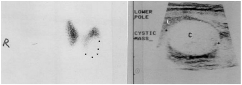
Figure 4. The left panel shows an anterior scintiscan of a euthyroid patient who had a firm nodule in the left thyroid lobe. The nodule is “cold”. * * * = nodule. The right panel shows a sonogram of the neck in the longitudinal plane revealing that the nodule is a smooth-walled cystic structure without internal echoes. between the + + symbols. Note the dark dense echoes distal to the cyst. C = cyst, L = thyroid lobe.